Trafficking of the Potato spindle tuber viroid between tomato and Orobanche ramosa

Journ
alof
Cell
Scie
nce
WD40-repeat protein 62 is a JNK-phosphorylatedspindle pole protein required for spindle maintenanceand timely mitotic progression
Marie A. Bogoyevitch1, Yvonne Y. C. Yeap1, Zhengdong Qu2, Kevin R. Ngoei1, Yan Y. Yip1, Teresa T. Zhao1,Julian I. Heng2 and Dominic C. H. Ng1,*1Department of Biochemistry and Molecular Biology, Bio21 Molecular Science and Biotechnology Institute, University of Melbourne, Victoria 3010,Australia2The Australian Regenerative Medicine Institute, Monash University, Victoria 3010, Australia
*Author for correspondence ([email protected])
Accepted 4 July 2012Journal of Cell Science 125, 5096–5109� 2012. Published by The Company of Biologists Ltddoi: 10.1242/jcs.107326
SummaryThe impact of aberrant centrosomes and/or spindles on asymmetric cell division in embryonic development indicates the tight regulation ofbipolar spindle formation and positioning that is required for mitotic progression and cell fate determination. WD40-repeat protein 62(WDR62) was recently identified as a spindle pole protein linked to the neurodevelopmental defect of microcephaly but its roles in mitosishave not been defined. We report here that the in utero electroporation of neuroprogenitor cells with WDR62 siRNAs induced their cell cycle
exit and reduced their proliferative capacity. In cultured cells, we demonstrated cell-cycle-dependent accumulation of WDR62 at the spindlepole during mitotic entry that persisted until metaphase–anaphase transition. Utilizing siRNA depletion, we revealed WDR62 function instabilizing the mitotic spindle specifically during metaphase. WDR62 loss resulted in spindle orientation defects, decreased the integrity of
centrosomes displaced from the spindle pole and delayed mitotic progression. Additionally, we revealed JNK phosphorylation of WDR62 isrequired for maintaining metaphase spindle organization during mitosis. Our study provides the first functional characterization of WDR62and has revealed requirements for JNK/WDR62 signaling in mitotic spindle regulation that may be involved in coordinating neurogenesis.
Key words: Centrosome, Spindle pole, WD-repeat proteins, Mitosis, Cell division, Neurogenesis
IntroductionThe bipolar spindle is a highly organized microtubule-based
superstructure with essential functions in faithful sister chromatid
segregation during cell division (Compton, 2000). During mitosis,
the minus ends of spindle microtubules in anti-parallel
arrangements are focused at the poles and anchored at
centrosomes. Centrosomes consist of paired centrioles surrounded
by proteinaceous pericentriolar material (PCM). In the early
stages of mitosis, the separation of duplicated centrosomes is
accompanied by a considerable expansion of the PCM via the
recruitment of c-tubulin- and c-tubulin-associated proteins (e.g.
NEDD1, pericentrin and cdk5rap2) that facilitates astral
microtubule nucleation and bipolar spindle assembly (Fong et al.,
2008; Haren et al., 2009; Luders et al., 2006; Oshimori et al., 2009;
Zimmerman et al., 2004). Moreover, MTOC activity and proper
positioning of centrosomes are required to specify the plane of
division and dictate precise cellular segregation into daughter cells
(Lesage et al., 2010; Megraw et al., 2011). Importantly, defects in
centrosome-directed functions result in aberrant bipolar spindle
formation, cell cycle progression and are linked to human disease
(Nigg and Raff, 2009; Zyss and Gergely, 2009). Proteins with
critical roles in centrosome/spindle regulation have been revealed
by genetic studies linking centrosomal protein mutations with
autosomal recessive primary microcephaly (MCPH) in which
defects in neuronal progenitor cell division are regarded as
underlying disease development (Thornton and Woods, 2009).
WD40-repeat protein 62 (WDR62) was first characterized as
involved in the assembly of signaling complexes associated with
cytoplasmic stress granule formation (Wasserman et al., 2010).
More recently, WDR62 gene mutations were linked to MCPH and
more severe brain malformations, thus implicating critical
contributions by WDR62 to cortical development (Bilguvar
et al., 2010; Nicholas et al., 2010; Yu et al., 2010). WDR62 is
170 kDa protein characterized by 13 annotated WD40 domain
repeats that span the N-terminal half of the protein (Wasserman
et al., 2010). WD40 repeat proteins facilitate protein–protein
interactions and are involved in large protein complex formation
(Stirnimann et al., 2010). WDR62 binds components of the c-Jun
N-terminal kinase (JNK) pathway to potentiate stress-stimulated
signal transduction (Cohen-Katsenelson et al., 2011; Wasserman
et al., 2010). The observed diverse intracellular distribution of
WDR62 suggests pleiotropic functions that may be dependent on
cellular context (Bilguvar et al., 2010; Nicholas et al., 2010;
Wasserman et al., 2010). For example, WDR62 is localized to
stress granules in response to cell stress (Wasserman et al., 2010).
In post-mitotic neurons WDR62 is localized to the nucleus,
whilst in neuronal progenitors undergoing mitosis, WDR62 is
present at centrosomes/spindle poles (Bilguvar et al., 2010;
Nicholas et al., 2010). Global proteomic analyses also identified
WDR62 as a mitotically regulated protein (Dephoure et al., 2008;
Santamaria et al., 2011). Although these observations are
consistent with a cell cycle regulatory function that may be
5096 Research Article

Journ
alof
Cell
Scie
nce
indispensable for cell divisions associated with neurogenesis, theprecise contributions of WDR62 in cell cycle regulation are
unknown.
In this study, we have shown for the first time that WDR62depletion with siRNA resulted in reduced cell proliferation in the
developing embryonic mouse brain. Exploiting human cellcultures to define underlying biochemical mechanistic links, werevealed WDR62 to be a mitotic phosphoprotein localized to
spindle poles from prophase to metaphase in a process thatrequires microtubule-dependent transport. Importantly, WDR62was required for proper progression through mitosis and its
depletion led to spindle orientation defects, metaphase spindleabnormalities, centrosome–spindle uncoupling and reducedcentrosome integrity. Furthermore, we demonstrated thatWDR62 phosphorylation by JNK in mitosis was involved in
the regulation of metaphase spindle architecture. Our studiesprovide the first functional analyses of WDR62 in neurogenesis,centrosome/spindle organization and cell cycle regulation with
important implications for centrosome-associated pathologiescharacterized by microcephaly.
ResultsWDR62 knockdown in vivo results in reduced proliferationof neuroprogenitors
WDR62 was recently identified as the second most commonlymutated gene linked to primary microcephaly or microcephaly
accompanied by severe cortical malformations (Bilguvar et al.,2010; Nicholas et al., 2010; Yu et al., 2010) albeit that itsfunctions during brain development are unknown. The detection
of WDR62 in neural precursors of the developing cerebral cortex(Nicholas et al., 2010) suggests its importance in regulatingneuroprogenitor cell cycle progression. To investigate this, weperformed in utero electroporation of embryonic mouse (E14)
brain to co-introduce a GFP expression construct together withexperimentally validated WDR62 siRNAs or non-targetingcontrol siRNAs (Fig. 1A). We then examined the proliferative
properties of the cortical progenitor cells. Twenty-four hourspost-electroporation, a single dose of BrdU was administered tolabel cells undergoing S-phase DNA replication before the
embryonic brains were finally harvested at 48 h (E16) to identifyprogenitor cells expressing Ki67 or phospho-histone H3 (pHH3)as markers of actively cycling and mitotic cells, respectively(Fig. 1).
We revealed that the treatment of cortical progenitors withWDR62 siRNA in vivo caused a significant decrease in Ki67
marker expression compared with control siRNA treatment(Fig. 1B,C). Moreover, WDR62 knockdown reduced theproliferative capacity of the progenitor compartment, as judgedby a significant increase in the cell cycle exit index [defined as the
fraction of GFP+BrdU+ cells which do not immunostain for Ki67versus all GFP+BrdU+ cells (GFP+BrdU+Ki672/GFP+BrdU+)]compared with control treatment (Fig. 1D). Similarly, we found
a significant decrease in the fraction of WDR62-siRNA-treatedcells that express the mitotic marker pHH3 (Fig. 1F,G), as well asa concomitant increase in their cell cycle exit (Fig. 1H).
Importantly, the incorporation of BrdU by cortical cells was notsignificantly affected by siRNA treatment (Fig. 1E,I), and weexcluded apoptosis as a contributing factor in our analysis by
showing no differences in cleaved caspase 3 staining in thedifferent cell populations (data not shown). Thus, the depletion ofWDR62 in vivo resulted in reduced proliferation and enhanced cell
cycle exit indicative of altered neuroprogenitor self-renewal duringcortical development.
Cell-cycle-dependent WDR62 localization as a spindlepole protein
A requirement for WDR62 in sustaining the proliferative capacityof neuroprogenitors suggests important cell cycle regulatoryfunctions and is consistent with the shared centrosome
association observed for the different MCPH proteins. Inaddition, the multi-compartment localization of WDR62(Bilguvar et al., 2010; Nicholas et al., 2010; Wasserman et al.,
2010; Yu et al., 2010) prompted our analysis of the intracellulardistribution of endogenous WDR62 throughout the cell cycle. Asthree commercially available antibodies detected human but not
murine WDR62 protein by immunofluorescence, we analyzedhuman cell cultures. Immunofluorescence analysis in HeLacells using antibodies recognizing distinct WDR62 epitopes
consistently revealed a cytosolic and nuclear distribution ofWDR62 during interphase (Fig. 2A; supplementary material Fig.S1A,B). During interphase, WDR62 was only weakly associatedwith centrosomes marked by c-tubulin co-staining (supplementary
material Fig. S1A). In contrast, we observed prominent WDR62colocalization with c-tubulin in mitotic cells indicatingcentrosome/spindle accumulation of WDR62 under these
conditions (Fig. 2A). All three WDR62 antibodies consistentlyrevealed dynamic WDR62 association with centrosomes in mitoticcells emphasizing the specificity of this localization (Fig. 2A;
supplementary material Fig. S1A,B). With staining for nuclearpore complex proteins, we demonstrated that the initial stages ofWDR62 centrosomal accumulation during prophase precedednuclear envelope breakdown (Fig. 2B). Further higher
magnification imaging and detailed dissection of the kinetics ofWDR62 centrosomal association revealed WDR62 accumulationaround c-tubulin-stained centrosomes beginning at prophase
(Fig. 2C). This culminated in prominent WDR62 localization atthe spindle pole that enveloped centrosomes during metaphase(Fig. 2C). In contrast, during anaphase and telophase, WDR62 no
longer showed obvious centrosomal association (Fig. 2A). Theseresults highlight a cell-cycle-dependent distribution of WDR62 tothe spindle pole.
To extend this analysis of mitotic WDR62 distribution, weevaluated WDR62 colocalization with other key centrosomalproteins. During prometaphase/metaphase, WDR62 surrounded
but did not show strict colocalization with the PCM proteinsNEDD-1, pericentrin, centrosome and Golgi-localized PKN-associated protein (CG-NAP) or cdk5 regulatory subunit
associated protein 2 (cdk5rap2; Fig. 2D). In contrast, WDR62colocalized with the microtubule cross-linker, nuclear mitoticapparatus (NUMA) protein, and the dynactin complex component,p150Glued, at spindle poles (Fig. 2E). Taken together, these results
identify WDR62 as a prominent spindle pole protein with dynamiclocalization that peaks during prometaphase/metaphase.
WDR62 depletion delays completion of mitosis
To define the potential roles of WDR62 in cell division, as
suggested by its spatial and temporal regulation during mitosis,we depleted WDR62 in HeLa cells with siRNA. Four individualsiRNAs all substantially silenced WDR62 when compared to a
non-targeting Con siRNA whereas levels of a- and c-tubulinwere unchanged (Fig. 3A), and depletion was sustained for atleast 72 h post-transfection (supplementary material Fig. S2A).
WDR62 function in mitosis 5097

Journ
alof
Cell
Scie
nce
Fig. 1. WDR62 depletion decreased cell proliferation within the embryonic cortex. (A) Neuro2a cells were transfected with individual mouse WDR62-targeting siRNA
(t1, t2, t3 and t4), a combined mouse WDR62 siRNA pool, non-targeting siRNA (Con siRNA) or not treated with siRNA (no siRNA) and immunoblotted for WDR62 and a-
tubulin. (B) Images of the coronal sections of embryonic mouse cerebral cortex electroporated with control or mouse WDR62 siRNA, together with GFP to label
electroporated cells. Embryos were pulse labeled with BrdU 24 h post-electroporation, and brain sections finally stained for Ki67 to identify actively proliferating cells at
time of harvest (48 h post-electroporation). Magnified panels (below) of the ventricular zone (VZ) highlight proliferating (GFP+/BrdU+/Ki67+, filled arrowheads) and non-
proliferating cells (GFP+/BrdU+/Ki672, open arrowheads). (C) Quantification of cell proliferation (GFP+/Ki67+) in brain sections from control and mouse WDR62-siRNA-
treated embryos. (D) The cell cycle exit index in response to WDR62 depletion was determined by counting GFP+/BrdU+/Ki672 cells and expressing this as a proportion of
GFP+/BrdU+ cells. (E) BrdU labeling index in control and WDR62-siRNA-treated brain sections used in the analysis depicted in B. (F) Coronal sections of embryos
electroporated with control or mouse WDR62 siRNA were stained for phospho-histone H3 (pHH3). Magnified panels (below) of the VZ highlight cells in M phase (GFP+/
BrdU+/pHH3+, filled arrowheads) and non-mitotic cells (GFP+/BrdU+/pHH32, open arrowheads). (G) The mitotic index in response to WDR62 depletion was determined by
the proportion of cells double labeled for GFP and pHH3 (GFP+/pHH3+). (H) A cell cycle exit index analysis performed with pHH3 and BrdU immunostaining was
determined by counting GFP+/BrdU+/pHH32 cells within the population of GFP+/BrdU+ cells (statistical analysis could not be performed because 100% of cells were
GFP+BrdU+pHH32 following WDR62 siRNA treatment). (I) BrdU labeling index in control and WDR62-siRNA-treated brain sections used in the analysis depicted in (F).
Scale bar: 100 mm, while all graphs plot means 6 s.e.m. (*P,0.05, **P,0.01, ns denotes not statistically significant).
Journal of Cell Science 125 (21)5098

Journ
alof
Cell
Scie
nce
All subsequent findings utilized WDR62 depletion by WDR62
target 1 siRNA but were re-confirmed with at least two other
independent siRNAs (typically WDR62 target 2 and target 3
siRNA) to ensure specificity of the knockdown phenotype.
Interestingly, proliferation and cell viability were not
markedly altered despite substantially reduced WDR62 levels
(supplementary material Fig. S2B,C). Thus, WDR62 loss did not
induce cell cycle arrest or prevent division in HeLa cells. In
contrast, we found that WDR62 depletion altered the duration of
mitosis.
When the effect of WDR62 loss was evaluated in synchronized
HeLa cells, we observed delayed mitotic progression following
double thymidine block (DTB) release. Levels of cyclin B1 and
phosphorylated cdc25C are typically elevated during mitotic
entry and persist until the metaphase–anaphase transition
(Hutchins and Clarke, 2004; Takizawa and Morgan, 2000), and
so we used these as indicators of cell cycle progression. In
WDR62-siRNA-treated cells cyclin B1 degradation and
dephosphorylation of cdc25C were delayed, extending to
11–12 h post-release compared to the return of these mitotic
markers to interphase levels by 10 h post-release in Con-siRNA-
treated cells (Fig. 3B). Similarly, in cells synchronized by
nocodazole treatment, elevated cyclinB1 and phospho-cdc25C
levels were extended to 6 h post-release from nocodazole
compared to the loss of these mitotic markers by 4 h post-
nocodazole release in Con-siRNA-treated cells (Fig. 3C). Time-
lapse imaging of WDR62-siRNA-treated cells further showed
that the duration of mitosis was prolonged (Fig. 3D,E) with
WDR62-siRNA-treated HeLa cells taking on average nearly
twice as long (96 min) to complete mitosis when compared with
Con-siRNA-treated cells (54 min; Fig. 3E). Some WDR62-
depleted cells took .200 min (approximately four times
average duration of control cells) to complete mitosis (Fig. 3E)
without cell cycle arrest. Furthermore, substantial movement and
rotation of the metaphase plate was observed suggestive of
spindle orientation defects during periods of prolonged mitosis
(Fig. 3D, broken white lines). Reflecting this mitotic delay, a
modest increase in mitotic index in WDR62-siRNA-treated cells
Fig. 2. Accumulation of WDR62 at
spindle poles during mitosis. (A) WDR62
subcellular localization in HeLa cells at
different mitotic stages. DAPI-stained
condensed chromatin and c-tubulin
immunostaining revealed centrosomes and
spindle poles. Scale bar: 20 mm. (B) WDR62
association with centrosomes occurred prior
to nuclear envelope breakdown as
demonstrated by co-staining with nuclear
pore complex proteins (Mab414 NPC). Scale
bar: 20 mm. (C) Higher magnification
images of WDR62 localization around c-
tubulin-stained centrosomes from prophase
to metaphase. Scale bar: 2 mm. Insets are
lower magnification images showing entire
cell and mitotic stage. (D) Co-staining of
WDR62 with PCM markers (NEDD1,
pericentrin, CG-NAP and Cdk5rap2).
(E) WDR62 co-localization with spindle
pole markers (NUMA and p150Glued). Insets
in D and E are high magnification images of
centrosomes. Scale bars: 20 mm.
WDR62 function in mitosis 5099

Journ
alof
Cell
Scie
nce
compared to controls was observed (Fig. 3F). Thus, knockdownof WDR62 delays the completion of mitosis indicating a
requirement for WDR62 in regulating timely mitotic progression.
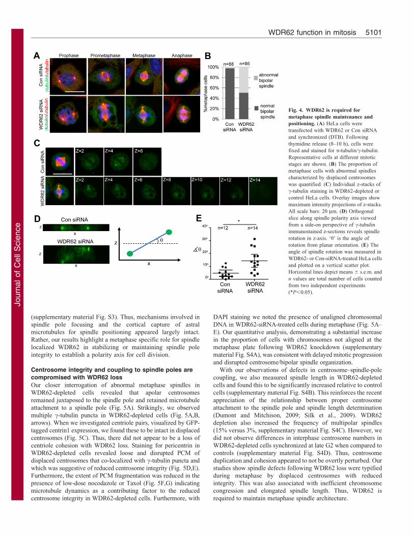
Metaphase spindle defects with WDR62 knockdownTo interrogate the features underlying delayed mitoticprogression and spindle orientation defects, we examined the
changes in mitotic spindle organization as a result of WDR62loss. On closer examination of spindle assembly in prophase andprometaphase, intact centrosomes anchoring astral microtubules
(as noted by a- and c-tubulin localizations) were observed forboth control and WDR62-depleted cells (Fig. 4A). Strikingly,WDR62 depletion resulted in abnormal metaphase spindles
characterized by a displacement of centrosomes from the spindle(Fig. 4A). Quantitation of the metaphase cell populationindicated that approximately half of WDR62-depleted cells
exhibited an abnormal bipolar spindle (Fig. 4B). In addition,
while metaphase spindles in cultured cells commonly attain aplanar orientation, with the division plane parallel to surface of
the culture dish, the metaphase spindle polarity axis in WDR62-depleted cells exhibited substantial rotation in the z-axis
(Fig. 4C). The rotation of the spindle in the z-axis was moreclearly seen through orthogonal sectioning through the z-stacks
(Fig. 4D) and our measurements of the angle of rotation fromplanar orientation showed a greater degree of spindle rotation inthe z-axis in WDR62-depleted cells compared to controls
(Fig. 4E). The localization of NUMA and dynein/dynactinmotors at spindle poles and the cell cortex are critical for pole
focusing activities and the cortical capture of astral microtubulesfor spindle positioning, respectively (Radulescu and Cleveland,
2010). However, the cortical and spindle pole localization ofNUMA and p150Glued was unaltered by WDR62 loss
Fig. 3. WDR62 depletion perturbs mitotic
progression. (A) HeLa cells were transiently
transfected with individual human WDR62-
targeting siRNA (t1, t2, t3 and t4), a WDR62
siRNA pool, non-targeting siRNA (Con siRNA) or
not treated with siRNA (no siRNA) and
immunoblotted for WDR62. a- and c-tubulin were
blotted as controls. (B) HeLa cells transfected with
WDR62 or Con siRNA were synchronized at G1/S
(DTB). Cell cycle progression following thymidine
release (2–14 h as indicated) was determined by
immunoblotting for cyclin B1 and cdc25C
phosphorylation. (C) HeLa cells transfected with
WDR62 or Con siRNA were synchronized at M-
phase [nocodazole (NZ) treated; 350 nM, 16 h].
Mitotic progression following NZ release (0.5–10 h
as indicated) was determined by immunoblotting
for cyclin B1 and phospho-cdc25C. a-tubulin was
blotted for protein loading and WDR62 levels
determined to confirm knockdown. (D) HeLa cells,
treated with Con siRNA or WDR62 siRNA, were
synchronized (DTB) and imaged under phase-
contrast optics at 6-min intervals from 7 h post
thymidine release. Representative images of mitotic
progression are shown. White dotted lines indicate
the position of the metaphase plate. (E) Duration of
mitotis in WDR62-depleted and control HeLa cells,
measured from nuclear envelope breakdown until
two daughter cells were observed, are shown on a
vertical scatter plot. Horizontal lines indicate mean
values and n values are the total cells counted from
three independent experiments (*P,0.01).
(F) WDR62 was depleted in HeLa cells for 48 h
and the proportion of cells in M phase identified by
DAPI staining. Values are means 6 s.e.m. from
three independent experiments.
Journal of Cell Science 125 (21)5100
Journ
alof
Cell
Scie
nce
(supplementary material Fig. S3). Thus, mechanisms involved in
spindle pole focusing and the cortical capture of astral
microtubules for spindle positioning appeared largely intact.
Rather, our results highlight a metaphase specific role for spindle
localized WDR62 in stabilizing or maintaining spindle pole
integrity to establish a polarity axis for cell division.
Centrosome integrity and coupling to spindle poles are
compromised with WDR62 loss
Our closer interrogation of abnormal metaphase spindles in
WDR62-depleted cells revealed that apolar centrosomes
remained juxtaposed to the spindle pole and retained microtubule
attachment to a spindle pole (Fig. 5A). Strikingly, we observed
multiple c-tubulin puncta in WDR62-depleted cells (Fig. 5A,B,
arrows). When we investigated centriole pairs, visualized by GFP-
tagged centrin1 expression, we found these to be intact in displaced
centrosomes (Fig. 5C). Thus, there did not appear to be a loss of
centriole cohesion with WDR62 loss. Staining for pericentrin in
WDR62-depleted cells revealed loose and disrupted PCM of
displaced centrosomes that co-localized with c-tubulin puncta and
which was suggestive of reduced centrosome integrity (Fig. 5D,E).
Furthermore, the extent of PCM fragmentation was reduced in the
presence of low-dose nocodazole or Taxol (Fig. 5F,G) indicating
microtubule dynamics as a contributing factor to the reduced
centrosome integrity in WDR62-depleted cells. Furthermore, with
DAPI staining we noted the presence of unaligned chromosomal
DNA in WDR62-siRNA-treated cells during metaphase (Fig. 5A–
E). Our quantitative analysis, demonstrating a substantial increase
in the proportion of cells with chromosomes not aligned at the
metaphase plate following WDR62 knockdown (supplementary
material Fig. S4A), was consistent with delayed mitotic progression
and disrupted centrosome/bipolar spindle organization.
With our observations of defects in centrosome–spindle-pole
coupling, we also measured spindle length in WDR62-depleted
cells and found this to be significantly increased relative to control
cells (supplementary material Fig. S4B). This reinforces the recent
appreciation of the relationship between proper centrosome
attachment to the spindle pole and spindle length determination
(Dumont and Mitchison, 2009; Silk et al., 2009). WDR62
depletion also increased the frequency of multipolar spindles
(15% versus 3%, supplementary material Fig. S4C). However, we
did not observe differences in interphase centrosome numbers in
WDR62-depleted cells synchronized at late G2 when compared to
controls (supplementary material Fig. S4D). Thus, centrosome
duplication and cohesion appeared to not be overtly perturbed. Our
studies show spindle defects following WDR62 loss were typified
during metaphase by displaced centrosomes with reduced
integrity. This was also associated with inefficient chromosome
congression and elongated spindle length. Thus, WDR62 is
required to maintain metaphase spindle architecture.
Fig. 4. WDR62 is required for
metaphase spindle maintenance and
positioning. (A) HeLa cells were
transfected with WDR62 or Con siRNA
and synchronized (DTB). Following
thymidine release (8–10 h), cells were
fixed and stained for a-tubulin/c-tubulin.
Representative cells at different mitotic
stages are shown. (B) The proportion of
metaphase cells with abnormal spindles
characterized by displaced centrosomes
was quantified. (C) Individual z-stacks of
c-tubulin staining in WDR62-depleted or
control HeLa cells. Overlay images show
maximum intensity projections of z-stacks.
All scale bars: 20 mm. (D) Orthogonal
slice along spindle polarity axis viewed
from a side-on perspective of c-tubulin
immunostained z-sections reveals spindle
rotation in z-axis. ‘h’ is the angle of
rotation from planar orientation. (E) The
angle of spindle rotation was measured in
WDR62- or Con-siRNA-treated HeLa cells
and plotted on a vertical scatter plot.
Horizontal lines depict means 6 s.e.m. and
n values are total number of cells counted
from two independent experiments
(*P,0.05).
WDR62 function in mitosis 5101

Journ
alof
Cell
Scie
nce
WDR62 is required for metaphase spindle maintenance but
not bipolar spindle formation
WDR62 spindle pole localization during early mitotic stages
parallels centrosome maturation and the enhanced nucleation of
astral microtubules that facilitate formation of a bipolar spindle.
To identify WDR62 functions in microtubule nucleation and
bipolar spindle formation, we employed microtubule regrowth
assays. WDR62 centrosomal localization was lost following
microtubule depolymerization by cold treatment and was rapidly
returned (,2 min) during astral microtubule regrowth (Fig. 6A).
This further reinforced WDR62 as a microtubule-associated
spindle pole protein. Surprisingly, at 10 min regrowth, WDR62
was also localized to regions of acentrosomal microtubule
formation originating from mitotic chromatin. We confirmed
WDR62 localization to calcium-resistant kinetochore
microtubules (supplementary material Fig. S5A). WDR62
localization then coalesced at the poles as spindle bipolarity
was re-established at 30 min (Fig. 6A). WDR62 was therefore
associated with sites of microtubule nucleation at both the
centrosomes and chromosomes.
We next established WDR62 requirements for microtubule
regrowth. In interphase cells, no overt defects in centrosome-
nucleated microtubule formation in WDR62-siRNA-treated cells
were observed (supplementary material Fig. S5B). This was not
surprising as WDR62 was not principally located at the MTOC
during this cell cycle stage. Intriguingly, in mitotic cells, the
extent of microtubule nucleation and regrowth at both
centrosomal and non-centrosomal sites did not differ when
WDR62-depleted and control HeLa cells were compared
(Fig. 6B). However, as the bipolar spindle was re-established
(45 min), defects in centrosome integrity and positioning were
observed in WDR62-siRNA-treated cells (Fig. 6B). The
proportion of WDR62-depleted cells with spindle abnormalities
observed in our microtubule regrowth assays were consistent
with our measurements during metaphase (Fig. 6C compared to
Fig. 4B).
To determine whether spindle defects arose due to microtubule
forces associated with bipolar spindle formation, we examined
centrosome integrity in DTB-synchronized HeLa cells
subsequently released in the presence of monastrol, an inhibitor
Fig. 5. Centrosome integrity is reduced
with WDR62 depletion. WDR62-depleted
or control HeLa cells in metaphase were
stained with (A) a- and c-tubulin or
(B) WDR62 and c-tubulin. Arrows
indicate c-tubulin puncta. DAPI staining of
DNA is shown in overlay images. (C)
HeLa cells were transfected with WDR62
or Con siRNA together with Centrin1–GFP
to label centrioles in addition to staining
for c-tubulin. (D,E) WDR62-depleted or
control HeLa cells in metaphase were
stained with pericentrin and (D) c-tubulin
or (E) a-tubulin. Insets in D,E are higher
magnification images of the centrosomal
region. All scale bars: 20 mm. (F) WDR62-
depleted cells were synchronized (DTB)
and treated with nocodazole (NZ, 5 nM) or
Taxol (TX, 5 nM) at 7 h post-thymidine
release. The number of cells in metaphase
with loose/fragmented PCM, as revealed
by pericentrin staining, was quantified and
expressed as a proportion of total
metaphase cells. Con siRNA treatment was
included for comparison. n values are the
total number of cells counted from three
independent experiments.
(G) Representative images of pericentrin
staining in WDR62-depleted cells treated
with nocodazole or Taxol.
Journal of Cell Science 125 (21)5102

Journ
alof
Cell
Scie
nce
of kinesin-5 motor proteins required for establishing spindle
tension and bipolarity (Mayer et al., 1999). Monastrol treatment
arrested both control and WDR62-siRNA-treated cells in mitosis
with monopolar spindles (Fig. 6D). Detection of c-tubulin
showed that centrosome integrity was unperturbed in WDR62-
depleted cells arrested in monastrol (Fig. 6D). Taken together,
our study indicates that microtubule nucleation and spindle
assembly does not require WDR62. In contrast, reduced
centrosome integrity and spindle attachment defects occur
subsequent to bipolar spindle formation and this is consistent
with the previously defined requirement for microtubule
dynamics (Fig. 5F).
WDR62 is phosphorylated in mitosis
In considering regulatory mechanisms that control WDR62
function or localization, the phosphorylation of centrosomal
and spindle proteins is critical for mitotic regulation (Luders et al.,
2006; Oshimori et al., 2006). WDR62 is likely targeted for
phosphorylation as previous studies have identified WDR62 as a
substrate of JNK and polo-like kinase 1 (Plk1) in the context of
cell stress and mitosis, respectively (Santamaria et al., 2011;
Wasserman et al., 2010). Indeed, M-phase arrest with nocodazole
decreased the mobility of WDR62 in SDS-PAGE (Fig. 7A).
Furthermore, WDR62 band detection in nocodazole-arrested
cells was weaker but l-phosphatase treatment rendered the
migration and detection of the WDR62 band in lysates from the
M-phase-arrested cells indistinguishable from asynchronous (AS)
or S-phase-arrested (using hydroxyurea; HU) cells consistent
with a phosphorylation modification (Fig. 7A). We obtained
similar results regardless of the specific WDR62 antibody used
(data not shown). As identical cell lysates were used for the l-
phosphatase or mock treatments, the reduced detection of
Fig. 6. WDR62 is required for spindle/
centrosome maintenance after spindle bipolarity
is established. (A) G1/S synchronized HeLa cells
(DTB) were released into nocodazole (350 nM)
before microtubules were depolymerized at 4 C
(30 min). Following nocodazole washout, cells were
incubated at 37 C for various time intervals before
staining for WDR62 and microtubule regrowth with
a-tubulin. Scale bar: 2 mm. (B) Microtubule
regrowth in WDR62-depleted HeLa cells was
analyzed as in A and compared to control (Con
siRNA) cells. In addition, centrosomes were
revealed by c-tubulin co-staining. Scale bar: 20 mm.
(C) Following MT regrowth (45 mins, 37 C) the
proportion of cells with normal, abnormal or multi-
polar spindles was determined. n values are total
number of cells counted from three independent
experiments. (D) HeLa cells synchronized to G1/S
(DTB) were released into monastrol (100 mM)
before fixing and immunostaining for WDR62 and
a- or c-tubulin. Scale bars: 20 mm.
WDR62 function in mitosis 5103

Journ
alof
Cell
Scie
nce
WDR62 during nocodazole arrest indicated reduced antibody
affinity for phosphorylated WDR62 on SDS-PAGE, rather than
changes in WDR62 protein levels. However, l-phosphatase
treatment did not alter our detection of WDR62 subcellular
localization indicating that phosphorylation did not impact on our
immunofluorescence analyses in fixed cells (data not shown).
The mobility shift of WDR62 in response to nocodazole-induced
mitotic arrest or hyperosmolarity was more easily visualized in
SDS-PAGE resolved with polyacrylamide-bound Mn2+-Phos-tag
(Fig. 7B) (Kinoshita et al., 2006). In addition, while not evident
on standard SDS-PAGE, the phosphorylation of WDR62 in
untreated AS cells was revealed by l-phosphatase treatment and
electrophoresis on a Phos-tag gel (Fig. 7B). These results confirm
WDR62 phosphorylation under basal conditions which was
further increased with cell stress or M phase synchronization.
The M-phase arrest of cells treated with nocodazole was
verified by increased cyclin B1 and phospho-cdc25C levels
(Fig. 7A). Further, nocodazole washout increased WDR62 band
mobility and detection in parallel with reduced cyclinB1 andphospho-cdc25C (Fig. 7C). Finally, as G1/S synchronized cells
by DTB were tracked following thymidine block release, the
WDR62 band shift closely correlated with mitotic progression asdetermined by biochemical markers (cyclin B1 and phospho-
cdc25C) and FACS analysis of the cell cycle (Fig. 7D,E). These
results revealed phosphorylation of WDR62 during mitotic
progression that coincided with spindle pole localization.
JNK mediated WDR62 phosphorylation is required forspindle regulation
As WDR62 was previously shown to be a JNK-interacting
protein (Wasserman et al., 2010), we investigated JNK regulation
of WDR62 and its contributions to mitosis. WDR62phosphorylation in M-phase-arrested cells was partially
reversed by JNK inhibition whereas cdc25C phosphorylation
Fig. 7. WDR62 is phosphorylated during mitosis.
(A) Protein lysates from asynchronous (AS), S-phase-
arrested (HU, 2 mM, 16 h) or mitotically arrested (NZ,
350 nM, 16 h) HeLa cells were treated with l-
phosphatase, mock treated without enzyme or left
untreated (TCL) before blotting with the indicated
antibodies. Arrows indicate decreased WDR62 band
migration on SDS-PAGE. (B) Protein lysates from AS-,
NZ- (350 nM, 16 h) or sorbitol-treated (Sorb, 0.5 M,
60 min) HeLa cells were treated with l-phosphatase or
left untreated (TCL) before resolving on Phos-tag gels
or standard SDS-PAGE followed by immunoblotting.
Arrows indicated reduced mobility of phosphorylated
WDR62. (C) HeLa cells were arrested at M phase (NZ,
350 nM, 16 h) before release into normal serum
medium for the indicated times. WDR62 band mobility,
cyclin B1, a-tubulin and phospho-cdc25C were
determined by immunoblot analysis. Arrows indicate
increased WDR62 gel migration and carets (,) antibody
detection with time following NZ release. (D) HeLa
cells were synchronized at G1/S (DTB). WDR62, cyclin
B1 and cdc25C levels were then analyzed by
immunoblotting. (E) Cell cycle distribution was
determined by FACS at time 0 min before release and at
regular time intervals following thymidine release.
Representative histograms are shown.
Journal of Cell Science 125 (21)5104
Journ
alof
Cell
Scie
nce
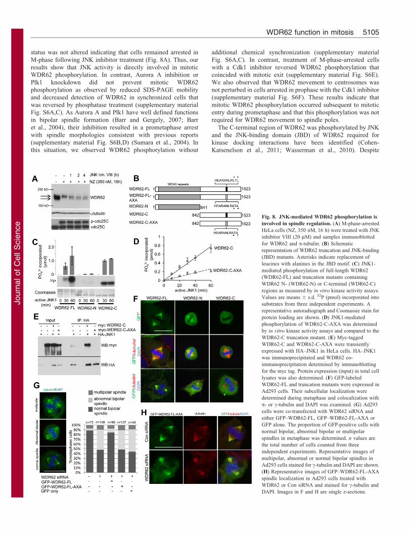
status was not altered indicating that cells remained arrested in
M-phase following JNK inhibitor treatment (Fig. 8A). Thus, our
results show that JNK activity is directly involved in mitotic
WDR62 phosphorylation. In contrast, Aurora A inhibition or
Plk1 knockdown did not prevent mitotic WDR62
phosphorylation as observed by reduced SDS-PAGE mobility
and decreased detection of WDR62 in synchronized cells that
was reversed by phosphatase treatment (supplementary material
Fig. S6A,C). As Aurora A and Plk1 have well defined functions
in bipolar spindle formation (Barr and Gergely, 2007; Barr
et al., 2004), their inhibition resulted in a prometaphase arrest
with spindle morphologies consistent with previous reports
(supplementary material Fig. S6B,D) (Sumara et al., 2004). In
this situation, we observed WDR62 phosphorylation without
additional chemical synchronization (supplementary material
Fig. S6A,C). In contrast, treatment of M-phase-arrested cells
with a Cdk1 inhibitor reversed WDR62 phosphorylation that
coincided with mitotic exit (supplementary material Fig. S6E).
We also observed that WDR62 movement to centrosomes was
not perturbed in cells arrested in prophase with the Cdk1 inhibitor
(supplementary material Fig. S6F). These results indicate that
mitotic WDR62 phosphorylation occurred subsequent to mitotic
entry during prometaphase and that this phosphorylation was not
required for WDR62 movement to spindle poles.
The C-terminal region of WDR62 was phosphorylated by JNK
and the JNK-binding domain (JBD) of WDR62 required for
kinase docking interactions have been identified (Cohen-
Katsenelson et al., 2011; Wasserman et al., 2010). Despite
Fig. 8. JNK-mediated WDR62 phosphorylation is
involved in spindle regulation. (A) M-phase-arrested
HeLa cells (NZ, 350 nM, 16 h) were treated with JNK
inhibitor VIII (20 mM) and samples immunoblotted
for WDR62 and a-tubulin. (B) Schematic
representation of WDR62 truncation and JNK-binding
(JBD) mutants. Asterisks indicate replacement of
leucines with alanines in the JBD motif. (C) JNK1-
mediated phosphorylation of full-length WDR62
(WDR62-FL) and truncation mutants containing
WDR62 N- (WDR62-N) or C-terminal (WDR62-C)
regions as measured by in vitro kinase activity assays.
Values are means 6 s.d. 32P (pmol) incorporated into
substrates from three independent experiments. A
representative autoradiograph and Coomassie stain for
protein loading are shown. (D) JNK1-mediated
phosphorylation of WDR62-C-AXA was determined
by in vitro kinase activity assays and compared to the
WDR62-C truncation mutant. (E) Myc-tagged
WDR62-C and WDR62-C-AXA were transiently
expressed with HA–JNK1 in HeLa cells. HA–JNK1
was immunoprecipitated and WDR62 co-
immunoprecipitation determined by immunoblotting
for the myc tag. Protein expression (input) in total cell
lysates was also determined. (F) GFP-labeled
WDR62-FL and truncation mutants were expressed in
Ad293 cells. Their subcellular localization were
determined during metaphase and colocalization with
a- or c-tubulin and DAPI was examined. (G) Ad293
cells were co-transfected with WDR62 siRNA and
either GFP–WDR62-FL, GFP–WDR62-FL-AXA or
GFP alone. The proportion of GFP-positive cells with
normal bipolar, abnormal bipolar or multipolar
spindles in metaphase was determined. n values are
the total number of cells counted from three
independent experiments. Representative images of
multipolar, abnormal or normal bipolar spindles in
Ad293 cells stained for c-tubulin and DAPI are shown.
(H) Representative images of GFP–WDR62-FL-AXA
spindle localization in Ad293 cells treated with
WDR62 or Con siRNA and stained for c-tubulin and
DAPI. Images in F and H are single z-sections.
WDR62 function in mitosis 5105

Journ
alof
Cell
Scie
nce
these advances, the biological function of WDR62phosphorylation is undetermined. We generated truncation and
JBD mutants of WDR62 (Fig. 8B) and confirmed that JNKphosphorylated the C-terminal half (WDR62-C, amino acids842–1523) of WDR62 but not the N-terminal region containingthe WD40 domains (WDR62-N, amino acids 1–841; Fig. 8C).
Furthermore, replacement of critical leucines with alanine inthe WDR62 JBD (AXA, L1299A/L1301A) inhibited JNKphosphorylation of WDR62-C in vitro and completely
abrogated interaction with JNK when ectopically expressed inHeLa cells (Fig. 8D,E). We then compared the spindlelocalization properties of WDR62 mutants expressed as GFP
fusion proteins. However, due to difficulties with aggregation ofGFP-labeled full-length WDR62 expressed in HeLa cells, weanalyzed Ad293 cells which displayed a greater proportionof cells that retained diffuse cytoplasmic distribution
(supplementary material Fig. S7A). Interestingly, expression ofthe GFP-labeled WDR62-N truncation mutant led to a greatlyenhanced proportion of cells with aggregates while GFP–
WDR62-C expression was predominantly cytoplasmic despitehigher expression levels (supplementary material Fig. S7A,B).Thus, the WDR62 N-terminal region containing WD40 repeats
was responsible for the propensity of the ectopically expressedprotein to aggregate. However, in cells with non-aggregate GFP–WDR62-N, we observed GFP–WDR62-N localization to the
mitotic spindle albeit with reduced efficiency compared to full-length GFP–WDR62 (GFP–WDR62-FL; Fig. 8F). In contrast,GFP–WDR62-C failed to localize to the mitotic spindle(Fig. 8F). These observations indicate a requirement for the N-
terminal domains of WDR62 in its mitotic localization.
We lastly sought to identify the contribution of JNK-mediatedWDR62 phosphorylation to spindle regulation. Chemical
inhibition of JNK during mitosis caused spindle abnormalitiesand centrosome attachment defects reminiscent of WDR62depletion (supplementary material Fig. S7C). To determine the
contribution of JNK signaling to WDR62 specifically, weperformed rescue experiments. WDR62 depletion in Ad293cells resulted in abnormal metaphase spindles as similarlyobserved in HeLa cells (Fig. 8G). Importantly, the concomitant
expression of siRNA-resistant GFP–WDR62-FL reversed thefrequency of the spindle defects in WDR62-depleted cellsconfirming siRNA effects were specific to WDR62 depletion. In
contrast, expression of the JBD mutant (WDR62-FL-AXA) inWDR62-depleted cells was without effect (Fig. 8G) despitelocalization to the mitotic spindle (Fig. 8H) and comparable
expression levels (supplementary material Fig. S7B). Thus, ourstudies show JNK-mediated phosphorylation of the WDR62 C-terminus is dispensable for its spindle localization but required for
WDR62 functions in maintaining metaphase spindle architecture.
DiscussionOur combined in vitro and in vivo studies are consistent in
identifying a mitotic role for WDR62 that is critical forneurogenesis. Specifically, we have provided the firstfunctional characterization of WDR62 in mitosis and revealed a
key role in metaphase spindle organization required for normalmitotic progression. WDR62 has a role in stabilizing themetaphase spindle for timely transition to anaphase but is not
required for proliferation per se in cultured cells. In contrast, wedemonstrate that WDR62 depletion impacts on the pools of self-renewing neural progenitors leading to their reduced proliferation
in embryonic brains consistent with previously reported
expression of WDR62 in mitotic neuronal precursor cells
(Nicholas et al., 2010). One possibility is that defects in the
timing of mitosis and/or spindle orientation following WDR62-
depeletion impacts specifically on cortical neurogenesis. Indeed,
centrosome-directed spindle orientation has been suggested as a
critical determinant for asymmetric division of neuroprogenitors,
a necessary property for their self-renewal within the germinal
compartment of the embryonic nervous system (Lesage et al.,
2010). This is compatible with the notion that centrosome-
associated mitotic defects during neurogenesis may underlie the
clinical manifestation of WDR62 mutations in MCPH (Nicholas
et al., 2010; Yu et al., 2010).
Our analysis of WDR62 intracellular distribution and cellular
functions reinforces the association of MCPH with centrosome-
associated proteins and highlights the role of spindle pole
integrity in neurogenesis (Thornton and Woods, 2009). The
prominent spindle pole localization of WDR62 is shared by
another MCPH protein, abnormal spindle microcephaly related
gene (ASPM) (Higgins et al., 2010; Pulvers et al., 2010). In
contrast, other MCPH proteins, microcephalin, cdk5rap2,
CENPJ, CEP152, and STIL are principally centrosomal in their
localization (Delattre et al., 2004; Fong et al., 2008; Varmark
et al., 2007; Zhong et al., 2006). Interestingly, ASPM and
WDR62 are most frequently mutated in MCPH accounting for
50% and 10% of cases, respectively (Thornton and Woods,
2009). We have demonstrated spindle orientation defects as a
consequence of WDR62 depletion in cultured human cells. The
depletion of ASPM in cultured cells has revealed similar
requirements for spindle organization and spindle positioning
(Higgins et al., 2010). Moreover, the loss of ASPM in embryonic
mice resulted in premature cell cycle exit due to altered cleavage
plane orientation in dividing neuroprogenitors (Fish et al., 2006).
The shared depletion phenotypes of WDR62 and ASPM suggest
that mitotic spindle and cleavage plane positioning may represent
a mechanism that links these proteins that are most commonly
mutated in MCPH.
The molecular basis that underlies the development of MCPH
may also involve the disruption and/or dysfunction of
centrosomes (Thornton and Woods, 2009). The depletion of
PCM proteins cdk5rap2 or pericentrin, with the latter also
genetically linked to primordial dwarfism with associated
microcephaly, similarly altered neuroprogenitor division in
embryonic mouse brains with reduced cell proliferation and
enhanced cell cycle exit (Buchman et al., 2010; Lizarraga et al.
2010). However, unlike MCPH proteins (cdk5rap2, CENPJ,
STIL, Cep152, Cep135 and Cep63) with well characterized
functions in centriole duplication (Barrera et al. 2010; Delattre
et al., 2004; Hatch et al., 2010; Hussain et al., 2012; Sir et al.,
2011; Vulprecht et al., 2012), we have demonstrated that WDR62
is not principally a centrosome protein and its depletion did not
obviously impact on centrosome number or structure during
interphase. We cannot completely exclude WDR62 functions as
an integral centrosomal protein. However, the WDR62 depletion
phenotype was observed specifically during metaphase and
coincided with its most prominent spindle localization. Thus,
centrosome/spindle defects in WDR62 knockdown cells were
likely the result of the loss of spindle localized WDR62 during
mitosis. Specifically, we revealed defects with centrosome–
spindle-pole coupling in cells lacking WDR62.
Journal of Cell Science 125 (21)5106

Journ
alof
Cell
Scie
nce
Centrosome–spindle-pole attachment defects were previouslyobserved with the disruption of centrosomal cdk5rap2, NUMA or
dynein/dynactin subunits (Barr et al., 2010; Morales-Mulia andScholey, 2005; Silk et al., 2009). Interestingly, we noted thatcentrosome displacement occurred subsequent to bipolar spindleformation suggesting that microtubule forces may underlie
centrosome uncoupling. However, WDR62 depletion did notresult in unfocused spindle poles. In addition, the spindle poleand cortical localization of NUMA and dynein were unaltered by
WDR62 loss. Our study therefore suggests that the disruption ofcentrosome–spindle coupling is linked to microtubule forcesassociated with establishing spindle bipolarity and spindle
orientation but not readily explained by the mislocalization ofpreviously identified factors critical for spindle–centrosomeattachment.
We have additionally revealed that the PCM of uncoupled
centrosomes appeared diffuse with multiple puncta positivelystained for pericentrin. It is possible that PCM recruitment to MTminus ends on the uncoupled spindle pole may also lead to such a
phenotype. However, disrupted centrosomes were smaller in sizeand as we did not find defects in centrosome maturation or astralmicrotubule formation, our findings suggest reduced centrosome
integrity in response to WDR62 knockdown. As withcentrosome/spindle pole uncoupling, defects in centrosomeintegrity were observed subsequent to bipolar spindle formation
and required microtubule dynamics, which suggests thatcentrosome uncoupling and fragmentation events may berelated. However, previous studies that have inactivatedcdk5rap2, NUMA or dynein have shown that loss of spindle
pole attachment does not necessarily lead to reduced centrosomeintegrity (Barr et al., 2010; Morales-Mulia and Scholey, 2005;Silk et al., 2009). Notably, NUMA and dynein/dynactin motors
perform multiple functions at several mitotic locations(Radulescu and Cleveland, 2010). Thus, the differences inspindle/centrosome defects may reflect different mechanisms
underlying WDR62 function in metaphase spindle maintenancecompared to previously characterized spindle pole proteins. Wepropose that the spindle localization of WDR62 is required tomaintain tight centrosome–spindle-pole coupling, centrosome
integrity and the maintenance of spindle orientation that isinvolved in determining cleavage plane position and divisionsymmetry. Disruption of these processes with WDR62 loss or
mutation may impact on self-renewing divisions in theembryonic brain that ultimately manifests as microcephaly.
As WDR62 is a multi-functional protein with dynamic
intracellular distribution dependent on cellular context, thisraises the significant question of the range of regulatorymechanisms for its actions. Our study has now provided first
insights into the mechanisms that regulate mitotic WDR62activity. WDR62 is mitotically phosphorylated. However,WDR62 movement to centrosomes, prior to mitotic entry,preceded its phosphorylation. This indicates that mitotic
phosphorylation is not likely involved in regulating WDR62movement. In support of this, the N-terminal region of WDR62,lacking C-terminal phosphorylation sites, is sufficient for spindle
localization. Rather, WDR62 phosphorylation appears morelikely to be involved in functional regulation.
We revealed JNK contribution to mitotic WDR62
phosphorylation. WDR62 was initially characterized as acomponent of the JNK signaling pathway (Wasserman et al.,2010) and our findings demonstrate that this is maintained during
mitosis. JNK expression and activity regulates various cell cycle
stages (Kennedy and Davis, 2003; MacCorkle and Tan, 2005).
Specifically, JNK is activated during early mitotic stages and
targets substrates such as cdc25C, histone H3 and Cdh1 to
facilitate mitotic progression (Gutierrez et al., 2010a; Gutierrez
et al., 2010b; Lee and Song, 2008). JNK has also previously been
reported to be centrosome/spindle pole associated during cell
division (Huang et al., 2011; MacCorkle-Chosnek et al., 2001).
Importantly, we have now shown that JNK phosphorylation of
WDR62 is involved in spindle regulation.
There are multiple serine/threonine residues at the C-terminal
region of WDR62 that are likely JNK target sites (Wasserman
et al., 2010). Future detailed analyses should reveal those sites
critical for the mitotic activity of WDR62. However, we have
taken advantage of the previously characterized WDR62 JBD
(Cohen-Katsenelson et al., 2011) and utilized mutations to this
motif (AXA) as a strategy to abrogate JNK signaling to WDR62.
This leaves unchanged the proline-directed serine/threonine
residues that may be additionally targeted by other mitotically
regulated kinases (e.g. cdks). WDR62-FL-AXA mutants failed to
rescue spindle abnormalities caused by endogenous WDR62
depletion highlighting JNK contribution to spindle regulation
through the targeting of WDR62. It is likely that WDR62 is
targeted by multiple kinases, particularly as JNK inhibition only
partially reverses WDR62 phosphorylation in response to M-
phase arrest. Although Plk1 and Aurora A inhibition did not
attenuate mitotic WDR62 phosphorylation, at least on residues
responsible for gel shift, this does not exclude roles for these
mitotic kinases in WDR62 regulation. However, investigations
into the precise contributions of Plk1, Aurora A and Cdk1 are
complicated by their critical requirements for bipolar spindle
formation and mitotic entry.
In targeting mitotically regulated WDR62, we have now
defined functions in centrosome/spindle organization, mitotic
regulation and neurogenesis. Our study has provided the first in
vitro and in vivo functional analysis of WDR62 and revealed
novel contributions of a JNK/WDR62 signaling mechanism
towards mitotic spindle and cell cycle regulation. Moreover,
studies in transgenic mice have demonstrated that JNK was
required for early brain development (Kuan et al., 1999). Thus,
we have revealed a novel signaling mechanism linking JNK
activity and neurogenesis that warrants further investigation.
Materials and MethodsAntibodies and reagents
Polyclonal WDR62 antibodies were from Bethyl Laboratories (A301-560A) orNovus Biologicals (NB100-77302). CG-NAP and p150Glued antibodies were fromBD Biosciences. NUMA, dynein intermediate chain (DIC), and mouse monoclonal(Mab414) antibodies to Nuclear Pore Complex proteins were from Abcam. a-Tubulin antibody was obtained from Santa Cruz Biotechnology. Plk1, phospho-TCTP (Ser46), cyclin B1 and cdc25C antibodies were from Cell SignalingTechnology. Antibodies to Ki67 and phosphorylated histone H3 were from Leicaand Merck-Millipore, respectively. Cdk1 inhibitor I and Aurora kinase inhibitor IIIwere from Calbiochem. Phos-tag reagent was from Wako Chemicals. Cell cultureand transfection reagents including DMEM and fetal bovine serum were fromInvitrogen-GibcoBRL. All other reagents, including thymidine, nocodazole,hydroxyurea, monoclonal and polyclonal c-tubulin and monoclonal WDR62(W3269) antibodies were obtained from Sigma-Aldrich. Unless specifiedotherwise, immunoblot and immunofluorescence analysis were performed withanti-WDR62 from Bethyl Laboratories.
In utero electroporation and brain slice analysis
Mice were maintained within the animal facilities at Monash University, with allanimal procedures approved by the relevant Animal Ethics Committee. In utero
electroporation was performed on pregnant, time-mated female C57/B6 mice as
WDR62 function in mitosis 5107

Journ
alof
Cell
Scie
nce
previously described (Heng et al., 2008). Mouse embryos (E14) wereelectroporated with WDR62 siRNA or a non-targeting control siRNA (10 mM ofsiRNA in solution) together with a GFP expression plasmid (1 mg/ml). BrdU pulselabeling (100 mg/kg) was performed by injecting pregnant dams 24 h post-electroporation. Following an additional 24 h, the embryonic brains were dissectedand fixed in 4% paraformaldehyde/PBS solution overnight, before washing withPBS followed by incubation in 20% sucrose/PBS solution. Finally, the brains wereembedded in OCT and sectioned at 16 mm thickness using a cryostat (Leica) andimmunostained using previously defined protocols (Heng et al., 2008). Theventricular zone/subventricular zone and intermediate zones were identified bynuclei density and images were acquired on a confocal microscope. Cell countswere performed on representative fields followed by statistical analysis(GraphPad). Data presented for the in utero electroporation experiments isplotted from three or four brains per condition.
Immunofluorescence and live-cell microscopy
Cell culture and treatments were performed on uncoated glass coverslips. Cellswere washed in PBS before fixation either with 4% (w/v) paraformaldehyde(20 min, room temperature) or cold methanol (5 min, 220 C). Sample preparationwas as previously described (Ng et al., 2011). Images were acquired on a LeicaTCS SP2 or SP5 confocal microscope using 10061.35 NA objectives. Unlessspecified otherwise, images are maximum intensity projections of z-stacks of 8–14sections of 0.4 mm thickness that encompasses the entire spindle. Z-stack spindlerotation analysis was performed with ImageJ using the Z-function plug-in. Spindlelength measurements were performed on single stack images using Metamorphsoftware by measuring the linear distance between the apexes of bipolar spindlespoles.
Plasmids and protein purification
SiRNA-resistant full-length WDR62 (WDR62-FL) was codon optimized andsynthesized by GeneArt (Invitrogen). WDR62 truncation mutants (WDR62-N,WDR62-C) were generated by PCR and, along with full-length WDR62,labeled with GFP or Myc tag by cloning into pEGFP-N3 or pXJ40-Mycvectors, respectively. WDR62-FL-AXA and WDR62-C-AXA (L1299A/L1301A)mutations were generated by site-directed mutagenesis. For recombinant proteinexpression, WDR62 cDNAs were cloned into pGEX-6P-1 to generate GSTfusions. Where specified, the GST tag was removed with Precision Proteasecleavage. HA–JNK1 was made by subcloning PCR amplified Jnk1a1 into pXJ41-HA. Active JNK1 was generated using baculoviral expression in insect cells asdescribed previously (Ngoei et al., 2011). All constructs were subjected torestriction digestion and full sequencing analysis prior to use. GFP–centrin1encoded in pCMV6 vector was obtained from Origene.
RNAi
ON-TARGETPlus human WDR62 siRNA, mouse WDR62 siRNA pool, humanPlk1 siRNA pool, non-targeting control siRNA (D-001810-01-20) and non-targeting siRNA pool (D-001810-10-20) were obtained from Dharmacon andresuspended in RNAase free water at 100 mM. siRNA were transiently transfectedusing LipofectamineTM 2000 according to the manufacturer’s protocol.
Cell culture and transient transfection
HeLa and Ad293 were maintained in DMEM and genetically modified U2OS-GFP–TUB1A (Sigma-Aldrich) was grown in McCoy’s 5A medium. All growthmedia were supplemented with 10% fetal calf serum and 100 U/ml penicillin/streptomycin and cells cultured in a humidified 5% CO2 environment. Liposomal-mediated transfection was performed with LipofectamineTM 2000 and antibiotic-free Opti-MEM medium according to manufacturer’s instructions.
Cell synchronization and cell cycle arrest
HeLa cells were synchronized by DTB or nocodazole arrest. Briefly, sub-confluentHeLa cells were cultured in the presence of thymidine (2 mM) for 20 h beforewashing with PBS (three washes) and release into normal growth medium for 6 h.Cells were then cultured in the presence of thymidine (2 mM) for a further 20 hbefore final release from late G1 arrest into normal growth medium. Cells werethen collected at specified times regular post-release for subsequent analysis. M-phase or S-phase arrests were with nocodazole (350 nM) or hydroxyurea (2 mM)treatment respectively for 16 h. Kinase inhibitors were applied at the time orsubsequent to cell cycle arrest as specified.
Cell lysates and immunoblots
Total cell lysates were prepared in RIPA buffer [50 mM Tris-HCl, pH 7.3,150 mM NaCl, 0.1 mM EDTA, 1% (w/v) sodium deoxycholate, 1% (v/v) TritonX-100, 0.2% (w/v) NaF and 100 mM Na3VO4] supplemented with proteaseinhibitors and immunoblotted as previously described (Ng et al., 2010) with theexception that protein lysates were resolved on SDS-PAGE without sample boilingdue to the heat sensitivity of WDR62 (Wasserman et al. 2010). For phosphatasetreatment, lysates were prepared in RIPA buffer lacking EDTA, NaF and Na3VO4.
Lysates (100 mg) were treated with l-phosphatase (400 U, New England Biolabs)for 20 min at 30 C. For Phos-tag SDS-PAGE, gels were supplemented with adinuclear manganese complex of acrylamide-pendant Phos-tag ligand (Mn2+-Phos-tag; 50 mM).
In vitro kinase assay
Purified recombinant protein substrate (10 mg) was incubated with active kinase(10 ng), and 32P-radiolabeled ATP ([c32-P]ATP, 1 mCi/reaction) in a kinasereaction buffer (20 mM HEPES pH 7.6, 20 mM MgCl2.6H2O, 75 mM ATP,20 mM b-glycerophosphate and supplemented with 25 mM Na3VO4 and 100 mMDTT) over a 120 min time course at 30 C. Reactions were stopped with theaddition of Laemmli sample buffer. Samples were then resolved by SDS-PAGE,stained with Gelcode Blue Stain reagent and analyzed by autoradiography andCerenkov counting.
Microtubule regrowth assays, Eg5 inhibition and CaCl2 extraction
HeLa cells were synchronized (DTB) and released into nocodazole (350 nM, 8–10 h). Nocodazole was removed by washing (36) with cold PBS then cold growthmedium added. Mitotic microtubules were completely depolymerized by coldtreatment (4 C, 30 min). Microtubule regrowth was initiated by incubating cells at37 C for between 2 and 60 min before fixation. For kinesin Eg5 inhibition, HeLacells were synchronized (DTB) and released into monastrol (100 mM, 8 h). Toreveal kinetochore microtubules, synchronized cells were extracted with CalciumExtraction Buffer (1 mM CaCl2, 100 mM PIPES pH 6.8, 0.1 mM MgCl2 and 0.1%v/v Triton X-100) for 3 min before fixing and immunofluorescence analysis.
Cell viability and cell proliferation
Cell viability was determined by labeling metabolically active cells with theyellow tetrazolium salt, XTT {sodium 39-[1-(phenylaminocarbonyl)-3,4-tetrazolium]-bis (4-methoxy-6-nitro) benzene sulfonic acid hydrate}, using theXTT Kit (Roche) according to manufacturer specifications. WDR62 protein levelswere blotted in parallel to confirm WDR62 depletion. Cell numbers were countedusing a hemocytometer and Trypan Blue exclusion.
AcknowledgementsWe thank Prof. Sharad Kumar (Centre for Cancer Biology/SouthAustralia Pathology) for the NEDD1 antibody and Prof. Leann Tilley(University of Melbourne) for critical reading of this manuscript.
FundingThis work was supported by a Faculty of Medicine, Dentistry andHealth Science (MDHS) CR Roper Fellowship [to D.C.H.N.]; anNational Health and Medical Research Council Career DevelopmentFellowship [grant number APP1011505 to J.I.H.]; and NationalHealth and Medical Research Council Project Grants [grant numbers628335 and 566804 to D.C.H.N and M.A.B., respectively.].Deposited in PMC for release after 6 months.
Supplementary material available online at
http://jcs.biologists.org/lookup/suppl/doi:10.1242/jcs.107326/-/DC1
ReferencesBarr, A. R. and Gergely, F. (2007). Aurora-A: the maker and breaker of spindle poles.
J. Cell Sci. 120, 2987-2996.
Barr, F. A., Sillje, H. H. and Nigg, E. A. (2004). Polo-like kinases and the orchestration
of cell division. Nat. Rev. Mol. Cell Biol. 5, 429-441.
Barr, A. R., Kilmartin, J. V. and Gergely, F. (2010). CDK5RAP2 functions in
centrosome to spindle pole attachment and DNA damage response. J. Cell Biol. 189,
23-39.
Barrera, J. A., Kao, L. R., Hammer, R. E., Seemann, J., Fuchs, J. L. and Megraw,
T. L. (2010). CDK5RAP2 regulates centriole engagement and cohesion in mice. Dev.
Cell 18, 913-926.
Bilguvar, K., Ozturk, A. K., Louvi, A., Kwan, K. Y., Choi, M., Tatli, B., Yalnizoglu,
D., Tuysuz, B., Caglayan, A. O., Gokben, S. et al. (2010). Whole-exome sequencing
identifies recessive WDR62 mutations in severe brain malformations. Nature 467,
207-210.
Buchman, J. J., Tseng, H. C., Zhou, Y., Frank, C. L., Xie, Z. and Tsai, L. H. (2010).
Cdk5rap2 interacts with pericentrin to maintain the neural progenitor pool in the
developing neocortex. Neuron 66, 386-402.
Cohen-Katsenelson, K., Wasserman, T., Khateb, S., Whitmarsh, A. J. and
Aronheim, A. (2011). Docking interactions of the JNK scaffold protein WDR62.
Biochem. J. 439, 381-390.
Compton, D. A. (2000). Spindle assembly in animal cells. Annu. Rev. Biochem. 69, 95-
114.
Journal of Cell Science 125 (21)5108

Journ
alof
Cell
Scie
nce
Delattre, M., Leidel, S., Wani, K., Baumer, K., Bamat, J., Schnabel, H., Feichtinger,R., Schnabel, R. and Gonczy, P. (2004). Centriolar SAS-5 is required for centrosomeduplication in C. elegans. Nat. Cell Biol. 6, 656-664.
Dephoure, N., Zhou, C., Villen, J., Beausoleil, S. A., Bakalarski, C. E., Elledge, S. J.
and Gygi, S. P. (2008). A quantitative atlas of mitotic phosphorylation. Proc. Natl.
Acad. Sci. USA 105, 10762-10767.Dumont, S. and Mitchison, T. J. (2009). Compression regulates mitotic spindle length
by a mechanochemical switch at the poles. Curr. Biol. 19, 1086-1095.Fish, J. L., Kosodo, Y., Enard, W., Paabo, S. and Huttner, W. B. (2006). Aspm
specifically maintains symmetric proliferative divisions of neuroepithelial cells. Proc.
Natl. Acad. Sci. USA 103, 10438-10443.Fong, K. W., Choi, Y. K., Rattner, J. B. and Qi, R. Z. (2008). CDK5RAP2 is a
pericentriolar protein that functions in centrosomal attachment of the gamma-tubulinring complex. Mol. Biol. Cell 19, 115-125.
Gutierrez, G. J., Tsuji, T., Chen, M., Jiang, W. and Ronai, Z. A. (2010a). Interplaybetween Cdh1 and JNK activity during the cell cycle. Nat. Cell Biol. 12, 686-695.
Gutierrez, G. J., Tsuji, T., Cross, J. V., Davis, R. J., Templeton, D. J., Jiang, W. and
Ronai, Z. A. (2010b). JNK-mediated phosphorylation of Cdc25C regulates cell cycleentry and G(2)/M DNA damage checkpoint. J. Biol. Chem. 285, 14217-14228.
Haren, L., Stearns, T. and Luders, J. (2009). Plk1-dependent recruitment of gamma-tubulin complexes to mitotic centrosomes involves multiple PCM components. PLoS
ONE 4, e5976.Hatch, E. M., Kulukian, A., Holland, A. J., Cleveland, D. W. and Stearns, T. (2010).
Cep152 interacts with Plk4 and is required for centriole duplication. J. Cell Biol. 191,721-729.
Heng, J. I., Nguyen, L., Castro, D. S., Zimmer, C., Wildner, H., Armant, O.,
Skowronska-Krawczyk, D., Bedogni, F., Matter, J. M., Hevner, R. et al. (2008).Neurogenin 2 controls cortical neuron migration through regulation of Rnd2. Nature
455, 114-118.Higgins, J., Midgley, C., Bergh, A. M., Bell, S. M., Askham, J. M., Roberts, E.,
Binns, R. K., Sharif, S. M., Bennett, C., Glover, D. M. et al. (2010). Human ASPMparticipates in spindle organisation, spindle orientation and cytokinesis. BMC Cell
Biol. 11, 85.Huang, X., Tong, J. S., Wang, Z. B., Yang, C. R., Qi, S. T., Guo, L., Ouyang, Y. C.,
Quan, S., Sun, Q. Y., Qi, Z. Q. et al. (2011). JNK2 participates in spindle assemblyduring mouse oocyte meiotic maturation. Microsc. Microanal. 17, 197-205.
Hussain, M. S., Baig, S. M., Neumann, S., Nurnberg, G., Farooq, M., Ahmad, I.,Alef, T., Hennies, H. C., Technau, M., Altmuller, J. et al. (2012). A truncatingmutation of CEP135 causes primary microcephaly and disturbed centrosomalfunction. Am. J. Hum. Genet. 90, 871-878.
Hutchins, J. R. and Clarke, P. R. (2004). Many fingers on the mitotic trigger: post-translational regulation of the Cdc25C phosphatase. Cell Cycle 3, 40-44.
Kennedy, N. J. and Davis, R. J. (2003). Role of JNK in tumor development. Cell Cycle
2, 199-201.Kinoshita, E., Kinoshita-Kikuta, E., Takiyama, K. and Koike, T. (2006). Phosphate-
binding tag, a new tool to visualize phosphorylated proteins. Mol. Cell. Proteomics 5,749-757.
Kuan, C. Y., Yang, D. D., Samanta Roy, D. R., Davis, R. J., Rakic, P. and Flavell,
R. A. (1999). The Jnk1 and Jnk2 protein kinases are required for regional specificapoptosis during early brain development. Neuron 22, 667-676.
Lee, K. and Song, K. (2008). Basal c-Jun N-terminal kinases promote mitoticprogression through histone H3 phosphorylation. Cell Cycle 7, 216-221.
Lesage, B., Gutierrez, I., Martı, E. and Gonzalez, C. (2010). Neural stem cells: theneed for a proper orientation. Curr. Opin. Genet. Dev. 20, 438-442.
Lizarraga, S. B., Margossian, S. P., Harris, M. H., Campagna, D. R., Han, A. P.,
Blevins, S., Mudbhary, R., Barker, J. E., Walsh, C. A. and Fleming, M. D. (2010).Cdk5rap2 regulates centrosome function and chromosome segregation in neuronalprogenitors. Development 137, 1907-1917.
Luders, J., Patel, U. K. and Stearns, T. (2006). GCP-WD is a gamma-tubulin targetingfactor required for centrosomal and chromatin-mediated microtubule nucleation. Nat.
Cell Biol. 8, 137-147.MacCorkle, R. A. and Tan, T. H. (2005). Mitogen-activated protein kinases in cell-
cycle control. Cell Biochem. Biophys. 43, 451-462.MacCorkle-Chosnek, R. A., VanHooser, A., Goodrich, D. W., Brinkley, B. R. and
Tan, T. H. (2001). Cell cycle regulation of c-Jun N-terminal kinase activity at thecentrosomes. Biochem. Biophys. Res. Commun. 289, 173-180.
Mayer, T. U., Kapoor, T. M., Haggarty, S. J., King, R. W., Schreiber, S. L. and
Mitchison, T. J. (1999). Small molecule inhibitor of mitotic spindle bipolarityidentified in a phenotype-based screen. Science 286, 971-974.
Megraw, T. L., Sharkey, J. T. and Nowakowski, R. S. (2011). Cdk5rap2 exposes thecentrosomal root of microcephaly syndromes. Trends Cell Biol. 21, 470-480.
Morales-Mulia, S. and Scholey, J. M. (2005). Spindle pole organization in DrosophilaS2 cells by dynein, abnormal spindle protein (Asp), and KLP10A. Mol. Biol. Cell 16,3176-3186.
Ng, D. C., Zhao, T. T., Yeap, Y. Y., Ngoei, K. R. and Bogoyevitch, M. A. (2010). c-Jun N-terminal kinase phosphorylation of stathmin confers protection against cellularstress. J. Biol. Chem. 285, 29001-29013.
Ng, D. C., Ng, I. H., Yeap, Y. Y., Badrian, B., Tsoutsman, T., McMullen, J. R.,
Semsarian, C. and Bogoyevitch, M. A. (2011). Opposing actions of extracellularsignal-regulated kinase (ERK) and signal transducer and activator of transcription 3(STAT3) in regulating microtubule stabilization during cardiac hypertrophy. J. Biol.
Chem. 286, 1576-1587.Ngoei, K. R., Catimel, B., Church, N., Lio, D. S., Dogovski, C., Perugini, M. A.,
Watt, P. M., Cheng, H. C., Ng, D. C. and Bogoyevitch, M. A. (2011).Characterization of a novel JNK (c-Jun N-terminal kinase) inhibitory peptide.Biochem. J. 434, 399-413.
Nicholas, A. K., Khurshid, M., Desir, J., Carvalho, O. P., Cox, J. J., Thornton, G.,Kausar, R., Ansar, M., Ahmad, W., Verloes, A. et al. (2010). WDR62 is associatedwith the spindle pole and is mutated in human microcephaly. Nat. Genet. 42, 1010-1014.
Nigg, E. A. and Raff, J. W. (2009). Centrioles, centrosomes, and cilia in health anddisease. Cell 139, 663-678.
Oshimori, N., Ohsugi, M. and Yamamoto, T. (2006). The Plk1 target Kizuna stabilizesmitotic centrosomes to ensure spindle bipolarity. Nat. Cell Biol. 8, 1095-1101.
Oshimori, N., Li, X., Ohsugi, M. and Yamamoto, T. (2009). Cep72 regulates thelocalization of key centrosomal proteins and proper bipolar spindle formation. EMBO
J. 28, 2066-2076.Pulvers, J. N., Bryk, J., Fish, J. L., Wilsch-Brauninger, M., Arai, Y., Schreier, D.,
Naumann, R., Helppi, J., Habermann, B., Vogt, J. et al. (2010). Mutations inmouse Aspm (abnormal spindle-like microcephaly associated) cause not onlymicrocephaly but also major defects in the germline. Proc. Natl. Acad. Sci. USA
107, 16595-16600.Radulescu, A. E. and Cleveland, D. W. (2010). NuMA after 30 years: the matrix
revisited. Trends Cell Biol. 20, 214-222.Santamaria, A., Wang, B., Elowe, S., Malik, R., Zhang, F., Bauer, M., Schmidt, A.,
Sillje, H. H., Korner, R., and Nigg, E. A. (2011). The Plk1-dependentphosphoproteome of the early mitotic spindle. Mol. Cell Proteomics 10, M110004457.
Silk, A. D., Holland, A. J. and Cleveland, D. W. (2009). Requirements for NuMA inmaintenance and establishment of mammalian spindle poles. J. Cell Biol. 184, 677-690.
Sir, J. H., Barr, A. R., Nicholas, A. K., Carvalho, O. P., Khurshid, M., Sossick, A.,
Reichelt, S., D’Santos, C., Woods, C. G. and Gergely, F. (2011). A primarymicrocephaly protein complex forms a ring around parental centrioles. Nat. Genet. 43,1147-1153.
Stirnimann, C. U., Petsalaki, E., Russell, R. B. and Muller, C. W. (2010). WD40proteins propel cellular networks. Trends Biochem. Sci. 35, 565-574.
Sumara, I., Gimenez-Abian, J. F., Gerlich, D., Hirota, T., Kraft, C., de la Torre, C.,Ellenberg, J. and Peters, J. M. (2004). Roles of polo-like kinase 1 in the assembly offunctional mitotic spindles. Curr. Biol. 14, 1712-1722.
Takizawa, C. G. and Morgan, D. O. (2000). Control of mitosis by changes in thesubcellular location of cyclin-B1-Cdk1 and Cdc25C. Curr. Opin. Cell Biol. 12, 658-665.
Thornton, G. K. and Woods, C. G. (2009). Primary microcephaly: do all roads lead toRome? Trends Genet. 25, 501-510.
Varmark, H., Llamazares, S., Rebollo, E., Lange, B., Reina, J., Schwarz,H. and Gonzalez, C. (2007). Asterless is a centriolar protein required forcentrosome function and embryo development in Drosophila. Curr. Biol. 17, 1735-1745.
Vulprecht, J., David, A., Tibelius, A., Castiel, A., Konotop, G., Liu, F., Bestvater, F.,Raab, M. S., Zentgraf, H., Izraeli, S. et al. (2012). STIL is required for centrioleduplication in human cells. J. Cell Sci. 125, 1353-1362.
Wasserman, T., Katsenelson, K., Daniliuc, S., Hasin, T., Choder, M. and Aronheim,A. (2010). A novel c-Jun N-terminal kinase (JNK)-binding protein WDR62 isrecruited to stress granules and mediates a nonclassical JNK activation. Mol. Biol.
Cell 21, 117-130.Yu, T. W., Mochida, G. H., Tischfield, D. J., Sgaier, S. K., Flores-Sarnat, L., Sergi,
C. M., Topcu, M., McDonald, M. T., Barry, B. J., Felie, J. M. et al. (2010).Mutations in WDR62, encoding a centrosome-associated protein, cause micro-cephaly with simplified gyri and abnormal cortical architecture. Nat. Genet. 42,1015-1020.
Zhong, X., Pfeifer, G. P. and Xu, X. (2006). Microcephalin encodes a centrosomalprotein. Cell Cycle 5, 457-458.
Zimmerman, W. C., Sillibourne, J., Rosa, J. and Doxsey, S. J. (2004). Mitosis-specific anchoring of gamma tubulin complexes by pericentrin controls spindleorganization and mitotic entry. Mol. Biol. Cell 15, 3642-3657.
Zyss, D. and Gergely, F. (2009). Centrosome function in cancer: guilty or innocent?Trends Cell Biol. 19, 334-346.
WDR62 function in mitosis 5109
Copyright © 2022 FDOKUMEN