of the Police forces,CentralApecurity breast by a ribbon 35 ...
TH
EJ
OU
RN
AL
OF
CE
LL
BIO
LO
GY
The Rockefeller University Press $30.00J. Cell Biol. Vol. 184 No. 3 391–397www.jcb.org/cgi/doi/10.1083/jcb.200809090 JCB 391
JCB: REPORT
Correspondence to Joachim Seemann: [email protected]
Abbreviations used in this paper: NAGT I, N -acetylglucosaminyl transferase I; PDI, protein disulfi de isomerase; RLG, rat liver Golgi.
Introduction The Golgi apparatus in mammalian cells consists of stacks of
fl attened cisternae that are linked together laterally into a single
continuous ribbon in the perinuclear region. As the central hub
of the secretory pathway, the Golgi receives newly synthesized
proteins from the ER and sorts them into their correct cellular
destinations. The orientation of the Golgi ribbon directs exo-
cytosis toward a particular area of the plasma membrane and
thereby facilitates the establishment of cell polarity, for in-
stance, during wound healing ( Preisinger et al., 2004 ), neural
development ( Horton et al., 2005 ), and immune response
( Stinchcombe et al., 2006 ).
During cell division, the single Golgi ribbon needs to be
segregated into the two daughter cells. To achieve partitioning,
the Golgi fragments at the onset of mitosis, and later reforms
in each daughter cell (for review see Lowe and Barr, 2007 ).
Two different mechanisms have been proposed for the par-
titioning of the Golgi. In one view, Golgi membranes are
absorbed into and partitioned with the ER ( Zaal et al., 1999 ).
The second view argues that the Golgi remains distinct from
the ER and that the two compartments are inherited indepen-
dently ( Bartz and Seemann, 2008 ). In this scenario, the spindle
has been proposed as the machinery responsible for Golgi par-
titioning based on the observed accumulation of Golgi mem-
branes at the spindle poles ( Shima et al., 1998 ), although a pool
of membranes is dispersed throughout the cytoplasm ( Jesch
et al., 2001 ).
To elucidate the role of the spindle in Golgi partitioning,
we established an assay in which the entire spindle is segregated
into only one daughter cell. Upon division, a Golgi ribbon re-
formed in the karyoplasts, whereas the stacked cisternae were
scattered throughout the cytoplasts. We could rescue ribbon as-
sembly in the cytoplasts by microinjecting the Golgi extract to-
gether with tubulin or by lowering the division temperature, at
which partial spindle materials were transferred into the cyto-
plasts. We propose that Golgi factors required for ribbon assem-
bly rely on the spindle for partitioning, but functional stacks are
inherited independently. This reveals two levels of regulation
underlying Golgi inheritance. Coupling of these factors with the
spindle would ensure that daughter cells receive the information
to assemble the Golgi ribbon, which is vital for cellular func-
tions that require polarized secretion and directional migration.
Results and discussion To evaluate the role of the spindle in Golgi partitioning, we fi rst
examined the spatial correlation between Golgi membranes and
the spindle. Consistent with previous studies ( Shima et al., 1998 ;
Jokitalo et al., 2001 ), we found Golgi membranes concentrated
in metaphase around the two spindle poles in a variety of cell
lines, including HeLa, Vero, LLC-PK1, normal rat kidney, and
PtK1 (unpublished data). The association was prominent in
The mammalian Golgi ribbon disassembles during mi-
tosis and reforms in both daughter cells after division.
Mitotic Golgi membranes concentrate around the
spindle poles, suggesting that the spindle may control Golgi
partitioning. To test this, cells were induced to divide asym-
metrically with the entire spindle segregated into only one
daughter cell. A ribbon reforms in the nucleated karyoplasts,
whereas the Golgi stacks in the cytoplasts are scattered.
However, the scattered Golgi stacks are polarized and
transport cargo. Microinjection of Golgi extract together
with tubulin or incorporation of spindle materials rescues
Golgi ribbon formation. Therefore, the factors required for
postmitotic Golgi ribbon assembly are transferred by the
spindle, but the constituents of functional stacks are parti-
tioned independently, suggesting that Golgi inheritance is
regulated by two distinct mechanisms.
The mitotic spindle mediates inheritance of the Golgi ribbon structure
Jen-Hsuan Wei and Joachim Seemann
Department of Cell Biology, University of Texas Southwestern Medical Center, Dallas, TX 75390
© 2009 Wei and Seemann This article is distributed under the terms of an Attribution–Noncommercial–Share Alike–No Mirror Sites license for the fi rst six months after the publica-tion date (see http://www.jcb.org/misc/terms.shtml). After six months it is available under a Creative Commons License (Attribution–Noncommercial–Share Alike 3.0 Unported license, as described at http://creativecommons.org/licenses/by-nc-sa/3.0/).
on October 18, 2013
jcb.rupress.orgD
ownloaded from
Published February 2, 2009
http://jcb.rupress.org/content/suppl/2009/02/02/jcb.200809090.DC1.html Supplemental Material can be found at:
on October 18, 2013
jcb.rupress.orgD
ownloaded from
Published February 2, 2009
on October 18, 2013
jcb.rupress.orgD
ownloaded from
Published February 2, 2009
on October 18, 2013
jcb.rupress.orgD
ownloaded from
Published February 2, 2009
on October 18, 2013
jcb.rupress.orgD
ownloaded from
Published February 2, 2009
on October 18, 2013
jcb.rupress.orgD
ownloaded from
Published February 2, 2009
on October 18, 2013
jcb.rupress.orgD
ownloaded from
Published February 2, 2009
on October 18, 2013
jcb.rupress.orgD
ownloaded from
Published February 2, 2009
on October 18, 2013
jcb.rupress.orgD
ownloaded from
Published February 2, 2009
JCB • VOLUME 184 • NUMBER 3 • 2009 392
survived on average 36 h after division and, in some cases, were
still alive and seemingly healthy when corresponding karyo-
plasts underwent a second round of cell division.
The scattered Golgi and the absence of microtubules in
the cytoplasts are reminiscent of cells treated with nocodazole,
which causes the Golgi ribbon to fragment into stacks that are
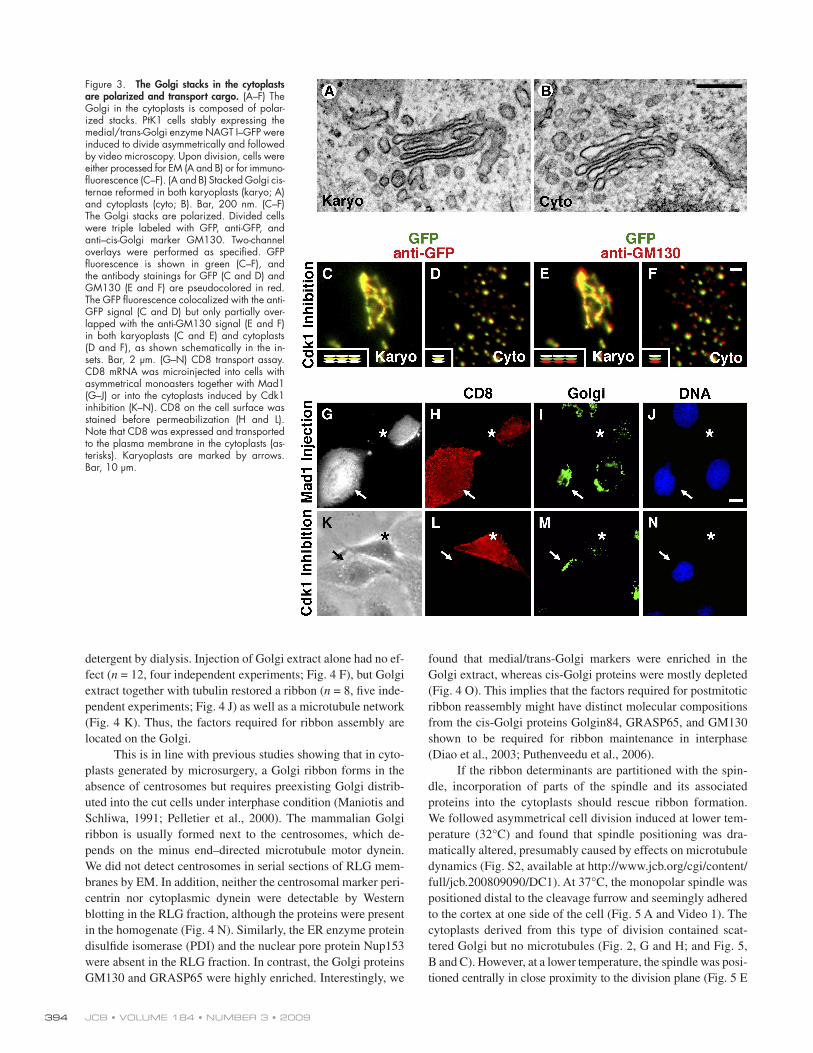
dispersed throughout the cell ( Shima et al., 1998 ). Our EM
analysis revealed that the Golgi in both karyoplasts and cyto-
plasts was stacked with an average of 3.4 ± 0.7 (karyoplast;
n = 22) and 3.3 ± 0.6 (cytoplast; n = 23) cisternae per stack
( Fig. 3, A and B ). The cisternae were stacked to a comparable
extent and separated by 11.8 ± 3.8 (karyoplast) and 12.9 ± 3.7 nm
(cytoplast). To determine whether the stacks were polarized,
we took advantage of a well-established fl uorescence mi-
croscopy method ( Shima et al., 1997 ). PtK1 cells stably ex-
pressing the medial/trans-Golgi enzyme NAGT I – GFP were
induced to divide asymmetrically and immunolabeled for GFP
and the cis-Golgi marker GM130 ( n = 15). In these triple-
labeled cells, the GFP fl uorescence was fi rst overlaid with the
anti-GFP signal ( Fig. 3, C and D ) and was then overlaid with
the anti-GM130 signal ( Fig. 3, E and F ). The anti-GFP stain-
ing served as a control, showing a complete overlap with the GFP
fl uorescence ( Fig. 3, C and D ). In contrast, the cis – anti-GM130
signal in the karyoplasts was found adjacent to the medial/
trans-GFP fl uorescence with only a partial overlap ( Fig. 3 E ),
suggesting that the ribbon in the karyoplasts preserved a
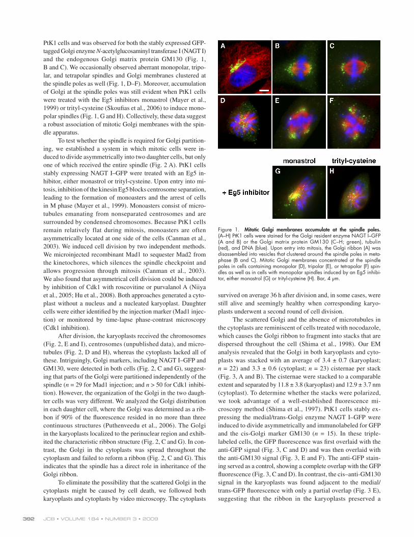
PtK1 cells and was observed for both the stably expressed GFP-
tagged Golgi enzyme N -acetylglucosaminyl transferase I (NAGT I)
and the endogenous Golgi matrix protein GM130 ( Fig. 1,
B and C ). We occasionally observed aberrant monopolar, tripo-
lar, and tetrapolar spindles and Golgi membranes clustered at
the spindle poles as well ( Fig. 1, D – F ). Moreover, accumulation
of Golgi at the spindle poles was still evident when PtK1 cells
were treated with the Eg5 inhibitors monastrol ( Mayer et al.,
1999 ) or trityl-cysteine ( Skoufi as et al., 2006 ) to induce mono-
polar spindles ( Fig. 1, G and H ). Collectively, these data suggest
a robust association of mitotic Golgi membranes with the spin-
dle apparatus.
To test whether the spindle is required for Golgi partition-
ing, we established a system in which mitotic cells were in-
duced to divide asymmetrically into two daughter cells, but only
one of which received the entire spindle ( Fig. 2 A ). PtK1 cells
stably expressing NAGT I – GFP were treated with an Eg5 in-
hibitor, either monastrol or trityl-cysteine. Upon entry into mi-
tosis, inhibition of the kinesin Eg5 blocks centrosome separation,
leading to the formation of monoasters and the arrest of cells
in M phase ( Mayer et al., 1999 ). Monoasters consist of micro-
tubules emanating from nonseparated centrosomes and are
surrounded by condensed chromosomes. Because PtK1 cells
remain relatively flat during mitosis, monoasters are often
asymmetrically located at one side of the cells ( Canman et al.,
2003 ). We induced cell division by two independent methods.
We microinjected recombinant Mad1 to sequester Mad2 from
the kinetochores, which silences the spindle checkpoint and
allows progression through mitosis ( Canman et al., 2003 ).
We also found that asymmetrical cell division could be induced
by inhibition of Cdk1 with roscovitine or purvalanol A ( Niiya
et al., 2005 ; Hu et al., 2008 ). Both approaches generated a cyto-
plast without a nucleus and a nucleated karyoplast. Daughter
cells were either identifi ed by the injection marker (Mad1 injec-
tion) or monitored by time-lapse phase-contrast microscopy
(Cdk1 inhibition).
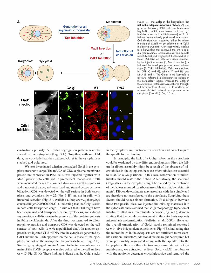
After division, the karyoplasts received the chromosomes
( Fig. 2, E and I ), centrosomes (unpublished data), and micro-
tubules ( Fig. 2, D and H ), whereas the cytoplasts lacked all of
these. Intriguingly, Golgi markers, including NAGT I – GFP and
GM130, were detected in both cells ( Fig. 2, C and G ), suggest-
ing that parts of the Golgi were partitioned independently of the
spindle ( n = 29 for Mad1 injection; and n > 50 for Cdk1 inhibi-
tion). However, the organization of the Golgi in the two daugh-
ter cells was very different. We analyzed the Golgi distribution
in each daughter cell, where the Golgi was determined as a rib-
bon if 90% of the fl uorescence resided in no more than three
continuous structures ( Puthenveedu et al., 2006 ). The Golgi
in the karyoplasts localized to the perinuclear region and exhib-
ited the characteristic ribbon structure ( Fig. 2, C and G ). In con-
trast, the Golgi in the cytoplasts was spread throughout the
cytoplasm and failed to reform a ribbon ( Fig. 2, C and G ). This
indicates that the spindle has a direct role in inheritance of the
Golgi ribbon.
To eliminate the possibility that the scattered Golgi in the
cytoplasts might be caused by cell death, we followed both
karyoplasts and cytoplasts by video microscopy. The cytoplasts
Figure 1. Mitotic Golgi membranes accumulate at the spindle poles. (A – H) PtK1 cells were stained for the Golgi resident enzyme NAGT I – GFP (A and B) or the Golgi matrix protein GM130 (C – H; green), tubulin (red), and DNA (blue). Upon entry into mitosis, the Golgi ribbon (A) was disassembled into vesicles that clustered around the spindle poles in meta-phase (B and C). Mitotic Golgi membranes concentrated at the spindle poles in cells containing monopolar (D), tripolar (E), or tetrapolar (F) spin-dles as well as in cells with monopolar spindles induced by an Eg5 inhibi-tor, either monastrol (G) or trityl-cysteine (H). Bar, 4 μ m.
393SPINDLE-DEPENDENT GOLGI RIBBON FORMATION • Wei and Seemann
in the cytoplasts are functional for secretion and do not require
the spindle for partitioning.
In principle, the lack of a Golgi ribbon in the cytoplasts
could be explained by two different mechanisms. First, the fail-
ure in ribbon assembly might be a result of the absence of mi-
crotubules in the cytoplasts because microtubules are essential
to establish a Golgi ribbon. In this case, reformation of micro-
tubules should restore the ribbon. Alternatively, the scattered
Golgi stacks in the cytoplasts might be caused by the exclusion
of the factors required for ribbon assembly (i.e., ribbon determi-
nants). Ribbon determinants may associate with the spindle and
are therefore not transferred to the cytoplasts. Supplying these
factors should rescue ribbon formation. To distinguish between
these two possibilities, we injected the missing materials into
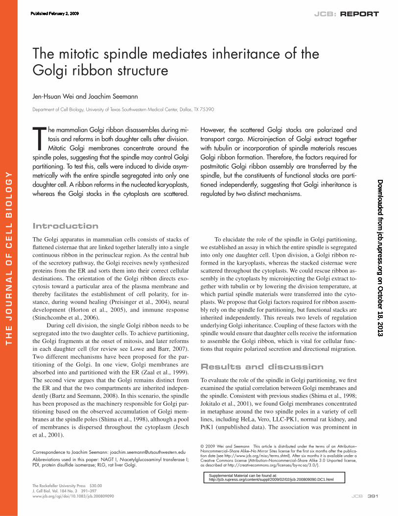
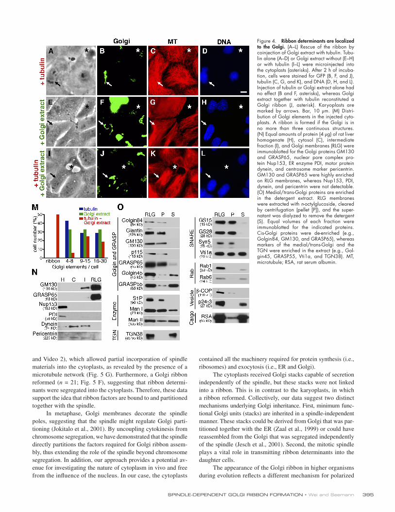
the cytoplasts and examined the Golgi morphology. Injection of
tubulin resulted in a microtubule network ( Fig. 4 C ), demon-
strating that the cellular environment in the cytoplasts supports
microtubule polymerization ( Pelletier et al., 2000 ). However,
the overall organization of Golgi stacks remained scattered
( n = 14, fi ve independent experiments; Fig. 4 B ), indicating that
the microtubules in the cytoplasts are not suffi cient to reassem-
ble a ribbon. Therefore, additional factors might be required but
were presumably segregated along with the spindle into the
karyoplasts. Because these factors may associate with Golgi
membranes, we extracted rat liver Golgi (RLG) membranes
with the nonionic detergent n -octylglucoside and removed the
cis-to-trans polarity. A similar segregation pattern was ob-
served in the cytoplasts ( Fig. 3 F ). Together with our EM
data, we conclude that the scattered Golgi in the cytoplasts is
stacked and polarized.
We next investigated whether the stacked Golgi in the cyto-
plasts transports cargo. The mRNA of CD8, a plasma membrane
protein not expressed in PtK1 cells, was injected together with
Mad1 protein into cells with asymmetrical monoasters. Cells
were incubated for 4 h to allow cell division, as well as synthesis
and transport of cargo, and were fi xed and stained before permea-
bilization. CD8 was detected on the cell surface in both karyo-
plasts and cytoplasts ( n = 22; Fig. 3 H ) but not in cells with
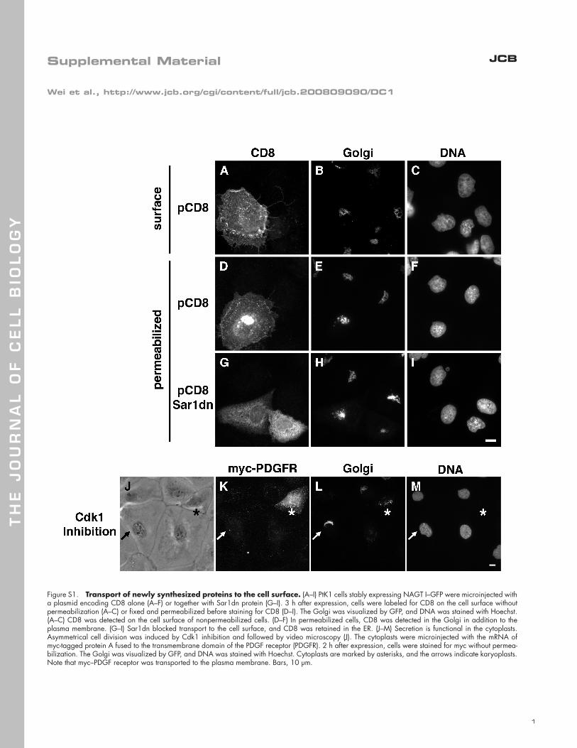
impaired secretion (Fig. S1, available at http://www.jcb.org/cgi/
content/full/jcb.200809090/DC1), indicating that the Golgi stacks
in both cells transported cargo. To rule out that CD8 might have
been expressed and transported before cytokinesis, we induced
asymmetrical cell division in the presence of the protein synthesis
inhibitor cycloheximide. After the drug was removed to allow
protein expression and transport, CD8 was detected on the cell
surface of both cells ( n = 9; unpublished data). In another ap-
proach, we injected CD8 mRNA into the cytoplasts generated by
Cdk1 inhibition. CD8 appeared on the cell surface of the cyto-
plasts but not on the noninjected karyoplasts ( n = 8; Fig. 3 L ).
Similarly, myc-tagged protein A fused to the transmembrane do-
main of the PDGF receptor was also delivered to the cell surface
( n = 15; Fig. S1 K). These fi ndings indicate that the Golgi stacks
Figure 2. The Golgi in the karyoplasts but not in the cytoplasts reforms a ribbon. (A) Dia-gram of the assay. PtK1 cells stably express-ing NAGT I – GFP were treated with an Eg5 inhibitor (monastrol or trityl-cysteine) for 2 h to induce asymmetrically positioned monoasters. Cell division was triggered either by micro-injection of Mad1 or by addition of a Cdk1 inhibitor (purvalanol A or roscovitine), leading to a karyoplast that received the entire spin-dle (centrosomes, chromosomes, and spindle microtubules) and a cytoplast that lacked all of these. (B – I) Divided cells were either identifi ed by the injection marker (B; Mad1 injection) or followed by time-lapse phase-contrast micros-copy (F; Cdk1 inhibition). Cells were stained for GFP (C and G), tubulin (D and H), and DNA (E and I). The Golgi in the karyoplasts (arrows) reformed a characteristic ribbon in the perinuclear region, whereas the Golgi in the cytoplasts (asterisks) was scattered through-out the cytoplasm (C and G). In addition, no microtubule (MT) network was present in the cytoplasts (D and H). Bar, 10 μ m.
JCB • VOLUME 184 • NUMBER 3 • 2009 394
found that medial/trans-Golgi markers were enriched in the
Golgi extract, whereas cis-Golgi proteins were mostly depleted
( Fig. 4 O ). This implies that the factors required for postmitotic
ribbon reassembly might have distinct molecular compositions
from the cis-Golgi proteins Golgin84, GRASP65, and GM130
shown to be required for ribbon maintenance in interphase
( Diao et al., 2003 ; Puthenveedu et al., 2006 ).
If the ribbon determinants are partitioned with the spin-
dle, incorporation of parts of the spindle and its associated
proteins into the cytoplasts should rescue ribbon formation.
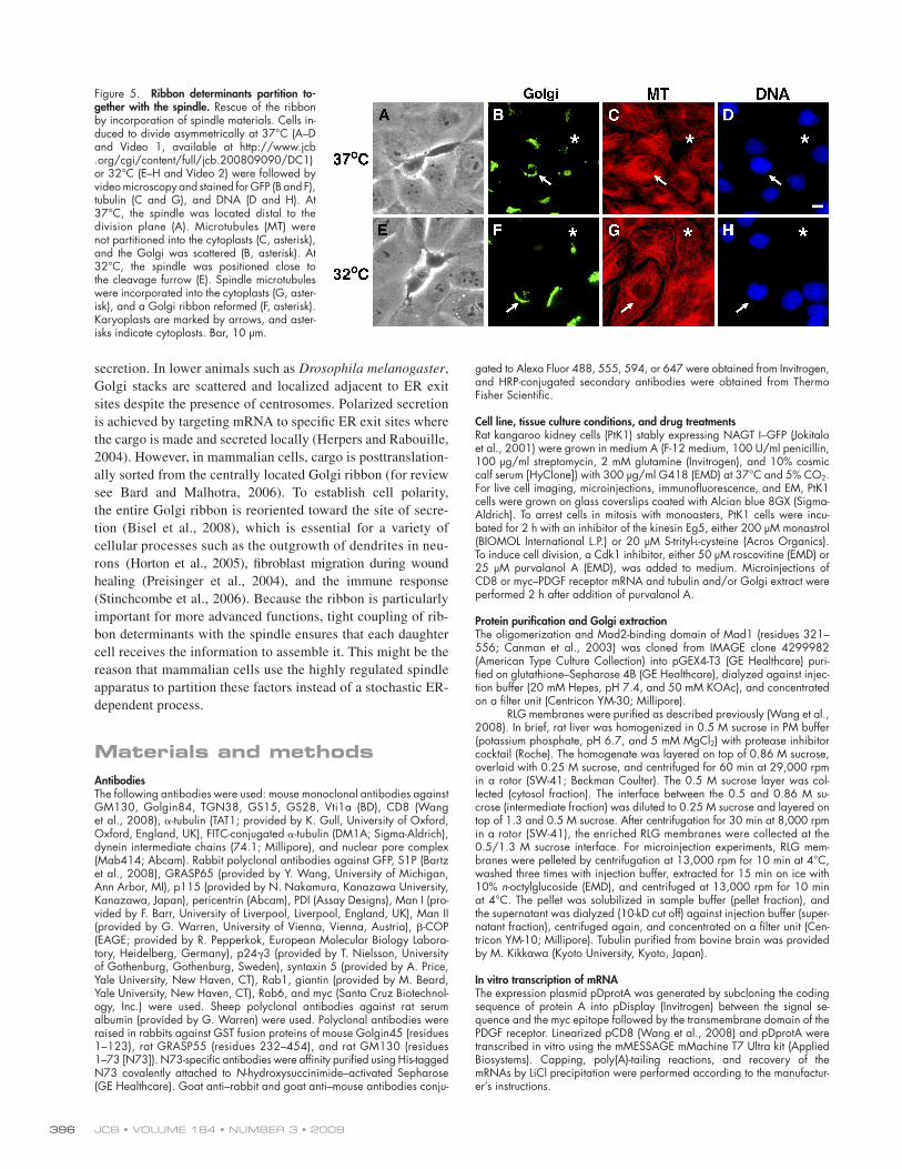
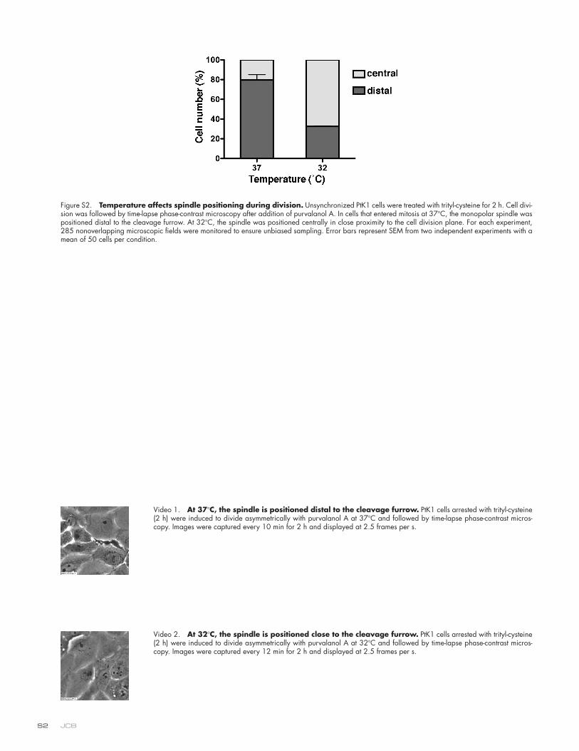
We followed asymmetrical cell division induced at lower tem-
perature (32 ° C) and found that spindle positioning was dra-
matically altered, presumably caused by effects on microtubule
dynamics (Fig. S2, available at http://www.jcb.org/cgi/content/
full/jcb.200809090/DC1). At 37 ° C, the monopolar spindle was
positioned distal to the cleavage furrow and seemingly adhered
to the cortex at one side of the cell ( Fig. 5 A and Video 1). The
cytoplasts derived from this type of division contained scat-
tered Golgi but no microtubules ( Fig. 2, G and H ; and Fig. 5,
B and C ). However, at a lower temperature, the spindle was posi-
tioned centrally in close proximity to the division plane ( Fig. 5 E
detergent by dialysis. Injection of Golgi extract alone had no ef-
fect ( n = 12, four independent experiments; Fig. 4 F ), but Golgi
extract together with tubulin restored a ribbon ( n = 8, fi ve inde-
pendent experiments; Fig. 4 J ) as well as a microtubule network
( Fig. 4 K ). Thus, the factors required for ribbon assembly are
located on the Golgi.
This is in line with previous studies showing that in cyto-
plasts generated by microsurgery, a Golgi ribbon forms in the
absence of centrosomes but requires preexisting Golgi distrib-
uted into the cut cells under interphase condition ( Maniotis and
Schliwa, 1991 ; Pelletier et al., 2000 ). The mammalian Golgi
ribbon is usually formed next to the centrosomes, which de-
pends on the minus end – directed microtubule motor dynein.
We did not detect centrosomes in serial sections of RLG mem-
branes by EM. In addition, neither the centrosomal marker peri-
centrin nor cytoplasmic dynein were detectable by Western
blotting in the RLG fraction, although the proteins were present
in the homogenate ( Fig. 4 N ). Similarly, the ER enzyme protein
disulfi de isomerase (PDI) and the nuclear pore protein Nup153
were absent in the RLG fraction. In contrast, the Golgi proteins
GM130 and GRASP65 were highly enriched. Interestingly, we
Figure 3. The Golgi stacks in the cytoplasts are polarized and transport cargo. (A – F) The Golgi in the cytoplasts is composed of polar-ized stacks. PtK1 cells stably expressing the medial/trans-Golgi enzyme NAGT I – GFP were induced to divide asymmetrically and followed by video microscopy. Upon division, cells were either processed for EM (A and B) or for immuno-fl uorescence (C – F). (A and B) Stacked Golgi cis-ternae reformed in both karyoplasts (karyo; A) and cytoplasts (cyto; B). Bar, 200 nm. (C – F) The Golgi stacks are polarized. Divided cells were triple labeled with GFP, anti-GFP, and anti – cis-Golgi marker GM130. Two-channel overlays were performed as specifi ed. GFP fl uorescence is shown in green (C – F), and the antibody stainings for GFP (C and D) and GM130 (E and F) are pseudocolored in red. The GFP fl uorescence colocalized with the anti-GFP signal (C and D) but only partially over-lapped with the anti-GM130 signal (E and F) in both karyoplasts (C and E) and cytoplasts (D and F), as shown schematically in the in-sets. Bar, 2 μ m. (G – N) CD8 transport assay. CD8 mRNA was microinjected into cells with asymmetrical monoasters together with Mad1 (G – J) or into the cytoplasts induced by Cdk1 inhibition (K – N). CD8 on the cell surface was stained before permeabilization (H and L). Note that CD8 was expressed and transported to the plasma membrane in the cytoplasts (as-terisks). Karyoplasts are marked by arrows. Bar, 10 μ m.
395SPINDLE-DEPENDENT GOLGI RIBBON FORMATION • Wei and Seemann
contained all the machinery required for protein synthesis (i.e.,
ribosomes) and exocytosis (i.e., ER and Golgi).
The cytoplasts received Golgi stacks capable of secretion
independently of the spindle, but these stacks were not linked
into a ribbon. This is in contrast to the karyoplasts, in which
a ribbon reformed. Collectively, our data suggest two distinct
mechanisms underlying Golgi inheritance. First, minimum func-
tional Golgi units (stacks) are inherited in a spindle-independent
manner. These stacks could be derived from Golgi that was par-
titioned together with the ER ( Zaal et al., 1999 ) or could have
reassembled from the Golgi that was segregated independently
of the spindle ( Jesch et al., 2001 ). Second, the mitotic spindle
plays a vital role in transmitting ribbon determinants into the
daughter cells.
The appearance of the Golgi ribbon in higher organisms
during evolution refl ects a different mechanism for polarized
and Video 2), which allowed partial incorporation of spindle
materials into the cytoplasts, as revealed by the presence of a
microtubule network ( Fig. 5 G ). Furthermore, a Golgi ribbon
reformed ( n = 21; Fig. 5 F ), suggesting that ribbon determi-
nants were segregated into the cytoplasts. Therefore, these data
support the idea that ribbon factors are bound to and partitioned
together with the spindle.
In metaphase, Golgi membranes decorate the spindle
poles, suggesting that the spindle might regulate Golgi parti-
tioning ( Jokitalo et al., 2001 ). By uncoupling cytokinesis from
chromosome segregation, we have demonstrated that the spindle
directly partitions the factors required for Golgi ribbon assem-
bly, thus extending the role of the spindle beyond chromosome
segregation. In addition, our approach provides a potential av-
enue for investigating the nature of cytoplasm in vivo and free
from the infl uence of the nucleus. In our case, the cytoplasts
Figure 4. Ribbon determinants are localized to the Golgi. (A – L) Rescue of the ribbon by coinjection of Golgi extract with tubulin. Tubu-lin alone (A – D) or Golgi extract without (E – H) or with tubulin (I – L) were microinjected into the cytoplasts (asterisks). After 2 h of incuba-tion, cells were stained for GFP (B, F, and J), tubulin (C, G, and K), and DNA (D, H, and L). Injection of tubulin or Golgi extract alone had no effect (B and F, asterisks), whereas Golgi extract together with tubulin reconstituted a Golgi ribbon (J, asterisk). Karyoplasts are marked by arrows. Bar, 10 μ m. (M) Distri-bution of Golgi elements in the injected cyto-plasts. A ribbon is formed if the Golgi is in no more than three continuous structures. (N) Equal amounts of protein (4 μ g) of rat liver homogenate (H), cytosol (C), intermediate fraction (I), and Golgi membranes (RLG) were immunoblotted for the Golgi proteins GM130 and GRASP65, nuclear pore complex pro-tein Nup153, ER enzyme PDI, motor protein dynein, and centrosome marker pericentrin. GM130 and GRASP65 were highly enriched on RLG membranes, whereas Nup153, PDI, dynein, and pericentrin were not detectable. (O) Medial/trans-Golgi proteins are enriched in the detergent extract. RLG membranes were extracted with n -octylglucoside, cleared by centrifugation (pellet [P]), and the super-natant was dialyzed to remove the detergent (S). Equal volumes of each fraction were immuno blotted for the indicated proteins. Cis-Golgi proteins were de-enriched (e.g., Golgin84, GM130, and GRASP65), whereas markers of the medial/trans-Golgi and the TGN were enriched in the extract (e.g., Gol-gin45, GRASP55, Vti1a, and TGN38). MT, microtubule; RSA, rat serum albumin.
JCB • VOLUME 184 • NUMBER 3 • 2009 396
gated to Alexa Fluor 488, 555, 594, or 647 were obtained from Invitrogen, and HRP-conjugated secondary antibodies were obtained from Thermo Fisher Scientifi c.
Cell line, tissue culture conditions, and drug treatments Rat kangaroo kidney cells (PtK1) stably expressing NAGT I – GFP ( Jokitalo et al., 2001 ) were grown in medium A (F-12 medium, 100 U/ml penicillin, 100 μ g/ml streptomycin, 2 mM glutamine (Invitrogen), and 10% cosmic calf serum [HyClone]) with 300 μ g/ml G418 (EMD) at 37 ° C and 5% CO 2 . For live cell imaging, microinjections, immunofl uorescence, and EM, PtK1 cells were grown on glass coverslips coated with Alcian blue 8GX (Sigma-Aldrich). To arrest cells in mitosis with monoasters, PtK1 cells were incu-bated for 2 h with an inhibitor of the kinesin Eg5, either 200 μ M monastrol (BIOMOL International L.P.) or 20 μ M S-trityl- L -cysteine (Acros Organics). To induce cell division, a Cdk1 inhibitor, either 50 μ M roscovitine (EMD) or 25 μ M purvalanol A (EMD), was added to medium. Microinjections of CD8 or myc – PDGF receptor mRNA and tubulin and/or Golgi extract were performed 2 h after addition of purvalanol A.
Protein purifi cation and Golgi extraction The oligomerization and Mad2-binding domain of Mad1 (residues 321 – 556; Canman et al., 2003 ) was cloned from IMAGE clone 4299982 (American Type Culture Collection) into pGEX4-T3 (GE Healthcare) puri-fi ed on glutathione – Sepharose 4B (GE Healthcare), dialyzed against injec-tion buffer (20 mM Hepes, pH 7.4, and 50 mM KOAc), and concentrated on a fi lter unit (Centricon YM-30; Millipore).
RLG membranes were purifi ed as described previously ( Wang et al., 2008 ). In brief, rat liver was homogenized in 0.5 M sucrose in PM buffer (potassium phosphate, pH 6.7, and 5 mM MgCl 2 ) with protease inhibitor cocktail (Roche). The homogenate was layered on top of 0.86 M sucrose, overlaid with 0.25 M sucrose, and centrifuged for 60 min at 29,000 rpm in a rotor (SW-41; Beckman Coulter). The 0.5 M sucrose layer was col-lected (cytosol fraction). The interface between the 0.5 and 0.86 M su-crose (intermediate fraction) was diluted to 0.25 M sucrose and layered on top of 1.3 and 0.5 M sucrose. After centrifugation for 30 min at 8,000 rpm in a rotor (SW-41), the enriched RLG membranes were collected at the 0.5/1.3 M sucrose interface. For microinjection experiments, RLG mem-branes were pelleted by centrifugation at 13,000 rpm for 10 min at 4 ° C, washed three times with injection buffer, extracted for 15 min on ice with 10% n -octylglucoside (EMD), and centrifuged at 13,000 rpm for 10 min at 4 ° C. The pellet was solubilized in sample buffer (pellet fraction), and the supernatant was dialyzed (10-kD cut off) against injection buffer (super-natant fraction), centrifuged again, and concentrated on a fi lter unit (Cen-tricon YM-10; Millipore). Tubulin purifi ed from bovine brain was provided by M. Kikkawa (Kyoto University, Kyoto, Japan).
In vitro transcription of mRNA The expression plasmid pDprotA was generated by subcloning the coding sequence of protein A into pDisplay (Invitrogen) between the signal se-quence and the myc epitope followed by the transmembrane domain of the PDGF receptor. Linearized pCD8 ( Wang et al., 2008 ) and pDprotA were transcribed in vitro using the mMESSAGE mMachine T7 Ultra kit (Applied Biosystems). Capping, poly(A)-tailing reactions, and recovery of the mRNAs by LiCl precipitation were performed according to the manufactur-er ’ s instructions.
secretion. In lower animals such as Drosophila melanogaster ,
Golgi stacks are scattered and localized adjacent to ER exit
sites despite the presence of centrosomes. Polarized secretion
is achieved by targeting mRNA to specifi c ER exit sites where
the cargo is made and secreted locally ( Herpers and Rabouille,
2004 ). However, in mammalian cells, cargo is posttranslation-
ally sorted from the centrally located Golgi ribbon (for review
see Bard and Malhotra, 2006 ). To establish cell polarity,
the entire Golgi ribbon is reoriented toward the site of secre-
tion ( Bisel et al., 2008 ), which is essential for a variety of
cellular processes such as the outgrowth of dendrites in neu-
rons ( Horton et al., 2005 ), fi broblast migration during wound
healing ( Preisinger et al., 2004 ), and the immune response
( Stinchcombe et al., 2006 ). Because the ribbon is particularly
important for more advanced functions, tight coupling of rib-
bon determinants with the spindle ensures that each daughter
cell receives the information to assemble it. This might be the
reason that mammalian cells use the highly regulated spindle
apparatus to partition these factors instead of a stochastic ER-
dependent process.
Materials and methods Antibodies The following antibodies were used: mouse monoclonal antibodies against GM130, Golgin84, TGN38, GS15, GS28, Vti1a (BD), CD8 ( Wang et al., 2008 ), � -tubulin (TAT1; provided by K. Gull, University of Oxford, Oxford, England, UK), FITC-conjugated � -tubulin (DM1A; Sigma-Aldrich), dynein intermediate chains (74.1; Millipore), and nuclear pore complex (Mab414; Abcam). Rabbit polyclonal antibodies against GFP, S1P ( Bartz et al., 2008 ), GRASP65 (provided by Y. Wang, University of Michigan, Ann Arbor, MI), p115 (provided by N. Nakamura, Kanazawa University, Kanazawa, Japan), pericentrin (Abcam), PDI (Assay Designs), Man I (pro-vided by F. Barr, University of Liverpool, Liverpool, England, UK), Man II (provided by G. Warren, University of Vienna, Vienna, Austria), � -COP (EAGE; provided by R. Pepperkok, European Molecular Biology Labora-tory, Heidelberg, Germany), p24 � 3 (provided by T. Nielsson, University of Gothenburg, Gothenburg, Sweden), syntaxin 5 (provided by A. Price, Yale University, New Haven, CT), Rab1, giantin (provided by M. Beard, Yale University, New Haven, CT), Rab6, and myc (Santa Cruz Biotechnol-ogy, Inc.) were used. Sheep polyclonal antibodies against rat serum albumin (provided by G. Warren) were used. Polyclonal antibodies were raised in rabbits against GST fusion proteins of mouse Golgin45 (residues 1 – 123), rat GRASP55 (residues 232 – 454), and rat GM130 (residues 1 – 73 [N73]). N73-specifi c antibodies were affi nity purifi ed using His-tagged N73 covalently attached to N -hydroxysuccinimide – activated Sepharose (GE Healthcare). Goat anti – rabbit and goat anti – mouse antibodies conju-
Figure 5. Ribbon determinants partition to-gether with the spindle. Rescue of the ribbon by incorporation of spindle materials. Cells in-duced to divide asymmetrically at 37 ° C (A – D and Video 1, available at http://www.jcb.org/cgi/content/full/jcb.200809090/DC1) or 32 ° C (E – H and Video 2) were followed by video microscopy and stained for GFP (B and F), tubulin (C and G), and DNA (D and H). At 37 ° C, the spindle was located distal to the division plane (A). Microtubules (MT) were not partitioned into the cytoplasts (C, asterisk), and the Golgi was scattered (B, asterisk). At 32 ° C, the spindle was positioned close to the cleavage furrow (E). Spindle microtubules were incorporated into the cytoplasts (G, aster-isk), and a Golgi ribbon reformed (F, asterisk). Karyoplasts are marked by arrows, and aster-isks indicate cytoplasts. Bar, 10 μ m.
397SPINDLE-DEPENDENT GOLGI RIBBON FORMATION • Wei and Seemann
References Bard , F. , and V. Malhotra . 2006 . The formation of TGN-to-plasma-membrane
transport carriers. Annu. Rev. Cell Dev. Biol. 22 : 439 – 455 .
Bartz , R. , and J. Seemann . 2008 . Mitotic regulation of SREBP and ATF6 by sep-aration of the Golgi and ER. Cell Cycle . 7 : 2100 – 2105 .
Bartz , R. , L.P. Sun , B. Bisel , J.H. Wei , and J. Seemann . 2008 . Spatial separation of Golgi and ER during mitosis protects SREBP from unregulated activa-tion. EMBO J. 27 : 948 – 955 .
Bisel , B. , Y. Wang , J.H. Wei , Y. Xiang , D. Tang , M. Miron-Mendoza , S. Yoshimura , N. Nakamura , and J. Seemann . 2008 . ERK regulates Golgi and centrosome orientation towards the leading edge through GRASP65. J. Cell Biol. 182 : 837 – 843 .
Canman , J.C. , L.A. Cameron , P.S. Maddox , A. Straight , J.S. Tirnauer , T.J. Mitchison , G. Fang , T.M. Kapoor , and E.D. Salmon . 2003 . Determining the position of the cell division plane. Nature . 424 : 1074 – 1078 .
Diao , A. , D. Rahman , D.J. Pappin , J. Lucocq , and M. Lowe . 2003 . The coiled-coil membrane protein golgin-84 is a novel rab effector required for Golgi ribbon formation. J. Cell Biol. 160 : 201 – 212 .
Herpers , B. , and C. Rabouille . 2004 . mRNA localization and ER-based protein sorting mechanisms dictate the use of transitional endoplasmic reticulum-golgi units involved in gurken transport in Drosophila oocytes. Mol. Biol. Cell . 15 : 5306 – 5317 .
Horton , A.C. , B. R á cz , E.E. Monson , A.L. Lin , R.J. Weinberg , and M.D. Ehlers . 2005 . Polarized secretory traffi cking directs cargo for asymmetric den-drite growth and morphogenesis. Neuron . 48 : 757 – 771 .
Hu , C.K. , M. Coughlin , C.M. Field , and T.J. Mitchison . 2008 . Cell polarization during monopolar cytokinesis. J. Cell Biol. 181 : 195 – 202 .
Jesch , S.A. , A.J. Mehta , M. Velliste , R.F. Murphy , and A.D. Linstedt . 2001 . Mitotic Golgi is in a dynamic equilibrium between clustered and free vesicles independent of the ER. Traffi c . 2 : 873 – 884 .
Jokitalo , E. , N. Cabrera-Poch , G. Warren , and D.T. Shima . 2001 . Golgi clusters and vesicles mediate mitotic inheritance independently of the endoplas-mic reticulum. J. Cell Biol. 154 : 317 – 330 .
Lowe , M. , and F.A. Barr . 2007 . Inheritance and biogenesis of organelles in the secretory pathway. Nat. Rev. Mol. Cell Biol. 8 : 429 – 439 .
Maniotis , A. , and M. Schliwa . 1991 . Microsurgical removal of centrosomes blocks cell reproduction and centriole generation in BSC-1 cells. Cell . 67 : 495 – 504 .
Mayer , T.U. , T.M. Kapoor , S.J. Haggarty , R.W. King , S.L. Schreiber , and T.J. Mitchison . 1999 . Small molecule inhibitor of mitotic spindle bipolarity identifi ed in a phenotype-based screen. Science . 286 : 971 – 974 .
Niiya , F. , X. Xie , K.S. Lee , H. Inoue , and T. Miki . 2005 . Inhibition of cyclin-dependent kinase 1 induces cytokinesis without chromosome segrega-tion in an ECT2 and MgcRacGAP-dependent manner. J. Biol. Chem. 280 : 36502 – 36509 .
Pelletier , L. , E. Jokitalo , and G. Warren . 2000 . The effect of Golgi depletion on exocytic transport. Nat. Cell Biol. 2 : 840 – 846 .
Preisinger , C. , B. Short , V. De Corte , E. Bruyneel , A. Haas , R. Kopajtich , J. Gettemans , and F.A. Barr . 2004 . YSK1 is activated by the Golgi matrix protein GM130 and plays a role in cell migration through its substrate 14-3-3 � . J. Cell Biol. 164 : 1009 – 1020 .
Puthenveedu , M.A. , C. Bachert , S. Puri , F. Lanni , and A.D. Linstedt . 2006 . GM130 and GRASP65-dependent lateral cisternal fusion allows uniform Golgi-enzyme distribution. Nat. Cell Biol. 8 : 238 – 248 .
Shima , D.T. , K. Haldar , R. Pepperkok , R. Watson , and G. Warren . 1997 . Partitioning of the Golgi apparatus during mitosis in living HeLa cells. J. Cell Biol. 137 : 1211 – 1228 .
Shima , D.T. , N. Cabrera-Poch , R. Pepperkok , and G. Warren . 1998 . An or-dered inheritance strategy for the Golgi apparatus: visualization of mitotic disassembly reveals a role for the mitotic spindle. J. Cell Biol. 141 : 955 – 966 .
Skoufi as , D.A. , S. Debonis , Y. Saoudi , L. Lebeau , I. Crevel , R. Cross , R.H. Wade , D. Hackney , and F. Kozielski . 2006 . S-trityl-l-cysteine is a reversible, tight-binding inhibitor of the human kinesin eg5 that specifi cally blocks mitotic progression. J. Biol. Chem. 281 : 17559 – 17569 .
Stinchcombe , J.C. , E. Majorovits , G. Bossi , S. Fuller , and G.M. Griffi ths . 2006 . Centrosome polarization delivers secretory granules to the immunologi-cal synapse. Nature . 443 : 462 – 465 .
Wang , Y. , J.H. Wei , B. Bisel , D. Tang , and J. Seemann . 2008 . Golgi cisternal un-stacking stimulates COPI vesicle budding and protein transport. PLoS ONE . 3 : e1647 .
Zaal , K.J. , C.L. Smith , R.S. Polishchuk , N. Altan , N.B. Cole , J. Ellenberg , K. Hirschberg , J.F. Presley , T.H. Roberts , E. Siggia , et al . 1999 . Golgi mem-branes are absorbed into and reemerge from the ER during mitosis. Cell . 99 : 589 – 601 .
Microinjection and live cell imaging For time-lapse microscopy and microinjection experiments, cells were in-cubated in medium A containing 50 mM Hepes, pH 7.4. Microinjection was performed with a microinjector (Transjector 5246 or FemtoJet; Ep-pendorf) and a micromanipulator (5171; Eppendorf) connected to a microscope (Axiovert 200M; Carl Zeiss, Inc.; Bartz and Seemann, 2008 ). The reagents were injected at the following concentrations: 1.5 mg/ml Mad1 or Sar1dn ( Bartz and Seemann, 2008 ), 0.2 mg/ml pCD8, 0.5 mg/ml CD8 or myc – PDGF receptor mRNA, 6 mg/ml tubulin, and 20 mg/ml Golgi extract. Mad1 was injected together with 2.5 mg/ml biotin-dextran as an injection marker and visualized by streptavidin – Alexa Fluor 350 (Invitrogen). All microinjection experiments were performed at least three times.
For time-lapse microscopy, coverslips were mounted into a chamber (Ludin; Life Imaging Services). The temperature was maintained using an incubator (XL-3; Carl Zeiss, Inc.) at 37 ° C unless otherwise stated. Phase-contrast images were taken at intervals of 3 – 20 min with an A-Plan 20 × /0.3 PH1 objective (Carl Zeiss, Inc.) and a microscope (Axiovert 200M). Either a camera (Orca-285; Hamamatsu Photonics) and Openlab software (version 4.0.2; PerkinElmer) or a camera (DXM1200F; Nikon) in combination with MetaMorph software (version 7.1.3; MDS Analytical Technologies) was used.
Immunofl uorescence and EM analysis Cells were either fi xed in 3.7% formaldehyde in PBS for 15 min and per-meabilized in methanol at � 20 ° C for 15 min or fi xed and permeabilized in methanol at � 20 ° C for 15 min and incubated with appropriate primary antibodies followed by secondary antibodies. DNA was stained with DRAQ5 (Biostatus) after RNase treatment or with Hoechst 33342 (Invitro-gen). After staining, cells were mounted in Mowiol 4 – 88 (EMD). Upon the acquisition of time-lapse phase-contrast images, cells were fi xed and stained in the chamber (Ludin) and repositioned onto the microscope stage to the original positions recorded during live cell imaging. Immunofl uores-cence analysis was performed with a Plan Neofl uar 40 × /1.3 differential interference contrast objective (Carl Zeiss, Inc.) and a camera (Orca-285) with the Openlab software (version 4.0.2). The contrast of the images was adjusted for the Golgi structures in the cytoplasts.
For quantitative analysis of the ribbon structure, the analyze parti-cles function of ImageJ (National Institutes of Health) was used, and a fi xed threshold was applied to all raw images ( Puthenveedu et al., 2006 ). The Golgi was determined as a ribbon if 90% of the fl uorescence intensity re-sided in no more than three continuous elements.
EM analysis was performed essentially as described previously ( Wang et. al, 2008 ). Asymmetrical cell division was induced by treat-ment with trityl-cysteine (2 h) followed by purvalanol A (2 h) and moni-tored by video microscopy. Upon division, the cells surrounding the karyoplasts and cytoplasts were scraped off with a microinjection needle. The remaining cells were fi xed in 2.5% glutaraldehyde in 0.1 M sodium cacodylate, pH 7.4, for 30 min, treated with 1% osmium tetroxide and 1.5% potassium cyanoferrate in 0.1 M cacodylate for 30 min, and em-bedded in Epon 812 (Electron Microscopy Sciences). Upon removal of the coverslips by hydrofl uoric acid, thin sections were cut and stained with 2% uranyl acetate and lead citrate. Sections were analyzed on an electron microscope (Tecnai G2 Spirit; FEI Company), and images were captured on a charge-coupled device camera (USC1000 2k; Gatan). Stacks were defi ned as three or more cisternae separated by < 20 nm.
Online supplemental material Fig. S1 shows transport of newly synthesized proteins to the cell surface. Fig. S2 shows that temperature affects spindle positioning during division. Video 1 shows that PtK1 cells were induced to divide asymmetrically at 37 ° C. Video 2 shows that PtK1 cells were induced to divide asymmetrically at 32 ° C. Online supplemental material is available at http://www.jcb.org/cgi/content/full/jcb.200809090/DC1.
We thank C. Gilpin, T. Januszewski, and the University of Texas Southwest-ern EM facility for EM assistance, Y. Wang, D. Mundy, and B. Bisel for criti-cal reading of the manuscript, M. Kikkawa for purifi ed tubulin, and H. Yu for advice.
J. Seemann is a Virginia Murchison Lithicum Scholar in Medical Re-search and is supported by a grant from the American Heart Association (8065090F).
Submitted: 12 September 2008 Accepted: 7 January 2009
TH
E J
OU
RN
AL
OF
CE
LL
BIO
LO
GY
JCB
1
Supplemental Material
Wei et al., http://www.jcb.org/cgi/content/full/jcb.200809090/DC1
Figure S1. Transport of newly synthesized proteins to the cell surface. (A–I) PtK1 cells stably expressing NAGT I–GFP were microinjected with a plasmid encoding CD8 alone (A–F) or together with Sar1dn protein (G–I). 3 h after expression, cells were labeled for CD8 on the cell surface without permeabilization (A–C) or fixed and permeabilized before staining for CD8 (D–I). The Golgi was visualized by GFP, and DNA was stained with Hoechst. (A–C) CD8 was detected on the cell surface of nonpermeabilized cells. (D–F) In permeabilized cells, CD8 was detected in the Golgi in addition to the plasma membrane. (G–I) Sar1dn blocked transport to the cell surface, and CD8 was retained in the ER. (J–M) Secretion is functional in the cytoplasts. Asymmetrical cell division was induced by Cdk1 inhibition and followed by video microscopy (J). The cytoplasts were microinjected with the mRNA of myc-tagged protein A fused to the transmembrane domain of the PDGF receptor (PDGFR). 2 h after expression, cells were stained for myc without permea-bilization. The Golgi was visualized by GFP, and DNA was stained with Hoechst. Cytoplasts are marked by asterisks, and the arrows indicate karyoplasts. Note that myc–PDGF receptor was transported to the plasma membrane. Bars, 10 µm.
JCB S2
Figure S2. Temperature affects spindle positioning during division. Unsynchronized PtK1 cells were treated with trityl-cysteine for 2 h. Cell divi-sion was followed by time-lapse phase-contrast microscopy after addition of purvalanol A. In cells that entered mitosis at 378C, the monopolar spindle was positioned distal to the cleavage furrow. At 328C, the spindle was positioned centrally in close proximity to the cell division plane. For each experiment, 285 nonoverlapping microscopic fields were monitored to ensure unbiased sampling. Error bars represent SEM from two independent experiments with a mean of 50 cells per condition.
Video 1. At 378C, the spindle is positioned distal to the cleavage furrow. PtK1 cells arrested with trityl-cysteine (2 h) were induced to divide asymmetrically with purvalanol A at 378C and followed by time-lapse phase-contrast micros-copy. Images were captured every 10 min for 2 h and displayed at 2.5 frames per s.
Video 2. At 328C, the spindle is positioned close to the cleavage furrow. PtK1 cells arrested with trityl-cysteine (2 h) were induced to divide asymmetrically with purvalanol A at 328C and followed by time-lapse phase-contrast micros-copy. Images were captured every 12 min for 2 h and displayed at 2.5 frames per s.
Copyright © 2022 FDOKUMEN