Spindle cell carcinoma of maxilla: Histomorphological and immunohistochemical analysis of a case
7
-
Upload
independent -
Category
Documents
-
view
0 -
download
0
Transcript of Spindle cell carcinoma of maxilla: Histomorphological and immunohistochemical analysis of a case

Journal of Oral and Maxillofacial Pathology Vol. 18 Issue 2 May - Aug 2014 256
INTRODUCTION
Spindle cell lesions of head and neck include a diverse group of clinically and biologically heterogenous lesions ranging from benign-reactive entities to malignancies.[1] In this array, spindle cell carcinoma (SpCC) stands out as a rare malignant biphasic tumor comprising both epithelial and mesenchymal components, which is of presumed epithelial origin.[2,3] The WHOclassificationhasalsoplacedthisentityundermalignantepithelial tumors of squamous cell carcinoma (SCC). The diagnostic criteria state that the presence of malignant spindle cells in addition to demonstration of invasive or in situ SCC or any evidence of epithelial differentiation of spindle cells are required to reveal SpCC.[4] However, about one-third of thecasesaremonophasic,makingdiagnosisdifficult.[5] We report one such unusual case that occurred in the maxilla of a 40-year old patient and depicted exclusively spindle cell morphology. Exclusion of lesions like pleomorphic
sarcoma(PS),fibrosarcoma,rhabdomyosarcoma(RMS)andangiosarcoma (AS) by IHC was mandatory. The histogenesis of spindle cells in SpCC and the various differential diagnoses were reviewed.
CASE REPORT
A 40-year male patient reported to SCB Dental College, Cuttack, with a complaint of swelling in the anterior part of mouth. History revealed a rapidly growing mass in the anterior palatal gingiva with duration of 3 months. Histopathological study done elsewhere had diagnosed it as a pyogenic granuloma. Patient had habit of chewing tobacco since 10 years. Intra-oral examination revealed an exophytic, polypoidal pinkish red mass in the maxillary anterior gingiva measuring about 6 × 3.5 × 3.5 cm with an ulcerated surface [Figure 1]. Such aggressive clinical features along with history of rapid growth prompted us to review the previous histopathology slide that displayed foci of poorly differentiated polygonal squamous epithelial cells with abundant cytoplasm and round, vesicular nuclei, spindle cells, few bizarre cells and increased number of mitoticfiguresinthesection[Figure2].Therewereareasinthesection that mimicked granulation tissue [Figure 3]. Based on these features, a poorly differentiated carcinoma was diagnosed, although the possibility of a SpCC was kept in mind. There was no history of radiotherapy. Orthopantomogram showed presence
Spindle cell carcinoma of maxilla: Histomorphological and immunohistochemical analysis of a case
Rachna Rath, Bijay K Das, SN Das, Manas Baisakh1
Department of Oral and Maxillofacial Pathology, SCB Dental College and Hospital, Cuttack, 1Consultant Oncopathologist, Apollo Hospitals, Bhubaneshwar, Odisha, India
Address for correspondence: Dr. Rachna Rath, Assistant Professor, Department of Oral and Maxillofacial Pathology, SCB Dental College and Hospital, Cuttack ‑ 753 007, Odisha, India. E‑mail: [email protected]
Received: 25‑09‑2013 Accepted: 02-07-2014
ABSTRACTThe intriguing array of spindle cell lesions occurring, especially in the head and neck region, poses a critical diagnostic challenge not only to the histopathologist but also ultimately to the clinicians for planning an appropriate treatment protocol. Overlapping spectrum of clinico-radiographic and microscopic features further compounds this problem. In such situations, the aid of ancillary techniques like immunohistochemistry (IHC) is sought to clinch the diagnosis. But is the diagnosis based on IHC most decisive? Probably the answer lies in the Pandora’s Box. This paper analyzed the potential doubts, apprehensions and the reliability that is singularly based on the morphological spindle appearance of an abnormal cell that has deviated from the usual clinical and microscopic presentation and thus posed a diagnostic dilemma warranting more questions than the answers. We review the etiopathogenesis of this entity and its differential diagnosis from few other spindle cell lesions of head and neck with special reference to use of immunohistochemical markers, with a case study.Key words: Immunohistochemistry, spindle cell lesion, spindle cell carcinoma, sarcomatoid carcinoma
Access this article online
Quick Response Code:Website:
www.jomfp.in
DOI:
10.4103/0973-029X.140772
CASE REPORT

Journal of Oral and Maxillofacial Pathology: Vol. 18 Issue 2 May ‑ Aug 2014
Spindle cell carcinoma with histological differentials Rath, et al. 257
of a radiolucent lesion in anterior maxilla [Figure 4]. Following baseline investigations partial maxillectomy was performed along with selective lymph node neck dissection involving level I and II. Grossing showed a multilobulated mass of dimension 3.8 × 3.5 × 3.5 cm with partly resorbed alveolar and palatal bone. Microscopically, sections from different areas of the lesion revealed a highly cellular tumor with invading sheets of spindle cells with oval or round nuclei with vesicular chromatin, arranged in a fascicular and storiform pattern below a normal appearingstratifiedsquamousepithelium.Therewascellularpleomorphism, atypical and increasedmitotic figures, giantcells,bizarrecellsandfewmixedinflammatorycellsinafibrousconnective tissue stroma of increased vascularity [Figure 5]. However, there was no evidence of invasive island of epithelial cells or dysplastic epithelium, even in serial sections. The tumor was provisionally diagnosed as a spindle cell sarcoma. The spindle cell morphology and varied morphological presentation raised wide-ranging possibilities in diagnosis starting from PS, AS to RMS [Figures 6-8], respectively. Due to confusion and disparity over the diagnosis, immunohistochemistry (IHC) was done with markers pancytokeratin (Dako, AE1/AE3 clone),
epithelial membrane antigen (EMA, Dako, E29 clone), CD 10, vimentin, smooth muscle actin (SMA), S-100, HMB-45, CD 68, desmin (Biogenex, V9 and 33 clones, respectively) and CD34 (Dako, QBEnd10 clone). The spindle cells showed diffuse and intense cytoplasmic positivity with both AE1/AE3 and vimentin [Figures 9 and 10]. They showed negative staining for the other markers. Thus, tumor was diagnosed as SpCC. The nodes were free of any metastasis. The patient has remained on a 1-year uneventful follow-up since then without any recurrence.
DISCUSSION
The accurate diagnosis of spindle cell lesions of head and neck has always remained an enigma for the histopathologist. These lesions invariably occur in the skin, soft tissue of scalp, orbit, neck and along the upper aerodigestive tract.[1-3] Spindle cell carcinoma is most predominant among them, occurring commonly in the larynx, hypopharynx and uncommonly in the oral cavity with less than 1% involving the lips, tongue, mandibular alveolar ridge and very few in maxilla.[6-8] It usually occurs in the 6th-7th decades of life. The predisposing factors for SpCC include smoking, alcohol consumption and previous irradiation of head
Figure 1: Clinical image shows polypoidal growth in anterior alveolus
Figure 3: Photomicrograph shows spindle cells in loose stroma mimicking granulation tissue (H&E stain, x100)
Figure 4: Orthopantomogram shows radiolucent lesion in anterior maxilla
Figure 2: Photomicrograph shows poorly differentiated squamous cells with abundant cytoplasm and enlarged nuclei with vesicular chromatin (H&E stain, x200)

Journal of Oral and Maxillofacial Pathology: Vol. 18 Issue 2 May ‑ Aug 2014
Spindle cell carcinoma with histological differentials Rath, et al. 258
Figure 5: Photomicrograph shows proliferating sheets of spindle cells below a normal appearing stratified squamous epithelium (H&E stain, ×40)
Figure 8: Photomicrograph shows pleomorphic and anaplastic spindle cells resembling pleomorphic RMS (H&E stain, ×400)
Figure 6: Photomicrograph shows pleomorphic spindle cells in storiform pattern with giant cells, resembling pleomorphic sarcoma (H&E stain, x100)
Figure 7: Photomicrograph shows atypical plump spindle cells with gaping vascular channels, akin to an AS (H&E stain, x200)
Figure 9: Photomicrograph showing diffuse and moderately intense positivity of spindle cells for pan cytokeratin (IHC stain, x400)
Figure 10: Photomicrograph showing intense cytoplasmic positivity for vimentin in the spindle cells (IHC stain, x100)

Journal of Oral and Maxillofacial Pathology: Vol. 18 Issue 2 May ‑ Aug 2014
Spindle cell carcinoma with histological differentials Rath, et al. 259
and neck.[9] According to Lewis et al., only 18% of SpCC in aerodigestive tract received radiotherapy.[10] In a series of 103 similar oral cases, none of the patients had the history of radiation. The author suggested that this could be due to preferential surgery done in this locale instead of adjuvant radiotherapy.[9] Spindle cell carcinoma mostly manifests as a rapidly growing exophytic, polypoidal mass or non-healing ulcer with symptoms of pain and swelling.[2,3] Many consider it as an unusual form of poorly differentiated SCC with aggressive behavior.[4,5] This case was unique by way of its occurrence at a younger age, in the maxilla and without prior history of radiotherapy.
The term SpCC was coined by Shervin et al.[4] Despite immunohistochemical, ultrastructural and genetic studies, the histogenesis of SpCC remains controversial.[10-12] The terminologiesalsoreflectthevaryinginterpretationofthespindlecell component. One of the theories suggests that the spindle cells and epithelial cells arise simultaneously from separate stem cells. Proponents of this theory argued that there is independent metastasis of the sarcomatous element and difference in immunohistochemical staining pattern between the epithelial and spindle cells.[11] But this concept of a collision tumor or carcinosarcoma seemed rather unlikely as areas of transition exist between the two cell types.[4] The term pseudosarcoma arose from the theory that the spindle component was a benign stromal proliferation to the SCC, but this was again doubtful as the spindle cells were found to be non-diploid suggesting their neoplastic nature.[2,4] The monoclonal origin of SpCC and dedifferentiation theory of epithelial cells to spindle cells was supported by immunohistochemical studies where both the epithelial and spindle cells show positivity for cytokeratin and EMA.[4,6,11] Double labeling indicated cytokeratin and vimentin positivity in individual spindle cells thus illustrating theversatilityoftheintermediatefilament.[12,13] Ultrastructural studies too have ascertained presence of desmosomes and tonofilaments, thus justifying the term sarcomatoidcarcinoma.[14] The basic defect is reported to be a dysfunctional cadherin-catenin complex resulting in alteration of the keratin filamentnetwork that shifts themorphology fromsquamoidto more spindled form. In addition, it is suggested that there is functional loss of genes that control epithelial differentiation and conversion to spindle morphology is a recessive entry.[13,14]
In spite of debatable histogenesis, it is agreed that SpCC has both characteristic malignant epithelial and spindle cells, the latter appearing bland and regular or with marked pleomorphism, in a variety of architectural patterns including storiform, fascicular, or more definable sarcomatousdifferentiation.[9,10] Multinucleated giant cells (MNGC) and numerousmitoticfiguresarealsopresent.[15] The squamous component appears in the form of either focal dysplasia, carcinoma in situ or frank SCC between the spindle cells and thus aids in diagnosis. Direct transition between the two cell types may be seen.[5,6] Thompson et al. in their study found squamoid differentiation in 79.6% of SpCC cases.[6] Other studies show that the squamous component can be found in
60-90% cases only.[1] Our case showed sheet like proliferation of pleomorphic spindle cells without any obvious squamoid component. The lone presence of spindle cells might mislead the pathologist toward the diagnosis of other spindle cell lesions.[1,2,6] An abnormal cell can take on a number of bizarre morphologies once there is loss of cellular control in them. Should a morphological feature like spindling of an abnormal cell be relied upon to term a tumor as SpCC? Is the mere presence of an epithelial-like or spindle-like morphology of an abnormal cell enough to designate a tumor as a carcinoma or sarcoma? Should we not go beyond these ambiguous histomorphological boundaries and look for other specific innate and qualifying characteristics like productof the tumor cells or ultrastructural elements that would be precisely diagnostic for lesions like the so called SpCC? In such a scenario, IHC detection of cytokeratin and vimentin is valuable, both of which are expressed in SpCC and may be considered diagnostic, especially in those cases where clear epithelial component is absent.[1,6,10] It has been seen that even pancytokeratin positivity in spindle cells occurs in 26-62% cases.[6,10,13,16] Therefore, reasonable queries may pertain for those minorities of cases where there is no routine light microscopic or immunohistochemical evidence of epithelial differentiation, toward diagnosis.[1]
Vimentin has been regarded as the intermediate filamentfor mesenchymal cells, whereas keratin as the same for epithelial cells. However, now it is known that many epithelial cells in solid neoplasms like renal, ovarian and thyroid adenocarcinoma co-express vimentin and keratin.[17-19] This co-expression of the markers is quite widespread and vimentin is of little importance in differentiating epithelial from mesenchymal neoplasms.[17,18] It has also been seen that mesenchymal cells considered keratin–negative and some sarcomas can express cytokeratins.[20] Thus, the utility of employing these markers in diagnosis is substantially reduced and inconclusive. Kramer has reviewed the various hypotheses behind the bimorphic differentiation of epithelial and mesenchymal parts in a single neoplasm and suggests that in light of these extraordinary IHCfindings, themostattractive hypothesis to explain biphenotypic tumors is that they arise from uncommitted multipotential stem cells that differentiate in a non-random fashion where even primitive mesenchymal cells can acquire epithelial morphology and express cytokeratin and show ultrastructural evidence of epithelial differentiation.[17,21] Therefore, it is time that we may transcend the morphology-based diagnosis and move on to genetic and molecular methods where meteoric clarity of evidence may surface to aid in the diagnosis.[6,10] However, for routine cases these methods may not be cost-effective and are time consuming. In such a context, IHC may be decisive in most cases.[1,6,10]
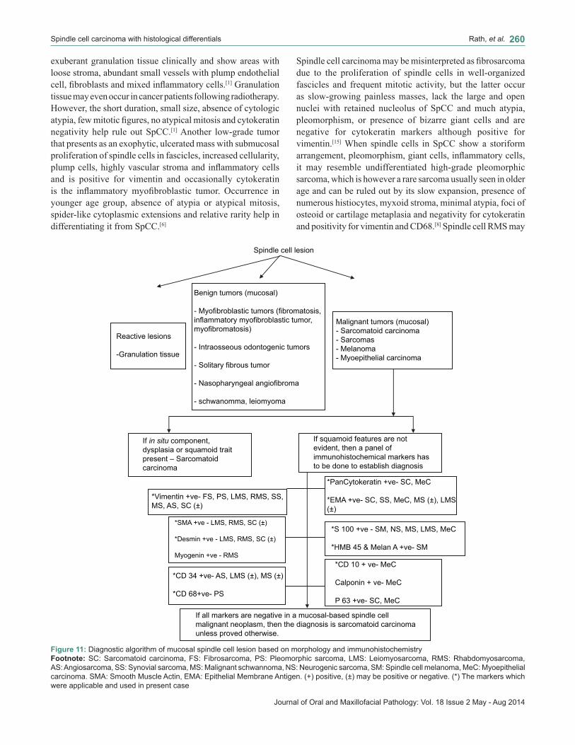
Due to the morphological diversity seen in monophasic spindled SpCC, there are a few common differentials that merit discussion. Spindle cell carcinoma may closely mimic
Journal of Oral and Maxillofacial Pathology: Vol. 18 Issue 2 May ‑ Aug 2014
Spindle cell carcinoma with histological differentials Rath, et al. 260
exuberant granulation tissue clinically and show areas with loose stroma, abundant small vessels with plump endothelial cell,fibroblastsandmixedinflammatorycells.[1] Granulation tissue may even occur in cancer patients following radiotherapy. However, the short duration, small size, absence of cytologic atypia,fewmitoticfigures,noatypicalmitosisandcytokeratinnegativity help rule out SpCC.[1] Another low-grade tumor that presents as an exophytic, ulcerated mass with submucosal proliferation of spindle cells in fascicles, increased cellularity, plumpcells,highlyvascularstromaandinflammatorycellsand is positive for vimentin and occasionally cytokeratin is the inflammatorymyofibroblastic tumor. Occurrence inyounger age group, absence of atypia or atypical mitosis, spider-like cytoplasmic extensions and relative rarity help in differentiating it from SpCC.[6]
Spindlecellcarcinomamaybemisinterpretedasfibrosarcomadue to the proliferation of spindle cells in well-organized fascicles and frequent mitotic activity, but the latter occur as slow-growing painless masses, lack the large and open nuclei with retained nucleolus of SpCC and much atypia, pleomorphism, or presence of bizarre giant cells and are negative for cytokeratin markers although positive for vimentin.[15] When spindle cells in SpCC show a storiform arrangement,pleomorphism,giantcells,inflammatorycells,it may resemble undifferentiated high-grade pleomorphic sarcoma, which is however a rare sarcoma usually seen in older age and can be ruled out by its slow expansion, presence of numerous histiocytes, myxoid stroma, minimal atypia, foci of osteoid or cartilage metaplasia and negativity for cytokeratin and positivity for vimentin and CD68.[8] Spindle cell RMS may
Figure 11: Diagnostic algorithm of mucosal spindle cell lesion based on morphology and immunohistochemistryFootnote: SC: Sarcomatoid carcinoma, FS: Fibrosarcoma, PS: Pleomorphic sarcoma, LMS: Leiomyosarcoma, RMS: Rhabdomyosarcoma, AS: Angiosarcoma, SS: Synovial sarcoma, MS: Malignant schwannoma, NS: Neurogenic sarcoma, SM: Spindle cell melanoma, MeC: Myoepithelial carcinoma. SMA: Smooth Muscle Actin, EMA: Epithelial Membrane Antigen. (+) positive, (±) may be positive or negative. (*) The markers which were applicable and used in present case

Journal of Oral and Maxillofacial Pathology: Vol. 18 Issue 2 May ‑ Aug 2014
Spindle cell carcinoma with histological differentials Rath, et al. 261
compriseanaplasticspindlecellswithbizarreconfiguration,MNGC,tadpolecells,frequentmitoticfiguresallofwhichmayoverlap with a monophasic SpCC. However, the relative rarity of spindle cell RMS in oral cavity, presence of striated cells, positiveimmunostainingwithdesmin,muscle‑specificactinand myoglobin or ultrastructural demonstration of skeletal muscle differentiation may be diagnostic.[15] The presence of spindle cells in a background of increased vascularity with distinct endothelium-lined vascular channels in an SpCC may be misdiagnosed as AS. However, the latter occur in older age, show tendency for anastomosis of vascular channels and proliferation of endothelial cell islands into vascular lumina. Besides they show positive immunostaining with CD34, CD31 and are negative for cytokeratin.[15] The spindle cell morphology of leiomyosarcoma (LMS) may mimic an SpCC with presence of increased mitosis, hyperchromatic nuclei but rare occurrence of mucosal LMS, fascicular arrangement of spindle cells with abundant eosinophilic cytoplasm, blunt ended and palisaded nuclei discern it from an SpCC. Also LMSshowsstrongpositivityforSMA,muscle‑specificactinand desmin, which is absent in SpCC. Recently P63 has been indicated as a useful marker in diagnosis of SpCC.[16] Although other spindle cell tumors like myoepithelial carcinoma, spindle cell melanoma, malignant schwannoma, synovial sarcoma subsist, they are comparatively rare. However, they need to be ruled out before diagnosing sarcomatoid carcinoma; therefore, an algorithm for diagnosing mucosal spindle cell lesions has been provided [Figure 11].
CONCLUSION
With increase in treatment including radiotherapy cases, SpCC would be frequently encountered in the clinical setting. Potentially aggressive with tendency to recur and metastasize, when diagnosed it has to be treated like an SCC in the same stage. Although it has an unpredictable biological behavior, tumors that are deeply invasive tend to have a poor prognosis than early stage tumors. A keen eye on the histomorphology, attention to clinical detail and judicious use of immunostains would help locate this ambiguous entity in the spindle cell lesion jargon.
REFERENCES
1. Lewis JS Jr. Spindle cell lesions- neoplastic or non neoplastic: Spindle cell carcinoma and other atypical spindle cell lesions of the head and neck. Head Neck Pathol 2008;2:103-10.
2. Ellis GL, Corio RL. Spindle cell carcinoma of the oral cavity. A clinicopathologic assessment of 59 cases. Oral Surg Oral Med Oral Pathol 1980;50:523-33.
3. Munakata R, Cheng J, Nakajima T, Saku T. Spindle cell carcinoma of the gingiva. Report of an autopsy case. J Oral Pathol Med 1998;27:180-4.
4. Prakash N, Harish Kumar MS, Sharada P, Pradeep GL. Spindle cell carcinoma of the oral cavity. A case report of a rare entity and review of literature. World J Dent 2010;1:55-8.
5. Oktay M, Kokenek-Unal TD, Ocal B, Saylam G, Korkmaz MH, Alper M. Spindle cell carcinoma of the tongue: A rare tumor in an unusual location. Patholog Res Int 2011;2011:572381.
6. Thompson LD, Wieneke JA, Miettinen M, Heffner DK. Spindle cell (Sarcomatoid) carcinomas of the larynx: A clinicopathologic study of 187 cases. Am J Surg Pathol 2002;26:13-70.
7. Stelow EB, Mills SE. Squamous cell carcinoma variants of upper aerodigestive tract. Am J Clin Pathol 2005;124:S96-109.
8. Parikh N, Desai N. Spindle cell carcinoma of the oral cavity: A case report of a rare entity and review of literature. J Acad Adv Dent Res 2011;2:31-6.
9. Viswanathan S, Rahman K, Pallavi S, Sachin J, Patil A, Chaturvedi P, et al. Sarcomatoid (Spindle Cell) Carcinoma of the head and neck mucosal region: A clinicopathologic review of 103 cases from a tertiary referral centre. Head Neck Pathol 2010;4:267-75.
10. Lewis JE, Olsen KD, Sebo TJ. Spindle cell carcinoma of the larynx: Review of 26 cases including DNA content and Immunohistochemistry. Hum Pathol 1997;28:664-73.
11. Mijima Y, Onizawa K, Kamma H, Yoshida H. Carcinosarcoma of tongue. Oral Oncol Extra 2006;42:78-81.
12. Gupta R, Singh S, Hedau S, Nigam S, Das BC, Singh I, et al. Spindle cell carcinoma of head and neck: An immunohistochemical and molecular approach to its pathogenesis. J Clin Pathol 2007;60:472-5.
13. Zarbo RJ, Crissman JD, Venkat H, Weiss MA. Squamous cell carcinoma of the upper aerodigestive tract mucosa: An immunohistologic and ultrastructural study of 18 biphasic tumors and comparison with 7 monophasic spindle cell tumors. Am J Surg Pathol 1986;10:741-53.
14. Battifora H. Spindle cell carcinoma: Ultrastructural evidence of squamous origin and collagen production by the tumor cells. Cancer 1976;37:2275-82.
15. Gnepp DR. Diagnostic Surgical Pathology of the head and neck. 2nd ed.Philadelphia: Saunders; 2009.
16. Lewis Js, Ritter JH, El-Moftys. Alternative epithelial markers in sarcomatoid carcinoma of the head and neck, lung and bladder- p63, MUC-31, TTF-1. Mod Pathol 2005;18:1471-81.
17. Kramer K, Hicks DG, Palis J, Rosier RN, Oppenheimer J, Faller MD, et al. Epitheloid Osteosarcomas of bone. Immunocytochemical evidence of mesenchymal differentiation in a primary osseous neoplasm. Cancer 1993;71:2977-82.
18. Kasper M, Stosiek P. The expression of vimentin in epithelial cells from human nasal mucosa. Eur Arch Otorhinolaryngol 1990;248:53-6.
19. Azumi N, Battifora H. The distribution of vimentin and keratin in epithelial and nonepithelial neoplasms. A comprehensive immunohistochemical study on formalin and alcohol fixedtumors. Am J Clin Pathol 1987;88:286-96.
20. Layfield LJ, Emerson L, Crim JR, Randall L. Squamousdifferentiation and cytokeratin expression in an osteosarcoma. A case report and review of the literature. Clin Med Pathol 2008;1:55-9.
21. Okada K, Hasegawa T, Yokoyama R, Boppa Y, Itoi E. Osteosarcoma with cytokeratin expression: A clinicopathological study of six cases with an emphasis of differential diagnosis from metastatic cancer. J Clin Pathol 2003;56:742-6.
How to cite this article: Rath R, Das BK, Das SN, Baisakh M. Spindle cell carcinoma of maxilla: Histomorphological and immunohistochemical analysis of a case. J Oral Maxillofac Pathol 2014;18:256-61.
Source of Support: Nil. Conflict of Interest: None declared.