Renal cell carcinoma with angioleiomyoma-like stroma: clinicopathological, immunohistochemical, and...
16
Renal cell carcinoma with angioleiomyoma-like stroma: clinicopathological, immunohistochemical, and molecular features supporting classification as a distinct entity Sean R Williamson 1,2 , Liang Cheng 1,3 , John N Eble 1 , Lawrence D True 4 , Nilesh S Gupta 2 , Mingsheng Wang 1 , Shaobo Zhang 1 and David J Grignon 1 1 Department of Pathology and Laboratory Medicine, Indiana University School of Medicine, Indianapolis, IN, USA; 2 Department of Pathology and Laboratory Medicine, Henry Ford Health System, Detroit, MI, USA; 3 Department of Urology, Indiana University School of Medicine, Indianapolis, IN, USA and 4 Department of Pathology, University of Washington School of Medicine, Seattle, WA, USA Rare renal epithelial neoplasms have been recognized to have an angioleiomyoma or leiomyoma-like proliferation of stromal smooth muscle; however, the nature of these tumors and their relationships to other renal cell carcinomas are poorly understood. We analyzed 23 such tumors for their clinicopathological, immunohistochemical, and cytogenetic features using fluorescence in situ hybridization. Twelve showed a homogeneous combination of features and were reclassified as renal cell carcinoma with angioleiomyoma-like stroma. These were composed of neoplastic glandular structures lined by cells with mixed clear, pale, and eosinophilic cytoplasm forming occasional papillary tufts. The stroma resembled smooth muscle and often extended away from the epithelial component, entrapping perinephric fat or non-neoplastic renal elements. Immunohistochemistry showed the epithelium to have reactivity for: carbonic anhydrase IX, CD10, vimentin, cytokeratin 7, cytokeratin 34bE12, and PAX8 but not a-methylacyl-coA-racemase. The stroma labeled for smooth muscle (smooth muscle actin 3 þ , desmin 1 þ , caldesmon 3 þ ) but not epithelial antigens. Neither component showed substantial reactivity for HMB45, melan-A, cathepsin K, or TFE3 protein. An interrupted, conspicuous layer of CD34-positive endothelial cells rimmed the epithelium, imparting a two-cell layer pattern resembling myoepithelial or basal cells. Chromosome 3p deletion and trisomy 7 and 17 were uniformly absent. Follow-up was available for three patients, none of whom experienced malignant behavior. Eleven tumors were excluded from this category and considered to be clear cell renal cell carcinoma with a reactive proliferation of smooth muscle (n ¼ 4) or tangential sectioning of the pseudocapsule (n ¼ 2), renal cell carcinoma unclassified (n ¼ 4), or clear cell papillary renal cell carcinoma (n ¼ 1). In summary, renal cell carcinoma with angioleiomyoma-like stroma is a distinct neoplasm with characteristic morphological, immunohistochemical, and molecular features, unrelated to clear cell renal cell carcinoma. The immunoprofile overlaps partly with that of clear cell papillary renal cell carcinoma, though morphology and reactivity for CD10 are points of contrast. Modern Pathology advance online publication, 5 September 2014; doi:10.1038/modpathol.2014.105 Recently, a small subgroup of renal epithelial neoplasms has been recognized to contain a stromal proliferation of smooth muscle resembling leiomyo- ma or angioleiomyoma. 1–4 Several names have been used for such tumors, including renal angiomyoadeno- matous tumor, 3 renal cell carcinoma associated with prominent angioleiomyoma-like proliferation, 2 and Correspondence: Dr SR Williamson, MD, Department of Pathol- ogy and Laboratory Medicine, Henry Ford Health System, Henry Ford Hospital, Department of Pathology—K6, 2799 West Grand Boulevard, Detroit, MI 48202, USA. E-mail: [email protected] Received 21 September 2013; revised 17 April 2014; accepted 18 June 2014; published online 5 September 2014 Modern Pathology (2014), 1–16 & 2014 USCAP, Inc. All rights reserved 0893-3952/14 $32.00 1 www.modernpathology.org
-
Upload
independent -
Category
Documents
-
view
2 -
download
0
Transcript of Renal cell carcinoma with angioleiomyoma-like stroma: clinicopathological, immunohistochemical, and...
Renal cell carcinoma withangioleiomyoma-like stroma:clinicopathological, immunohistochemical,and molecular features supportingclassification as a distinct entitySean R Williamson1,2, Liang Cheng1,3, John N Eble1, Lawrence D True4, Nilesh S Gupta2,Mingsheng Wang1, Shaobo Zhang1 and David J Grignon1
1Department of Pathology and Laboratory Medicine, Indiana University School of Medicine, Indianapolis,IN, USA; 2Department of Pathology and Laboratory Medicine, Henry Ford Health System, Detroit, MI, USA;3Department of Urology, Indiana University School of Medicine, Indianapolis, IN, USA and 4Department ofPathology, University of Washington School of Medicine, Seattle, WA, USA
Rare renal epithelial neoplasms have been recognized to have an angioleiomyoma or leiomyoma-like
proliferation of stromal smooth muscle; however, the nature of these tumors and their relationships to other
renal cell carcinomas are poorly understood. We analyzed 23 such tumors for their clinicopathological,
immunohistochemical, and cytogenetic features using fluorescence in situ hybridization. Twelve showed a
homogeneous combination of features and were reclassified as renal cell carcinoma with angioleiomyoma-like
stroma. These were composed of neoplastic glandular structures lined by cells with mixed clear, pale, and
eosinophilic cytoplasm forming occasional papillary tufts. The stroma resembled smooth muscle and often
extended away from the epithelial component, entrapping perinephric fat or non-neoplastic renal elements.
Immunohistochemistry showed the epithelium to have reactivity for: carbonic anhydrase IX, CD10, vimentin,
cytokeratin 7, cytokeratin 34bE12, and PAX8 but not a-methylacyl-coA-racemase. The stroma labeled for smooth
muscle (smooth muscle actin 3þ , desmin 1þ , caldesmon 3þ ) but not epithelial antigens. Neither component
showed substantial reactivity for HMB45, melan-A, cathepsin K, or TFE3 protein. An interrupted, conspicuous
layer of CD34-positive endothelial cells rimmed the epithelium, imparting a two-cell layer pattern
resembling myoepithelial or basal cells. Chromosome 3p deletion and trisomy 7 and 17 were uniformly
absent. Follow-up was available for three patients, none of whom experienced malignant behavior. Eleven
tumors were excluded from this category and considered to be clear cell renal cell carcinoma with a reactive
proliferation of smooth muscle (n¼ 4) or tangential sectioning of the pseudocapsule (n¼ 2), renal cell
carcinoma unclassified (n¼ 4), or clear cell papillary renal cell carcinoma (n¼ 1). In summary, renal cell
carcinoma with angioleiomyoma-like stroma is a distinct neoplasm with characteristic morphological,
immunohistochemical, and molecular features, unrelated to clear cell renal cell carcinoma. The immunoprofile
overlaps partly with that of clear cell papillary renal cell carcinoma, though morphology and reactivity for CD10
are points of contrast.Modern Pathology advance online publication, 5 September 2014; doi:10.1038/modpathol.2014.105
Recently, a small subgroup of renal epithelialneoplasms has been recognized to contain a stromalproliferation of smooth muscle resembling leiomyo-ma or angioleiomyoma.1–4 Several names have beenused for such tumors, including renal angiomyoadeno-matous tumor,3 renal cell carcinoma associated withprominent angioleiomyoma-like proliferation,2 and
Correspondence: Dr SR Williamson, MD, Department of Pathol-ogy and Laboratory Medicine, Henry Ford Health System, HenryFord Hospital, Department of Pathology—K6, 2799 West GrandBoulevard, Detroit, MI 48202, USA.E-mail: [email protected] 21 September 2013; revised 17 April 2014; accepted 18June 2014; published online 5 September 2014
Modern Pathology (2014), 1–16
& 2014 USCAP, Inc. All rights reserved 0893-3952/14 $32.00 1
www.modernpathology.org

clear cell renal cell carcinoma with smooth musclestroma.4 As only a small number of tumors withthese features have been reported and their clinico-pathological, immunohistochemical, and molecularfeatures are incompletely understood, it is unclearwhether these lesions comprise a single distincttype of renal neoplasm or whether these reportsencompass multiple entities. The relationships ofthese tumors to clear cell renal cell carcinoma4 andclear cell papillary renal cell carcinoma5–11 (alsoknown as clear cell tubulopapillary renal cellcarcinoma)6,12 are also poorly understood.13 Toelucidate the characteristics of these tumors andassess whether they represent a distinct subcategoryof renal neoplasia, we studied the clinicopatho-logical, immunohistochemical, and molecularfeatures of renal epithelial neoplasms noted tohave a smooth muscle stromal component fromour pathology archives.
Materials and methods
Pathology and Light Microscopy
Renal epithelial neoplasms with a stromal compo-nent resembling smooth muscle were retrieved viaan electronic database search of the pathologyarchives and the contributing authors’ consultationfiles and via retrospective review of a consecutiveseries of 341 clear cell renal cell carcinomas(a 3-year period) with special attention to thepresence of stromal smooth muscle. Tumors thatdemonstrated one or more tissue blocks with stromaresembling smooth muscle were selected for analysisof their morphological features, immunohistochemicalstaining characteristics, and abnormalities ofchromosomes 3p, 7, and 17 by fluorescence in situhybridization (FISH). Other tumors with a mixtureof epithelial and stromal components were notstudied, such as mixed epithelial and stromal tumorof the kidney, cystic nephroma, angiomyolipomawith epithelial cysts, or synovial sarcoma of thekidney. Reclassification of the tumors was attemptedbased on their constellation of morphological,immunohistochemical, and molecular features.
Clinicopathological characteristics were assessed,including those listed in Table 1. Hematoxylin andeosin and Masson trichrome-stained sections wereprepared from the available paraffin blocks. Thevolume of the tumor comprised of cysts was esti-mated as a percentage of the entire tumor volume.Other architectural features were assessed, includ-ing the presence of branched tubular structures,papillary structures, ‘secretory cells’ with nucleialigned above the basement membrane,10 entrap-ment of non-neoplastic renal tubules or glomeruliwithin the smooth muscle proliferation, extensionof the smooth muscle proliferation into adiposetissue, extension of the epithelial componentinto adipose tissue, vascular invasion by either Table
1C
lin
icop
ath
olo
gic
al
ch
ara
cte
rist
ics
of
ren
alcell
carc
inom
as
wit
han
gio
leio
myom
a-l
ike
stro
ma
Pati
en
tT
um
or
Age
Gen
der
Siz
eS
tage
Sid
eG
rad
e3p
FIS
HT
riso
my
7or
17
Foll
ow
-up
mon
ths
Foll
ow
-up
statu
sC
yst
icC
yto
pla
sm
Tw
o-c
ell
layer
patt
ern
Pap
illa
ryst
ructu
res
Bra
nch
ed
tubu
les
Sm
ooth
mu
scle
involv
es
fat
En
trap
ped
non
-neop
last
ictu
bu
les
Blo
od
vess
els
Calc
ific
a-
tion
Oss
ific
a-
tion
11
76
M3.5
T1a
R2
No
dele
tion
NA
58
NE
M,
uro
thel
ial
CA
wit
hli
ver
mets
5%
C,
FL
Yes
1þ
Yes
Yes
No
Th
ick,
slit
-lik
e,
hem
an
gio
ma-l
ike
No
No
22
78
M2.8
T1a
R3
No
dele
tion
No
NA
NA
40%
C,
FL
Yes
No
No
Yes
Yes
Th
ick,
slit
-lik
eN
oY
es
33
73
F3.0
T1a
L2
No
dele
tion
No
NA
NA
No
C,
FL
Yes
No
No
Yes
Yes
Th
ick,
slit
-lik
eN
oN
o4
459
M2.8
T1a
R3
No
dele
tion
No
4C
on
trala
tera
ltu
mor
(no.
4)
rese
cted
at
4m
on
ths
30%
C,
E,
FL
Yes
2þ
Yes
Yes
Yes
Sli
t-li
ke
No
Yes
45
60
1.8
T1a
L3
No
dele
tion
No
NA
NA
No
C,
E,
FL
Yes
2þ
Yes
Yes
Yes
Th
ick,
slit
-lik
eN
oY
es
56
66
F2.7
T1a
R2
No
dele
tion
No
2N
ED
5%
C,
FL
Yes
2þ
Yes
Yes
Yes
Th
ick,
slit
-lik
eY
es
Yes
67
68
M2.4
T1a
R2
No
dele
tion
No
NA
NA
No
C,
E,
FL
Yes
1þ
Yes
Yes
Yes
Sli
t-li
ke
Yes
No
78
76
F2.2
T1a
L2
No
dele
tion
No
NA
Iden
tifi
ed
at
au
top
syN
oC
,F
LY
es
No
No
No
Yes
Th
ick,
slit
-lik
eN
oN
o
89
45
F1.8
T1a
L2
NA
NA
NA
NA
No
C,
FL
Yes
No
No
Yes
No
Th
ick,
slit
-lik
e,
hem
an
gio
ma-l
ike
No
No
910
59
M1.7
T1a
R2
No
dele
tion
No
NA
NA
No
C,
FL
Yes
2þ
No
Yes
Yes
Th
ick,
slit
-lik
eN
oN
o10
11
62
F1.7
T1a
L3
No
dele
tion
No
38
NE
DN
oC
,E
,F
LY
es
3þ
Yes
No
Yes
Sli
t-li
ke,
hem
an
gio
ma-l
ike
No
No
11
12
41
M1.0
T1a
All
ogra
ft2
No
dele
tion
No
26
NE
D5%
C,
FL
Yes
1þ
Yes
No
No
Sli
t-li
ke
No
No
Abbre
via
tion
s:C
,cle
ar
cyto
pla
sm;C
A,carc
inom
a;E
,eosi
nop
hil
iccyto
pla
sm;F,fe
male
;F
L,p
ale
,fl
occu
len
tcyto
pla
sm;F
ISH
,fl
uore
scen
ce
insi
tuh
ybri
diz
ati
on
;L
,le
ft;M
,m
ale
;N
A,n
ot
avail
able
;N
ED
,n
oevid
en
ce
of
resi
du
al,
recu
rren
tor
meta
stati
cd
isease
;N
EM
,n
oevid
en
ce
of
meta
stati
cre
nal
cell
carc
inom
a;
R,
righ
t.
Renal cell carcinoma with angioleiomyoma-like stroma
2 SR Williamson et al
Modern Pathology (2014), 1–16

component, and a dual cell layer pattern with theepithelial elements rimmed by a conspicuous layerof capillaries. Cytological features were assessed,including nuclear grade,14 presence of ill-definedapical cytoplasm,3 and cytoplasmic staining charac-teristics. Other features studied included the lightmicroscopic appearance of the stromal component(smooth muscle-like, hyalinized, or edematous,with or without hemangioma-like areas), ossifica-tion, and other forms of calcification. Morphologicalfeatures were compared with a control group of 55clear cell papillary renal cell carcinomas from 34patients, the features of which were previouslypublished,10 with particular emphasis on thepresence of well-formed bundles of smooth musclewithin the tumor stroma, extension of smooth muscleinto adipose tissue, prominence of blood vesselswithin the stroma, and entrapment or dilation ofnon-neoplastic renal tubules within the tumorstroma.
Immunohistochemistry
Antibodies directed against a-methylacyl-coA-race-mase (AMACR/P504S, 13H4; Dako Corp), carbonicanhydrase IX (polyclonal rabbit; Abcam Inc.),cathepsin K (3F9; Abcam), CD10 (56C6; Dako Corp),CD34 (QBEnd; Dako Corp), cytokeratin (CK) 34bE12(34bE12; Dako Corp), CK7 (OV-TL 12/30; DakoCorp), desmin (D33; Dako Corp), caldesmon (h-CD;Dako Corp), human melanosome (HMB45; DakoCorp), melan-A (A103; Dako Corp), PAX8 (polyclo-nal; Cell Marque), smooth muscle actin (1A4 clone;Dako Corp), TFE3 (MRQ-37; Cell Marque), andvimentin (V9; Dako Corp) were utilized in a Dakoautomated instrument. Positive and negative con-trols gave appropriate results for each procedure.
In the epithelial component, the extent of im-munohistochemical staining was estimated micro-scopically as a percentage of all neoplastic cellsshowing a positive staining reaction. Particular patternsof labeling, such as the ‘cup-shaped’ staining patternof carbonic anhydrase IX seen in clear cell papillaryrenal cell carcinoma,8,15 were additionally noted.For antibodies with a nuclear labeling pattern (PAX8and TFE3) and for evaluation of the stromal com-partment, the staining reaction was considered 3þif it yielded diffusely and intensely positive labelingin essentially all cells from low magnification (� 4objective). The staining reaction was considered 2þif labeling was easily recognized from low magni-fication but not diffuse or 1þ if labeling was focal orweak, requiring high magnification to be identified.Immunohistochemical staining results were comparedwith those previously published for clear cell papillaryrenal cell carcinoma6,8,10 and clear cell renal cellcarcinoma.16,17 Staining reactions for caldesmon,CD34, and cytokeratin 34bE12 were also studied in acontrol group of 10 tumors: 5 clear cell papillary
renal cell carcinomas and 5 clear cell renal cellcarcinomas.
FISH
FISH evaluation of chromosomes 3p, 7, and 17 wasperformed as described previously.18,19 Briefly,multiple tissue sections of 4-mm thickness wereprepared from formalin-fixed paraffin-embeddedtissue blocks. A hematoxylin and eosin-stainedslide from each block was examined to identify theneoplastic cells. The slides were deparaffinizedwith two washes of xylene, 15 min each, andsubsequently washed twice with absolute ethanol,10 min each, and then air-dried in a fume hood.Next, the slides were treated with 0.1 mM citric acid(pH 6.0) (Zymed, South San Francisco, CA, USA) at95 1C for 10 min and rinsed in distilled water for3 min followed by a wash of 2xSSC (standard salinecitrate) for 5 min. Digestion of the tissue wasperformed by applying 0.4 ml of pepsin (5 mg/mlin 0.1 N HCl/0.9 NaCl) (Sigma, St Louis, MO, USA)at 37 1C for 40 min. The slides were rinsed withdistilled water for 3 min, washed with 2xSSC for5 min, and air-dried. Centromeric chromosomeenumeration probes (CEP) were utilized for chromo-some 3 (CEP3, Orange), chromosome 7 (CEP7,Green), and chromosome 17 (CEP17, Orange) fromVysis (Downers Grove, IL, USA). FISH for chromo-somes 7 and 17 was performed with centromerica-satellite DNA probe cocktail containing CEP7 andCEP17. Deletion of chromosome 3p was assessedusing a probe cocktail containing probe to chro-mosome 3p25 (3pTel25, Green, Vysis) and CEP3.Probes CEP7–CEP17 were diluted with tDenHyb1and probes CEP3–3p25 were diluted with tDenHyb2 (Insitus, Alburquerque, NM, USA) in a ratio of1:100.
The hybridized slides were examined using aZeiss Axioplan 2 microscope (ZEISS, Gottingen,Germany) with the following filters from Chroma(Chroma, Brattleboro, VT, USA): SP-100 for 40, 6-diamidino-2-phenylindole, FITC MF-101 for Spec-trum Green (3pTel25, CEP7), and Gold 31003 forSpectrum Orange (CEP3, CEP17). The images wereacquired with a charged coupled device camera andanalyzed with the MetaSystem Isis Software (Meta-System, Belmont, MA, USA). Four sequential focusstacks with 0.4-mm intervals were acquired and thenintegrated into a single image to reduce thickness-related artifacts.
The method of analysis was described pre-viously.20,21 In brief, for each slide, 100–150 nucleifrom tumor tissue were scored for signals fromprobes under the fluorescence microscope with� 1000 magnification. Non-neoplastic renal cortexwas examined as an internal control. The method ofanalysis for 3p25 deletion was based upon previousstudies of deletion of chromosomes 1p and 19q inoligodendrogliomas. The cutoff value for 3p deletion
Modern Pathology (2014), 1–16
Renal cell carcinoma with angioleiomyoma-like stroma
SR Williamson et al 3

was defined as a 3p25/CEP3 ratio of r0.7, as pre-viously described.7,18,22–24 Definitions of chromo-somal trisomy of chromosomes 7 and 17 were basedon the Gaussian model and related to the non-neoplastic controls. The cutoff values for each probewere set at mean plus three standard deviations(meanþ 3 s.d.) of the control values.25,26
Results
Clinicopathological Characteristics of Renal CellCarcinomas with Angioleiomyoma-Like Stroma
Twelve tumors from 11 patients were reclassified asrenal cell carcinoma with angioleiomyoma-likestroma, based on a unique combination of morpho-logical, immunophenotypical, and molecular find-ings (Table 1). None of the tumors that were selectedfor analysis based on retrospective review of 341consecutive clear cell renal cell carcinomas wasultimately reclassified into this category. Tumorsranged in size from 1.0 to 3.5 cm (median 2.3). Thepatients’ ages ranged from 41 to 78 years (median66). Six patients were men, and five were women.The nuclear grade was 2 (8 of 12, 67%) or 3 (4 of 12,33%). One tumor occurred in an allograft kidney.Follow-up of 41 year was available for threepatients, ranging in duration from 26 to 58 months,of whom all were alive without evidence of residual,recurrent, or metastatic disease. One patient (patient4) was noted at the time of initial resection to have acontralateral renal tumor (tumor 5) that was resected4 months after the first, although no additionalfollow-up was available after the second resection.Both tumors demonstrated similar morphologicaland immunohistochemical features.
Grossly, these tumors formed pink-tan or whitefibrous-appearing nodules and lacked the golden-yellow, heterogeneous and hemorrhagic cut surfacethat is typical of clear cell renal cell carcinoma(Figure 1a). Microscopically, a cystic componentwas most often absent (n¼ 7). The stromal compo-nent not only microscopically contained areas witha smooth muscle-like appearance in all cases butalso commonly contained areas of hyalinization oredema. Three tumors contained hemangioma-likefoci within the stroma, one prominently and twofocally. At least a minor vascular component formedby slit-like vascular spaces dispersed in the smoothmuscle stroma was present in all tumors, similar tothe solid pattern of angioleiomyoma.27 In sometumors (8/12), the stroma also contained vascularchannels with thicker, muscular walls, similar to thevenous pattern of angioleiomyoma.27 The smoothmuscle proliferation commonly extended into theadjacent adipose tissue (n¼ 9, 75%, Figures 1b andc) and often entrapped adjacent non-neoplasticrenal tubules (n¼ 9, 75%), some of which weredilated, imparting a cystic nephroma-like appear-ance (Figure 1c).
The epithelial component was composed ofclusters of glandular structures dispersed in themuscular stroma (Figure 1d). These were lined bycells with cytoplasm that varied from optically clearto pale with a flocculent appearance and eosino-philic within the same tumor (Figures 1e and f).In contrast to the stromal component, epithelialelements did not extend into adipose tissue in anytumor. Glands with a branched configuration werepresent in seven tumors and a ‘secretory cell’ patternwith nuclei aligned at the apical aspect of the cellswas present only focally in one tumor. Smallpapillary tufts were present in three tumors(Figure 2a), occasional foci of branching papillaein four tumors (Figure 2b), and areas composedentirely of papillary structures were present in asingle tumor. All showed a dual-cell layer pattern atleast focally (Figure 2c), composed of epithelialnests or glands tightly rimmed by a prominent layerof CD34-positive endothelial cells (Figure 2d, seeresults of immunohistochemical staining below),imparting a resemblance to a basal cell or myoe-pithelial cell layer. Similarly, hemangioma-like vas-cular proliferations (Figure 2e) were highlighted byimmunohistochemical staining for CD34 (Figure 2f).The apical aspect of the cytoplasm lining theglandular structures had an ill-defined appearancein some areas of all tumors, in three focally and innine more diffusely. Two tumors had dystrophiccalcification in the hyaline stroma and four had areasof osseous metaplasia. In contrast, only a singletumor from the control group of 55 clear cellpapillary renal cell carcinomas10 morphologicallydemonstrated bundles of smooth muscle within thecentral tumor stroma, and none exhibited bundles ofsmooth muscle radiating away from the epithelialcomponent into the adjacent adipose tissue.
Histochemical and Immunohistochemical StainingCharacteristics of Renal Cell Carcinomas withAngioleiomyoma-Like Stroma
Histochemical staining with the Masson trichrometechnique showed a mixture of smooth muscle andcollagen staining patterns in the stroma of all of therenal cell carcinomas with angioleiomyoma-likestroma, even in areas where the light microscopicappearance was fibrous or edematous. Results ofimmunohistochemical staining are detailed in Table 2:Antibodies to carbonic anhydrase IX (Figures 3a andb), CK7 (Figure 3c), CD10 (Figure 3d), and cytokeratin34bE12 (Figure 3e) labeled the epithelial cells andnot the stromal cells in all tumors (median 95, 90,40, and 55% of tumor cells, respectively). There wasa membranous pattern of labeling for carbonicanhydrase IX in all tumors, and eight showed the‘cup-shaped’ staining pattern characteristic of clearcell papillary renal cell carcinoma,8 five focally andthree more diffusely. Entrapped non-neoplasticrenal tubules when present showed a negative
Modern Pathology (2014), 1–16
Renal cell carcinoma with angioleiomyoma-like stroma
4 SR Williamson et al

reaction for carbonic anhydrase IX. Staining forCD10 was not restricted to a cystic component as hasbeen reported in clear cell papillary renal cell carci-
noma.10 Staining for AMACR was typically absentin both epithelium and stroma but often highlightedthe entrapped non-neoplastic renal tubules. The five
Figure 1 Grossly (a), renal cell carcinomas with angioleiomyoma-like stroma formed a solid, white-tan, fibrous-appearing nodulewithout the golden-yellow heterogeneous cut surface typical of clear cell renal cell carcinoma. Microscopically (b), the tumors werecomposed of a neoplastic proliferation of glands in a smooth muscle stroma. The smooth muscle proliferation often extended into andentrapped lobules of adjacent adipose tissue (black arrows) and entrapped non-neoplastic tubules that were often dilated (white arrow).Higher magnification (c) demonstrates a cystic nephroma-like area of entrapped non-neoplastic tubules (right) and the smooth muscleproliferation encasing lobules of adipose tissue (left). The epithelial component (d) formed glandular structures distributed in the smoothmuscle proliferation, characteristically rimmed by a prominent layer of endothelial cells mimicking a basal cell or myoepithelial celllayer. Cytoplasm of the epithelial cells often varied from clear (e) to pale, flocculent and eosinophilic (f), even within the same tumor.
Modern Pathology (2014), 1–16
Renal cell carcinoma with angioleiomyoma-like stroma
SR Williamson et al 5

control cases of clear cell papillary renal cell carci-noma also showed positive staining for cytokeratin34bE12, ranging from 35 to 75% of tumor cells. In
contrast, only one of five clear cell renal cellcarcinomas showed focal labeling for cytokeratin34bE12 in p5% of tumor cells.
Figure 2 In renal cell carcinomas with angioleiomyoma-like stroma, small papillary tufts often protruded into the glandular structures(a). Some exhibited more complex branching papillary structures (b). A prominent layer of endothelial cells rimmed the tubules andnests, imparting a two-cell layer appearance (c). The endothelial nature of these cells was confirmed by immunohistochemical stainingfor CD34 (d). Similarly, a subset of tumors contained hemangioma-like proliferations of capillaries (e), highlighted by immunostaining forCD34 (f).
Modern Pathology (2014), 1–16
Renal cell carcinoma with angioleiomyoma-like stroma
6 SR Williamson et al

Antibody to vimentin usually labeled both the epi-thelial and stromal components of the renal cell carci-nomas with angioleiomyoma-like stroma (Figure 3f,9/10). All renal cell carcinomas with angioleiomyo-ma-like stroma showed intense, typically diffusenuclear labeling with antibody to PAX8 in the epithe-lium only. Antibody to cathepsin K labeled macro-phages within glandular lumina and in seven of theten stained tumors labeled the stromal cells mini-mally (1þ ), sometimes including the capillary net-work surrounding the tumor cells. Staining for CD34showed an interrupted or ‘dotted’ pattern of capillaryendothelial cells tightly rimming the neoplasticepithelial component in all tumors (Figure 2d). Inthe control group of clear cell papillary renal cell carci-nomas, one tumor demonstrated this pattern diffusely,whereas three showed a similar pattern mainly aroundthe cells lining cysts. In clear cell renal cell carcino-mas, the cystic component of only one tumor showedthis pattern. Other areas of all three types of tumor(renal cell carcinoma with angioleiomyoma-like stro-ma, clear cell papillary renal cell carcinoma, and clearcell renal cell carcinoma) demonstrated a morecontinuous reticular network of labeling for CD34around the tumor cell nests, particularly in areascomposed of compact architecture. No tumor showedepithelial labeling for cathepsin K or any labeling forHMB45, melan-A, or TFE3 protein.
Immunohistochemistry for smooth muscle actinwas usually diffusely and strongly positive in thestromal component (Figure 4a), whereas desminshowed a more variable staining reaction, often show-ing limited staining, most commonly 1þ (Figure 4b).The stromal component showed intense and diffusepositive reactivity for caldesmon (Figure 4c, all 2þ to3þ ), which highlighted the extension of smoothmuscle into the perinephric fat, when present. Areasthat were edematous with a less conspicuous smoothmuscle component often still demonstrated a diffuseand strong staining reaction for caldesmon, similar tothe findings using the trichrome stain (Figure 4d). Inthe control group of five clear cell papillary renal cellcarcinomas, staining for caldesmon also often showeda positive reaction, although more limited in extent(absent in one tumor, 1þ in two tumors, and 2þ intwo tumors). The control group of five clear cell renalcell carcinomas showed a negative reaction in thetumor stroma, although there was labeling of thetumor pseudocapsule and the walls of intratumoralblood vessels in both clear cell papillary renal cellcarcinoma and clear cell renal cell carcinoma. Otherimmunohistochemical markers were used as part ofthe diagnostic evaluation of a subset of cases andavailable for review, as detailed in Table 2.
FISH Results in Renal Cell Carcinomas withAngioleiomyoma-Like Stroma
Material was available (paraffin blocks or unstainedslides) in 11 of the 12 renal cell carcinomas withT
able
2Im
mu
noh
isto
ch
em
ical
stain
ing
ch
ara
cte
rist
ics
of
ren
alcell
carc
inom
as
wit
han
gio
leio
myom
a-l
ike
stro
ma
Tu
mor
CA
IX%
(ep
ith
eli
um
)
CA
IXp
att
ern
(ep
ith
eli
um
)C
K7
(ep
ith
eli
um
)C
D10
(ep
ith
eli
um
)A
MA
CR
SM
A(s
trom
a)
Desm
in(s
trom
a)
Cald
esm
on
(str
om
a)
Vim
en
tin
34bE
12
(ep
ith
eli
um
)PA
X8
HM
B45
Mela
n-
AC
ath
ep
sin
KT
FE
3C
D34
Oth
er
1100%
M,
focal
CS
90%
15%
—3þ
1þ
3þ
þE
S5%
2þ
E—
—1þ
S—
Dot-
like
MIT
F�
2N
AN
AN
AN
AN
A2þ
NA
NA
NA
NA
NA
NA
NA
NA
NA
NA
ER
an
dP
R�
340%
M,
focal
CS
100%
30%
Weak,
E3þ
1þ
3þ
þE
S90%
3þ
E—
—1þ
S—
Dot-
like
p63�
465%
CS
80%
50%
—3þ
2þ
3þ
þE
S40%
3þ
E—
—1þ
S—
Dot-
like
CD
117�
5100%
CS
100%
30%
—3þ
2þ
3þ
þE
S80%
3þ
E—
—1þ
S—
Dot-
like
695%
CS
75%
80%
Weak,
E3þ
2þ
3þ
NA
65%
2þ
E—
——
—D
ot-
like
p63,E
R,P
R�
7100%
M95%
90%
1þ
S3þ
3þ
3þ
þE
S50%
3þ
E—
——
—D
ot-
like
ER
an
dP
Rfo
cal
inp
eri
ph
era
lst
rom
a8
95%
M,
focal
CS
75%
25%
—3þ
—3þ
þE
S40%
2þ
E—
—1þ
S—
Dot-
like
960%
M50%
30%
NA
3þ
1þ
NA
þS
NA
NA
——
NA
NA
Dot-
like,
focal
AE
1/3þ
65%
ep
ith
eli
um
,E
R�
,P
Rfo
cal
inp
eri
ph
era
lst
rom
a10
100%
M95%
50%
—3þ
1þ
3þ
þE
S80%
3þ
E—
—1þ
S—
Dot-
like
11
95%
M,
focal
CS
95%
40%
5%
E3þ
1þ
2þ
þE
S60%
3þ
E—
——
—D
ot-
like
12
100%
M,
focal
CS
75%
50%
—3þ
1þ
2þ
þE
S20%
3þ
E—
—1þ
S—
Dot-
like
Abbre
via
tion
s:A
MA
CR
,a-
meth
yla
cyl-
coA
-racem
ase
;C
AIX
,carb
on
ican
hyd
rase
IX;
CK
,cyto
kera
tin
;C
S,
‘cu
p-s
hap
ed
’;þ
E,
posi
tive
inep
ith
eli
um
on
ly;
ER
,est
rogen
recep
tor;þ
ES
,p
osi
tive
inep
ith
eli
um
an
dst
rom
a;
M,
mem
bra
nou
s;M
ITF,
mic
roop
hth
alm
iatr
an
scri
pti
on
facto
r;P
R,
pro
gest
ero
ne
recep
tor;þ
S,
posi
tive
inst
rom
aon
ly;
SM
A,
smooth
mu
scle
acti
n.
Modern Pathology (2014), 1–16
Renal cell carcinoma with angioleiomyoma-like stroma
SR Williamson et al 7

Figure 3 Immunohistochemical staining of renal cell carcinomas with angioleiomyoma-like stroma (a) characteristically yielded positivelabeling of the epithelial component for carbonic anhydrase IX (b), typically with a diffuse, strong membranous pattern. Cytokeratin 7 (c)was also positive in the epithelium, although often o100% of cells. CD10 typically yielded apical and lateral membranous staining (d),more diffusely than is expected for clear cell papillary renal cell carcinoma. Cytokeratin 34bE12 (e) also showed a positive reaction in theepithelial component in a variable percentage of the tumor cells. Antibody to vimentin (f) variably labeled both the epithelial and stromalcells.
Modern Pathology (2014), 1–16
Renal cell carcinoma with angioleiomyoma-like stroma
8 SR Williamson et al

angioleiomyoma-like stroma for assessment of chro-mosome 3p status by FISH and in 10 for assessmentof chromosomes 7 and 17. None showed deletion of
chromosome 3p in either the epithelial component(Figure 4e) or the stromal component (Figure 4f),evidenced by two pairs of centromeric chromosome
Figure 4 The stroma of renal cell carcinomas with angioleiomyoma-like stroma characteristically showed diffuse, strong labeling withantibody to smooth muscle actin (a), although reactivity for desmin was more variable and often focal (b). Labeling for caldesmon (c) wastypically strong and diffuse, supporting the morphological impression of smooth muscle differentiation. Trichrome stain similarlyshowed a mixture of collagen and smooth muscle in the stroma (d). Fluorescence in situ hybridization uniformly showed no deletion ofchromosome 3p in either the epithelial (e) or stromal (f) component.
Modern Pathology (2014), 1–16
Renal cell carcinoma with angioleiomyoma-like stroma
SR Williamson et al 9

3 probe signals and chromosome 3p probe signals,and none showed trisomy of chromosome 7 or 17.
Excluded Tumors
After reclassification, 11 tumors were excluded fromthe category of renal cell carcinoma with angioleio-myoma-like stroma, as summarized in Table 3. Intwo of these excluded tumors, a smooth musclecomponent was present in only one tissue block andinterpreted as tangential sectioning of the tumorpseudocapsule in a clear cell renal cell carcinoma(Figures 5a and b). In this situation, morphologicaland immunohistochemical (carbonic anhydraseIXþ , CD10þ , CK7� , cytokeratin 34bE12� ) fea-tures were otherwise typical of clear cell renalcell carcinoma, although the pseudocapsule labeledfor markers of smooth muscle differentiation byimmunohistochemistry (smooth muscle actin 3þ ,desmin 1þ , caldesmon 3þ ). In another fourtumors, smooth muscle bundles dissected into thetumor in more than one tissue block, althoughmorphological and immunohistochemical featureswere again similar to those of clear cell renal cellcarcinoma (Figure 5c). Chromosome 3p deletion waspresent in the epithelial component of two of thethree of these tumors evaluated by FISH, and threeof these four tumors invaded the renal capsule orrenal sinus, suggesting that the presence of smoothmuscle bundles was a reactive phenomenon.
Three excluded tumors were originally consid-ered renal cell carcinomas with angioleiomyoma-like stroma based on their morphological featuresbut were recategorized as renal cell carcinoma,unclassified, based on the results of immunohisto-chemical and molecular analysis. In one (patient 18,Figure 5d), immunohistochemical staining didnot reveal smooth muscle differentiation in thespindle cell component (smooth muscle actin 1þ ,
desmin� , caldesmon� ). In contrast to the renalcell carcinomas with angioleiomyoma-like stroma,the spindle cell component of this tumor labeled forcarbonic anhydrase IX in a patchy distribution,CD10 diffusely, and focally PAX8. Chromosome 3pdeletion was not detected. The patient had a lymphnode metastasis at the time of nephrectomy andadditionally developed a distant metastasis to thesoft tissue of the orbit 12 months after nephrectomy.The latter was composed predominantly of spindle-shaped cells that were similarly positive by im-munohistochemistry for CD10 and PAX8. Based onthese features, this tumor was recategorized as renalcell carcinoma, unclassified, although an unusualpattern of sarcomatoid clear cell renal cell carcinomacould not be entirely excluded. Two tumors (frompatients 19 and 20, Figure 5e) were composedentirely of a nested arrangement of cells with clearcytoplasm resembling clear cell renal cell carcinomabut with a prominent expanded stromal compart-ment (smooth muscle actin 3þ , desmin 1þ ,caldesmon 2þ ). The immunophenotype of theepithelial component was similar to that of therenal cell carcinomas with angioleiomyoma-likestroma (carbonic anhydrase IXþ , CD10þ , CK7þ ,cytokeratin 34bE12þ ). However, labeling for carbo-nic anhydrase IX was not diffuse (5 and 35% ofcells), and chromosome 3p deletion was present inthe epithelial component (70% of cells for bothtumors).
One additional tumor originally considered renalcell carcinoma, unclassified, (patient 21, Figure 5f)had a papillary architecture lined by cells with clearto eosinophilic, flocculent cytoplasm. Immunohis-tochemical and molecular results were similar tothose of the renal cell carcinomas with angioleio-myoma-like stroma (carbonic anhydrase IXþ ,CD10þ , CK7þ , cytokeratin 34bE12þ , 3p deletionabsent); however, due to the contrasting morphology(exclusively papillary), this tumor could not be
Table 3 Tumors excluded from the category of renal cell carcinoma with angioleiomyoma-like stroma
Patient Original diagnosis Reclassification
12 Clear cell renal cell carcinoma Clear cell renal cell carcinoma (tangential sectioning of pseudocapsule)13 Clear cell renal cell carcinoma Clear cell renal cell carcinoma (tangential sectioning of pseudocapsule)14 Clear cell renal cell carcinoma Clear cell renal cell carcinoma with reactive smooth muscle (invades renal sinus)15 Clear cell renal cell carcinoma Clear cell renal cell carcinoma with reactive smooth muscle (invades renal capsule)16 Clear cell renal cell carcinoma Clear cell renal cell carcinoma with reactive smooth muscle (invades renal capsule)17 Clear cell renal cell carcinoma Clear cell renal cell carcinoma with reactive smooth muscle18 Renal cell carcinoma with
angioleiomyoma-like stromaUnclassified renal cell carcinoma
19 Renal cell carcinoma withangioleiomyoma-like stroma
Unclassified renal cell carcinoma
20 Renal cell carcinoma withangioleiomyoma-like stroma
Unclassified renal cell carcinoma
21 Unclassified renal cell carcinoma Unclassified renal cell carcinoma22 Clear cell papillary renal cell
carcinomaClear cell papillary renal cell carcinoma vs collision tumor
Modern Pathology (2014), 1–16
Renal cell carcinoma with angioleiomyoma-like stroma
10 SR Williamson et al

confidently placed in the same category. One tumorwas originally considered a clear cell papillary renalcell carcinoma. Morphology and immunoprofile intwo of the sections resembled the other renal cellcarcinomas with angioleiomyoma-like stroma; how-ever, features in the remaining sections resembledclear cell papillary renal cell carcinoma. No single
slide showed a transition between these morpholo-gical patterns. Due to the uncertainty of distinguish-ing between these two entities and the inability toexclude the possibility of two juxtaposed tumors, adiagnosis of renal cell carcinoma with angioleio-myoma-like stroma could not be established withcertainty in this case.
Modern Pathology (2014), 1–16
Renal cell carcinoma with angioleiomyoma-like stroma
SR Williamson et al 11
Discussion
Recently, it has been recognized that a small groupof renal epithelial neoplasms contain a prominentproliferation of stromal smooth muscle.1–4 However,the relationship of these tumors to other renalepithelial neoplasms, particularly clear cell renalcell carcinoma4 and clear cell papillary renal cellcarcinoma,5–11 is incompletely understood.13 Toshed light on these relationships, in this studywe assessed clinicopathological, immunohistoche-mical, and molecular features of renal epithelialneoplasms with a prominent stromal smooth musclecomponent. We found a subset of tumors tocomprise a homogeneous group that we regard as adistinct subcategory of renal neoplasia.
Previously, Michal et al1 reported an exampleof a renal tumor composed of cytologically blandepithelial elements dispersed in muscular andvascular stroma, which they termed ‘benign renalangiomyoadenomatous tumor.’ In a subsequentseries of five similar tumors, Michal et al3 foundthese neoplasms to label for CK7 but not CD10 byimmunohistochemistry and lack key molecularalterations of clear cell renal cell carcinoma,leading to the conclusion that they are distinctneoplasms. In another study, Kuhn et al2 reportedfive renal cell carcinomas that were noted to have astroma with a prominent ‘angioleiomyoma-like’appearance. The authors concluded that this angio-leiomyoma-like component was unrelated to angio-myolipoma or tuberous sclerosis, and the epithelialcomponent was interpreted as usual clear cell renalcell carcinoma. However, based on overlappingmorphological and immunohistochemical features,others have hypothesized that these renal cellneoplasms with smooth muscle stroma are part ofa morphological spectrum of clear cell papillaryrenal cell carcinoma.13,28 In a contrasting study,Shannon et al4 found three renal cell carcinomaswith smooth muscle stroma to be positive for bothCK7 and CD10 by immunohistochemistry. By FISH,two demonstrated monosomy of chromosome 3 andone exhibited chromosome 3p deletion, leading theauthors to suggest that these tumors are a variant of
clear cell renal cell carcinoma.4 Based on theheterogeneous constellation of features yielded bythese and other reports,29–31 it has been unclearwhether these tumors encompass a single distinctentity or multiple subtypes of renal neoplasia.
In this study, we sought to reclassify renal epi-thelial neoplasms with a prominent smooth musclecomponent based on a panel of morphological,immunohistochemical, and molecular features. Weidentified a subgroup of tumors that demonstrated aremarkably homogeneous constellation of features,which we regarded as a distinct subcategory of renalneoplasm. As the morphological features of theseneoplasms most closely resembles that described byKuhn et al2 and as these tumors appear to be distinctfrom clear cell renal cell carcinoma, we chose to usethe nomenclature ‘renal cell carcinoma with angio-leiomyoma-like stroma’ for these tumors. Thesetumors were characterized by a prominent stromalsmooth muscle proliferation that was uniformlypositive for markers of smooth muscle differen-tiation by immunohistochemistry, best evidenced bydiffuse and strong staining for caldesmon (2þ to3þ ), often showing only minor labeling for desmin.Similar to the neoplasms described by Kuhn et al,2
the smooth muscle proliferation often extendedinto perinephric adipose tissue (75%), though theepithelial component uniformly did not, leading usto regard all tumors as stage pT1a. Within thekidney, this proliferation also commonly entrappednon-neoplastic renal elements.2 In some cases,entrapped non-neoplastic tubules became dilated,resembling cystic nephroma or mixed epithelial andstromal tumor. A prominent smooth muscle-likecomponent was uniformly present as the basis forinclusion in this study; however, other areas of thestroma were edematous or hyalinized. Arguingagainst the possibility of a ‘collision’ betweenangiomyolipoma and renal cell carcinoma,2 thestroma lacked labeling for HMB45, melan-A, andcathepsin K.32
The epithelial component of the renal cellcarcinomas with angioleiomyoma-like stroma wascomposed of glandular elements lined by cells withvariably clear to eosinophilic or flocculent cyto-
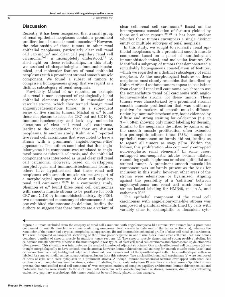
Figure 5 Tumors excluded from the category of renal cell carcinoma with angioleiomyoma-like stroma: Two tumors had a prominentcomponent of smooth muscle-like stroma containing numerous blood vessels in only one of the tumor sections (a), whereas theremainder of the tumor had a typical morphological appearance (b) and immunohistochemical profile of clear cell renal cell carcinoma.This was interpreted as tangential sectioning of the tumor pseudocapsule in one tissue block. Four clear cell renal cell carcinomascontained bundles of smooth muscle in multiple tumor sections (c). The smooth muscle demonstrated strong positive labeling forcaldesmon (inset); however, otherwise the immunoprofile was typical of clear cell renal cell carcinoma and chromosome 3p deletion wasoften present. This situation was interpreted as the result of invasion of adjacent structures. One unclassified renal cell carcinoma (d) wasthought morphologically to have smooth muscle stroma; however, immunohistochemical staining for smooth muscle actin (inset) andcaldesmon (not pictured) highlighted only the intratumoral blood vessels and not the spindle-shaped cells. The spindle-shaped cells alsolabeled for some epithelial antigens, supporting exclusion from this category. Two unclassified renal cell carcinomas (e) were composedof nests of cells with clear cytoplasm in a prominent stroma. Although immunohistochemical features overlapped with renal cellcarcinoma with angioleiomyoma-like stroma, extent of labeling for carbonic anhydrase IX was less and chromosome 3p deletion waspresent. One unclassified renal cell carcinoma (f) was composed entirely of complex papillary architecture. Immunohistochemical andmolecular features were similar to those of renal cell carcinoma with angioleiomyoma-like stroma; however, due to the contrastingexclusively papillary morphology, this tumor could not be confidently placed in that category.
Modern Pathology (2014), 1–16
Renal cell carcinoma with angioleiomyoma-like stroma
12 SR Williamson et al
plasm. Often the epithelium demonstrated a tuftedgrowth into the lumen of the glands with anill-defined apical cytoplasmic border, similar to the‘blister-like’ appearance described by Michal et al.3
Surrounding the epithelial elements, a layer ofcapillaries was often prominent, imparting a dual-cell layer pattern resembling a basal cell ormyoepithelial cell layer. The endothelial nature ofthese cells was supported by immunohistoche-mistry for CD34, with some areas showing aninterrupted or discontinuous capillary networksurrounding the glands.
The immunohistochemical profile of the epithe-lial component of these tumors was also uniform:epithelial cells were labeled with antibodies tocarbonic anhydrase IX (with a membranous or‘cup-shaped’8 pattern), CK7, CD10, vimentin, cyto-keratin 34bE12, and PAX8. Immunohistochemicalstaining with antibody to AMACR typically yieldeda negative reaction, with focal or weak labeling in aminority of tumors. Negative immunohistoche-mistry for HMB45, melan-A, cathepsin K, andTFE3 protein also argues against the possibilitythat these tumors are a manifestation of trans-location-associated renal cell carcinoma.33–35
Comparison to Clear Cell Papillary Renal CellCarcinoma
It has been hypothesized that these renal cellcarcinomas with smooth muscle stroma are part ofthe morphological spectrum of clear cell papillaryrenal cell carcinoma.13 In keeping with such apossibility, we found the immunohistochemicalprofile of these two tumor types to overlap, includ-ing labeling for carbonic anhydrase IX (sometimeswith a ‘cup-shaped’ staining pattern8), CK7,6,8,10 andcytokeratin 34bE128 and negative reactivity forAMACR.6,8,10 As a point of contrast, the tumors inthis study showed more diffuse labeling for CD10than expected of clear cell papillary renal cellcarcinoma.10 In our experience, clear cell papillaryrenal cell carcinoma usually shows an entirelynegative reaction for CD10 or apical labeling ofonly the cyst-lining cells; however, others haveoccasionally observed focal labeling for CD10 innon-cystic areas.6,8,10 Genetically, absence of chro-mosome 3p deletion is also a point of similaritybetween these tumor types that contrasts both toclear cell renal cell carcinoma.7,36 However, featurescontrasting these tumors included a higher nucleargrade in renal cell carcinoma with angioleiomyoma-like stroma (grade 2-3) compared with clear cellpapillary renal cell carcinoma (grade 1–2) and theabsence of the ‘secretory cell’ pattern with nucleialigned above the basement membrane in the former.In contradistinction to clear cell papillary renal cellcarcinoma, none of the tumors in this study wereassociated with end-stage renal disease (includingacquired cystic kidney disease),9 although one
occurred in an allograft kidney. Renal cellcarcinoma with angioleiomyoma-like stroma alsoappears quite rare compared with clear cellpapillary renal cell carcinoma: in this study, noneof the tumors that were ultimately classified as renalcell carcinoma with angioleiomyoma-like stromawere identified via a retrospective review of 341consecutive cases of clear cell renal cell carcinoma,whereas 10 tumors from the same series werereclassified as clear cell papillary renal cellcarcinoma, the latter accounting for approximately3% of total renal cell carcinomas from a 3-yearperiod.10 Therefore, if indeed these two neoplasmsrepresent opposing ends of the spectrum of a singledisease process, very few appear to occur at one end.If so, the presence of prominent smooth muscle thatis ready recognizable morphologically appears to beassociated with several other unique features,including increased labeling for CD10, a highernuclear grade, infrequent cysts, and the absence ofthe ‘secretory cell’10 pattern.
Comparison to Clear Cell Renal Cell Carcinoma
The absence of chromosome 3p deletion by FISHand positive immunohistochemical labeling for CK7and cytokeratin 34bE12 observed in this studysupport the idea that these tumors are not a mani-festation of clear cell renal cell carcinoma. However,some patterns of clear cell renal cell carcinomamimic the morphology of renal cell carcinoma withangioleiomyoma-like stroma.28,37 In one suchscenario, we found that areas of tangential section-ing of the tumor pseudocapsule may impart asimilar appearance to the prominent smoothmuscle stroma of these tumors. In such cases(n¼ 2), the immunohistochemical staining patternof clear cell renal cell carcinoma was observed(carbonic anhydrase IXþ , CD10þ , CK7� ,cytokeratin 34bE12� ), despite that the tumorpseudocapsule indeed resembled smooth muscleby light microscopy and immunohistochemistry. Ahelpful clue to this possibility is the presence ofsuch a smooth muscle proliferation in only a singletissue block of a neoplasm that otherwise resemblesclear cell renal cell carcinoma. Another four tumorshad bundles of smooth muscle extending into thetumor in multiple sections, again coupled with thetypical morphology and immunoprofile of clearcell renal cell carcinoma (carbonic anhydrase IXþ ,CD10þ , CK7� , cytokeratin 34bE12� ) and oftendeletion of chromosome 3p. Three of four of thesetumors demonstrated invasion of the renal capsuleor renal sinus, suggesting that this phenomenon maybe a reactive change to invasion of adjacent struc-tures. In both of these situations, the characteristicimmunohistochemical profile of clear cell renal cellcarcinoma may be helpful in pointing to the correctdiagnosis.
Modern Pathology (2014), 1–16
Renal cell carcinoma with angioleiomyoma-like stroma
SR Williamson et al 13

Tumors that could not be Confidently Classified
Five other tumors were excluded from the categoryof renal cell carcinoma with angioleiomyoma-likestroma that could not be confidently classified: ofthese, one tumor exhibited a mixed morphology andimmunoprofile of clear cell papillary renal cellcarcinoma and renal cell carcinoma with angioleio-myoma-like stroma. As discussed above, the rela-tionship between the tumors reported here and clearcell papillary renal cell carcinoma remains incom-pletely understood. The renal cell carcinomas withangioleiomyoma-like stroma in this study exhibiteda higher nuclear grade (2–3) than typically seen inclear cell papillary renal cell carcinoma (usually 1–2); however, it seems highly unlikely that theyrepresent a high-grade ‘transformation’ of clear cellpapillary renal cell carcinoma, as all were small(p3.5 cm) and confined to the kidney. One possibleexplanation for the mixed features in this one tumor(a larger mass measuring 7.5 cm) is juxtaposition oftwo separate tumors that were not recognized grosslyas distinct. In support of this possibility, the kidney inthis case harbored multifocal tumors,10 and no singletissue section showed a transition between morpho-logies. Another tumor that could not be confidentlyclassified (patient # 21) exhibited some of the featuresof renal cell carcinoma with angioleiomyoma-likestroma, including prominent stromal smooth muscle(caldesmonþ ) and a similar immunohistochemicaland molecular profile in the epithelial component(carbonic anhydrase IXþ , CK7þ , cytokeratin34bE12þ , CD10þ , AMACR� , 3p deletion absent);however, in contrast to the other renal cell carcinomaswith angioleiomyoma-like stroma, this neoplasm wascomposed exclusively of papillary architecture.Tumors with these features may indeed be withinthe spectrum of renal cell carcinoma with angio-leiomyoma-like stroma, though in the absence of acharacteristic molecular alteration it is difficult tofully support such an assertion in light of thesomewhat differing morphology.
As in the tumors reported by Shannon et al,4 twotumors in this study that were excluded from thecategory of renal cell carcinoma with angioleio-myoma-like stroma demonstrated chromosome 3pdeletion and labeling for CK7 (patients 19 and 20).Labeling for carbonic anhydrase IX was also lessthan is typical of clear cell renal cell carcinoma(5–35%) in these two tumors. The significance ofthese findings is not entirely clear. It has beensuggested that previously reported examples of CK7-positive clear cell renal cell carcinomas38 are in factclear cell papillary renal cell carcinomas.12 However,as clear cell papillary renal cell carcinomas lackchromosome 3p deletion and these tumors did notexhibit such morphology, the possibility thatanother subtype of CK7-positive ‘clear cell’ renalneoplasm exists remains open to further study.
Based on these results, we suspect that a substantialfraction of the tumors previously categorized as
‘renal angiomyoadenomatous tumor’, ‘renal cell car-cinoma associated with prominent angioleiomyoma-like proliferation’, and ‘clear cell renal cell carcinomawith smooth muscle stroma’2–4,13,28–31,37,39 could bealternatively categorized as clear cell papillaryrenal cell carcinoma, clear cell renal cell carci-noma with prominent bundles of smooth muscle(reactive or related to the tumor pseudocapsule),or renal cell carcinoma, unclassified. In supportof this hypothesis, after thorough morphological,immunohistochemical, and molecular study, almost50% of the tumors originally considered forinclusion in this study were thought not to fit intothe category of renal cell carcinoma with angio-leiomyoma-like stroma, even some of which wereoriginally diagnosed as such. Therefore discri-minating between subtypes of renal tumors withprominent stroma can be a particular diagnosticchallenge, requiring integration of the entireconstellation of features.
Clinical Significance
Although follow-up information was available forvery few patients in this study, none of the tumorsreclassified as renal cell carcinoma with angioleio-myoma-like stroma pursued an aggressive course.Likewise, to our knowledge no tumor with thesefeatures reported to date has demonstrated evidenceof malignant behavior. Our results also suggest thatawareness of the mimics of renal cell carcinomawith angioleiomyoma-like stroma is important.One neoplasm in this study was originally thoughtto belong to this category, composed of cellswith clear cytoplasm forming nests and tubularstructures in a spindle cell and vascular stroma(patient # 18). After immunohistochemical stain-ing (caldesmon� , CK7� , cytokeratin 34bE12� ,PAX8þ spindle cell component, AMACRþ ) andevaluation in the context of the other tumors, itbecame apparent that this neoplasm was betterregarded as unclassified renal cell carcinoma,possibly with an unusual manifestation of sarcoma-toid change. Contrasting to the behavior of the renalcell carcinomas with angioleiomyoma-like stroma,this patient had metastases to a regional lymph nodeand developed distant metastasis to the soft tissue ofthe orbit 12 months after diagnosis.
Summary
In this study, we examined a series of 23 renalcell neoplasms with a prominent smooth musclestromal component. Of these, 12 demonstrated ahomogeneous constellation of morphological andimmunohistochemical features, supporting classifi-cation into a distinct category that we regarded asrenal cell carcinoma with angioleiomyoma-likestroma. These tumors demonstrated prominentstromal smooth muscle (caldesmon þ ) that often
Modern Pathology (2014), 1–16
Renal cell carcinoma with angioleiomyoma-like stroma
14 SR Williamson et al

extended away from the epithelial component toentrap non-neoplastic renal tubules or interdigitatewith perinephric fat. The epithelial component wascharacteristically comprised of glandular structureswith variable clear to pale, flocculent, or eosinophi-lic cytoplasm, often with small tufts protruding intothe lumina. The immunohistochemical and mole-cular profile of the epithelial component was alsodistinct (carbonic anhydrase IXþ , CD10þ , CK7þ ,cytokeratin 34bE12þ , AMACR� , 3p deletionabsent) and contrasted to clear cell renal cellcarcinoma. Some features, particularly the labelingfor carbonic anhydrase IX, CK7, and cytokeratin34bE12 and absence of chromosome 3p deletion,raise the possibility of a relationship to clear cellpapillary renal cell carcinoma; however, theincreased labeling for CD10 and distinct morpholo-gical features, including well-formed bundles ofstromal smooth muscle, are points of contrast.Malignant potential of these tumors is not yetestablished; however, awareness of morphologicalmimics, some of which are unequivocally malig-nant, is important to avoid misdiagnosis.
Disclosure/conflict of interest
The authors declare no conflict of interest.
References
1 Michal M, Hes O, Havlicek F. Benign renal angiomyoa-denomatous tumor: a previously unreported renaltumor. Ann Diagn Pathol 2000;4:311–315.
2 Kuhn E, De Anda J, Manoni S, et al. Renal cellcarcinoma associated with prominent angioleiomyo-ma-like proliferation: Report of 5 cases and review ofthe literature. Am J Surg Pathol 2006;30:1372–1381.
3 Michal M, Hes O, Nemcova J, et al. Renal angiomyoa-denomatous tumor: morphologic, immunohistochem-ical, and molecular genetic study of a distinct entity.Virchows Arch 2009;454:89–99.
4 Shannon BA, Cohen RJ, Segal A, et al. Clear cell renalcell carcinoma with smooth muscle stroma. HumPathol 2009;40:425–429.
5 Adam J, Couturier J, Molinie V, et al. Clear-cellpapillary renal cell carcinoma: 24 cases of a distinctlow-grade renal tumour and a comparative genomichybridization array study of seven cases. Histopathol-ogy 2011;58:1064–1071.
6 Aydin H, Chen L, Cheng L, et al. Clear cell tubulopa-pillary renal cell carcinoma: a study of 36 distinctivelow-grade epithelial tumors of the kidney. Am J SurgPathol 2010;34:1608–1621.
7 Gobbo S, Eble JN, Grignon DJ, et al. Clear cell papillaryrenal cell carcinoma: a distinct histopathologic andmolecular genetic entity. Am J Surg Pathol 2008;32:1239–1245.
8 Rohan SM, Xiao Y, Liang Y, et al. Clear-cell papillaryrenal cell carcinoma: molecular and immunohistochemi-cal analysis with emphasis on the von Hippel-Lindaugene and hypoxia-inducible factor pathway-relatedproteins. Mod Pathol 2011;24:1207–1220.
9 Tickoo SK, dePeralta-Venturina MN, Harik LR, et al.Spectrum of epithelial neoplasms in end-stage renaldisease: an experience from 66 tumor-bearing kidneyswith emphasis on histologic patterns distinct fromthose in sporadic adult renal neoplasia. Am J SurgPathol 2006;30:141–153.
10 Williamson SR, Eble JN, Cheng L, et al. Clear cellpapillary renal cell carcinoma: differential diagnosisand extended immunohistochemical profile. ModPathol 2013;26:697–708.
11 Bhatnagar R, Alexiev BA. Renal-cell carcinomas inend-stage kidneys: a clinicopathological study withemphasis on clear-cell papillary renal-cell carcinomaand acquired cystic kidney disease-associated carci-noma. Int J Surg Pathol 2012;20:19–28.
12 Srigley JR, Delahunt B, Eble JN, et al. The InternationalSociety of Urological Pathology (ISUP) VancouverClassification of Renal Neoplasia. Am J Surg Pathol2013;37:1469–1489.
13 Verine J. Renal angiomyoadenomatous tumor: morpholo-gic, immunohistochemical, and molecular genetic studyof a distinct entity. Virchows Arch 2009;454:479–480.
14 Delahunt B, Cheville JC, Martignoni G, et al. TheInternational Society of Urological Pathology (ISUP)grading system for renal cell carcinoma and otherprognostic parameters. Am J Surg Pathol 2013;37:1490–1504.
15 Chen YB, Tickoo SK. Spectrum of preneoplastic andneoplastic cystic lesions of the kidney. Arch PatholLab Med 2012;136:400–409.
16 Al-Ahmadie HA, Alden D, Fine SW, et al. Role ofimmunohistochemistry in the evaluation of needlecore biopsies in adult renal cortical tumors: an ex vivostudy. Am J Surg Pathol 2011;35:949–961.
17 Williamson SR, Halat S, Eble JN, et al. Multilocularcystic renal cell carcinoma: similarities and differ-ences in immunoprofile compared with clear cellrenal cell carcinoma. Am J Surg Pathol 2012;36:1425–1433.
18 Gobbo S, Eble JN, MacLennan GT, et al. Renal cellcarcinomas with papillary architecture and clear cellcomponents: the utility of immunohistochemical andcytogenetical analyses in differential diagnosis. Am JSurg Pathol 2008;32:1780–1786.
19 Cheng L, MacLennan GT, Zhang S, et al. Evidence forpolyclonal origin of multifocal clear cell renal cellcarcinoma. Clin Cancer Res 2008;14:8087–8093.
20 Halat S, Eble JN, Grignon DJ, et al. Multilocular cysticrenal cell carcinoma is a subtype of clear cell renal cellcarcinoma. Mod Pathol 2010;23:931–936.
21 Williamson SR, Zhang S, Eble JN, et al. Clear cellpapillary renal cell carcinoma-like tumors in patientswith von Hippel-Lindau disease are unrelated tosporadic clear cell papillary renal cell carcinoma. AmJ Surg Pathol 2013;37:1131–1139.
22 Weller M, Berger H, Hartmann C, et al. Combined 1p/19qloss in oligodendroglial tumors: predictive or prognosticbiomarker? Clin Cancer Res 2007;13:6933–6937.
23 Wiens AL, Cheng L, Bertsch EC, et al. Polysomy ofchromosomes 1 and/or 19 is common and associatedwith less favorable clinical outcome in oligodendro-gliomas: fluorescent in situ hybridization analysis of84 consecutive cases. J Neuropathol Exp Neurol2012;71:618–624.
24 Brunelli M, Fiorentino M, Gobbo S, et al. Many facetsof chromosome 3p cytogenetic findings in clear cellrenal carcinoma: the need for agreement in assessment
Modern Pathology (2014), 1–16
Renal cell carcinoma with angioleiomyoma-like stroma
SR Williamson et al 15

FISH analysis to avoid diagnostic errors. HistolHistopathol 2011;26:1207–1213.
25 Cossu-Rocca P, Eble JN, Delahunt B, et al. Renalmucinous tubular and spindle carcinoma lacks thegains of chromosomes 7 and 17 and losses of chromo-some Y that are prevalent in papillary renal cellcarcinoma. Mod Pathol 2006;19:488–493.
26 Jones TD, Eble JN, Wang M, et al. Molecular geneticevidence for the independent origin of multifocalpapillary tumors in patients with papillary renal cellcarcinomas. Clin Cancer Res 2005;11:7226–7233.
27 Hisaoka M, Quade B. Angioleiomyoma, In: FletcherCDM, Bridge JA, Hogendoorn PC, Mertens F (eds).WHO Classification of Tumours of Soft Tissue andBone, 4th edn. IARC Press: Lyon; 2013, pp 120–121.
28 Petersson F, Grossmann P, Hora M, et al. Renal cellcarcinoma with areas mimicking renal angiomyoade-nomatous tumor/clear cell papillary renal cell carci-noma. Hum Pathol 2013;44:1412–1420.
29 Iczkowski KA, Shanks JH, Burdge AH, et al. Renal cellcarcinoma with clear cells, smooth muscle stroma, andnegative for 3p deletion: a variant of renal angiomyoa-denomatous tumour? A case report. Histopathology2013;62:522–524.
30 Singh C, Kendi AT, Manivel JC, et al. Renal angio-myoadenomatous tumor. Ann Diagn Pathol 2012;16:470–476.
31 Petersson F, Yan B, Huang J, et al. Low-grade renalcarcinoma with histologic features overlapping withrenal angiomyoadenomatous tumor and featuringpolysomy 7 and 17 and a mutation in the vonHippel-Lindau gene: report of a hybrid tumor and afew comments on renal angiomyoadenomatous tumor
and papillary renal tumors with clear cells. Ann DiagnPathol 2011;15:213–216.
32 Martignoni G, Bonetti F, Chilosi M, et al. Cathepsin Kexpression in the spectrum of perivascular epithelioidcell (PEC) lesions of the kidney. Mod Pathol2012;25:100–111.
33 Argani P, Hicks J, De Marzo AM, et al. Xp11translocation renal cell carcinoma (RCC): extendedimmunohistochemical profile emphasizing novel RCCmarkers. Am J Surg Pathol 2010;34:1295–1303.
34 Argani P, Olgac S, Tickoo SK, et al. Xp11 translocationrenal cell carcinoma in adults: expanded clinical,pathologic, and genetic spectrum. Am J Surg Pathol2007;31:1149–1160.
35 Camparo P, Vasiliu V, Molinie V, et al. Renaltranslocation carcinomas: clinicopathologic, immuno-histochemical, and gene expression profiling analysisof 31 cases with a review of the literature. Am J SurgPathol 2008;32:656–670.
36 Cheng L, Williamson SR, Zhang S, et al. Under-standing the molecular genetics of renal cell neoplasia:implications for diagnosis, prognosis and therapy.Expert Rev Anticancer Ther 2010;10:843–864.
37 Kuroda N, Hosokawa T, Michal M, et al. Clear cellrenal cell carcinoma with focal renal angiomyoadeno-matous tumor-like area. Ann Diagn Pathol 2011;15:202–206.
38 Mai KT, Kohler DM, Belanger EC, et al. Sporadic clearcell renal cell carcinoma with diffuse cytokeratin 7immunoreactivity. Pathology 2008;40:481–486.
39 Kuroda N, Michal M, Hes O, et al. Renal angiomyoa-denomatous tumor: fluorescence in situ hybridization.Pathol Int 2009;59:689–691.
Modern Pathology (2014), 1–16
Renal cell carcinoma with angioleiomyoma-like stroma
16 SR Williamson et al