Telomere Maintenance Mechanisms in Cancer: Clinical Implications
14
Send Orders for Reprints to [email protected] Current Pharmaceutical Design, 2014, 20, 6361-6374 6361 1873-4286/14 $58.00+.00 © 2014 Bentham Science Publishers Telomere Maintenance Mechanisms in Cancer: Clinical Implications Roger R. Reddel* Children's Medical Research Institute, 214 Hawkesbury Road, Westmead, New South Wales 2145, Australia; Sydney Medical School, University of Sydney, New South Wales 2006, Australia Abstract: The presence of immortal cell populations with an up-regulated telomere maintenance mechanism (TMM) is an almost univer- sal characteristic of cancers, whereas normal somatic cells are unable to prevent proliferation-associated telomere shortening and have a limited proliferative potential. TMMs and related aspects of telomere structure and function therefore appear to be ideal targets for the development of anticancer therapeutics. Such treatments would be targeted to a specific cancer-related molecular abnormality, and also be broad-spectrum in that they would be expected to be potentially applicable to most cancers. However, the telomere biology of normal and malignant human cells is a relatively young research field with large numbers of unanswered questions, so the optimal design of TMM-targeted therapeutic approaches remains unclear. This review outlines the opportunities and challenges presented by telomeres and TMMs for clinical management of cancer. Keywords: Alternative lengthening of telomeres, cancer, diagnosis, prognosis, telomerase, telomeres, therapy. INTRODUCTION More than 50 years have elapsed since the discovery that nor- mal human somatic cells have a finite proliferative capacity (subse- quently termed the Hayflick limit), in contrast to the apparently unlimited growth potential – or immortality – of cell lines derived from tumors [1, 2]. This discovery suggested the very attractive possibility that cellular immortality may result from molecular ab- normalities that clearly distinguish cancer cells from their normal counterparts, and may therefore represent an ideal opportunity for targeted anticancer therapies that have few if any side-effects on normal cells (reviewed in ref. [3]). The molecular alterations re- quired for immortalization of human cells are complex. Two major tumor suppressor pathways, involving pRb and p53, play a role in activating the cellular senescence program at the end of the normal replicative life span, and loss of function of these pathways is usu- ally required for immortalization of human cells (reviewed in [3]). A major determinant of cellular mortality is the telomere shortening that accompanies normal proliferation [4], so a key event in acquisi- tion of cellular immortality is the up-regulation of a telomere length maintenance mechanism (TMM) [5]. This review is limited to the aspects of immortalization related to the TMMs, and the opportuni- ties these present for clinical management of cancer. 1. TELOMERE STRUCTURE AND FUNCTION 1A. The Sequence and Structure of Telomeric DNA Human telomeres contain repetitive DNA, predominantly tan- dem arrays of the 5'-TTAGGG-3' hexanucleotide [6], often termed the canonical telomeric sequence. The proximal end of human te- lomeres (i.e., the end closest to the centromere) also contains vari- ant repeats such as TTGGGG, TGAGGG and TCAGGG [7, 8]. These variants are less common in the remainder (i.e., distal por- tion) of the telomere, which predominantly contains canonical re- peats and hexanucleotides with base substitutions at positions 1 and 3, such as GTAGGG and TTCGGG [9]. The termini of telomeres consist of 25-200 nucleotides of sin- gle-stranded DNA - usually the G-rich strand (referred to as the “G- overhang”). It is presumed that most of the remainder of the *Address correspondence to this author at Children's Medical Research Institute, 214 Hawkesbury Road, Westmead, New South Wales 2145, Australia; Tel: +612 8865 2901; Fax: +612 8865 2860; E-mail: [email protected] telomere forms duplex DNA by Watson-Crick base pairing, but the G-rich strand is capable of Hoogsteen base pairing, which forms planar G-quartet structures that stack on each other to form G- quadruplexes. Antibodies recognizing G-quadruplexes have dem- onstrated that these structures occur at human telomeres [10], and it is possible that they help protect the telomere against DNA repair. Another form of higher-order structure that is proposed to help protect telomeres against DNA repair is the t-loop [11]. The te- lomere is able to fold back on itself, so that the single-stranded telomeric overhang can invade duplex telomeric DNA and anneal with the complementary strand to create a loop structure, a process which is facilitated by the TRF2 protein [12]. Visualization of t- loops by super-resolution light microscopy has demonstrated that the point of invasion can be located at almost any point along the duplex DNA, resulting in t-loops of highly variable sizes [13]. Formation of these circular DNA structures may be an important contributor to the protection of telomere ends from DNA repair. 1B. Telomere Binding Proteins Telomeric DNA is bound by a protein complex, shelterin, which contains six proteins, TRF1, TRF2, TIN2, RAP1, TPP1 and POT1 (reviewed in ref. [14]). These proteins prevent telomeres being recognized by the cell as a DNA break and repaired by non- homologous end joining (NHEJ) or by homologous recombination (HR)-mediated repair. Repression of DNA repair at chromosome ends is essential for maintaining the organization of the genome into separate chromosomes, and failure of this repression results in genomic instability. 1C. Proliferation-Dependent Telomere Shortening It has been observed that cell proliferation in vitro is accompa- nied by telomere shortening [15, 16] (Fig. 1), which reflects the gradual overall decrease in telomere length in normal human so- matic tissues with increasing age [17]. This is due in part to the inability of the normal processes of semi-conservative DNA repli- cation to copy the termini of linear DNA molecules by lagging- strand synthesis [18, 19], referred to as the “end replication prob- lem”). It is also partly due to enzymatic processes that generate or elongate the single-stranded overhang at telomeric termini [20, 21]. The template available for replicating telomeric DNA thus steadily decreases in length with each cell cycle.
-
Upload
independent -
Category
Documents
-
view
2 -
download
0
Transcript of Telomere Maintenance Mechanisms in Cancer: Clinical Implications

Send Orders for Reprints to [email protected]
Current Pharmaceutical Design, 2014, 20, 6361-6374 6361
1873-4286/14 $58.00+.00 © 2014 Bentham Science Publishers
Telomere Maintenance Mechanisms in Cancer: Clinical Implications
Roger R. Reddel*
Children's Medical Research Institute, 214 Hawkesbury Road, Westmead, New South Wales 2145, Australia;
Sydney Medical School, University of Sydney, New South Wales 2006, Australia
Abstract: The presence of immortal cell populations with an up-regulated telomere maintenance mechanism (TMM) is an almost univer-sal characteristic of cancers, whereas normal somatic cells are unable to prevent proliferation-associated telomere shortening and have a limited proliferative potential. TMMs and related aspects of telomere structure and function therefore appear to be ideal targets for the development of anticancer therapeutics. Such treatments would be targeted to a specific cancer-related molecular abnormality, and also be broad-spectrum in that they would be expected to be potentially applicable to most cancers. However, the telomere biology of normal and malignant human cells is a relatively young research field with large numbers of unanswered questions, so the optimal design of TMM-targeted therapeutic approaches remains unclear. This review outlines the opportunities and challenges presented by telomeres and TMMs for clinical management of cancer.
Keywords: Alternative lengthening of telomeres, cancer, diagnosis, prognosis, telomerase, telomeres, therapy.
INTRODUCTION
More than 50 years have elapsed since the discovery that nor-mal human somatic cells have a finite proliferative capacity (subse-quently termed the Hayflick limit), in contrast to the apparently unlimited growth potential – or immortality – of cell lines derived from tumors [1, 2]. This discovery suggested the very attractive possibility that cellular immortality may result from molecular ab-normalities that clearly distinguish cancer cells from their normal counterparts, and may therefore represent an ideal opportunity for targeted anticancer therapies that have few if any side-effects on normal cells (reviewed in ref. [3]). The molecular alterations re-quired for immortalization of human cells are complex. Two major tumor suppressor pathways, involving pRb and p53, play a role in activating the cellular senescence program at the end of the normal replicative life span, and loss of function of these pathways is usu-ally required for immortalization of human cells (reviewed in [3]). A major determinant of cellular mortality is the telomere shortening that accompanies normal proliferation [4], so a key event in acquisi-tion of cellular immortality is the up-regulation of a telomere length maintenance mechanism (TMM) [5]. This review is limited to the aspects of immortalization related to the TMMs, and the opportuni-ties these present for clinical management of cancer.
1. TELOMERE STRUCTURE AND FUNCTION
1A. The Sequence and Structure of Telomeric DNA
Human telomeres contain repetitive DNA, predominantly tan-dem arrays of the 5'-TTAGGG-3' hexanucleotide [6], often termed the canonical telomeric sequence. The proximal end of human te-lomeres (i.e., the end closest to the centromere) also contains vari-ant repeats such as TTGGGG, TGAGGG and TCAGGG [7, 8]. These variants are less common in the remainder (i.e., distal por-tion) of the telomere, which predominantly contains canonical re-peats and hexanucleotides with base substitutions at positions 1 and 3, such as GTAGGG and TTCGGG [9].
The termini of telomeres consist of 25-200 nucleotides of sin-gle-stranded DNA - usually the G-rich strand (referred to as the “G-overhang”). It is presumed that most of the remainder of the
*Address correspondence to this author at Children's Medical Research Institute, 214 Hawkesbury Road, Westmead, New South Wales 2145, Australia; Tel: +612 8865 2901; Fax: +612 8865 2860; E-mail: [email protected]
telomere forms duplex DNA by Watson-Crick base pairing, but the G-rich strand is capable of Hoogsteen base pairing, which forms planar G-quartet structures that stack on each other to form G-
quadruplexes. Antibodies recognizing G-quadruplexes have dem-onstrated that these structures occur at human telomeres [10], and it is possible that they help protect the telomere against DNA repair.
Another form of higher-order structure that is proposed to help protect telomeres against DNA repair is the t-loop [11]. The te-lomere is able to fold back on itself, so that the single-stranded telomeric overhang can invade duplex telomeric DNA and anneal with the complementary strand to create a loop structure, a process which is facilitated by the TRF2 protein [12]. Visualization of t-loops by super-resolution light microscopy has demonstrated that the point of invasion can be located at almost any point along the duplex DNA, resulting in t-loops of highly variable sizes [13]. Formation of these circular DNA structures may be an important contributor to the protection of telomere ends from DNA repair.
1B. Telomere Binding Proteins
Telomeric DNA is bound by a protein complex, shelterin, which contains six proteins, TRF1, TRF2, TIN2, RAP1, TPP1 and POT1 (reviewed in ref. [14]). These proteins prevent telomeres being recognized by the cell as a DNA break and repaired by non-homologous end joining (NHEJ) or by homologous recombination (HR)-mediated repair. Repression of DNA repair at chromosome ends is essential for maintaining the organization of the genome into separate chromosomes, and failure of this repression results in genomic instability.
1C. Proliferation-Dependent Telomere Shortening
It has been observed that cell proliferation in vitro is accompa-nied by telomere shortening [15, 16] (Fig. 1), which reflects the gradual overall decrease in telomere length in normal human so-matic tissues with increasing age [17]. This is due in part to the inability of the normal processes of semi-conservative DNA repli-cation to copy the termini of linear DNA molecules by lagging-strand synthesis [18, 19], referred to as the “end replication prob-lem”). It is also partly due to enzymatic processes that generate or elongate the single-stranded overhang at telomeric termini [20, 21]. The template available for replicating telomeric DNA thus steadily decreases in length with each cell cycle.
6362 Current Pharmaceutical Design, 2014, Vol. 20, No. 41 Roger R. Reddel
1D. Telomere Capping and the Connection between Telomere
Shortening and Senescence
A telomere that is fully protected, presumably by a combination of its higher-order DNA structure and its binding proteins, avoids eliciting a DNA damage response (DDR) or unwanted DNA repair, and is referred to as "capped". It has been proposed that the te-lomere needs to become temporarily uncapped to allow access of telomerase [22]. Telomeres can become fully uncapped by experi-mental disruption of specific shelterin components, resulting in an ATM- or ATR-dependent DDR and end-to-end fusion of chromo-somes [23-25]. Uncapped telomeres are recognized by the co-localization of DDR proteins, such as phosphorylated histone H2AX (i.e., �H2AX) and chromosome ends, which are referred to as Telomere dysfunction-Induced Foci (TIFs) [23].
Replicating young human cells also exhibit a small number of telomeric DDR foci, and the number increases as the cells continue proliferating and their telomeres continue to shorten; in contrast to TIFs in cells with uncapped telomeres, these foci are not associated with end-to-end fusions. The cells finally arrest in G1 phase of the cell cycle and become senescent when the number of telomeric DDR foci reaches 4 or 5 [26]. If senescence is bypassed by loss of function of the p53 and pRb tumor suppressor pathways, continued proliferation is associated with further telomere shortening and further accumulation of telomeric DDR foci, until eventually there is widespread end-to-end fusion of chromosomes (indicating that many of the telomeres have become fully uncapped) and cell death - a state referred to as culture crisis [26]. Therefore, it was deduced that there must be a telomere conformation intermediate between fully capped and fully uncapped, which elicits a DDR but represses DNA repair, and that integrated signaling from 4 or 5 of these in-termediate-state telomeres (IST) results in senescence [26] (Fig. 2).
Telomere shortening is not the only stimulus for telomeres to adopt the IST conformation. When mitosis is prolonged exces-sively, ISTs are induced, and cells which eventually escape from mitosis undergo cell cycle arrest in the subsequent G1 phase, unless they lack wild-type p53 function, in which case they continue divid-ing and become aneuploid [27]. The signaling pathway for IST-induced G1 arrest involves ATM, but does not involve phosphory-lation of CHK2, and is thus distinct from the genomic DDR [28]. In both the case of replication-associated telomere shortening and of
prolonged mitosis, it is presumably advantageous for the organism if the cell is permitted to continue through the cell cycle until it reaches G1, at which point the senescence program can be activated (Fig. 2). It will be very interesting to determine whether there are other abnormalities of cellular function where the same telomeric signaling pathway is used to allow the cell to continue through the cell cycle until it reaches G1 and becomes senescent, what upstream signaling events result in adoption of the IST conformation, and how the molecular players in an IST-associated DDR response differ from those at an uncapped telomere or a DDR elsewhere in the genome.
1E. Telomere Lengthening by Telomerase
Gradual attrition of chromosome ends would eventually result in loss of vital genomic material, so a mechanism for preventing this in the germ line is essential. The best known of the telomere lengthening mechanisms is telomerase, a ribonucleoprotein (RNP) enzyme that synthesizes new telomeric DNA to compensate for replication-associated telomere attrition by reverse transcribing a template region within its RNA moiety [29]. Telomerase mostly synthesizes the canonical repeat, but the presence throughout the distal telomere of repeats differing from this sequence at positions 1 and 3 indicates that telomerase sometimes substitutes bases at spe-cific locations on its template [9]; the functional consequences of these substitutions remain to be determined.
Telomerase activity can be detected in human embryonic stem cells at levels which are sufficient to prevent telomere shortening [30]. Moreover, induced pluripotent stem cells are able to up-regulate telomerase levels sufficiently to elongate their telomeres compared to the normal cells from which they are derived [31]. In multipotent stem cells, such as those of the hematopoietic system, telomerase is under tight control and does not completely prevent telomere shortening [32]. Most somatic tissues have very low levels of telomerase and undergo telomere shortening throughout life. A rare exception to this is that CD19+CD27+ (memory) B-lymphocyte cells appear to have sufficient telomerase activity to undergo telomere lengthening during their ontogeny [33].
In contrast to normal somatic tissues, the great majority (~85%) of human cancers have detectable levels of telomerase [34], and in cancer-derived cell lines this appears to be sufficient to prevent telomere shortening. However, even in cell lines where telomerase
Fig. (1). Telomeres undergo gradual attrition during cellular proliferation. Telomeres (lighter bars; darker bars represent non-telomeric DNA) contain tandemly repeated arrays of the hexameric sequence, 5'-TTAGGG-3'. Telomeres are mostly double-stranded, but they terminate in a region of single-stranded (usually G-rich) DNA. In cultured human fibroblasts, telomeres shorten by approximately 50-150 base pairs per cell division. This ultimately results in a DNA damage response (DDR) focus, and, when a sufficient number of such foci accumulate, the cell undergoes permanent withdrawal from the cell cycle (i.e., becomes senescent).
Centromere (5'�TTAGGG�3')n
(3'�AATCCC�5')nn
DDR GrowthArrest

Telomere-Targeted Cancer Therapeutics Current Pharmaceutical Design, 2014, Vol. 20, No. 41 6363
has maintained telomere length for many hundreds of population doublings (PD), the number of active telomerase molecules has been estimated at 20-50 copies per cell [35], which means that there are fewer active telomerase molecules than there are telomere ends and that this is one of the least abundant molecules in the cell. This presents major challenges for studying this enzyme, e.g., for study-ing its intracellular location using antibody staining, and for obtain-ing sufficient enzyme for structural studies.
Telomerase is a complex molecule with several subunits, and additional associated proteins are involved in its biogenesis, all of which are potential targets for development of therapeutics that decrease telomerase activity. The molecules which are essential for its catalytic activity are the reverse transcriptase (TERT) and RNA (an H/ACA RNA which is variously referred to as TR, TER or TERC) moieties (Fig. 3). Mass spectrometry of active human te-lomerase showed that it contains two molecules each of TERT, TR and a pseudouridylase H/ACA ribonucleoprotein protein, dyskerin (also named NAP57) [35]. It has been found that both TERT cata-lytic sites must be active in order for the complex to be active, im-plying that telomerase functions as a dimer, and this was supported by electron microscopy data at 23-Å resolution showing that the enzyme is bi-lobar [36]. Other proteins that associate with telom-erase and are required for its assembly include two ATPases – reptin and pontin [37] – and three additional H/ACA RNP proteins – NOP10, NHP2, and GAR1 (reviewed in [38]). A further protein that may be transiently involved in telomerase assembly is NAF1, which initially binds dyskerin and escorts it to the nascent H/ACA RNA before being replaced by GAR1 to yield mature H/ACA RNPs in Cajal bodies and nucleoli [39].
The processes involved in relocation of telomerase from the nucleolus, where it is assembled [40], to its substrate, the telomere, also present a number of potential opportunities for therapeutic intervention. The processes, often collectively referred to as telom-erase recruitment (reviewed in [41]), are as follows. The assembled telomerase travels to, and accumulates in Cajal Bodies in a process that is dependent on the RNA binding protein, TCAB1 [42], which then plays a direct role in the transport of telomerase to the te-
lomere [43]. At the telomere, telomerase cannot act on its substrate unless the cis-acting inhibitory effects of TRF1 are relieved [44] and telomerase is able to interact with TPP1 and dock at the end of the telomeric single-stranded overhang [45]. The factors which control these mechanisms, as well as the processivity of telomerase (i.e., its ability to continue adding telomeric repeats to a telomere terminus), and the mechanisms whereby it relocates to other te-lomeres, are all incompletely understood at present.
1F. Mechanisms of Telomerase Up-Regulation in Cancer
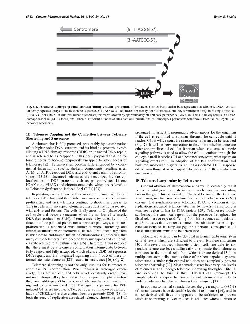
Details of the mechanisms involved in the up-regulation of telomerase in cancer are continuing to emerge (Fig. 3). Many stud-ies have focussed on the role of transcriptional control of TERT expression or on phosphorylation-induced increases in TERT activ-ity ([46-50]; reviewed in refs. [51-53]). Positively acting transcrip-tion factors and co-regulators include the MYC oncogene, the E6 protein of oncogenic human papillomaviruses, NF-�B and �-catenin. Up-regulated expression may also occur through loss of pRB, WT1, Menin and TGF� pathway tumor suppressor function. Protein kinase C� and AKT can phosphorylate TERT.
Mutations within the core promoter region of TERT are com-mon, and these may increase TERT transcriptional activity by cre-ating de novo consensus binding motifs for ETS transcription fac-tors [54, 55]. TERT promoter mutations are most common in mela-nomas, bladder carcinomas, liposarcomas, hepatocellular carcino-mas, squamous cell carcinomas of the tongue, medulloblastomas, and gliomas, especially primary glioblastoma multiforme [54-58]. TERT promoter mutations appear to be a useful urinary biomarker for early detection and monitoring of bladder cancer [59]. Another mechanism of up-regulation is translocation of the TERT gene to an immunoglobulin gene or other loci in B-cell neoplasms, which presumably contributes to the increased TERT transcription and telomerase activity observed in these tumors [60].
There is evidence that the levels of TERT and TR are both lim-iting for telomerase activity (reviewed in [61]), so it is reasonable to assume that up-regulation of telomerase requires an increase in both of these telomerase subunits. PAX8 is able to coordinately up-
Fig. (2). Telomeres can adopt at least three different conformational states. The fully capped state inhibits both the DDR and DNA repair by non-homologous end-joining (NHEJ) which would result in end-to-end joining of chromosomes and therefore genomic instability. Normal telomere shortening results in an intermediate-state telomere which inhibits NHEJ but not DDR. When the threshold number of telomeric DDR foci is exceeded, the cell continues through mitosis and then arrests in G1 phase. An abnormal cell state, such as excessively slow transit through mitosis, also elicits intermediate-state telomeres (ISTs) by an unknown mechanism, with the result that the cell continues to proceed through mitosis until it reaches G1 and can then undergo an orderly exit from the cell cycle. Excessive telomere shortening results in fully uncapped telomeres which elicit both DDR and NHEJ and result in cell death; when most of the cells in a culture enter this state, this is referred to as "culture crisis".
Abnormal�Fullycell�statesCapped
Telomere
Inter�mediateTelomere
Excessiveshortening
G1�ArrestFully
Uncapped
Telomere
g
Crisis
Telomere
6364 Current Pharmaceutical Design, 2014, Vol. 20, No. 41 Roger R. Reddel
regulate both TERT and TR expression, and the PAX8 expression level correlates with telomerase activity in gliomas [62]. The copy number of the TERT and TR genes is commonly increased in hu-man cancers (reviewed in ref. [63]).
1G. Non-catalytic Functions of Telomerase
Evidence has been presented that telomerase has roles in cell proliferation, genome stability and protection against apoptosis in addition to its role in catalyzing synthesis of telomeric DNA. The TERT protein was found to have a mitochondrial targeting se-quence in its N-terminus, and mitochondrial extracts were found to contain telomerase activity [64]. Although one study found that mitochondrial telomerase sensitized cells to oxidative stress and induction of apoptosis [64], other groups have reported that over-expression of TERT lowered mitochondrial production of reactive oxygen species, and protected mitochondria from oxidative stress, nuclei from DNA damage, and cells from apoptosis [65-68]. In another study, over-expression of the major splice variant of TERT (termed the �-deletion variant), which lacks the catalytic domain, protected breast cancer cells from cisplatin-induced apoptosis [69].
Another proposed non-catalytic role of TERT is induction of signaling via the Wnt pathway in mouse and human cells, by inter-acting with BRG1, a chromatin remodeling protein and by acting as a transcriptional co-factor in a complex with �-catenin [70, 71]. However, in different human cell lines, no association was found between TERT and BRG1 or �-catenin, and no consistent effects on Wnt pathway signaling were seen [72].
1H. Telomere Lengthening by ALT
Some immortalized cell lines maintain telomere length for hun-dreds of population doublings in the absence of telomerase activity, and it was therefore deduced that they must have an alternative
lengthening of telomeres (ALT) mechanism [73, 74]. In contrast to telomerase, which uses an RNA template for de novo synthesis of telomeric DNA [29], ALT involves synthesis of new telomeric DNA from a DNA template via HR (reviewed in ref. [75] (Fig. 4)). The template may be the telomere of another chromosome [76, 77] or another region of the same telomere via t-loop formation or sister telomere recombination [78]. Many molecular details of the ALT mechanism remain unknown, but it has been proposed that various HR proteins are involved [79-88].
ALT activity is detectable in normal mouse tissues [89], where it is clearly insufficient to maintain telomere length. In human cells where ALT activity is up-regulated sufficiently to maintain te-lomere length, there is a characteristic telomere phenotype. The telomeres are highly heterogeneous in length, but the average length (>17 kb) is about double that of most cells where telomere length is maintained by telomerase [73]. A large proportion of the telomeres in ALT cells evoke a DDR, and most likely are in an IST conformation [90]. There are substantial quantities of extra-chromosomal telomeric repeat (ECTR) DNA, which may be linear [91, 92], circular and double-stranded (t-circles) [93, 94], circular and at least partly single-stranded (referred to as C- or G-circles, if the C- or G-rich strand is intact, respectively) [95, 96], or high mo-lecular weight and complex in structure [96]. C-circles can be de-tected as self-priming templates for rolling circle amplification by �29 polymerase [95, 97], and the C-circle level appears to correlate with the level of ALT activity [95].
Some of the ECTR DNA, as well as chromosomal telomeric DNA, is contained within PML nuclear bodies [98-100]. Because telomeric DNA and telomere binding proteins are not normally found within PML bodies, these structures are referred to as ALT-associated PML bodies (APBs). APBs can be used to determine whether tumors are ALT-positive [98, 101, 102]. Early studies
Fig. (3). Telomerase up-regulation in cancer cells can occur by a variety of mechanisms. The telomerase catalytic subunit (TERT) and the RNA template molecule (TR) are both present at very low copy number in normal cells, and it is likely that the expression of both must be increased in order to express suffi-cient telomerase to prevent telomere shortening in cancer cells. Increased transcriptional activation (for both TERT and TR), and loss of transcriptional repres-sors (for TERT) have been found in some cancer cells. Many telomerase-positive cancer cells have a mutation in the TERT gene promoter/enhancer region, which appears to result in increased trans-activation of this gene. The activity of TERT may also be increased by certain kinases. Increased copy number of TERT and/or TERC has been identified in many cancers.
Trans� Kinases
Trans�repressors
Transactivators
TERTControl Coding
TERTActivatingMutation
regionelements
TERTTRl
CodingControl
elements region
Trans�activators

Telomere-Targeted Cancer Therapeutics Current Pharmaceutical Design, 2014, Vol. 20, No. 41 6365
showed that HR proteins, such as RPA, RAD51, RAD52, and NBS1, localize within APBs [98, 103] and many more APB com-ponents have subsequently been added to the list. Compared to other immortalized cells, there is a markedly increased level of recombination at telomeres [104, 105], but not at most other loca-tions in the genome [104, 106]. However, a subset of minisatellite sequences undergo increased recombination for reasons that are not understood [107-109].
Types of tumors where ALT is very common include osteosar-comas, undifferentiated pleomorphic sarcomas, leiomyosarcomas, and grade 2 and 3 astrocytic brain tumors, but ALT is also common in some other types of soft tissue sarcomas, grade 4 astrocytic brain tumors (glioblastomas), and neuroblastomas, and occurs at low frequency in many types of carcinomas [101, 110-121] (reviewed in ref. [122]). A small proportion of ALT-positive tumors also have telomerase activity [110-113, 123]. The correlation between ALT and prognosis (discussed further in section 2B below) varies among different tumor types; for example, the presence of ALT in glioblas-tomas is associated with a better outcome, whereas in liposarcomas ALT is associated with a significantly worse outcome [112, 113, 117, 124].
1I. Mechanisms of ALT Up-Regulation in Cancer
Somatic cell hybridization analyses demonstrated that normal telomerase-negative human cells, as well as telomerase-positive immortalized cells, contain repressors of ALT activity [73, 125, 126]. The observation that the gene encoding one or other member of the ATRX/DAXX protein complex is mutated in many ALT-positive cancers and cell lines [120, 127-133] suggests that loss of ATRX/DAXX function is required for ALT to be activated.
Among many functions attributed to ATRX/DAXX, this com-plex is thought to be involved in chromatin remodeling, including telomeric deposition of the variant histone H3.3. Some pediatric glioblastomas have mutations both in H3.3 and ATRX [133], sug-gesting that the major selection pressure for loss of ATRX/DAXX is not related to its H3.3 function. There are other indications, how-ever, that the architecture of telomeric chromatin is substantially altered in cells with up-regulated ALT activity (reviewed in ref. [134]). The telomeric DNA sequence becomes altered as variant
repeats which are normally present in the most proximal portion of the telomere become spread throughout the telomere, presumably initiated when a proximal telomere is used as the template for ALT-mediated DNA synthesis [9, 77, 135]. Shelterin is therefore par-tially displaced from ALT telomeres by proteins such as the nuclear receptors, COUP-TF2 and TR4 [135]. Shelterin desaturation most likely contributes to the large number of telomeric DDR foci in ALT cells, which can be partially suppressed by over-expression of TRF2 [90], and would be expected to decrease the repression of HR at telomeres [136] and result in an increase in stalled replication forks within telomeres [137], thus stimulating telomeric recombina-tion. For reasons that are not understood, telomeric chromatin ap-pears to be less compacted in cells that utilize ALT compared to those that utilize telomerase, which may also favor interactions among telomeres [138].
1J. Telomere Trimming
In addition to the gradual telomere attrition that accompanies cellular proliferation, there is a more rapid telomere shortening process termed telomere trimming (reviewed in ref. [139]), which resembles telomere rapid deletion in yeast [140]. First observed in human cancer cell lines with telomeres that were over-lengthened by exogenous telomerase, telomere trimming appears to be a well-regulated mechanism for rapid correction of over-lengthening via excision of circularized telomeric DNA [141]. In cells with up-regulated telomerase activity, this would help maintain telomere length close to a set point [139]. Telomere trimming also occurs in cells of the male germ line and in normal lymphocytes following mitogen-stimulated up-regulation of telomerase activity [142]. Te-lomere length is therefore subject to multiple lengthening and shortening processes (Fig. 5).
2. CLINICAL IMPLICATIONS OF TELOMERE MAINTE-
NANCE MECHANISMS FOR CANCER
2A. Diagnosis
The standard method of detecting telomerase in tumors is the Telomere Repeat Amplification Protocol (TRAP) assay [143]; this is a highly sensitive, semi-quantitative assay in which telomerase present in tumor lysates extends an oligonucleotide substrate and
Fig. (4). Telomerase and ALT both result in de novo synthesis of telomeric DNA. Unlike telomerase, which reverse transcribes new telomeric sequence from the template region (black bar) of its RNA subunit, ALT involves formation of a DNA recombination intermediate and the use of a DNA template for synthesis of new telomeric sequence; see text for details.
Telomerase
TERT
DyskerinTR
ALT

6366 Current Pharmaceutical Design, 2014, Vol. 20, No. 41 Roger R. Reddel
the products are detected by PCR. Tumor tissues may contain in-hibitors of the TRAP assay which cause false-negative results un-less the lysate is diluted appropriately. A possible alternative is to remove inhibitors from the sample by immunoprecipitation with an antibody against TERT prior to the TRAP assay [144]. Direct te-lomerase assays which have no PCR amplification step are quanti-tative, but are less sensitive by several orders of magnitude [145, 146]. In some studies, the presence of telomerase activity is inferred from TERT and/or TERC transcript levels measured by reverse transcription PCR, or by in situ hybridization in the case of TERC. Immunohistochemical detection of telomerase in tumor tissues is problematic, most likely due to the exceptionally low abundance of this molecule, even in cancer cells [35].
Use of telomerase detection for cancer diagnosis has been re-viewed elsewhere (e.g., ref. [147]). Despite the potential promise of being able to diagnose cancer via detection of an enzyme activity which is up-regulated in 85% of all cancers [34], telomerase assays have not yet been adopted for routine cancer diagnosis. A system-atic review and meta-analysis of the correlation between telomerase activity (measured by TRAP or telomerase component transcript levels) and histopathologic diagnosis in 2395 breast lesions found that 82% of breast cancers and 18% of benign lesions were positive for telomerase activity [148]. It is possible that a better correlation would be obtained if technical improvements in telomerase assays are able to decrease false negative and false positive results, if measurements of TERT mRNA levels distinguish between full-length transcripts and splice variants that do not encode catalyti-cally active TERT protein, and if the presence of ALT is taken into account.
The most reliable methods for detecting ALT in tumor samples are assays for APBs [98, 101], C-circles [95], or telomere length [110]. APBs can be visualized in fresh-frozen tumors, cytology specimens, or formalin-fixed paraffin-embedded tumor samples after deparaffinization and an antigen retrieval step, by immu-nostaining for PML and either immunostaining for a shelterin pro-tein or Fluorescence in situ Hybridization (FISH) with a telomeric probe. The amount of telomeric DNA in APBs is often much greater than at a single chromosome end, so ALT may also be de-tected through the presence of very bright foci after telomeric FISH [127]. C-circles can be quantitated in genomic DNA extracted from tumors by rolling circle amplification with �29DNA polymerase in the absence of any added primer, followed by detection of telomeric DNA [95]. Telomeres in ALT cells are typically very heterogene-
ous in length, but long on average, and this can be detected in good-quality genomic DNA from tumors by Southern blotting of terminal restriction fragments [110]. Alternatively, content of telomeric DNA may be measured in small samples of genomic DNA by quan-titative PCR (qPCR), and this may be combined with qPCR detec-tion of C-circles amplified by �29 DNA polymerase [97].
Reliable methods for detecting telomerase and ALT are likely to become an important pre-requisite for the use of treatments tar-geting one or other of these mechanisms. Moreover, in principle, a method for detecting both tumor-derived telomerase and markers of ALT activity in blood or other bodily fluids could be a simple screening test for cancer. It has been demonstrated that C-circles from ALT-positive osteosarcomas can be detected in blood samples [95], so it is possible that this could be used for surveillance in clinical situations where there is a high risk of developing an ALT-positive tumor, or to monitor response to treatment of an ALT-positive tumor. However, the presence of telomerase activity in normal leukocytes makes it difficult to use telomerase activity as-says in an analogous manner, so use of TMM detection as the basis of a blood test for cancer would require some technical advances.
2B. Prognosis
In view of the technical difficulties in quantitating telomerase activity, it is perhaps not surprising that studies of the correlation between telomerase activity levels and patient outcome have yielded divergent results. Using breast cancer again as an example, several studies have shown that high levels of telomerase activity or subunit transcripts are associated with poor prognosis [149-153]. In contrast, a more recent study found that telomerase level was not associated with disease outcome overall, although an association may have been masked by the types of treatment [154]. Similarly, higher telomerase levels have been found to be associated with better [155] or worse [156] outcomes in stage II colorectal cancer, and worse outcomes in colorectal cancer overall [156, 157].
The correlation between ALT and prognosis varies among dif-ferent tumor types. In osteosarcomas, a tumor type where ALT is the more common TMM, it is telomerase which appears to be the adverse prognostic indicator, based on the separate observations that progression-free and overall survival was worse in telomerase-positive tumors compared to telomerase-negative [158], absence of both telomerase and ALT was associated with better clinical out-come [111], and there was no significant difference between sur-vival of patients with ALT and non-ALT tumors [101]. For malig-
Fig. (5). Multiple factors contribute to telomere length dynamics in normal and cancer cells. Telomeres undergo proliferation-associated attrition, which may be counteracted by lengthening via telomerase and/or ALT. Over-lengthening may be corrected rapidly by telomere trimming. In immortalized cells, telomere length is maintained within a range around a maintenance set point, whereas in normal, mortal cells telomere length is not maintained overall and may decrease sufficiently to induce senescence. Cells in which tumor suppressor pathways, p53 and pRb, are inactivated may fail to enter senescence and their telomeres may continue shortening until they are no longer able to inhibit DNA repair by end-joining, at which point (crisis) they undergo cell death.
i /
Trimming
Setpoint/Threshold
ngth�
Maintenanceom
ere�Len
Senescence
Telo
Crisis

Telomere-Targeted Cancer Therapeutics Current Pharmaceutical Design, 2014, Vol. 20, No. 41 6367
nant fibrous histiocytomas, presence of ALT was associated with decreased survival, and this was not affected by co-existent telom-erase activity [159], although a smaller study did not find a differ-ence in survival between ALT and non-ALT tumors [101]. In liposarcomas, presence of ALT was more strongly associated with decreased survival than was telomerase activity [113, 124]. In con-trast, presence of ALT in glioblastoma multiforme has been associ-ated with better patient outcome [101, 112, 117], and telomerase was found to confer a worse outcome in pediatric high-grade glio-mas [160].
Although the functional consequences of telomerase and ALT up-regulation are similar in that they both prevent overall telomere shortening in tumors, these TMMs differ in several regards which may account for their differential prognostic significance in various tumor types. As described above, the genetic and epigenetic events responsible for activation of these mechanisms differ (possibly in a manner that is specific for the cell of origin). Moreover, some of the functional consequences may differ: up-regulated telomerase may possibly have non-canonical functions (also as described above), and ALT has been associated with complex karyotypes and chro-mosomal instability which may drive tumor progression [113, 161, 162].
2C. Opportunities for TMM-Targeted Therapies
There are many aspects of telomere maintenance in cancers that present potential opportunities for developing targeted therapies. The most obvious of these are the catalytic events involved in de novo synthesis of telomeric DNA by telomerase or ALT, but other changes in cancer cells that are associated with activation of these mechanisms should also be considered as potential targets.
2C(i). Inhibition of Telomerase
The development of telomerase inhibitors has been the subject of many reviews, including refs. [163-174]. The telomerase-catalyzed telomere lengthening reaction proceeds via multiple steps, which include binding to its substrate, reverse transcription of telomeric DNA, and translocation of the enzyme on its extended substrate to continue the lengthening reaction (which is required for its processivity). All of these steps are potentially able to be inhib-
ited, and inhibitors of the reverse transcription step could target either the template, as is the case for GRN163L (Imetelstat; a lipi-dated 13-mer oligonucleotide, which has entered clinical trials), or the catalytic subunit. A compound which has been used quite exten-sively for in vitro studies, BIBR1532, is a non-competitive telom-erase inhibitor which appears to inhibit its processivity [175, 176].
Telomerase-mediated lengthening could potentially be inhibited by targeting other steps in the overall process, starting with expres-sion of the subunits and their assembly into the active enzyme com-plex (Fig. 6). The TERT gene is subject to alternative splicing with the majority of splice variants expected to result in catalytically inactive protein, if indeed they are translated [177], so modulation of splicing may also be a useful therapeutic strategy [178], as could selective targeting of TERT protein for proteasome-mediated deg-radation [179]. Mutations in two of the proteins involved in telom-erase assembly, NHP2 and NOP10, result in telomere shortening in vivo, indicating that these proteins could potentially be targeted as a means of decreasing telomerase levels. In addition, telomerase must be transported to the telomere via processes that involve TCAB1 and Cajal bodies [43]. TCAB1 mutations are known to cause exces-sively short telomeres and the associated clinical manifestations of short telomere syndrome in humans [180]. This suggests that target-ing of TCAB1 and other aspects of telomerase transport is likely to decrease telomerase activity in tumours, and that it therefore could be an effective therapeutic intervention.
Access of telomerase to its substrate is controlled by an incom-pletely-understood, and presumably intricately-regulated set of processes in which TIN2-anchored TPP1 is involved in recruiting telomerase [45, 181], and another shelterin component, TRF1, plays an inhibitory role [182]. The inhibitory role of TRF1 is relieved by tankyrase-induced poly ADP-ribosylation of TRF1 [183], which suggests that inhibitors of tankyrase may impede access of telom-erase to telomeres. The telomeric transcript, TERRA, also appears to be involved in excluding telomerase from telomeres, and this can be alleviated by hnRNPA1 [184, 185], which is therefore another potential drug target. Finally, the number of active telomerase molecules in cancer cells appears to be smaller than the number of telomeres [35], which suggests that telomerase needs to undock
Fig. (6). The action of telomerase may be inhibited at multiple points in its "life cycle". These include synthesis of the individual components of active telomerase (TERT, dyskerin and TR), assembly into the enzyme complex, transport to a telomere, docking with the telomere, extending the telomere via its catalytic function, and movement to another telomere.
4.�Docking5.�Catalysis 6.�Transport�to�
another�telomereTERT
Dyskerin
1.�Synthesis
3.�TransportTERT2.�Assembly
TRTERT
DyskerinDyskerin
TERTTERTTR
y

6368 Current Pharmaceutical Design, 2014, Vol. 20, No. 41 Roger R. Reddel
from one telomere and move to another telomere by a process which is essentially unknown and which is another potential target for anticancer therapeutics.
2C(ii). ALT Inhibitors
The ultimate steps in the ALT process involve HR and DNA synthesis, which are processes vital for repair and replication of DNA in normal cells. Although there may be a therapeutic window where cancer cells, especially those which have lost p53 function, are more vulnerable to inhibition of these functions than normal cells, the likelihood of toxic side-effects of such inhibition is high. Many molecular details of ALT remain to be discovered, however, and it is possible that key steps, e.g., those involved in the juxtapo-sition of telomeres that must be required for one telomere to use a non-homologous telomere as a copy template, involve molecules which are not expressed, or are expressed at much lower levels in normal somatic cells, and which therefore would be suitable targets for development of ALT inhibitors. Failing this, it still may be pos-sible to exploit the causes and/or consequences of ALT up-regulation, as described below.
2C(iii). Immunotherapy
Peptides generated by degradation of hTERT are presented on the cell surface via the major histocompatibility complex (MHC) class I pathway; cytotoxic T-lymphocytes (CTL) can recognise these peptides and kill the cells that present them, which makes telomerase a potential target for the development of immunothera-pies (reviewed in refs. [186, 187]). Cancer vaccines containing these peptides have been subjected to clinical trials and have shown acceptable safety and tolerability, and have generated strong im-mune responses in a substantial proportion of patients, as well as some encouraging effects on tumors [188-191]. If proteins are dis-covered that are expressed in ALT cells to a much greater extent than in normal somatic cells, it might also be possible to develop suitable immunotherapeutic approaches for tumors that depend on ALT.
2C(iv). Targeting Normal or Abnormal Telomere Sequence and
Structure
It is possible to design drugs to bind to specific DNA structures, and given the G-rich nature of the telomeric sequence there have been extensive efforts to develop drugs that target telomeric G-quadruplex DNA (reviewed in ref. [192]). Somewhat paradoxically, there is evidence that such drugs cause telomere uncapping (re-viewed in ref. [193]). Although it had been assumed that G-quadruplex stabilizing ligands would act as telomerase inhibitors, it has been found that ciliate telomerase is able to extend G-
quadruplex DNA [194, 195], and it remains to be determined whether human telomerase also has this capability, and for which types of G-quadruplexes. Nevertheless, it remains a very interesting possibility that drugs targeting this, or some other aspect of te-lomeric DNA will have more adverse effects on cells that depend on an activated TMM than on normal somatic cells.
Cells with up-regulated telomerase are not known to contain abnormalities of telomere structure that could be targeted, but, as described above, the telomeres of cells immortalized by up-regulation of ALT contain abnormalities both of telomeric DNA sequence and of associated proteins (reviewed in ref. [134] (Fig. 7)). Variant telomeric repeats are spread throughout the telomeres of ALT cells, there are proteins which are not normally telomeric present at the telomeres of ALT cells, and the telomeric chromatin appears to be less compacted than normal [9, 77, 135, 138, 196]. In principle, any of these abnormalities are potential targets for devel-opment of ALT-specific therapeutics. The very large numbers of DDR foci which are present at the telomeres of ALT cells [90] suggest that these cells may be very vulnerable to further perturba-tion of telomeric chromatin or conformation.
2C(v). Trans-Acting Factors
The genetic and epigenetic changes that result in telomerase or ALT being up-regulated in cancer cells through the gain or loss of trans-acting factors may result in therapeutically exploitable differ-ences from normal cells. For example, if tumor cells up-regulate TERT by increased expression of trans-activating, or decreased expression of trans-repressing transcription factors that act on TERT gene enhancer elements, it may be possible to kill those cells by transducing them with transgenes which are driven by the rele-vant TERT enhancers. Examples of transgenes that may serve this purpose include those encoding: a prodrug-activating enzyme which can convert a systemically administered non-toxic drug pre-cursor into an actively toxic form; a protein which causes cell death; or an immune-stimulatory factor which stimulates or en-hances an anti-cancer immune response (reviewed in ref. [197]). Similarly, oncolytic viruses could be modified by incorporation of TERT enhancer elements so that they replicate selectively in cancer cells. As an example, an adenovirus (designated Telomelysin, OBP-301) has been constructed in which the E1A and E1B genes are driven by a TERT promoter/enhancer so that it will replicate selec-tively in cancer cells and thereby cause viral cytotoxicity to those cells [198]. The success of this type of approach will depend on the extent to which telomerase is up-regulated by alteration of TERT trans-acting factors, rather than the other mechanisms for up-
Fig. (7). Telomeric chromatin is altered in ALT cells. Variant repeat DNA sequences which are normally only abundant in the proximal region of the te-lomere (i.e., the portion closest to the centromere) become spread throughout the remainder of the telomere. This results in increased binding of other proteins such as nuclear receptors, and a relative decrease in the number of shelterin molecules.
ALTShelterin
TTAGGG
VariantRepeatsp

Telomere-Targeted Cancer Therapeutics Current Pharmaceutical Design, 2014, Vol. 20, No. 41 6369
regulating telomerase discussed above, such as TERT promoter mutations or TERT and/or TERC gene amplification.
Activation of ALT in tumors is commonly associated with loss of expression of the ATRX or DAXX genes, which encode proteins with multiple proposed functions that include chromatin remodel-ing, especially at the telomere, and resistance to various cellular stresses [120, 127-133]. It may therefore be possible to devise syn-thetic lethal approaches to killing ATRX- or DAXX-null ALT cells using cellular stressors for which ATRX and DAXX normally play a protective role.
2D. Challenges for TMM-Targeted Therapies
2D(i). Length of Time to Act
The normal rate of telomere attrition in the absence of a TMM is usually in the range of 50 – 150 base pairs per cell division, so it is expected that there will be a long lag time before TMM inhibitors cause cellular senescence or cell death. A possible exception to this might be in cells where one or more telomeres are already critically short. However, although the telomeres of almost every individual cell within an ALT cell line population range in length from very long to extremely short, when ALT is inhibited by genetic manipu-lation there are cells that are able to continue dividing for a further 60 – 80 population doublings [80]. This suggests that TMM inhibi-tors will be more useful for prevention of tumor recurrence than as front-line therapies, and that it will be important to learn how to use TMM inhibitors in combination with treatments that cause rapid tumor debulking. In vitro evidence supports the concept that TMM inhibitors will enhance the response to existing anticancer drugs [199], and that they may delay or prevent recurrence of treated tu-mors by depleting cancer stem cells (reviewed in ref. [171]).
In contrast, drugs or oncolytic viruses that exploit a vulnerabil-ity caused by up-regulation of a TMM may act much more rapidly. Moreover, the finding that mammalian cells undergo a well-controlled version of telomere rapid deletion (reviewed in ref. [139]), raises the possibility that a detailed understanding of how this process is regulated may lead to the ability to selectively induce it in cancer cells, which may help overcome the lag time before TMM inhibitors start to have a clinically useful effect.
2D(ii). Potential Side-Effects
Invaluable insights into telomere biology and the potential side-effects of TMM inhibitors have been provided by inherited muta-tions that result in excessive telomere shortening (reviewed in refs. [200-202]). There are now nine genes associated with short te-lomere syndromes, with more remaining to be identified. These genes encode telomerase protein and RNA subunits (TERT, DKC1, TERC), proteins involved in telomerase biogenesis (NHP2, NOP10), transport of telomerase (TCAB1), telomere binding (TINF2), and telomere processing (CTC1), and a helicase with telomeric functions (RTEL1). There is a wide spectrum of clinical manifestations, and common causes of mortality include bone mar-row insufficiency and pulmonary fibrosis. Studies of these syn-dromes have revealed that TERT and TERC are haploinsufficient, and that a 50% reduction in telomerase activity can cause severe disease. Moreover, the clinical manifestations in family members inheriting these mutations have an earlier onset and tend to be more severe in successive generations, indicating that telomerase activity is important for maintaining telomere length in the human germ line.
These data suggest that a sustained pharmacological inhibition of telomerase activity (perhaps over a period of several years) could lead to substantial side-effects in tissues such as the bone marrow and lungs, and in the germ line. It is therefore of interest that re-versible anemia, neutropenia and thrombocytopenia were noted in Phase I trials of a telomerase inhibitor, but it is not clear to what extent these toxicities were due to concurrent treatment with stan-dard chemotherapy [171]. Although there is evidence that ALT
activity also occurs in normal mammalian somatic cells [89], as yet there are no genetic or other data which provide insights into the likely side-effects of inhibiting ALT.
2D(iii). Drug Resistance
Some tumors have evidence of both telomerase and ALT activ-ity [110-113, 159]. It is not yet known whether both TMMs may be present within individual cancer cells, or whether there are sub-populations within the tumors that use one or other TMM. How-ever, in vitro studies in which exogenous telomerase was expressed in ALT cells have demonstrated that both TMMs can co-exist within an individual cell [203-207]. It might be expected that use of a single TMM inhibitor to treat such tumors would be ineffective, leading either to continued telomere maintenance via the other TMM in the case of cancers with dual-positive cells, or to selection for the overgrowth of cells using the other TMM in the case of tu-mors with mixed subpopulations. Moreover, in tumors that are de-pendent on a single TMM, inhibition of that TMM exerts a strong selection pressure for the activation of the other [208]. It might therefore be anticipated that successful treatment of cancer by tar-geting TMMs will require both telomerase and ALT inhibitors for dual-TMM tumors, and in single-TMM tumors will require either the use of both telomerase and ALT inhibitors upfront to prevent the emergence of drug-resistance, or initial treatment with the rele-vant TMM inhibitor, keeping inhibitors of the other TMM in re-serve to treat drug-resistant cells if and when they emerge (Fig. 8).
Fig. (8). Telomerase (TEL) and ALT may both need to be targeted for
cancer therapy, either simultaneously or sequentially. A significant minor-ity of cancers have both telomerase and ALT activity, either because they contain a mixture of cells with either TMM or, perhaps, because they con-tain cells which have both TMMs. Treatment of telomerase-positive cancers with potent telomerase inhibitors may be expected to exert a strong selection pressure for the cells to activate ALT, and treatment of ALT cancers with ALT inhibitors will potentially select for activation of telomerase.
2D(iv). Are there Cancers Without a TMM?
Although immortalization is regarded as an essentially univer-sal feature of cancer [209, 210], there may be circumstances where activation of a TMM and acquisition of limitless growth potential is not required for oncogenesis [3]. It has been speculated that these circumstances may include a tumor arising in cells that start out with long telomeres, lack of the usual adverse tumor factors (e.g.,
Mixed�population Dual�TMM
ALTALTTELALTTEL
Telomerase
TEL ALTTreatment�with�Telomerase
ALT
Inhibitor
ALT
ALT TELTreatment�with�ALT�Inhibitor

6370 Current Pharmaceutical Design, 2014, Vol. 20, No. 41 Roger R. Reddel
poor oxygenation) which result in a low cell surviving fraction, and when there is an unusually small requirement for clonal evolution within the tumor (e.g., due to inherited mutations, or a virus that supplies the equivalent of multiple oncogenic mutations) [3]. There are a number of tumor types where TMM analyses have identified a subset of tumors which have neither telomerase activity nor evi-dence of ALT (e.g., refs. [111-113, 211]), and it is currently unclear whether this is due to a false-negative TMM assay (e.g., due in-complete correlation between ALT and markers such as APBs [109] or due to the presence in tumor tissues of as-yet unidentified inhibitors of the standard telomerase assay), or to the presence of another ALT mechanism, or whether these tumors do not have any TMM. This is an important question to address, because therapeu-tics that target a TMM are not expected to be effective against TMM-negative tumors.
CONCLUSION
The almost universal dependence of cancer cells on a TMM for immortalization, and the lack of cellular immortalization in normal somatic cells, suggests that the TMMs may be ideal targets for de-velopment of anti-cancer therapeutics. However, evidence is emerg-ing from genetic studies that telomerase has an important role in the continued proliferation of a number of normal tissue compartments, and it needs to be investigated whether the same applies to ALT. This may indicate the likely toxicities of sustained use of TMM inhibitors. An alternative treatment strategy may be to target vul-nerabilities that are created in cancer cells because of the genetic or epigenetic changes required to up-regulate the TMM, or because of the consequences of the up-regulated TMM. In addition, it will be important to determine the most effective ways to use TMM-targeted therapies in combination with other types of treatments, especially with regard to timing and duration of treatment.
Although good progress has been made to date in understanding telomere structure and function in normal and cancer cells, there are many unanswered questions. It is critically important that telomere biology continues to be studied so that attempts to develop thera-pies targeting cancer cell telomeres are rationally based and proceed more rapidly than would otherwise be possible.
CONFLICTS OF INTEREST
The author declares no financial conflicts of interest. Financial support for work in the author's laboratory is listed under Acknow-ledgements.
ACKNOWLEDGEMENTS
The author thanks Tracy Bryan for comments on the manu-script. Work in the author's laboratory is supported by a Program Grant PG11-08 from Cancer Council New South Wales, Project Grants 1034564, 1051897, 1051898 and 1070881 from National Health and Medical Research Council Australia, Cancer Institute New South Wales, and the Judith Hyam Memorial Trust Fund for Cancer Research.
REFERENCES
[1] Hayflick L, Moorhead PS. The serial cultivation of human diploid cell strains. Exp Cell Res 1961; 25: 585-621.
[2] Hayflick L. The limited in vitro lifetime of human diploid cell strains. Exp Cell Res 1965; 37: 614-36.
[3] Reddel RR. The role of senescence and immortalization in carcinogenesis. Carcinogenesis 2000; 21: 477-84.
[4] Allsopp RC, Vaziri H, Patterson C, et al. Telomere length predicts replicative capacity of human fibroblasts. Proc Natl Acad Sci USA 1992; 89: 10114-8.
[5] Counter CM, Avilion AA, LeFeuvre CE, et al. Telomere shortening associated with chromosome instability is arrested in immortal cells which express telomerase activity. EMBO J 1992; 11: 1921-9.
[6] Moyzis RK, Buckingham JM, Cram LS, et al. A highly conserved repetitive DNA sequence, (TTAGGG)n, present at the telomeres of human chromosomes. Proc Natl Acad Sci USA 1988; 85: 6622-6.
[7] Allshire RC, Dempster M, Hastie ND. Human telomeres contain at least three types of G-rich repeat distributed non-randomly. Nucleic Acids Res 1989; 17: 4611-27.
[8] Baird DM, Jeffreys AJ, Royle NJ. Mechanisms underlying telomere repeat turnover, revealed by hypervariable variant repeat distribution patterns in the human Xp/Yp telomere. EMBO J 1995; 14: 5433-43.
[9] Lee M, Hills M, Conomos D, et al. Telomere extension by telomerase and ALT generates variant repeats by mechanistically distinct processes. Nucleic Acids Res 2014; 42: 1733-6.
[10] Biffi G, Tannahill D, McCafferty J, et al. Quantitative visualization of DNA G-quadruplex structures in human cells. Nat Chem 2013; 5: 182-6.
[11] Griffith JD, Comeau L, Rosenfield S, et al. Mammalian telomeres end in a large duplex loop. Cell 1999; 97: 503-14.
[12] Stansel RM, de Lange T, Griffith JD. T-loop assembly in vitro involves binding of TRF2 near the 3' telomeric overhang. EMBO J 2001; 20: 5532-40.
[13] Doksani Y, Wu JY, de Lange T, et al. Super-resolution fluorescence imaging of telomeres reveals TRF2-dependent T-loop formation. Cell 2013; 155: 345-56.
[14] Palm W, de Lange T. How shelterin protects mammalian telomeres. Annu Rev Genet 2008; 42: 301-34.
[15] Hastie ND, Dempster M, Dunlop MG, et al. Telomere reduction in human colorectal carcinoma and with ageing. Nature 1990; 346: 866-8.
[16] Harley CB, Futcher AB, Greider CW. Telomeres shorten during ageing of human fibroblasts. Nature 1990; 345: 458-60.
[17] Daniali L, Benetos A, Susser E, et al. Telomeres shorten at equivalent rates in somatic tissues of adults. Nat Commun 2013; 4: 1597.
[18] Olovnikov AM. A theory of marginotomy. The incomplete copying of template margin in enzymic synthesis of polynucleotides and biological significance of the phenomenon. J Theor Biol 1973; 41: 181-90.
[19] Watson JD. Origin of concatemeric T7 DNA. Nat New Biol 1972; 239: 197-201.
[20] Makarov VL, Hirose Y, Langmore JP. Long G tails at both ends of human chromosomes suggest a C strand degradation mechanism for telomere shortening. Cell 1997; 88: 657-66.
[21] Wellinger RJ, Ethier K, Labrecque P, et al. Evidence for a new step in telomere maintenance. Cell 1996; 85: 423-33.
[22] Blackburn EH. Switching and signaling at the telomere. Cell 2001; 106: 661-73.
[23] Takai H, Smogorzewska A, de Lange T. DNA damage foci at dysfunctional telomeres. Curr Biol 2003; 13: 1549-56.
[24] Denchi EL, de Lange T. Protection of telomeres through independent control of ATM and ATR by TRF2 and POT1. Nature 2007; 448: 1068-71.
[25] Guo X, Deng Y, Lin Y, et al. Dysfunctional telomeres activate an ATM-ATR-dependent DNA damage response to suppress tumorigenesis. EMBO J 2007; 26: 4709-19.
[26] Kaul Z, Cesare AJ, Huschtscha LI, et al. Five dysfunctional telomeres predict onset of senescence in human cells. EMBO Rep 2012; 13: 52-9.
[27] Hayashi MT, Cesare AJ, Fitzpatrick JA, et al. A telomere-dependent DNA damage checkpoint induced by prolonged mitotic arrest. Nat Struct Mol Biol 2012; 19: 387-94.
[28] Cesare AJ, Hayashi MT, Crabbe L, et al. The telomere deprotection response is functionally distinct from the genomic DNA damage response. Mol Cell 2013; 51: 141-55.
[29] Greider CW, Blackburn EH. A telomeric sequence in the RNA of Tetrahymena telomerase required for telomere repeat synthesis. Nature 1989; 337: 331-7.
[30] Zeng S, Liu L, Sun Y, et al. Telomerase-mediated telomere elongation from human blastocysts to embryonic stem cells. J Cell Sci 2014; 127: 752-62.
[31] Marion RM, Strati K, Li H, et al. Telomeres acquire embryonic stem cell characteristics in induced pluripotent stem cells. Cell Stem Cell 2009; 4: 141-54.
[32] Lansdorp PM. Role of telomerase in hematopoietic stem cells. Ann N Y Acad Sci 2005; 1044: 220-7.
Telomere-Targeted Cancer Therapeutics Current Pharmaceutical Design, 2014, Vol. 20, No. 41 6371
[33] Martens UM, Brass V, Sedlacek L, et al. Telomere maintenance in human B lymphocytes. Br J Haematol 2002; 119: 810-8.
[34] Shay JW, Bacchetti S. A survey of telomerase activity in human cancer. Eur J Cancer 1997; 33: 787-91.
[35] Cohen SB, Graham ME, Lovrecz GO, et al. Protein composition of catalytically active human telomerase from immortal cells. Science 2007; 315: 1850-3.
[36] Sauerwald A, Sandin S, Cristofari G, et al. Structure of active dimeric human telomerase. Nat Struct Mol Biol 2013; 20: 454-60.
[37] Venteicher AS, Meng Z, Mason PJ, et al. Identification of ATPases pontin and reptin as telomerase components essential for holoenzyme assembly. Cell 2008; 132: 945-57.
[38] Egan ED, Collins K. Biogenesis of telomerase ribonucleoproteins. RNA 2012; 18: 1747-59.
[39] Darzacq X, Kittur N, Roy S, et al. Stepwise RNP assembly at the site of H/ACA RNA transcription in human cells. J Cell Biol 2006; 173: 207-18.
[40] Lee JH, Lee YS, Jeong SA, et al. Catalytically active telomerase holoenzyme is assembled in the dense fibrillar component of the nucleolus during S phase. Histochem Cell Biol 2014; 141: 137-52.
[41] Stern JL, Bryan TM. Telomerase recruitment to telomeres. Cytogenet Genome Res 2008; 122: 243-54.
[42] Venteicher AS, Abreu EB, Meng Z, et al. A human telomerase holoenzyme protein required for Cajal body localization and telomere synthesis. Science 2009; 323: 644-8.
[43] Stern JL, Zyner KG, Pickett HA, et al. Telomerase recruitment requires both TCAB1 and Cajal bodies independently. Mol Cell Biol 2012; 32: 2384-95.
[44] Ancelin K, Brunori M, Bauwens S, et al. Targeting assay to study the cis functions of human telomeric proteins: evidence for inhibition of telomerase by TRF1 and for activation of telomere degradation by TRF2. Mol Cell Biol 2002; 22: 3474-87.
[45] Abreu E, Aritonovska E, Reichenbach P, et al. TIN2-tethered TPP1 recruits human telomerase to telomeres in vivo. Mol Cell Biol 2010; 30: 2971-82.
[46] Wu KJ, Grandori C, Amacker M, et al. Direct activation of TERT transcription by c-MYC. Nat Genet 1999; 21: 220-4.
[47] Yin L, Hubbard AK, Giardina C. NF-kB regulates transcription of the mouse telomerase catalytic subunit. J Biol Chem 2000; 275: 36671-5.
[48] Lin SY, Elledge SJ. Multiple tumor suppressor pathways negatively regulate telomerase. Cell 2003; 113: 881-9.
[49] Hoffmeyer K, Raggioli A, Rudloff S, et al. Wnt/b-catenin signaling regulates telomerase in stem cells and cancer cells. Science 2012; 336: 1549-54.
[50] Zhang Y, Toh L, Lau P, et al. Telomerase reverse transcriptase (hTERT) is a novel target of Wnt/b-catenin pathway in human cancer. J Biol Chem 2012; 287: 32494-511.
[51] Liu JP. Studies of the molecular mechanisms in the regulation of telomerase activity. FASEB J 1999; 13: 2091-104.
[52] Oh S, Song Y, Yim J, et al. The Wilms' tumor 1 tumor suppressor gene represses transcription of the human telomerase reverse transcriptase gene. J Biol Chem 1999; 274: 37473-8.
[53] Horikawa I, Barrett JC. Transcriptional regulation of the telomerase hTERT gene as a target for cellular and viral oncogenic mechanisms. Carcinogenesis 2003; 24: 1167-76.
[54] Huang FW, Hodis E, Xu MJ, et al. Highly recurrent TERT promoter mutations in human melanoma. Science 2013; 339: 957-9.
[55] Horn S, Figl A, Rachakonda PS, et al. TERT promoter mutations in familial and sporadic melanoma. Science 2013; 339: 959-61.
[56] Vinagre J, Almeida A, Populo H, et al. Frequency of TERT promoter mutations in human cancers. Nat Commun 2013; 4: 2185.
[57] Killela PJ, Reitman ZJ, Jiao Y, et al. TERT promoter mutations occur frequently in gliomas and a subset of tumors derived from cells with low rates of self-renewal. Proc Natl Acad Sci USA 2013; 110: 6021-6.
[58] Allory Y, Beukers W, Sagrera A, et al. Telomerase reverse transcriptase promoter mutations in bladder cancer: high frequency across stages, detection in urine, and lack of association with outcome. Eur Urol 2014; 65: 360-6.
[59] Papadopoulos N, Kinde I, Munari E, et al. TERT promoter mutations occur early in urothelial neoplasia and are biomarkers of early disease and disease recurrence in urine. Cancer Res 2013; 73: 7162-7.
[60] Nagel I, Szczepanowski M, Martin-Subero JI, et al. Deregulation of the telomerase reverse transcriptase (TERT) gene by chromosomal translocations in B-cell malignancies. Blood 2010; 116: 1317-20.
[61] Reddel RR. Senescence: an antiviral defense that is tumor suppressive. Carcinogenesis 2010; 31: 19-26.
[62] Chen YJ, Campbell HG, Wiles AK, et al. PAX8 regulates telomerase reverse transcriptase and telomerase RNA component in glioma. Cancer Res 2008; 68: 5724-32.
[63] Cao Y, Bryan TM, Reddel RR. Increased copy number of the TERT and TERC telomerase subunit genes in cancer cells. Cancer Sci 2008; 99: 1092-9.
[64] Santos JH, Meyer JN, Skorvaga M, et al. Mitochondrial hTERT exacerbates free-radical-mediated mtDNA damage. Aging Cell 2004; 3: 399-411.
[65] Ahmed S, Passos JF, Birket MJ, et al. Telomerase does not counteract telomere shortening but protects mitochondrial function under oxidative stress. J Cell Sci 2008; 121: 1046-53.
[66] Haendeler J, Drose S, Buchner N, et al. Mitochondrial telomerase reverse transcriptase binds to and protects mitochondrial DNA and function from damage. Arterioscler Thromb Vasc Biol 2009; 29: 929-35.
[67] Singhapol C, Pal D, Czapiewski R, et al. Mitochondrial telomerase protects cancer cells from nuclear DNA damage and apoptosis. PLoS One 2013; 8: e52989.
[68] Indran IR, Hande MP, Pervaiz S. hTERT overexpression alleviates intracellular ROS production, improves mitochondrial function, and inhibits ROS-mediated apoptosis in cancer cells. Cancer Res 2011; 71: 266-76.
[69] Listerman I, Sun J, Gazzaniga FS, et al. The major reverse transcriptase-incompetent splice variant of the human telomerase protein inhibits telomerase activity but protects from apoptosis. Cancer Res 2013; 73: 2817-28.
[70] Park JI, Venteicher AS, Hong JY, et al. Telomerase modulates Wnt signalling by association with target gene chromatin. Nature 2009; 460: 66-72.
[71] Hrdlickova R, Nehyba J, Bose HR, Jr. Regulation of telomerase activity by Interferon regulatory factors 4 and 8 in immune cells. Mol Cell Biol 2009; 29: 929-41.
[72] Listerman I, Gazzaniga FS, Blackburn EH. An investigation of the effects of the telomerase core protein TERT on Wnt signaling in human breast cancer cells. Mol Cell Biol 2014; 34: 280-9.
[73] Bryan TM, Englezou A, Gupta J, et al. Telomere elongation in immortal human cells without detectable telomerase activity. EMBO J 1995; 14: 4240-8.
[74] Rogan EM, Bryan TM, Hukku B, et al. Alterations in p53 and p16INK4 expression and telomere length during spontaneous immortalization of Li-Fraumeni syndrome fibroblasts. Mol Cell Biol 1995; 15: 4745-53.
[75] Cesare AJ, Reddel RR. Alternative lengthening of telomeres: models, mechanisms and implications. Nat Rev Genet 2010; 11: 319-30.
[76] Dunham MA, Neumann AA, Fasching CL, et al. Telomere maintenance by recombination in human cells. Nat Genet 2000; 26: 447-50.
[77] Varley H, Pickett HA, Foxon JL, et al. Molecular characterization of inter-telomere and intra-telomere mutations in human ALT cells. Nat Genet 2002; 30: 301-5.
[78] Muntoni A, Neumann AA, Hills M, et al. Telomere elongation involves intra-molecular DNA replication in cells utilizing Alternative Lengthening of Telomeres. Hum Mol Genet 2009; 18: 1017-27.
[79] Tarsounas M, Munoz P, Claas A, et al. Telomere maintenance requires the RAD51D recombination/repair protein. Cell 2004; 117: 337-47.
[80] Jiang WQ, Zhong ZH, Henson JD, et al. Suppression of alternative lengthening of telomeres by Sp100-mediated sequestration of MRE11/RAD50/NBS1 complex. Mol Cell Biol 2005; 25: 2708-21.
[81] Zhong ZH, Jiang WQ, Cesare AJ, et al. Disruption of telomere maintenance by depletion of the MRE11/RAD50/NBS1 complex in cells that use alternative lengthening of telomeres. J Biol Chem 2007; 282: 29314-22.
[82] Compton SA, Choi JH, Cesare AJ, et al. Xrcc3 and Nbs1 are required for the production of extrachromosomal telomeric circles in human alternative lengthening of telomere cells. Cancer Res 2007; 67: 1513-9.
6372 Current Pharmaceutical Design, 2014, Vol. 20, No. 41 Roger R. Reddel
[83] Potts PR, Yu H. The SMC5/6 complex maintains telomere length in ALT cancer cells through SUMOylation of telomere-binding proteins. Nat Struct Mol Biol 2007; 14: 581-90.
[84] Temime-Smaali N, Guittat L, Wenner T, et al. Topoisomerase IIIa is required for normal proliferation and telomere stability in alternative lengthening of telomeres. EMBO J 2008; 27: 1513-24.
[85] Bhattacharyya S, Keirsey J, Russell B, et al. Telomerase associated protein 1, HSP90 and topoisomerase IIa associate directly with the BLM helicase in immortalized cells using ALT and modulate its helicase activity using telomeric DNA substrates. J Biol Chem 2009; 284: 14966-77.
[86] Zeng S, Yang Q. The MUS81 endonuclease is essential for telomerase negative cell proliferation. Cell Cycle 2009; 8: 2157-60.
[87] Saharia A, Stewart SA. FEN1 contributes to telomere stability in ALT-positive tumor cells. Oncogene 2009; 28: 1162-7.
[88] Fan Q, Zhang F, Barrett B, et al. A role for monoubiquitinated FANCD2 at telomeres in ALT cells. Nucleic Acids Res 2009; 37: 1740-54.
[89] Neumann AA, Watson CM, Noble JR, et al. Alternative lengthening of telomeres in normal mammalian somatic cells. Genes Dev 2013; 27: 18-23.
[90] Cesare AJ, Kaul Z, Cohen SB, et al. Spontaneous occurrence of telomeric DNA damage response in the absence of chromosome fusions. Nat Struct Mol Biol 2009; 16: 1244-51.
[91] Ogino H, Nakabayashi K, Suzuki M, et al. Release of telomeric DNA from chromosomes in immortal human cells lacking telomerase activity. Biochem Biophys Res Commun 1998; 248: 223-7.
[92] Tokutake Y, Matsumoto T, Watanabe T, et al. Extra-chromosome telomere repeat DNA in telomerase-negative immortalized cell lines. Biochem Biophys Res Commun 1998; 247: 765-72.
[93] Cesare AJ, Griffith JD. Telomeric DNA in ALT cells is characterized by free telomeric circles and heterogeneous t-loops. Mol Cell Biol 2004; 24: 9948-57.
[94] Wang RC, Smogorzewska A, de Lange T. Homologous recombination generates T-loop-sized deletions at human telomeres. Cell 2004; 119: 355-68.
[95] Henson JD, Cao Y, Huschtscha LI, et al. DNA C-circles are specific and quantifiable markers of alternative-lengthening-of-telomeres activity. Nat Biotechnol 2009; 27: 1181-5.
[96] Nabetani A, Ishikawa F. Unusual telomeric DNAs in human telomerase-negative immortalized cells. Mol Cell Biol 2009; 29: 703-13.
[97] Lau LM, Dagg RA, Henson JD, et al. Detection of alternative lengthening of telomeres by telomere quantitative PCR. Nucleic Acids Res 2013; 41: e34.
[98] Yeager TR, Neumann AA, Englezou A, et al. Telomerase-negative immortalized human cells contain a novel type of promyelocytic leukemia (PML) body. Cancer Res 1999; 59: 4175-9.
[99] Molenaar C, Wiesmeijer K, Verwoerd NP, et al. Visualizing telomere dynamics in living mammalian cells using PNA probes. EMBO J 2003; 22: 6631-41.
[100] Fasching CL, Neumann AA, Muntoni A, et al. DNA damage induces alternative lengthening of telomeres (ALT) associated promyelocytic leukemia bodies that preferentially associate with linear telomeric DNA. Cancer Res 2007; 67: 7072-7.
[101] Henson JD, Hannay JA, McCarthy SW, et al. A robust assay for alternative lengthening of telomeres in tumors shows the significance of alternative lengthening of telomeres in sarcomas and astrocytomas. Clin Cancer Res 2005; 11: 217-25.
[102] Venturini L, Erdas R, Costa A, et al. ALT-associated promyelocytic leukaemia body (APB) detection as a reproducible tool to assess alternative lengthening of telomere stability in liposarcomas. J Pathol 2008; 214: 410-4.
[103] Wu G, Lee WH, Chen PL. NBS1 and TRF1 colocalize at promyelocytic leukemia bodies during late S/G2 phrases in immortalized telomerase-negative cells. Implication of NBS1 in alternative lengthening of telomeres. J Biol Chem 2000; 275: 30618-22.
[104] Londono-Vallejo JA, Der-Sarkissian H, Cazes L, et al. Alternative lengthening of telomeres is characterized by high rates of telomeric exchange. Cancer Res 2004; 64: 2324-7.
[105] Bailey SM, Brenneman MA, Goodwin EH. Frequent recombination in telomeric DNA may extend the proliferative life of telomerase-negative cells. Nucleic Acids Res 2004; 32: 3743-51.
[106] Bechter OE, Zou Y, Shay JW, et al. Homologous recombination in human telomerase-positive and ALT cells occurs with the same frequency. EMBO Rep 2003; 4: 1138-43.
[107] Tsutsui T, Kumakura S, Tamura Y, et al. Immortal, telomerase-negative cell lines derived from a Li-Fraumeni syndrome patient exhibit telomere length variability and chromosomal and minisatellite instabilities. Carcinogenesis 2003; 24: 953-65.
[108] Jeyapalan JN, Varley H, Foxon JL, et al. Activation of the ALT pathway for telomere maintenance can affect other sequences in the human genome. Hum Mol Genet 2005; 14: 1785-94.
[109] Jeyapalan JN, Mendez-Bermudez A, Zaffaroni N, et al. Evidence for alternative lengthening of telomeres in liposarcomas in the absence of ALT-associated PML bodies. Int J Cancer 2008; 122: 2414-21.
[110] Bryan TM, Englezou A, Dalla-Pozza L, et al. Evidence for an alternative mechanism for maintaining telomere length in human tumors and tumor-derived cell lines. Nat Med 1997; 3: 1271-4.
[111] Ulaner GA, Huang HY, Otero J, et al. Absence of a telomere maintenance mechanism as a favorable prognostic factor in patients with osteosarcoma. Cancer Res 2003; 63: 1759-63.
[112] Hakin-Smith V, Jellinek DA, Levy D, et al. Alternative lengthening of telomeres and survival in patients with glioblastoma multiforme. Lancet 2003; 361: 836-8.
[113] Costa A, Daidone MG, Daprai L, et al. Telomere maintenance mechanisms in liposarcomas: association with histologic subtypes and disease progression. Cancer Res 2006; 66: 8918-24.
[114] Villa R, Daidone MG, Motta R, et al. Multiple mechanisms of telomere maintenance exist and differentially affect clinical outcome in diffuse malignant peritoneal mesothelioma. Clin Cancer Res 2008; 14: 4134-40.
[115] Matsuo T, Shimose S, Kubo T, et al. Alternative lengthening of telomeres as a prognostic factor in malignant fibrous histiocytomas of bone. Anticancer Res 2010; 30: 4959-62.
[116] Omori Y, Nakayama F, Li D, et al. Alternative lengthening of telomeres frequently occurs in mismatch repair system-deficient gastric carcinoma. Cancer Sci 2009; 100: 413-8.
[117] McDonald KL, McDonnell J, Muntoni A, et al. Presence of Alternative Lengthening of Telomeres mechanism in patients with glioblastoma identifies a less aggressive tumor type with longer survival. J Neuropathol Exp Neurol 2010; 69: 729-36.
[118] Subhawong AP, Heaphy CM, Argani P, et al. The alternative lengthening of telomeres phenotype in breast carcinoma is associated with HER-2 overexpression. Mod Pathol 2009; 22: 1423-31.
[119] Heaphy CM, Subhawong AP, Hong SM, et al. Prevalence of the alternative lengthening of telomeres telomere maintenance mechanism in human cancer subtypes. Am J Pathol 2011; 179: 1608-15.
[120] de Wilde RF, Heaphy CM, Maitra A, et al. Loss of ATRX or DAXX expression and concomitant acquisition of the alternative lengthening of telomeres phenotype are late events in a small subset of MEN-1 syndrome pancreatic neuroendocrine tumors. Mod Pathol 2012; 25: 1033-9.
[121] Lee YK, Park NH, Lee H. Prognostic value of alternative lengthening of telomeres-associated biomarkers in uterine sarcoma and uterine carcinosarcoma. Int J Gynecol Cancer 2012; 22: 434-41.
[122] Henson JD, Reddel RR. Assaying and investigating Alternative Lengthening of Telomeres activity in human cells and cancers. FEBS Lett 2010; 584: 3800-11.
[123] Gocha AR, Nuovo G, Iwenofu OH, et al. Human sarcomas are mosaic for telomerase-dependent and telomerase-independent telomere maintenance mechanisms: implications for telomere-based therapies. Am J Pathol 2013; 182: 41-8.
[124] Venturini L, Motta R, Gronchi A, et al. Prognostic relevance of ALT-associated markers in liposarcoma: a comparative analysis. BMC Cancer 2010; 10: 254.
[125] Perrem K, Bryan TM, Englezou A, et al. Repression of an alternative mechanism for lengthening of telomeres in somatic cell hybrids. Oncogene 1999; 18: 3383-90.
[126] Ishii Y, Tsuyama N, Maeda S, et al. Telomerase activity in hybrids between telomerase-negative and telomerase-positive immortal human cells is repressed in the different complementation groups but not in the same complementation group of immortality. Mech Ageing Dev 1999; 110: 175-93.
Telomere-Targeted Cancer Therapeutics Current Pharmaceutical Design, 2014, Vol. 20, No. 41 6373
[127] Heaphy CM, de Wilde RF, Jiao Y, et al. Altered telomeres in tumors with ATRX and DAXX mutations. Science 2011; 333: 425.
[128] Lovejoy CA, Li W, Reisenweber S, et al. Loss of ATRX, genome instability, and an altered DNA damage response are hallmarks of the Alternative Lengthening of Telomeres pathway. PLoS Genet 2012; 8: e1002772.
[129] Khuong-Quang DA, Buczkowicz P, Rakopoulos P, et al. K27M mutation in histone H3.3 defines clinically and biologically distinct subgroups of pediatric diffuse intrinsic pontine gliomas. Acta Neuropathol 2012; 124: 439-47.
[130] Bower K, Napier CE, Cole SL, et al. Loss of wild-type ATRX expression in somatic cell hybrids segregates with activation of Alternative Lengthening of Telomeres. PLoS One 2012; 7: e50062.
[131] Jiao Y, Killela PJ, Reitman ZJ, et al. Frequent ATRX, CIC, FUBP1 and IDH1 mutations refine the classification of malignant gliomas. Oncotarget 2012; 3: 709-22.
[132] Nguyen DN, Heaphy CM, de Wilde RF, et al. Molecular and morphologic correlates of the Alternative Lengthening of Telomeres phenotype in high grade astrocytomas. Brain Pathol 2013; 23: 237-43.
[133] Schwartzentruber J, Korshunov A, Liu XY, et al. Driver mutations in histone H3.3 and chromatin remodelling genes in paediatric glioblastoma. Nature 2012; 482: 226-31.
[134] Conomos D, Pickett HA, Reddel RR. Alternative lengthening of telomeres: remodeling the telomere architecture. Front Oncol 2013; 3: 27.
[135] Conomos D, Stutz MD, Hills M, et al. Variant repeats are interspersed throughout the telomeres and recruit nuclear receptors in ALT cells. J Cell Biol 2012; 199: 893-906.
[136] Sfeir A, Kabir S, van Overbeek M, et al. Loss of Rap1 induces telomere recombination in the absence of NHEJ or a DNA damage signal. Science 2010; 327: 1657-61.
[137] Sfeir A, Kosiyatrakul ST, Hockemeyer D, et al. Mammalian telomeres resemble fragile sites and require TRF1 for efficient replication. Cell 2009; 138: 90-103.
[138] Episkopou H, Draskovic I, van Beneden A, et al. Alternative Lengthening of Telomeres is characterized by reduced compaction of telomeric chromatin. Nucleic Acids Res 2014; 42: 4391-405.
[139] Pickett HA, Reddel RR. The role of telomere trimming in normal telomere length dynamics. Cell Cycle 2012; 11: 1309-15.
[140] Li B, Lustig AJ. A novel mechanism for telomere size control in Saccharomyces cerevisiae. Genes Dev 1996; 10: 1310-26.
[141] Pickett HA, Cesare AJ, Johnstone RL, et al. Control of telomere length by a trimming mechanism that involves generation of t-circles. EMBO J 2009; 28: 799-809.
[142] Pickett HA, Henson JD, Au AY, et al. Normal mammalian cells negatively regulate telomere length by telomere trimming. Hum Mol Genet 2011; 20: 4684-92.
[143] Kim NW, Piatyszek MA, Prowse KR, et al. Specific association of human telomerase activity with immortal cells and cancer. Science 1994; 266: 2011-5.
[144] Au AY, Hackl T, Yeager TR, et al. Telomerase activity in pleural malignant mesotheliomas. Lung Cancer 2011; 73: 283-8.
[145] Cristofari G, Reichenbach P, Regamey PO, et al. Low- to high-throughput analysis of telomerase modulators with Telospot. Nat Methods 2007; 4: 851-3.
[146] Cohen SB, Reddel RR. A sensitive direct human telomerase activity assay. Nat Methods 2008; 5: 355-60.
[147] Hiyama E, Hiyama K. Clinical utility of telomerase in cancer. Oncogene 2002; 21: 643-9.
[148] Winnikow EP, Medeiros LR, Edelweiss MI, et al. Accuracy of telomerase in estimating breast cancer risk: a systematic review and meta-analysis. Breast 2012; 21: 1-7.
[149] Clark GM, Osborne CK, Levitt D, et al. Telomerase activity and survival of patients with node-positive breast cancer. J Natl Cancer Inst 1997; 89: 1874-81.
[150] Roos G, Nilsson P, Cajander S, et al. Telomerase activity in relation to p53 status and clinico-pathological parameters in breast cancer. Int J Cancer 1998; 79: 343-8.
[151] Elkak A, Mokbel R, Wilson C, et al. hTERT mRNA expression is associated with a poor clinical outcome in human breast cancer. Anticancer Res 2006; 26: 4901-4.
[152] Salhab M, Jiang WG, Newbold RF, et al. The expression of gene transcripts of telomere-associated genes in human breast cancer: correlation with clinico-pathological parameters and clinical outcome. Breast Cancer Res Treat 2008; 109: 35-46.
[153] Poremba C, Heine B, Diallo R, et al. Telomerase as a prognostic marker in breast cancer: high-throughput tissue microarray analysis of hTERT and hTR. J Pathol 2002; 198: 181-9.
[154] Lu L, Zhang C, Zhu G, et al. Telomerase expression and telomere length in breast cancer and their associations with adjuvant treatment and disease outcome. Breast Cancer Res 2011; 13: R56.
[155] Kawanishi-Tabata R, Lopez F, Fratantonio S, et al. Telomerase activity in stage II colorectal carcinoma. Cancer 2002; 95: 1834-9.
[156] Bertorelle R, Briarava M, Rampazzo E, et al. Telomerase is an independent prognostic marker of overall survival in patients with colorectal cancer. Br J Cancer 2013; 108: 278-84.
[157] Tatsumoto N, Hiyama E, Murakami Y, et al. High telomerase activity is an independent prognostic indicator of poor outcome in colorectal cancer. Clin Cancer Res 2000; 6: 2696-701.
[158] Sanders RP, Drissi R, Billups CA, et al. Telomerase expression predicts unfavorable outcome in osteosarcoma. J Clin Oncol 2004; 22: 3790-7.
[159] Matsuo T, Shay JW, Wright WE, et al. Telomere-maintenance mechanisms in soft-tissue malignant fibrous histiocytomas. J Bone Joint Surg Am 2009; 91: 928-37.
[160] Dorris K, Sobo M, Onar-Thomas A, et al. Prognostic significance of telomere maintenance mechanisms in pediatric high-grade gliomas. J Neurooncol 2014; 117: 67-76.
[161] Scheel C, Schaefer KL, Jauch A, et al. Alternative lengthening of telomeres is associated with chromosomal instability in osteosarcomas. Oncogene 2001; 20: 3835-44.
[162] Sakellariou D, Chiourea M, Raftopoulou C, et al. Alternative lengthening of telomeres: recurrent cytogenetic aberrations and chromosome stability under extreme telomere dysfunction. Neoplasia 2013; 15: 1301-13.
[163] Kelland LR. Telomerase: biology and phase I trials. Lancet Oncol 2001; 2: 95-102.
[164] Saretzki G. Telomerase inhibition as cancer therapy. Cancer Lett 2003; 194: 209-19.
[165] Shay JW, Wright WE. Telomerase therapeutics for cancer: challenges and new directions. Nat Rev Drug Discov 2006; 5: 577-84.
[166] Olaussen KA, Dubrana K, Domont J, et al. Telomeres and telomerase as targets for anticancer drug development. Crit Rev Oncol Hematol 2006; 57: 191-214.
[167] Mocellin S, Pooley KA, Nitti D. Telomerase and the search for the end of cancer. Trends Mol Med 2013; 19: 125-33.
[168] Harley CB. Telomerase and cancer therapeutics. Nat Rev Cancer 2008; 8: 167-79.
[169] Dikmen ZG, Ozgurtas T, Gryaznov SM, et al. Targeting critical steps of cancer metastasis and recurrence using telomerase template antagonists. Biochim Biophys Acta 2009; 1792: 240-7.
[170] Buseman CM, Wright WE, Shay JW. Is telomerase a viable target in cancer? Mutat Res 2012; 730: 90-7.
[171] Ouellette MM, Wright WE, Shay JW. Targeting telomerase-expressing cancer cells. J Cell Mol Med 2011; 15: 1433-42.
[172] Ruden M, Puri N. Novel anticancer therapeutics targeting telomerase. Cancer Treat Rev 2013; 39: 444-56.
[173] Williams SC. No end in sight for telomerase-targeted cancer drugs. Nat Med 2013; 19: 6.
[174] Puri N, Girard J. Novel therapeutics targeting telomerase and telomeres. J Cancer Sci Ther 2013; 5: e127.
[175] Damm K, Hemmann U, Garin-Chesa P, et al. A highly selective telomerase inhibitor limiting human cancer cell proliferation. EMBO J 2001; 20: 6958-68.
[176] Pascolo E, Wenz C, Lingner J, et al. Mechanism of human telomerase inhibition by BIBR1532, a synthetic, non-nucleosidic drug candidate. J Biol Chem 2002; 277: 15566-72.
[177] Kilian A, Bowtell DD, Abud HE, et al. Isolation of a candidate human telomerase catalytic subunit gene, which reveals complex splicing patterns in different cell types. Hum Mol Genet 1997; 6: 2011-9.
[178] Wong MS, Chen L, Foster C, et al. Regulation of telomerase alternative splicing: a target for chemotherapy. Cell Rep 2013; 3: 1028-35.
[179] Kim JH, Park SM, Kang MR, et al. Ubiquitin ligase MKRN1 modulates telomere length homeostasis through a proteolysis of hTERT. Genes Dev 2005; 19: 776-81.
[180] Zhong F, Savage SA, Shkreli M, et al. Disruption of telomerase trafficking by TCAB1 mutation causes dyskeratosis congenita. Genes Dev 2011; 25: 11-6.
6374 Current Pharmaceutical Design, 2014, Vol. 20, No. 41 Roger R. Reddel
[181] Zhong FL, Batista LF, Freund A, et al. TPP1 OB-fold domain controls telomere maintenance by recruiting telomerase to chromosome ends. Cell 2012; 150: 481-94.
[182] van Steensel B, de Lange T. Control of telomere length by the human telomeric protein TRF1. Nature 1997; 385: 740-3.
[183] Smith S, Giriat I, Schmitt A, et al. Tankyrase, a poly(ADP-ribose) polymerase at human telomeres. Science 1998; 282: 1484-7.
[184] Flynn RL, Centore RC, O'Sullivan RJ, et al. TERRA and hnRNPA1 orchestrate an RPA-to-POT1 switch on telomeric single-stranded DNA. Nature 2011; 471: 532-6.
[185] Redon S, Zemp I, Lingner J. A three-state model for the regulation of telomerase by TERRA and hnRNPA1. Nucleic Acids Res 2013; 41: 9117-28.
[186] Vonderheide RH. Telomerase as a universal tumor-associated antigen for cancer immunotherapy. Oncogene 2002; 21: 674-9.
[187] Liu JP, Chen W, Schwarer AP, et al. Telomerase in cancer immunotherapy. Biochim Biophys Acta 2010; 1805: 35-42.
[188] Inderberg-Suso EM, Trachsel S, Lislerud K, et al. Widespread CD4+ T-cell reactivity to novel hTERT epitopes following vaccination of cancer patients with a single hTERT peptide GV1001. Oncoimmunology 2012; 1: 670-86.
[189] Kotsakis A, Vetsika EK, Christou S, et al. Clinical outcome of patients with various advanced cancer types vaccinated with an optimized cryptic human telomerase reverse transcriptase (TERT) peptide: results of an expanded phase II study. Ann Oncol 2012; 23: 442-9.
[190] Georgoulias V, Douillard JY, Khayat D, et al. A multicenter randomized phase IIb efficacy study of Vx-001, a peptide-based cancer vaccine as maintenance treatment in advanced non-small-cell lung cancer: treatment rationale and protocol dynamics. Clin Lung Cancer 2013; 14: 461-5.
[191] Brunsvig PF, Aamdal S, Gjertsen MK, et al. Telomerase peptide vaccination: a phase I/II study in patients with non-small cell lung cancer. Cancer Immunol Immunother 2006; 55: 1553-64.
[192] Balasubramanian S, Neidle S. G-quadruplex nucleic acids as therapeutic targets. Curr Opin Chem Biol 2009; 13: 345-53.
[193] Bryan TM, Baumann P. G-quadruplexes: from guanine gels to chemotherapeutics. Mol Biotechnol 2011; 49: 198-208.
[194] Oganesian L, Moon IK, Bryan TM, et al. Extension of G-quadruplex DNA by ciliate telomerase. EMBO J 2006; 25: 1148-59.
[195] Oganesian L, Graham ME, Robinson PJ, et al. Telomerase recognizes G-quadruplex and linear DNA as distinct substrates. Biochemistry 2007; 46: 11279-90.
[196] Dejardin J, Kingston RE. Purification of proteins associated with specific genomic loci. Cell 2009; 136: 175-86.
[197] Dorer DE, Nettelbeck DM. Targeting cancer by transcriptional control in cancer gene therapy and viral oncolysis. Adv Drug Deliv Rev 2009; 61: 554-71.
[198] Kyo S, Takakura M, Fujiwara T, et al. Understanding and exploiting hTERT promoter regulation for diagnosis and treatment of human cancers. Cancer Sci 2008; 99: 1528-38.
[199] Cerone MA, Londono-Vallejo JA, Autexier C. Telomerase inhibition enhances the response to anticancer drug treatment in human breast cancer cells. Mol Cancer Ther 2006; 5: 1669-75.
[200] Savage SA, Bertuch AA. The genetics and clinical manifestations of telomere biology disorders. Genet Med 2010; 12: 753-64.
[201] Gramatges MM, Bertuch AA. Short telomeres: from dyskeratosis congenita to sporadic aplastic anemia and malignancy. Transl Res 2013; 162: 353-63.
[202] Armanios M, Price C. Telomeres and disease: an overview. Mutat Res 2012; 730: 1-2.
[203] Cerone MA, Londono-Vallejo JA, Bacchetti S. Telomere maintenance by telomerase and by recombination can coexist in human cells. Hum Mol Genet 2001; 10: 1945-52.
[204] Ford LP, Zou Y, Pongracz K, et al. Telomerase can inhibit the recombination-based pathway of telomere maintenance in human cells. J Biol Chem 2001; 276: 32198-203.
[205] Grobelny JV, Kulp-McEliece M, Broccoli D. Effects of reconstitution of telomerase activity on telomere maintenance by the alternative lengthening of telomeres (ALT) pathway. Hum Mol Genet 2001; 10: 1953-61.
[206] Perrem K, Colgin LM, Neumann AA, et al. Coexistence of alternative lengthening of telomeres and telomerase in hTERT-transfected GM847 cells. Mol Cell Biol 2001; 21: 3862-75.
[207] Stewart SA, Hahn WC, O'Connor BF, et al. Telomerase contributes to tumorigenesis by a telomere length-independent mechanism. Proc Natl Acad Sci USA 2002; 99: 12606-11.
[208] Hu J, Hwang SS, Liesa M, et al. Antitelomerase therapy provokes ALT and mitochondrial adaptive mechanisms in cancer. Cell 2012; 148: 651-63.
[209] Hanahan D, Weinberg RA. The hallmarks of cancer. Cell 2000; 100: 57-70.
[210] Hanahan D, Weinberg RA. Hallmarks of cancer: the next generation. Cell 2011; 144: 646-74.
[211] Gupta J, Han LP, Wang P, et al. Development of retinoblastoma in the absence of telomerase activity. J Natl Cancer Inst 1996; 88: 1152-7.
Received: May 7, 2014 Accepted: June 26, 2014