Novel E3 Ubiquitin Ligases That Regulate Histone Protein Levels in the Budding Yeast Saccharomyces...
10
Novel E3 Ubiquitin Ligases That Regulate Histone Protein Levels in the Budding Yeast Saccharomyces cerevisiae Rakesh Kumar Singh*, Melanie Gonzalez, Marie-Helene Miquel Kabbaj, Akash Gunjan* Department of Biomedical Sciences, College of Medicine, Florida State University, Tallahassee, Florida, United States of America Abstract Core histone proteins are essential for packaging the genomic DNA into chromatin in all eukaryotes. Since multiple genes encode these histone proteins, there is potential for generating more histones than what is required for chromatin assembly. The positively charged histones have a very high affinity for negatively charged molecules such as DNA, and any excess of histone proteins results in deleterious effects on genomic stability and cell viability. Hence, histone levels are known to be tightly regulated via transcriptional, posttranscriptional and posttranslational mechanisms. We have previously elucidated the posttranslational regulation of histone protein levels by the ubiquitin-proteasome pathway involving the E2 ubiquitin conjugating enzymes Ubc4/5 and the HECT ( Homologous to E6-AP C- Terminus) domain containing E3 ligase Tom1 in the budding yeast. Here we report the identification of four additional E3 ligases containing the RING ( Really Interesting New Gene) finger domains that are involved in the ubiquitylation and subsequent degradation of excess histones in yeast. These E3 ligases are Pep5, Snt2 as well as two previously uncharacterized Open Reading Frames (ORFs) YKR017C and YDR266C that we have named Hel1 and Hel2 (for Histone E3 Ligases) respectively. Mutants lacking these E3 ligases are sensitive to histone overexpression as they fail to degrade excess histones and accumulate high levels of endogenous histones on histone chaperones. Co-immunoprecipitation assays showed that these E3 ligases interact with the major E2 enzyme Ubc4 that is involved in the degradation related ubiquitylation of histones. Using mutagenesis we further demonstrate that the RING domains of Hel1, Hel2 and Snt2 are required for histone regulation. Lastly, mutants corresponding to Hel1, Hel2 and Pep5 are sensitive to replication inhibitors. Overall, our results highlight the importance of posttranslational histone regulatory mechanisms that employ multiple E3 ubiquitin ligases to ensure excess histone degradation and thus contribute to the maintenance of genomic stability. Citation: Singh RK, Gonzalez M, Kabbaj M-HM, Gunjan A (2012) Novel E3 Ubiquitin Ligases That Regulate Histone Protein Levels in the Budding Yeast Saccharomyces cerevisiae. PLoS ONE 7(5): e36295. doi:10.1371/journal.pone.0036295 Editor: Mary Bryk, Texas A&M University, United States of America Received December 9, 2011; Accepted March 29, 2012; Published May 3, 2012 Copyright: ß 2012 Singh et al. This is an open-access article distributed under the terms of the Creative Commons Attribution License, which permits unrestricted use, distribution, and reproduction in any medium, provided the original author and source are credited. Funding: These authors have no support or funding to report. Competing Interests: The authors have declared that no competing interests exist. * E-mail: [email protected] (RKS); [email protected] (AG) Introduction Histones are essential basic proteins that package the genomic DNA of all eukaryotes into nucleosomes to form chromatin [1,2,3]. In doing so, histones also regulate access to the genetic information contained within the DNA and thereby affect all aspects of DNA metabolism. Most histones are encoded by multiple genes in all eukaryotes [4] that have the potential for generating histones in excess of the requirement for chromatin assembly [5]. Any excess of the positively charged histones can allow them to potentially associate non-specifically with negatively charged molecules such as DNA in the cell, resulting in deleterious effects on cell viability and genomic stability [6,7,8]. As such, in organisms such as the Xenopus that undergo rapid embryonic cell cycles in the absence of transcription, large quantities of histones are stored bound to chaperone proteins such as Nucleoplasmin in their oocytes to provide adequate quantities of histones for chromatin assembly during DNA synthesis [9,10]. A related strategy has also been reported in Drosophila and other fly embryos wherein histones appear to be stored in lipid droplets [11]. In the budding yeast and mammalian cells, histone chaperones such as Asf1 ( Anti- Silencing Function 1), CAF-1 ( Chromatin Assembly Factor-1), Spt16 ( Su ppressor of Ty 16) and Nap1 ( Nucleosome Assembly Protein 1) can bind to and buffer against only a limited excess of histone proteins [6,12,13,14,15,16]. Therefore, to avoid the problems associated with excess histone accumulation, most eukaryotic cells rely largely on the strict regulation of their histone protein levels via transcriptional, posttranscriptional, translational and posttranslational mechanisms [5,17]. Proteins are the structural and functional workhorses of all cells. In order to respond effectively to constantly changing environ- mental cues, protein levels must be regulated by eliminating the pools that have either been damaged or are simply no longer required for normal cell function. Therefore, cells have evolved regulated proteolysis to fine-tune their metabolic processes [18,19]. This regulated proteolysis involves a fairly well defined choreography known as the ubiquitin-proteasome pathway. Ubiquitin is a short 76-amino acid peptide that is universally conserved among eukaryotes (hence the name ubiquitin). Ubiq- uitin is attached to substrate proteins to mark them for destruction or transduce other signals in the cell. Several ubiquitin molecules (at least four) need to be covalently attached to the same lysine residue in the target protein for it to be recognized and degraded by the multifunctional protease known as the 26S proteasome [20]. The conjugation of ubiquitin to proteins requires the sequential action of three enzymes. The ubiquitin-activating enzyme (Enzyme 1 or simply E1; Uba1 in the budding yeast) PLoS ONE | www.plosone.org 1 May 2012 | Volume 7 | Issue 5 | e36295
-
Upload
independent -
Category
Documents
-
view
3 -
download
0
Transcript of Novel E3 Ubiquitin Ligases That Regulate Histone Protein Levels in the Budding Yeast Saccharomyces...

Novel E3 Ubiquitin Ligases That Regulate Histone ProteinLevels in the Budding Yeast Saccharomyces cerevisiaeRakesh Kumar Singh*, Melanie Gonzalez, Marie-Helene Miquel Kabbaj, Akash Gunjan*
Department of Biomedical Sciences, College of Medicine, Florida State University, Tallahassee, Florida, United States of America
Abstract
Core histone proteins are essential for packaging the genomic DNA into chromatin in all eukaryotes. Since multiple genesencode these histone proteins, there is potential for generating more histones than what is required for chromatinassembly. The positively charged histones have a very high affinity for negatively charged molecules such as DNA, and anyexcess of histone proteins results in deleterious effects on genomic stability and cell viability. Hence, histone levels areknown to be tightly regulated via transcriptional, posttranscriptional and posttranslational mechanisms. We have previouslyelucidated the posttranslational regulation of histone protein levels by the ubiquitin-proteasome pathway involving the E2ubiquitin conjugating enzymes Ubc4/5 and the HECT (Homologous to E6-AP C-Terminus) domain containing E3 ligase Tom1in the budding yeast. Here we report the identification of four additional E3 ligases containing the RING (Really InterestingNew Gene) finger domains that are involved in the ubiquitylation and subsequent degradation of excess histones in yeast.These E3 ligases are Pep5, Snt2 as well as two previously uncharacterized Open Reading Frames (ORFs) YKR017C andYDR266C that we have named Hel1 and Hel2 (for Histone E3 Ligases) respectively. Mutants lacking these E3 ligases aresensitive to histone overexpression as they fail to degrade excess histones and accumulate high levels of endogenoushistones on histone chaperones. Co-immunoprecipitation assays showed that these E3 ligases interact with the major E2enzyme Ubc4 that is involved in the degradation related ubiquitylation of histones. Using mutagenesis we furtherdemonstrate that the RING domains of Hel1, Hel2 and Snt2 are required for histone regulation. Lastly, mutantscorresponding to Hel1, Hel2 and Pep5 are sensitive to replication inhibitors. Overall, our results highlight the importance ofposttranslational histone regulatory mechanisms that employ multiple E3 ubiquitin ligases to ensure excess histonedegradation and thus contribute to the maintenance of genomic stability.
Citation: Singh RK, Gonzalez M, Kabbaj M-HM, Gunjan A (2012) Novel E3 Ubiquitin Ligases That Regulate Histone Protein Levels in the Budding YeastSaccharomyces cerevisiae. PLoS ONE 7(5): e36295. doi:10.1371/journal.pone.0036295
Editor: Mary Bryk, Texas A&M University, United States of America
Received December 9, 2011; Accepted March 29, 2012; Published May 3, 2012
Copyright: � 2012 Singh et al. This is an open-access article distributed under the terms of the Creative Commons Attribution License, which permitsunrestricted use, distribution, and reproduction in any medium, provided the original author and source are credited.
Funding: These authors have no support or funding to report.
Competing Interests: The authors have declared that no competing interests exist.
* E-mail: [email protected] (RKS); [email protected] (AG)
Introduction
Histones are essential basic proteins that package the genomic
DNA of all eukaryotes into nucleosomes to form chromatin
[1,2,3]. In doing so, histones also regulate access to the genetic
information contained within the DNA and thereby affect all
aspects of DNA metabolism. Most histones are encoded by
multiple genes in all eukaryotes [4] that have the potential for
generating histones in excess of the requirement for chromatin
assembly [5]. Any excess of the positively charged histones can
allow them to potentially associate non-specifically with negatively
charged molecules such as DNA in the cell, resulting in deleterious
effects on cell viability and genomic stability [6,7,8]. As such, in
organisms such as the Xenopus that undergo rapid embryonic cell
cycles in the absence of transcription, large quantities of histones
are stored bound to chaperone proteins such as Nucleoplasmin in
their oocytes to provide adequate quantities of histones for
chromatin assembly during DNA synthesis [9,10]. A related
strategy has also been reported in Drosophila and other fly embryos
wherein histones appear to be stored in lipid droplets [11]. In the
budding yeast and mammalian cells, histone chaperones such as
Asf1 (Anti-Silencing Function 1), CAF-1 (Chromatin Assembly
Factor-1), Spt16 (Suppressor of Ty 16) and Nap1 (Nucleosome
Assembly Protein 1) can bind to and buffer against only a limited
excess of histone proteins [6,12,13,14,15,16]. Therefore, to avoid
the problems associated with excess histone accumulation, most
eukaryotic cells rely largely on the strict regulation of their histone
protein levels via transcriptional, posttranscriptional, translational
and posttranslational mechanisms [5,17].
Proteins are the structural and functional workhorses of all cells.
In order to respond effectively to constantly changing environ-
mental cues, protein levels must be regulated by eliminating the
pools that have either been damaged or are simply no longer
required for normal cell function. Therefore, cells have evolved
regulated proteolysis to fine-tune their metabolic processes
[18,19]. This regulated proteolysis involves a fairly well defined
choreography known as the ubiquitin-proteasome pathway.
Ubiquitin is a short 76-amino acid peptide that is universally
conserved among eukaryotes (hence the name ubiquitin). Ubiq-
uitin is attached to substrate proteins to mark them for destruction
or transduce other signals in the cell. Several ubiquitin molecules
(at least four) need to be covalently attached to the same lysine
residue in the target protein for it to be recognized and degraded
by the multifunctional protease known as the 26S proteasome
[20]. The conjugation of ubiquitin to proteins requires the
sequential action of three enzymes. The ubiquitin-activating
enzyme (Enzyme 1 or simply E1; Uba1 in the budding yeast)
PLoS ONE | www.plosone.org 1 May 2012 | Volume 7 | Issue 5 | e36295

hydrolyzes ATP to convert ubiquitin into an activated form, which
is covalently linked at its carboxyl terminus to a cysteine (Cys)
residue of the E1 enzyme via a high-energy thiol-ester linkage [21].
The activated ubiquitin molecule is then transferred to the second
enzyme of this pathway, an ubiquitin conjugating enzyme (Ubc or
E2; Ubc1–Ubc13 in the budding yeast), and the activated form is
maintained through the formation of a thiol-ester linkage with a
Cys residue in the E2 enzyme. The third enzyme in the process,
the ubiquitin ligase (E3; approximately 80 E3 ligases in yeast),
supports the transfer of ubiquitin to substrates and generally
provides the specificity [20,22,23]. E3 ubiquitin ligases have been
broadly classified into three main categories, namely RING (Really
Interesting New Gene) finger domain containing E3 ligases [24],
the U-box domain containing E3 ligases that carry a modified
RING domain [25] and HECT (homologous to E6-AP C-
terminus) domain containing E3 ligases [26]. The RING domain
is a Cys rich sequence motif that can bind two zinc atoms and the
majority of the E3 ligases belong to this class [24]. The consensus
RING motif has been defined as a unique pattern of Cys and
histidine (His) residues at defined positions in a peptide sequence
that is Cys-X2-Cys-X9–39-Cys-X1–3-His-X2–3-Cys-X2-Cys-X4–48-
Cys-X2-Cys (and is often abbreviated to Cys3-His-Cys4), where X
can be any amino acid. The RING domain has been shown to
facilitate the interaction between the E2 and the substrate being
ubiquitylated without ever being covalently attached to ubiquitin.
U-box E3 ligases contain a U-box motif which is similar to RING
motif except that it lacks the canonical Cys residues for zinc
coordination [22,25]. The consensus 75-amino acid U-box
domain is poorly conserved and is characterized by the same
sequence of -helices, ß-sheets and unstructured loops found in
RING fingers. The HECT E3 ligases form a relatively small family
of conserved E3 ligases that are present from yeast to humans
[26,27]. These ligases are characterized by the HECT domain, a
C-terminal region of approximately 350 amino acids with
significant similarity to the C-terminus of E6-AP. Unlike RING
domain and U-box E3 ligases, the HECT E3 ligases interact with
the cognate E2 ubiquitin conjugating enzymes leading to covalent
attachment of the ubiquitin moiety by a thiol-ester bond to a
conserved Cys residue in HECT domain. Subsequently this
ubiquitin is transferred directly from the E3 ligase to the substrate
protein. Elongation of the initial ubiquitin into polyubiquitin
chains is carried out by the concerted actions of E2 and E3
enzymes. Even though some E2s might provide the chain-type
specificity, most of the substrate specificity is achieved by the E3
ligases. Not surprisingly, cells have a large number of proteins in
their proteome (1–2% of the total number of proteins) that are
putative E3 ligases [23]. The importance of E3 ligases is
underscored by the fact that a number of human diseases
including cancer, neurodegenerative processes, metabolic disor-
ders, familial Parkinson’s disease and Angelman’s syndrome have
been found to have defects in this proteolytic pathway
[28,29,30,31]. Further, some of the most important cell cycle
regulators are under very tight regulation and this is achieved by
employing multiple E3s for this task. The most studied protein to
fall in this class is the tumor suppressor p53, which is known to be
ubiquitylated by at least 11 different E3 ligases (Mdm2, Pirh2,
Cop1, TOPORS, Synoviolin, CHIP, UBC13, CARP1, CARP2,
p300/CBP and ARFBP1) [32]. Even though these E3 ligases have
other targets, they all contribute individually in regulating p53
levels, which is vital for proper cell cycle progression.
We have been engaged for the past decade in trying to
understand the posttranslational regulation of histone protein
levels using the budding yeast Saccharomyces cerevisiae as a model
system. Histones are generally considered to be extremely stable
proteins with half-lives in the order of several months in
mammalian cells [33]. However, these half-lives are likely to
mainly reflect the contribution of chromatin bound histones, as we
have previously shown that non-chromatin bound histones are
rapidly degraded with a half-life of 30–40 minutes in yeast cells
[6,7]. We went on to demonstrate that the phosphorylation,
ubiquitylation and subsequent degradation of excess histones in
yeast occurs via the ubiquitin-proteasome pathway. We identified
the DNA damage checkpoint kinase Rad53 as the master
regulator of this pathway which also employs the E2 ubiquitin
conjugating enzyme Ubc4/5 and the HECT domain E3 ligase
Tom1 for the degradation related polyubiquitylation of excess
histones. Interestingly HUWE1 (HECT, UBA and WWE domain
containing 1), the human homolog of Tom1, has been reported to
ubiquitylate all four core histones in vitro [34] as well as control the
levels or activities of important cellular regulators such as Cdc6
[35], Mcl-1 [36], Myc [37] and p53 [38]. tom1 deletion strains are
defective in efficient degradation of ectopically expressed histone
H3 [7]. Subsequent experiments carried out by us strongly
suggested that additional E3 ligases maybe involved in the
degradation of excess histones in budding yeast. Here we report
the identification and characterization of four additional histone
E3 ligases, namely Pep5, Snt2 and two novel E3 ligases that
correspond to previously uncharacterized ORFs. These ORFs
(YKR017c on chromosome XI and YDR266c on chromosome
IV) encode RING finger domain containing proteins. Based on
their involvement in ubiquitylating histones, we propose the name
HEL1 and HEL2 respectively (for Histone E3 Ligase) for these two
ORFs. Using a combination of genetic and biochemical assays we
show that these genes encode genuine ubiquitin E3 ligases and
histones serve as at least one of their major substrates. As such,
these E3 ligases play an important role in regulating histone
protein levels and are likely to contribute to the maintenance of
genomic stability in the budding yeast.
Results
Identification of four RING finger containing predicted E3ligases that are sensitive to histone overexpression
Although we had previously demonstrated that tom1 mutants
were defective in the degradation of exogenous histones [7], when
followed for longer time points, the tom1 mutants appeared to
degrade the exogenously expressed histones, albeit with much
slower kinetics compared to the wild type cells (Figure 1A).
Additionally, the tom1 mutant was not as sensitive to genotoxic
agents as the ubc4 ubc5 double deletion strain that lacks both the E2
enzymes responsible for the degradation related ubiquitylation of
histones in yeast [7]. These data strongly suggested that additional
E3 ligases may be involved in the degradation of excess histones in
budding yeast. The yeast proteome has approximately 80 E3
ligases [23] and we used a simple screen based on the sensitivity of
yeast mutants defective in the histone degradation pathway to
histone overexpression as described previously [7] to identify
additional histone E3 ligases. For this, we acquired all the non-
essential deletions strains corresponding to the predicted E3 ligases
from the yeast genome deletion collection (Open Biosystems).
Additionally, we acquired temperature sensitive mutants for a few
of the essential E3 ligases as well. All the mutant strains along with
the isogenic wild type strain were transformed with either the
empty vector (pYES2-HTH) or a galactose inducible HIS10-TEV-
HA tagged histone H3 overexpression construct (pYES2-HTH-
HHT2) described previously [7]. The transformants were grown
overnight on a minimal liquid media with raffinose as the sole
source of carbon, but without uracil to maintain selection for the
Novel E3 Ligases Involved in Histone Degradation
PLoS ONE | www.plosone.org 2 May 2012 | Volume 7 | Issue 5 | e36295
plasmid. Then 10-fold serial dilutions corresponding to each strain
were plated on glucose or galactose containing plates lacking uracil
and incubated at 30uC (semi-permissive temperature for the
temperature sensitive mutants) for 2–3 days prior to being
photographed. The growth of each strain was compared between
galactose (in the presence of histone overexpression) and glucose
plates (no histone overexpression). In addition to the tom1 deletion
strain that served as a positive control in our screen, four putative
E3 deletion strains were sensitive to histone overexpression to
varying degrees (Figure 1B). Two of the strains corresponded to
deletions of previously characterized genes PEP5 and SNT2, while
the remaining two strains carried deletions of previously unchar-
acterized ORFs and we named them hel1 and hel2 for Histone E3
Ligase. Since histone overexpression causes cytotoxicity via
multiple mechanisms in yeast cells [8] we needed to further
characterize the four genes identified in our initial screen to
determine if they encoded bona fide histone E3 ligases.
hel1, hel2, pep5 and snt2 mutants are defective in histonedegradation
If hel1, hel2, pep5 and snt2 mutants were sensitive to histone
overexpression due to a defect in histone degradation, then this
defect could be detected using our histone degradation assays
described previously [6,7]. For this assay, G1 arrested cells were
induced with galactose for 90 minutes to express HA tagged
histone H3, after which cells were switched to glucose media to
stop the synthesis of the tagged H3. Samples were harvested at 30-
minute or indicated intervals and processed for Western blotting
using the H3-C antibody as described previously [6,7], except that
the signals were detected using fluorescently labeled secondary
antibodies on a Li-COR Odyssey imager. Compared to the wild
type BY4741 strain, the hel1, hel2, pep5 and snt2 deletion strains
were clearly defective in degrading ectopically expressed histone
H3 to varying degrees, with pep5 being the most defective and
nearly indistinguishable from rad53 mutants (Figure 2A). The
levels of endogenous chromatin bound histone H3 do not change
during the course of the experiment and serve as an excellent
internal loading control. Next, we investigated if the RING
domains of the putative histone E3 ligases were required for the
degradation of excess histones. For this we used site-directed
mutagenesis to replace the critical Cys and His residues in the
RING domains of Hel1, Hel2 and Snt2 with Alanine (Ala) residues
as described in Materials and Methods. These hel1-r1, hel1-r2, hel1-
r1r2, hel2-r and snt2-r2 mutants carrying point mutations in their
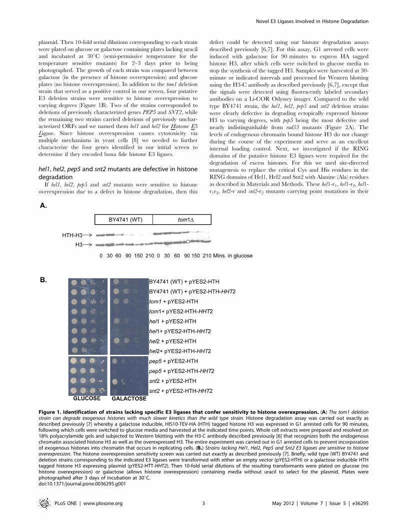
Figure 1. Identification of strains lacking specific E3 ligases that confer sensitivity to histone overexpression. (A) The tom1 deletionstrain can degrade exogenous histones with much slower kinetics than the wild type strain. Histone degradation assay was carried out exactly asdescribed previously [7] whereby a galactose inducible, HIS10-TEV-HA (HTH) tagged histone H3 was expressed in G1 arrested cells for 90 minutes,following which cells were switched to glucose media and harvested at the indicated time points. Whole cell extracts were prepared and resolved on18% polyacrylamide gels and subjected to Western blotting with the H3-C antibody described previously [6] that recognizes both the endogenouschromatin associated histone H3 as well as the overexpressed H3. The entire experiment was carried out in G1 arrested cells to prevent incorporationof exogenous histones into chromatin that occurs in replicating cells. (B.) Strains lacking Hel1, Hel2, Pep5 and Snt2 E3 ligases are sensitive to histoneoverexpression. The histone overexpression sensitivity screen was carried out exactly as described previously [7]. Briefly, wild type (WT) BY4741 anddeletion strains corresponding to the indicated E3 ligases were transformed with either an empty vector (pYES2-HTH) or a galactose inducible HTHtagged histone H3 expressing plasmid (pYES2-HTT-HHT2). Then 10-fold serial dilutions of the resulting transformants were plated on glucose (nohistone overexpression) or galactose (allows histone overexpression) containing media without uracil to select for the plasmid. Plates werephotographed after 3 days of incubation at 30uC.doi:10.1371/journal.pone.0036295.g001
Novel E3 Ligases Involved in Histone Degradation
PLoS ONE | www.plosone.org 3 May 2012 | Volume 7 | Issue 5 | e36295

respective RING domains were as defective in excess histone
degradation as the hel1, hel2 and snt2 deletion strains (Figure 2B),
strongly suggesting that their RING domains are important for
histone protein regulation.
hel1, hel2, pep5 and snt2 mutants accumulate excesshistones on the histone chaperone Asf1
We have previously shown that yeast strains defective in the
histone degradation pathway such as rad53, ubc4, ubc5 and tom1
accumulate excess endogenous histones on histone chaperones
such as Asf1 which typically binds small amounts of histones under
normal conditions but becomes saturated with histones upon
replication arrest or DNA damage [6,7]. If hel1, hel2, pep5 and snt2
are indeed defective in degrading endogenous histones, they
should also exhibit histone chaperones overloaded with endoge-
nous histones under normal conditions. Wild-type and E3-mutant
strains carrying FLAG3 tagged Asf1 (Asf1-FLAG) were treated
with or without methyl methane sulfonate (MMS) to induce
alkylation damage, following which Asf1-FLAG was immunopre-
cipitated using FLAG-M2 affinity resin. As expected, in the
absence of MMS, small amounts of H3 were associated with Asf1–
FLAG in wild-type cells, which increased dramatically upon MMS
treatment (Figure 3). In contrast, hel1, hel2, pep5 and snt2 deletion
strains showed high levels of H3 associated with Asf1–FLAG both
in the absence and presence of MMS-induced DNA damage. The
near saturation of Asf1 with histones in each of these mutants even
in the absence of DNA damage strongly suggests that the
corresponding E3 ligases contribute independently of each other
in the posttranslational regulation of endogenous histone protein
levels.
Hel1, Hel2, Pep5 and Snt2 interact with Ubc4 andhistones
All the new histone E3 ligases identified in our screen are RING
finger domain containing proteins. RING domains are known to
directly interact with Ubc (E2) enzymes as well as the substrate,
thereby helping the Ubc enzymes transfer the ubiquitin to the
substrate proteins [24,39]. Hence, if the putative RING finger
histone E3 ligases identified by us are bona fide E3 ligases, they
should interact physically with their substrate histones as well as
the E2 enzymes involved in the degradation related ubiquitylation
of histones. We tested this idea directly by checking if Hel1, Hel2,
Pep5 and Snt2 interact with the major histone ubiquitin
conjugating enzyme Ubc4 and histones. For this we transformed
a strain expressing MYC13-tagged Ubc4 [7] with constructs
expressing HIS6-HA-Protein A (HIS6-HA-PrA) tagged E3 ligases.
Then we prepared whole cell extracts and immunoprecipitated
(IPed) either Ubc4-MYC using anti-MYC EZ View beads (Sigma)
(Figure 4A) or HA-tagged E3 ligases using magnetic beads
conjugated to HA.11 antibodies (Figure 4B). All IPed material
was resolved on precast gradient gels. Western blotting with MYC
and HA antibodies clearly shows that all the four E3 ligases co-
immunoprecipitated (co-IPed) with Ubc4-MYC (Figure 4A).
Similar results were obtained using reciprocal immunoprecipita-
tion experiments (data not shown). Further, both histones H3 and
H4 co-IPed with all the four E3 ligases (Figure 4B). Overall, these
Figure 2. Strains lacking putative E3 ligases Hel1, Hel2, Pep5 and Snt2 are defective in degrading excess histones. (A) Strains carryingdeletions of hel1, hel2, pep5 and snt2 are deficient in degrading exogenously expressed histones. The exogenous histone degradation assay was carriedout in the indicated strains carrying the pYES2-HTH-HHT2 plasmid as described in Figure 1A. The rad53 deletion strain serves as a positive control inthis assay as we have previously shown that it is defective in the degradation of exogenously expressed histones [6]. The endogenous chromatinbound histone H3 is not degraded in this assay and serves as a loading control. Glu = glucose. (B) The RING domains of Hel1, Hel2 and Snt2 are requiredfor efficient degradation of excess histones. Strains carrying mutations in the RING (r) domains of Hel1, Hel2 and Snt2 were generated as described inthe Materials and Methods section. These mutant strains were then transformed with the pYES6/CT-HA-HHT2 plasmid carrying a Blasticidin resistancemarker and a galactose inducible, HA-tagged histone H3 gene (HA-H3). The excess histone degradation assay was carried out as described in (A)except that the duration of the experiment was limited to 90 minutes and the exogenous HA-H3 was detected using HA.11 antibodies, while theendogenous H3 was detected using the H3-C antibody described previously [6,7].doi:10.1371/journal.pone.0036295.g002
Novel E3 Ligases Involved in Histone Degradation
PLoS ONE | www.plosone.org 4 May 2012 | Volume 7 | Issue 5 | e36295
interactions demonstrate that the new RING finger containing
histone E3 ligases can potentially mediate the ubiquitin transfer
from the E2 enzyme Ubc4 to the substrate histones by acting as a
scaffold to bring these proteins together.
Hel1, Hel2, Pep5 and Snt2 promote ubiquitylation ofhistones in vitro
To further demonstrate that the E3 ligases identified by us were
bona fide histone E3 ligases, next we tested if they can
polyubiquitylate histones in vitro. For this we set up in vitro histone
ubiquitylation reactions using recombinant E1, E2, ubiquitin and
histone H4 along with purified TAP-tagged E3 ligases as described
previously [7]. Reactions were stopped by the addition of Sodium
Dodecyl Sulfate (SDS) loading buffer and boiling prior to analysis
by Western blotting using histone H4 antibodies. The formation of
high-molecular weight species clearly demonstrates that histone
H4 is efficiently ubiquitylated in vitro using Hel1, Hel2, Pep5 and
Snt2 E3 ligases (Figure 5). Further, the inclusion of Rad53
significantly stimulated the activity of all these E3 ligases in the in
vitro ubiquitylation reactions, especially in the case of Hel1
(Figure 5, compare lanes 3 to 4, 5 to 6 and 7 to 8), suggesting
that the newly identified E3 ligases may recognize the same
Rad53-dependent phosphorylation as degradation signals on
histones as the Tom1 ligase identified previously [7].
Hel1, Hel2, Pep5 and Snt2 do not regulate the levels ofthe E2 enzyme Ubc4
Although the in vivo and in vitro data presented here so far
strongly suggest that the newly identified E3 ligases are likely to be
directly involved in histone ubiquitylation and degradation, it is
also formally possible that one or more of these ligases may be
indirectly affecting histone levels by controlling the abundance of
Ubc4, the major E2 enzyme involved in the ubiquitylation
mediated degradation of histones. To investigate this possibility,
we measured the levels of full-length MYC13-tagged Ubc4, as well
as its ubiquitylated forms and degradation products in wild type
and E3 mutant strains. Although we readily detected multiple
bands migrating slower than full-length Ubc4-MYC, we did not
observe any differences in the levels of the full-length or
ubiquitylated forms, as well as in the degradation products of this
protein upon the deletion of any of the newly identified E3 ligases
(Figure 6). This data strongly suggests that these E3 ligases do not
regulate Ubc4 protein levels, although it is still formally possible
that these E3 ligases may regulate the levels of Tom1 or each
other, thereby indirectly influencing histone levels.
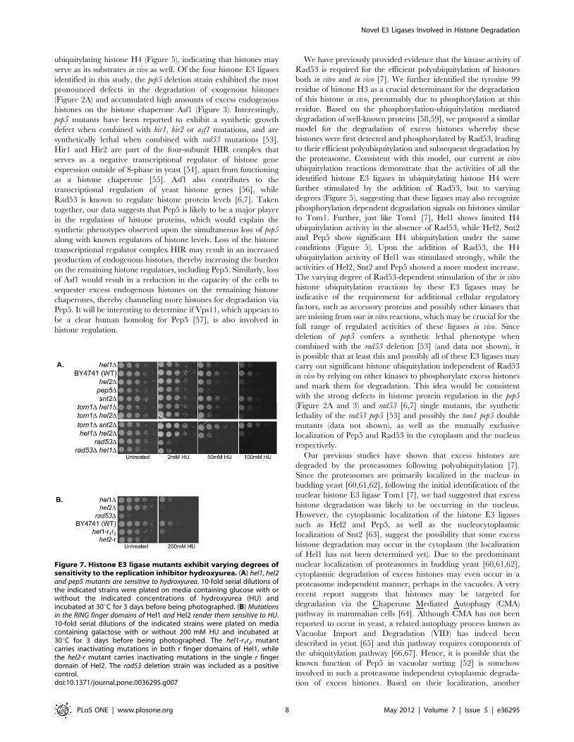
Histone E3 ligases exhibit varying degrees of sensitivityto genotoxic agents such as the replication inhibitorhydroxyurea
DNA damage in eukaryotes occurs in context of chromatin.
Therefore, histone proteins and chromatin structure is likely to
influence DNA damage and repair. DNA repair enzymes need to
gain access to the damaged DNA in order to repair it. We have
previously shown that yeast strains lacking factors involved in the
regulation of histone protein levels such as the E3 ligase Tom1 are
sensitive to genotoxic agents to varying degrees [7]. To assess if the
new histone E3 ligases identified in our screen were also sensitive
to genotoxic agents, we plated 10-fold serial dilutions of mutant
and wild type strains on plates containing the replication inhibitor
hydroxyurea (HU). We found the pep5 deletion strain to be very
sensitive to HU, though not as sensitive as the rad53 deletion
strain, while the hel1 and hel2 deletion mutants were sensitive to HU
to a lesser extent (Figure 7A). The snt2D mutant was not sensitive
at all to HU, suggesting the possibility that different histone E3
ligases may be employed for the degradation of excess histones in
different contexts. Combining the tom1 deletion mutant which is
slightly sensitive to HU [7] with the hel1, hel2 and snt2 deletion
mutants individually did not further exacerbate the HU sensitivity
of any of these E3 ligases (Figure 7A), suggesting that they may be
working in the same pathway. Similar results were obtained for the
hel1 hel2 and the rad53 hel1 double mutants, as the HU sensitivity of
the hel2 and rad53 single mutants was not enhanced any further in
these double mutants (Figure 7A). However, all our attempts to
generate the tom1D pep5D double mutant have failed so far,
suggesting that this combination may be synthetically lethal (data
not shown) and that these two E3 ligases may work in parallel
pathways that are redundant with each other. We also investigated
if the RING domains of Hel1 and Hel2 played any role in
resistance of yeast cells to HU. Mutation of the critical residues
within the RING finger domains of Hel1 and Hel2 rendered them
as sensitive to HU as the complete loss of the proteins (Figure 7B),
once again suggesting that the RING finger domains are essential
for the function of these E3 ligases.
Discussion
In this study we have identified and characterized four putative
E3 ligases that contribute to the posttranslational regulation of
histone protein levels in the budding yeast. As opposed to the
previously identified HECT-domain containing histone E3 ligase
Tom1 [7], all the four new histone E3 ligases carry RING finger
domains that enable them to polyubiquitylate excess histones for
subsequent degradation. Non-degradation related polyubiquityla-
tion of histones H2B has also been reported to occur in the
budding yeast [40]. As such, although we have focused here on the
involvement of these new E3 ligases in the degradation related
ubiquitylation of histones, we acknowledge the possibility that they
may also be involved in histone ubiquitylation that is not related to
degradation. The five histone E3 ligases identified so far in the
posttranslational regulation of histone proteins represent roughly
7% of the 80 predicted E3 ligases in the budding yeast [23].
However, it is possible that this list may expand further upon the
analysis of the remaining essential predicted E3 ligases that we
have not screened so far. This involvement of multiple E3 ligases
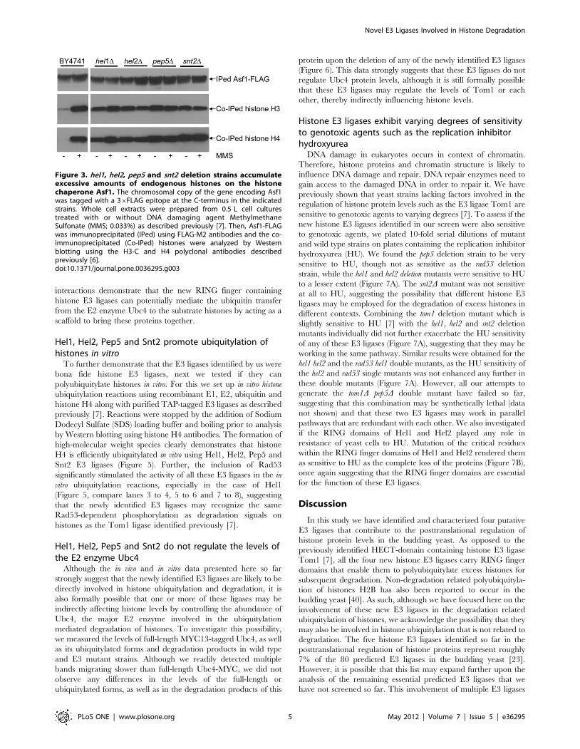
Figure 3. hel1, hel2, pep5 and snt2 deletion strains accumulateexcessive amounts of endogenous histones on the histonechaperone Asf1. The chromosomal copy of the gene encoding Asf1was tagged with a 36FLAG epitope at the C-terminus in the indicatedstrains. Whole cell extracts were prepared from 0.5 L cell culturestreated with or without DNA damaging agent MethylmethaneSulfonate (MMS; 0.033%) as described previously [7]. Then, Asf1-FLAGwas immunoprecipitated (IPed) using FLAG-M2 antibodies and the co-immunoprecipitated (Co-IPed) histones were analyzed by Westernblotting using the H3-C and H4 polyclonal antibodies describedpreviously [6].doi:10.1371/journal.pone.0036295.g003
Novel E3 Ligases Involved in Histone Degradation
PLoS ONE | www.plosone.org 5 May 2012 | Volume 7 | Issue 5 | e36295

in the regulation of histone protein levels is reminiscent of the
multiple E3 ligases that independently contribute to the regulation
of p53 tumor suppressor protein levels in mammalian cells [32].
Overall, the identification of multiple E3 ligases, each of which
appears to contribute independently to the regulation of histone
protein levels, highlights the importance of the posttranslational
degradation of histones in yeast cells.
Two of the E3 ligases identified in our screen, Hel1 and Hel2,
correspond to previously uncharacterized budding yeast ORFs
YKR017C and YDR266C respectively. Hel1 is only one of two
yeast E3 ligases that belong to the RBR (RING-in between-RING)
or TRIAD sub-class of E3 ligases that are characterized by the
presence of two RING finger domains with a novel Cys-rich Cys6-
His-Cys in-between-RING (IBR) domain present in between the
two RING domains [41,42]. The other RBR E3 ligase in the
budding yeast, Itt1, is not sensitive to histone overexpression and
as such it is unlikely to be involved in the regulation of histone
protein levels (data not shown). The RBR family of E3 ligases
include human E3 ligases such as the human homolog of Drosophila
Ariadne (HHARI; also known as ARIH1) that shares homology
with Hel1, as well as TRIAD1 (or ARIH2) and Parkin (or
PARK2), the latter of which has been implicated in a subset of
Parkinson’s disease [43]. It remains to be determined if these
human RBR E3 ligases that share similarities with Hel1 can
ubiquitylate histones. Recent mechanistic insight into these RBR
E3 ligases suggest that they may function as hybrid RING/HECT
E3 ligases whereby the first RING domain interacts with the E2
enzyme while the second RING domain participates in the
transfer of the ubiquitin moiety to the substrate [44]. Not
surprisingly, the second RING domain of HHARI appears to
have a very different structure compared to canonical RING
domains [45]. Mutations in any one of the two RING domains of
Hel1 result in similar defects in histone degradation (Figure 2B)
and confer the same degree of DNA damage sensitivity as the null
mutant (Figure 7B), further confirming that both the RING
domains are required for the function of RBR E3 ligases. Overall,
our studies on Hel1 constitute the first characterization of an RBR
Figure 4. Hel1, Hel2, Pep5 and Snt2 interact with the E2 enzyme Ubc4 as well as histones. (A) Hel1, Hel2, Pep5 and Snt2 interact with Ubc4.Whole cell extracts prepared from 0.5 liter cultures of the indicated strains were used to IP Ubc4-MYC essentially as described for Asf1-FLAG IPs inFigure 3, but using anti-MYC EZ view beads (Sigma). Co-IPed HA-tagged E3 enzymes were detected using HA.11 antibodies. Apart from thepredominant bands corresponding to the full length proteins (indicated by the asterisks), additional bands of weaker intensity are detected and theseare likely to be either degradation products (faster migrating) or ubiquitylated (slower migrating) forms of these proteins. No co-IPed proteins weredetected in the parental untagged strain carrying Ubc4-MYC but lacking HA-tagged proteins. 1% of the whole cell extracts were loaded on a separategel to determine that roughly equal amounts of proteins were present in the input fraction by measuring the amount of histone H4. (B) Hel1, Hel2,Pep5 and Snt2 interact with histones. The HA-tagged E3 enzymes were IPed from 1 liter cultures of the indicated strains using HA.11 antibodies. Co-IPed endogenous histones H3 and H4 was detected using the polyclonal antibodies described previously [6]. No co-IPed histones were detected inthe ‘‘no tag’’ control. The amount of Ubc4-MYC was measured in 1% of the input fraction to demonstrate that roughly equal amounts of cell extractswere used for the IP reactions. (C) Hel1 and Hel2 interact with Rad53. MYC tagged Hel1 or Hel2 were IPed using MYC antibody beads from 3 litercultures of strains carrying FLAG-tagged Rad53. IPed and Co-IPed proteins were detected by Western Blotting using appropriate antibodies. Therelative amount of histone H3 present in each of the ‘‘input’’ extracts used for the IP is shown to demonstrate that same amount of material was usedfor each IP. (D) Snt2 interacts with Rad53 whereas Pep5 does not. Tandem Affinity Purification (TAP) tagged Pep5 or Snt2 was IPed from 3 liter culturesof strains carrying FLAG-tagged Rad53. Any co-IPed Rad53 was detected using FLAG antibodies. The similar levels of Rad53-FLAG present in theextracts confirm that the same amount of extracts was used for each IP. No evidence for any interaction between Rad53 and Pep5 was observed evenupon scaling up the Pep5-TAP IP by 3-fold (data not shown).doi:10.1371/journal.pone.0036295.g004
Novel E3 Ligases Involved in Histone Degradation
PLoS ONE | www.plosone.org 6 May 2012 | Volume 7 | Issue 5 | e36295

E3 ligase in yeast and suggest that histones are a potential
substrate.
Hel2 is a canonical RING domain containing E3 ligase that has
been previously shown to be phosphorylated by the DNA damage
checkpoint kinase and histone protein regulator Rad53 [46]. Not
surprisingly, in vitro ubiquitylation of histone H4 in the presence of
Hel2 is significantly stimulated by the presence of Rad53 (Figure 5).
Further, hel2 mutants are sensitive to the replication inhibitor HU
and this maybe in part due to its role in regulating histone protein
levels, although it is quite likely that there are additional substrates
that are ubiquitylated in vivo by Hel2 in response to replication
arrest and we are currently investigating this. The human protein
with the highest similarity to Hel2 is an uncharacterized zinc
finger protein 598 (ZNF598), although known E3 ligases such as
KPC1 (Kip1 Ubiquitination-Promoting Complex 1; also known as
RNF123) also show limited similarity to Hel2 [47]. It is unclear if
these human E3 enzymes are capable of ubiquitylating histones.
Snt2 is a poorly characterized protein. It was initially identified
and named for the presence of the DNA binding SANT (SWI3,
ADA2, N-CoR and TFIIIB) domain and has been predicted to
function as a transcriptional regulator based largely on in silico
analyses, although this prediction is yet to be verified experimen-
tally [48,49]. Snt2 also has three PHD (Plant Homeo-Domain)
fingers consisting of the Cys4-His-Cys3 motif which is typically
involved in binding methylated lysine residues on histones,
although the Snt2 PHD fingers show weak or nonspecific binding
to methylated histone H3 peptides at best [50]. The PHD finger is
very similar to the Cys3-His-Cys4 containing RING finger domain
and two of the PHD fingers of Snt2 are also RING fingers
potentially capable of E3 ligase activity. Our data clearly show that
the snt2 deletion strain is sensitive to histone overexpression
(Figure 1B), defective in histone degradation (Figure 2A) and
accumulates excess endogenous histones (Figure 3). Further, point
mutations in the second RING domain of Snt2 result in a defect in
excess histone degradation (Figure 2B). More importantly, Snt2
interacts with Ubc4, histones and Rad53 (Figure 4) and exhibits
robust histone ubiquitylation activity in vitro that is further
stimulated by Rad53 (Figure 5). Hence, based on these data we
conclude that Snt2 is a genuine histone E3 ligase and contributes
to the regulation of histone protein levels.
Pep5 (also known as Vps11) was initially isolated as a protease
deficient mutant [51]. Pep5 was subsequently shown to be
involved in protein trafficking and vacuole biogenesis pathways
[52], although it has not been studied as a RING E3 ligase. In our
in vitro ubiquitylation assay, Pep5 shows robust activity in
Figure 5. Hel1, Hel2, Pep5 and Snt2 can efficiently ubiquitylate histone H4 in vitro in a reaction that is stimulated by Rad53.Recombinant and purified components were used to reconstitute the ubiquitylation of histone H4 in vitro exactly as described previously [7]. Thereaction products were resolved on an 18% polyacrylamide gel and processed for Western blotting with H4 antibodies. Addition of commerciallyavailable recombinant yeast Uba1 (E1), human UbcH5A (E2; homolog of yeast Ubc4) and HIS6-tagged ubiquitin to recombinant human histone H4with or without Rad53 did not result in appreciable histone modifications, apart from some monoubiquitylation (lanes 2 and 1 respectively). Only theaddition of the E3 ligases Hel1 (H1), Hel2 (H2), Pep5 (P5) and Snt2 (S2) purified from yeast extracts via TAP epitope tags resulted in considerable highmolecular weight histone modifications, which were further stimulated to varying degrees by the addition of recombinant Rad53 to the reactionmixture.doi:10.1371/journal.pone.0036295.g005
Figure 6. The E3 ligases Hel1, Hel2, Pep5, Snt2 and Tom1 donot regulate the protein levels of Ubc4, the major E2 enymeinvolved in excess histone degradation. Whole cell lysates wereprepared as described previously [69] from wild type and the indicatedE3 ligase deletion strains carrying MYC-tagged Ubc4. The lysates wereresolved on a 12% polyacrylamide gel and processed for Westernblotting using MYC antibodies. No significant differences are observedin the levels of full-length Ubc4-MYC, or in its slower migrating modifiedforms and faster migrating degradation products.doi:10.1371/journal.pone.0036295.g006
Novel E3 Ligases Involved in Histone Degradation
PLoS ONE | www.plosone.org 7 May 2012 | Volume 7 | Issue 5 | e36295
ubiquitylating histone H4 (Figure 5), indicating that histones may
serve as its substrates in vivo as well. Of the four histone E3 ligases
identified in this study, the pep5 deletion strain exhibited the most
pronounced defects in the degradation of exogenous histones
(Figure 2A) and accumulated high amounts of excess endogenous
histones on the histone chaperone Asf1 (Figure 3). Interestingly,
pep5 mutants have been reported to exhibit a synthetic growth
defect when combined with hir1, hir2 or asf1 mutations, and are
synthetically lethal when combined with rad53 mutations [53].
Hir1 and Hir2 are part of the four-subunit HIR complex that
serves as a negative transcriptional regulator of histone gene
expression outside of S-phase in yeast [54], apart from functioning
as a histone chaperone [55]. Asf1 also contributes to the
transcriptional regulation of yeast histone genes [56], while
Rad53 is known to regulate histone protein levels [6,7]. Taken
together, our data suggests that Pep5 is likely to be a major player
in the regulation of histone proteins, which would explain the
synthetic phenotypes observed upon the simultaneous loss of pep5
along with known regulators of histone levels. Loss of the histone
transcriptional regulator complex HIR may result in an increased
production of endogenous histones, thereby increasing the burden
on the remaining histone regulators, including Pep5. Similarly, loss
of Asf1 would result in a reduction in the capacity of the cells to
sequester excess endogenous histones on the remaining histone
chaperones, thereby channeling more histones for degradation via
Pep5. It will be interesting to determine if Vps11, which appears to
be a clear human homolog for Pep5 [57], is also involved in
histone regulation.
We have previously provided evidence that the kinase activity of
Rad53 is required for the efficient polyubiquitylation of histones
both in vitro and in vivo [7]. We further identified the tyrosine 99
residue of histone H3 as a crucial determinant for the degradation
of this histone in vivo, presumably due to phosphorylation at this
residue. Based on the phosphorylation-ubiquitylation mediated
degradation of well-known proteins [58,59], we proposed a similar
model for the degradation of excess histones whereby these
histones were first detected and phosphorylated by Rad53, leading
to their efficient polyubiquitylation and subsequent degradation by
the proteasome. Consistent with this model, our current in vitro
ubiquitylation reactions demonstrate that the activities of all the
identified histone E3 ligases in ubiquitylating histone H4 were
further stimulated by the addition of Rad53, but to varying
degrees (Figure 5), suggesting that these ligases may also recognize
phosphorylation dependent degradation signals on histones similar
to Tom1. Further, just like Tom1 [7], Hel1 shows limited H4
ubiquitylation activity in the absence of Rad53, while Hel2, Snt2
and Pep5 show significant H4 ubiquitylation under the same
conditions (Figure 5). Upon the addition of Rad53, the H4
ubiquitylation activity of Hel1 was stimulated strongly, while the
activities of Hel2, Snt2 and Pep5 showed a more modest increase.
The varying degree of Rad53-dependent stimulation of the in vitro
histone ubiquitylation reactions by these E3 ligases may be
indicative of the requirement for additional cellular regulatory
factors, such as accessory proteins and possibly other kinases that
are missing from our in vitro reactions, which may be crucial for the
full range of regulated activities of these ligases in vivo. Since
deletion of pep5 confers a synthetic lethal phenotype when
combined with the rad53 deletion [53] (and data not shown), it
is possible that at least this and possibly all of these E3 ligases may
carry out significant histone ubiquitylation independent of Rad53
in vivo by relying on other kinases to phosphorylate excess histones
and mark them for degradation. This idea would be consistent
with the strong defects in histone protein regulation in the pep5
(Figure 2A and 3) and rad53 [6,7] single mutants, the synthetic
lethality of the rad53 pep5 [53] and possibly the tom1 pep5 double
mutants (data not shown), as well as the mutually exclusive
localization of Pep5 and Rad53 in the cytoplasm and the nucleus
respectively.
Our previous studies have shown that excess histones are
degraded by the proteasomes following polyubiquitylation [7].
Since the proteasomes are primarily localized in the nucleus in
budding yeast [60,61,62], following the initial identification of the
nuclear histone E3 ligase Tom1 [7], we had suggested that excess
histone degradation was likely to be occurring in the nucleus.
However, the cytoplasmic localization of the histone E3 ligases
such as Hel2 and Pep5, as well as the nucleocytoplasmic
localization of Snt2 [63], suggest the possibility that some excess
histone degradation may occur in the cytoplasm (the localization
of Hel1 has not been determined yet). Due to the predominant
nuclear localization of proteasomes in budding yeast [60,61,62],
cytoplasmic degradation of excess histones may even occur in a
proteasome independent manner, perhaps in the vacuoles. A very
recent report suggests that histones may be targeted for
degradation via the Chaperone Mediated Autophagy (CMA)
pathway in mammalian cells [64]. Although CMA has not been
reported to occur in yeast, a related autophagy process known as
Vacuolar Import and Degradation (VID) has indeed been
described in yeast [65] and this pathway requires components of
the ubiquitylation pathway [66,67]. Hence, it is possible that the
known function of Pep5 in vacuolar sorting [52] is somehow
involved in such a proteasome independent cytoplasmic degrada-
tion of excess histones. Based on their localization, another
Figure 7. Histone E3 ligase mutants exhibit varying degrees ofsensitivity to the replication inhibitor hydroxyurea. (A) hel1, hel2and pep5 mutants are sensitive to hydroxyurea. 10-fold serial dilutions ofthe indicated strains were plated on media containing glucose with orwithout the indicated concentrations of hydroxyurea (HU) andincubated at 30uC for 3 days before being photographed. (B) Mutationsin the RING finger domains of Hel1 and Hel2 render them sensitive to HU.10-fold serial dilutions of the indicated strains were plated on mediacontaining galactose with or without 200 mM HU and incubated at30uC for 3 days before being photographed. The hel1-r1r2 mutantcarries inactivating mutations in both r finger domains of Hel1, whilethe hel2-r mutant carries inactivating mutations in the single r fingerdomain of Hel2. The rad53 deletion strain was included as a positivecontrol.doi:10.1371/journal.pone.0036295.g007
Novel E3 Ligases Involved in Histone Degradation
PLoS ONE | www.plosone.org 8 May 2012 | Volume 7 | Issue 5 | e36295
possibility is that Tom1 targets excess histones in the nucleus for
degradation, while Hel2 and Pep5 are involved in the cytoplasmic
degradation of excess histones, whereas Snt2 contributes to histone
degradation in both cellular compartments. Future studies will
determine if this is indeed the case.
Materials and Methods
All yeast strains used are listed in Table S1. These strains were
either obtained from Open Biosystems budding yeast genome
deletion collection or were generated in the lab as needed using
standard yeast manipulations [68]. MYC13-tagged Ubc4 and the
plasmid pRS416-RAD53-FLAG for the expression of FLAG-
tagged Rad53 have been described previously [7]. TAP-tagged
strains were obtained from Open Biosystems. Plasmids pYES2-
HTH and pYES2-HTH-HHT2 have been described previously
[7]. Plasmid BG1805 based constructs expressing C-terminal
HIS6-HA-Protein A (HIS6-HA-PrA) tagged Hel1, Hel2, Pep5 and
Snt2 under the control of a galactose inducible promoter were also
obtained from Open Biosystems. Mutagenesis of the HEL1, HEL2
and SNT2 RING finger domains was carried out on the plasmid
BG1805 based constructs expressing Hel1, Hel2 and Snt2 using
Stratagene’s QuikChange Multi Site-Directed Mutagenesis kit
following the manufacturer’s instructions. Briefly, the crucial Cys
and His residues in the RING (r) finger domains were mutated to
Ala residues. The hel1-r1 mutant carries the Cys-195, His-197,
Cys-200RAla mutations in r1, while the hel1-r2 of carries the His-
356, Cys-359, Cys-362RAla mutations in r2. The hel1-r1r2 mutant
carries both the r1 and r2 mutations simultaneously. The hel2-r
mutant carries the Cys-79, His-81, Cys-87RAla mutations in its r
finger domain. The snt2-r2 carries the Cys1061, His1066,
Cys1069RAla mutations in its second r2 finger domain. All
mutations were confirmed by sequencing the entire mutant gene.
The plasmids bearing the r finger mutants of the E3 ligases were
transformed into cells lacking the endogenous gene for the
respective E3 ligase to perform the experiments shown in
figures 2B and 7B. All other methods used such as the histone
degradation assay, histone overexpression sensitivity assay, West-
ern blotting, co-immunoprecipitation assay and in vitro ubiquityla-
tion assay have been described extensively in our previous
publications [6,7].
Supporting Information
Table S1 List of strains used in this study.
(PDF)
Acknowledgments
We wish to thank Alain Verreault and Yanchang Wang for reagents. We
thank Dr. Johanna Paik for critical reading of this manuscript.
Author Contributions
Conceived and designed the experiments: AG RKS. Performed the
experiments: RKS MG MHMK AG. Analyzed the data: RKS AG. Wrote
the paper: AG RKS.
References
1. Richmond TJ, Davey CA (2003) The structure of DNA in the nucleosome core.
Nature 423: 145–150.
2. van Holde KE (1988) Chromatin; van Holde KE, editor. New York: Springer-
Verlag.
3. Wolffe AP (1995) Chromatin: Structure and Function. San Diego: Academic
Press. 299 p.
4. Marzluff WF, Gongidi P, Woods KR, Jin J, Maltais LJ (2002) The human and
mouse replication-dependent histone genes. Genomics 80: 487–498.
5. Singh RK, Paik J, Gunjan A (2009) Generation and management of excess
histones during the cell cycle. Front Biosci 14: 3145–3158.
6. Gunjan A, Verreault A (2003) A Rad53 kinase-dependent surveillance
mechanism that regulates histone protein levels in S. cerevisiae. Cell 115:
537–549.
7. Singh RK, Kabbaj MH, Paik J, Gunjan A (2009) Histone levels are regulated by
phosphorylation and ubiquitylation-dependent proteolysis. Nat Cell Biol 11:
925–933.
8. Singh RK, Liang D, Gajjalaiahvari UR, Kabbaj MH, Paik J, et al. (2010) Excess
histone levels mediate cytotoxicity via multiple mechanisms. Cell Cycle 9:
4236–4244.
9. Woodland HR, Adamson ED (1977) The synthesis and storage of histones
during the oogenesis of Xenopus laevis. Developmental Biology 57: 118–135.
10. Dingwall C, Laskey RA (1990) Nucleoplasmin: the archetypal molecular
chaperone. Seminars in Cell Biology 1: 11–17.
11. Cermelli S, Guo Y, Gross SP, Welte MA (2006) The lipid-droplet proteome
reveals that droplets are a protein-storage depot. Current Biology 16:
1783–1795.
12. Andrews AJ, Chen X, Zevin A, Stargell LA, Luger K (2010) The histone
chaperone Nap1 promotes nucleosome assembly by eliminating nonnucleosomal
histone DNA interactions. Molecular Cell 37: 834–842.
13. Groth A, Corpet A, Cook AJ, Roche D, Bartek J, et al. (2007) Regulation of
replication fork progression through histone supply and demand. Science 318:
1928–1931.
14. Groth A, Ray-Gallet D, Quivy JP, Lukas J, Bartek J, et al. (2005) Human Asf1
regulates the flow of S phase histones during replicational stress. Mol Cell 17:
301–311.
15. Morillo-Huesca M, Maya D, Munoz-Centeno MC, Singh RK, Oreal V, et al.
(2010) FACT prevents the accumulation of free histones evicted from
transcribed chromatin and a subsequent cell cycle delay in G1. PLoS Genet
6: e1000964.
16. Liang D, Burkhart SL, Kabbaj MH, Gunjan A (2012) Histone dosage regulates
DNA damage sensitivity in a checkpoint independent manner via the
homologous recombination pathway. Nucleic Acids Res (In Press).
17. Marzluff WF, Wagner EJ, Duronio RJ (2008) Metabolism and regulation of
canonical histone mRNAs: life without a poly(A) tail. Nat Rev Genet 9:
843–854.
18. Hershko A, Ciechanover A (1998) The ubiquitin system. Annu Rev Biochem 67:
425–479.
19. Hochstrasser M (1996) Ubiquitin-dependent protein degradation. Annu Rev
Genet 30: 405–439.
20. Mani A, Gelmann EP (2005) The ubiquitin-proteasome pathway and its role in
cancer. Journal of Clinical Oncology 23: 4776–4789.
21. Haas AL, Warms JV, Hershko A, Rose IA (1982) Ubiquitin-activating enzyme.
Mechanism and role in protein-ubiquitin conjugation. Journal of Biological
Chemistry 257: 2543–2548.
22. Ardley HC, Robinson PA (2005) E3 ubiquitin ligases. Essays in Biochemistry 41:
15–30.
23. Li W, Bengtson MH, Ulbrich A, Matsuda A, Reddy VA, et al. (2008) Genome-
wide and functional annotation of human E3 ubiquitin ligases identifies
MULAN, a mitochondrial E3 that regulates the organelle’s dynamics and
signaling. PLoS One 3: e1487.
24. Deshaies RJ, Joazeiro CA (2009) RING domain E3 ubiquitin ligases. Annual
Review of Biochemistry 78: 399–434.
25. Hatakeyama S, Nakayama KI (2003) U-box proteins as a new family of
ubiquitin ligases. Biochemical and Biophysical Research Communications 302:
635–645.
26. Rotin D, Kumar S (2009) Physiological functions of the HECT family of
ubiquitin ligases. Nat Rev Mol Cell Biol 10: 398–409.
27. Huibregtse JM, Scheffner M, Beaudenon S, Howley PM (1995) A family of
proteins structurally and functionally related to the E6-AP ubiquitin-protein
ligase. Proceedings of the National Academy of Sciences of the United States of
America 92: 5249.
28. Ciechanover A, Schwartz AL (2002) Ubiquitin-mediated degradation of cellular
proteins in health and disease. Hepatology 35: 3–6.
29. Ciechanover A (2003) The ubiquitin proteolytic system and pathogenesis of
human diseases: a novel platform for mechanism-based drug targeting.
Biochemical Society Transactions 31: 474–481.
30. Scheffner M, Staub O (2007) HECT E3s and human disease. BMC Biochem 8
Suppl 1: S6.
31. Ardley HC, Robinson PA (2004) The role of ubiquitin-protein ligases in
neurodegenerative disease. Neurodegener Dis 1: 71–87.
32. Brooks CL, Gu W (2011) p53 regulation by ubiquitin. FEBS Letters 585:
2803–2809.
33. Commerford SL, Carsten AL, Cronkite EP (1982) Histone turnover within
nonproliferating cells. Proc Natl Acad Sci U S A 79: 1163–1165.
Novel E3 Ligases Involved in Histone Degradation
PLoS ONE | www.plosone.org 9 May 2012 | Volume 7 | Issue 5 | e36295
34. Liu Z, Oughtred R, Wing SS (2005) Characterization of E3Histone, a novel
testis ubiquitin protein ligase which ubiquitinates histones. Mol Cell Biol 25:2819–2831.
35. Hall JR, Kow E, Nevis KR, Lu CK, Luce KS, et al. (2007) Cdc6 stability is
regulated by the Huwe1 ubiquitin ligase after DNA damage. Molecular Biologyof the Cell 18: 3340–3350.
36. Zhong Q, Gao W, Du F, Wang X (2005) Mule/ARF-BP1, a BH3-only E3ubiquitin ligase, catalyzes the polyubiquitination of Mcl-1 and regulates
apoptosis. Cell 121: 1085–1095.
37. Adhikary S, Marinoni F, Hock A, Hulleman E, Popov N, et al. (2005) Theubiquitin ligase HectH9 regulates transcriptional activation by Myc and is
essential for tumor cell proliferation. Cell 123: 409–421.38. Chen D, Kon N, Li M, Zhang W, Qin J, et al. (2005) ARF-BP1/Mule is a
critical mediator of the ARF tumor suppressor. Cell 121: 1071–1083.39. Zheng N, Wang P, Jeffrey PD, Pavletich NP (2000) Structure of a c-Cbl-UbcH7
complex: RING domain function in ubiquitin-protein ligases. Cell 102:
533–539.40. Geng F, Tansey WP (2008) Polyubiquitylation of histone H2B. Mol Biol Cell 19:
3616–3624.41. van der Reijden BA, Erpelinck-Verschueren CA, Lowenberg B, Jansen JH
(1999) TRIADs: a new class of proteins with a novel cysteine-rich signature.
Protein Science 8: 1557–1561.42. Marin I, Ferrus A (2002) Comparative genomics of the RBR family, including
the Parkinson’s disease-related gene parkin and the genes of the ariadnesubfamily. Molecular Biology and Evolution 19: 2039–2050.
43. Marin I, Lucas JI, Gradilla AC, Ferrus A (2004) Parkin and relatives: the RBRfamily of ubiquitin ligases. Physiol Genomics 17: 253–263.
44. Wenzel DM, Lissounov A, Brzovic PS, Klevit RE (2011) UBCH7 reactivity
profile reveals parkin and HHARI to be RING/HECT hybrids. Nature 474:105–108.
45. Capili AD, Edghill EL, Wu K, Borden KL (2004) Structure of the C-terminalRING finger from a RING-IBR-RING/TRIAD motif reveals a novel zinc-
binding domain distinct from a RING. Journal of Molecular Biology 340:
1117–1129.46. Ptacek J, Devgan G, Michaud G, Zhu H, Zhu X, et al. (2005) Global analysis of
protein phosphorylation in yeast. Nature 438: 679–684.47. Kamura T, Hara T, Matsumoto M, Ishida N, Okumura F, et al. (2004)
Cytoplasmic ubiquitin ligase KPC regulates proteolysis of p27(Kip1) at G1phase. Nat Cell Biol 6: 1229–1235.
48. Yang Y, Zhang Z, Li Y, Zhu XG, Liu Q (2010) Identifying cooperative
transcription factors by combining ChIP-chip data and knockout data. CellResearch 20: 1276–1278.
49. Ward LD, Bussemaker HJ (2008) Predicting functional transcription factorbinding through alignment-free and affinity-based analysis of orthologous
promoter sequences. Bioinformatics 24: i165–171.
50. Shi X, Kachirskaia I, Walter KL, Kuo JH, Lake A, et al. (2007) Proteome-wideanalysis in Saccharomyces cerevisiae identifies several PHD fingers as novel
direct and selective binding modules of histone H3 methylated at either lysine 4or lysine 36. Journal of Biological Chemistry 282: 2450–2455.
51. Jones EW (1977) Proteinase mutants of Saccharomyces cerevisiae. Genetics 85:23–33.
52. Srivastava A, Woolford CA, Jones EW (2000) Pep3p/Pep5p complex: a putative
docking factor at multiple steps of vesicular transport to the vacuole of
Saccharomyces cerevisiae. Genetics 156: 105–122.
53. Pan X, Ye P, Yuan DS, Wang X, Bader JS, et al. (2006) A DNA integrity
network in the yeast Saccharomyces cerevisiae. Cell 124: 1069–1081.
54. Osley MA (1991) The regulation of histone synthesis in the cell cycle. Annu Rev
Biochem 60: 827–861.
55. Green EM, Antczak AJ, Bailey AO, Franco AA, Wu KJ, et al. (2005)
Replication-independent histone deposition by the HIR complex and Asf1.
Current Biology 15: 2044–2049.
56. Sutton A, Bucaria J, Osley MA, Sternglanz R (2001) Yeast ASF1 protein is
required for cell cycle regulation of histone gene transcription. Genetics 158:
587–596.
57. Huizing M, Didier A, Walenta J, Anikster Y, Gahl WA, et al. (2001) Molecular
cloning and characterization of human VPS18, VPS 11, VPS16, and VPS33.
Gene 264: 241–247.
58. Deshaies RJ, Ferrell JE Jr. (2001) Multisite phosphorylation and the countdown
to S phase. Cell 107: 819–822.
59. Verma R, Annan RS, Huddleston MJ, Carr SA, Reynard G, et al. (1997)
Phosphorylation of Sic1p by G1 Cdk required for its degradation and entry into
S phase. Science 278: 455–460.
60. Russell SJ, Steger KA, Johnston SA (1999) Subcellular localization, stoichiom-
etry, and protein levels of 26 S proteasome subunits in yeast. Journal of
Biological Chemistry 274: 21943–21952.
61. Enenkel C, Lehmann A, Kloetzel PM (1998) Subcellular distribution of
proteasomes implicates a major location of protein degradation in the nuclear
envelope-ER network in yeast. EMBO Journal 17: 6144–6154.
62. Laporte D, Salin B, Daignan-Fornier B, Sagot I (2008) Reversible cytoplasmic
localization of the proteasome in quiescent yeast cells. Journal of Cell Biology
181: 737–745.
63. Huh WK, Falvo JV, Gerke LC, Carroll AS, Howson RW, et al. (2003) Global
analysis of protein localization in budding yeast. Nature 425: 686–691.
64. Cook AJ, Gurard-Levin ZA, Vassias I, Almouzni G (2011) A specific function for
the histone chaperone NASP to fine-tune a reservoir of soluble H3–H4 in the
histone supply chain. Molecular Cell 44: 918–927.
65. Alibhoy AA, Chiang HL (2011) Vacuole import and degradation pathway:
Insights into a specialized autophagy pathway. World J Biol Chem 2: 239–245.
66. Shieh HL, Chen Y, Brown CR, Chiang HL (2001) Biochemical analysis of
fructose-1,6-bisphosphatase import into vacuole import and degradation vesicles
reveals a role for UBC1 in vesicle biogenesis. Journal of Biological Chemistry
276: 10398–10406.
67. Katzmann DJ, Babst M, Emr SD (2001) Ubiquitin-dependent sorting into the
multivesicular body pathway requires the function of a conserved endosomal
protein sorting complex, ESCRT-I. Cell 106: 145–155.
68. Longtine MS, McKenzie A 3rd, Demarini DJ, Shah NG, Wach A, et al. (1998)
Additional modules for versatile and economical PCR-based gene deletion and
modification in Saccharomyces cerevisiae. Yeast 14: 953–961.
69. Kushnirov VV (2000) Rapid and reliable protein extraction from yeast. Yeast
16: 857–860.
Novel E3 Ligases Involved in Histone Degradation
PLoS ONE | www.plosone.org 10 May 2012 | Volume 7 | Issue 5 | e36295