Low Oil-Water Ratio Invert Emulsion Mud for Unconventional ...
Upload
uni-tuebingenCategory
view
0download
0
Cell, Vol. 84, 687–697, March 8, 1996, Copyright 1996 by Cell Press
Mother Cell–Specific HO Expressionin Budding Yeast Depends on the UnconventionalMyosin Myo4p and Other Cytoplasmic Proteins
Ralf-Peter Jansen, Celia Dowzer,* Christine Michaelis, whose location in Drosophila embryos determines sub-sequent cell fate are often mRNAs (e.g., bicoid andMarta Galova, and Kim Nasmythoskar) or proteins that regulate mRNA translation (e.g.,Research Institute of Molecular PathologyNanos; Curtis et al., 1995). Another good example is theA-1030 Viennadifferent accumulation of GLP-1 protein in the two cellAustriablastomeres of Caenorhabditis elegans; this asymmetryis also due to differences in mRNA translatability (Evanset al., 1994).
SummaryStudies using inhibitors have implicated the cytoskel-
eton in the segregation of developmental determinants.Certain cell types giverise to progeny that adoptdiffer-
Destruction of the actin microfilament network of C.ent patterns of gene expression in the absence of any elegans with cytotochalasin B disrupts the asymmetricdifferences in their environment. Cells of budding segregation of germline P granules (Hill and Strome,yeast give birth to mother and daughtercells thatdiffer 1988), whereas disruption of microtubules in Drosophilain that only mother cells express the HO endonuclease oocytes by colchicine treatment abolishes the posteriorgene and thereby switch mating types. We describe localization of oskar mRNA (Ferrandon et al., 1994; Clarkthe identification of five genes, called SHE1–SHE5, that et al., 1994). However, the results obtained with suchencode cytoplasmicproteins required for mother-spe- drastic treatments are hard to interpret. Cytoskeletalcific HO expression. She1p, which is identical to the drugs could interfere with the cell fate of daughter cellsminimyosin Myo4p, and She3p are not, however, either by directly affecting the distribution of determi-mother-specific proteins. On the contrary, they accu- nants or by indirectly upsetting the structure of the cy-mulate in growing buds. She proteins might be re- toskeleton of the mother cell, upon which the asymmetryquired for the transport of factors that promote HO of the cell is dependent. Genetic studies in Drosophilarepression from the mother cell into its bud. In an have recently implicated filamentous actin in the segre-accompanying paper, we show that SHE genes are gation of determinants. Tropomyosin, which stabilizesneeded for the accumulation in daughter nuclei of actin filaments, is required for the posterior localizationAsh1p, a repressor of HO. of oskar mRNA in oocytes (Erdelyi et al., 1995). Here
we describe genetic evidence that determinants can beIntroduction segregated by myosin-based motors. We have found
that the minimyosin Myo4p and several other cyto-plasmic proteins are required for mother cell–specificThe characteristics of different cell types in multicellularHO gene expression in the budding yeast Saccharo-organisms are largely determined by differences in theirmyces cerevisiae.patterns of gene expression. How cells give birth to
The HO endonuclease induces mating-type switchingdaughter cells with differing gene expression during em-in S. cerevisiae by creating a double-stranded breakbryogenesis is a crucial problem. Many studies haveat the MAT locus. The HO gene is only transcribed inshown that gene expression can be influenced by exter-“mother” cells, i.e., cells that have previously buddednal factors. Sister cells frequently find themselves inand given birth to a “daughter” cell. In mother cells, HOdifferent environments, in which one or the other is ex-is transcribed transiently during the cell cycle, shortlyposed to different factors secreted by neighboring cells.before budding and DNA replication (Nasmyth, 1993).However, in the development of several organisms thereHO activation depends on at least ten genes, termedare many examples of sister cells adopting different cellSWI1 through SWI10. SWI4 and SWI6 encode subunitsfates in the absence of any differences in their surround-of a cell cycle–regulated transcription factor, SCB-bind-ings (Horvitz and Herskowitz, 1992). Whether this is dueing factor (SBF), that activates a number of genes at theto differential segregation between sister cells of cyto-G1/S boundary (Koch and Nasmyth, 1994). Other SWIplasmic or nuclear factors is in many cases not known.genes, for example SWI1, SWI2, SWI3, and SWI10, en-The problem of identifying such factors has so far onlycode components of a large, multisubunit complex,been amenable to genetic analyses, mainly in flies andwhich is needed for the expression of many yeast genesworms.(Peterson and Herskowitz, 1992).Examples of cytoplasmic factors whose cellular distri-
A crucial role in directing mother cell–specific HO ex-bution eventually influences gene expression are thosepression has been established for the transcription fac-that determine the major body axes in Drosophila. Dur-tor encoded by SWI5 (Nasmyth, 1993). Swi5p binds toing oogenesis, bicoid and oskar mRNAs are localizedtwo sites in a region of the HO promoter, called URS1,to the anterior and posterior ends of the egg, respec-that directs mother-specific HO expression. However,tively, and this later leads to different patterns of geneSwi5p is not a mother cell–specific determinant, and itsexpression in nuclei that end up in the anterior or poste-properties cannot explain the asymmetry of HO activa-rior of the cytoplasm (St Johnston, 1995). The factorstion. It is synthesized during G2 and M phases, butremains in the cytoplasm until late anaphase, uponwhich it enters both mother and daughter nuclei and is*Present address: Asia Pacific Fermentation Industries, 5 Belvoir
Road, Cambridge CB4 1JQ, United Kingdom. rapidly degraded (Nasmyth et al., 1990; Tebb et al.,
Cell688
1993). The transient accumulation of Swi5p in motherand daughter cell nuclei as cells enter G1 leads to theactivation of several other genes whose transcription isSwi5p dependent (Koch and Nasmyth, 1994). HO ex-pression, in contrast, does not occur at this time be-cause it must await the subsequent activation of SBFat the G1 to S phase transition.
A second gene that is required for mother-specificHO expression, SDI1/SIN3 is also unlikely to encode thedeterminant of asymmetric HO expression. Mutationsthat affect Sin3p allow HO to be expressed in the ab-sence of Swi5p and cause HO to be expressed in daugh-ter cells (Nasmyth et al., 1987; Sternberg et al., 1987).However, like Swi5p, Sin3 protein is found in bothmother and daughter cell nuclei at the end of mitosis(Wang et al., 1990).
We describe here the identification of five new genes,called SHE1–SHE5 (for Swi5p-dependent HO expres-sion) that are specifically required for HO expressionin mother cells. These SHE genes encode cytoplasmicproteins, which in thecase of She1p and She3p accumu-late preferentially in growing buds. She1p is identical to
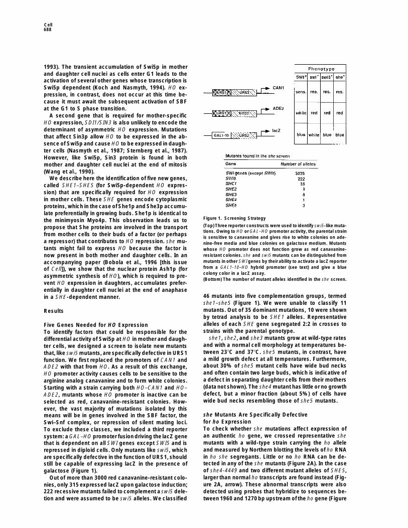
Figure 1. Screening Strategythe minimyosin Myo4p. This observation leads us to(Top) Three reporter constructs were used to identify swi5-like muta-propose that She proteins are involved in the transporttions. Owing to HO or GAL–HO promoter activity, the parental strain
from mother cells to their buds of a factor (or perhaps is sensitive to canavanine and gives rise to white colonies on ade-a repressor) that contributes to HO repression. she mu- nine-free media and blue colonies on galactose medium. Mutantstants might fail to express HO because the factor is whose HO promoter does not function grow as red canavanine-
resistant colonies. she and swi5 mutants can be distinguished fromnow present in both mother and daughter cells. In anmutants in other SWI genes by their ability to activate a lacZ reporteraccompanying paper (Bobola et al., 1996 [this issuefrom a GAL1-10–HO hybrid promoter (see text) and give a blueof Cell]), we show that the nuclear protein Ash1p (forcolony color in a lacZ assay.asymmetric synthesis of HO), which is required to pre-(Bottom) The number of mutant alleles identified in the she screen.
vent HO expression in daughters, accumulates prefer-entially in daughter cell nuclei at the end of anaphase
46 mutants into five complementation groups, termedin a SHE-dependent manner.she1–she5 (Figure 1). We were unable to classify 11mutants. Out of 35 dominant mutations, 10 were shownResultsby tetrad analysis to be SHE1 alleles. Representativealleles of each SHE gene segregated 2:2 in crosses toFive Genes Needed for HO Expressionstrains with the parental genotype.To identify factors that could be responsible for the
she1, she2, and she3 mutants grow at wild-type ratesdifferential activity of Swi5p at HO in mother and daugh-and with a normal cell morphology at temperatures be-ter cells, we designed a screen to isolate new mutantstween 238C and 378C. she5 mutants, in contrast, havethat, like swi5 mutants, arespecifically defective in URS1a mild growth defect at all temperatures. Furthermore,function. We first replaced the promoters of CAN1 andabout 30% of she5 mutant cells have wide bud necksADE2 with that from HO. As a result of this exchange,and often contain two large buds, which is indicative ofHO promoter activity causes cells to be sensitive to thea defect in separating daughter cells from their mothersarginine analog canavanine and to form white colonies.(data not shown). The she4 mutant has little or no growthStarting with a strain carrying both HO–CAN1 and HO–defect, but a minor fraction (about 5%) of cells haveADE2, mutants whose HO promoter is inactive can bewide bud necks resembling those of she5 mutants.selected as red, canavanine-resistant colonies. How-
ever, the vast majority of mutations isolated by thisshe Mutants Are Specifically Defectivemeans will be in genes involved in the SBF factor, thefor ho ExpressionSwi–Snf complex, or repression of silent mating loci.To check whether she mutations affect expression ofTo exclude these classes, we included a third reporteran authentic ho gene, we crossed representative shesystem: a GAL–HO promoter fusion driving the lacZ genemutants with a wild-type strain carrying the ho allelethat is dependent on all SWI genes except SWI5 and isand measured by Northern blotting the levels of ho RNArepressed in diploid cells. Only mutants like swi5, whichin ho she segregants. Little or no ho RNA can be de-are specifically defective in the function of URS1, shouldtected in any of the she mutants (Figure 2A). In the casestill be capable of expressing lacZ in the presence ofof she4-4449 and two different mutant alleles of SHE5,galactose (Figure 1).larger than normal ho transcripts are found instead (Fig-Out of more than 3000 red canavanine-resistant colo-ure 2A, arrow). These abnormal transcripts were alsonies, only 315 expressed lacZ upon galactose induction;detected using probes that hybridize to sequences be-222 recessive mutants failed tocomplement a swi5 dele-
tion and were assumed to be swi5 alleles. We classified tween 1960 and 1270 bp upstream of the ho gene (Figure

A Myosin Required for Asymmetric HO Expression in Yeast689
Figure 2. she Mutants Do Not Express ho
(A) RNA from exponentially growing SHE1
(K699), swi5D (K1750), she1-1024 (K4918),she2-652 (K4919), she3-1440 (K4917), she4-4449 (K4925), she5-2944 (K4913), she5-3219(K4914), she1D (K5209), she3D (K5234), andshe5D (K5206) cells was analyzed by North-ern blot. The filter was probed successivelyfor ho, CLN2, SWI5, and CMD1. The arrowindicatesthe position of a large transcript thatcross-hybridizes with probes against ho.(B) RNA from wild-type (K699) cells, wild-typecells treated with the pheromone a factor,she5-2944 (K4913) cells, and she5-3219(K4914) cells was probed successively withprobes against the ho promoter (hybridizingto 21905 bp to 21221 bp of ho) and FUS1.(C) RNA of exponentially growing SHE1,swi5D, ace2D (K3772), swi5D ace2D (K3773),the indicated she mutant, and ace2D she dou-ble mutant cells was analyzed by Northernblotting. Filters were probed for EGT2, SIC1,and CMD1.
2B) and are due to transcription initiating far upstream direct role for She4p or She5p in the activity of Swi5por Ace2p.of the normal ho initiation site. Similar transcripts appear
in wild-type cells treated with pheromone (Figure 2B).Induction of the upstream transcripts in she4 and she5 SHE Genes Affect Mating-Type Switching
Only in Mother Cellsmutants is not due to constitutive activation of phero-mone signal transduction, because the FUS1 gene is not We introduced the she mutations into a HO strain and
measured theireffects on mating-type switching by ped-induced in the absence of pheromone in she mutants.she mutations did not affect expression of CLN2, an- igree analysis (Table 1). We found that 70% of mother
cells and no daughters switch in wild-type cells. Muta-other gene activated by SBF in late G1 (Koch and Nas-myth, 1994). More importantly, given that the mutants tions in SHE1, SHE2, SHE3, and SHE4 reduced the fre-
quency of mother cell switching to less than 6% andhad been identified as having swi5-like defects in URS1function, they did not affect expression of SWI5 (Figure had no effect in daughters. Pedigree analysis of she5
mutants was not possible owing to their cell separation2A). It seemed possible, nevertheless, that SHE genesmight be needed for Swi5p activity. We therefore tested defect. Although the effect of she mutations is not as
extreme as that caused by a swi5 mutation, the SHEwhether they were required for the expression of otherSWI5-regulated genes. Transcription of EGT2, which is genes are clearly essential for effective mating-type
switching.involved in cell separation, and SIC1, which encodes aninhibitor of cyclin B/Cdc28 kinases, depends on SWI5 We next addressed whether SHE genes were involved
in a process that is specific to mother cells. If the func-and a related gene, ACE2 (Koch and Nasmyth, 1994;Kovacech and Schuster, submitted). EGT2 and SIC1 tion of SHE gene products were mother cell specific,
then SHE genes should not be necessary for mating-transcriptsare modestly reduced in ace2 and swi5 singlemutants, but are almost absent in ace2 swi5 double type switching in daughter cells caused by constitutive
expression of SWI5 from the RP39 promoter (Lydall etmutants. We found that the levels of EGT2 and SIC1transcripts were either only modestly or not at all af- al., 1991). We found that she mutations caused little or
no reduction in daughter cell switching of RP39–SWI5fected by she1, she2, or she3 mutations. The she muta-tions also had either little or no effect on EGT2 or SIC1 mutant cells (Table 1). In RP39–SWI5 she double mu-
tants, mothers and daughters switchwith equal frequen-RNA levels in strains lacking ACE2, in which EGT2 andSIC1 expression is particularly dependent on SWI5 (Fig- cies. This suggests that SHE genes might be important
for generating the differences between mothers andure 2C). The ho transcription defect in she mutants can-not therefore be due to a general reduction in the activity daughters that restrict HO expression to mother cells.
RP39–SWI5 also causes germinating spores to switchof Swi5p in the nucleus. In she4 and she5 mutants,the levels of EGT2 and SIC1 transcripts were, however, mating types. None of the mutations she1, she2, or she3
reduced the frequency of spore switching, but she1 andmodestly reduced. Owing to the greater pleiotropy ofthese mutations, it is not clear whether this indicates a she2 mutations actually increased RP39–SWI5-induced

Cell690
that encodes a septin protein required for cytokinesisTable 1. Mating-Type Switching in she Mutants and sheRP39–SWI5 Double Mutants (Sanders and Field, 1994). bni mutants, like she5 mu-
tants, are defective in some aspect of cytokinesis. BNI1Mating-Type Switching (%)encodes a 220 kDa protein that contains sequence mo-
Strain Mother Daughter Spores tifs found in the FH1/2 protein family, which includesSHE1SWI1 70 (33 of 47) 0 (0 of 42) 0 (0 of 24) proteins involved in polarity establishment or cytokine-swi5-100 0 (0 of 88) 0 (0 of 70) 0 (0 of 30) sis (Castrillion and Wasserman, 1994; Emmons et al.,she1-1024 1.5 (1 of 77) 0 (0 of 68) 0 (0 of 27) 1995).she2-652 5.5 (4 of 73) 0 (0 of 58) 0 (0 of 27) The she2-652 mutation was complemented by plas-she3-1440 0.7 (1 of 151) 0 (0 of 120) 0 (0 of 36)
mids containing sequences from chromosome XI (Dujonshe4-4449 3.0 (4 of 140) 0 (0 of 104) 0 (0 of 35)et al., 1994). The complementing activity resided in
RP39–SWI5 70 (32 of 46) 20 (8 of 42) 50 (19 of 36) YKL130c, which we have renamed SHE2. SHE2 has theRP39–SWI5 potential to encode a 28 kDa protein with no homologyshe1-456 14 (16 of 112) 15 (17 of 116) 85 (62 of 72) to any other protein.RP39–SWI5 she3-1440 was complemented by plasmids carryingshe2-652 18 (11 of 63) 19 (11 of 58) 81 (48 of 59)
a part of chromosome II with three open reading framesRP39–SWI5(ORFs) (Becam et al., 1994). Disruption of ORF YBR1005she3-1440 28 (38 of 130) 28 (35 of 128) 55 (42 of 76)destroyed the complementing activity. We renamed this
The frequency of switching in mother and daughter cells and inORF SHE3. It encodes a 47 kDa polypeptide with nospores is shown. Percentages are at left, and the actual numberssignificant homology to other proteins. The amino-termi-of switches per division are within parentheses. Mating-type switch-nal portion is predicted to form a coiled-coil domaining in she and swi5 single mutants is shown in the upper group; the
lower group is mating-type switching in RP39–SWI5 and RP39–SWI5 extending over the first 200 amino acids.she double mutants. The following homozygous diploid strains were Three plasmids were isolated that complemented theused: H990 (HO), RPY34 (HO swi5::URA3), K1752 (HO RP39–SWI5), she4-4449 mutant. The activity was localized to a 3 kbK4872 (HO, she1-1024), K4873 (HO, she2-652), K4874 (HO she3-
fragment carrying a 2376 bp ORF of chromosome XV1440), K4949 (HO she4-4449), K5108 (HO RP39–SWI5 she1-456),adjacent to PEP12. SHE4 encodes a 791 amino acidK5109 (HO RP39–SWI5 she2-652), and K5110 (HO RP39–SWI5 she3-protein with no significant homology to other proteins.1440).
To confirm that we had cloned the correct genes andto establish their null phenotypes, we replaced most ofthe coding sequences from each SHE gene with URA3.
spore switching. We currently have no explanation for None of the five SHE genes is essential for mitoticthis increase. Our results indicate that she mutations do growth. Tetrad analysis of crosses between SHE disrup-not reduce HO expression either in daughter cells or in tions and the original she mutations confirmed that theygerminating spores when induced by RP39–SWI5, which were allelic. ho RNA is not detected by Northern blotcontrasts with their dramatic effect of reducing HO ex- analysis in cells deleted for SHE1/MYO4, SHE2, SHE3,pression in mother cells. SHE4, and SHE5/BNI1 (Figure 2A; data not shown). In
contrast with she4-4449 and she5-2944 mutant alleles,SHE1 and SHE5 Genes Encode strains deleted for SHE4 and SHE5 did not cause theCytoskeletal Proteins appearance of RNAs from upstream ho promoter se-To analyze further the function of She proteins, we iso- quences. The appearance of upstream ho transcripts inlated the SHE1–SHE5 genes from a genomic library by our she4 and she5 mutants is therefore specific for thesevirtue of their ability to complement the adenine auxotro- alleles and cannot be responsible for their lack of hophy of she mutants. This revealed that two genes, SHE1 expression.and SHE5, had previously been described, whereasSHE2 and SHE3 had been sequenced as part of theYeast Genome Project, but had not been characterized. She1p, She3p, and She5p Localize to Buds
Given the mother cell specificity of SHE genes, we askedWe were surprised to find that none of the SHE genesencodes proteins that resemble transcription factors. whether any of the SHE gene products accumulated
preferentially in mother cells. We determined the cellularThe only plasmid complementing a she1-456 mutationcarried the MYO4 gene encoding a class V minimyosin location of She proteins using epitope-tagged versions
of SHE1/MYO4, SHE2, SHE3, and SHE5/BNI1. Endoge-(Haarer et al., 1994) that is closely related to MYO2 (57%amino acid identity), another class V myosin. Myo2p is nous SHE genes were replaced with versions that en-
coded three (in the case of SHE2) or six (in the case ofrequired for vesicle transport from mother cells to theirbuds (Govindan et al., 1995). No function has hitherto SHE1/MYO4, SHE3, and SHE5/BNI1) copies of the
c-Myc epitope tag (Evan et al., 1985) at their carboxylbeen assigned to Myo4p. Disruption of MYO4 has noapparent phenotype, either alone or in combination with termini. All four epitope-tagged SHE genes were fully
functional, since they sustained wild-type levels of hoother mutations that affect vesicle transport (Haarer etal., 1994). transcription or HO–ADE2 function (data not shown).
SHE2–myc3, SHE3–myc6, and SHE1/MYO4–myc6We obtained two plasmids that complemented ashe5-2944 mutant. Both contained the BNI1 gene. bni1 produced proteins of around 34, 57, and 180 kDa, re-
spectively, values consistent with their predicted molec-mutations have been identified on the basis of theirlethality in combination with a temperature-sensitive al- ular masses (Figure 3A). Several bands are visible in
blots produced from BNI1–myc6 cells, the uppermostlele of the CDC12 gene (Fares and Pringle, submitted)
A Myosin Required for Asymmetric HO Expression in Yeast691
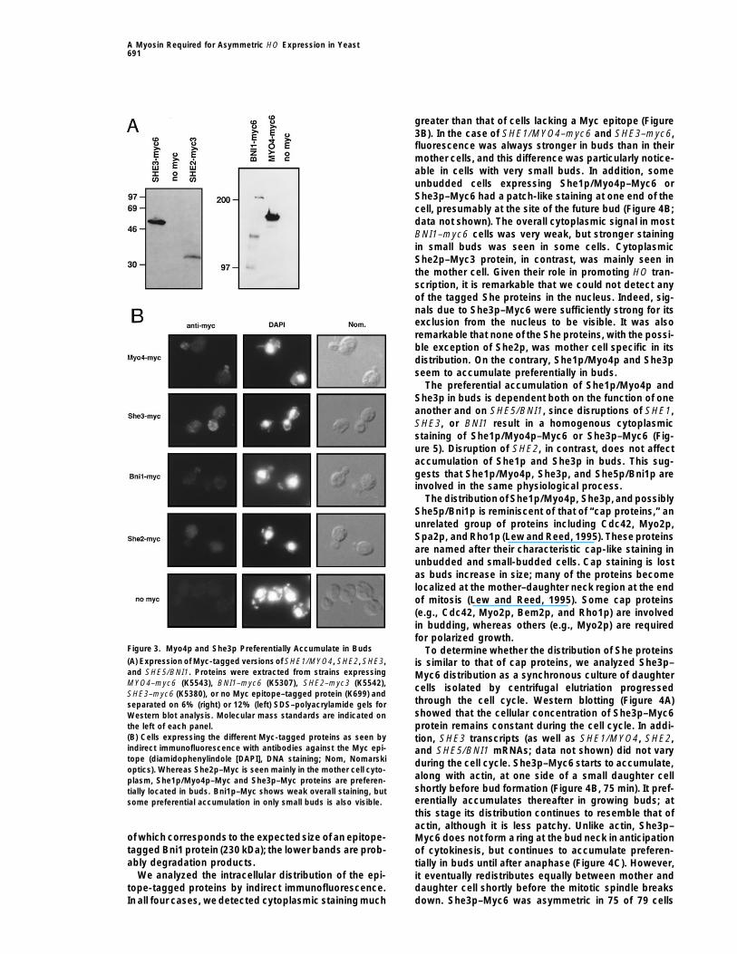
greater than that of cells lacking a Myc epitope (Figure3B). In the case of SHE1/MYO4–myc6 and SHE3–myc6,fluorescence was always stronger in buds than in theirmother cells, and this difference was particularly notice-able in cells with very small buds. In addition, someunbudded cells expressing She1p/Myo4p–Myc6 orShe3p–Myc6 had a patch-like staining at one end of thecell, presumably at the site of the future bud (Figure 4B;data not shown). The overall cytoplasmic signal in mostBNI1–myc6 cells was very weak, but stronger stainingin small buds was seen in some cells. CytoplasmicShe2p–Myc3 protein, in contrast, was mainly seen inthe mother cell. Given their role in promoting HO tran-scription, it is remarkable that we could not detect anyof the tagged She proteins in the nucleus. Indeed, sig-nals due to She3p–Myc6 were sufficiently strong for itsexclusion from the nucleus to be visible. It was alsoremarkable that none of the She proteins, with the possi-ble exception of She2p, was mother cell specific in itsdistribution. On the contrary, She1p/Myo4p and She3pseem to accumulate preferentially in buds.
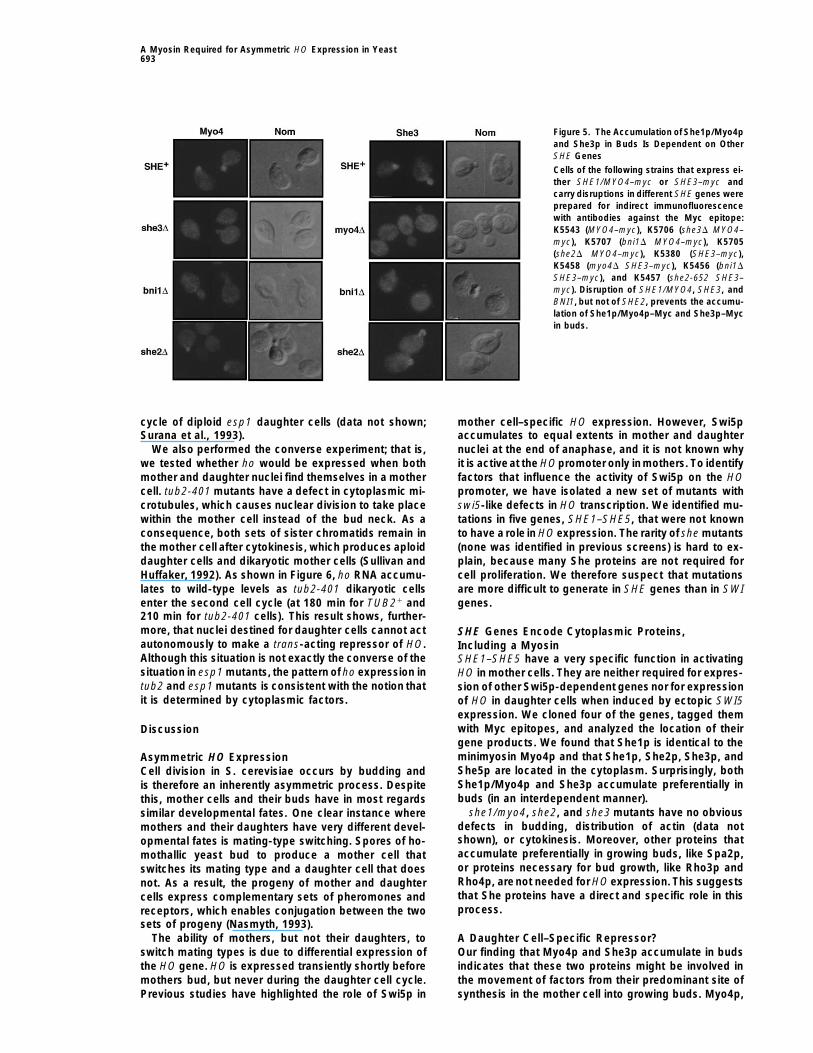
The preferential accumulation of She1p/Myo4p andShe3p in buds is dependent both on the function of oneanother and on SHE5/BNI1, since disruptions of SHE1,SHE3, or BNI1 result in a homogenous cytoplasmicstaining of She1p/Myo4p–Myc6 or She3p–Myc6 (Fig-ure 5). Disruption of SHE2, in contrast, does not affectaccumulation of She1p and She3p in buds. This sug-gests that She1p/Myo4p, She3p, and She5p/Bni1p areinvolved in the same physiological process.
The distribution of She1p/Myo4p, She3p, and possiblyShe5p/Bni1p is reminiscent of that of “cap proteins,” anunrelated group of proteins including Cdc42, Myo2p,Spa2p, and Rho1p (Lew and Reed, 1995). These proteinsare named after their characteristic cap-like staining inunbudded and small-budded cells. Cap staining is lostas buds increase in size; many of the proteins becomelocalized at the mother–daughter neck region at the endof mitosis (Lew and Reed, 1995). Some cap proteins(e.g., Cdc42, Myo2p, Bem2p, and Rho1p) are involvedin budding, whereas others (e.g., Myo2p) are requiredfor polarized growth.
Figure 3. Myo4p and She3p Preferentially Accumulate in Buds To determine whether the distribution of She proteins(A) Expression of Myc-tagged versions of SHE1/MYO4, SHE2, SHE3, is similar to that of cap proteins, we analyzed She3p–and SHE5/BNI1. Proteins were extracted from strains expressing Myc6 distribution as a synchronous culture of daughterMYO4–myc6 (K5543), BNI1–myc6 (K5307), SHE2–myc3 (K5542), cells isolated by centrifugal elutriation progressedSHE3–myc6 (K5380), or no Myc epitope–tagged protein (K699) and
through the cell cycle. Western blotting (Figure 4A)separated on 6% (right) or 12% (left) SDS–polyacrylamide gels forshowed that the cellular concentration of She3p–Myc6Western blot analysis. Molecular mass standards are indicated onprotein remains constant during the cell cycle. In addi-the left of each panel.
(B) Cells expressing the different Myc-tagged proteins as seen by tion, SHE3 transcripts (as well as SHE1/MYO4, SHE2,indirect immunofluorescence with antibodies against the Myc epi- and SHE5/BNI1 mRNAs; data not shown) did not varytope (diamidophenylindole [DAPI], DNA staining; Nom, Nomarski during the cell cycle. She3p–Myc6 starts to accumulate,optics). Whereas She2p–Myc is seen mainly in the mother cell cyto-
along with actin, at one side of a small daughter cellplasm, She1p/Myo4p–Myc and She3p–Myc proteins are preferen-shortly before bud formation (Figure 4B, 75 min). It pref-tially located in buds. Bni1p–Myc shows weak overall staining, buterentially accumulates thereafter in growing buds; atsome preferential accumulation in only small buds is also visible.this stage its distribution continues to resemble that ofactin, although it is less patchy. Unlike actin, She3p–
of which corresponds to the expectedsize of an epitope- Myc6 does not form a ring at the bud neck in anticipationtagged Bni1 protein (230 kDa); the lower bands are prob- of cytokinesis, but continues to accumulate preferen-ably degradation products. tially in buds until after anaphase (Figure 4C). However,
We analyzed the intracellular distribution of the epi- it eventually redistributes equally between mother andtope-tagged proteins by indirect immunofluorescence. daughter cell shortly before the mitotic spindle breaks
down. She3p–Myc6 was asymmetric in 75 of 79 cellsIn all four cases, we detected cytoplasmic staining much

Cell692
with an elongated but undivided nucleus and in 67 of118 binucleate cells with intact spindles, but was in only5 of 60 binucleate cells lacking spindles. An analysis ofasynchronous cultures suggests that She1p/Myo4p–Myc6 has a pattern similar to that of She3p–Myc6 (datanot shown).
The changes in localization during the cell cycle ofShe1p/Myo4p and She3p resembles, but is not identicalto, other cap proteins. To assess the specificity of thedefect of the she mutants in HO transcription, we dis-rupted one of the two copies of SPA2, RHO3, and RHO4in a diploid strain homozygous for the HO–ADE2 reportergene. All four spores from each tetrad derived fromstrains heterozygous for any of the three disruptionswere capable of growth in the absence of adenine. HOexpression does not, therefore, depend onSPA2, RHO3,or RHO4 (data not shown).
Daughter, but Not Mother, Cell CytoplasmRepresses HOWhy should HO expression in mother cells depend ona myosin and other cytoskeletal proteins that accumu-late in buds? One explanation is that HO is repressedin daughter cells by a factor that is transported from themother cell cytoplasm into the bud by She1p/Myo4pand other She proteins. Thus, asymmetric HO expres-sion may be due to the generation of differences be-tween the cytoplasm of mothers and their daughtersand not to differences between their nuclei.
This notion predicts that HO would not be expressedfrom a mother cell nucleus that finds itself in a daughtercell. The behavior of esp1 mutants provides a means oftesting this prediction. Despite a defect in anaphase,esp1-1 mutant cells reenter the next cell cycle. Curi-ously, their single undivided nucleus is transported tothe bud in 90% of these defective divisions, with theresult that cytokinesis produces an “aploid” mother celland a “diploid” daughter cell (McGrew et al., 1992). Wecompared ho RNA levels in wild-type and esp1-1 mu-tants following incubation at 378C of unbudded G1daughter cells isolated by centrifugal elutriation (Figure6). ho RNA accumulated to high levels in wild-type cells,but not in esp1 mutants, as cells entered the secondcell cycle (at 150 min for ESP11 and 240 min for esp1-1 cells that are delayed for the entry into the secondcell cycle). The failure of esp1 mutants to express ho isnot due to a general failure to reactivate SBF-regulatedgenes in the second cycle because HCS26 RNAs reac-cumulated to wild-type level upon entry into the cell
(B) Aliquots of cells were fixed at the indicated timepoints and ana-lyzed by indirect immunofluorescence with anti-Myc antibodies andwith tetramethylrhodamine B isothiocyanate–phalloidin (stainingF-actin). Note that the asymmetric distribution of She3p–Myc disap-pears at the end of mitosis.(C) Cells undergoing mitosis were simultaneously stained with anti-
Figure 4. She3p Is Asymmetrically Distributed throughout Most of tubulin, anti-Myc, and DAPI to visualize the distribution of tubulin,the Cell Cycle DNA, and She3p–Myc at different stages of mitosis ([a], [e], and [i]
are metaphase; [b], [c], [f], [g], [k], and [l] are anaphase; and [d], [h],(A) The ratio of She3p–Myc to total protein does not change duringand [m] are telophase). The asymmetric distribution of She3p–Myc isthe cell cycle. Early G1 daughter cells (K5380, SHE3–myc6) werelost during anaphase (fully elongated spindle and separated nuclei).collected by centrifugal elutriation and incubated in YEP–raffinoseNote that some of the anti-Myc signal is the result of cross-reactivityat 308C. A Western blot probed with anti-Myc and anti-Swi6p anti-with the anti-tubulin staining.bodies is shown. Numbers at the top indicate time.
A Myosin Required for Asymmetric HO Expression in Yeast693
Figure 5. The Accumulation of She1p/Myo4pand She3p in Buds Is Dependent on OtherSHE Genes
Cells of the following strains that express ei-ther SHE1/MYO4–myc or SHE3–myc andcarry disruptions in different SHE genes wereprepared for indirect immunofluorescencewith antibodies against the Myc epitope:K5543 (MYO4–myc), K5706 (she3D MYO4–myc), K5707 (bni1D MYO4–myc), K5705(she2D MYO4–myc), K5380 (SHE3–myc),K5458 (myo4D SHE3–myc), K5456 (bni1D
SHE3–myc), and K5457 (she2-652 SHE3–myc). Disruption of SHE1/MYO4, SHE3, andBNI1, but not of SHE2, prevents the accumu-lation of She1p/Myo4p–Myc and She3p–Mycin buds.
cycle of diploid esp1 daughter cells (data not shown; mother cell–specific HO expression. However, Swi5paccumulates to equal extents in mother and daughterSurana et al., 1993).
We also performed the converse experiment; that is, nuclei at the end of anaphase, and it is not known whyit is activeat the HOpromoter only inmothers. To identifywe tested whether ho would be expressed when both
mother and daughter nuclei find themselves in a mother factors that influence the activity of Swi5p on the HOpromoter, we have isolated a new set of mutants withcell. tub2-401 mutants have a defect in cytoplasmic mi-
crotubules, which causes nuclear division to take place swi5-like defects in HO transcription. We identified mu-tations in five genes, SHE1–SHE5, that were not knownwithin the mother cell instead of the bud neck. As a
consequence, both sets of sister chromatids remain in to have a role in HO expression. The rarity of she mutants(none was identified in previous screens) is hard to ex-the mother cell after cytokinesis, which produces aploid
daughter cells and dikaryotic mother cells (Sullivan and plain, because many She proteins are not required forcell proliferation. We therefore suspect that mutationsHuffaker, 1992). As shown in Figure 6, ho RNA accumu-
lates to wild-type levels as tub2-401 dikaryotic cells are more difficult to generate in SHE genes than in SWIgenes.enter the second cell cycle (at 180 min for TUB21 and
210 min for tub2-401 cells). This result shows, further-more, that nuclei destined for daughter cells cannot act SHE Genes Encode Cytoplasmic Proteins,autonomously to make a trans-acting repressor of HO. Including a MyosinAlthough this situation is not exactly the converse of the SHE1–SHE5 have a very specific function in activatingsituation in esp1 mutants, the pattern of ho expression in HO in mother cells. They are neither required for expres-tub2 and esp1 mutants is consistent with the notion that sion of other Swi5p-dependent genes nor for expressionit is determined by cytoplasmic factors. of HO in daughter cells when induced by ectopic SWI5
expression. We cloned four of the genes, tagged themwith Myc epitopes, and analyzed the location of theirDiscussiongene products. We found that She1p is identical to theminimyosin Myo4p and that She1p, She2p, She3p, andAsymmetric HO ExpressionShe5p are located in the cytoplasm. Surprisingly, bothCell division in S. cerevisiae occurs by budding andShe1p/Myo4p and She3p accumulate preferentially inis therefore an inherently asymmetric process. Despitebuds (in an interdependent manner).this, mother cells and their buds have in most regards
she1/myo4, she2, and she3 mutants have no obvioussimilar developmental fates. One clear instance wheredefects in budding, distribution of actin (data notmothers and their daughters have very different devel-shown), or cytokinesis. Moreover, other proteins thatopmental fates is mating-type switching. Spores of ho-accumulate preferentially in growing buds, like Spa2p,mothallic yeast bud to produce a mother cell thator proteins necessary for bud growth, like Rho3p andswitches its mating type and a daughter cell that doesRho4p, arenot needed for HO expression. This suggestsnot. As a result, the progeny of mother and daughterthat She proteins have a direct and specific role in thiscells express complementary sets of pheromones andprocess.receptors, which enables conjugation between the two
sets of progeny (Nasmyth, 1993).The ability of mothers, but not their daughters, to A Daughter Cell–Specific Repressor?
Our finding that Myo4p and She3p accumulate in budsswitch mating types is due to differential expression ofthe HO gene. HO is expressed transiently shortly before indicates that these two proteins might be involved in
the movement of factors from their predominant site ofmothers bud, but never during the daughter cell cycle.Previous studies have highlighted the role of Swi5p in synthesis in the mother cell into growing buds. Myo4p,

Cell694
unlike its relative Myo2p, is not required for bud growth(Haarer et al., 1994). Myo2p (Lillie and Brown, 1994) isthought to use actin cables running from the mother tothe bud to transport secretory vesicles from mother cellsinto their buds.Given the similarity of Myo2p and Myo4p,we suppose that Myo4p transports substances in a simi-lar manner. If so, these two proteins would seem totransport very different cargos, because HO expressionis highly dependent on MYO4, while the defective bud-ding of myo2-66 mutants is not exacerbated by deletionof MYO4.
To explain the dependence of HO transcription onSHE genes, we propose that the function of Swi5p asa transcription factor is inhibited by a protein whoseexclusion from mothers depends on transport mediatedby She1p/Myo4p. In an accompanying paper, we de-scribe the identification of a protein with these proper-ties. Ash1p is a protein necessary to repress HO indaughter cells and whose preferential accumulation indaughter cell nuclei at the end of mitosis is dependenton SHE genes (Bobola et al., 1996; Sil and Herskowitz,1996 [this issue of Cell]). Our demonstration that HOcannot be expressed in esp1 mutants in which bothmother and daughter chromatids find themselves in thedaughter cell suggests that the cytoplasm of daughtercells contains factors that promote HO repression. Suchfactors might be required for the synthesis or accumula-tion of Ash1p and might be transported into daughtercells by She1p/She3p.
She Protein FunctionsIf Myo4p is used to move determinants from the motherto the daughter cell, what might be the role of the otherShe proteins? she5/bni1 mutants have wide bud necks,are slow in undergoing cytokinesis, are altered in budsite positioning, and are lethal in combination with muta-tions in CDC12 (Fares and Pringle, submitted). The she5/bni1 mutant alleles that we isolated produce an abnor-mal transcript from the HO locus that initiates far up-stream of the transcription start. Surprisingly, the abnor-mal HO transcripts are absent from she5/bni1D cells.This allele-specific phenotype suggests that the cyto-plasmic She5p/Bni1p could have a more direct role indefining the transcription start site of HO. Alternatively,the abnormal transcript might merely be a coincidentalpleiotropy that is unrelated to the defect in HO expres-sion. We considered, but can reject, the notion that HOfails to be expressed in she5 mutants for the trivial rea-son that mothers fail to separate their cytoplasm fromthat of daughters. We show in the accompanying paper
nucleus. Note that the weak ho signal in the first cell cycle of theelutriated ESP1 daughter cells (75 min) is due to a contamination
Figure 6. Nuclei Located in Daughter Cells Cannot Express ho with mother cells (5%).(B) tub2-401 (K5429) and TUB2 (K1168) cells were synchronized by(A) Early G1 daughter cells of esp1-1 (K2788) and ESP1 (K699) strains
were isolated by centrifugal elutriation and released into YEP– addition of a factor and released into YEPD at 188C (restrictivetemperature). Shown are a Northern blot, probed for ho and CLN2raffinose at 378C (restrictive temperature). RNA was isolated at the
indicated timepoints after release and probed for ho, CMD1 (data mRNAs, and a budding index (top); the ratio of ho to CMD1 asquantified by a Molecular Dynamics phosphoimager (middle); andnot shown), and HCS26 mRNAs. Shown are a Northern blot and
budding index (top); the ratio of ho to CMD1 as quantified by a a schematic presentation of the morphological phenotype of tub2-401 cells at 188C (bottom). Closed circles (bottom) represent nuclei.Molecular Dynamics phosphoimager (middle); and a schematic pre-
sentation of the morphological phenotype of esp1-1 cells at 378C Note that ho is not expressed in the first G1 phase (30 min) afterrelease from an a factor arrest.(bottom). The closed circle in the schematic (bottom) represents the
A Myosin Required for Asymmetric HO Expression in Yeast695
was necessary to allow complementation testing, since a matD(Bobola et al., 1996) that Ash1p still accumulates asym-strain mates to MATa, and the resulting diploid strain does notmetrically and mothers still express HO in cells that failrepress HO. After EMS mutagenesis to 75% survival (Cvrckova andto undergo cytokinesis owing to a cdc12-1 mutation.Nasmyth, 1993), cells were grown for two generations in YEP–
Overexpression of BNI1 in strains carrying mutations in dextrose (YEPD) to avoid phenotypic lag. Cells (6 3 106) were platedthe gene for the actin-binding protein profilin causes onto selective medium containing 0.03% canavanine, 10 mg/l ade-
nine, 4% glucose, and all amino acids except arginine. After 3 daysslow growth (J. Pringle, personal communication);at 258C, about 25,000 mutants had formed colonies, of which 3,400She5p/Bni1p might, therefore, interact with the actinshowed a red color indicating a lack of both CAN1 and ADE2 expres-cytoskeleton.The functions of She2p, She3p, and She4psion. These were transferred onto YEP–Gal media to induce lacZremain mysterious. The dependence of She1p/Myo4pexpression from the GAL–HO–lacZ reporter gene. Mutants that
accumulation in buds on SHE3 and BNI1 suggests that formed blue colonies in this assay were considered to carry muta-both might be involved in transport mediated by Myo4p. tions in genes involved in mother/daughter control of HO. We
crossed 315 such mutants to a parent strain of the opposite matingtype to test for dominance. We mated 280 recessive mutants withOn the Mechanism of SegregatingMATa and matD versions of a strain carrying a SWI5 deletion. TheDevelopmental Determinantscorresponding diploids were tested for complementation of adenine
During the development of most organisms, cell prolifer- auxotrophy. We found that 57 mutants complemented the SWI5ation relies on the precise segregation of sister chroma- deletion strains and placed 46 of them in complementation groups
by testing the diploids for adenineauxotrophy. Using this screen, wetids to daughter cells and the more or less equal divisionidentified five complementation groups, which were named SHE1 toof cytoplasmic components. It is becoming increasinglySHE5. Pairwise crosses between 10 out of 35 dominant mutantsclear that the unequal segregation of very specific cellshowed them to be allelic to each other and to she1. Two mutantscomponents has an essential role in many cases of cellfrom each complementation group (only one in the case of she4)
differentiation. How some cellular components are were crossed back three times to nonmutagenized parent strainsasymmetrically segregated while most are segregated prior to further analysis.evenly is a question of great interest. It was not known at
Mating-Type Switching Analysisthe outset of this study whether nuclear or cytoskeletalStrains K4824 (HO–ADE2 HO–CAN1 she1-1024), K4822 (HO–ADE2processes were responsible for mother cell–specific HOHO–CAN1 she3-1440), K4819 (HO–ADE2 HO–CAN1 she2-652), andexpression. Our results clearly point to the latter.K4924 (HO–ADE2 HO–CAN1 she4-4449) were crossed to a HO1
The dependence of posterior oskar mRNA accumula-strain, and progeny were selected that carried a she mutation and
tion on tropomyosin in Drosophila oocytes (Erdelyi et the functional HO gene. Pedigree analysis on these strains wasal., 1995) suggests that actin cytoskeletal components performed as described previously (Strathern and Herskowitz,
1979). Homothallic she mutant strains were crossed to a homothallichave an important role in the asymmetric distributionstrain expressing SWI5 from the constitutive RP39 promoter. Cellsof developmental determinants. The identification of athat carried both a she mutation and the RP39–SWI5 fusion werespecific myosin subtype (Myo4p) in the asymmetric ac-chosen for pedigree analysis.cumulation of Ash1p is direct evidence that actin-based
motors have a crucial role in such processes. It willCloning and Disruption of the SHE Genes
be intriguing to address whether Myo4p and other She The SHE genes were isolated by virtue of their ability to complementproteins are involved in generating differences between the adenineauxotrophy of she mutants. A genomic library (Cvrckova
and Nasmyth, 1993) was transformed into K4793 (she1-456), K4819mothers and daughters that affect processes besides(she2-652), K4822 (she3-1440), and K4795 (she5-2944), and trans-HO expression. One candidate is the different life spansformants were selected on plates lacking adenine and leucine.of mothers and their daughters.
The only plasmid complementing a she1-456 mutation containedan 8 kb insert. Sequencing showed that it carried the full MYO4
Experimental Procedures gene. To verify that the complementing activity resided within theMYO4 gene, we introduced a frameshift by filling in the unique XhoI
Culture Conditions, Strains, and Media site of MYO4 with Klenow polymerase. The resulting plasmid couldUnless otherwise stated, yeast strains are derived from W303a not complement a she1-456 mutation. For disruption of MYO4, an(which is equivalent to K699; Schwob et al., 1994). Strains K1168 internal SalI–HpaI fragment was replaced by the URA3 gene. The(MATa ura3-52 ho) and K5429 (MATa ura3-52 his4-539 lys2-801 disrupted allele was released from the vector and transformed intotub2-401 ho) are congenic to S288C. Only the relevant genotype is K842 (diploid wild-type strain) and K4452 (MATa HO–ADE2 HO–shown. Cells were grown in yeast extract–peptone (YEP) medium CAN1).as described by Schwob et al. (1994). The she2-652 mutant was complemented by two plasmids. Se-
quencing of the 5 and 5.2 kb inserts revealed that both carried apart of chromosome XI containing the ORFs YKL130c to YKL133c.Mutant Isolation and Characterization
We generated yeast strains that allowed us to select for mutants A 1.1 kb HindIII fragment with only ORF YKL130c and adjacent 59
and 39 untranslated region (UTR) sequences was able to restorethat cannot express HO. The complete HO promoter was fused toboth the ADE2 and CAN1 ORFs and integrated by targeted gene HO–ADE2 expression in K4819. We therefore refer to ORF YKL130c
as SHE2. Disruption of SHE2 was performed by replacing an internalreplacement into the HO and CAN1 loci, respectively. CAN1 causescells to be sensitive to the arginine analog canavanine. Cells ex- NsiI–StuI fragment with URA3 and transformation of the resulting
construct into K4452 and K842.pressing ADE2 form white colonies, whereas mutants that cannotexpress it form red colonies on media with limited amounts of ade- A she3-1140 mutant was complemented by four plasmids that
carried different inserts with a common 2.2 kb SacI–XbaI fragmentnine. Details concerning the construction of HO–ADE2 and HO–CAN1 are available upon request. A GAL–HO–lacZ fusion (Nasmyth, containing the complementing activity. Sequencing of this common
fragment showed that it carried a part of yeast chromosome II with1987) on a YIplac204 vector was integrated at the URA3 locus, andthe resulting strain was transformed either with the HIS3 gene or the full ORF YBR1005 and the amino-terminal third of YBR1004.
Disruption of YBR1005 with a URA3 marker at a unique HpaI sitewith a matD:LEU2 construct to obtain strains K4535 (MATa HIS3HO–ADE2 HO–CAN1 URA3::GAL1-10–HO–lacZ ) and K4570 led to loss of the complementing activity, which indicates that
YBR1005 is SHE3. For deletion of SHE3, a 1.8 kb PCR fragment(matD::LEU2 HO–ADE2 HO–CAN1 URA3::GAL1-10–HO–lacZ) thatwere used for EMSmutagenesis. Deletion of the MAT locus in K4570 with new XbaI and EcoRI sites at the 59 and 39 ends of SHE3 was
Cell696
subcloned into pUC19, and an internal 0.7 kb XhoI–HpaI fragment Acknowledgmentswas replaced by URA3.
The three plasmids that complemented a she4-4449 mutant car- Correspondence should be addressed to K. N. We would like tothank R. Siegmund for constructing the HO–ADE2 fusion and I.ried inserts of different sizes, which contained a common 3 kb frag-
ment. This fragment was sequenced and found to contain a part of Howell-Stevenson for invaluable help during the design of the HO–CAN1 reporter gene. We acknowledge Drs. M. Snyder, T. Huffaker,chromosome XV with an unknown ORF of 2376 bp, which was
designated SHE4. The gene was subcloned into pTZ18 and dis- Y. Matsui, C. Peterson, J. Fares, J. Pringle, S. Kron, and D. Kornitzerfor providing yeast strains and plasmids, G. Ammerer for 9E10 anti-rupted by replacing an internal 1.1kb ClaI–XhoI fragment with URA3.
The disruption construct was released from the vector and trans- bodies, and E. Hurt for help with sequence analysis. Thanks go toC. Koch, E. Schwob, and M. Cotten for helpful discussions of theformed into K4452 and K842.
Two plasmids were able to rescue a she5-2944 mutant. The plas- manuscript. We would like to thank J. Pringle and T. Schuster forproviding data prior to publication. R.-P. J. was supported by themids contained 6.5 and 12 kb inserts that showed several similar
restriction fragments. The 12 kb insert carried the full BNI1 gene Boehringer Ingelheim Fonds, C. M. by a European Molecular BiologyOrganization fellowship, and K. N. by the Austrian Industrial Re-and additional 59 and 39 sequence. The 6.5 kb insert carried the 59
UTR and most of the BNI1 coding sequence, lacking only the 39 176 search Promotion Fund.bp of the BNI1 ORF. For BNI1 disruption, we used a constructaccording to Fares and Pringle (submitted). Received November 17, 1995; revised January 19, 1996.
To demonstrate linkage between the cloned SHE genes and theshe mutations, we crossed strains carrying deletion alleles with the Referencescorresponding she mutant strains. Tetrad analysis showed that in allcases no spore of each tetrad could express the HO–ADE2 reporter Becam, A.-M., Cullin, C., Grzybowska, E., Lacroute, F., Nasr, F.,gene, demonstrating tight linkage of the she mutant and the corre- Ozier-Kalogeropoulos, O., Palucha, A., Slonimski, P.P., Zagulski,M.,sponding she disruption. and Herbert, C.J. (1994). The sequence of 29.7 kb from the right
To analyze the effect of deletions of cap protein genes, we trans- arm of chromosome II reveals 13 complete open reading frames,formed spa2::URA3 (Gehrung and Snyder, 1990), rho3::URA3, and of which ten correspond to new genes. Yeast 10, 1–11.rho4::HIS3 (Matsui and Toh-E, 1992) disruption constructs into
Bobola, N., Jansen, R.-P., Shin, T. H., and Nasmyth, K. (1996). Asym-K5044 (HO–ADE2/HO–ADE2 HO–CAN1/HO–CAN1). Tetrad dissec-
metric accumulation of Ash1p in postanaphase nuclei depends ontion of diploids with one disrupted copy revealed that all spores
a myosin and restricts yeast mating-type switching to mother cells.could express ADE2 and CAN1 from the HO promoter.
Cell 84, this issue.
Castrillion, D.H., and Wasserman, S.A. (1994). diaphanous is re-Localization of the She Proteins quired for cytokinesis in Drosophila and shares domains of similarityTo introduce an epitope tag at the carboxyl termini of She proteins, with the products of the limb deformity gene. Development 120,we created BamHI (in the case of BNI1 and SHE3) or XbaI sites (in 3367–3377.the case of MYO4 and SHE2) in front of the corresponding stop Clark, I., Giniger, E., Ruohola-Baker, H., Jan, L.Y., and Jan, Y.N.codon by PCR-mediated mutagenesis. BamHI or XbaI fragments of
(1994). Transient posterior localization of a kinesin fusion proteinthe c-Myc epitope cassette (provided by S. Kron) that contained
reflects anteroposterior polarity of the Drosophila oocyte. Curr. Biol.three Myc epitopes were cloned into the new site. SHE2–myc3 and
4, 289–300.SHE3–myc6 constructs were excised and integrated at the genomic
Curtis, D., Lehmann, R., and Yamore, P. (1995). Translational regula-locus by replacing the she2::URA3 and she3::URA3 disruptions intion in development. Cell 81, 171–178yeast. Positive clones were selected on 5-fluoroorotic acid–
containing medium. To introduce the tagged versions of MYO4 and Cvrckova, F., and Nasmyth, K., (1993). Yeast G1 cyclins CLN1 andBNI1 at the genomic locus, we initially had to clone a URA3 marker CLN2 and a GAP-like protein have a role in bud formation. EMBOinto the 39 end of the genes (detailed construct description is avail- J. 12, 5277–5286.able upon request). In a subsequent step, the marked alleles were Dujon, B., Alexandraki, D., Andre, B., Ansorge, W., Baladron, V.,replaced by the epitope-tagged versions. In all four cases, 5-fluoro- Ballesta, J.P.G., Banrevi, A., Bolle, P.A., Bolotin-Fukuhara, M., Bos-orotic acid–positive clones were checked by Western blot analysis sler, P., et al. (1994). Complete DNA sequence of yeast chromosomefor the expression of epitope-tagged proteins (Schwob et al., 1994). XI. Nature 369, 371–378.Strains K5307 (BNI1–myc6), K5380 (SHE3–myc6), K5542 (SHE2–
Emmons, S., Phan, H., Calley, J., Chen, W., James, B., and Manseau,myc3), and K5543 (MYO4–myc6) were found to be positive andL. (1995). cappucino, a Drosophila maternal effect gene required forwere used for indirect immunofluorescence microscopy. Cells werepolarity of the egg and embryo, is related to the vertebrate limbprepared for immunofluorescence according to Nasmyth et al.deformity locus. Genes Dev. 9, 2482–2494.(1990), except that Zymolase 100T (75 mg/ml) was used for sphero-
plasting and the methanol–acetone fixation step was omitted when Erdelyi, M., Michon, A.-M., Guichet, A., Glotzer, J.B., and Ephrussi,costaining with phalloidin anti-actin was done. The anti-Myc anti- A. (1995). Requirement for Drosophila cytoplasmic tropomyosin inbody used was purified from 9E10 hybridoma supernatant. Pictures oskar mRNA localization. Nature 377, 524–527.were taken on Kodak T-MAX400 film or with a CCD camera mounted Evan, G., Lewis, G.K., Ramsay, G., and Bishop, J.M. (1985). Isolationto a Zeiss Axiophot microscope. of monoclonal antibodies specific for human c-myc proto-oncogene
product. Mol. Cell. Biol. 5, 3610–3616.
Synchronous Cultures, Northern Blot, Evans, T.C., Crittenden, S.L., Kodoyianni, V., and Kimble, J. (1994).and Yeast Transformation Translational control of maternal glp-1 mRNA establishes an asym-RNA extraction from yeast and Northern blot analysis were per- metry in the C. elegans embryo. Cell 77, 183–194.formed as described previously (Tebb et al., 1993). Probes against Ferrandon, D., Elphick, L., Nusslein-Volhard, C., and St Johnston,ho, CLN2, SWI5, SIC1, and CMD1 have been described elsewhere D. (1994). Staufen protein associates with the 39UTR of bicoid mRNA(Tebb et al., 1993; Schwob et al., 1994). The probe against EGT2 to form particles that move in a microtubule-dependent manner.was a 1.4 kb fragment (2215 bp to 11187 bp; obtained from B. Cell 79, 1221–1232.Kovacech). The probe against the HO 59 UTR was a 0.7 kb fragment
Gehrung, S., and Snyder, M. (1990). The SPA2 gene of Saccharo-(21905 bp to 21221 bp). Yeast transformation was performed asmyces cerevisiae is important for pheromone-induced morphogene-described by Nasmyth et al. (1990). All of the above-mentioned genesis and efficient mating. J. Cell Biol. 111, 1451–1464.replacements were confirmed by Southern blot analysis. Synchroni-
zation of yeast cells was performed by centrifugal elutriation under Govindan, B., Bowser, R., and Novick, P. (1995). The role of Myo2,a yeast class V myosin, in vesicular transport. J. Cell Biol. 128,conditions described by Schwob et al. (1994) or by arrest–release
experiments using a factor (Nasmyth et al., 1990). 1055–1068.
A Myosin Required for Asymmetric HO Expression in Yeast697
Haarer, B.K., Petzold, A., Lillie, S.H., and Brown, S.S. (1994). Identifi- regulator of HO, contains four paired amphipathic helix motifs. Mol.Cell. Biol. 10, 5927–5936.cation of MYO4, a second class V myosin gene in yeast. J. Cell Sci.
107, 1055–1064.
Hill, D.P., and Strome, S. (1988). An analysis of the role of microfila- GenBank Accession Numberments in the establishment and maintenance of asymmetry inCaenorhabditis elegans zygotes. Dev. Biol. 125, 75–84. The accession number for the SHE4 sequence described in this
paper is X93598.Horvitz, H.R., and Herskowitz, I. (1992). Mechanisms of asymmetriccell division: two Bs or not two Bs, that is the question. Cell 68,237–255.
Koch, C., and Nasmyth, K. (1994). Cell cycle regulated transcriptionin yeast. Curr. Opin. Cell Biol. 6, 451–459.
Lew, D.J., and Reed, S.I. (1995). Cell cycle control of morphogenesisin budding yeast. Curr. Opin. Genet. Dev. 5, 17–23.
Lillie, S.H., and Brown, S.S. (1994). Immunofluorescence localizationof the unconventional myosin, Myo2p, and the putative kinesin-related protein, Smy1p, to the same regions of polarized growth inSaccharomyces cerevisiae. J. Cell Biol. 125, 825–842.
Lydall, D., Ammerer, G., and Nasmyth, K. (1991). A new role forMCM1 in yeast: cell cycle regulation of SWI5 transcription. GenesDev. 5, 2405–2419.
Matsui, Y., and Toh-E, A. (1992). Isolation and characterization oftwo novel ras superfamily genes in Saccharomyces cerevisiae. Gene114, 43–49.
McGrew, J.T., Goetsch, L., Byers, B., and Baum, P. (1992). Require-ment for ESP1 in the nuclear division of Saccharomyces cerevisiae.Mol. Biol. Cell 3, 1443–1454.
Nasmyth, K. (1987). The determination of mother cell-specific matingtype switching in yeast by a specific regulator of HO transcription.EMBO J. 6, 243–248.
Nasmyth, K. (1993). Regulating the HO endonuclease in yeast. Curr.Opin. Genet. Dev. 3, 286–294.
Nasmyth, K., Stillman, D., and Kipling, D. (1987). Both positive andnegative regulators of HO transcription are required for mother cell–specific mating-type switching in yeast. Cell 48, 579–587.
Nasmyth, K., Adolf, G., Lydall,D., and Seddon, A. (1990). The identifi-cation of a second cell cycle control on the HO promoter in yeast:cell cycle regulation of SWI5 nuclear entry. Cell 62, 631–647.
Peterson, C.L., and Herskowitz, I. (1992). Characterization of theyeast SWI1, SWI2, and SWI3 genes, which encode a global activatorof transcription. Cell 68, 573–583.
Sanders, S.L., and Field, C.M. (1994). Septins in common? Curr.Biol. 4, 907–910.
Schwob, E., Bohm, T. Mendenhall, M.D., and Nasmyth, K. (1994).The B-type cyclin kinase inhibitor p40SIC1 controls the G1 to Stransition in S. cerevisiae. Cell 79, 233–244.
Sil, A., and Herskowitz, I. (1996). Identification of an asymmetricallylocalized determinant, Ash1p, required for lineage-specific tran-scription of the yeast HO gene. Cell 84, this issue.
Sternberg, P.W., Stern, M.J., Clark, I., and Herskowitz, I. (1987).Activation of the yeast HO gene by release from multiple negativecontrols. Cell 48, 567–577.
St Johnson, D. (1995). The intracellular localization of messengerRNAs. Cell 81, 161–170.
Strathern, J.N., and Herskowitz, I. (1979). Asymmetry and direction-ality in production of new cell types during clonal growth: the switch-ing pattern of homothallic yeast. Cell 17, 371–381.
Sullivan, D.S., and Huffaker, T.C. (1992). Astral microtubules are notrequired for anaphase B in Saccharomyces cerevisiae. J. Cell Biol.119, 379–388.
Surana, U., Amon, A., Dowzer, C., McGrew, J., Byers, B., and Nas-myth, K. (1993). Destruction of the CDC28/CLB mitotic kinase is notrequired for the metaphase to anaphase transition in budding yeast.EMBO J. 12, 1969–1978.
Tebb, G., Moll, T., Dowzer, C., and Nasmyth, K. (1993). SWI5 instabil-ity may be necessary but is not sufficient for asymmetric HO expres-sion in yeast. Genes Dev. 7, 517–528.
Wang, H., Clark, I., Nicholson, P.R., Herskowitz, I., and Stillman,D.J. (1990). The Saccharomyces cerevisiae SIN3 gene, a negative
Copyright © 2022 FDOKUMEN