Mammalian retinal horizontal cells are unconventional GABAergic neurons
Upload
independentCategory
view
5download
0
1
An unconventional road for the secretion of transglutaminase in pollen
tubes?
Stefano Del Duca, Donatella Serafini-Fracassini, Giampiero Cai 1
Dipartimento di Scienze Biologiche, Geologiche e Ambientali (BiGeA), Università degli Studi di Bologna, via
Irnerio, 40126 Bologna (Italy) 1 Dipartimento di Scienze della Vita, Università degli Studi di Siena, via Mattioli 4, 53100 Siena (Italy)
Keywords: transglutaminase, secretion, pollen tube, cytoskeleton
Abbreviations: TGase, transglutaminase; MVB, Multi Vesicular Body; TGN, Trans Golgi Network; BFA,
Brefeldin A; AGP, arabino-galactan proteins
Summary
The transglutaminase (TGase) is present in the pollen tube where it most likely participates in the
regulation of different activities including the organization of cytoskeletal elements (microtubules and actin
filaments). In addition to a cytosolic form of TGase, new data suggest the existence of TGase forms
associated with the internal membranes and with the cell wall of pollen tubes. This different localization
extends the functional range of pollen TGase but also raises the question how TGase can be precisely (and
in harmony with the pollen tube growth) redistributed in different cellular compartments. The discovery
that TGase exists as different isoforms may suggest a pathway to achieve this result.
2
In a recent work, Del Duca and coworkers 1 have analyzed the distribution of transglutaminase (TGase) in
the pollen tube of pear tree, mainly in relation to the dynamics of the cytoskeleton and membrane traffic.
The enzyme was localized in association with different cellular compartments, including the cytosol, the
internal membrane system and the cell wall. In view of the fact that TGase is associated with several
cellular compartments, it is important to understand why and how the enzyme is differentially distributed,
if a relationship exists between the TGase forms present in different districts and if each form of TGase
performs a specific activity. In fact, TGase often have more features depending on their cellular localization 2. In addition, by enzymatically adding amino groups to different substrates, TGases can exert different
roles, from regulatory mechanisms to processes involving programmed cell death. The specific role of
TGase in pollen tube is not yet clear. Several publications have highlighted that TGase can enzymatically
modify both tubulin and actin of pollen tubes, suggesting that it is involved in the regulation of the
dynamics of actin filaments and microtubules. This function could be exerted by the cytosolic TGase that,
presumably after activation by changes in Ca2+ concentration, could thus introduce post-translational
modifications in both actin and tubulin monomers. It is not yet clear if these modifications are reversible;
otherwise, the role of TGase would consequently be restricted to processes related to the disorganization
of the cytoskeletal apparatus, as those occurring during the response to self-incompatibility. Currently,
other potential cytoplasmic substrates of pollen TGase are unknown and it is consequently difficult to make
further assumptions about its role. Quite different is the possible role of membrane TGase and, more
generally, of non-cytosolic TGase, including therefore also the cell wall (extracellular) TGase. Data published
in some papers indicated consistently that the pollen TGase is also associated with the membrane
compartments of pollen tubes, including the Golgi apparatus and plasma membrane 3,1. Some papers
report the presence of TGase in association with the chloroplast fraction of plant cells (in which the enzyme
could exert protective functions on the photosystems of the photosynthetic apparatus) 4. However, this
feature has to be excluded in pollen tubes in view of the limited number of plastidial elements and absence
of photosynthetic metabolic functions. The association of TGase with the Golgi apparatus, apart from
suggesting that TGase is transported to the plasma membrane or toward the vacuolar compartment, also
suggests that the enzyme is secreted outside the cell via the vesicular system. Consequently, the Golgi-
TGase association could be the first step of a secretory process that leads TGase in the extracellular matrix
(cell wall).
Association of TGase with the membrane system seems to require the post-translational
modification of the enzyme. Two-dimensional electrophoretic analysis of TGase extracted from different
cellular compartments showed that TGase exhibits an isoforms pattern typical for each compartment. The
use of different isoforms in dependence of different compartments is not an atypical feature but it seems
commonly used to mark the same protein in different environments. A relatively recent example is sucrose
synthase, a key enzyme of the plant metabolism that can exist in differently phosphorylated forms
depending on the localization 5. We do not know the type of post-translational modification (and even
where and when it occurs) to which TGase is subjected; nevertheless, it seems clear that a specific kind of
post-translational modification is a prerequisite to fix the enzyme in different localizations.
The association between TGase and the membrane system is an issue not easy to decipher. The
optimal isolation of single membrane compartments is not technically simple and can therefore result in
interpretations not completely accurate. However, the association between TGase and Golgi was
demonstrated by immunological and enzyme assays 1. Conventional immunofluorescence microscopy
showed that TGase distributes as dots in the cytoplasm of growing pollen tubes (Figure 1A), a labeling
pattern that generally evokes the distribution of organelles and, more specifically, of Golgi bodies. A more
detailed analysis by immuno-electron microscopy allowed us to detect the presence of TGase in association
with other membrane compartments of the pollen tube of pear tree, including the rough endoplasmic
reticulum (Figure 1B). In addition to the presence of TGase in the internal membranes of pollen tubes, the
enzyme was also localized in association with the membrane of hypothetical secretory vesicles (Figure 1C).
3
The association of TGase with the membrane of vesicular structures suggests that part of the enzyme is
transported to the plasma membrane where it can exert its specific function. Examples of TGase in
association with the plasma membrane are numerous (at least in animal cells) and the functions performed
by these enzymes at the plasma membrane can be quite diverse 6.
The presence of TGase in the cell wall of pollen tubes reinforces the idea that TGase is synthesized
on the endoplasmic reticulum, exported to Golgi and then to the vesicular system of secretion. However,
the secretory process could not end exactly as expected for the pollen tube system. Generally, it is believed
that the reticulum-Golgi transport system produces secretory vesicles that accumulate at the pollen tube
apex (the so-called clear zone) and that fuse with the plasma membrane. This linear pathway can be
disturbed by the application of inhibitory substances, such as Brefeldin A (BFA), that induce the formation
of a membrane complex in the pollen tube sub-apex (the so-called BFA compartment) 7. In our tests with
BFA, we never observed the formation of such a complex containing TGase suggesting that TGase (if
secreted through the reticulum-Golgi pathway) may use a vesicular system different from that usually used
in pollen tubes. The possibility that TGase uses a different secretory pathway is also evidenced by further
data of electron microscopy (Figure 1C) revealing the association of TGase with vesicular structures of
different sizes but not localized in the apical region of pollen tubes. These vesicles could be a vehicle for
transporting TGase to areas of the plasma membrane separate from the apical region. A common finding
with other proteins is the fact that the transport of TGase is still dependent on the integrity of actin
filaments. However, this evidence is reasonable and not unexpected because the coordination of
membrane trafficking in plant cells requires the dynamic activity of actin filaments. It is therefore possible
to hypothesize alternative mechanisms of secretion. Non-Golgi-based secretion of TGase is a documented
alternative secretion pathway. The so-called mechanism of "membrane blebbing or microvesicle shedding"
provides that cytosolic TGase is packaged directly into membrane-derived protrusions; these protrusions
come off and the formerly cytosolic components are released when the vesicular protrusions break 8. Based
on these observations (and on information available in the literature), we can hypothesize the following
mechanism for TGase secretion in the pollen tube (Figure 1D). The membrane TGase (green) could be
classically synthesized in the endoplasmic reticulum and exported to the Golgi apparatus and then to the
Trans-Golgi Network (TGN) from where it would be packaged into secretory vesicles. In this way, TGase is
secreted externally in the cell wall and the new position is stabilized by a further post-translational
modification (blue TGase, which could also serve to activate the enzyme). Once in the cell wall, TGase could
be released outside in the pollen-style extracellular matrix, as suggested in apple 3, but TGase could be also
eventually recycled by endocytotic events that may occur both in the apical region of pollen tubes and in
more distal regions. The TGN-recycled TGase, according to the most reliable models, might be either
secreted again or directed towards the Multi-Vesicular-Body (MVB) for degradation 9. Currently we cannot
discriminate between recycling and degradation of TGase, although we believe that the latter is more likely
because in line with what found in animal cells and because the degradation process would allow
maintaining constant the levels of TGase in the apical and subapical region. Considering the uncertainties
still existing on this route, it is also plausible and intriguing to believe that cytosolic TGase (in red) is directly
incorporated and packaged within secretory structures that deliver TGase to the extracellular matrix.
Regardless of the type of secretion, the presence of extracellular TGase asks the question about the
function exerted locally by this enzyme. The cell wall of pollen tubes contains, like other plant cells,
polysaccharides and proteins; the latter can be generally categorized into two main groups: enzymes that
modify the cell wall (such as pectin methyl-esterase) and proteins combined with polysaccharide chains of
variable length, such as arabino-galactan proteins (AGPs). Identifying the specific target of TGase is not
simple. It is clear that the cell wall proteins may represent an ideal target for the enzyme suggesting that
such protein substrates are consequently controlled by TGase. Currently, there is no evidence in favor of
this hypothesis. The identification of protein substrates would require locating the products of the TGase
activity, for example by enzymatically combining tagged-substrates (for example, labeled polyamines) to
protein targets and then using these tags for "fish" the target proteins from cell wall extracts. The protein
substrates may also be represented by the protein skeleton of AGPs. In tomato, one AGP (LeAGP-1) was
4
identified and isolated as a trimer, whose assembly was supposedly caused by a TGase activity 10. In pear
pollen tube, double labeling assays showed that TGase co-localized (although partially) with MAC207-
labelled AGPs 1. The substrates of TGase may also be represented by polysaccharides of the cell wall. As
already mentioned 1, the pectin skeleton could theoretically be a plausible target since previous works
showed that (acid) pectins can interact with (basic) polyamines, the latter being natural substrates of TGase 11. Although these findings do not prove that pectins are targets of TGase, they suggest that this enzyme
may play a role in cell wall organization.
Acknowledgements
We sincerely thank Mrs. Claudia Faleri (Dipartimento Scienze della Vita, University of Siena) for permission
to use the immunofluorescence and immuno-gold microscopy images.
5
References
1. Del Duca S, Faleri C, Iorio RA, Cresti M, Serafini-Fracassini D, Cai G. Distribution of transglutaminase in pear pollen tubes in relation to cytoskeleton and membrane dynamics. Plant Physiol 2013; doi: http://dx.doi.org/10.1104/pp.112.?212225 :
2. Beninati S, Piacentini M. The transglutaminase family: an overview: minireview article. Amino Acids 2004; 26:367-372.
3. Di Sandro A, Del Duca S, Verderio E, Hargreaves AJ, Scarpellini A, Cai G, Cresti M, Faleri C, Iorio RA, Shigehisa H, Furutani Y, Coutts IGC, Griffin M, Bonner PL, Serafini-Fracassini D. An extracellular transglutaminase is required for apple pollen tube growth. Biochem J 2010; 429:261-271.
4. Della Mea M, Di Sandro A, Dondini L, Del DS, Vantini F, Bergamini C, Bassi R, Serafini-Fracassini D. A Zea mays 39-kDa thylakoid transglutaminase catalyses the modification by polyamines of light-harvesting complex II in a light-dependent way. Planta 2004; 219:754-764.
5. Persia D, Cai G, Del Casino C, Faleri C, Willemse MTM, Cresti M. Sucrose synthase is associated with the cell wall of tobacco pollen tubes. Plant Physiol 2008; 147:1603-1618.
6. Fesus L, Piacentini M. Transglutaminase 2: an enigmatic enzyme with diverse functions. Trends Biochem Sci 2002; 27:534-539.
7. Parton RM, Fischer-Parton S, Trewavas AJ, Watahiki MK. Pollen tubes exhibit regular periodic membrane trafficking events in the absence of apical extension. J Cell Sci 2003; 116:2707-2719.
8. Aumuller G, Wilhelm B, Seitz J. Apocrine secretion - fact or artifact? Ann Anatomy - Anatomischer Anzeiger 1999; 181:437-446.
9. Wang H, Zhuang XH, Hillmer S, Robinson DG, Jiang LW. Vacuolar Sorting Receptor (VSR) proteins reach the plasma membrane in germinating pollen tubes. Mol Plant 2011; 4:845-853.
10. Gao M, Kieliszewski MJ, Lamport DTA, Showalter AM. Isolation, characterization and immunolocalization of a novel, modular tomato arabinogalactan-protein corresponding to the LeAGP-1 gene. Plant J 1999; 18:43-55.
11. D'Orazi D, Bagni N. In vitro interactions between polyamines and pectic substances. Biochem Biophys Res Commun 1987; 148:1259-1263.
6
Figure legend
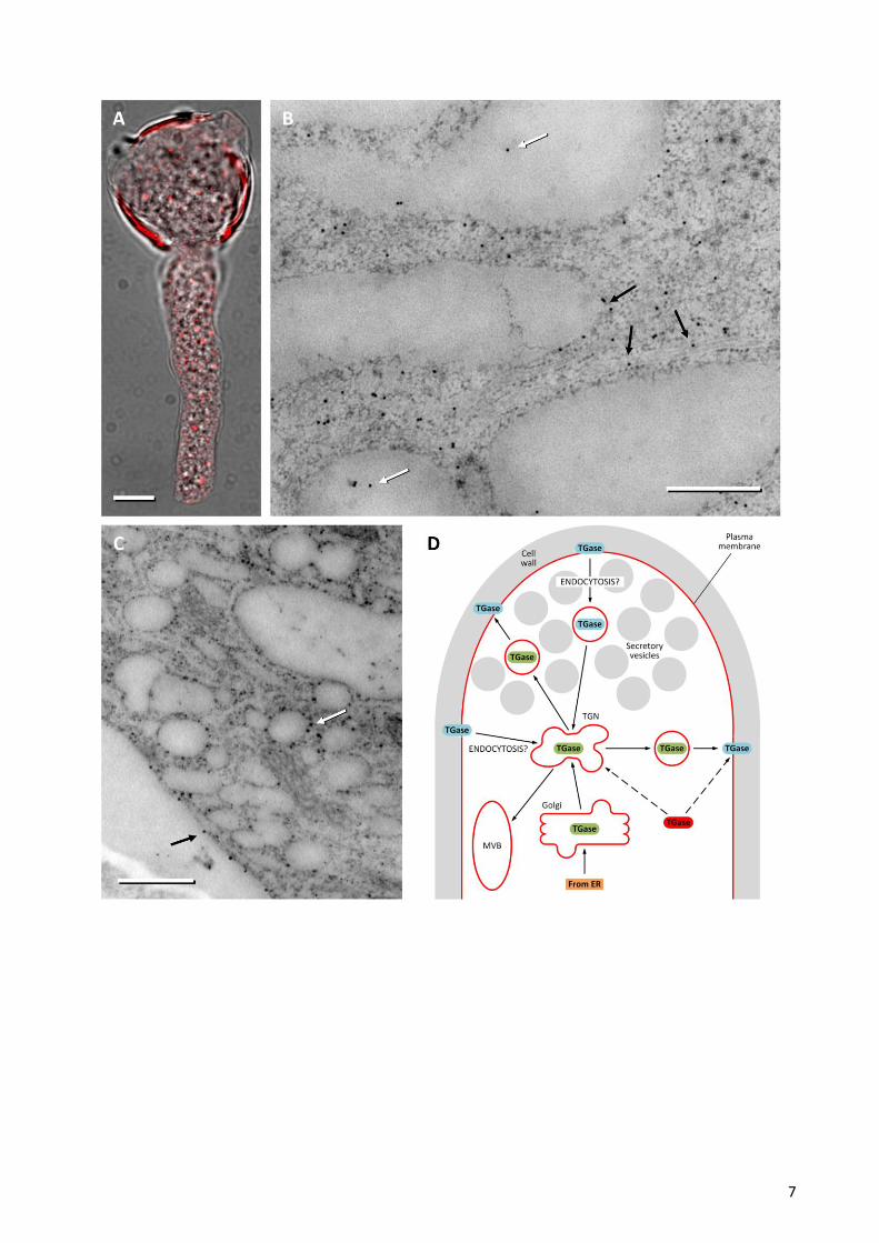
Figure 1. Analysis by immuno-electron microscopy of TGase in pear pollen tube. (A) Immunofluorescence
microscopy of TGase (red) in the pollen tube of pear. The fluorescence image is superimposed on the same
pollen tube shown in bright field microscopy. Bar: 10 μm. (B) Localization of TGase by immuno-gold in not-
apical regions of the pollen tube. The enzyme is shown within membrane structures (white arrows) but also
in association with the surface of internal membranes (possibly including the endoplasmic reticulum, black
arrows). Bar: 500 nm. (C) Distribution of TGase near the cell cortex. TGase can be observed in association
with the surface of vesicular structures (white arrow) and with the plasma membrane of pollen tubes (black
arrows). Bar: 500 nm. (D) Model of the hypothetical secretory process of TGase. The enzyme (in green) may
be secreted through a conventional mechanism that starts in the endoplasmic reticulum (ER), goes through
the Golgi and the Trans Golgi Network (TGN), ending up with the secretory vesicle-mediated exocytosis.
Secretory vesicles may not necessarily fuse with the apical membrane. In the cell wall, TGase would be
subject to post-translational modifications that mark and/or activate the enzyme (in blue). Either excess or
functionally useless TGase may be removed by endocytotic processes to be degraded in the Multi Vesicular
Body (MVB). Considering data in the literature, we cannot exclude that the cytosolic TGase (in red) is
directly incorporated into the membranes and then secreted.
7
Copyright © 2022 FDOKUMEN