A POTENTIAL ROLE FOR TRANSGLUTAMINASE - MSpace
97
THE BIOCHEMISTRY OF ATHEROSCLEROSIS; A POTENTIAL ROLE FOR TRANSGLUTAMINASE RICKY I. WIEBE A Thesis Submitted to the Faculty of Graduate Studies in Partial Fulfillment of the Requirements for the Degree of . MASTER OF SCIENCE Department of Biochemistry and Molecular Biology University of Manitoba Winnipeg, Manitoba Copyright @ August, 1991 BY
-
Upload
khangminh22 -
Category
Documents
-
view
0 -
download
0
Transcript of A POTENTIAL ROLE FOR TRANSGLUTAMINASE - MSpace

THE BIOCHEMISTRY OF ATHEROSCLEROSIS;
A POTENTIAL ROLE FOR TRANSGLUTAMINASE
RICKY I. WIEBE
A Thesis
Submitted to the Faculty of Graduate Studiesin Partial Fulfillment of the Requirements
for the Degree of
. MASTER OF SCIENCE
Department of Biochemistry and Molecular Biology
University of Manitoba
Winnipeg, Manitoba
Copyright @ August, 1991
BY

E*E i¡åä1'.i"*Canadian fheses Serv¡ce
Ottaw4 CeoadaKIA ON4
B¡b{¡othèque nat¡onatedu Canada
Sefvir:e des tÉ3€s c¿nadr'ennes
rsBN ø-315-76988-2
The_ author has grarìted an inevocable non.exclus¡ve ficence allowiqg the NationaiUbrêryof Canada to reproduce, 6*l, csñutL ors€l¡copres of h¡s/her thes¡s by any means and inTy f9m or fomaE maldng-ür¡s t es¡" *rr,lrbL[o tnterested persons.
The author retains ownersh¡p of the copyrightin
. his/her thesis. Neithei ttre tnãs¡s nor
substantial extracts from it may Oepr¡nted orotherwise reproduced withoút trijnàr penmission.
L'auteuraaccordé une l¡cerice inévocabte etnol .ex$u-sîve permettant à la B¡bt¡otfrèquenakjnale du Ca¡¡ada de reproduire, prêtbr,disbibuerou vendre aes cohes ¿à ä thèsede quelque nranière et sous qr"lq* formequ9 ce, soit pour mettre des exemàfaires decette thèse à la disposition des pu*nn*.intéressées.
L'auteurcons€rve fa propriété du dro{td auteurqui protege sa thèse- Ni ta thèse n¡¿es ãxtra¡tssubstantiels de celleci ne ¿o¡vent êtreimprimés ou autrement reproduit
'""n"
"onautorisation.
eanadä

THE BIOCHEMISTRY OF ATITEROSCLEROSIS;
A POTENTIAL ROLE FOR TRANSGLUTA}IINASE
RICKY I. WIEBE
A thesis subnì¡ned ro thc Faculty of Craduate Studies ofthe university of Manitoba in partial'fulfillinent of tlle requirenrents
of the degree of
MASTER OF SCIENCE
o 1991
Permission has been grarrred ro the LIBRARy OF THE UNIVER-S¡TY OF MANITOBA to lend or selt copies of this rhesis. to
the NATIONAL LIBRARY OF CANADA ro rnicrolilm rtris
thesis a¡td to lend or æll copies oí the lìtm, and UNIVERSITYMICROFILMS to publisir an absrracr of rhis rhesis.
The author res€rves other publication rights, and neither thc
thesis nor extensiye extracts frorn it may be pnnteC or other-
wise reproduced without the author's writ.ten permission.

¡
ACKN OWLEDG EM ENTS
The author is grateful for the financial suppoft from the Medical Research
Council of Canada, the Manitoba Heart and Stroke Foundation and the
Manitoba Health Research Council.
Also greatly appreciated is the technical assistance and the wit and humour of
Alan Tarr who provided the encouragement, the laughs and was always there
to grab a "'feine"
The author's deepest gratitude goes to Dr. J. M. Bowness. The encouragement,
insight, direction, that proved invaluable, were always available. Special
thanks go to him as well for the patience he showed in the preparation of this
thesis.
ii
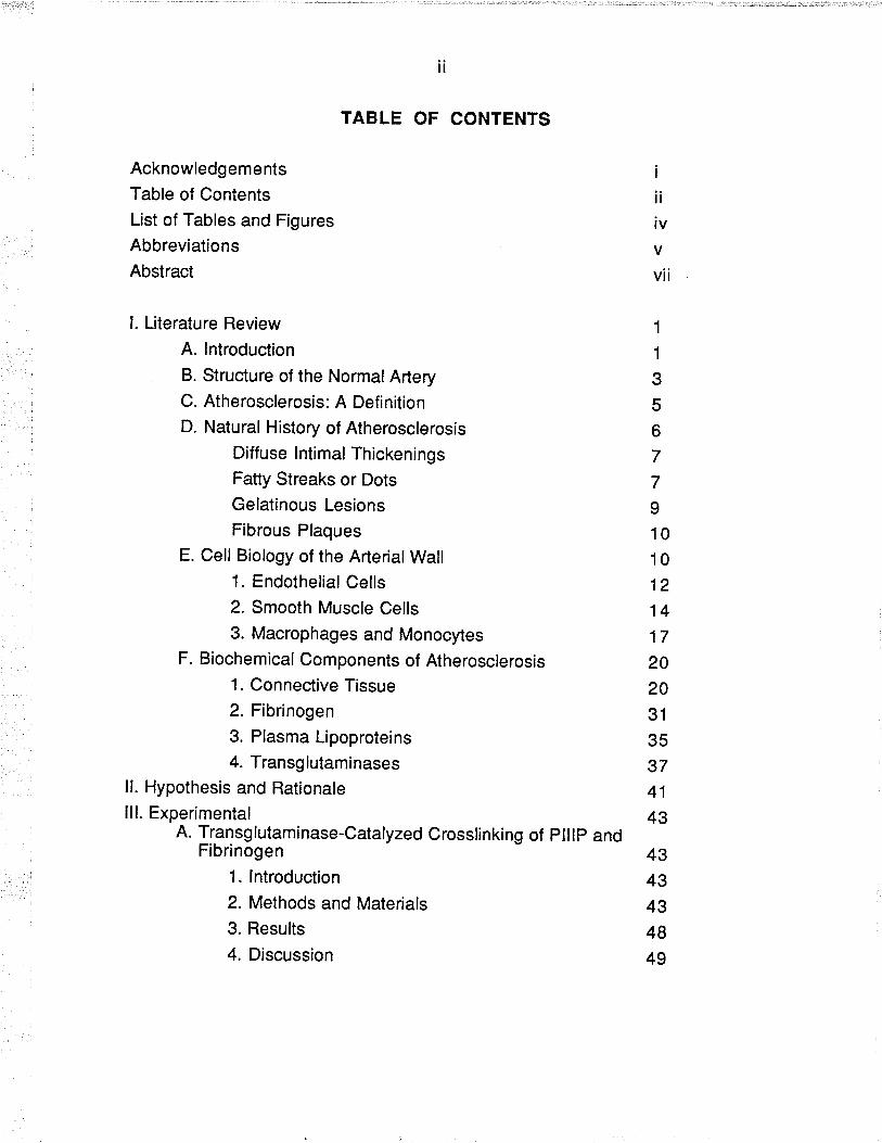
TABLE OF CONTENTS
Acknowledgements
Table of ContentsList of Tables and Figures
AbbreviationsAbstract
l. Literature Review
A. lntroduc{ion
B. Structure of the Normal Artery
C. Atherosclerosis: A Definition
D. Natural History of AtherosclerosisDiffuse lntimal ThickeningsFatty Streaks or Dots
Gelatinous LesionsFibrous Plaques
E. Cell Biology of the Arterial Wall1. Endothelíal Cells2. Smooth Muscle Cells
3. Macrophages and MonocytesF. Biochemical Components of Atherosclerosis
1. Connective Tissue
2. Fibrinogen
3. Plasma Lipoproteins4. Transglutaminases
ll. Hypothesis and Rationale
lll. Experimental
I
ii
iv
v
vii
1
1
3
5
6
7
7
I10
10
12
14
17
20
20
31
35
3741
43A. I¡ansglutaminase-Catalyzed Crosslinking of plllp andFibrinogen -
4g1. lntroduction 4g2. Methods and Materials 493. Results 4g4. Discussion 49

ilt
B. lncreased Transglutamínase in the Aortas of Cholesterol-Fed Rabbits
1. lntroduction
2. Methods and Materials
3. Results
4. Discussion
C. Binding of PlllP or Putrescine in Smooth Muscle Cultures1. lntroduction
2. Methods and Materials
3. Results
4. Discussion
lV. SummaryV. References
51
51
51
54
60
6363636571
74
77

IV
TABLES AND FIGURES
Table 1. Genetically distinct collagens. 22Table 2. Composition of human serum lipoproteins. g6
Table 3. Precipitation of sDS-insoluble materiar after incubatíonwith transglutaminase. 47
Table 4. specific activities of transglutaminases in three layers ofnormal and atherosclerotic rabbit aortas. Sg
Table 5. Total counts in TCA precipitates of confruent sMc layerslabeled with 3H-putrescine. 66
Figure 1. Hypothesis. 2Figure 2. Schematic representation of the normal artery. 4Figure 3. Schematic representation of a fibrous plaque. 11
Figure 4. Transglutaminase-catalyzed crosslink. ggFigure 5. SDS-PAGE of fibrinogen and plllp preparations. 44Figure 6. Transglutaminase-catalyzed crosslinking shown by
chromatography on Sepharose CL-28 in 0.1% SDS. 46Figure 7. Transglutaminase activity in three rayers of normal and
atherosclerotic rabbit aortas. 57Figure L Tissue transglutaminase antigen in three layers of normal
and atherosclerotic rabbit aortas. SgFigure 9. Fluorograph of SDS-PAGE of ceil extracts after labeling
with 3H-putrescine. 67Figure 10. SDS-PAGE of lyophilized medium after incubation of
12sl-plllp with smooth muscle cells. 6gFigure 11. SDS-PAGE of lyophilízed medium after incubation of
1251-Plltp with smooth muscle cells. 69Figure 12. SDS-PAGE of lyophilized medium after incubation of
1251-Plllp with smooth muscle cells. ZO


vl
HDL......... ............hi9h density tipoprotein
HS............ .............heparan sulfate
1EL............ ............interna1 elastic lamina
LDL.......... ............1ow density lipoprotein
MDFC...... ............macrophage-derived foam cell
MG8G...... ............methylglyoxyt bis-(guanylhydrazone)
min........... ............minute
tv|W........... ............molecular weight
PAGE....... ............polyacrylamide-gel electrophoresis
PBS......... ............phosphate buffered saline
PC|11......... ............procollagen type ill
PDGF....... ............p1ate1et-derived growth factor
PG............ ............proteoglycan
PG12......... ............prostacyclin
P|11P......... ............aminopropeptide of type lll collagen
PMSF phenylmethylsulfonylf tuoride
RER.......... ............rough endoplasmic reticulum
s................ ............seconds
SDS......... .............sodium dodecyl sulfate
SE............ ............standard error
SG............ ............structural glycoprotein
SMC ..smooth muscle cell
TCA.......... ............trichloroacetic acid
TGase...... .............transglutaminase
TG F-p............ transforming growth factor-p
VLDL .very low density lipoprotein
vymlo volume fraction of myofilaments

vil
ABSTRACT
A rev¡ew of the literature on the development of the lesions ofatherosclerosis suggests a role for transglutaminase-catalyzed crosslinking ofprocollagen type lll and fibrinogen or low density lipoprotein in the arterial wall.
Bovine type lll 3H-procollagen was shown by chromatography underdissociating conditions to form very high molecular weight complexes withexcess bovine fibrinogen. Larger complexes of this type formed with fibrinogenor fibrin monomers can be separated by centrifugation and were found to beinsoluble upon washing with 1% SDS.
Rabbits were fed for 10-12 weeks on a normal diet or the normal dietsupplemented with 1% cholesterol and 6to peanut oil. The aortas of theseanimals were separated into three layers which were homogenized andextracted. The extracts and the insoluble residues were assayed fortransglutaminase activity and tissue transglutaminase antigen. Whencompared to normal aortas, the inner and middle layers of the aortas withlesions, from cholesterol-fed rabbits, showed higher transglutaminase activitiesin the buffer-soluble fraction without corresponding increase in antigen. Thebuffer-insoluble fraction, which contained a lower specific activitytransglutaminase than the butfer-soluble, showed higher concentrations of bothactivity and antigen in the inner and middle layers of the atherosclerotic aortas.
3H-Putrescine incorporation into cultured smooth muscle cells showedthe presence of 14 kDa and very high molecularweight labeled compounds bySDS-PAGE after reduction. The amount of labeling was increased whenascorbate or butyrate were added. When smooth muscle cell cultures wereincubated with the 125¡-¿¡¡¡opropeptide of type lll procollagen the mediumshowed the presence of high molecular weight complexes on sDS-PAGEunder reducing conditions. These complexes were not present when 1¡s 125¡-
propeptide was incubated in medium without cells or in medium preconditionedwith smooth muscle cells.
The results of these stud¡es are consistent with the hypothesis thattransglutaminase, present on the surface of íntimal macrophages and smoothmuscle cells, catalyzes the formation of crosslinks between procollagen l¡ or itsaminopropeptide and fibrinogen or low density lipoprotein in thesubendothelium. This process would anchor fibrinogen and low densitylipoprotein to the connectíve tissue and the modification of low density

vilt
lipoprotein would cause increased accumulation by macrophages and foamcell formation. The sequestration of the propeptide of procollagen lll wouldinhibit its feedback inhíbition of collagen synthesis and cause the increasedcollagen accumulation found in advanced atherosclerotic lesions.

1
I. LITERATUHE REVIEWA. lntroduction
Cardiovascular disease is the major cause of death in the Western world,
and atheroscferosis is the chief cause. Atherosclerosis is a slowly progressive
disease whích may begin in childhood and does not fully manifest itself until
middle age, or later; therefore it is difficult to isolate the initiating events or
mechanisms in the development of atherosclerotic lesions. The research in this
field over the past 3 decades has produced volumes of information about the
cellular and molecular aspects of the vascular wall, providing numerous insights
into the causes of the progression of the disease; however, the processes
involved in atherogenesis and the relevance and relative importance of various
mechanisms in the initiation and progressíon of lesions of atherosclerosis are
still largely unknown.
The predominant theory explaining lesion progression has been put forth
by Ross [1]. This theory states that the lesions are due to proliferation of arterial
smooth muscle cells (SMC) and progress to mature fibrous plaques as a result
of the arteries' response to some endothelial injury associated with various risk
factors. The major biochemical risk factors associated with the development of
atherosclerotic lesions are high concentrations of fibrinogen (FGN) and low
density lipoprotein (LDL) in the plasma and the arterial wall [2]. Clinical trials
have shown that reducing circulating levels of LDl-cholesterol are correlated
with a reduction in the incidence of cardiovascular disease[109]. lncreased
production of connective tissue components, specifically collagen, by the arterial
SMC in the intíma is the hallmark of developing atherosclerotic lesions
1134,1351, along with intra- and extracellular accumulations of LDL
1228,241,244,245,248,2531 and extracellular accumulations of FGN 1227-zg5l.

2
It seems quite logical that for effective prevention of a disease, such as
atherosclerosis, one of the basic prerequisites is a knowledge of its cause. lt is
the processes and mechanisms of the development of the comptex lesions of
INTIMAL
TRANSGLUTAMINASE]CATALYZED
CROSSLINKING
ARTERIALFIBRINOGEN
OR FIBRIN
-PROCOLI-AGEN TYPE III(oR PiltP)
Figure 1. Hypothesis. See discussion ín text.
,/
EXCESS\
ARTERIALLDL
IMPAIREDHEALING
3
atherosclerosis that are in question. What are the mechanisms by which LDL
and FGN are retained within the arterial wall and why are the natural abilities of
the artery to clear the intima of these excess plasma proteins impaired? These
are the questions that will be addressed in this thesis.
lf atherosclerosis is assumed to proceed due to an altered response of the
vessel wall to injury, then perhaps connections can be drawn with studies of
wounding in skin. Studies have shown that the enzyme transglutaminase
(TGase) plays a role in the healing of wounded skin [3,4]. lt has also been
shown that an early response to wounding of skin is increased production of type
lll procollagen (PClll) [163], and that the aminopropeptide of type lll procollagen
(Pl¡lP) is a very good substrate for TGase 12771. This thesís wíll argue that high
concentratíons of FGN and LDL are bound up in the arterial wall connective
tíssue matrix to PClll or PlllP, produced by proliferating SMC, through a TGase-
calalyzed reaction, producing an altered response to injury in the artery (Fig. 1).
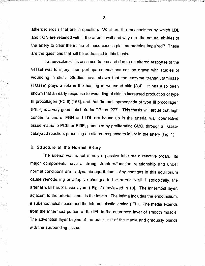
B. Structure of the Normal Artery
The arterial wall is not merely a passive tube but a reactive organ. ltsmajor components have a strong structure/function relationship and under
normal conditions are in dynamic equilibrium. Any changes in this equilibrium
cause remodelling or adaptive changes in the arterial wall. Histologically, the
arterial wall has 3 basic layers ( Fig. 2) [reviewed in 10]. The innermost layer,
adjacent to the aderial lumen is the intima. The intima includes the endothelium,
a subendothelial space and the internal elastic lamina (lEL). The media extends
from the innermost port¡on of the IEL to the outermost layer of smooth muscle.
The adventítial layer begins at the outer limit of the media and gradually blends
with the surrounding tissue.

4
Endothelium
lnternal ElasticLamina
Figure 2. Schematic representation of the normal artery. The normalartery consists of 3 layers: intima (l), media (M) and adventitia (A). Theendothelium, a subendothelial space and the internal elastic lamina constitutethe intima (see discussion).
lNTlMA. The intima is probably the most metabolically active layer in the aorta.
The endothelium is a single smooth layer of cells, which was thought for many
years to be an impermeable barrier. lt is a semi-permeable covering which
provides protection for the underlyíng portions of the vessel wall, maintaining
haemostasis. The subendothelium in early life is composed entirely of loose
connective tissue, but, with age, medial smooth muscle cells gradually migrate
into this region. The IEL ís a connective tissue layer, consisting of collagens,
proteoglycans and elastin, separating the media from the intima. The IEL serves
as a gel-sieve or filter of plasma macromolecules that enter the intima lZ4iJ,allowing through smaller molecules and only small amounts of large molecules.

5
MEDIA. The components of this layer are similar in both elastic and muscular
arteries. Mammalian arteries contain only SMC in the medial layer. The
orientation of the SMC with respect to each other and the artery determine
whether the artery is elastic or muscular. Layers of SMC are sandwiched
between elastic plates and tissue space containing collagen, constituting the
basic structure of elastic a¡'terial media. The connective tissue space between
laminae of elastin and their collagen content increase with age.
ADVENTITIA. Most of the arterial adventitia consists of a loose arrangement of
collagen, elastic fibres, fibroblasts, blood vessels, lymphatics and nerves. The
adventítia is not significantly involved in the process of atherosclerosis and will
not be discussed further.
C. Atherosclerosis: A Definition
Atherosclerotic lesions have been observed in humans for centuries [5,6].
Lobstein coined the generic term "arteriosclerosis" to describe scarring and
calcification of arteries. ln 1904 Marchand first used the term "atherosclerosis" to
designate a disease characterized by lipid-rich arterial lesions, intimal
thickening, fibrosís and calcification.
Virchow started the debate on the mechanisms of the development of the
lesions of atherosclerosis by introducing his imbibition theory in the mid-1g00's.
He suggested that the atheromatous lesions were due to the imbibition of
plasma proteins into the arterial wall. The theories have progressed to the
present most widely accepted theory of the response to injury hypothesis [1],
which emphasizes the importance of intimal SMC proliferation as the key event
in the development of atherosclerotic lesíons. Schwartz and colleagues [7]
describe atherosclerosis as a "result of a multiplicity of interactive cascades
among injurious stimuli and the healing responses of the arterial wall, occurring

6
concurrently within a hyperlipidemíc environment" [p. 23G]. These interactíons
involve a number of co-factors: the endogenous cells of the arterial wall
(endothelial and SMC), elements of the blood (monocytes and platelets), plasma
prote¡ns (LDL and FGN) and connective tissue of the arter¡al wall (collagen,
elastin and proteoglycans).
There are, generally,4 characteristic features of atherosclerotic lesions
[reviewed in 1,5-9]: increased infiltration of macrophages/monocytes into the
subendothelial space; migration and focal proliferation of arterial SMC in the
intima; increased production of extracellular connective tissue elements; and
deposition of lipid (from LDL) and FGN/fibrin in the intima accompanied by
accumulation of intracellular lipid by intimal macrophages and SMC. Following
these conditions, later events include death of íntimal SMC and macrophages,
calcification, necrosis, ulceration and thrombosis.
D. Natural History of Atherosclerosis
Knowledge about the natural history of atherosclerotic lesions has been
difficult to obtain sínce the lesions often begin in early childhood and progress at
varying rates throughout a person's life. The age at which atherogenesis begins
also varies among the different arteries, but despite the varying ages of onset the
processes of development appear to be the same in all arteries. Most insight
ínto the natural history of these lesions is derived from examination of arteries of
persons who have died from causes other than coronary heart disease (CHD) or
from experimental animal models. This section will discuss the general
progression of the lesions and describe the general morphology of the various
lesions.

7
Diffuse lntimal Thickening (DlT). Also known as the fibromuscuar plaque,this
seems to be a part of normal development of the arteries. [10,1 1] ln early life, the
intíma ís composed of lamellar units, as discussed earlier. A musculo-elastic
layer, composed of a loose network of elastic fibres and longitudinally oriented
SMC, is present in the subendothelial space at birth. Later in life, a second
layer, called the elastic-hyperplastic layer, forms medial to the first layer. This
layer is composed of elastic fibres, lamellae and a few SMC. These two layers
continue to increase in size and by the age of 20 years, the thickness of the
intima is about equal to the media. These two layers are joined by a third inner
layer, the connective tissue intimal layer, at about the age of 40 years. This
connective tissue layer's chief constituents are collagen, glycosaminoglycans
(GAG) and some SMC. ln animals, such as rat and rabbit, the increased
thickness of the intima is due to an increase in the size and number of lamellar
units. [10]
These DlTs tend to develop in the branching sites of arteries as an
adaptive response to blood pressure and vessel size.[12] The DlTs seem to be
sites of predilection for the formation of atheromatous plaques, but they do not
necessarily always form lesions; however, arteries commonly found to be the
seat of severe atherosclerosis show prominent degrees of Dlr [13].
The majority of the cells in DlTs are SMC. Gerrity and co-workers [14]
have shown that the SMC in the intima origínate in the media and migrate into
the subendothelial space. These SMC show higher growth rates and produce
more interstitial collagens than those in the media. lt is this focal proliferation
and synthesis of extracellular matrix that produces the DlT.
Fatty Streak or Dot. These are the first clearly recognizable changes of
atherosclerosis to occur. They appear in the intima and inner media as scattered

I
deposits of lipid in SMc, extracellular spaces and macrophages. These
deposits, one type of which ís commonly found in children, appear as opaque
yellow dots or streaks without staining and are easily visualized with sta¡ns such
as Sudan lV, and are aptly named fatty streaks (FS) orfatty dots. [15] Many of
the SMC and macrophages exist ín the FS as cells whose cytopfasms are filled
with lipid droplets, mostly cholesteryl esters and free cholesterol. These fat{illed
cells are termed macrophage-derived foam cells (MDFC) or SMO-derived foam
cells (SMC-FC), because of their appearance when viewed from a microscope.
There are at least 3 different types of FS found in human arteries
[reviewed in 15]. The firsttype occurs largely in childhood and adolescence. lt
consists of predominantly intracellular lipid, no new connective tissue and no
extracellular lipid. The second type is found in young adults and consists of
extracellular lipid deposits in areas of few cells, numerous SMC and
macrophages and increased extracellular connective tissue. The final type is
found mainly in middle aged and elderly persons, consisting of diffuse infiltration
of lipid, droplets of plasma material concentrated close to elastic fibres, few cells,
no pools of extracellular lipid. lt is suggested that the second type of FS may be
a precursor to more advanced lesions, but there is no evidence that the third type
of lesion undergoes transition.
The cellular events associated with the development of FS have been
studied in rabbit [16-19] and non-human primate [20-24j models under various
levels of hypercholesterolemia; these yield a similar chain of events. lt has been
found that the initial events are a special type of inflammatory response. There is
monocyte adherence to the endothelium of the arterial wall followed by
penetration of the endothelium and migration into the subendothelial space.
However, recent evidence suggests that lipid deposition into the subendothelium
may occur prior to the adherence and entry of monocytes. [25,26] Monocytes

I
then differentiate to become macrophages and begin to take up lipid in the form
of native or modified LDL, producing the MDFC. The macrophages are joined by
migrating SMC from the media and these begin to take up lipid as well. The
expanding and maturing FSs increases in thickness and have multiple layers of
lipid-laden macrophages and SMC, most commonly by consisting of alternating
layers of lipid-laden macrophages and lipid laden SMC sandwiched between
layers of macrophages.
There is no hard evidence that the FS progresses to the more advanced
fibrous plaque [21]; however, FS are often found at the same sites, in early life,
as the SMC-rich fibrous plaques in older persons 127,281. Also, many lesions in
the arteries of young adults are at an intermedíate point between FS and fibrous
plaques with respect to histologic and biochemical characteristics [28-30].
Gelatinous Lesrons. Gelatinous lesions appear visually as translucent, blister-
like elevations on the arterial lumen [31-35]. They are loose, focal proliferations
of SMC and collagen with high concentrations, about 4 times that in normal
arterial wall, of LDL, FGN and other plasma proteins [31-33]. Some gelatinous
lesions have large pools of plasma insudate, while other lesions of this type are
composed of densely packed fibres and cells. Smith and Staples [33] found that
most human gelatinous lesions have high levels of fibrin/FGN and LDL in lipid-
rich areas. The lipid in these areas can only be released by incubation with
proteases, such as plasmin. Antibody studíes have revealed that the FGN/fibrin
in gelatinous lesions consists of very fine, diffusely distributed material within the
altered intima [36]. Haust [34] has shown that the SMC in the altered intima are
not altered from those in the normal intima. There also appears to be apreferential retention of certain proteins in the arterial wall, since proteins such
as prothrombin, antithrombin, a2 macroglobulin, albumin, LDL and FGN are

10
dístributed differently in the gelatinous lesion [33]. Despite the variety of
morphologies among individual lesions, they all possess focal edema in the
subendothelíal area and some necrotic material, suggestíng that these lesions
may be precursors to the more complicated lesions of atherosclerosis [31-35].
Fibrous Plaques. The fibrous plaques are raised, somewhat rounded lesions
with a pale-gray or off-white color and are sometimes called atheromatous or
fibrofatty lesions. The plaques exhibit histologic variability among individual
plaques, but a typical plaque consists of a fibrous cap, a deeper cell-rich area
and a necrotic core (Fig. 3) [37,38]. The fibrous plaque is composed mainly of
multiple layers of a special type of SMC, different than that found in normal
intima or media, and a few leukocytes and macrophages in dense connective
tissue, composed of elastin, collagens and proteoglycans (PG). Underneath the
fibrous cap is acellular area, consisting of a mixture of SMC, macrophages and
perhaps a few leukocytes, all surrounded by connective tissue. These cells may
contain lipid droplets in their cytoplasms. The deepest part of the lesion is a
necrotic core, consisting of necrotic cellular debris, cholesterol crystals,
extracellular lipid deposits and calcification.
It is of little doubt that the further development of these lesions leads to
the clinical manifestation of the disease. The lesion continues to grow and may
rupture the endothelial cell covering, exposing the subendothelium, which may
cause thrombosis and occlusion of the artery.
E. Cell Biology of the Arter¡al Wall
There are essentially 3 different cell types in the arterial wall that are
associated with the development of atherosclerosis: endothel¡al cells, smooth
muscle cells and monocytes and macrophages . Macrophages are not original

11
III
M
ôA{
'o=i ff.¿:=
...ä\ @Yv e;-_--/ \ _- -\--/
3J"^.*PHAGES
<>G
ffilr"oorH MuscLEëJ cerus
. LYMPHOCYTE O cAPtLLARY
* COLLAGEN
ä'Nu båPd9.rrc
æ CHOLESTEROLCRYSTAL
Figure 3. Schematic representation of a cross-sect¡on througha fibrous plaque (1, intima; M, media; A adventitia, medial SMC not drawn in).
The endothelium in this example is intact. The fibrous cap contains SMC and
collagenous bands over a core region abundant in extracellular lipid and foam
cells. Adventitial capillaries are gravitating to the core. [from reference g]

12
cells of the arterial wall; however they migrate into the arterial intima and soon
become an integral part of the development of atheromatous plaque. The key
cell type in the development of the lesions of atherosclerosis is the arterial SMC
[1] and this will be discussed more than the others.
1. Endothelial Cells
The arterial endothelium is a single layer of polygonal cells lining the
entire length of the lumen of blood vessels. Recent research on vascular
endothelium has established endothelial cells as ac'tive cells which perform a
variety of critical functions, including modulating permeability characteristics of
tissues, maintaining haemostasis and thromboresistance, synthesizing and
metabolizing a variety of biologícally active molecules and regulating vascular
tone, inflammatory and immune functions and vascular growth [39-41].
Endothelium responds to various stimuli by altering function, metabolism and
structure, directly influencing the response to injurious stimuli. The endothelial
cells also act to influence both macrophages and SMC by producing vasoactive
agents 142,431, growth factors [44-46,50] and growth inhibitors [47,49]. These
actions and the regulation of thrombosis are the functions most implicated in the
development of the atheromatous plaque and will be discussed in this section.
Most studies on endothelial cells are performed on cultured cells,
providing a somewhat limited model of endothelial response to stimuli. Cultured
confluent endothelial cells grow in an obligate monolayer, suggesting the
importance of cell-cell interaction [53]. Human endothelial cells in vivo usually
turnover at relatively low rates [51,52]., while cultured endothelial cells have a
rate of turnover which is more closely associated with a state of injury in vivo
than a state of quiescence [52]; therefore, cells grown on plastic, in culture, may
be in an abnormal state, or injured state [46,50]. DNA hybridization studies have

13
shown that cultured bovine aortic and human umbilical-veín endothelial cells
have 83x and 10x more mRNA for platelet-derived growth factor (PDGF) than
cells rn vivo [50]. The cultured cells produced PDGF on a continuous basís,
while in vivo, cells produce low levels of PDGF [a6]. PDGF is a cationic protein
(28kD-32kD) which is also stored in the alpha granules of platelets and released
upon activation of the platelets [reviewed in 1]. lt binds with high affinity to SMC,
fibroblasts and other cells derived from mesenchyme, but not endothelial cells.
This growth factor is uníque amongst other growth factors in that it is not only a
mitogen but also a chemoattractant. This property could allow PDGF to attract
SMC from the media into the intima.
There may be a link between the release of mitogens such as PDGF by
the endothelium and the coagulation pathway. The exposure of endothelial cells
in vitro to activated Factor X, of the coagulation cascade, or y-thrombin induces
the release of increased amounts of PDGF [sa]. This suggests that aperturbation of the endothelium by activation of the coagulation cascade may
cause the release of mitogens into the subendothelium.
As mentioned, the endothelium modulates haemostasis and thrombosis.
Under conditions of stress, thrombin is generated through the coagulation
cascade. The anti-coagulant activity of the endothelium is initiated by the
binding of thrombin to the membrane receptor, thrombomodulin, present in
unstimulated endothelial cells [55,56]. The binding to thrombomodulin activates
Protein C,and in the presence of Protein S, degrades Factors V and Vlll, limiting
coagulation [57,58]. Activated Protein C also enhances fibrinolytic ac{ivity and
internalization and subsequent neutralization of thrombin [59]. Fibrinolysis is the
ultimate defense mechanism against excessive fibrin formation. The reaction is
triggered by the release of plasminogen activators which catalyze the formation
of plasmin, a potent serine protease which catalyzes the lysis of fibrin [60].

14
Another important mitogen produced by endothelial cells is prostacyclin
(PG12). PG12 inhibits platelet aggregation by stimulating adenylate cyclase
ac{ivity, increasing cAMP levels in platelets [61]. PGle allows platelets to stick to
the arterial wall and interact with it while still limiting thrombus formation, since it
inhíbits platelet aggregation at lower levels than levels needed to inhíbit platelet
adhesion [62]. tncubation of endothelial cells with high LDL concentrations
causes a decrease of PGl2 production by the cells and incubation with
atherogenic LDL also influences PG12 synthesis [63]. Also, PGle was less
effective in inhibiting platelet aggregation in the presence of increased LDL [64].
Endothelial cells also produce connective tissue components, such as
procollagens, GAGs, and fibronectin. ln arteries denuded by balloon catheter,
SMC proliferation extends only to the region to be recovered by endothelium
last, suggesting the endothelium secretes an inhibitor of SMC proliferation into
the subendothelium [48]. Cultured endothelial cells produce a heparin-like GAG
which inhibits SMC growth.
2. Smooth Muscle Cells
lnflammatíon and wound healing outside of the artery is otten associated
with the proliferation of fibroblasts, which deposit collagen and other substances
necessary for the remodelling of the tissue. ln the íntima of the artery, it is the
SMC which serve this role. SMC have long been recognized as .multifunctional
mesenchyme cells" [89]. SMC are the only cell type present in the media of the
mammalian artery and are responsible for maintaining tension, via contraction-
relaxation, and arterial integrity, by proliferation and synthesis of extracellular
matrix. SMC proliferation is the key event which determines how extensive an
atheromatous plaque becomes and whether there are clinical consequences [1].
As mentioned, SMC are found in DlT, fatty streaks and gelatinous tesions. SMC
15
are the predominant cell type in the fibrous plaques and control the further
development of these lesions. SMC in the aner¡al intima not only possess the
ability to proliferate, but can also produce large amounts of connective tissue
[65-67] and can accumulate lipid and form foam cells. SMC contaín LDL
receptors [68,69] and receptors for grov'rth factors, such as PDGF [70] and TGF,p
17lJ.
Early studies have shown that in response to balloon catheter denuding of
aortic endothelium there is early proliferation of SMC in the media, followed by
migration into the intima and further proliferation 172-741. The cells that migrate
and are highly proliferative contain large amounts of rough endoplasmic
reticulum (RER) free ribosomes and mitochondria up to 3 weeks post injury
175,761. When the endothelial layer is reestablished, the SMC of the neointima
appear to be normal; 3H-thymidine levels, in labelled SMC, reach a maximum
level shortly after denudation and return to baseline several weeks later.
Several studies have shown that SMC exist in at least two different
phenotypes, a "contractile" phenotype at one extreme and a "synthetic"
phenotype at the other 177-801. ln fact Campbell and Campbell [80] suggest that
SMC exist in a spectrum of phenotypes ín the arterial wall, since Bjorkerud and
Ekroth [81] found 2 distinct subpopulations of cells in cultures of SMC from
human arteries: a group of low adhesive cells and a group of high adhesive
cells. They suggested these high adhesive cells were involved in the repair and
remodelling of the artery. There is a gradient in cell morphology from normal
SMC of the media to cells with greater differentiation closer to the site of injury.
The cells at the site of injury undergo mitosis at a much higher rate than those in
the media. lt appears that this change of phenotype from contrac{ile to synthetíc
is associated with some type of injury. Macrophages, also associated with the
process of wound healing and atherogenesís, have been shown to stimulate

16
increased proliferation of contractile SMC in culture and stimulate a change to
synthetic phenotype [88].
These two distinct phenotypes have been studied extensively and
express functional, biochemical and cytoskeletal differences. Differences are
evident between the SMC of normal media and those of the atheromatous
plaques and the DlTs in regions of atherosclerotic plaques [80,82,83]. These
differences have been studied and documented Í77-801. lt is known that SMC in
culture remain in a contractile phenotype if the cells are seeded in such a high
concentration that they reach confluency by day 1; however, once the cells
undergo at least 5 cell doublings, they change, irreversibly, to a synthetíc
phenotype. The contractile phenotype is not induced to proliferate in response
to serum factors. One of the first observations of the two phenotypes was the
large amounts of RER, free ribosomes and Golgi apparatus present in synthetic
cells. The synthetic SMC also have a lower volume fraction of myofilaments (low
vymlo), compared to contractile cells (high vymyo). There are also changes in
actin, vimentin and desmin content per cell and a switch from cr-actín to B-actin
predominance associated with the change from contractile to synthetíc [7g,84].
Labelling cells of both phenotypes with 3H-proline shows a 4OOT. increase in
collagen labelling in the synthetic phenotype as compared to the contractile
phenotype [77]. The synthetic SMC produce up to 30x more collagen than the
contractile in culture [85]. This is to be expected, since synthetic cells have more
synthetic organelles than contractile and less structural proteins. The two
phenotypes also differ in their ability to degrade lipid. Campbell and co-workers
[86] have shown that 1251-lOt is degraded by synthetic SMC at one-fifth the rate
of contractile in cultured pig and rabbit aortic SMC cells. Synthetic phenotype
cells accumulate 7x more lipid than contractile cells in the presence of elevated
P-VLDL (B-migrating very low density lipoproteín) [86,87]. These differences are

17
due to the increase of the specific activity of acid cholesteryl esterase and acyl-
CoA cholesterol acyltransferase upon phenotypic change to synthetic SMC.
Ross [1] implicates the activity of growth factors and other mitogens as the
major cause of SMC migration and proliferation in the arterial wall. The major
mitogen implicated is PDGF from endothelial cells, macrophages and SMC
themselves [90-93]. Hosang and Rouge [95] have recently shown that not only
do SMC possess PDGF receptors, but they express at least 2 different receptors
for PDGF and they secrete a form of PDGF. PDGF's effect on SMC includes
stimulation of DNA synthesis and prolíferation [90,91,97,98] and chemotaxis
[93,96,98,99]. Other factors thought to be involved in the development of
atherosclerosis are TGF-P and endothelin [97,100-107]. TGF-B is released by
activated platelets, macrophages and endothelial cells [101-103] and causes an
increase in the synthesis and secretion of connective tissue by SMC t1O4l. TGF-
p is also chemotactic for SMC ,but TGF-p inhibits migration of cultured rat aortic
SMC in the presence of PDGF [104]. Endothelin is a mitogen for SMC, secreted
by the endothelium, which stimulates the proliferation of SMC [97,106,107].
3. Monocytes and Macrophages
Experimental evidence, in animal models 116-241, shows that one of
earliest visible cellular interactions in the development of atherosclerosis is the
attachment of monocytes, from the plasma, to endothelial cells. Peripheral blood
monocytes have been shown to preferentially adhere to injured or regenerating
endothelial cells [110,121]. Hansson and co-workers 11121have shown that lgG
binds to endothelium in areas with a predilectíon to the development of
atheromatous plaques. The presence of lgG on the surface of the endothelium
promotes the recruitment and subsequent activation and differentiation of blood
monocytes. Another possible mechanism of recruitment of monocytes to the

18
subendothelium is the presence of chemotactic agents, such as a peptide
produced by SMC and endothelial cells [116,119]. SMC and endothelial cells
secrete a peptide into the culture medium that is chemotactic to macrophages
Modified LDL partícles, which accumulate in the arterial wall, have also been
shown to be chemotactic for macrophages [1 17].
ln wound healing, the macrophage is believed to behave not only as a
scavenger of dead material but also as the cell which, by its secretions,
stimulates the ingrowth of fibroblasts and endothelial sprouts for repair of the
wound [113]. Leibovich and Ross 11141 have shown that the inhibition of
macrophages in wound repair causes a decrease in the proliferation of
fibroblasts and synthesis of connective tissue. Activated macrophages secrete
mitogens and chemotactic substances for fibroblasts, endothelíal cells and SMC,
including PDGF, TNF and TGF-Ê; they also secrete enzymes and proteins such
as collagenases, elastases, lipases, fibronectin (FN) and pG [109,11s].Macrophages, through secretion of various toxic substances, such as superoxide
anions, lysosomaf hydrolases and oxídized lipids, are able to injure neighboring
cells. The actions of macrophages in the process of atherosclerosis are farther
reaching than simply scavenging excess LDL and FGN from the arterial wall.
As monocytes infiltrate into the arterial intima, they almost immedíately
differentiate into macrophages. The most prominent abnormal structures in the
earliest lesíon are the large numbers of lipid{illed macrophages. The lipid-filled
macrophages, or MDFC, are formed by ingestion of excessive amounts of native
or modified LDL that infiltrate the arterial wall. Some monocytes from early lipid-
rich lesions migrate back into the blood stream, suggestíng that migration into
the subendothelial space and ingestion of excess macromolecules is a normal
response [118]. Lesions in which cell immigration equals cell emigration appear
to be stable and progress no further. ln hypercholesterolemic monkeys, pigs and

19
rabbits, it is the macrophages' normal function as scavengers that contributes to
the development of the early lesions of atherosclerosis [120].
Macrophages can accumulate lipids by several mechanisms [125].
Macrophages can internalize lipoproteins via receptors for both native and
modified LDL 11221. The receptor for native LDL is down regulated by cellular
cholesterol levels and is, therefore, not responsible for the excess lipid
accumulation associated with FC formation. The receptor for modified LDL,
commonly called the scavenger receptor, is not down regulated and has been
characterized [123,124]. Cross competition studies suggest that macrophages
express several scavenger receptors 1126,1271. The scavenger receptor
recognizes several lipoproteins, including p-vLDL,normal vLDL and
chylomicron remnants, to produce lipid accumulation ín macrophages.
Lipoproteins can also aggregate with other lipoprotein particles or interact with
plaque components (for example, connective tissue components) and be
internalized by phagocytosís. Unesterified cholesterol can associate with
molecules that are ligands for receptors expressed by macrophages and be
internalized. Cholesteryl ester droplets derived from lysed FC or denatured LDL
are ínternalized by phagocytosis.
Monocytes respond to a number of endogenous chemotactic substances
including collagen and its constituent chains, fibronectin and elastin-derived
pept¡des [128-130]. lt is suggested that macrophages slide along coltagen
fibrils, using their scavenger receptors to take up any lipoproteins that stick to the
collagen. They also produce leukotriene-84, the most potent chemoattractant
known, as a product of the lipoxygenase system.

20
F. Biochemical components of the Atherosclerotic Lesion
There are many factors associated with the development of the
atherosclerotic lesion. The cellular and morphological characteristics have been
discussed. To understand the mechanisms of atherogenesis one must look
more closely at the bíochemical aspects of the disease. The key players in
atherogenesis are the connective tissue components, FGN and lipoproteins.
TGase is also repoñed to play a role [232,268,269).
1. Connective Tissue
Associated with the proliferation of SMC in the arterial intima is the
increased synthesis of extracellular matrix, most notably procollagens. The
extracellular matrix of the arterial wall is made up mostly of collagen, elastin, pG
and structural glycoproteins. This section will discuss each of these, with special
attention to collagen, and their putative role in atherogenesis.
COLLAGEN. Collagen, the major constituent of the arterial extracellular matrix,
has been studied extensively and recently, has been thoroughly reviewed [191-
1331. There are presently at least 14 differenttypes of collagen that have been
identified. The separation of the various types of collagen has depended on the
physical-chemical propertíes, the differential solubility in neutral salt solutions
and dilute acid, the susceptibility to proteolytic enzymes and the structural
differences of the various collagen types. A recent partial classification of the
types of collagen can be found in Table 1.
Collagens are distinguished from other proteins by their relative
resistance to most proteases, their susceptibility to the collagenases and by the
characteristic structural repeating sequence Gly-X-y, where proline and
hydroxyproline fill X and Y frequently. The sequence Gly-Pro-(OH)Pro occurs in

21
1Ol" o'tthe molecular sequence. Collagen comprises 20% of the dry weight of
large arteries and up to 5O% dry weíght in smaller vessels. The amount of
collagen in the arterial intima increases over 10x during atherogenesis.
The collagens of the arterial wall have been studied and reviewed
recently [134,135]. This discussion will be limited to the more abundant collagen
types in the arterial wall, types I and lll.
The fíbrous collagen molecule (types l-lll) consists of a rigid triple
helix, formed from 3 left-handed helical chains. The molecule, therefore, is a
hybrid of two like chains and one different chain or, three chains which are
identical. These individual chains are termed q-chains (see Table 1). Several
years ago, it was shown that there are precursors to the mature collagen
molecule called procollagens, which could be separated from collagen and
isolated by precipitating with ammonium sulphate, then ethanol, followed by
elution from DEAE-cellulose by an NaCl gradient in 2M urea [136]. Other
separation techniques exploit the differential solubility in neutral salt solutions
and weak acetic acid, the susceptibility to proteolytic cleavage and the structural
differences of various collagens.
The ability to separate and isolate the different collagen types, has
allowed the study of the biosynthesís of the collagens. The steps in the
biosynthesis of collagen have been extensively reviewed [131-193,197]. The
biosynthesis is begun in the same manner as most other secretory proteins
[138], with an hydrophobic signal peptide whích is fed through the RER
membrane and ís subsequently cleaved by a signal peptidase.
As translation continues, and peptide appears in the lumen of the
RER, hydroxylation of ceftain proline and lysine residues takes place. These
hydroxylations are catalyzed by three separate enzymes, prolyl 4-hydroxylase,

22
Table 1. Genetically Distinct Coltagens*
Type I [o1(t)]2cr2(l) Most tissues except cartilage; major
[a1(r)]3 ::iå:ffill .t bone, tendon, skin
Type ll [a1(¡)]3 Cartilaginous tissue.
rvpe rrr ra1(*)r3 [ti:,l'äi'r,'.ïi:Jårå!J:,'3:Hlland gut.
Type lv [ø1(tv)]2ø2(tv) Majo.r component of basementmembrane.
Type V [a1(V)]2a2(V) Minor component of most tissues
[o1(V)]3 excePt cartilage'
lcrs(V)lsa1 (V)a2(V)cr3(V)
Type Vl [ø1(lv)]2a2(Vl) Minor component ¡n most tissues.
Type Vll [a1(Vll)]3 Amniotic membrane and skín;associated with all stratif iedepithelia.
Type Vlll [a1(V¡l)]3 Variety of cells, especiailyendothelial cells.
Type lX a1(lX)a2(tX)aB(tX) Car.titaginous tissues.
Type X Cartilage hypertrophic celts.
*[from reference 242)

23
prolyl 3-hydroxylase and lysyl hydroxylase, producing 4-hydroxyproline, 3-
hydroxyproline and hydroxylysine almost exclusively in collagen molecules
[139,140]. These enzymes require ascorbic acid, cr-ketoglutarate, and ferrous
ion , as well as a specific amino acid sequence to catalyse the reactions. Triple-
helical formation prevents the hydroxylation reactions from occuring. The
function of the 4-hydroxyproline residues appears to be to stabilize the collagen
triple helix under physiologic conditions. The role of the 3-hydroxyproline
residues is uncertain. The function of the hydroxylysine residues is two-fold,
they act as sites of attachment for carbohydrate units and stabilize the
intermolecular collagen crosslinks [reviewed in 1 gg,1 4B].
Once the hydroxylysine residues are formed, further modifications of the
collagen peptide chains can occur through the action of two specific
glycosyltransferases, located in the RER and the smooth ER of the cell [141].
The propeptides of the procollagen molecules contain asparagine-linked
carbohydrate units 11 421.
The propeptides of types I and lll procollagens contain intra-chaín
disulfide bonds. The aminopropeptide of type lll procollagen (PlllP) and its C-
terminal propeptide differ from those of other collagens, in that they have inter-
chain disulfide linkages, as well as intra-chain linkages [143-146]. The triple-
helix can form once the C-terminal propept¡de has been translated, with the
inter-chain disulfide bonds in the C-terminus most likely providing the nucleation
sites for helix formation [133,147J.
Once secreted into the extracellular space, the procollagen molecules are
converted proteolytically to collagen, and the collagen molecules assemble into
fibres in the matrix [133]. The conversion is catalyzed by two types of enzyme,
procollagen N-proteinase and procollagen C-proteinase. The actual number of
procollagen proteinases is large, since the N-proteinase is collagen type-

24
spec¡fic. This is obvious from the difference of isolated aminopropeptides from
type I and lll collagens 1144,149J. There appears to be no obligatory sequence
for the removal of the two propeptides of type I cof lagen [150,155]. However, the
N-terminal propeptides of type lll procollagen are removed very slowly
[151,152,154,155]. In fact, many tissues contain significant amounts of partially
processed PClll with a complete N-terminal propeptide and no C-terminal
propeptíde [150,152,154,155]. The PlllP molecules were found to be bound to
the collagen fibrils, suggesting this peptide may regulate the growth of the fibril
by steric hinderance. PlllP may act as a linkage between fully mature collagen
fibrils and other components of the ECM. Wiestner and colleagues tlSg] have
also determined that the presence of free aminopropeptides of both pcl and
PClll specifically inhibit collagen biosynthesis in cultured fibroblasts, suggesting
a feedback ínhibition mechanism for collagen biosynthesis.
The ability to isolate specific types of collagen and establ¡sh the
mechanisms of the processing of collagens, has allowed an approach to the
characterization, localization and metabolism of each type. This work, to date is
not complete; however, the amino acid sequences and structures are known for
PCI and PClll [144,156].
Changes in the ratio of synthesis of types I and ltl collagens occur during
changes of metabolic activity in tissues during embryonic development, wound
healing and fibrotic diseases [163]. There are some alterations in collagen
metabolísm in atherosclerosis demonstrated by changes in the proportion of type
llype lll collagen synthesis [157-162]. The proportions of type land type lll
collagen are well defined in the normal human aorta: 60% type I and B0% type lll
[158-160]; however, the ratios present in the human atherosclerotic aorta are not
well established. There appear to be variances in the proportions of collagen
types as the plaque ages. Some authors [159,161,162] have found increased

25
type lll collagen levels in recent developing atherosclerotic plaques. The
deposition of increasing amounts of type I collagen seems to be associated with
with the later stage of the disease [162].
The collagens of the blood vessels are largely insoluble in neutral salt or
weak acid solutions. Limited pepsin digestion is usually used to solibulize a
proportion of the total collagen in tissues [157,159]. This suggested a large
proportion of the collagen in the arterial wall was type lll collagen; however, type
lll collagen appears to be preferentially digested by pepsin [160]. A more
accepted method of enraction to determine relative proportions of collagens is to
fractionate and quantitate characteristic CNBr peptides of the o1(l) and a1(lll)
chaíns [159]. A third method used for quantitation of collagen in tissues is by
immunofluorescence [1 52].
PROTEOGLYCANS. Most studies on proteoglycan (PG) structure have been
done for cartilage; however, PG structures from other tíssues are very similar
[164]' The structure of PG is described as a "bottle brush" configuration that can
reach MWs of 106, or more. Each pG may be made up of s0 or more
glycosaminoglycan chains (GAG) attached to a protein core, formíng a pG
monomer. The GAG consists of an hexosamine, glucosamine or galactosamine,
alternating with another sugar, glucuronic acid, iduronic acid or galactose. The
GAG may vary in size and sulfate content within a single PG unit, producing a
high degree of heterogeneity.
PG are only minor components of the vascular tissue but are important in
maintaining structural integrity of the vessel wall and influencing collagen
fibrillogenesis, blood coagulation, platelet aggregation, binding of lipoprotein
lipase (LPL) and regulating fluíd and electrolyte balance in the arterial wall. The
PG of the arterial wall and their function have been extensively reviewed [165-

26
167,169,1791. A brief review of the characteristics of the arterial PG and their
putative role in atherogenesis will be discussed here.
PG are found mostly in the subendothelíal space and make up part of the
lEL. PG are present in the gel-sieve matrix in the subendothelial region of the
vessel wall, atfectíng the passage of macromolecules from the plasma. pG
capacity for hydrophilic trapping, charge characteristics and ínteraction with
collagen,elastín and fibronectin (FN) allows the molecules to form an effective
gel-sieve environment in the subendothelium.
GAG have been isolated after extensive degradation of the tissue. There
are known to be Ttypes of GAG in mammalian tissues [164], but there are only
two known to exist in the bovine and human aorta: a chondroitin sulfate (CS)/
dermatan sulfate (DS) hybrid (DS-cs) and heparan sutfate (HS) t165-1671.
Endothelial cells are known to synthesize and secrete both HS and DS pG.
They secrete at least 3 specíes of DS, including a high MW DSPG that can be
covalently crosslinked to FN by a tissue TGase-catalyzed reaction t1761. SMC
synthesize and secrete OSPG and DSPG along with some HSPG. The
synthesis of CSPG by endothelial cells and SMC is known to be stimulated by
macrophage-derived rGF-P [166] and incubation with LDL [180].
Generally, with the possible exception of hyaluronic acid (HA), connective
tissue GAG occur in the native state covalently bound to a protein core to form
the PG monomer. CS, HS and heparin are linked to protein by O-glycosidic
bonds between the D-xylose and the hydroxyl group of serine. DS are linked to
the protein backbone through xylose-lysyl linkages. The CS-DS PG are present
in greater amounts and can be extracted from tissue with dissociative solvents,
such as 4 M guanidine-HOl or MgCl2 [170]. HS is not as easily extracted by
dissociative solvents, with only 10o/" of the total amount extracted by this
method; the remaining HS is firmly bound to elastic tissue and must be isolated

27
by elastase digestion. The only difficulty of elastase digestion is that the
protease is nonspecific and can degrade the PG protein core. HS has been
isolated by sequential extraction of bovine aorta by NaCl, followed by digestion
with collagenase, then elastase, resulting in a PG with MW of 300 000 containing
19% protein and 18/" uronate [168].
The molecular sieving capabilities of PG allows selective retention of
macromolecules and cations, such as apo-B-containing lipoproteins, FGN lípid
and calcium, all implicated in atherogenesis [165-167]. Recent studies have
demonstrated that there are modifications of content, composition and molecular
size of PG occuring in the lesíons of atherosclerosis 1171-1751. Some studies
have shown that plaques in human arteries contain less uronic acid than normal
areas, suggesting a total decrease in the amount of PG associated with lesion
progression 1172,175). Cherchi et al [175] found that there is a relative increase
ín the GAG DS and, especially, HS in the lesioned areas of human arteries
compared to normal regions. They also showed that there ís an increased
resistance to extraction, suggesting that increased interaction with other
components of the anerial wall occur in the development of atherosclerosis.
PG modifications occurring in atherosclerotic regions increase
interactions with LDL, emphasizing a role for LDL-PG interactions in
atherosclerosís [176-178]. Papain digests of PG from atherosclerotic and normal
regions of human afteries showed that components from atheroslcerotic samples
react more strongly with LDL than those from normal areas of the same artery.
GAG form both soluble and insoluble complexes selectively with plasma apo-B-
containing lipoproteins [165-167,181]. LDL degradation by mouse peritoneal
macrophages is increased several fold compared with controls when cells were
incubated with LDL complexed with CS-DS PG aggregates from bovine aorta
[181J. ln humans, early lesions were found to contain lipoprotein-CS complexes

28
and pept¡des derived from collagen and elastin, while the complexes of later
lesions were CS-HA 1176-178). As well, Falcone et al 1182) showed that
insoluble complexes of LDL, heparin, FGN and collagen were taken up more
rapidly and to a greater exfent by macrophages than normal LDL.
ELASTIN. Elastic fibres have been recognized as an histological entíty for at
least a century. Elastic fibres are composed of two morphologic components, a
central amorphous material composed of elastin and microfibrils composed of a
structural glycoprotein [188]. The primary material of elastic fibres is elastin.
These elastic fibres are associated with collagenous material, in most tissues.
ln the human body elastin is largely an insoluble protein, causing
difficulties in its isolation and characterization. Early extraction techniques
involved vigorous extraction and denaturation with hot alkali. The discovery of
the more soluble precursor, tropoelastin, allowed the chemical nature of elastin
to be studíed [183]. Tropoelastin is a monomer,when secreted from the arterial
SMC, with a MW of 70 000. lt is soluble in alcohols and spontaneously
precipitates when warmed, in aqueous solutions, to temperatures greater than
1soO. Elastin has an extremely high content of small hydrophobic amino acids,
such as glycine, alanine and valine, with no methionine, tryptophan or histidine
[184].
The tropoelastin molecule is rapidly crosslinked into insoluble polymers.
The characteristic crosslink in the elastic fibres is called desmosine. The
desmosines are adducts of four adjacent, but not contiguous, lysyl residues on
two tropoelastin chains [188]. The synthesis of elastin can be quantitated by
measuring the amount of incorporation of 3H-lysíne into the desmosines. During
translation,ceñain lysyl and prolyl residues are hydroxylated by lysyl hydroxylase
and prolyl hydroxylase, respectively. Crosslink formatíon is mediated by a

29
copper-dependent enzyme, lysyl oxidase, through the oxidative deamination of
lysyl residues, since the crosslinking of elastin and collagens are inhibited by Ê-
aminopropionitrile, a potent inhibitor of lysyl oxidase. Elastin then assembles
into elastic fibres upon the bindíng of a structural glycoprotein (microfibrils) to the
elastin polymer surface [185,186]. These microfibrillar structural glycoproteins
serve as a nucleation site for condensation of tropoelastin molecules into mature
elastic fibres.
Elastíns have been shown to be integral in maintaining the integrity and
function of blood vessels [187]. The fibres provide resilience and elasticity to
many tissues, including the vessel wall.
Although elastin synthesis can be stimulated by injury in adult tissues,
elastic tissues are not well equipped to undergo regenerative process [18g,1g0].
ln atherosclerotic lesions, there are lower amounts of elastin than in normal
aortic tissue [1 91 ,1 92]. This decrease in elastin could be due to the increase in
degradative enzymes, such as elastase, in the arter¡al wall as lesions progress
[192,194]. There is an increased deposition of microfibrils with little or no elastin
by SMC in atherosclerotic lesions. The elastin that is secreted is abnormal
[191,193]. Kramsch and Hollander [193] found that atherosclerotíc elastin had
an increased amount of polar amino acids, as compared to normal. This is
thought to be one of the factors contributing to the increased interaction of LDL
and elastin extracted from atherosclerotic a¡,teries [193,195] and the association
of lipid droplets with elastin in electronmicrographs of lesions t196l. Also, large
amounts of lipíd-protein complexes containing apo-B can be released from
atherosclerotic tissue after elastase treatment I1g7l. The role of these
interac{ions in the progression of atherosclerosis is unsure, since LDL may not
migrate far enough into the arterial wallto react with much elastin, due to the gel-
sieve matrix of the lEL.

30
STRUCTURAL GLYCOPROTEINS. ln the early síxties, it became clear that
besides the preceding 3 constituents of the connective tissue matrix, there were
locally synthesized constituents which did not fit in any of these categories of
macromolecules. Since these glycoproteins appeared to play a structural role in
the matrix, they were termed 'structural glycoproteins' (SG). The microfibrillar
component of elastic tissues was one of the first SGs to be recognized. Most SG
were thought to play a crucial role in cell-matrix interactions. The best
understood example of an SG is fibronectin (FN). SG, specifícally FN, have
been extensively reviewed recenily [1gB-201]; therefore the characteristics of FN
will be only briefly discussed, along with its putative role in the progression of
atherosclerosis.
FN is a high MW glycoprotein present in various body fluids and widely
distributed in tissues. The structure of FN is known both from protein chemical
studies and from gene sequencing [201 ,2OZ). This revealed the presence of
several isoforms recognized by specific monoclonal antibodies.
FN was shown to interac{ with specific cell membrane receptors, called
integrins, through a limited amino acid sequence: Arg-Gly-Asp, or more
commonly known as R-G-D [201]. These adhesion receptors have been shown
to be involved in several important recognition interactions, such as those
between platelets and FGN, leukocytes or endothelial cells and thrombin,
collagen and other agonists I20g,ZO4).
FN is known to specifically bind to matrix and cell surface molecules such
as collagen, HS, HA and gangliosides [212-214]. lt is also a substrate of plasma
TGase (Factor Xllla) [215] and tíssue TGase t2161.
FN interacts with several components of the hemostatic process. plasma
FN can be crosslinked by Factor Xllla to the a-chain of fibrin during wound

31
healing 12171. The TGase activity can also crosslink FN to collagen, especially
type lll [2231. The crosslinked FN mediates cell adhesion to the fibrin [218]. FN
is also thought to act as an opsonin for macrophages by aiding in the clearance
of cellular and bacterial debris from the wound site [219]. FN can interact with
the previously mentioned components and can, through its interaction with FN
receptors on macrophages, clear debris through the mononuclear phagocytic
system. FN is primarily associated with type lll collagen in granulation tissue
12201. this is expected since, of all collagens, FN has the strongest interaction
with type lll collagen in vitro 12211. FN is secreted by and expressed on the
surface of activated platelets, enhancíng the spreading of platelets on collagen
surfaces 12221.
FN is synthesized and secreted by several types of cultured cells, most
notably by fibroblasts, endothelial and SMC, as well as by macrophages [199-
2021. FN is also especially abundant in the matrix of these cells as they occur in
the arterial wall in vivo [205-208]. Radiolabelled FN has been detected in the
early atheroma in vivo [209] and in the progressing lesions l2og,z1o,211]. FN
was localized in the arterial wall, by immunofluorescence, to be not in the matrix
but immediately circling SMC 12111. Approximately equal amounts of soluble
and FN covalently-bound to collagen are found in the normal intima and the
developing plaques; however, concentration of collagen-bound FN in fibrous
plaques is 2x less than that found in normal intima and developing lesions t210].
The presence of FN in the early lesions and in the cellular regions of fibrous
plaques may indicate a proliferative stage in the plaque formation.
2. Fibrinogen (FGN)
FGN is one of the acute phase proteins, and produced mainly in the liver.
High circulating and intimal concentrations of FGN are known to be risk factors in
32
the development of cHD 1224,234,235). The atherogenicity of FGN may be due
to the lack of an effective method of clearing FGNifibrin from the intima.
FGN is a glycoprotein with MW of 340 000 and is composed of two
identical subunits 1225). Each subunit contains 3 polypeptide chains, Ao, BB and
T, connected by disulfide linkages, producing the final structure [AcrBpy]2. These
two subunits form three domains: a 32 kD central domain, composed of the
amino terminal ends of all 6 polypeptide chains and two globular terminal
domaíns or lobes, at the carboxy terminus, connected to the central domain by
two coiled coil structures. The structure and biosynthesis of FGN has been
extensively reviewed Í2251and will not be dealt with further.
The conversion of plasma FGN to the insoluble matrix of a fibrin blood clot
is a multistep process involving limited proteolysis and aggregation l221l. The
final steps of this process will be discussed. Upon the proteolytic ac{ivation of
thrombin, a proteolytic enzyme, the process of fibrin formation begins. FGN has
two proteolytic susceptible sites at the N-terminal ends of the cr and p chains of
both subunits. These two sites are cleavage points for two peptides, termed
fibrinopeptide A (FPA) and B (FPB). Thrombin-catalyzed release of FpA is the
first step in fibrin formation. The release of FPA exposes an essential N-terminal
polymerizatíon site on the cr-chain. This site interacts with a complementary site
on the y lobe of the distal domain of another FGN molecule. This other molecule
must also have at least one FPA cleaved, it interacts with the y lobe of the first
molecule. These interactions continue until the FGN molecules, minus FpA, are
assembled into two-stranded fibrin protofibrils (fibrin l). The release of the 14
amino acid FPB exposes a second site. This site can interact with acomplementary site on the B lobe of the distal domain of nearby molecules, to
produce assembly ínto fibres (fibrin ll). lt is the interactions after FpB release that
can produce lateral associations and branching.

33
During the formation of fibrin, Faclor Xllla catalyzes a crosslinking reac{ion
between the repeatíng units of polymerized fibrin. These crosslinks occur
between anti-parallel y-chains in adjacent molecules to form y1 dimers. This
crosslinking tends to stab¡lize the fibrin clot to provide the scatfolding necessary
for tissue repair. FN is also preferentially crosslinked to 1-chain polymers by
Factor Xl I la-catalyzed reactions 12261.
Atherosclerotic lesions have long been known to contain large amounts of
FGN/fibrin. Smith el al12271 have stated that it is a gross over simplification to
think of atherosclerotic development as simply a case of lipid infiltration; lipids
probably play a role in lesion progression, but FGN/fibrin may play a larger role
in atherogenesis, since deposition of LDL was seen only in association with
deposition of FGN [2].
Smith el al12271 developed a techníque for quantitating FGN and fibrin or
other insoluble fibrin-like antigens in arterial intíma samptes. They used
electrophoresis directly from the tissue into antibody-contaíning gel for
measurement of FGN and then exhaustive plasmin treatment, to degrade fibrin-
related residual material, followed by immunoelectrophoresis of the fibrin-
derived fragments. From these results, they reported that the normal intima
contained about 2/" librin and FGN:fibrin was 1 :1.5. There was a small increase
in fibrin in gelatinous lesions, but a large increase in FGN. The edges of larger
plaques showed 3x normal FGN and 5x normal fibrin, with fibrin making up 10%
of the dry tissue weight. About 80% ol the soluble FGN-related antigen (FRA),
was clottable with thrombin. The finding of large amounts of fibrin or some
insolubfe form of FGN bound to collagenous material in these plaques led to the
hypothesis that FGN is bound up in the aderiat wail by TGase IZïZ).
Bini and colleagues [231] did further studies on FGN, fibrin land ll in
normal and atherosclerotic arteries using monoclonal antibodies to various

34
reg¡ons of FGN. They found that FGN and fibrin I were in the intima and
subintima around foam cells, in areas of connective tissue and around vascular
cells (SMC and macrophages) in the early lesions. Fibrous plaques showed
FGN and fibrin I within tissue or on the luminal surface; fibrin ll was seen around
cells or in bundles in the intima and media. ln advanced lesions FGN and fibrin I
were seen in large foci in connective tissue and around SMC and macrophages.
FGN and fibrin I and ll were all found around cholesterol crystals and calcium
deposits. These results showed that with increasing severity of lesions there
was an increase in fibrin ll and also in plasmin degradation products of
FGN/fibrin.
Smith [233] showed that at least half of the FRA in atherosclerotic lesions
is intact FGN, the remaínder were fragments of high MWs, with characteristic
patterns in different lesions. Further study by Smith and co-workers l¿Zgjisolated FRA from intimal extracts by affinity chromatography on Sepharose-
antiFGN columns, followed by elution with I M urea and separation by SDS-
PAGE. They found that ín gelatinous lesions, there is increased FGN entry into
the arterial wall and reduced fibrinolysis. The fibrin in lesions is highly
crosslinked. The FGN degradation products found in the arterial wall appeared
to be formed by the simultaneous action of thrombin, TGase and plasmin on
FGN, or that fibrin was formed prior to initiation of plasmin degradation.
FGN imbedded in the arter¡al wall exerts several effects on itsenvironment. FGN fragments have been shown to be chemotactic for monocytes
and macrophages [236] and cultured SMC t2391. lshida and Tanaka [237] have
shown that fibrin increases SMC proliferation, in cufture, but FGN degradation
products decrease SMC mitotic activity: Lorenzet et al [240] showed that plasmin
digests of FGN caused the release of growth factors into the culture medium of

35
SMC but intact FGN had no effect. FGN and its low MW degradation producrts
can damage endothelial cells in culture [298].
3. Plasma Lipoproteins
One of the most obvious characleristics of atherosclerotíc lesions, of all
types, is the accumulation of lipid, both intra- and extracellularly. Biochemical
and ultrastructural evidence suggests that most of the lipid is derived from
plasma LDL1244,2451.
Essentially all plasma lipids are associated with proteins to form water-
soluble macromolecular complexes, called lipoproteins. They have been
isolated by density gradient centrifugation and have been named according to
their densities: very low density (VLDL), low density LDL and high density (HDL).
Each lipoprotein type has a characteristic composition of protein, phospholipids,
unesterified and esterified cholesterol and triglycerides (Table Z). This
discussion will focus on the atherogenic lipoproteins, specifically LDL.
High serum LDl-cholesterol levels constitute a known risk factor for CHD.
The relationship between total cholesterol and CHD is correlated with elevated
serum LDL [109]. Clinical trials in lipid research have suggested that lower LDL
levels are beneficial since lower LDL levels are correlated w¡th a reduction in the
incidence of clinical manifestations of atherosclerosis [10g].
The physiological role of circulating LDL is to províde cells with
cholesterol12471. This is accomplished by LDL binding to specific cell receptors.
The cells take up the LDL particles, hydrolyse their cholesteryl esters in
lysosomes and use the unesterified cholesterol for membrane synthesis. lntimal
cells must acquire nutrients, including cholesterol, directly from the plasma;
therefore LDL particles enter the intima to provide cholesterol to SMC. The LDL
concentration in normal intima is twice that of the circulating plasm al241l. The

36
Table 2. Composition of human serum lipoproteins. Each value ispercent dry weight per particle.
major factors that appear to contribute to the hyperlipidemic environment of the
aderial intima are the IEL barrier and the presence of a dense extracellular
matrix 1241J. ln order to become atherogenic, native LDL must be chemically or
physically modified, since cell receptors for native LDL are down-regulated by
cellular cholesterol levels 11221.
The major classes of lipid in arteries are free cholesterol, cholesteryl
esters and phospholipids 12481. There is a continuous increase in all 3 forms of
lipid in the artery during the first 15 years of life. Cholesteryl esters have not
been shown to have an essential cellular function, instead they seem to be a
storage form of cholesterol. There is a change, with age, in fatty acid content of
cholesteryl ester from saturated fatty acids, such as palmitate and stearate , to
unsaturated, such as linoleate and arachidonate. ln atherosclerotic plaques,
Lipoprotein
particle
Chylomicrons
Protein Phospholipid Unesterified
cholesterol
2.O
Esterif¡ed
cholesterol
5.0
Triglycerides
2.O 7.0 84.0
VLDL 8.0 18.0 7.0 12.0 50.0
LDL 21.0 22.0 8.0 37.O 1 1.0
HDL 55.0 23.0 2.9 12.0 4.1
*[from reference 246]

37
there is a very large increase in cholesteryl ester content , as well as an increase
in free cholesterol; suggesting , that most of the cholesterol ester in fatty streaks
represents cholesterol taken up and reesterified by cells in the lesions.
It has been suggested by several studíes [reviewed in 248) that native
LDL must be modified to become atherogenic. Modified LDL isolated from
atherogenic serum possesses the capacity to induce accumulation of
intracellular cholesterol and to stimulate DNA and extracellular matrix synthesis
in cell cultures 1249-2511. Some of the possible modifications to LDL include,
oxidation, acetylation, self aggregation and conjugation wíth connective tissue
components and FGN degradation products 12481. ln diabetic patients,
lipoproteins have also been shown to be non-enzymatically glycated, altering
their clearance from circulation [300]. There is considerably less amounts of
saline-extractable apo-B in atherosclerotic lesions, than in normal arteries 12441.
This decrease could be due to the uptake of modified LDL by cells in the lesions
and the formation of bound LDL in the arterial intima. Of all protease digestions
tried in one study, only plasmín digestion released maximum amounts of
lípoprotein [253]. Most of these modifications appear to occur on the e-amino
groups of lysyl residues in the apo-B portion of the LDL particle l2s2).
4. Transglutaminases (TGases).
Transglutaminase (EC 2.3.2.13) structure and actions have been
extensively reviewed; therefore the reactions catalyzed by the enzyme will only
be brief Iy discussed along with the significance of its action in
atherosclerosis.[254-256]. TGases are Ca2+-dependent enzymes which
catalyze the formation of an e-(y-glutamyl)lysine bond between the ycarboxyl
group of peptide-bound glutamine residues of one protein and the e-amino
group of a lysine residue in another protein or some amines (eg. putrescine,

38
I-CH2-CH2-G@ + HzN-(CHz)4-CH
ttNHa
Figure 4. Transglutaminase-catalyzed crosstink. Transglutaminasescatalyze the formation of an iso-peptide bond between the y-glutamyl group ofpeptide-bound glutamine and the e-amino of lysine residues or other amines.
spermidine) (see Figure 4) ï254,2551. TGases are widely distributed in most
organs, tissues and body fluíds. These include liver or tissue TGase, which is
found in many tissues, epidermal and hair follicle TGase, coagulation Factor Xlll
and prostate TGase. The different enzymes are distinguishable by their physical
characteristics and the substrate sites they bind to.
Factor Xlll is probably the best characterized and its role is best
understood of the TGases. lt is a plasma protein made of 4 subunits a2b2, with a
MW of 320 000. The two a subunits are the catalytic subunits and are activated
by proteolytic cleavage of activation peptides on these subunits by thrombin.
Tissue TGase is found in many tissues, including liver and erythrocytes, has a
different amino acid sequence than Factor Xlll and it exists as a monomer with a
MW of 75-80 000.
I
CHI
Glutamine Lysine
IltCH - CH2-CH2-G@ - NH -(CH2)a-CH + NHglt

39
The reaction catalyzed by Fastor Xllla is well known to produce crosslinks
between the y-chains of two fibrin molecules to form ry dimers stabilizing the
fibrin plug. This discussion will focus on tissue TGase, termed TGase from this
point.
The cellular functions and the endogenous substrates of the ubiquitous
tissue TGase are not well understood. lt has been suggested that TGase may
play a role in the modification of membranes and membrane-associated proteins
1257,2581. lt has also been suggested that TGase is involved in receptor-
mediated endocytosis [259]. Other possible functions of TGase are regulation of
cell proliferation, ageing, stimulus-secretion coupling, cell differentiation and
formation of the intracellular matrix [reviewed in 261]. Fesus and Thomazy [261]have suggested that TGase may also play a role in programmed cell death
(apoptosis). One other role TGase may play is in the metabolism of connective
tissue ín wound healing 1278,2791.
The determination of function and substrates of the enzyme is further
hampered by the possibility that there are two types of TGase. lnitially, TGase
was thought to occur solely as a cytosolic enzyme. Evidence now suggests that
the enzyme may be membrane-bound on the membrane surface [260-267].
TGase activity has been known to be present in the arterial wall for a
number of years and it has been implicated in the development of
atherosclerosis [268,269]. The enzyme is present in many of the cells
associated with lesion formation. Korner and colleagues lZTOl have
characterized a TGase present in endothelial cells. TGase has also been
localized in cultured SMC, endothelial cells and in supernatants of the
homogenates of the human saphenous vein 12711. TGase has also been located
and studíed in macrophages [272-275j.

40
As mentioned, the endogenous substrates of TGase are not known;
however several proteins possess a specific substrate site for TGase and can be
crosslinked to other proteins in vitro and in cell culture. FGN/fibrín have been
known to be substrates for Fac{or Xllla but they also possess a substrate site for
TGase. This substrate site is close to the C-terminal end of the cr-chain.
Recently, Shainoff and co-workers 12761 used a novel immunoelectrophoretic
technique to characterize FGN and fibrin in plasma and atherosclerotic lesions.
They discovered several ct-linked FGN products were present in plasma and
lesions. FGN and FN were shown to be crosslinked by TGase on the surface of
hepatocytes in suspension [265,266]. FN has been shown to be a substrate for
TGase and may be able to act as a carrier for the enzyme in the plasma after
release of the enzyme through cell lysis 12671. Cocuzzi and colleagues have
shown the apo-B protein of lipoprotein part¡cles to be post-translationally
modified by TGase [280]. PlllP, another protein present in the arterial wall, is
known to be a very good substrate for TGas e [277]. This site has been
characterized and shown to be centred around the glutaminyl residue at position
14 in the 15 kDa peptide by incorporation of 3H-putrescine, an amine donor.
PlllP is a better substrate than casein, previously thought to be one of the best
substrates for TGase. However, PlllP is not a glutaminyl donor for Factor Xllla.

41
II. HYPOTHESIS AND RATIONALE
The predominant view of the mechanism of the development of the
atherosclerotic lesion is explained by Ross 11l as the 'response-to-inury
hypothesis'. He states that repeated trauma to the endothelial layer of arteries,
by various mechanisms, causes the mass infiltration of monocytes into the
subendothelium and proliferation and migration of intimal and medial SMC in
response to various mitogens and chemoattractants. These cells begin to
accumulate lipid and secrete various substances. The SMC change their
phenotype from a contractile to a more fíbroblast-like synthetic phenotype,
producing large amounts of connective tissue, especíally collagens. For reasons
unknown FGN and LDL remain bound in the arterial wall, usually to the ECM.
This accumulation results in an impaired healing response by the endothelium
and the rest of the arterial wall, causing the formation of complex lesions.
Skin wound healing studies showed increased TGase activity in rat
wounds compared to normal tissue Í2791. These studies also showed increases
in the amount of PlllP in early stages of healing in skin wounds as compared to
normal tissue [288]. lf the development of atherosclerotic lesions is a response
to injury the arterial wall may behave as skin does during wound healing, with
SMC playing the role of fibroblasts in ECM production and cellular repair.
The hypothesis to be examined in this thesis is that high concentrations of
FGN and LDL in the arterial wall are crosslinked by SMC and/or macrophage
TGase, present on the cell surface, to PtllP and/or PCill actively secreted in large
amounts by SMC. This covalent linkage is thought to impair the clearance of
FGN from the arterial wall and modify LDL so that it is recognized by the
scavenger receptor on macrophages causing intracellular lipid accumulation
and foam cell formation. ln addition, feedback inhibition of collagen synthesis by
PlllP [153] may be abolished, producing fibrosis.

42
ln Part A of this thesis the first approach to testing this hypothesis has
been to incubate PlllP and PClll, excellent substrates for TGase, with TGase and
FGN or fibrin in vitro to determine if the formation of insoluble complexes can
occur. ln solution these complexes can be detected by chromatography in SDS.
lnsoluble complexes can be isolated by centrifugation after boilíng in SDS.
lf rGase plays a role in atherogenesis, it may increase in the
atherosclerotic artery compared to normal. Benko and Laki !2g4l showed that
TGase activity increased in aortic homogenates of rabbits fed a high cholesterol
d¡et as when compared with those aortas from rabbits fed a normal diet. ln
contrast, another study reported that buffer soluble extracts of advanced human
lesions compared to adjacent normal-appearing tissue showed no difference or
less TGase activity [289]. These studies did not separate the layers, nor did they
try different extraction methods to more closely identify the source of the TGase
activity. Moreover it was unsure whether this activity was tissue TGase or some
other type. Part B determines the amount of TGase activity and tissueTGase
antigen in extracts of three layers of the atherosclerotic artery. Since human
tissue samples are difficult to obtaín, it was decided to use diet induced
atherosclerosis [292] in the rabbit model. ln the early stages of development
these lesions are cellularly and biochemically similar to human atherosclerosis
[111]. The aorta can be carefully separated into S layers and activity assays
performed on each layer. A monoclonal antibody to tissue TGase [291] was
available to determíne if activity is due to TGase and not Factor Xllla.
Putrescine incorporation into protein has been used previously to identify
the endogenous substrates of TGase [297,298]. ln Part C this method has been
tried with SMC. lncubation of SMC r,t¡¡1¡ 125¡-plllP plus FGN has been tried as a
search procedure for specific covalent complex formation catalyzed by TGase.

43
III. EXPERIMENTAL
A. Transglutaminase-catalyzed crosslinking of FGN and plllp.
l. lntroduction
High concentrations of circulating FGN have been identified as a risk
factor for ischemic heart disease. While there is evídence that it is found in a
bound form in the arterial wall 12,224,234,235] it is not clear how it is bound and
how it may influence the development of atherosclerosis. Recently, increased
serum concentrations of PlllP antigen have been reported in a number of
patients wíth coronary artery disease Í2811. ln this section, TGase-catalysed
interactions of FGN and PClll and Plllp are demonstrated in vitro.
2. Materials and Methods
3H-Procoltagen ttt and Fibrinogen Fresh bovine aortae were transported on Íce
from the packing plant and small ponions of the intima-media layer were minced
and used as explants for culturing aortic SMC by standard procedures [282,2g3].The cells were labelled with 20 pOi/ml of 2,3-3H-prolíne (Amersham) in DMEM
(Gibco) after preincubation with sodium ascorbate and p-aminoproprionitrile
12821. 3H-pClll was isolated by precipitation with ammonium sulphate and
chromatography on DEAE-cellulose ï2821. SDS-PAGE showed the presence of
high MW material after pepsin digestion; this was converted to a single labelled
band, after reduction, whích corresponded in position with the a1(l) band of a
collagen type I standard (Fig. 5). Together with the location of the labelled band
before pepsin digestion, this identifies the labelled material as pClll or pGlll
lacking the c-terminal extension (PNlll). since the reactions to be

44
A
| . iwn&,b.,4 *rtt@1 w
Figure 5. SDS-PAGE of FGN and 3n-pclll preparations. Get A (3_
10T" gradient): lane 1, 20 pg FGN; rane 2, 40 pg FGN. Gel B (4%16%
discontinuous): lane 1,3H-pclll reduced; lane 2, pepsin digest of sH-pclllreduced; lane 3, same as lane 2 but unreduced; lane 4, c,;2(l) and c¡1 (l) collagen
chains (90 kDa), ovalbumin (4s kDa), chymotrypsinogen A (24 kDa) and
cytochrome C (12.2 kDa). A1 and 2 and 84 were visualized by Coomassie blue
stain and 81-3 by fluorography.

45
studied here involve only the N-terminal extension, these two procollagen forms
were not separated or distinguished. Some unlabeled protein was also present
in the preparation. The content of POlll was determined by densitometry of a gel
stained with Coomassie blue.
Unlabeled POlll was prepared from fetal calf skin by the method of Timpl
and co-workers [145].
Bovine FGN (Sigma) was dissolved and then dialysed against O.1M
Tris/HCl, pH 8.3 (TGase butfer) with 1mM EDTA added, then stored at -20oC in
small aliquots. The purity of the FGN preparation !s shown by the presence of
only 3 stained bands representing the a, p and 7 chains (Fig. sA). some of this
material was refractionated by glycine precipitation but'this was not found to
increase the purity or alter the results obtained. ln Table S, lines 1-4 were
obtained with refractionated material and lines 5 and 6 with unfractionated.
Tissue TGase was prepared from rabbit liver by an established procedure
12841. The preparation was concentrated to give a protein concentration of 3mg/ml and then stored at -20oC in 50 pl aliquots.
Chromatography. A 1.6 x 45 cm column of Sepharose CL-28 (pharmacia) was
equilibrated with the running buffer,0.15M Nacl and 0.1% sDS in 50mM
Tris/HCl, pH 8.0. Materials to be tested for crosslinking (Fig. 6) were incubated in
microcentrifuge tubes in a total volume of 0.3 ml TGase buffer, containing
1mM.DTT, SmM CaCl2 and 5 pl of 4x diluted liver TGase, for t h at g7oo. The
samples were then diluted to 1 ml containing 1OmM EDTA, 1% sDS and somM
DTT and boiled in a water bath for 2 min. After cooling, bromophenol blue was
added as a marker and the sample applied to the column. Elution was with the
running buffer applied to the column in an upward direction by a peristaltic pump
at 12 ml/h- Fractions of 1 ml were collected and assayed by scintillation counting
by adding Beckman Ready protein+ scintillant to the samples.

El-oo.qqE
1 600
1 200
800
400
o
2400
600
800
0.0 o.2 0.4 0.6
KavFigure 6. TGase-catalysed crosslinking shown by chromatographyon Sepharose CL-28 in 0.1"/o SDS.A.3H-PClll (40 000 dpm,0.1 pg) incubated with rGase and 40 t¡g FGN ( H )or 50 ¡rg plasma proteins ( o.<
).
B. 3tt-pc¡¡l (as in A) incubated with rGase alone (r-o ) or plus s pg FGN( H ). Arrow marks elution point of reduced sH-pClll alone.
Kau = (ve-vo) / (V¡-V6), where vs is the void vorume of the column, vs is theeffluent volume collected up to a specific point and V1 is the total volume of thecolumn.
0
1.00.8

47
Table 3. Precipitation of SDS-insoluble material after incubation
with TGase. lncuÞation was with materials listed as described in 'Methods'.
a P<0.01 in paír t-test agaínst incubations without TGase.
Þ P<O.O1 in pair t-test against incubations without FGN.
lncubation Mixture 7" dom in llet (mean + SD
3¡t-Pcill: With TGase Without TGase
1. (0.04 pg)+FGN (40 rc)+ Thrombin
2. (0.04 pg)+Fibrin
3. (0.04 pg)+FGN (40 pg)
4. (0.04 pg) alone
5. (3.5 pg)+FGN (10 pg)
+ Thrombin
47.7 +2.43ab
28.4 ¡ 4.O6ab
14.0 + 3.43ab
5.9 + 1.45
17.0 + 1.384ö
2.7 * 1 .09
3.1 + 0.28
3.8 r 1.13
4.4 t 0.69
7.8 + 0.82
6
5
6
5
6. (3.5 rq) alone 6.0 + 0.97 6.3 + 0.85 5

48
Precipítation Assays. The materials listed in Table 3 were incubated for 2 h at
37oC in a total volume of 0.3 ml TGase buffer containing 1mM DTT and SmM
CaCle. 16 000 dpm of 3tt-pOlll was used in each incubation with unlabeled
PClll added when amounts greater than 0.04 U.g were used. 5 pl of 4x diluted
TGase and 2 NIH uníts of thrombin were used where listed. Fibrin, where listed,
was prepared by preincubatíon of 40 pg of FGN with 2 NIH units of thrombin. Ail
incubations were terminated by adding 30 pl of 0.1M EDTA. For controls
(without TGase), 5 pl of dlluted TGase was added at this stage. The mixtures
were then centrifuged at 8000 x g for 10 min. The supernatants were removed
and pellets washed with 1% SDS in TGase butfer. After centrifuging, the washes
were pooled with the first supe¡natants and the precipitates were dissolved in 1
ml TGase buffer with 1% SDS and 50 mM, DTT; by boiling for 2 min. The pooted
supernatants and the dissolved precipitates were counted after the addition of
scintillant.
3. Results
Reduced 3H-PClll gives a single symmetrical peak (arrow in Fig 68) when
chromatographed in SDS on Sepharose CL-28. After íncubation with TGase,
some higher MW material is produced, as shown by the shoulders in the open
circle profile of Fig. 68. However, incubation with FGN plus TGase (Fig. 6A)
produces very high MW material (close to Ve) which is not produced when 3H-
PClll is íncubated with BSA or a plasma protein preparation (Pentex) (Fig. 6A).
The plasma proteins produce a peak al 0.42-0.45 Kav which is also found with
FGN (Fí9. 64, 40 ¡rg and Fig. 68, s pg), but this peak may be due to the presence
of FGN in the plasma protein sample.
A small amount of the 3H-PClll preparation sediments in SDS after
centrifugation, with or without TGase incubation (lines 4 and 6 in Table 3). A
largerfraction is sedimented when 1000-fold excess of FGN is used with TGase

49
plus thrombin (line 1). Preformed fibrin or FGN alone produce less
sedimentation. Presumably much of the high MW mater¡al seen in Fig. 6 is not
sedimented under these conditions.
4. Discussion
As mentioned, the amínopropeptide of type lll collagen contains a specific
glutamine which forms part of an excelfent substrate site for tissue TGase; FGN
and fibrin are poor substrates for this enzyme 1254,2771. Fig. 6 shows that there
is a fairly specific TGase-catalysed interaction between PClll or Plltp and excess
FGN; presumably FlllP is the glutaminyl donor. The absence of a peak at Vo,
when POlll is incubated w¡tn BSA'or other plasma proteins, indicates that only
FGN produces very high MW material which is not sptit up by boiling in SDS and
DTT and therefore is presumably stabilized by covalent crosslinks. Table 3
shows that a non-reduced high MW comptex can be sedimented by
centrifugation even in the presence of SDS. lncreased resistance to
solibulízation by denaturing media, such as SDS, is typically associated with
crosslinking by TGase [285]. Preformed fibrin produces more labelled
precipitate than FGN with 3H-PC|ll, but even more is formed when FGN and
thrombin are present in the incubation medium with TGase. This indicates that
P0lll ís more readily crosslinked to fibrin monomers or oligomers than to FGN or
fibrin polymers. A 2.85{old excess of fibrin monomer was the minimum for which
significant differences were found between incubations with and without TGase
(Table 3,line 5). A 1000-fold excess produces more labelled precipitate (Table B,
line 1), but in no case is all the 3H-pC¡l¡ sedimented. This may be due to the
presence of soluble non-sedimentable complexes, such as those seen in Fig. 6,
as well as to the presence of unbound 3n-pCIl¡.

50
Further experimentation showed that TGase catalyses the formation of
high MW crosslinks between 3H-pCI¡l or 1251-PlllP and IDL or LDL, but not HDL
[286,287). These high MW crosslínks of FlllP were found in the floating fraction
of sera of infarct patients after density gradient ultracentrifugation. These
interactions, along with those with FGN could provide a link between FGN and
LDL whích is consistent with the response to injury hypothesis of atherosclerosis
I1 l.
skin wound healing studies l27g,z$Bl suggest that pcill may be
temporarily crosslinked to fibrin or other extracellular structures in the early
stages of fibre repair. Type lll collagen is known to secur with type | ín the intimal
and medial layers of arteries, as well as in atherosclerotic plaques [133,1 94,157-
1621, but the concentration of POlll in the intima is unknown. l.lp to 0.1 mg/g has
been obtained from fetal calf skin [145J but the concentration is known to be
lower in adult skin and it is unlikely to be more than this in the intima. The
concentrations of LDL, FGF{ and fibrin in the arterial intima are known to be
increased in atherosclerosis [33] and may be up to 50x the estimate of 0.1mg/g
for PClll. Under these conditions, it appears that crosslinking by TGase is
possible. The increase in serum concentration of PlltP antigen in some patients
with CHD [281] is consistent with an increase in biosynthesis of pgl¡ in
atherosclerosis. lt has not been establ¡shed whether this increase in serum
antigen is due to the presence of free PlllP or to a form which is joined to some
other molecule.

51
B. lncreased rGase in the Aortas of cholesterol-fed Rabbits.
1. lntroduction
It has been reported that TGase activity in homogenates of aortas from
cholesterol-fed rabbits is higher than in aortas from rabbits on a normal diet [294].
However in human advanced atherosclerotic plaques from the thoracic aorta, the
soluble TGase activity was 25T" lower than in adjacent normal tissue t28gl.
It has been shown in Section !ll:A [296] that TGase in vitro can form high
MW compounds involving FGN or low density lipoprote¡n and PlllP. lt is
hypothesized that this process occurs locally in the arterial wall, promoting the
development of atherosclerotic lesions, if the concentrations of both the enzyme
and the blood-borne risk factors are abnormalty high. Since elevated
concentrations of FGN and low density lipoprotein are known to be associated
with the atherosclerotic process [2] it seemed important to obtain further
information about TGase levels.
ln the present work, three layers of normal and cholesterol-fed rabbit
aortas have been examined by two different assay procedures: TGase activity by
3H-putrescine incorporation into casein 12771and tissue TGase antigen by an
e.l.i.s.a. method using a monoclonal antibody [2gO].
2. Materials and Methods
1 ,4-3H-Putrescine dihydrochloride (17 Ci/mmol)was purchased from
Amersham Canada, Ltd. Casein (Hammarsten) was from BDH Chemicals Ltd.
Dithiothreitol (DTT) was purchased from Boehringer-Mannheim Canada Ltd. .
DMEM and FBS were from Flow Laboratories . All other chemicals and supplies
were from Sigma or Canlab , except where mentioned in the text. Rabbit liver
TGase was prepared by standard procedures [284] . Monoclonal antibody to

52
gu¡nea pig liver TGase [291] was a generous gitt from Dr. P. J. Birckbichler of the
Samuel Roberts Noble Foundation, Oklahoma, U. S. A.
Animals and Tissue Preparatian. Male New Zealand white rabbits, 3.0-4.0 kg,
were fed either a norrnal diet of Punatone rabbit pellets (Winkler Feed Service
Ltd.) or the normal diet suprplemented with one percent cholesterol and six
percent peanut oil for ten to twelve weeks 12921. After this period of time, the
rabbits were anaesthetized with sodium pentobarbital by intravenous injectíon
and killed by heart puncture and rernoval of blood. The aortas were removed,
dissected free of fat and fascia, and washed with isotonic saline at 37oC to
remove all blood. They wêre then cut open longitudinally and pinned down,
luminal side up. The inner layer was peeled off wíth forceps and placed into a
polycarbonate tube on ice. The remaining middle layer was scraped away from
the outer layer with a scalpel blade and placed into a separate tube, while the
outer layer was cut up and placed into another tube. The ínner layer contained
all of the endothelium and the intima, but the peeling procedure also left some of
the media in this layer. The middle layer was more fragile and less coherent
than the outer layer and therefore the former presumably corresponds with the
smooth muscle and elastic tissue adventitia.
The weight of each tissue layer was determined and 0.1 M Tris-HCl, pH
8.3, containing 1 mM EDTA was added to give a tissue concentration of 40
mg/ml buffer. The tissues were then homogenized three tímes for 30 s each
using a Polytron homogenizer (Brinkmann lnstruments) at speed setting seven in
a 4oC cold room. The homogenates were allowed to extract at 4oC for one hour,
then centrifuged for 20 min and 12 000 x g and the supernatant removed (buffer
extract). The pellets were re-extracted in the same way except that the buffer
contained 0.05% (viv) Triton X-100. The supernatant was removed and called
the Triton extract. The remaining pellet, bufferand Triton X,100 insoluble, was

53
resuspended in butfer and called the resídue frac{ion. The extracts and fractions
were stored at -20oC when not in use.
Tàase Activity Assay. T'he buffcr extract, Triton extract and residues were
assayed for TGase activity by incorporation of 3H-putrescíne into casein, using
previously described procedurev[2V7,279], with some modifications. The assay
mixture,0.3 ml in a 1.5 ml microcentrifugetube, contained 100 pg casein, 1 pCi
3H-putrescine,0.1 mM putrescíne, z mM DTT, 1o mM cacl2,50 prM MGBG and
0.1 ml of sample extract, all ir¡ 0.1 M Tris-HCl, pH 8.3. Controls contained S0 mM
EDTA in addition to the rest of the assay mixture. Standards of S pl of 2 xdiluted
rabbit liver TGase (approximately 1.2 pg prote¡n) were run in place of the sample
with each set of assays to check reproducibility.
The assay mixtures were incubated at 37oC for g0 min before addition of
100 pL of SOY" (w/v) trichloroacetic acid. The mixtures were allowed to
precipitate overnight at 4oC. The supernatant was discarded and the pellet
washed with 1 ml of 1O% (w/v) TCA, 1 ml ethanol:ether (1 :1 ;v/v) and finally 1 ml
ether. After removal of the ether, the pellets were allowed to dry, resuspended ín
0.5 M NaOH and boiled 5 min before being transferred to mini-scintillation vials
(Beckmann lnstruments), mixed with 4 ml Beckman Ready protein scintillation
fluid and 100 ¡rL glacial acetic acid and counted in a Beckman LS ggol
scintillation counter. Control values were subtracted from test values to yield the
results shown in Figure 7.
TGase Antigen Assay. The procedure was a modification of a previously
descríbed inhibition e.l.i.s.a. [290] , using a monoclonal antibody to guinea pig
liver TGase [291].
The hybridoma liquid (ascites fluid) containing the monoclonal antibody
was diluted five times with DMEM containing 10% (v/v) FBS and 10 pL of the
diluted antibody was placed into sterile tubes (Falcon 205) with 90 pL of the

54
sample and 150 pL of DMEM with 1O% (vlv) FBS. The rabbit liver enzyme (0.5
mg protein/ml) was diluted 2000 times in 0.1 M Tris-HCl, pH 8.3 and standards
were set up containíng 0, 5, 10, 20, 25, 30 and 40 ¡rL of the d¡luted enzyme and
Tris-HCl buffer to give vok¡rne of 9CI p[-. The tubes were then shaken for 2 h at
20oC before standing overnight at 4oC.
The following day, the enzynre preparation (0.5 mg/ml protein) was diluted
3000times with PBS (0.15 M Nacl, 10 rnM sodium phosphate,pHT.4) and so
pL of the diluted enzyme was plaoed in each well of a polyvinyl microtitre plate
(Falcon 3913). The plate was shaken at 20oC for one hour, the liquid discarded
and replaced with 200 pL of 5o/o (w/v) BSA in PBS in each well, then incubated
for one hour an 20oC. The liquid was discarded and 100 pL of the antibody-
sample mixture was added to dupticate wells and incubated, with shaking, at
20oC for 3 h. The liquid was then discarded, the wells washed B x with pBS and
100 ¡tl of a 200 x dilution of the B-D-galactopyranosidase-linked second antibody
(from a hybridoma screening kit, Bethesda Research Laboratories Life
Technologies, lnc.) in PBS was added to each well. The plates were incubated
at 25oC for 2 h. The liquid was then discarded, the wells washed 3 x with pBS
and 100 pl 1.0 mgiml p-nitrophenyl p-D-galactopyranoside was added to each
well. The plates were incubated at room temperature for t h, then S0 pl o,f 11"
sodium carbonate was added to each well and colour development was read at
410 nm on an MR700 plate reader (Dynatech Laboratories, lnc. ín the laboratory
of Dr. R. Yatscoff). The tests were compared to the standard curve and
expressed as ng antigen/mg wet tissue weight (Figure g).

55
3. Results
Animals. A total ol 23 rabbits were used for the study; I rabbits fed a normal diet
and the remaining 15 fed the high eholesterol diet. Two of the 15 rabbits became
jaundiced and were not assayed.
The aortas of the cholesterol-fed rabbits appeared more fatty than those of
the normal rabbits and all had raísed lesions on the luminal surface. tn
accordance with previous fíndings l2g2l, the rabbits developed varying degrees
of atherosclerosis and two groups co¡.¡ld be distinguished on the basis of visual
inspectíon of the aorta without staining: group l, having less than 25lo of lhe
luminal surface lesisned and group ll having more lhen 25"/" of the luminal
surface lesioned (up to 80%). The distribution of the lesions observed without
stainíng corresponded very closely to that described by Duff & McMillan [293] in
atherosclerotic rabbit aortas stained with Sudan lV. Thus, group I aortas
correspond with their grades 1 and 2 and group ll aortas correspond with their
grades 3 and 4. Because of the small size of many of the lesions it was not
possible to dissect them to obtain a comparison of the activity ín lesions with that
in non-lesioned areas. Seven rabbits fell into group I and six into group ll.
The weíghts (means É, SE) of the three layers (inner, middle and outer, in
that order) were 227.1 t9.2;1 19.3 t i6.2;89.2 * 1 1 .2 mg for the normal aortas,
243.3 * 14.9; 127.5 X24.O;58.1 t S.1 forthe group latherosclerotic aortas and
363.1 s 18.5; 157.7 ¡17.7;87.9 ¡ 13.s for the group lt aortas. plasma
cholesterol concentrations in normal rabbits ranged trom 0.24 - 0.66 mg/ml,
whíle cholesterol-fed rabbits had a range of 13.20 - 34.99 mg/ml as measured by
the method of Webster [299]. No significant difference in concentration was
found between groups I and ll and there was no significant correlation between
cholesterol and TGase activity.

56
Activity and Antigen Assays. The activities shown in Figure 7 are entirely
calcium-dependent, since the relatively low incorporations of 3H-putrescine in
the presence of EDTA ( in all cases less than 500 dpm/mg wet tissue weight)
were subtrac{ed from eacl'¡ test result. The incorporation of sH-putrescine was
not due to the presence of amine oxíelase activity, since the inhibitor MGBG was
used in all assay míxtures. Separate experiments (results not shown)
demonstrated that the 3H-putrescine incorporation was inhibited at least BO% by
0.2 mM dansyl cadaverine, a potent inhibitor of TGase. These findings indicate
that the observed activity is similar to that for other transglutaminases ï254,2551.
Figure 7. TGase activity ln three layers of normat and
atherosclerotic rabbit aontas. B, buffer extract; T, Triton X-1OO extract; R,
residue. <25y" and >25Y", aortas with lesions on less or more lhan 25To of their
luminal surface. The bar graphs show means and the lines above them the SE.
"P<0.02 in pair t-test against normals.
Figure 8. Tissue TGase antigen in three layers of normal and
atherosclerotic rabbit aortas. Abbreviations are as in Fig. 7. Triton X-100
extracts were made in each case, but the assay results were too low to be
reliable.
*P<0.02 in pair t-test against normals.

BTR BTR<25% >25%
BT R
NormalBTR>25%
BTR<25%
-co) (o)C\
o:JØØ-
Y.\-
+to3o)E4ct-
No.to. io rìX¿(f)
Io
ET R
NormalBTR>25%
BTR<25%
BT R
Normal
l\o:JEl]L

.g)o3of,U)
.Ø¡f-,
+¿o
=
BO
oC',r.c¡ ec\co.9.FJcCÚ
o)c
60
40
20
@
o
ct)IL
BR BRNormal <25%
BR>25o/o
BR BRNormal <25o/o
BR>2íVo
BR BR BRNormal <25% >25%

59
The data for ng antigen in Figure I were obtained by comparison with a
standard curve for a purified rabbit liver TGase preparation which gave
maximum antigenic activity per ng of protein and reproducible readings. The
specific activities shown in Table 4 are derived from the actívity data of Figure 7
and the ng antigen data of Figure 8.
Due to the difficulty in obtaining comptete dispersion in the assay
mixtures, the activity and antigen assays on the insoluble residues probably
represent minimum values.
Table 4. Specific activities of TGase ln three layers of normal andatherosclerotic rabbit aortas.
The results shown are means t SE expressed as dpm/ng TGase antigen. Thenumber of tissue samples was I for the normal aortas, 7 for the aortas wilh <25"/oof the surface area covered with lesions and 6 for the >zsy" group.*P<0.05 and +P<0.01 in pair t-test against the normal groups.
TISSUE
Normal
< 25"/"
> 25/"
INNER MIDDLE OUTER
Buffer
201 + 46
357 + 125
357 !117
Residue
138 + 13
176 ! 41
74 !26.
Buffer
209 +56
584 + 242
591 Ð.20
Residue
89 + 12
151 É,25.
106 É,16
Buffer
387 ! 123
379 ! 126
585 + 145+
Residue
118+7
232 + 111
122 + 26

60
4. Discussion
The results of the present work confirm the finding of Benko and Laki l2g4lthat TGase activity in the aortas of cholesterol-fed rabbits ís higher than in normal
aortas. ln addition, they show that the increase is not localized to the inner layer,
whÍch contains the intima and the atherosclerotic lesions, but is also found in the
middle layers (Figure 7) which contain most of the media. A general effect of this
type is presumably due to stimulation by a factor or factors spreading from the
hyperlipidemic plasma of the atherosclerotic lesions ínto the medial portion of
the artery. lt is possible that the lesions themselves do not have raised TGase
levels, as has been reported for advanced human lesions by York and Benjamin
[289]. lt seems more likely, however, that increased TGase activities do occur in
the lesions, but are assoc¡ated only with the insoluble residues, as is suggested
by Figure 1 in the present work. Only supernatant extracls of the human lesions
were assayed by York and Benjamin [289], whereas Benko and Laki [294]used
tissue homogenates for their assays.
ln all three layers of both normal and atherosclerotic aortas a large
fraction of both the Tcase activity and the tissue TGase antigen was not
extractable with buffer or Triton X-100. ln every case the insoluble residues
contain the largest fraction of the antigen (Figure 8) and in one instance (the
inner layer wilh <25"/o luminal area lesioned) the largest fraction of the activity.
The effects of cholesterol feeding on the buffer-soluble TGase fraction
díffer from those on the buffer-insoluble fraction. The results of the e.l.i.s.a. for
tissue TGase show that this antigen is higher in the buffer-insoluble residues,
from the inner and middle layers of cholesterol-fed rabbits, than in the same
layers from normal animals. ln the buffer extracts of all three layers there is a
consistent trend to an increase in activíty with increased severity of
atherosclerosis (Figure 7). This effect is statistically significant in the case of the

61
middle layer. ln contrast, Figure I shows that there is no significant difference in
the antigen content of the buffer extracts from any layer of the normal and
atherosclerotic aoftas. Consequently, the activities per ng of antigen in the buffer
extracts are higher in the atherosclerotic aortas (Table 4). lt is possible that
Factor Xllla, a TGase which does not react with the antibody to tissue TGase ,
might infiltrate the atherosclerotic lesions and contribute to this increase.
However, the increase is statistically siEnificant only in the middle layer, which
would be expected to receive less infiltrate from the blood than the inner layer,
which is adjacent to the lumen of the aorta. In addition, for the insoluble
residues, there is a significant decrease in the activity per ng of antigen in
intimas with >25% lesions. ln all layers of the aortas, aetivities per unit of antigen
were lower for the insoluble residues than for the buffer extracts.
TGase aclivity and tissue TGase antigen have previously been reported in
endothelíal cells 12701and in cultured smooth muscle cells, endothelial cells and
in supernatants of homogenates of human saphenous vein 1271J. Only 4-22I" ot
the activity was found in the particulate fraction from endothelial cells. This is
considerably lower than the percentage found in the buffer-insoluble fraction of
the homogenates in the present work. The nature of the binding of TGase in the
buffer-insoluble residues ís not yet established. The possibility that it is a plasma
membrane component, as suggested by the work of Tyrell et al. [262,2g5] on
hepatocytes, does not explain the divergence between the present results and
those for endothelial cells. Juprelle-Soret et al [296] found that a cytosolic
enzyme from liver bound to plasma membranes mixed with collagen during the
in vitro homogenization procedure. lt is possible that this process may contribute
to the buffer-insoluble activity observed in the present work.
The relationship between the observed changes in TGase and the
process of atherosclerosís remain to be determined. Fesus et al [261] have

62
shown that increased levels of TGase are associated with programmed cell
death (apoptosis) in hepatocytes. As yet, there have been no studies of the
possible occurrence of apoptosis in arterial cells. lt has been shown that tissue
TGase in vitro will crosslink PlllP (which is a normal product of collagen
biosynthesis in the artery) to FGN or fibrin [Section lil.A]. Crosslinked
aminopropeptide compounds were also shown to bind to lipoproteins [287]. The
existence of these crosslinked complexes in the arterial wall, and their effects,
remaín to be demonstrated.

63
C. Binding of PlllP or Putrescine in SMC Gultures.
1. lntroduction
The endogenous substrates of tissue TGase are unknown. There are
several proteins which exhibit the characteristic binding site for TGase. One of
these proteins is PtltP, which is an excellent substrate for TGase ín vitro 12771.
Recent evidence suggests that TGase may not only be a cytosolic enzyme but
may be either membrane-bound intracellularly 1262.263,295,2961 or bound
extracellularly to the ECM or plasma membrane Í267,2961.
Cultured SMC contain both TGase activity and antig en [271]. lt is not
clear where thís enzyme is located; The previous study [Section lll.B] showed
the presence of a large amount of TGase activity and antigen in the particulate
fraction of aofta homogenates. lt is possible that this may be either membrane-
bound or ex:tracellular.
lncubation of cell cultures and tissues with radiolabeled putrescine and
other amines followed by homogenization and SDS-PAGE have been used in
previous studies to attempt to examine the endogenous substrate(s) of TGase
1297,298). The workdescribed in this section uses a similar methodto tookfor
substrates of TGase in cultured bovine aortic SMC. lt also studies ty¡s1¡s¡ 125¡-
PlllP can be crosslinked by SMC cultures under various conditions and
examines possible locations for the enzyme activity.
2. Methods and Materials
Cell Culture. Fresh bovine aortae were transported, on ice, from the packing
plant and small portions of the intima-media were minced and used as explants
to grow SMC by establíshed procedures [1] Cells were grown in Waymouth's
MB 752/1 medium (Gibco) to confluency, incubated in trypsin/EDTA (Boehringer

64
Mannheim) and passaged 1-100 mm plastic culture dishes to 3- or 4-100 mm
dishes (Falcon). All incubatíons were on cell cultures between the second and
eighth passages.
3H-Putrescine Labeling and Sample Preparation. (Fig. 9) Confluent and non-
confluent SMC cultures in 1OCI rnm plastic culture dishes were incubated with 40
pOi each of 3H-putrescine (Amersham) for48 h in DMEM, with orwithout 10%
calf serum. Some cultures were preincubaled 24 h with 1 mM butyrate and/or 50
pg/ml ascorbate. After incubation, cells were washed 3x with PBS then scraped
into tubes with 0.5 ml TGase buffer eontalning 0.2 mM PMSF and 5 mM EDTA.
The medium and washes were pooled and dialysed against TGase buffer
containing 5 mM EDTA and then scintillation counted.
The cells were freezelthawed twice and sonicated for 15 s at 40 watts
(Brinkman lnstuments). Small alíquots of the cell homogenate were counted in a
scintillation counter (Beckman) after the addition of Ready Protein+ scintillant
(Beckman). The remainder of eaeh homogenate was precipitated with the
addition of ice cold TCA to give a concentration of 25"/" (w/v). The precipitate
was centrifuged al 12 000 x g for 10 min, the supernatant discarded and the
pellet washed 3x with 10% TCA (w/v), once with ethanol:ether (1:1), and 2x with
ether and dried. The remaining protein was resolubilized by boiling in a small
volume of TGase buffercontaining 1% SDS and 50 mM DTT before, running on
a 3/"-10% Laemmli gradient SDS-PAGE. The gel was then soaked in Autofluor
(National Diagnostics) for 90 min and exposed to X-ray film at -80oC.
125\-PtttP Labeting. (Fig. 10-12) Confluent cultures of SMC in 100 mm ptastic
culture dishes, between passages 2 and 8, were incubated with 150-200 OOO
cpm of 125¡-p¡¡¡p (Behring) forg0 min eitheralone in serum-free DMEM (Gibco)
or with one of the following in DMEM: 50 ¡rg/ml FGN, s0 pg/ml BSA, 200 pg/ml
case¡n, 0.2 mM dansyl cadaver¡ne (Sigma). Some cultures were preincubated

65
with 1 mM n-butyric acíd (Sigma) for 90 min before the incubations. Total
incubation volume in each case was 2 ml. After incubation, medium was
removed with a pasteur pipet and cells were washed twice with 2 ml PBS and
pooled wíth the medium. EDTA was added (to 10 mM) to the medium pools to
terminate the incubation.
Some incubations were done with conditioned medium in the absence of
SMC. For these samples, a total of 2 ml of serum-free DMEM was added to cells
alone or with 0.2 mM dansyl cadaverine or 200 pg/ml casein. The medium was
removed after a 90 min incubation and centrifuged at 400 x g for 10 min to
remove cellular material. 1251-PlllP was added to the supernatants alone or with
50 pg/ml FGN and ineubated again for 90 min in sterile plastic culture tubes
(Falcon) in a 37oC water bath. Control incubations were done with
unconditioned medium or with PBS and CaCl2(FiE. 12, lanes s and 6). All
incubations were terminated with EDT'A as above.
Protease inhibitors (pepstatin A, 10 pg/ml; PMSF, to 0.2 mM; NEM, to 10
mM) were then added to the cell media from the íncubations and centr¡fuged at
400 x g for 10 min to remove cellular material. The supernatants were dialyzed
against water and lyophilized.
The lyophilized supernatants were resolubilized in TGase buffer with 1%
SDS and 50 mM DTT and boiled for 5 min prior to running on a Laemmli 3%-
12/" gradient SDS-PAGE. The gels were dried and exposed to X-ray film
(Kodak).
3. Results
The proteins labeled with 3H-putrescine ín confluent and non-confluent
SMC cultures were simílar but the high MW proteins with incorporated label
were increased in the confluent cultures (Fig. 9). lt was also found that
incorporation of 3H-putrescine into the cell layer was increased in the presence

66
of ascorbate and butyrate (l-able 5). ln all cases, the labeling of the medium after
dialysis was less than 8% of total label incorporation into (cells + medium)
protein.
Figure 9 shows the labeling of several low MW proteins in the regíon of
14,000 Da. There were also very high MW complexes labeled; some did not
enter lhe 37" acrylamide stacking gel and others did not enter the 10% running
gel.
The incubation of confluent Sfi/fG cultures with 12sl-plllP and either FGN
or BSA produced a numberof high hfiW bands in the medium (Fig. 1O). ln all
incubations with 1251-PlllP, incorporatíon of label into the cell layer was less than
10% ot total label added. ln Fig. 10 it appears that the presence of FGN (tane 2)
produced more of the high MW bands than BSA (lane 1), with a decreased
Table 5. Total counts in TCA precipitates of conftuent SMC layers
labeled w¡th sH-putrescine.
DMEM containing:
no additions
ascorbate
e
edprecipitate t SE
68 316 r 5190
115694È3307
178 772 +, 71 66
n=3 for all samples.
All samples were prepared as in'methods'.

67
62
, ue "\
a e: 6. r 1¿. @e ..
-14 kDa
Figure 9. Fluorograph of SDS-PAGE of cell extracts after labelingwith 3H-Putrescine. Confluent (lanes 1-3) and non-confluent (lanes 4-6)SMC cultures were incubated with 3H-putrescine and samples prepared asdescribed earlier. Cultures were incubated with 3H-putrescine in DMEM plus:lane 1, ascorbate with 10% serum; lane 2, 10"/o serum alone; lane 3, ascorbatewithout serum; lane 4, no additions; lane 5, ascorbate; lane 6, ascorbate andbutyrate.

68
3
ffi
Figure 10. Gradient sDs-PAGE of lyophilized medium afterincubation of 1251-PlltP with SMC. Lanes 1 and 2 show the products afterincubation of SMC with 12sl-P¡llp in serum-free DMEM, with the addition of S0
pg/ml BSA (lane 1) and 50 pg/ml FGN (lane 2). Lane 3 contains only a standardof 12sl-PlllP without incubation

69
4 5 6
ÊÁ # è *, ê*
Figure 11. Gradient sDs-pAGE of lyophilized medium afterincubation of 1251-Pltlp with SMC or medium. lncubations for lanes 1-6and 9 were with confluent SMC cultures in DMEM containing: lane 1, r.251-plllp
and 50 pg/ml FGN; lane 2, 125¡-p¡¡¡p and 50 pg/ml BSA; .lane 3, 12s¡-p¡¡¡p and 50pg/ml FGN (SMC were preincubated for 24 h with butyrate for lanes 3 and 4);lane 4, 125I-PlllP and 50 prg/ml FGN and 15 ng cold Plllp; lane 5, 125¡-p¡¡lp alone;lane 6, 12s¡-p¡¡¡p and 50 pg/ml casein. Lane 7,125¡-p¡¡lp and 200 pg/ml caseinwere incubated in conditioned medium without sMc. Lane g, 12sl-plllp wasincubated wih conditioned medium obtained by incubating SMC with 0.2 mMdansyl cadaverine. Lane g, 125¡-plllp was incubated with sMC in DMEMcontaininE 0.2 mM dansyl cadaverine. Lane 10 shows standard 12sl-plllpwithout any incubation.
1012ffi1e, W

70
Figure 12. Gradient sDS-PAGE of tyophirized medium af terincubation of 12s¡-plllP with SMC or medium. SMC were incubated with125¡-p¡¡¡p, as described on p. 64 and 65, with the addition of 50 ¡rg/ml FGN (lane1),50 pg/ml BSA (lane 2),50 pg/ml casein (lane 3) and 50 pg/mr FGN with abutyrate preincubation (lane 4). Lane 5 shows the product after the incubation of125l-PlllP plus 50 pg/ml FGN in conditioned media removed from ceils after gO
min incubation. Lane 6 shows the g0 min incubation of 1251-Plllp with 50 pg/mlFGN in PBS plus 5 mM CaOle in the absence of SMC. Lane 7 shows standard12st-PlltP.
7

71
amount of the lower MW. However, when this incubation was repeated (Fig. 11,
lanes 1 and 2) this difference is not apparent. When SMC were preincubated
with butyrate there was an increased incorporation of the 1251-plllP into large
complexes (Fig. 12,lane 4) compared to just FGN. Once again, this difference
was not evident in a repeated incubation (Fig. 11;lane 1-4). Addition of casein
(Fig. 11; lane 6) and cold PlllP (Fig. 11; lane s) appeared to produce the same
banding patterns as FGN in figures 11 and 12. When medium was removed
from the cultures and incubated wíth 1251- Pll¡P very little of the high MW
complexes were formed (Fig. 11, lane 7; Fig. 12, lane 5). lncubation s1 125¡-plllp
and FGN in PBS also showed no high MW band formation (Fig. 12, lane 6).
ln an effort to determine if these high MW complexes were TGase-
catalyzed the SMC were incubated with the inhibitor dansyl cadaverine. The
gels from these experiments (Fig. 11, lanes I and 9) showed the formation of a
large amount of high MW complexes in the dialysed media. The cells did not
respond well to these incubations. The cells were quite unstable and did not
adhere well to the culture dish. These suggested that the experimental
procedure should be revised, as discussed below.
4. Discussion
The endogenous substrates of TGases are unknown and several
investigators have attempted to determine these substrates by crosslinking
putrescine, or other amines, to various proteins and cellular systems
Í277,297,2981. Hand el al 12971 labelled normal hepatocytes and hepatocyte
carcinomas with 1aO-methylamine and, after SDS-PAGE, found 14 kD bands
along with very high MW complexes that did not enter the gels. Benínati et al
[298] labeled CHO cells in culture with 3H-putrescine and found two different
pathways of protein labeling, one via hypusine synthesis producing low MW (18

72
kDa) bands on SDS-PAGE and another through the covalent binding of the
putrescine label to protein through y-glutamyl bonding producing very hígh
molecular complexes. These y-glutamyl bonds were suggested to be produced
by TGase. The results shown in this study are consistent with those of Hand et al
[297] and Beninati et al [298]. The low MW bands in Figure 9 may be due to
hypusine synthesis and the high MW complexes due to TGase activity.
Birckbichler et al 1242) showed that TGase activity was increased in lung
fibroblast cultures when incubated with sodium butyrate. The present work
shows that there is greater incorporation of 3H-putrescine when the SMC are
preincubated with butyrate or ascorbate (Table 5). These incubations were done
in an effort to enhance the incorporation of label by increasing TGase activity
and determine if an increase in collagen synthesis would produce a notíceable
incorporation into collagen.
The putrescine labeling studies with SMC show that the banding patterns
on the gels were similar to those of other investigators with different cell types
1297,2981;therefore it seems likelythat similarproteins are labelled ín all the cell
types examined so far. The observation that 3H-putrescine labelling of high MW
bands is a greater fraction of the total label in confluent cultures than in non-
confluent cultures is also of interest (Fig. 9, lanes 3 and 5). This suggests that
TGase activity is associated with cell contact. Further work is required to follow
up this observation.
The results of the 1251-PlllP experiments show that SMC must be present
for high MW complex formation to occur (Fig. 10-12). The hígh MW complexes
are not formed in medium alone with 1251-plllp, nor is there an enzyme
catalyzing this reaction secreted into the medium during the incubation (Fig. 11,
laneT: Fig. 12, lanes 5 and 6); therefore the complexes must be formed by the

73
SMC. The results suggest that crosslinked complex formatíon during g0 minute
incubation may be catalyzed on the surface of the cell layer.
lncubation times were kept at a minimum to reduce incorporation of the
labeled P¡llP into the cell layer. lt is possible that the PlllP may have been
endocytosed into the cells as has been shown for liver endothelial cells l24gl.
Smedsrod 1243) recently showed that PlllP is removed from circulation through
receptor-mediated endocytosis by liver endothelial cells. Once internalized, the
peptide is degraded. The incubations were limited to 90 minutes to keep
endocytosis and degradation of the PIIIP to a minimum. lt is not known if SMC
possess such a receptor, nor is it known whether any high MW complexes are
produced during degradation in liver endothelial cells. However, degradation
would produce low MW bands on the gels and these are not seen in Figs. g-11.
Therefore, it is possible that covalent complex formation is catalyzed by an
enzyme on the surface of the SMC.
The addition of FGN, BSA or casein to cell incubations containing 125¡-
PlllP gave varied results and the experiments are therefore inconclusíve though
2 out of 3 experiments showed more higher MW material formed with FGN than
BSA. The addition of the potent TGase inhibitor dansyl cadaverine seems to
disrupt the cells, perhaps releasing large amounts of cytosolic enzyme into the
medium (Fig. 11, lanes I and 9). The enzyme in the medium may catalyze
covalent bond formation during dialysis, when no inhibitor was present. lt is
clear that in future work the experimental procedure should be revised by adding
an inhibitor of TGase which does not harm the cells for incubations and adding a
further ínhibitor to all the dialysis fluids.
The covalent incorporation of putrescíne into high MW substrates and the
formation of high MW PlllP complexes indicate that SMC in culture possess an
active TGase enzyme. This activity may be located on the surface of the SMC as

74
is suggested for several other cell types !2î2,269,26s,266,27s,29s,2961. This
may allow for the covalent incorporation of FGN and LDL into the arterial wall,
through linkage to PClll and PlllP in the ECM This process may contribute to the
development of atherosclerotic lesions.

75
IV. SUMMARY
Section lll.A showed by chromatography that bovine 3H-labeled
procollagen type lll (POlll) forms very high molecular weight compounds with
excess bovine fibrinogen after incubation with purified transglutaminase.
Equivalent complexes were formed by transglutaminase with 3H-PClll plus
plasma protein samples or BSA. Larger complexes formed with fibrinogen or
fibrin monomers can be separated by centrifugation and are insoluble even after
washing with 1% SDS.
Other work from this laboratory [286] showed by ultracentrifugation that
3H-pCIIl forms low density complexes after incubation with transglutaminase and
LDL, but not with HDL. SDS-PAGE also showed that 125|-aminopropeptide of
type lll procollagen (125¡-p¡¡lP) also forms high MW complexes after incubation
with transglutaminase and excess LDL but not with HDL. Bowness and Tarr
[287) later showed that human plasma samples contain similar hígh molecular
weight, low density fractions of PlllP antigen. These fractions were large in 2 ol lmyocardial infarct patients.
ln section lll.B transglutaminase activity and antigen were shown to be
higher in the aortas of cholesterol-fed rabbits than in rabbits fed a normal diet.
Three layers from aortas of rabbits fed a high cholesterol diet or a normal diet
were separated and assayed for transglutaminase activity and tissue
transglutaminase antigen. The ínner and middle layers of the aortas with
atherosclerotic lesions, from cholesterol-fed rabbits, showed increased
transglutaminase activity and antigen compared to aortas from normal rabbits.
All three layers showed a large amount of transglutaminase activity and antigen
that was not extractable by buffer or Triton X-100. The presence of a particulate
enzyme as described by Tyrell et al 1262,2951 or an association with collagen

76
during homogenization as described by Juprelle-Soret et al [296] may explain
the large amount of transglutaminase activity and antigen in the residue
fractions.
ln section lll.C it was shown that the protein bands labeled by 3H-
putrescine in smooth muscle cells were similar to those found for other cells by
other investigators. lncubation s1 r25¡-plllP with smooth muscle cells or culture
medium showed that the formation of hígh molecular weight material labeled
with 125|-p¡llp did not occur in the absence of smooth muscle cell culture. Other
workers have shown that the transglutaminase present in macrophages can
calalyze crosslinkíng of fibrinogen from the medium and at the cell surface l271l.These results suggest that the same may be true for smooth muscle cells. ln any
case, it is known that atherosclerotic lesions contain macrophages as well as
smooth muscle cells [1 ,16-24].
All these results obtained are consístent with the hypothesis shown in FÍg.
1 of section 1.4.1, though they do not prove it. Elevated concentrations of
fibrinogen and LDL in the arterial wall are known to be associated with the
atherosclerotic process [2]. lt ¡s hypothesized that with the normal low
concentrations of fibrinogen and LDL in the arter¡al wall transglutaminase does
not form significant amounts of crosslinks. However, under the conditions where
fibrinogen and LDL are greatly increased, transglutaminase may catalyze
crosslinks between fibrinogen or LDL and PClll or PlttP. lt is suggested that the
binding of the PlllP to LDL and fibrinogen would modify them, increasing their
accumulation by macrophages and smooth muscle cells and result in foam cell
formation. The sequester¡ng of PlllP in these complexes could also interfere with
the feedback inhibition of smooth muscle cell collagen production by plllp, thus
causing the accumulation of collagen found in atherosclerotic lesions.

77
These results provide no answers to the cure of cardíovascular disease,
but the results obtained here do advance our knowledge of the processes that
are ac'tive in the development of atherosclerosis. Further studies may include
the determination of the amount of transglutaminase-catalyzed crosslink present
in each layer of human atherosclerotic plaques. This would conclusively show
the amount of activity of transglutaminases in the arterial wall that is associated
with the development of atherosclerosis. Future studies may then involve the
use of specific inhibitors of tissue transglutamiunase to promote the arrest of the
development of atherosclerotic lesions or eventually their regression.
The work in this thesis contributed to presentations at the following meetings:Can. Fed. Bíol. Soc. (1988)
Can. Fed. Biol. Soc. (1989)
West. Connec. Tíss. Soc. (1990)
and publishing of the following papers:
Bowness, J.M., Tarr, A.H. and wiebe, R.l. (1989) Transglutaminase-catalyzedcrosslinkíng: a potential mechanism for the interaction of fibrinogen, lowdensity lipoprotein and arterial type lll procollagen. Thromb. Res. 54,357-367.
Wiebe, R.1., Tarr, A.H. and Bowness, J.M. (1991) lncreased transglutaminase in
the aortas of cholesterol-fed rabbits; occurence of buffer soluble andinsoluble forms and an inhibitor. Biochem. Cell Biot. (accepted forpublishing).

78
V. REFERENCES
1. Ross, R. (1986) N. Eng. J. Med.314,488.2. Smith, E.B. (1991) Biochem. Soc. Trans.lg, 23S.
3. Duckert, F. and Nyman, D. (1978) Thromb. Haemostas. 68, 3g1.4. Bowness, J.M., et al (1 988) Biochim. Biophys. Acta 967, 294.5. Velican, C and Velican, D (1989) Natural H¡story of Coronary
Atherosclerosis: CRC Press lnc., Boca Raton, Fla.
6. McGíll, H.C.J. (1988) CIin. Chem. 34, Bg3.
7. Schwartz, C.J., et al (1989) Am. J. Cardiol. 04, 23G.8. Munro, M.J. and Cotran, R.S. (1988) Lab. lnvest. Sg,Z4g.9. Campbell, J.H. and Campbell, G.R. (1989) Clin. Exper. Hyper. All, 901.10. Haust, M.B. (1972) Monographs in Atherosclerosis11. Stary, H.C. (1987) Atherosclerosis 64, 91.12. Velican, C. and Velican, D. (1979) Atherosclerosrs 33, 301.13. Velican, C. and Velican, D. (1976) Atherosclerosis 23,345.14. Gerrity, R.G., et al (1979) Am. J. pathol. 95,775.15. McGill, H.C.Jr. (1974) Atherosclerosis lll ,27.16. Rosenfeld, M.E., et al (1 987) Arteriosclerosis 7, g.
17. Mazzone, T., et al (1983) Proc. Natl. Acad. Sci. IJ.S.A. BO, SOg4.
18. Gerrity, R.6., et al (1985) Arteriosclerosis S, SS.
19. Rosenfeld, M.E., et al (1987) Afteriosclerosis 2,24.20. Faggioto,4., et al (1984) Arteriosclerosis 4,gZS.21. Faggioto, A. and Ross, R. (1984) Arteriosclerosis 4,941.22. Gerrily, R.G. (1981) Am. J. Pathot. 109, 181.
23. Gerrity, R.G.(1981) Am.J. Pathot. 109, 191.
24. Masuda, J. and Ross, R. (1990) Arteriosclerosis 10, 164.25. Quinn, M.T., et al (1987) proc. Natl. Acad. Sci. IJ.S.A. 94, 2995.26. Parthasarathy, S., et al (1989) Afteriosclerosis g, ggg.
27. McGill, H.C.Jr. (1984) Arteriosclerosis 4,445.28. Robertson, W.8., et al (1 963) Exp. MoL pathot. 51,29.29. Greer, J.C., et al (1968) taö. lnvest. 1B, 565.
30. Kalz, S.S. (1981) J. Biol. Chem. 250,1 ZztS.31. Smith, E.B. (1975) Adv. Exp. Med. Biot. 57,254.

79
32. Smith, E.B. and Smith, R.M. (1976) Atherosclerosis Hev. 1, 1 19.
33. Smith, E.B. and Staples, E.M. (1981) Lancet 1,1171.34. Haust, M.B. (1971) Am. J. Pathol. 86, 665.
35. Smith, E.B. (1977) Am. J. Pathol. 39, 521.
36. Woolf, (1961) Am.J. Pathol. 39, S21.
37. Ross, R. and Wight, T.N. (1984) Am. J. Pathol. 114,79.38. Jonasson, L., et al (1986) Arteriosclerosis 6, 191.
39. Jaffe, E.A. (1987) Human Pathol. 18,234.40. Wallis, W.J., et al (1986) Pafhol. lmmunopathol. Res. 5,13.41. Stern, D.M., et al (1988) Haemosfasrs 4-6,202.42. Moncado, S., et al (1977) Thromb. Æes. 11,923.
43. Furchgott, R.F. (1983) Circ. Æes. 53, 5S7.
44. Gajdusek, C.M., et al (1980) J. Cell Biol. 85,467.45. Dicorleto, P.E., et al (1993) J. Cell physiol. 114, 339.46. DiCorleto, P.E. and Bowen-Pope, D.F. (1993) proc. Natt. Acad. Sci. IJ.s.A.
80, 1919.
47. Castellot, J.J.Jr., et al (1981) J. Cell Biol. 90,372.48. Castellot, J.J.Jr., et al (1982) J. Biol. Chem. 257,11256.49. Fox, P.L. and DiOorleto, P.E. (1984) J. Cell Physiol. 121,298.50. Barrett, T.8., et al (1984) Proc. Natl. Acad. Sci. U.S.A. 81, 6772.51. schwartz, s.M. and Bend¡tt, E.P. (1976) Proc. Natt. Acad. scí. IJ.S.A.
73,651.
52. Schwartz, S.M. and Benditt, E.P. (1 977) Circ. Res. 41,249.53. Gimbrone, M.A.Jr. (1976) Prog. Hemost. Thromb. 3, i.54. Gajdusek, C.M., et al (1986) J. Cell Biot. 103,419.55. Esman, c.T. and owen, w.G. (1981) Proc. Natr. Acad. sci. IJ.S.A. T9,2249.56. Owen, W.G. and Esman, C.T. (1981) J. Biol. Chem. 256, SS32.
57. Walker, F.J., et al (1979) Biochim. Biophys. Acta 571, 033.58. Walker, F.J. (1981)J. Biol. Chem. 2SO, 11128.59. Savion, N., et al (9191) J. Biol. chem. 256, 4514.60. Collen, D.(1980) Thromb. Haemost. 49,77.61. Willis, 4.L., et al (1986) Lancef ii, 682.62. Moncada, S. (1982) Arteriosclerosis 2,193.63. Hopkins, N.K. and Gorman, R.R. (1981)J. Clin. tnvest. 67,540.64. Tremoli, E., et al (1gBS) Arteriosclerosis 5,179.65. Burke, J.M. and Ross, R. (1gzg) lnt. Rev. connect. rrss. Res. g, 119.

80
66. Muratak, K., et al (1986) Atherosclerosrs 60,251.67. Yla-Herttuala, S., et al (1986) Lab. lnvest. 54,402.68. Chait, 4., et al (1980) Proc. Natl. Acad. sci. tJ.s.A. 77, 4094.69. witte, L.D. and Cornicelli, J.A. (1980) Proc. Natl. Acad. Sci. IJ.S.A. 72, s962.70. Rubin, K., et al (1988) Lancet l, 1353.
71. Madri, J.A., et al (1990) Ann. N.Y. Acad. Scr. Sg3, 248.72. Clowes, A.W. and Schwartz, S.M. (1985) Circ. Res. 56, 1gg.
73. Webster, W.S., et al (1974) Am. J. Pathol. 76,245.74. Schwartz, S.M., et al (1985) Ann. N. y. Acad. Sci. 454,292.75. Clowes,4.W., et al (1983) tab. lnvest. 49,208.76. Gabbiani, G., et al (1982) Lab. lnvest. 47,265.77. Chanley-Campbell, G.R., et al (1991) J. cell Biot. gg,37g.
78. Stadler, E., et al (1989) J. Cradiovasc. pharm. 14, 51.79. campbell, G.R. and campbell, J.H. (1990) Ann. N. Y. Acad. scr. s9g, 143.80. Campbell, G.R. and Campbell, J.H. (1985) Exp. Mot. pathot. 42,189.81. Bjokerud, S. and Ekroth, R. (1980) Artery 8,929.82. Mosse, P.R.L., et al (1985) Lab. lnvest. 53, 556.
83. Mosse, P.R.L., et al (1986) Arteriosclerosis 6, GO4.
84. Skalli, O. andGabbiani, G. (1985) Pathol. Res. Pract. i80, gg8.
85. Ang, A.H., et al (1990) Biochem. J. 265, 461.
86. Campbell, J.H., et al (1983) Atherosclerosis 47,279.87. Campbell, G.R., et al (1 988) Arch. Pathol. Lab. Med. 112, 977.88. Rennick, R.E., et al (1988) Atherosclerosis 71,35.89. Wissler, R.W. (1968) J. Atherosc. Res. 8,201.90. Ross, R., et al (1990) Science 248, 1009.
91. Goodman, L.V. and Majach, R. (1989) J. Biot. Chem. 264,5241.92. Walker, L.N., et al (1986) Proc. Natl. Acad. Sci. |J.S.A. Bg, 2911.93. Fager, G., et al (1988) Exp. Cell Res. 176,319.94. Fager, R., et al (1989) ln vitro 25, 511.
95. Hosang, M. and Rouge, M. (1989) J. cardiovasc. pharmacor. 14, s22.96. Koyama, N., et al (1990) Biochem. Biophys. Res. Comm. 169,725.97. Luscher, T.F. (1990) Clin. Exper. Hypertens. A1Z,Bg7.98. Antio, 1., et al (1ggg) Artery 16,72.99. Gotendorst, G.R., et al (1992) J. cell physiot. 113,261.100. Barath, P., et al (1990) Am. J. cardiol. 65, 297.101. Assoian, R.K. and Sporn, M.B. (1986) J. Celt Biol. 1O2,1212.

81
1O2. Rappolee, D.4., et al (1988) Scrence 241,708.103. Antonelli-Orlidge, 4., et al (1989) Proc. Natl. Acad. Sci. LJ.S.A. 86, 4544.104. Merrilees, M.J. and Scott, L. (1990) Atherosclerosis 81,245.105. Merrilees, M.J. and Scott, L. (1990) Atherosclerosrs 81 ,255.106. Hirata, Y., et al (1989) Atherosclerosis Tg,22S.107. Nakaki, T., et al (1989) Biochem. Biophys. Res. Comm. tSB, gg0.
108. Leibovich, S.J., et al (1987) Nature 929, 630.
109. Lipid Research Clinics Program (1984) JAMA 2S1, 365.
1 10. DiOorleto, E. and de la Motte, C.A. (1985) J. CIin. lnvest. 75,1153.:. 111. Vesselinovitch, D., et al (1988) Arch. Pathol. Lab. Med.112, 1011.
112. Hansson, G.K., et al (1989) Arteriosclerosis g, 567.
, 1 13. Vranue, E.R. (1976) Am. J. Pathol. Bg, 996.114. Leibovich, S.J. and Ross, R. (197S) Am. J. pathol. 78,71.115. Nathan, C.F. (1987) J. Clin. lnvesf. 79,319.1 16. Valente, A.J., et al (1988) Bíochemistry 27, 4162.117. Steinberg, D., et al (1988) N. Eng. J. Med. 920,91S.118. Gerrity, R.G. (1981) Am.J. pathot. 109, 191.
1 19. Gerrity, R.G., et al (1 985) Arteriosclerosis S, SS.
' 120. Faggioto, A., et al (1984) Arteriosclerosis 4,323.121. Ball, R.Y., et al (1989) J. Cell Sci. 99, 169.
122. Brown, M.S. and Golstein, (1983) Annu. Rev. Biochem. 52,223.i¡: 123. Kodama, T., et al (1990) Nature 943, S31.
124. Roher, L., et al (1990) Nature 943, S70.
125. Hoff, H.F., et al (1990) Eur. Heaft J. 11E, 105.
' 126. Sparrow, C.p., et al (1ggg) J. Biot. Chem. 264,2599.127. Arai, H., et al (1989) Biochem. Biophys. Res. comm. 15g, 1375.128. Rostlethwait, A.E. and Kang, A.H. (1976) J. Exp. Med. 143. 1299.129. Yonemasa, K., et al (1988) Microbiol. lmmunol. 27,299.130. Senior, R.M., et al (1990) J. clin. lnvesf. 66,959.
, 131. Fleishmajer, R., et al (eds.) "structure and Molecular Biology and: Pathotogy of Collagen" (1990) Ann. N. y. Acad. Scr. Sg0.
132. Burgeson, R.E. and Morris, N.P. in "Connective Tissue Disease' (1987)Uiüo, J. and Perejda, ed.: Marcell-Dekker, lnc., N.y.
133. Nimini, M.E. (1988) 'Collagen" v.1,2,3: CRC Press, Boca Raton, Fla.134. Barnes, M.J. (1985) Coll. ReL Res. 5, 65.
135. Mayne, R. (1986) A¡teriosclerosrs 6, SBS.

82
136. Church, R.L. and Tanzer, M.L. (1975) FEBS ¿eff. 53, 10S.
137. Fessler, J.H., et al (1985) Ann. N. Y. Acad. Sci. 460, 181.
138. Blobel, G. (1981) Proc. Natl. Acad. Sci. U.S.A.77,1496.139. Kivirikko, K.l. and Myllyla, R. (1985) Ann. N. Y. Acad. Sci. 46O, iBT.140. Adams, E. and Frank, L. (1 980) Annu. Rev. Biochem. 49, 100S.
141 . Kivirikko, K.l. and Myllyla, R. (1 g7g) lnt. Rev. Conn lrss. Res. 8,28.142. shinkai, H. and Lapiere, c.M. (1983) Biochim. Biophys. Acta 7sB, 30.143. Kuhn, K., et al (1982) Conn. Irss. Æes. 10, 43.
144. Kuhn, 1(. (1984) Coll. ReL Æes. 4,309.145. Timpl, R., et al (1975) Z. Physiol. Chem. 356, 1793.
146. Cheung, P.T., et al (1983) J. Biol. Chem.2S8,lZ74.147. Fessler, 1., et al (1981) J. Biol. Chem. 256,2591.148. Rícard-Blum, S. and Ville, G. (1989) lnt. J. Biochem. 11, 1185.
149. Nusgens, 8.U., et al (1980) Biochem. J. 191, 699.
150. Fleishmajer, R., etal (1983) Proc. Natl. Acad. Sci. IJ.S.A.B0,3gS4.151. Nimini, M.E. (1983) Sem. Arthr. Rheum. 19, 1.
152. Fleischmajer, R., et al (1981) Proc. Natl. Acad. sci. IJ.S.A. 79,7360.153. Wiestner, M., et al (1979) J. Biol. Chem. 254,7016.154. Fleischmajer, R., et al (1985) Ann. N. Y. Acad. Sci. 460,246.155. Fleischmajer, R., et at (19811 Proc. Nail. Acad. Sci. IJ.S.A. 78, 7360.156. Miller, E.J. (1985) Ann. N. Y. Acad. Sci. 460, 1.
157. McOullaugh, K.G. and Ballan, G. (1915) Nature 258,73.158. Rauterberg, J., et al (1977) Z. Physiol. Chem. 3S8, 401.159. Morton, L.F. and Barnes, M.J. (1982) Atherosclerosis 42,41.160. Szyamanowicz, A., et al (1982) Artery 10,250.161. Ooshima, A. (1981) Science 213, 666.162. Labat-Robert, J., et at (1995) pathol. Biol. 33, 261.163. DeOrombruggle, 8., et al (1990) Ann. N. Y. Acad. Sci. Sg0, gg.
164. Silbert, J.E. in "Connective Tissue Disease": Uitto, J. and perejda, A.J., eds.(1987) Marcel-Dekker, lnc., New york.
165. Berenson, G.S., et al (1985) Ann. N. Y. Acad. Scr.4S4,6g.166. Wight, T.N. (1989) Arteriosclerosrs 9, 1.
167. Radhakrishnamurthy, B. (1990) Eur. Heart J. 11E, 149.
168. Víjayagopal, P. (1983) Biochim. Biophys. Acta 759,70.169. Oegema, T.R. (1 979) J. Biol. Chem. 254, 191r2.
170. McMurtrey, J. (1979) J. Biol. Chem. 254,1612.

83
171 . Wagner, W.D., et al (1 986) Arteriosclerosis 6, 407.172. Dalfrese, E.R. (1987) Exp. Mol. Pathol. 47, gGS.
'173. Berenson, G.S., et al (1988) Arch. Pathol. Lab. Med. 112,1002.174. Murata, K. and Yokoyama, Y. (1989) Atherosclerosis ZB, 69.175. Cherchi, G.M., et al (1990) Matríx 10, 962.176. Kínesella, M.G. and wight, T.N. (1990) J. Biol. chem. 26s, 17991.177. Wagner, W.D., et al (1989) Atherosclerosis 75,49.178. Srinivasan, S.R., et al (1989) Biochim. Biophys. Acta 106, 1Sg.
179. Vijayagopal, P., et al (1980) Lab. lnvest. 42,190.180. Vijayagopal, P., et al (1999) Biochem. J. 255, 639.181. Vijayagopal, P., et al (1 985) Biochim. Biophys. Acta gg7, 251.182. Falcone, D.J., et al (1984) J. Cell Biol. 99,1266.183. Smith, D.W., et af (1 968) Biochem. Biophys. Bes. Comm. 91, g0g.
184. Foster, J.4., et al (1980) Biocemistry 19,957.185. Siegel, R.C. (1979) lnt. Rev. Connec't. Tiss. Æes. B, 73.186. Ross, R. and Bornstein, P. (1969) J. Cell Biol. 4O,866.187. Page, R.C. and Benditt, E.P. (1972) Lab. lnvest. 26,22.188. Jacques, A. and Serafin-Fracassini, L. (1986) J. IJltrastruct. Mot. Struct.
Res. 95,218.189. Giro, M.G., et al (1995) J. CIin. lnvest. 75,672.190. Moczar, M. (1989) Matrix 9, 28.
191. Yea-Herttuba, S., et al (1986) tab. lnvest. 54,402.192. Katsuda, S., et al (19900 Exp. MoL pathol. 52,13.193. Kramsch, D.M. and Hollander, w. (1 g7g) J. ctin. tnvest. s2,296.194. Robert, L., et al (198a) Mech. Ageing Dev. 28,1SS.195. Podet, E.J., et al (1991 ) Ar-teriosclerosis 11, 1 16.
196. Bocan, T.M.A. (1980) Am. J. Pathot. 129,419.197. Srinivasan, S.R. (1981) Atherosclerosis gg, 137.198. Labat-Robert, J., et al (1986) in "Fronteirs of Matrix Biology", v1 1 , Skarger,
Basel.
199. Yamada, K.M. and Olden, K. (1 g7B) Nature 275, 17g.20O. Hynes, R.O. and Yamada, l(.M. (1982) J. Ceil Biot. gS, 969.201. Ruoslahti, E. (1988) Annu. Rev. Biochem. St,B7S.202. Hynes, R.O. (1 985) Annu. Rev. Cell Biot. 1, 67.203. Ruoslahti, E. and Pierschabacher, D. (1997) science zgg,4g1.204. Ginsberg, M.H., et al (1988) Thromb. Haemosf. 59, 1.

84
205. Couchman, J.R., et al (1979) Arch. Dermatol. Res. 266,295.206. Stenman, S. and Valheri, A. (1978) J. Exp. Med. 142,1054.207. Jensen, 8.4., et al (1 983) Hisfo chemistry 77 , 395.208. Shekhonin, 8.V., et al (1987) Atherosclerosis 67,9.209. Viehara, 4., et al (1988) J. Nucl. Med. T, 1264.210. Smith, E.B. and Ashal, K. (198G) Biochim. Biophys. Acta BB0,1O.211. Shekhonin,8.V., et al (1990) Atherosclerosis 82,219.212. Hahn, L.H.E. and Yamada, K.M. (1979) Cell 18,1043.213. Furie, M.8., et al (1990) J. Biol. chem. 255, 4391.214. Ruoslahti, E. (1979) J. Biol. Chem. 254,6054.215. Mosher, D.F., et al (1980) J. Biol. Chem. 2SS, 1 181 .
216. Birckbichler, P.J. and Patterson, M.K.Jr. (1978) Ann. N. y. Acad. Sci. 812,354.
217. Mosher, D.F. (1975) J. Biol. Chem. 250,6614.218. Grinnell, F., et al (1980) Cell 19, 517.
219. Saba, T.M. and Jaffe, E. (1980) Am. J. Med. ffi, 577.220. Grinnell, F., et al (1981) J. lnvest. Derm. 76, 191.
221. Engvall, E., et al (1978) J. Exp. Med. 142, 1S84.
222. Plow, E.F. and Ginsberg, M.H. (1981) J. Biol. Chem. 256,9477.223. Mosher, D.F. (1984) Mol. Cell. Biochem. 58,63.224. Ernst, E. (1990) J. lnt. Med. 227, 965.
225. Doolittle, R.F. (1994) Annu. Rev. Biochem. 53, 1g5.226. Okada, M., et at (1995) J. Biol. chem.260, 1 191.
227. Smith, E.8., et al (1976) Atherosclerosrs 23, 1g.
228. Smith, E.B. and Staples, E.M. (1981) Lancet i,1171.229. Smíth, 8.8., et al (1990) Arteríoscterosrs 10,263.230. Shekhonin, 8.V., et al (1990) Atherosclerosìs g2, Z1g.231. Bini, 4., et al (1989) Arteriosclerosis g, 104.232. Lakí, K., et al (1972) Ann. N. Y. Acad. Sci. 202,295.233. Smith, E.B. (1986) Ctín. Heamatot. 1S, gSS.
234. Wolhelmsen, L., et al (1984) N. Eng. J. Med. Blt, SO1.
235. Kannel, W.8., et al (1987) JAMA 2SB, 1183.236. Richardson, D.L., et al (1g76) Br. J. Haematot. 32,507.237. lshída, T. and Tanaka, l(. (1982) Atherosclerosis 44, 161.238. Watanabe, K. and Tanaka, l(. (1983) Atherosclerosis 49,57.239. Naito, M., et al (1989) FEBS Lett. 24Z,BSB.

85
240. Lorenzet, R., et al (1987) J. Cell Biol. 105,224.241 . Smith, E.B. (1990) Fur. Heart J. 11,872.242. Birckbichler, P.J., et al (1983) Biochim. Biophys. Acta 729,27.243. Smedsrod, B. (1988) Coll. ReL Res. 8, g7S.
244. Smith, E.B. (1974) Adv. Lipid Res. 12,1.245. Guyton, J.R., et al (1985) Arteríosclerosis 5,644.246. Shen, 8., et al (1976) J. Lipid Res. 11,125.247. Kovanen, P.T. (1990) Eur. HeaftJ. 11, E2gB.
248. Yla-Herttulla, S. (1991) Ann. N. Y. Acad. Sci. 623, 40.249. Orekhov, 4.N., et al (1988) Circ. Hes. 62,421.250. Tertov, V.V. (1989) Exp. Mol. Pathol. S0, gg7.
251. Orekhov, 4.N., et al (1990) circ. Æes. 66, 311.252. Weigensberg, B.l. (1985) Atherosclerosis SZ,87.253. Smith, E.8., et al (1990) Atherosclerosrs 84, 165.
254. Folk, J.E. (1980) Annu. Rev. Biochem. 4g,S1t.255. Lorand, L. and Conrad, S.M. (1984) Mol. Cell. Biochem. 58, 9.
256. lchinose, 4., et al (1990) J. BioL Chem. 265,19411.257. Lorand, L., et al (1987) Biochemistry 26, 308.258. Maccioni, R.B. and Arechaga, J.(198G) Exp. Cell Res. 167,266.259. Davies, P.A. and Murtaugh, M.P. (1984) Mol. Ceil. Biochem. 58,69.260. Chang, S.K. and Chung, S.l. (1986) J. Biol. Chem. ZE',,8112.261. Fesus, L. and Thomazy, V. (1988) Adv. Exp. Med. Biol. 291, 119.
262. Tyrell, D.J., et al (1988) J. Biol. Chem. 269, 1946.
263. Hormann, H., et al (1987) Thromb. Æes. 46, 39.264. Wiestner, M., et al (1979) J. Biol. Chem. 254,7016.265. Barsigian, C., et al (1988) J. Biol. Chem. 263, 1401S.266. Fellin, F.M., et al (1988) J. Biol. Chem.,269, 1791.267. Lorand, L., et al (1988) Proc. Natl. Acad. Sci. |.J.S.A. BS, 10S7.
268. Lorand, L., et al (1966) Biochem. Biophys. Res. Comm. 23, g2g.
269. Laki, K., et al (1972) Ann. N. Y. Acad. Sci. 202,235.270. Korner, G., et al (1989) Biochem. J. 262,699.271. Greenberg, C.S., et al (1987) Blood 70,702.272. Roth, W.J., et al (1 988) Adv. Exp. Med. Biot. 231, 161.273. Murtaugh, M.P., et al (1993) J. Biol. chem. 25g, 11074.274. Roth, W.J., et al (1983) J. Leuk. Biot. 42,9.275. Conkling, P.R., et al (1989) Thromb. Æes.55, 57.

86
276. Shainoff, J.R., et al (1991) J. Biol. Chem. 266,6429.277. Bowness, J.M., et al (1987) J. Biol. Chem. 262,1022.278. Lee, S.Y. and Chung, S.l.(1979) Thromb. Diath. Haemorrh. 42,379.279. Bowness, J.M., et al (1988) Biochim. Biophys. Acta 962,294.280. Cocuzzi, E., et al (1990) Biochem. J. 265,707.281. Bonnet, J., et al (1988) Europ. J. Clin. lnvest. 18, 18.
282. Sage, H. and Bornstein, P. (1982) Meth. Enzymol. 82, 96.
283. Ross, R. (1971) J. Cell Biol. 50,172.284. Abe, T., et al (1977) Biochemistry 16,5495.285. Folk, J.E. and Finlayson, J.S. (1977) Adv. Prot. Chem. 31,2.
286. Bowness, J.M., et al (1 989) Thromb. Res. 54,357.287. Bowness, J.M. and Tarr, A.H. (1990) Biochem. Biophys. Hes. Comm. 1TO,
51 9.
288. Bowness, J.M., et al (1 987) Connect. Tiss. Res. 16, 57.289. York, J.L. and Benjamin, W.S. (1981) Thromb. Res. 21,425.290. Lee, K.N., et al (1987) Biochim. Biophys. Acta 929,22.291. Birckbichler, P.J., et al (1985) Hybridoma 4, 179.292. Opsahl, W.P., et al (1987) Arteriosclerosis 7, 470.
293. Duff, G.L. and McMillan, G.C. (1949) J. Exp. Med. 89, 61 1.
294. Benko, A. and Laki, K. (1968) Biochem. Biophys. Res. Comm. 31,291.295. Tyrell, D.J., et al (1986) J. Biol. Chem. 261, 14833.296. Juprelle-Soret, M., et al (1988) Biochem. J. 250,421.297. Hand, D., et al (1990) Biochim. Biophys. Acta 1093, 57.
298. Beninati, S., et al (1988) Biochim. Biophys. Acta gS2,g2S.
299. Webster, D. (1962) CIin. Chem. Acta Z ,277.300. Sasaki, J., et al (1982) Biochim. Biophys. Acta 713, 199.