Biological Functionalities of Transglutaminase 2 and the Possibility of Its Compensation by Other...
14
Review Article Biological Functionalities of Transglutaminase 2 and the Possibility of Its Compensation by Other Members of the Transglutaminase Family Benedict Onyekachi Odii and Peter Coussons Biomedical Research Group, Department of Life Sciences, Faculty of Science & Technology, Anglia Ruskin University, East Road, Cambridge, CB1 1PT, UK Correspondence should be addressed to Benedict Onyekachi Odii; [email protected] Received 2 October 2013; Accepted 30 October 2013; Published 23 March 2014 Academic Editors: B. Poulain, M. Shimaoka, and M. M. M. Wilhelmus Copyright © 2014 B. O. Odii and P. Coussons. is is an open access article distributed under the Creative Commons Attribution License, which permits unrestricted use, distribution, and reproduction in any medium, provided the original work is properly cited. Transglutaminase 2 (TG2) is the most widely distributed and most abundantly expressed member of the transglutaminase family of enzymes, a group of intracellular and extracellular proteins that catalyze the Ca 2+ -dependent posttranslational modification of proteins. It is a unique member of the transglutaminase family owing to its specialized biochemical, structural and functional elements, ubiquitous tissue distribution and subcellular localization, and substrate specificity. e broad substrate specificity of TG2 and its flexible interaction with numerous other gene products may account for its multiple biological functions. In addition to the classic Ca 2+ -dependent transamidation of proteins, which is a hallmark of transglutaminase enzymes, additional Ca 2+ -independent enzymatic and nonenzymatic activities of TG2 have been identified. Many such activities have been directly or indirectly implicated in diverse cellular physiological events, including cell growth and differentiation, cell adhesion and morphology, extracellular matrix stabilization, wound healing, cellular development, receptor-mediated endocytosis, apoptosis, and disease pathology. Given the wide range of activities of the transglutaminase gene family it has been suggested that, in the absence of active versions of TG2, its function could be compensated for by other members of the transglutaminase family. It is in the light of this assertion that we review, herein, TG2 activities and the possibilities and premises for compensation for its absence. 1. Introduction e human transglutaminase 2 (TGM2) gene localizes to chromosome 20q11-12 and its exons span approximately 37 kb [1]. e protein, transglutaminase 2 (TG2, EC 2.3.2.13), is made up of 687 amino acids, with molecular mass of 77.3 kDa [2, 3]. Transglutaminase 2 (TG2) is also known as tissue transglutaminase (tTG), cytosolic, type II, or liver transglu- taminase. It is the most abundant and most studied of the nine members of the transglutaminase enzyme family, including TG1, TG3, and TG5 isoforms, which are predominantly expressed in epithelial tissue; TG4, which is expressed in the prostate gland; factor XIII (FXIII), which is expressed in the blood; TG6 which is expressed in the testis, lungs, and brain [4, 5]; TG7, which is ubiquitously expressed but most prominently distributed in the testis and lungs [4]. A further member of the TG2 family is band 4.2, which is an enzymatically inactive protein component of the erythro- cyte membrane that shares homology with many transglu- taminases but lost the characteristic transglutaminase activity as a result of an amino acid substitution (Cys-Ala) at the active site [4, 6]. It is expressed in the surface of erythrocyte membranes, bone marrow, foetal liver, and spleen and serves as a key component of erythrocyte skeletal network, where it maintains erythrocyte shape and mechanical properties [4, 6] (Table 1). Furthermore, shorter forms of TG2 with different prop- erties have been reported to be produced through the alternative splicing of transglutaminase 2 encoding gene (TGM2) [3, 7–9]. A total of four spliced forms of TG2 have been reported, including TG2-S, TG2-H2, TG2 V1 , and TG2 V2 , but their roles in vivo are yet to be defined (as reviewed Hindawi Publishing Corporation e Scientific World Journal Volume 2014, Article ID 714561, 13 pages http://dx.doi.org/10.1155/2014/714561
-
Upload
independent -
Category
Documents
-
view
0 -
download
0
Transcript of Biological Functionalities of Transglutaminase 2 and the Possibility of Its Compensation by Other...
Review ArticleBiological Functionalities of Transglutaminase 2 andthe Possibility of Its Compensation by Other Members ofthe Transglutaminase Family
Benedict Onyekachi Odii and Peter Coussons
Biomedical Research Group, Department of Life Sciences, Faculty of Science & Technology, Anglia Ruskin University,East Road, Cambridge, CB1 1PT, UK
Correspondence should be addressed to Benedict Onyekachi Odii; [email protected]
Received 2 October 2013; Accepted 30 October 2013; Published 23 March 2014
Academic Editors: B. Poulain, M. Shimaoka, and M. M. M. Wilhelmus
Copyright © 2014 B. O. Odii and P. Coussons. This is an open access article distributed under the Creative Commons AttributionLicense, which permits unrestricted use, distribution, and reproduction in any medium, provided the original work is properlycited.
Transglutaminase 2 (TG2) is the most widely distributed and most abundantly expressed member of the transglutaminase familyof enzymes, a group of intracellular and extracellular proteins that catalyze the Ca2+-dependent posttranslational modificationof proteins. It is a unique member of the transglutaminase family owing to its specialized biochemical, structural and functionalelements, ubiquitous tissue distribution and subcellular localization, and substrate specificity.The broad substrate specificity of TG2and its flexible interaction with numerous other gene products may account for its multiple biological functions. In addition to theclassic Ca2+-dependent transamidation of proteins, which is a hallmark of transglutaminase enzymes, additional Ca2+-independentenzymatic and nonenzymatic activities of TG2 have been identified.Many such activities have been directly or indirectly implicatedin diverse cellular physiological events, including cell growth and differentiation, cell adhesion andmorphology, extracellularmatrixstabilization, wound healing, cellular development, receptor-mediated endocytosis, apoptosis, and disease pathology. Given thewide range of activities of the transglutaminase gene family it has been suggested that, in the absence of active versions of TG2,its function could be compensated for by other members of the transglutaminase family. It is in the light of this assertion that wereview, herein, TG2 activities and the possibilities and premises for compensation for its absence.
1. Introduction
The human transglutaminase 2 (TGM2) gene localizes tochromosome 20q11-12 and its exons span approximately 37 kb[1]. The protein, transglutaminase 2 (TG2, EC 2.3.2.13), ismade up of 687 amino acids, withmolecular mass of 77.3 kDa[2, 3]. Transglutaminase 2 (TG2) is also known as tissuetransglutaminase (tTG), cytosolic, type II, or liver transglu-taminase. It is themost abundant andmost studied of the ninemembers of the transglutaminase enzyme family, includingTG1, TG3, and TG5 isoforms, which are predominantlyexpressed in epithelial tissue; TG4, which is expressed inthe prostate gland; factor XIII (FXIII), which is expressed inthe blood; TG6 which is expressed in the testis, lungs, andbrain [4, 5]; TG7, which is ubiquitously expressed but mostprominently distributed in the testis and lungs [4].
A further member of the TG2 family is band 4.2, which isan enzymatically inactive protein component of the erythro-cyte membrane that shares homology with many transglu-taminases but lost the characteristic transglutaminase activityas a result of an amino acid substitution (Cys-Ala) at theactive site [4, 6]. It is expressed in the surface of erythrocytemembranes, bone marrow, foetal liver, and spleen and servesas a key component of erythrocyte skeletal network, where itmaintains erythrocyte shape andmechanical properties [4, 6](Table 1).
Furthermore, shorter forms of TG2 with different prop-erties have been reported to be produced through thealternative splicing of transglutaminase 2 encoding gene(TGM2) [3, 7–9]. A total of four spliced forms of TG2 havebeen reported, including TG2-S, TG2-H2, TG2V1, and TG2V2,but their roles in vivo are yet to be defined (as reviewed
Hindawi Publishing Corporatione Scientific World JournalVolume 2014, Article ID 714561, 13 pageshttp://dx.doi.org/10.1155/2014/714561
2 The Scientific World Journal
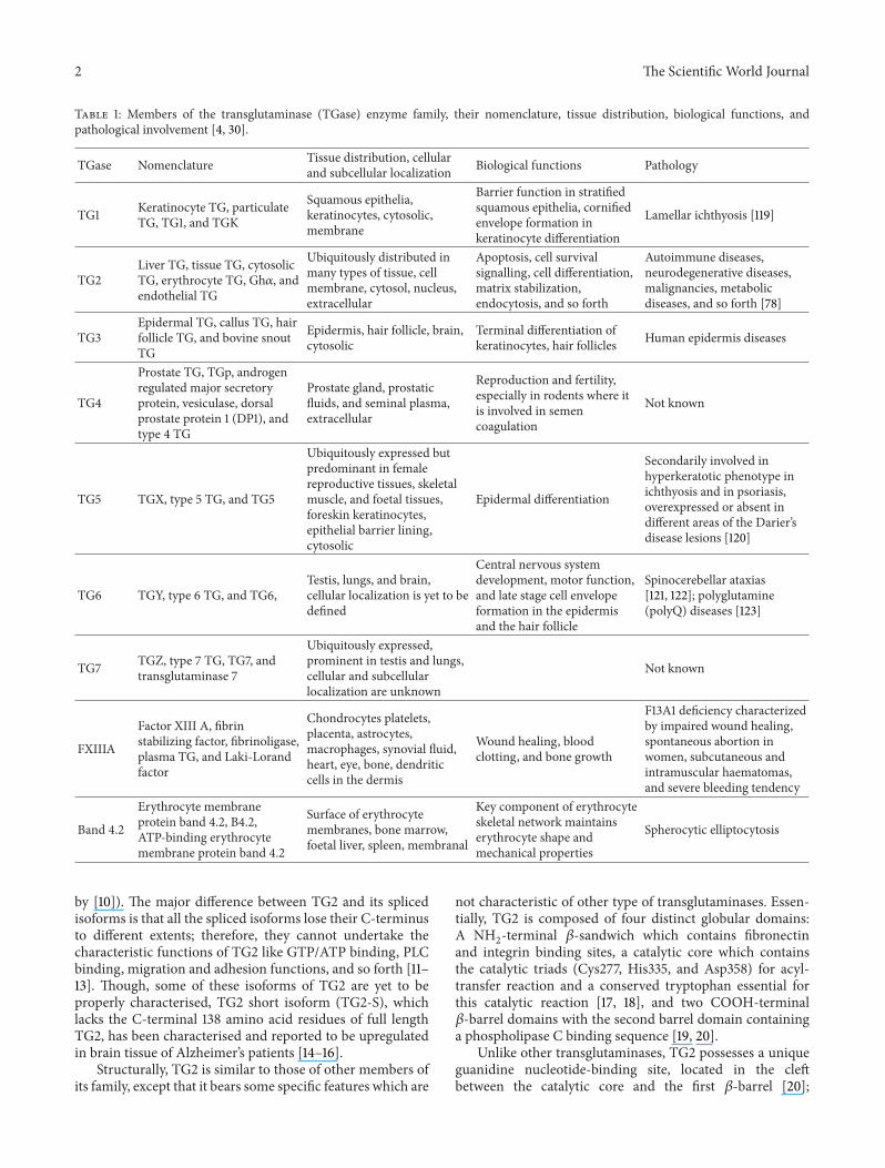
Table 1: Members of the transglutaminase (TGase) enzyme family, their nomenclature, tissue distribution, biological functions, andpathological involvement [4, 30].
TGase Nomenclature Tissue distribution, cellularand subcellular localization Biological functions Pathology
TG1 Keratinocyte TG, particulateTG, TG1, and TGK
Squamous epithelia,keratinocytes, cytosolic,membrane
Barrier function in stratifiedsquamous epithelia, cornifiedenvelope formation inkeratinocyte differentiation
Lamellar ichthyosis [119]
TG2Liver TG, tissue TG, cytosolicTG, erythrocyte TG, Gh𝛼, andendothelial TG
Ubiquitously distributed inmany types of tissue, cellmembrane, cytosol, nucleus,extracellular
Apoptosis, cell survivalsignalling, cell differentiation,matrix stabilization,endocytosis, and so forth
Autoimmune diseases,neurodegenerative diseases,malignancies, metabolicdiseases, and so forth [78]
TG3Epidermal TG, callus TG, hairfollicle TG, and bovine snoutTG
Epidermis, hair follicle, brain,cytosolic
Terminal differentiation ofkeratinocytes, hair follicles Human epidermis diseases
TG4
Prostate TG, TGp, androgenregulated major secretoryprotein, vesiculase, dorsalprostate protein 1 (DP1), andtype 4 TG
Prostate gland, prostaticfluids, and seminal plasma,extracellular
Reproduction and fertility,especially in rodents where itis involved in semencoagulation
Not known
TG5 TGX, type 5 TG, and TG5
Ubiquitously expressed butpredominant in femalereproductive tissues, skeletalmuscle, and foetal tissues,foreskin keratinocytes,epithelial barrier lining,cytosolic
Epidermal differentiation
Secondarily involved inhyperkeratotic phenotype inichthyosis and in psoriasis,overexpressed or absent indifferent areas of the Darier’sdisease lesions [120]
TG6 TGY, type 6 TG, and TG6,Testis, lungs, and brain,cellular localization is yet to bedefined
Central nervous systemdevelopment, motor function,and late stage cell envelopeformation in the epidermisand the hair follicle
Spinocerebellar ataxias[121, 122]; polyglutamine(polyQ) diseases [123]
TG7 TGZ, type 7 TG, TG7, andtransglutaminase 7
Ubiquitously expressed,prominent in testis and lungs,cellular and subcellularlocalization are unknown
Not known
FXIIIA
Factor XIII A, fibrinstabilizing factor, fibrinoligase,plasma TG, and Laki-Lorandfactor
Chondrocytes platelets,placenta, astrocytes,macrophages, synovial fluid,heart, eye, bone, dendriticcells in the dermis
Wound healing, bloodclotting, and bone growth
F13A1 deficiency characterizedby impaired wound healing,spontaneous abortion inwomen, subcutaneous andintramuscular haematomas,and severe bleeding tendency
Band 4.2
Erythrocyte membraneprotein band 4.2, B4.2,ATP-binding erythrocytemembrane protein band 4.2
Surface of erythrocytemembranes, bone marrow,foetal liver, spleen, membranal
Key component of erythrocyteskeletal network maintainserythrocyte shape andmechanical properties
Spherocytic elliptocytosis
by [10]). The major difference between TG2 and its splicedisoforms is that all the spliced isoforms lose their C-terminusto different extents; therefore, they cannot undertake thecharacteristic functions of TG2 like GTP/ATP binding, PLCbinding, migration and adhesion functions, and so forth [11–13]. Though, some of these isoforms of TG2 are yet to beproperly characterised, TG2 short isoform (TG2-S), whichlacks the C-terminal 138 amino acid residues of full lengthTG2, has been characterised and reported to be upregulatedin brain tissue of Alzheimer’s patients [14–16].
Structurally, TG2 is similar to those of other members ofits family, except that it bears some specific features which are
not characteristic of other type of transglutaminases. Essen-tially, TG2 is composed of four distinct globular domains:A NH
2-terminal 𝛽-sandwich which contains fibronectin
and integrin binding sites, a catalytic core which containsthe catalytic triads (Cys277, His335, and Asp358) for acyl-transfer reaction and a conserved tryptophan essential forthis catalytic reaction [17, 18], and two COOH-terminal𝛽-barrel domains with the second barrel domain containinga phospholipase C binding sequence [19, 20].
Unlike other transglutaminases, TG2 possesses a uniqueguanidine nucleotide-binding site, located in the cleftbetween the catalytic core and the first 𝛽-barrel [20];

The Scientific World Journal 3
this sequence is coded by exon 10 of the TG2 gene, whichis characterised by very poor sequence homology with thesame exons in other transglutaminases. Some GDP/GTP-interacting residues and those necessary for GTP hydrolysisare situated in other domains [21]. In the GDP-bound formof TG2, access to the transamidation active site is blocked bytwo loops, and the active site cysteine is attached to a tyrosineresidue by hydrogen bonding. In the latent conformation ofTG2, there is a significant interdomain interaction betweenthe catalytic domain 2 and domains 3 and 4, which reducesthe accessibility of the active centre [20].
Besides the primary transglutaminase enzymes’ activityof catalyzing the calcium-dependent posttranslational mod-ification of proteins, TG2 can also bind and hydrolyze GTP[22], exhibit protein disulphide isomerase activity [23], andfunction as a protein kinase independently of calcium [24].Furthermore, TG2 has calcium-independent nonenzymaticactivities, especially extracellularly, where it interacts witha number of cell surface proteins [25], taking part in celladhesion processes and stabilization of the extracellularmatrix. This catalogue of special activities and multiple func-tionalities is underlined by TG2’s structural uniqueness andcomplexity typified by its complex four-domain structure,constituted of an N-terminal 𝛽-sandwich, a catalytic core,and two C-terminal 𝛽-barrel domains [18]. Regardless ofthe wide range of biological functionalities associated withTG2 activities, amidst its unique cellular biochemistry, itsexact physiological function is still debated. Hence, the viewthat in the event of its absence, the physiological functionof TG2 could be compensated for by another member(s) ofthe transglutaminase family. It is against this background,that we considered the implications of TG2 expression andTG2-specific activities in biological processes. This is donewith a view to rationalizing its involvement in many cellularphysiological events and dissecting the premise that itsfunctional compensation is feasible or unlikely.
2. Transglutaminase 2-SpecificEnzymatic Activities
Catalysis of Ca2+-dependent posttranslational modificationof proteins is the major hallmark of transglutaminaseenzymes. The mechanism of this reaction is generally thesame for TG2 and other members of TG-enzyme familyand involves a two-step process, as recently reviewed inGundemir et al. [26]. The first step is the formation of athioester bond with the enzyme’s active cysteine site via thetransamidation of the 𝛾-carboxamide group of a peptidebond glutamyl substrate, which is accompanied by the releaseof ammonia as a by-product. This is followed by the transferof the acyl intermediate to a nucleophilic substrate, includingthe 𝜀-amino group of a peptide-bound lysine residue. Con-sequently, an intermolecular isopeptide 𝜀-(𝛾-glutamyl)lysinebond is formed, which results in the internal cross-linking ofmonomeric protein units [27]. In transamidation reactions,lysine can be replaced by lower molecular mass amines, suchas polyamines. Hence, in the presence of high concentrationof polyamines, such as spermine, TG2 can form covalent
cross-linking between two polypeptide chains, in the formof a dimer or an adduct as in the case of Gln–Gln or Gln–Gly- [27]. These bonds are resistant to chemical and physicaldegradation; hence, they are believed to be of biologicalsignificance especially in the stabilization of the extracellularmatrix (ECM) [25, 28]. In cellular physiology, the isopeptidebonds formed by TG2 activity have been suggested to befunctionally important in apoptosis, where they preventinflammation by ensuring that the intracellular contents ofdying cells are not released to the extracellular environment[29]. However, water can also act as a nucleophile and causedeamidation of protein-bound glutamine residues [27, 30].The conversion of the acyl-donor glutamine residue to aglutamate residue triggers deamination. Originally, it wasbelieved that the deamination reaction could only take placeunder conditions that do not favour transamidation [26, 31].However, studies have reported the tendency of TG2 tocarry out specific deamination [32], when structural featuresthat favour deamination over transamidation are present inprotein substrates [33].
3. Calcium-Independent NonenzymaticActivities of Transglutaminase 2
In addition to the general hallmark of calcium-dependenttransamidation activity, TG2 has other enzymatic activities,independent of Ca2+ as reviewed in Gundemir et al. [26]. Forinstance, TG2 can function as a protein kinase [24, 34–37],bind and hydrolyze GTP (GTPase and G-protein function)[11, 19, 22, 38–42], and exhibit protein disulfide isomeraseactivity in vitro [23] and in vivo [43–45], independent ofcalcium as reviewed in Belkin [25].
Over the past two decades, other functions of TG2that are independent of its enzymatic activities have beenestablished [46–52]. These calcium-independent, nonenzy-matic and transamidation-independent activities of TG2 areinvolved in many critical physiological processes underlyingmany key aspects of cell behaviour, including cell adhesion,growth, migration, differentiation, programmed cell death,and ECM assembly [49]. In turn, these cellular processesare vital to embryogenesis and tissue morphogenesis, woundhealing, and tissue repair, as well as tumor growth andmetastasis [53].
In 1992,Gentile and colleagues first suggested the involve-ment of transglutaminase 2 in the mediation of extracellularmatrix (ECM) adhesion [54]. They observed an astonishingeffect of TG2 overexpression on the growth of fibroblastsand their increased resistance to trypsinization. Subsequentconvincing proofs, at both the cellular and molecular levels,have supported TG2’s involvement in the mediation ofcellular interactions with the ECM and have demonstratedthat TG2 serves as an adhesion receptor for fibronectin (FN)on the cell surface [46, 55–57].
Transglutaminase 2 has a very high affinity for FN, towhich it has been shown to bind at a stoichiometry of 2 : 1[58], independently of either Ca2+ or the transamidatingand GTPase activities of TG2 [59]. The interaction of TG2
4 The Scientific World Journal
with FN has been shown to mediate ECM adhesion [46]and many other adhesion-dependent phenomena, such ascell migration, matrix assembly, and signalling [60, 61]. Thegelatin-binding domain (42 kD) serves as the only bindingsite for TG2 on FN and binds TG2 with similar affinity as thewhole FN molecule [62]. Additionally, the adhesive functionof TG2 is favored by the fact that the 42 kD gelatin-bindingdomain of FN has no interaction sites for the numerous FN-binding integrins, as well as other FN-associated adhesionreceptors [63]. Therefore, TG2 and integrin can indepen-dently bind distinct domains of FN, hence supporting amodel of cooperation rather than engaging in competition inthe cell adhesion process [49]. It has been shown in differentcell types that the binding of TG2 to the 42 kD fragmentof FN results in stable cell adhesion, limited spreading,and formation of specialized adhesive structures at the cell-substrate interface [56, 60].
Regardless of the coexistence of TG2 and integrin atdifferent FN-binding domains, where they are involved inthe cell adhesion process, TG2 also associates with integrinto maintain cell-extracellular matrix (ECM) interactions.Integrins represent a large class of transmembrane adhesionreceptors constituted by distinct noncovalently associatedand are composed of 𝛼 and 𝛽 subunits [64]. In all celltypes apart from red blood cells, 24 integrin heterodimersconstituted variously of 8 𝛽 subunits and 18 𝛼 subunits areexpressed, serving as receptors for a number of ECM ligandsand facilitate adhesion between cells [64, 65]. The role ofintegrin in wound healing, blood clotting and thrombosis,viral and bacterial infection, inflammation, tumor growth,and angiogenesis, as well as other pathological and physiolog-ical states, exemplifies the fundamental functions of integrinin cell-matrix adhesion [49].
Transglutaminase 2 has been shown to be associatedwith many integrin receptors, by binding to the extracellulardomains of the 𝛽1 and 𝛽3 integrin subunits in differentcell types [46, 56, 60]. The stable, noncovalent TG2-integrincomplexes are formed independently of the transamidat-ing activity of TG2, and there is no evidence of integrinserving as an enzymatic substrate of TG2 or other trans-glutaminases [46]. Furthermore, while performing somebiochemical experiments on cells that do not synthesizeFN, Akimov et al. [46], demonstrated that the TG2-integrininteraction is notmediated by fibronectin but is independent.They further observed that integrin-TG2 complexes have 1 : 1stoichiometry and found that cell-surface TG2 is bound tointegrin receptors, with the possibility of up to 40% of 𝛽1integrin being associated with TG2 in various cell types [46,60]. The ability of TG2 to form ternary adhesive complexeswith various isoforms of integrin and FN, where all thethree proteins successfully interact with each other, highlightsthe importance of TG2 in cell adhesion and indicates anunconventional role of TG2 as a coreceptor in cell-matrixinteractions [46]. The implications of these nonenzymatic,calcium- and transamidation-independent activities in somekey biological events are highlighted below.
4. Implications of Transglutaminase 2 inBiological Events: A Synoptic Update
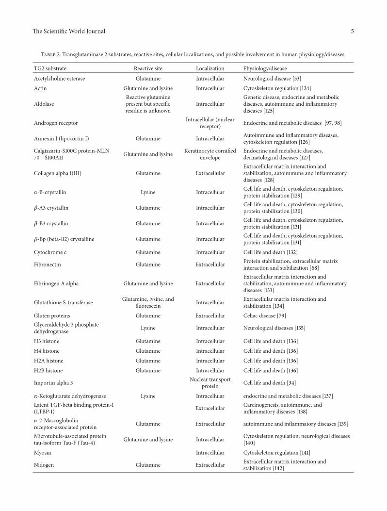
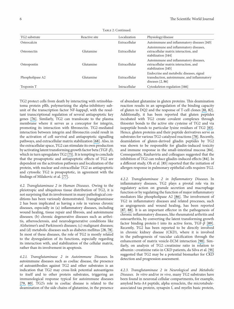
Transglutaminase 2 is a multifunctional protein with over 130substrates at various locations inside and outside the cell [66](Table 2). The broad range of specificity of TG2 for its targetsmay account for its pleiotropic functionality. However, toachieve a particular function necessitates that the selection ofa specific subset of protein substrate related to that particularbiological event must be tightly regulated by additionalfactors. The various physiological implications of TG2 typifythe relationships between its diverse biochemical activitiesand cellular functions and make it difficult to determine theexact role it plays in cell physiology and pathology.
4.1. Transglutaminase 2 in Cell Survival and Death Processes.With regard to cell death and survival, the role of TG2is extremely complex, and for almost three decades it hasremained under investigation, following the first reportof TG2’s involvement in apoptosis [67]. Transglutaminase2’s involvement in apoptosis could be better described asa double-edged sword as it can be both proapoptotic orantiapoptotic. Cells undergoing apoptosis show an increasedlevel of TG2 expression, whichmay prime the cell to undergoapoptosis. Its inhibition results in a decreased propensity ofcells to die by apoptosis [68, 69].
4.1.1. Proapoptotic Activity of TG2. The proapoptotic activityof TG2 is defined by its cross-linking configuration, whichrequires amillimolar concentration of calcium. Stressful con-ditions, such as ultraviolet radiation and chemotherapeuticagents, can generate reactive oxygen species (ROS)—withresultant induction of TG2. Further increase in such stressfulconditions may further trigger the release of Ca2+ from theendoplasmic reticulum (ER), resulting in the activation ofTG2 and extensive cross-linking of intracellular proteins,which, in turn, initiates the apoptotic process [68, 70]. Amajor physiological significance of TG2 involvement in apop-totic initiation is its mediation of the crosstalk between dyingand phagocytic cells to ensure tissue and cellular integrity. Inessence, the focal function of TG2 in apoptosis is to ensurethat, once the apoptotic process is initiated, it is completedwithout inflammation or tissue injury resulting from theprocess [71]. TG2 can achieve maintenance of a cellular envi-ronment devoid of inflammation whilst directly promotingapoptosis in some cell types [72] or indirectly promoting theactivation of TGF-𝛽 released by the macrophages, which canpromote the death of various cells [73, 74], to ensure thatall damaged cells are killed quickly without the occurrenceof necrosis. Additionally, TG2 can promote chemoattractantformation and the release of phosphatidylserine, to, respec-tively, aid macrophage migration to the site of apoptosis andthe recognition of apoptotic cells [71, 75].
4.1.2. Antiapoptotic Activity of TG2. The antiapoptotic effectof TG2 is independent of its transamidation and enzymaticcross-linking activities and does not require calcium. Nuclear
The Scientific World Journal 5
Table 2: Transglutaminase 2 substrates, reactive sites, cellular localizations, and possible involvement in human physiology/diseases.
TG2 substrate Reactive site Localization Physiology/disease
Acetylcholine esterase Glutamine Intracellular Neurological disease [53]
Actin Glutamine and lysine Intracellular Cytoskeleton regulation [124]
AldolaseReactive glutaminepresent but specificresidue is unknown
IntracellularGenetic disease, endocrine and metabolicdiseases, autoimmune and inflammatorydiseases [125]
Androgen receptor Intracellular (nuclearreceptor) Endocrine and metabolic diseases [97, 98]
Annexin I (lipocortin I) Glutamine Intracellular Autoimmune and inflammatory diseases,cytoskeleton regulation [126]
Calgizzarin-S100C protein-MLN70—S100A11 Glutamine and lysine Keratinocyte cornified
envelopeEndocrine and metabolic diseases,dermatological diseases [127]
Collagen alpha 1(III) Glutamine ExtracellularExtracellular matrix interaction andstabilization, autoimmune and inflammatorydiseases [128]
𝛼-B-crystallin Lysine Intracellular Cell life and death, cytoskeleton regulation,protein stabilization [129]
𝛽-A3 crystallin Glutamine Intracellular Cell life and death, cytoskeleton regulation,protein stabilization [130]
𝛽-B3 crystallin Glutamine Intracellular Cell life and death, cytoskeleton regulation,protein stabilization [131]
𝛽-Bp (beta-B2) crystalline Glutamine Intracellular Cell life and death, cytoskeleton regulation,protein stabilization [131]
Cytochrome c Glutamine Intracellular Cell life and death [132]
Fibronectin Glutamine Extracellular Protein stabilization, extracellular matrixinteraction and stabilization [68]
Fibrinogen A alpha Glutamine and lysine ExtracellularExtracellular matrix interaction andstabilization, autoimmune and inflammatorydiseases [133]
Glutathione S-transferase Glutamine, lysine, andfluorescein Intracellular Extracellular matrix interaction and
stabilization [134]
Gluten proteins Glutamine Extracellular Celiac disease [79]Glyceraldehyde 3 phosphatedehydrogenase Lysine Intracellular Neurological diseases [135]
H3 histone Glutamine Intracellular Cell life and death [136]
H4 histone Glutamine Intracellular Cell life and death [136]
H2A histone Glutamine Intracellular Cell life and death [136]
H2B histone Glutamine Intracellular Cell life and death [136]
Importin alpha 3 Nuclear transportprotein Cell life and death [34]
𝛼-Ketoglutarate dehydrogenase Lysine Intracellular endocrine and metabolic diseases [137]Latent TGF-beta binding protein-1(LTBP-1) Extracellular Carcinogenesis, autoimmune, and
inflammatory diseases [138]𝛼-2-Macroglobulinreceptor-associated protein Glutamine Extracellular autoimmune and inflammatory diseases [139]
Microtubule-associated proteintau-isoform Tau-F (Tau-4) Glutamine and lysine Intracellular Cytoskeleton regulation, neurological diseases
[140]
Myosin Intracellular Cytoskeleton regulation [141]
Nidogen Glutamine Extracellular Extracellular matrix interaction andstabilization [142]
6 The Scientific World Journal
Table 2: Continued.
TG2 substrate Reactive site Localization Physiology/diseaseOsteocalcin Extracellular Autoimmune and inflammatory diseases [143]
Osteonectin Glutamine ExtracellularAutoimmune and inflammatory diseases,extracellular matrix interaction, andstabilization [144]
Osteopontin Glutamine ExtracellularAutoimmune and inflammatory diseases,extracellular matrix interaction, andstabilization [145]
Phospholipase A2 Glutamine ExtracellularEndocrine and metabolic diseases, signaltransduction, autoimmune, and inflammatorydiseases [2, 86]
Troponin T Intracellular Cytoskeleton regulation [146]
TG2 protect cells from death by interacting with retinoblas-toma protein pRb, polymerizing the alpha-inhibitory sub-unit of the transcription factor NF-kappa𝛽, with the resul-tant transcriptional regulation of several antiapoptotic keygenes [76]. Similarly, TG2 can translocate to the plasmamembrane where it serves as a coreceptor for integrin,promoting its interaction with fibronectin. TG2-mediatedinteraction between integrin and fibronectin could result inthe activation of cell survival and antiapoptotic signallingpathways, and extracellular matrix stabilization [68]. Also, inthe extracellular space, TG2 can stimulate its own productionby activating latent transforming growth factor beta (TGF-𝛽),which in turn upregulates TG2 [71]. It is tempting to concludethat the proapoptotic and antiapoptotic effects of TG2 aredependent on the activation pathways and localization of theprotein, with nuclear and extracellular TG2 as antiapoptoticand cytosolic TG2 is proapoptotic, in agreement with thefindings of Milakovic et al. [77].
4.2. Transglutaminase 2 in Human Diseases. Owing to thepleiotropic and ubiquitous tissue distribution of TG2, it isnot surprising that its involvement inmany pathological con-ditions has been variously demonstrated. Transglutaminase2 has been implicated as having a role in various chronicdiseases, especially in (a) inflammatory diseases, includingwound healing, tissue repair and fibrosis, and autoimmunediseases; (b) chronic degenerative diseases such as arthri-tis, atherosclerosis, and neurodegenerative conditions likeAlzheimer’s and Parkinson’s diseases; (c) malignant diseases;and (d) metabolic diseases such as diabetes mellitus [28, 78].In most of these diseases, the role of TG2 is mostly relatedto the dysregulation of its functions, especially regardingits interaction with, and stabilization of the cellular matrix,rather than its involvement in apoptosis.
4.2.1. Transglutaminase 2 in Autoimmune Diseases. Inautoimmune diseases such as coeliac disease, the presenceof autoantibodies against TG2 and other substrates is anindication that TG2 may cross-link potential autoantigensto itself and to other protein substrates, triggering animmunological response typical for autoimmune diseases[79, 80]. TG2’s role in coeliac disease is related to thedeamination of the side chains of glutamine, in the presence
of abundant glutamine in gluten proteins. This deaminationreaction results in an upregulation of the binding capacityof gluten to DQ2 and the response of T-cell clones [81, 82].Additionally, it has been reported that gluten peptidesincubated with TG2 create covalent complexes throughthioester bonds to the active site cysteine of TG2 and viaisopeptide bonds to particular lysine residues of TG2 [83].Hence, gluten proteins and their peptide derivatives serve assubstrates for various TG2-catalysed reactions [78]. Recently,deamidation of gluten-derived gliadin peptides by TG2was shown to be responsible for gliadin-induced toxicityand immune response in the small-intestinal mucosa [84].Consequently, Rauhavirta and colleagues suggested that theinhibition of TG2 can reduce gliadin-induced effects [84]. Ina different study, Oh et al. [85] reported that the initiation ofallergen response in pulmonary epithelial cells requires TG2.
4.2.2. Transglutaminase 2 in Inflammatory Diseases. Ininflammatory diseases, TG2 plays a pivotal role via itsregulatory action on granule secretion and macrophagefunction or by regulating the function ofmajor inflammatorymediators like phospholipase A2 [86]. The involvement ofTG2 in inflammatory diseases and related processes, suchas angiogenesis and wound healing, has been reported[87, 88]. It is an important effector in the pathogenesis ofchronic inflammatory diseases, like rheumatoid arthritis andosteoarthritis, by converting the latent transforming growthfactor binding protein-1 into its active form, TGF-𝛽 [89].Recently, TG2 has been reported to be directly involvedin chronic kidney disease (CKD), where it is involvedin the pathogenesis of vascular calcification through theenhancement of matrix vesicle-ECM interaction [90]. Sim-ilarly, on analysis of TG2 : creatinine ratio in relation toalbumin : creatinine ratio in CKD patients, da Silva et al. [91]suggested that TG2 may be a potential biomarker for CKDdetection and progression assessment.
4.2.3. Transglutaminase 2 in Neurological and MetabolicDiseases. In vitro and/or in vivo, many TG2 substrates havebeen found in neuronal cellular compartments, for example,amyloid beta-A4 peptide, alpha synuclein, the microtubule-associated tau protein, synapsin I, and myelin basic protein,
The Scientific World Journal 7
as reviewed by Facchiano et al. [78]. TG2-mediated cross-linking is therefore believed to be implicated in neurode-generative diseases such as Huntington’s, Alzheimer’s, andParkinson’s diseases [79, 92] and in diseases related to neuro-transmitter release [93]. Similarly, the possible involvementof TG2 in neurotransmitter release and related pathologicalconditions, such as that caused by tetanus neurotoxin intoxi-cation, has been reported [94].
The covalent modification of TG2 substrates suchas glyceraldehyde-3-phosphate dehydrogenase (GAPDH),alpha-ketoglutarate dehydrogenase, phosphoglycerate dehy-drogenase, and fatty acid synthase [95], which are involvedin energy metabolism, could account for the role of TG2in metabolic diseases. Additionally, TG2-mediated covalentmodification of hormone receptors or hormone-bindingproteins indicates that TG2-catalysed cross-linking may beinvolved in controlling complex metabolic responses tohormones [96–98].The involvement of TG2 in the regulationof insulin secretion and diabetes mellitus has also beensuggested [99, 100].
4.2.4. Transglutaminase 2 in Cancer. In cancer, transglutami-nase 2 has been shown to play a major role in developmentof drug resistance and metastasis in many cancer types,including pancreatic carcinoma [101], ovarian carcinoma[102, 103], malignant melanoma [104], lung carcinoma [105],glioblastoma [106], and breast carcinoma [107]. When aber-rantly regulated, TG2 could aid tumor cells to evade apoptosisand have direct consequences on cancer drug resistance[108, 109] and metastatic progression [107]. For instance,Park et al. [110] reported that TG2-specific cross-linkingactivity resulted in the polymerization and inhibition ofnucleophosmin and concomitant increase in drug resistancepotential of cancer cells. Recent evidence shows that aberrantexpression of TG2 in mammary epithelial cells confers stemcell characteristics on the cells [111]. Similarly, Kumar andcolleagues reported that high basal expression of TG2 inbreast cancer cells promotes the development of stem cellfeatures but did not mediate their terminal differentiation[111]. Additionally, Caffarel et al. [112] observed that theactivation of TG2: integrin-𝛼5𝛽1 interactions through thestimulation of oncostatin M receptor in cervical squamouscell carcinoma induced promalignant changes.
Clinically, TG2 has been reported to serve as a predictiveindicator of anticancer therapeutic efficacy. For instance,Jeong et al. [113] suggested that TG2 expression is a promisingindicator of the effectiveness of epidermal growth factorreceptor tyrosine kinase inhibitor (EGFR-TKI) therapy inpatients suffering from nonsmall cell lung cancer. Similarly,Jasmeet et al. [114] reported that the accumulation of TG2 intumour stroma can serve as an independent risk factor for theidentification of invasive ductal carcinomas (IDCs) of breastand can establish breast cancer patients at high risk of recur-rence. They also observed that overexpression of TG2 canserve as an indicator of poor prognosis for IDC of the breast.Agnihotri et al. [115] proposed that inflammation-inducedprogression of breast cancer and acquisition of survival andinvasive capabilities by breast cancer cells are mediated by
TG2. In acute myeloid leukaemia, Pierce et al. [116] demon-strated that increased expression of TG2 precipitated a moreadvanced state of the disease in relapse patients.They furtherestablished that increased TG2 expression correlates with theexpression of proteins involved in apoptosis, motility, andextracellular matrix association and processes that have beenlinked with leukemia development and progression. This is atestament to the specialized ability of TG2 to interact withseveral proteins as substrates in various biological events,probably due to the unique biochemical structure of TG2 thatis uncharacteristic of other transglutaminase enzymes.
5. Compensation for Transglutaminase2 Functions
Regardless of the wide range of biological functionalitiesassociated with TG2 and amidst its unique cellular biochem-istry, its exact physiological functions are still debated. Thisdebate is complicated by the fact that homozygous deletion ofTG2 inmice does not result in an embryonic lethal phenotype[117, 118], suggesting that compensation for its absence maybe achieved by other family members. Such knockouts arenot however without associated pathology. For example,Bernassola et al. [100] observed that TG2-deficient micedisplayed significant changes such as characteristic glucoseintolerance and hyperglycaemia due to reduced insulin secre-tion, a condition equivalent to a subtype of diabetes calledmaturity-onset diabetes of the young (MODY). Moreover,a TG2-deficiency disease is yet to be identified in humansimplying the importance of its presence.
This notion is further supported by the observation thatTG2 is relatively more abundant than other members of thetransglutaminase family. Its wide tissue distribution and itspossession of a wide range of structural features that allowsfor flexibility in interaction with widely assorted proteins aresome of the factors that give TG2 a potentially wide range offunctions other than simple enzymatic activity.
Thus while there are increasing suggestions of possiblecompensation for the absence of TG2 by other membersof the transglutaminase family [100], the pleiotropic natureof this protein indicates that TG2 is actually involved inmore physiological processes than any other member of theenzyme family and, thus, while TG2 might feasibly com-pensate for other members of the family the suggestion thatalternative TG isoforms may act as a universal “back up”system for TG2 seems less likely.
From the above, it seems more appropriate to argue thatcompensation for TG2’s functions by other TGase isoformsmight not be possible, except for roles that are determinedby its calcium-dependent cross-linking and transamidatingactivities, which are common features of the transglutami-nase family. For example, TG2-mediated functions that areenzymatic but independent of calcium, such as its role asa G-protein, protein disulphide isomerase activity, kinasefunction, and regulation of energy metabolism, are unlikelyroles to be undertaken by any other member of the transglu-taminase family. This could be due to absence of appropriatestructural conformation in other transglutaminase enzymes
8 The Scientific World Journal
that could enable them to alternately assume such roles in theevent of TG2 absence.
Similarly, TG2-mediated integrin-fibronectin interactionis critical to many physiological events in the cell, includ-ing cell adhesion, growth, migration, differentiation, pro-grammed cell death, and ECM assembly [25, 49]. Suchinteraction is vital to many cellular processes and servesas one of the major routes for extracellular survival, sig-nalling activation, and consequent apoptotic evasion. It isnonenzymatic and independent of TG2 transamidation andcross-linking activities. Consequently, it is unlikely that anyothermember of the transglutaminase family can successfullycompensate for this function in the event of TG2 absence.
6. Conclusions
The abundance of TG2 in various cell types, its specializedstructural conformation, and its broad substrate specificityare some of the key factors justifying the enzyme’s implicationin myriads of biological events. From this review, it is evidentthat besides its calcium-dependent activities, TG2 can enzy-matically or nonenzymatically mediate key cell physiologicalevents. However, it has been increasingly suggested thatin the event of TG2 absence its biological functions couldbe successfully compensated for by other members of thetransglutaminase family. These suggestions have been madewithout recourse to the distinguishing features of TG2 amongthe transglutaminase family. It is necessary to carry outfurther investigations to ascertain the main reasons whyTG2 knockout is not embryonic lethal, instead of relyingon the assertion that its functions are compensated for byother transglutaminase enzymes. Finally, it is also our viewthat a systematic investigation should be carried out toestablish, with certainty, the possibility of and premise forthe replacement of TG2 function by any other member of thetransglutaminase family.
Conflict of Interests
There is no conflict of interests.
References
[1] V. Gentile, P. J. A. Davies, and A. Baldini, “The human tissuetransglutaminase gene maps on chromosome 20q12 by in situfluorescence hybridization,” Genomics, vol. 20, no. 2, pp. 295–297, 1994.
[2] L. Fesus and M. Piacentini, “Transglutaminase 2: an enigmaticenzyme with diverse functions,” Trends in Biochemical Sciences,vol. 27, no. 10, pp. 534–539, 2002.
[3] B. M. Fraij and R. A. Gonzales, “Organization and structureof the human tissue transglutaminase gene,” Biochimica et Bio-physica Acta, vol. 1354, no. 1, pp. 65–71, 1997.
[4] K.Mehta, “Mammalian transglutaminases: a family portrait,” inTransglutaminases: Family of Enzymes with Diverse Functions,K.Mehta and R. Eckert, Eds., vol. 38 of Progress in ExperimentalTumor Research, pp. 1–18, Karger Medical and Scientific Pub-lishers, 2005.
[5] H. Thomas, K. Beck, M. Adamczyk et al., “Transglutaminase 6:a protein associated with central nervous system development
and motor function,” Amino Acids, vol. 44, no. 1, pp. 161–177,2013.
[6] L. Lorand and R.M. Graham, “Transglutaminases: crosslinkingenzymes with pleiotropic functions,” Nature Reviews MolecularCell Biology, vol. 4, no. 2, pp. 140–156, 2003.
[7] B. M. Fraij, P. J. Birckbichler, M. K. Patterson Jr., K. N. Lee, andR. A. Gonzales, “A retinoic acid-inducible mRNA from humanerythroleukemia cells encodes a novel tissue transglutaminasehomologue,”The Journal of Biological Chemistry, vol. 267, no. 31,pp. 22616–22623, 1992.
[8] B. M. Fraij, “GTP hydrolysis by human tissue transglutaminasehomologue,”Biochemical andBiophysical ResearchCommunica-tions, vol. 218, no. 1, pp. 45–49, 1996.
[9] B. M. Fraij and R. A. Gonzales, “A third human tissue transglu-taminase homologue as a result of alternative gene transcripts,”Biochimica et Biophysica Acta, vol. 1306, no. 1, pp. 63–74, 1996.
[10] T.-S. Lai and C. S. Greenberg, “TGM2 and implications forhuman disease: role of alternative splicing,” Frontiers in Bio-science, vol. 18, pp. 504–519, 2013.
[11] M.-J. Im, M. A. Russell, and J.-F. Feng, “TransglutaminaseII: a new class of GTP-Binding protein with new biologicalfunctions,” Cellular Signalling, vol. 9, no. 7, pp. 477–482, 1997.
[12] S. N. P. Murthy, J. W. Lomasney, E. C. Mak, and L. Lorand,“Interactions of Gh/transglutaminase with phospholipase C𝛿1and with GTP,” Proceedings of the National Academy of Sciencesof the United States of America, vol. 96, no. 21, pp. 11815–11819,1999.
[13] T. S. Lai, Y. Liu,W. Li, andC. S. Greenberg, “Identification of twoGTP-independent alternatively spliced forms of tissue transg-lutaminase in human leukocytes, vascular smooth muscle, andendothelial cells,” FASEB Journal, vol. 21, no. 14, pp. 4131–4143,2007.
[14] B. A. Citron, K. S. SantaCruz, P. J. A. Davies, and B. W.Festoff, “Intron-exon swapping of transglutaminase mRNA andneuronal tau aggregation in Alzheimer’s disease,”The Journal ofBiological Chemistry, vol. 276, no. 5, pp. 3295–3301, 2001.
[15] B. A. Citron, Z. Suo, K. SantaCruz, P. J. A. Davies, F. Qin, andB. W. Festoff, “Protein crosslinking, tissue transglutaminase,alternative splicing and neurodegeneration,” NeurochemistryInternational, vol. 40, no. 1, pp. 69–78, 2002.
[16] M. A. Antonyak, J. M. Jansen, A. M. Miller, T. K. Ly, M. Endo,and R. A. Cerione, “Two isoforms of tissue transglutaminasemediate opposing cellular fates,” Proceedings of the NationalAcademy of Sciences of the United States of America, vol. 103, no.49, pp. 18609–18614, 2006.
[17] S. N. P. Murthy, S. Iismaa, G. Begg, D. M. Freymann, R. M.Graham, and L. Lorand, “Conserved tryptophan in the coredomain of transglutaminase is essential for catalytic activity,”Proceedings of the National Academy of Sciences of the UnitedStates of America, vol. 99, no. 5, pp. 2738–2742, 2002.
[18] R. Kiraly, M. A. Demeny, and L. Fesus, “Protein transamidationby transglutaminase 2 in cells: a disputed Ca2+-dependentaction of a multifunctional protein,” FEBS Journal, vol. 278, no.24, pp. 4717–4739, 2011.
[19] K.-C. Hwang, C. D. Gray, N. Sivasubramanian, and M.-J. Im,“Interaction site of GTP binding G(h) (transglutaminase II)with phospholipase C,”The Journal of Biological Chemistry, vol.270, no. 45, pp. 27058–27062, 1995.
[20] S. Liu, R. A. Cerione, and J. Clardy, “Structural basis for theguanine nucleotide-binding activity of tissue transglutaminaseand its regulation of transamidation activity,” Proceedings of
The Scientific World Journal 9
the National Academy of Sciences of the United States of America,vol. 99, no. 5, pp. 2743–2747, 2002.
[21] S. E. Iismaa, M. Wu, N. Nanda, W. B. Church, and R. M. Gra-ham, “GTP binding and signaling by G(h)/transglutaminase IIinvolves distinct residues in a unique GTP-binding pocket,”TheJournal of Biological Chemistry, vol. 275, no. 24, pp. 18259–18265,2000.
[22] H. Nakaoka, D.M. Perez, K. J. Baek et al., “G(h): a GTP-bindingprotein with transglutaminase activity and receptor signalingfunction,” Science, vol. 264, no. 5165, pp. 1593–1596, 1994.
[23] G. Hasegawa, M. Suwa, Y. Ichikawa et al., “A novel functionof tissue-type transglutaminase: protein disulphide isomerase,”Biochemical Journal, vol. 373, no. 3, pp. 793–803, 2003.
[24] S. Mishra and L. J. Murphy, “Tissue transglutaminase hasintrinsic kinase activity. Identification of transglutaminase 2as an insulin-like growth factor-binding protein-3 kinase,” TheJournal of Biological Chemistry, vol. 279, no. 23, pp. 23863–23868, 2004.
[25] A. M. Belkin, “Extracellular TG2: emerging functions andregulation,” FEBS Journal, vol. 278, no. 24, pp. 4704–4716, 2011.
[26] S. Gundemir, G. Colak, J. Tucholski, and G. V. W. Johnson,“Transglutaminase 2: a molecular Swiss army knife,” Biochimicaet Biophysica Acta, vol. 1823, no. 2, pp. 406–419, 2012.
[27] R. Porta, C. Esposito, S. Metafora et al., “Mass spectrometricidentification of the amino donor and acceptor sites in atransglutaminase protein substrate secreted from rat seminalvesicles,” Biochemistry, vol. 30, no. 12, pp. 3114–3120, 1991.
[28] M. Griffin, R. Casadio, and C. M. Bergamini, “Transglutami-nases: nature’s biological glues,” Biochemical Journal, vol. 368,no. 2, pp. 377–396, 2002.
[29] B. Nicholas, P. Smethurst, E. Verderio, R. Jones, and M. Griffin,“Cross-linking of cellular proteins by tissue transglutaminaseduring necrotic cell death: a mechanism for maintaining tissueintegrity,” Biochemical Journal, vol. 371, no. 2, pp. 413–422, 2003.
[30] C. Esposito and I. Caputo, “Mammalian transglutaminases:identification of substrates as a key to physiological functionand physiopathological relevance,” FEBS Journal, vol. 272, no.3, pp. 615–631, 2005.
[31] B. Fleckenstein, Ø. Molberg, S. Qiao et al., “Gliadin T cellepitope selection by tissue transglutaminase in celiac disease.Role of enzyme specificity and pH influence on the transami-dation versus deamidation reactions,” The Journal of BiologicalChemistry, vol. 277, no. 37, pp. 34109–34116, 2002.
[32] J. Stamnaes, B. Fleckenstein, and L. M. Sollid, “The propensityfor deamidation and transamidation of peptides by transg-lutaminase 2 is dependent on substrate affinity and reactionconditions,” Biochimica et Biophysica Acta, vol. 1784, no. 11, pp.1804–1811, 2008.
[33] D. M. Pinkas, P. Strop, A. T. Brunger, and C. Khosla, “Trans-glutaminase 2 undergoes a large conformational change uponactivation,” PLoS Biology, vol. 5, no. 12, article e327, 2007.
[34] T. Kuo, H. Tatsukawa, and S. Kojima, “New insights into thefunctions and localization of nuclear transglutaminase 2,” FEBSJournal, vol. 278, no. 24, pp. 4756–4767, 2011.
[35] S. Mishra, A. Saleh, P. S. Espino, J. R. Davie, and L. J. Murphy,“Phosphorylation of histones by tissue transglutaminase,” TheJournal of Biological Chemistry, vol. 281, no. 9, pp. 5532–5538,2006.
[36] S. Mishra and L. J. Murphy, “The p53 oncoprotein is a substratefor tissue transglutaminase kinase activity,” Biochemical andBiophysical Research Communications, vol. 339, no. 2, pp. 726–730, 2006.
[37] S. Mishra, G. Melino, and L. J. Murphy, “Transglutaminase 2kinase activity facilitates protein kinase A-induced phospho-rylation of retinoblastoma protein,” The Journal of BiologicalChemistry, vol. 282, no. 25, pp. 18108–18115, 2007.
[38] K. E. Achyuthan and C. S. Greenberg, “Identification of aguanosine triphosphate-binding site on guinea pig liver trans-glutaminase. Role of GTP and calcium ions in modulatingactivity,”The Journal of Biological Chemistry, vol. 262, no. 4, pp.1901–1906, 1987.
[39] S. Chen, F. Lin, S. Iismaa, K. N. Lee, P. J. Birckbichler, and R. M.Graham, “𝛼1-adrenergic receptor signaling via G(h) is subtypespecific and independent of its transglutaminase activity,” TheJournal of Biological Chemistry, vol. 271, no. 50, pp. 32385–32391,1996.
[40] J.-F. Feng, S. G. Rhee, and M.-J. Im, “Evidence that phos-pholipase 𝛿1 is the effector in the G(h) (transglutaminase II)-mediated signaling,” The Journal of Biological Chemistry, vol.271, no. 28, pp. 16451–16454, 1996.
[41] K. J. Baek, N. S. Kwon, H. S. Lee, M. S. Kim, P. Muralidhar, andM.-J. Im, “Oxytocin receptor couples to the 80 kDa Ghx familyprotein in human myometrium,” Biochemical Journal, vol. 315,no. 3, pp. 739–744, 1996.
[42] R. Vezza, A. Habib, andG. A. FitzGerald, “Differential signalingby the thromboxane receptor isoforms via the novel GTP-binding protein, G(h),”The Journal of Biological Chemistry, vol.274, no. 18, pp. 12774–12779, 1999.
[43] P. G. Mastroberardino, M. G. Farrace, I. Viti et al., “‘Tissue’transglutaminase contributes to the formation of disulphidebridges in proteins of mitochondrial respiratory complexes,”Biochimica et Biophysica Acta, vol. 1757, no. 9-10, pp. 1357–1365,2006.
[44] Z. Szondy, P. G. Mastroberardino, J. Varadi et al., “Tissue trans-glutaminase (TG2) protects cardiomyocytes against ischemia/reperfusion injury by regulating ATP synthesis,” Cell Death andDifferentiation, vol. 13, no. 10, pp. 1827–1829, 2006.
[45] W. Malorni, M. G. Farrace, P. Matarrese et al., “The adeninenucleotide translocator 1 acts as a type 2 transglutaminase sub-strate: implications for mitochondrial-dependent apoptosis,”Cell Death and Differentiation, vol. 16, no. 11, pp. 1480–1492,2009.
[46] S. S. Akimov, D. Krylov, L. F. Fleischmana, and A. M. Belkin,“Tissue transglutaminase is an integrin-binding adhesion core-ceptor for fibronectin,” Journal of Cell Biology, vol. 148, no. 4, pp.825–838, 2000.
[47] R. Dardik and A. Inbal, “Complex formation between tissuetransglutaminase II (tTG) and vascular endothelial growthfactor receptor 2 (VEGFR-2): proposed mechanism for mod-ulation of endothelial cell response to VEGF,” Experimental CellResearch, vol. 312, no. 16, pp. 2973–2982, 2006.
[48] A. Janiak, E. A. Zemskov, and A. M. Belkin, “Cell surface trans-glutaminase promotes RhoA activation via integrin clusteringand suppression of the Src-p190RhoGAP signaling pathway,”Molecular Biology of the Cell, vol. 17, no. 4, pp. 1606–1619, 2006.
[49] E.A. Zemskov,A. Janiak, J.Hang,A.Waghray, andA.M. Belkin,“The role of tissue transglutaminase in cell-matrix interactions,”Frontiers in Bioscience, vol. 11, no. 1, pp. 1057–1076, 2006.
[50] D. Telci, Z. Wang, X. Li et al., “Fibronectin-tissue transglu-taminase matrix rescues RGD-impaired cell adhesion throughsyndecan-4 and 𝛽1 integrin co-signaling,”The Journal of Biolog-ical Chemistry, vol. 283, no. 30, pp. 20937–20947, 2008.
[51] A. Scarpellini, R. Germack, H. Lortat-Jacob et al., “Heparan sul-fate proteoglycans are receptors for the cell-surface trafficking
10 The Scientific World Journal
and biological activity of transglutaminase-2,” The Journal ofBiological Chemistry, vol. 284, no. 27, pp. 18411–18423, 2009.
[52] E. A. Zemskov, E. Loukinova, I.Mikhailenko, R. A. Coleman,D.K. Strickland, and A. M. Belkin, “Regulation of platelet-derivedgrowth factor receptor function by integrin-associated cellsurface transglutaminase,” The Journal of Biological Chemistry,vol. 284, no. 24, pp. 16693–16703, 2009.
[53] D. Hand, D. Dias, and L. W. Haynes, “Stabilization of collagen-tailed acetylcholinesterase in muscle cells through extracellularanchorage by transglutaminase-catalyzed cross-linking,”Molec-ular and Cellular Biochemistry, vol. 204, no. 1-2, pp. 65–76, 2000.
[54] V. Gentile, V. Thomazy, M. Piacentini, L. Fesus, and P. J.A. Davies, “Expression of tissue transglutaminase in Balb-C3T3 fibroblasts: effects on cellular morphology and adhesion,”Journal of Cell Biology, vol. 119, no. 2, pp. 463–474, 1992.
[55] E. Verderio, B. Nicholas, S. Gross, and M. Griffin, “Regulatedexpression of tissue transglutaminase in Swiss 3T3 fibroblasts:effects on the processing of fibronectin, cell attachment, and celldeath,” Experimental Cell Research, vol. 239, no. 1, pp. 119–138,1998.
[56] A. M. Belkin, S. S. Akimov, L. S. Zaritskaya, B. I. Ratnikov, E. I.Deryugina, and A. Y. Strongin, “Matrix-dependent proteolysisof surface transglutaminase by membrane-type metallopro-teinase regulates cancer cell adhesion and locomotion,” TheJournal of Biological Chemistry, vol. 276, no. 21, pp. 18415–18422,2001.
[57] M. Kabir-Salmani, S. Shiokawa, Y. Akimoto, K. Sakai, K. Sakai,and M. Iwashita, “Tissue transglutaminase at embryo-maternalinterface,” Journal of Clinical Endocrinology and Metabolism,vol. 90, no. 8, pp. 4694–4702, 2005.
[58] E. K. LeMosy, H. P. Erickson,W. F. Beyer et al., “Visualization ofpurified fibronectin transglutaminase macrophage and humanendothelial cell tissue transglutaminase,”The Journal of Biologi-cal Chemistry, vol. 266, pp. 478–483, 1992.
[59] P. M. Turner and L. Lorand, “Complexation of fibronectin withtissue transglutaminase,” Biochemistry, vol. 28, no. 2, pp. 628–635, 1989.
[60] S. S. Akimov and A. M. Belkin, “Cell-surface transglutaminasepromotes fibronectin assembly via interaction with the gelatin-binding domain of fibronectin: a role in TGF-𝛽-dependentmatrix deposition,” Journal of Cell Science, vol. 114, no. 16, pp.2989–3000, 2001.
[61] E. A.M. Verderio, D. Telci, A. Okoye, G.Melino, andM. Griffin,“A novel RGD-independent cell adhesion pathway mediated byfibronectin-bound tissue transglutaminase rescues cells fromanoikis,” The Journal of Biological Chemistry, vol. 278, no. 43,pp. 42604–42614, 2003.
[62] J. T. Radek, J. Jeong, S. N. P. Murthy, K. C. Ingham, andL. Lorand, “Affinity of human erythrocyte transglutaminasefor a 42-kDa gelatin-binding fragment of human plasmafibronectin,” Proceedings of the National Academy of Sciences ofthe United States of America, vol. 90, no. 8, pp. 3152–3156, 1993.
[63] J. Hang, E. A. Zemskov, L. Lorand, and A. M. Belkin, “Identifi-cation of a novel recognition sequence for fibronectinwithin theNH2-terminal 𝛽-sandwich domain of tissue transglutaminase,”The Journal of Biological Chemistry, vol. 280, no. 25, pp. 23675–23683, 2005.
[64] R. O. Hynes, “Integrins: bidirectional, allosteric signalingmachines,” Cell, vol. 110, no. 6, pp. 673–687, 2002.
[65] M. J. Humphries, M. A. Travis, K. Clark, and A. P. Mould,“Mechanisms of integration of cells and extracellular matrices
by integrins,” Biochemical Society Transactions, vol. 32, no. 5, pp.822–825, 2004.
[66] E. Csosz, B.Mesko, and L. Fesus, “Transdabwiki: the interactivetransglutaminase substrate database on web 2.0 surface,”AminoAcids, vol. 36, no. 4, pp. 615–617, 2009.
[67] L. Fesus, V.Thomazy, and A. Falus, “Induction and activation oftiussue transglutaminase during programmed cell death,” TheFEBS Letters, vol. 224, no. 1, pp. 104–108, 1987.
[68] K.Mehta, J. Y. Fok, and L. S.Mangala, “Tissue transglutaminase:frombiological glue to cell survival cues,”Frontiers in Bioscience,vol. 11, no. 1, pp. 173–185, 2006.
[69] A. Verma and K. Mehta, “Tissue transglutaminase-mediatedchemoresistance in cancer cells,” Drug Resistance Updates, vol.10, no. 4-5, pp. 144–151, 2007.
[70] L. S. Mangala and K. Mehta, “Tissue transglutaminase (TG2) incancer biology,” Progress in Experimental Tumor Research, vol.38, pp. 125–138, 2005.
[71] L. Fesus and Z. Szondy, “Transglutaminase 2 in the balance ofcell death and survival,” The FEBS Letters, vol. 579, no. 15, pp.3297–3302, 2005.
[72] S. Oliverio, A. Amendola, C. Rodolfo, A. Spinedi, and M.Piacentini, “Inhibition of “tissue” transglutaminase increasescell survival by preventing apoptosis,” The Journal of BiologicalChemistry, vol. 274, no. 48, pp. 34123–34128, 1999.
[73] Z. Szondy, Z. Sarang, P. Molnar et al., “Transglutaminase 2-/- mice reveal a phagocytosis-associated crosstalk betweenmacrophages and apoptotic cells,” Proceedings of the NationalAcademy of Sciences of the United States of America, vol. 100, no.13, pp. 7812–7817, 2003.
[74] X. Huang and C. Lee, “From TGF-𝛽 to cancer therapy,” CurrentDrug Targets, vol. 4, no. 3, pp. 243–250, 2003.
[75] H. Nishiura, Y. Shibuya, and T. Yamamoto, “S19 ribosomal pro-tein cross-linked dimer causes monocyte-predominant infil-tration by means of molecular mimicry to complement C5a,”Laboratory Investigation, vol. 78, no. 12, pp. 1615–1623, 1998.
[76] J. E. Boehm, U. Singh, C. Combs, M. A. Antonyak, and R. A.Cerione, “Tissue transglutaminase protects against apoptosis bymodifying the tumor suppressor protein p110 Rb,” The Journalof Biological Chemistry, vol. 277, no. 23, pp. 20127–20130, 2002.
[77] T. Milakovic, J. Tucholski, E. McCoy, and G. V. W. Johnson,“Intracellular localization and activity state of tissue transg-lutaminase differentially impacts cell death,” The Journal ofBiological Chemistry, vol. 279, no. 10, pp. 8715–8722, 2004.
[78] F. Facchiano, A. Facchiano, and A. M. Facchiano, “The roleof transglutaminase-2 and its substrates in human diseases,”Frontiers in Bioscience, vol. 11, no. 2, pp. 1758–1773, 2006.
[79] S. Y. Kim, T. M. Jeitner, and P. M. Steinert, “Transglutaminasesin disease,” Neurochemistry International, vol. 40, no. 1, pp. 85–103, 2002.
[80] L. M. Sollid, O. Molberg, S. Mcadam, and K. E. A. Lundin,“Autoantibodies in coeliac disease: tissue transglutaminase guiltby association?” Gut, vol. 41, no. 6, pp. 851–852, 1997.
[81] H. Quarsten, O. Molberg, L. Fugger, S. N. McAdam, and L.M. Sollid, “HLA binding and T cell recognition of a tissuetransglutaminase-modified gliadin epitope,” European Journalof Immunology, vol. 29, no. 8, pp. 2506–2514, 1999.
[82] H. Arentz-Hansen, R. Korner, Ø. Molberg et al., “The intestinalT cell response to 𝛼-gliadin in adult celiac disease is focusedon a single deamidated glutamine targeted by tissue transglu-taminase,” Journal of Experimental Medicine, vol. 191, no. 4, pp.603–612, 2000.
The Scientific World Journal 11
[83] B. Fleckenstein, S. Qiao, M. R. Larsen, G. Jung, P. Roepstorff,and L. M. Sollid, “Molecular characterization of covalent com-plexes between tissue transglutaminase and gliadin peptides,”The Journal of Biological Chemistry, vol. 279, no. 17, pp. 17607–17616, 2004.
[84] T. Rauhavirta, M. Oittinen, R. Kivisto et al., “Are transglutami-nase 2 inhibitors able to reduce gliadin-induced toxicity relatedto celiac disease? A proof-of-concept study,” Journal of ClinicalImmunology, vol. 33, no. 1, pp. 134–142, 2013.
[85] K. Oh,M.W. Seo, G. Y. Lee et al., “Airway epithelial cells initiatethe allergen response through transglutaminase 2 by inducingIL-33 expression and a subsequent Th2 response,” RespiratoryResearch, vol. 14, article 35, 2013.
[86] E. Cordella-Miele, L. Miele, and A. B. Mukherjee, “A noveltransglutaminase-mediated post-translational modification ofphospholipase A2 dramatically increases its catalytic activity,”The Journal of Biological Chemistry, vol. 265, no. 28, pp. 17180–17188, 1990.
[87] J. Sohn, T. Kim, Y. Yoon, J. Kim, and S. Kim, “Novel trans-glutaminase inhibitors reverse the inflammation of allergicconjunctivitis,” Journal of Clinical Investigation, vol. 111, no. 1,pp. 121–128, 2003.
[88] E. A. M. Verderio, T. S. Johnson, and M. Griffin, “Transglu-taminases in wound healing and inflammation,” Progress inExperimental Tumor Research, vol. 38, pp. 89–114, 2005.
[89] I. Nunes, P. Gleizes, C. N. Metz, and D. B. Rifkin, “Latent trans-forming growth factor-𝛽 binding protein domains involvedin activation and transglutaminase-dependent cross-linking oflatent transforming growth factor-𝛽,” Journal of Cell Biology,vol. 136, no. 5, pp. 1151–1163, 1997.
[90] N. X. Chen, K. O’Neill, X. Chen, K. Kiattisunthorn, V. H.Gattone, and S. M. Moe, “Transglutaminase 2 acceleratesvascular calcification in chronic kidney disease,”The AmericanJournal of Nephrology, vol. 37, pp. 191–198, 2013.
[91] M. L. da Silva, M. El Nahas, and T. S. Johnson, “Urinary transg-lutaminase 2 as a potential biomarker of chronic kidney diseasedetection and progression,” The Lancet, vol. 381, supplement 1,article S33, 2013.
[92] C. D. Bailey, J. Tucholski, and G. V. W. Johnson, “Transglutami-nases in neurodegenerative disorders,” Progress in ExperimentalTumor Research, vol. 38, pp. 139–157, 2005.
[93] F. Deloye, F. Doussau, and B. Poulain, “Action mechanismsof botulinum neurotoxins and tetanus neurotoxins,” ComptesRendus des Seances de la Societe de Biologie et de ses Filiales, vol.191, no. 3, pp. 433–450, 1997.
[94] F. Facchiano and A. Luini, “Tetanus toxin potently stimulatestissue transglutaminase. A possible mechanism of neurotoxi-city,” The Journal of Biological Chemistry, vol. 267, no. 19, pp.13267–13271, 1992.
[95] S. Orru, I. Caputo, A. D’Amato, M. Ruoppololl, and C. Esposito,“Proteomics identification of acyl-acceptor and acyl-donorsubstrates for transglutaminase in a human intestinal epithelialcell line. Implications for celiac disease,”TheJournal of BiologicalChemistry, vol. 278, no. 34, pp. 31766–31773, 2003.
[96] K. Sakai, W. H. Busby Jr., J. B. Clarke, and D. R. Clem-mons, “Tissue transglutaminase facilitates the polymerizationof insulin-like growth factor-binding protein-1 (IGFBP-1) andleads to loss of IGFBP-1’s ability to inhibit insulin-like growthfactor-I-stimulated protein synthesis,” The Journal of BiologicalChemistry, vol. 276, no. 12, pp. 8740–8745, 2001.
[97] L. M. Mandrusiak, L. K. Beitel, X. Wang et al., “Transglu-taminase potentiates ligand-dependent proteasome dysfunc-tion induced by polyglutamine-expanded androgen receptor,”Human Molecular Genetics, vol. 12, no. 13, pp. 1497–1506, 2003.
[98] L. M. Mandrusiak, L. K. Beitel, X. Wang et al., “Transglu-taminase potentiates ligand-dependent proteasome dysfunc-tion induced by polyglutamine-expanded androgen receptor,”Human Molecular Genetics, vol. 12, no. 13, pp. 1497–1506, 2003.
[99] P. J. Bungay, J. M. Potter, and M. Griffin, “The inhibition ofglucose-stimulated insulin secretion by primary amines. A rolefor transglutaminase in the secretory mechanism,” BiochemicalJournal, vol. 219, no. 3, pp. 819–827, 1984.
[100] F. Bernassola, M. Federiciz, M. Corazzari et al., “Role oftransglutaminase 2 in glucose tolerance: knockout mice studiesand a putative mutation in a MODY patient,” FASEB Journal,vol. 16, no. 11, pp. 1371–1378, 2002.
[101] A. Verma, H. Wang, B. Manavathi et al., “Increased expressionof tissue transglutaminase in pancreatic ductal adenocarcinomaand its implications in drug resistance and metastasis,” CancerResearch, vol. 66, no. 21, pp. 10525–10533, 2006.
[102] M. Satpathy, L. Cao, R. Pincheira et al., “Enhanced peritonealovarian tumor dissemination by tissue transglutaminase,” Can-cer Research, vol. 67, no. 15, pp. 7194–7202, 2007.
[103] J. Y. Hwang, L. S. Mangala, J. Y. Fok et al., “Clinical andbiological significance of tissue transglutaminase in ovariancarcinoma,” Cancer Research, vol. 68, no. 14, pp. 5849–5858,2008.
[104] J. Y. Kok, S. Ekmekcioglu, and K. Mehta, “Implications of tissuetransglutaminase expression in malignant melanoma,”Molecu-lar Cancer Therapeutics, vol. 5, no. 6, pp. 1493–1503, 2006.
[105] K. S. Park,H.Kim, J. Lee et al., “Transglutaminase 2 as a cisplatinresistance marker in non-small cell lung cancer,” Journal ofCancer Research and Clinical Oncology, vol. 136, no. 4, pp. 493–502, 2010.
[106] L. Yuan, M. Siegel, K. Choi et al., “Transglutaminase 2 inhibitor,KCC009, disrupts fibronectin assembly in the extracellularmatrix and sensitizes orthotopic glioblastomas to chemother-apy,” Oncogene, vol. 26, no. 18, pp. 2563–2573, 2007.
[107] K. Mehta, J. Fok, F. R. Miller, D. Koul, and A. A. Sahin,“Prognostic significance of tissue transglutaminase in drugresistant andmetastatic breast cancer,”Clinical Cancer Research,vol. 10, no. 23, pp. 8068–8076, 2004.
[108] K. Mehta, “High levels of transglutaminase expression indoxorubicin-resistant human breast carcinoma cells,” Interna-tional Journal of Cancer, vol. 58, no. 3, pp. 400–406, 1994.
[109] J. S. K. Chen, N. Agarwal, and K. Mehta, “Multidrug-resistantMCF-7 breast cancer cells contain deficient intracellular cal-cium pools,” Breast Cancer Research and Treatment, vol. 71, no.3, pp. 237–247, 2002.
[110] K. Park, B. Han, K. H. Lee et al., “Depletion of nucleophosminvia transglutaminase 2 cross-linking increases drug resistancein cancer cells,”Cancer Letters, vol. 274, no. 2, pp. 201–207, 2009.
[111] A. Kumar, H. Gao, J. Xu, J. Reuben, D. Yu, and K. Mehta,“Evidence that aberrant expression of tissue transglutaminasepromotes stem cell characteristics in mammary epithelial cells,”PLoS ONE, vol. 6, no. 6, Article ID e20701, 2011.
[112] M. M. Caffarel, A. Chattopadhyay, A. M. Araujo, J. Bauer,C. G. Scarpini, and N. Coleman, “Tissue transglutaminasemediates the pro-malignant effects of oncostatin M receptorover-expression in cervical squamous cell carcinoma,” Journalof Pathology, vol. 231, no. 2, pp. 168–179, 2013.

12 The Scientific World Journal
[113] J. H. Jeong, B. C. Cho, H. S. Shim et al., “Transglutaminase2 expression predicts progression free survival in non-smallcell lung cancer patients treated with epidermal growth factorreceptor tyrosine kinase inhibitor,” Journal of Korean MedicalScience, vol. 28, no. 7, pp. 1005–1014, 2013.
[114] A. Jasmeet, G. Srivastava, A. Matta, M. C. Chang, P. G. Walfish,and R. Ralhan, “Transglutaminase 2 overexpression in tumorstroma identifies invasive ductal carcinomas of breast at highrisk of recurrence,” PLoS ONE, vol. 8, no. 9, Article ID e74437,2013.
[115] N.Agnihotri, S. Kumar, andK.Mehta, “Tissue transglutaminaseas a central mediator in inflammatory-induced progression ofbreast cancer,” Breast Cancer Research, vol. 15, article 202, 2013.
[116] A. Pierce, A. D. Whetton, S. Meyer et al., “Transglutaminase2 expression in acute myeloid leukemia: association withadhesion molecule expression and leukemic blast motility,”Proteomics, vol. 13, no. 14, pp. 2216–2224, 2013.
[117] V. de Laurenzi and G. Melino, “Gene disruption of tissuetransglutaminase,” Molecular and Cellular Biology, vol. 21, no.1, pp. 148–155, 2001.
[118] N. Nanda, S. E. Iismaa, W. A. Owens, A. Husain, F. Mackay,and R. M. Graham, “Targeted inactivation of Gh/tissue transg-lutaminase II,”The Journal of Biological Chemistry, vol. 276, no.23, pp. 20673–20678, 2001.
[119] E. Candi, G. Melino, A. Lahm et al., “Transglutaminase 1mutations in lamellar ichthyosis: loss of activity due to failureof activation by proteolytic processing,”The Journal of BiologicalChemistry, vol. 273, no. 22, pp. 13693–13702, 1998.
[120] E. Candi, S. Oddi, A. Paradisi et al., “Expression of transglutam-inase 5 in normal and pathologic human epidermis,” Journal ofInvestigative Dermatology, vol. 119, no. 3, pp. 670–677, 2002.
[121] J. L. Wang, X. Yang, K. Xia et al., “TGM6 identified as anovel causative gene of spinocerebellar ataxias using exomesequencing,” Brain, vol. 133, part 12, pp. 3510–3518, 2010.
[122] A. Sailer and H. Houlden, “Recent advances in the genetics ofcerebellar ataxias,”Current Neurology andNeuroscience Reports,vol. 12, no. 3, pp. 227–236, 2012.
[123] W. J. Guan, K. D. Xia, Y. T. Ma et al., “Transglutaminase6 interacts with polyQ proteins and promotes the formationof polyQ aggregates,” Biochemical and Biophysical ResearchCommunications, vol. 437, no. 1, pp. 94–100, 2013.
[124] Z. Nemes Jr., R. Adany, M. Balazs, P. Boross, and L. Fesus,“Identification of cytoplasmic actin as an abundant glutaminylsubstrate for tissue transglutaminase in HL-60 and U937 cellsundergoing apoptosis,”The Journal of Biological Chemistry, vol.272, no. 33, pp. 20577–20583, 1997.
[125] K. N. Lee, M. D. Maxwell, M. K. Patterson Jr., P. J. Birckbichler,and E. Conway, “Identification of transglutaminase substratesin HT29 colon cancer cells: use of 5-(biotinamido)pentylamineas a transglutaminase-specific probe,” Biochimica et BiophysicaActa, vol. 1136, no. 1, pp. 12–16, 1992.
[126] Y. Ando, S. Imamura, M. K. Owada, and R. Kannagi, “Calcium-induced intracellular cross-linking of lipocortin I by tissuetransglutaminase in A431 cells. Augmentation by membranephospholipids,”The Journal of Biological Chemistry, vol. 266, no.2, pp. 1101–1108, 1991.
[127] N. A. Robinson and R. L. Eckert, “Identification oftransglutaminase-reactive residues in S100Al1,” The Journal ofBiological Chemistry, vol. 273, no. 5, pp. 2721–2728, 1998.
[128] J. M. Orban, L. B. Wilson, J. A. Kofroth, M. S. El-Kurdi, T.M. Maul, and D. A. Vorp, “Crosslinking of collagen gels by
transglutaminase,” Journal of Biomedical Materials Research A,vol. 68, no. 4, pp. 756–762, 2004.
[129] P. J. T. A. Groenen, H. Bloemendal, and W. W. de Jong,“The carboxy-terminal lysine of 𝛼B-crystallin is an amine-donor substrate for tissue transglutaminase,” European Journalof Biochemistry, vol. 205, no. 2, pp. 671–674, 1992.
[130] P. J. T. A. Groenen, J. J. Grootjans, N. H. Lubsen, H. Bloemendal,andW.W. de Jong, “Lys-17 is the amine-donor substrate site fortransglutaminase in 𝛽A3-crystallin,” The Journal of BiologicalChemistry, vol. 269, no. 2, pp. 831–833, 1994.
[131] G. A. M. Berbers, R. W. Feenstra, R. van den Bos, W. A.Hoekman, H. Bloemendal, and W. W. de Jong, “Lens transg-lutaminase selects specific 𝛽-crystallin sequences as substrate,”Proceedings of the National Academy of Sciences of the UnitedStates of America, vol. 81, no. 22, pp. 7017–7020, 1984.
[132] S. J. Butler andM. Landon, “Transglutaminase-catalysed incor-poration of putrescine into denatured cytochrome. Preparationof a mono-substituted derivative reactive with cytochrome coxidase,” Biochimica et Biophysica Acta, vol. 670, no. 2, pp. 214–221, 1981.
[133] S. N. P. Murthy, J. H.Wilson, T. J. Lukas, Y. Veklich, J. W.Weisel,and L. Lorand, “Transglutaminase-catalyzed crosslinking of theA𝛼 and 𝛾 constituent chains in fibrinogen,” Proceedings of theNational Academy of Sciences of the United States of America,vol. 97, no. 1, pp. 44–48, 2000.
[134] J. van denAkker, E. VanBavel, R. vanGeel et al., “The redox stateof transglutaminase 2 controls arterial remodeling,” PLoS ONE,vol. 6, no. 8, Article ID e23067, 2011.
[135] S.Orru,M. Ruoppolo, S. Francese, L. Vitagliano,G.Marino, andC. Esposito, “Identification of tissue transglutaminase-reactivelysine residues in glyceraldehyde-3-phosphate dehydrogenase,”Protein Science, vol. 11, no. 1, pp. 137–146, 2002.
[136] E. Ballestar, C. Abad, and L. Franco, “Core histones are glu-taminyl substrates for tissue transglutaminase,” The Journal ofBiological Chemistry, vol. 271, no. 31, pp. 18817–18824, 1996.
[137] A. J. L. Cooper, K. R. Sheu, J. R. Burke et al., “Transglutaminase-catalyzed inactivation of glyceraldehyde 3-phosphate dehydro-genase and 𝛼-ketoglutarate dehydrogenase complex by polyg-lutamine domains of pathological length,” Proceedings of theNational Academy of Sciences of the United States of America,vol. 94, no. 23, pp. 12604–12609, 1997.
[138] E. Verderio, C. Gaudry, S. Gross, C. Smith, S. Downes, andM. Griffin, “Regulation of cell surface tissue transglutaminase:effects onmatrix storage of latent transforming growth factor-𝛽binding protein-1,” Journal of Histochemistry andCytochemistry,vol. 47, no. 11, pp. 1417–1432, 1999.
[139] L. K. Rasmussen, L. Ellgaard, P. H. Jensen, and E. S. Sørensen,“Localization of a single transglutaminase-reactive glutaminein the third domain of RAP, the 𝛼2-macroglobulin receptor-associated protein,” Journal of Protein Chemistry, vol. 18, no. 1,pp. 69–73, 1999.
[140] S. N. P. Murthy, J. H. Wilson, T. J. Lukas, J. Kuret, andL. Lorand, “Cross-linking sites of the human tau protein,probed by reactions with human transglutaminase,” Journal ofNeurochemistry, vol. 71, no. 6, pp. 2607–2614, 1998.
[141] L. Eligula, L. Chuang, M. L. Phillips, M. Motoki, K. Seguro, andA.Muhlrad, “Transglutaminase-induced cross-linking betweensubdomain 2 of g-actin and the 636–642 lysine-rich loop ofmyosin subfragment 1,” Biophysical Journal, vol. 74, no. 2, pp.953–963, 1998.
The Scientific World Journal 13
[142] D. Aeschlimann, M. Paulsson, and K. Mann, “Identification ofGln726 in nidogen as the amine acceptor in transglutaminase-catalyzed cross-linking of laminin-nidogen complexes,” TheJournal of Biological Chemistry, vol. 267, no. 16, pp. 11316–11321,1992.
[143] M. T. Kaartinen, A. Pirhonen, A. Linnala-Kankkunen, and P. H.Maenpaa, “Transglutaminase-catalyzed cross-linking of osteo-pontin is inhibited by osteocalcin,” The Journal of BiologicalChemistry, vol. 272, no. 36, pp. 22736–22741, 1997.
[144] D. Aeschlimann, O. Kaupp, and M. Paulsson, “Transglutam-inase-catalyzedmatrix cross-linking in differentiating cartilage:identification of osteonectin as a major glutaminyl substrate,”Journal of Cell Biology, vol. 129, no. 3, pp. 881–892, 1995.
[145] M. T. Kaartinen, S. El-Maadawy, N. H. Rasanen, and M. D.McKee, “Tissue transglutaminase and its substrates in bone,”Journal of Bone and Mineral Research, vol. 17, no. 12, pp. 2161–2173, 2002.
[146] L. Gorza, R. Menabo, M. Vitadello, C. M. Bergamini, and F. DiLisa, “Cardiomyocyte troponin T immunoreactivity ismodifiedby cross-linking resulting from intracellular calcium overload,”Circulation, vol. 93, no. 10, pp. 1896–1904, 1996.

Submit your manuscripts athttp://www.hindawi.com
Hindawi Publishing Corporationhttp://www.hindawi.com Volume 2014
Anatomy Research International
PeptidesInternational Journal of
Hindawi Publishing Corporationhttp://www.hindawi.com Volume 2014
Hindawi Publishing Corporation http://www.hindawi.com
International Journal of
Volume 2014
Zoology
Hindawi Publishing Corporationhttp://www.hindawi.com Volume 2014
Molecular Biology International
Hindawi Publishing Corporationhttp://www.hindawi.com
GenomicsInternational Journal of
Volume 2014
The Scientific World JournalHindawi Publishing Corporation http://www.hindawi.com Volume 2014
Hindawi Publishing Corporationhttp://www.hindawi.com Volume 2014
BioinformaticsAdvances in
Marine BiologyJournal of
Hindawi Publishing Corporationhttp://www.hindawi.com Volume 2014
Hindawi Publishing Corporationhttp://www.hindawi.com Volume 2014
Signal TransductionJournal of
BioMed Research International
Hindawi Publishing Corporationhttp://www.hindawi.com Volume 2014
Evolutionary BiologyInternational Journal of
Hindawi Publishing Corporationhttp://www.hindawi.com Volume 2014
Hindawi Publishing Corporationhttp://www.hindawi.com Volume 2014
Biochemistry Research International
ArchaeaHindawi Publishing Corporationhttp://www.hindawi.com Volume 2014
Hindawi Publishing Corporationhttp://www.hindawi.com Volume 2014
Genetics Research International
Hindawi Publishing Corporationhttp://www.hindawi.com Volume 2014
Advances in
Virolog y
Hindawi Publishing Corporationhttp://www.hindawi.com
Nucleic AcidsJournal of
Volume 2014
Stem CellsInternational
Hindawi Publishing Corporationhttp://www.hindawi.com Volume 2014
Hindawi Publishing Corporationhttp://www.hindawi.com Volume 2014
Enzyme Research
Hindawi Publishing Corporationhttp://www.hindawi.com Volume 2014
International Journal of
Microbiology