Strong Truncation Approximation in Tandem Queues with Blocking
Upload
independentCategory
view
0download
0
pubs.acs.org/JAFC Published on Web 10/08/2009 © 2009 American Chemical Society
10150 J. Agric. Food Chem. 2009, 57, 10150–10155
DOI:10.1021/jf902268z
Blocking Peptides Decrease Tissue TransglutaminaseProcessing of Gliadin in Vitro
KAROLINA HOFFMANN,*,† MARIE ALMINGER,† THOMAS ANDLID,† TINGSU CHEN,‡, )
OLOF OLSSON,§ AND ANN-SOFIE SANDBERG†
†Department of Chemical and Biological Engineering/Food Science, Chalmers University of Technology,SE-41296 G
::oteborg, Sweden, ‡Department of Cell and Molecular Biology, Gothenburg University,
SE-40530 G::oteborg, Sweden, and §Department of Plant and Environmental Sciences, Gothenburg
University, SE-40530 G::oteborg, Sweden. )Current address: Department of Pharmacology,
Medical School, Academic Health Center, 55455 Minneapolis, MN.
Tissue transglutaminase (tTG) plays an important role in celiac disease pathology as it catalyzes
deamidation and cross-linking of specific gluten peptides and converts them into potent epitopes
recognized by intestinal T-cells. We investigated whether synthetic peptides with high affinity to
gliadin could alter tTG activity on gliadin and whole gluten digest. The immobilized substrates were
incubated with synthetic peptides identified by the phage display technique and a control peptide
with no affinity to gliadin. Transglutaminase activity was measured with time resolved fluorescence.
The mean tTG activity, compared to that of the control without the peptides, was reduced by 31, 33,
and 36% for three selected gliadin-binding peptides, and 30% for the peptide pool (P e 0.001-0.004) when gliadin was the substrate. Finally, substrate specificity experiments suggested that
avenin was processed in a manner similar that used for gliadin during in vitro assays with tTG. The
results showed that the blocking peptides efficiently reduced tTG processing of gliadin in vitro, and
this strategy will be further investigated as an alternative therapy for celiac disease.
KEYWORDS: Celiac disease; gliadin; synthetic peptides; time resolved fluorescence; tissue transglut-aminase
INTRODUCTION
Celiac disease (CD) is a complex inflammatory condition thatis diagnosed in approximately 1% of European and Americanpopulations (1, 2). Clinical presentation varies from asympto-matic to severe gastrointestinal complaints including abdominalpain, diarrhea,weight loss, andmalabsorption related conditions.Histologically, the disease is presented with infiltration of intrae-pithelial lymphocytes into the intestinal mucosa, which leads tolesions of the small intestine with villous atrophy and cryptshyperplasia. Symptoms develop because of an inadequate im-mune response and include antibody production and cytotoxicreaction to specific peptides originating from ingested wheatgluten as well as proteins from related cereals (3-6).
Wheat gluten proteins, gliadins and glutenins, have a uniqueamino acid composition with high content of proline ((15%),hydrophobic amino acids ((19%), and glutamine ((35%).Moreover, they contain domains with numerous repetitive se-quences rich in those amino acids (7, 8). Because of this gluta-mine- and proline-rich structure, gluten proteins are resistantto complete digestion by gastric and pancreatic human pepti-dases (9, 10).
Tissue transglutaminase (tTG) is an enzyme (EC2.3.2.13) fromthe transglutaminase family of calcium dependent acyltrans-ferases (11). It can be found in extracellular compartments ofthe subepithelial region of the small intestine where it catalyzesthe formation of intermolecular, covalent cross-linking betweenprimary amines or proteins (12). During the first stage of thisreaction, a thiol ester is formed between a glutamine residue(an acyl-donor) from a gluten peptide and a cystine residue fromthe tTG active site, and ammonia is released as a reactionbyproduct. During the second stage of the reaction, the acylgroup is transferred to the acyl acceptor (often a lysine residue),and an isopeptide bond is formed. If the lysine residue is notpresent, tTGdeamidates glutamine to glutamic acid by catalyzingonly the first reaction stage after which the thiol ester is hydro-lyzed (12, 13).
The tTG enzyme has been identified as an autoantigen in CDand is now considered to play an important role during diseasepathology as it is targeted by highly specific antiendomysialantibodies, and is also involved in the generation of epitopesfor intestinal T-cells (12, 14-16). Gluten peptides that cross theintestinal mucosa barrier are preferentially deamidated by tTG,which leads to enhanced stimulation ofCD4þT-cells and triggersa cascade of immune responses that result in mucosal inflamma-tion (12). It has also been hypothesized that tTG can catalyze thecross-linking of gluten peptides with themucosal matrix proteins,which could result in a longer exposure of these antigens in themucosa of the small intestine. Moreover, high molecular weight
*To whom correspondence should be addressed. Department ofChemical and Biological Engineering/Food Science, Chalmers Uni-versity of Technology, SE-412 96, G
::oteborg, Sweden. Tel: þ46 31-
772-38-21. Fax: þ46 31-772-3830. E-mail: [email protected].
Article J. Agric. Food Chem., Vol. 57, No. 21, 2009 10151
complexes of tTG-gluten are targeted by disease specific IgAantibodies (14, 17).
So far, the only treatment available for CDpatients is a gluten-free diet. However, the abundance of gluten in several nonbakeryfood products as well as contamination of various foods withgluten during production processes makes the diet difficult tofollow (18, 19). This is why new complementary therapies allow-ing ingestion of low amounts of gluten are highly needed.Reduction of tTG-catalyzed gluten modification could possiblyhinder the activation of the immune system and minimize thedevelopment of inflammation. To test this hypothesis, in thepresent work we asked whether the tTG activity on gluten couldbe changed by blocking gluten sequences with synthetic peptidesselected for high affinity to wheat gliadin.
The effect of three blocking peptides on tTG-based intactgliadin modification was investigated in this study. Moreover,whole gluten digest obtained during simulated human gastro-intestinal digestion was incubated with the blocking peptides.Their potential to block gliadin sequences when interacting withgluten fragments was thereby investigated. Finally, an oat pro-lamin, avenin,was tested in a separate assay to examinewhether itcould act as a tTG substrate in vitro.
MATERIALS AND METHODS
Materials.Wheat gluten (G5004), pepsin from porcine gastricmucosa(P6887), pancreatin from porcine pancreas (P1750), porcine bile extract(B8631) N,N-dimethylcasein, bovine serum albumin (BSA), guinea pigliver transglutaminase (T5398), and Tween20 (T-20) were from SigmaAldrich (Stockholm,Sweden).OatmealRenHavre (SemperAB;Sundbyberg,Sweden) was purchased in the local supermarket. All chemicals werepurchased from Scharlau Chemie S.A., Sentmenat, Spain, except forCaCl2 3 2H2O (Merck Sharp & Dohme Sweden, Sollentuna, Sweden),polypropylene glycol P2000 (Fluka Chemie AG, Buchs, Switzerland),andDulbeco’s phosphate buffered saline (PBS) buffer without Ca andMg(PAA Laboratories GmbH, Pasching, Austria). Microplates forfluorescence measurements FluoroNunc MaxiSorp were purchased fromNunc (Roskilde, Denmark). 5-(Biotinamido)Pentylamine was obtainedfrom Pierce (Rockford, IL., USA). Eu-labeled streptavidin, DELFIAassay buffer, and DELFIA enhancement buffer were purchased fromPerkinElmer, Wallac Oy (Abo, Finland).
Blocking Peptide Synthesis. Gliadin binding peptide sequences wereoriginally identified from a phage display library Ph.D.-12 Phage DisplayPeptide Library kit (New England BioLabs, Beverly, MA, USA). Thislibrary contains filamentous M13 phage derivatives carrying random36 bp oligonucleotides cloned into theminor coat protein gene pIII.Whenexpressed, a 12-mer peptide will be expressed at theN-terminus of pIII andexposed on the phage surface. The selection of phages carrying peptidesbinding to gliadin was carried out using microtiter plates coated withgliadin extracted from wheat gluten according to Mimouni (20). Candi-date phages were tested byELISA, dot blot, andWestern blot (Chen et al.,unpublished results). Inserted DNAs from gliadin binding phages wereisolated and sequenced, and the corresponding peptides were synthesizedin vitrowith 95%purity (obtained fromXaiaCustomPeptides, G
::oteborg,
Sweden). The sequences were as follows: peptide P64WHWTWLSEYPPP;peptide P22 LETSKLPPPAFL; peptide P61 WHWRNPDFWYLK;control peptide PC31 with no affinity to gliadin AYYPQNHKSNAE(one-letter amino acid code; for three-letter amino acid codes, seeTable 1).
Protein Extraction. Wheat prolamin (gliadin) was extracted fromgluten during sequential extraction as described by Osborne (21) andmodified by Weiss (22). Gluten (1 g) was mixed with 4 mL of Tris-HClbuffer (50mM, pH 8.8), and the proteins were extracted for 1 h at 4 �C, ona shaker. The samples were centrifuged for 20 min, at 20000g at 4 �C, andthe supernatant with salt soluble proteins was obtained. The extractionwas repeated twice, and the supernatant was discarded. The pellet waswashed with ultrapure water, 4 mL of 75% ethanol was added, andgliadins were extracted for 2 h at ambient temperature. Subsequently, thesamples were centrifuged as described above, and clear supernatants werestored at -80 �C.
Oatmeal was ground to fine flour and defatted as described by Lendinget al. (23). Extraction of oat prolamin, avenin, was performed according toKim et al. (24). The defatted flour wasmixed with 52% ethanol (1:10w/v),and avenin was extracted for 1 h at ambient temperature. The sampleswere centrifuged for 20 min at 10000g, and the supernatant was collected.Avenin was precipitated from solution with 2 volumes of cold NaCl(1.5%). Proteins were recovered by centrifugation for 30min at 7000g and4 �C. The pellet was washed with ultrapure water in order to remove thesalt ions and dried. For further analyses, avenin was dissolved to thedesired concentration in 52% ethanol.
In Vitro Digestion. Wheat gluten digest was obtained during in vitrodigestion in a fermenter by simulation of human physiological conditionsand time dependent changes in pH in both gastric and intestinal phases(unpublished data). Shortly, the temperature was set to 37 �C, and wheatgluten was suspended in 500 mL water with 100 μL of P2000 in a 2-LLABFORS fermenter (Infors, Switzerland). To mimic gastric conditions,pepsin in 500 mL of gastric solution prepared according to Matuschek etal. (25) andHClwere secreted into the fermenter vessel for 90min to followa designed pH gradient from pH 5.5 to pH 2.0. The intestinal phase was60 min long, and pancreatin and bile salts in 150 mL of 0.1 M NaHCO3
were pumped into the fermenter for 30 min to mimic the duodenalconditions. Bicarbonate was secreted to adjust the pH from 2.0 to 5.0and subsequently to pH 6.8.
The concentration of all extracted proteins and gluten digest wasmeasured with bicinchoninic acid (BCA)-based protein assay (Pierce;Rockford, IL., USA) with BSA used as a standard, and the results wereexpressed as μg/mL.
Electrophoresis. Sodium dodecyl sulfate-polyacrylamide gel electro-phoresis (SDS-PAGE) analysis of extracted prolamins fromwheat glutenand oat was performed according to Laemmli (26). All the gels, molecularweight standards (MWS), low and broad range, and solutions were fromBIO-RAD Laboratories (Sundbyberg, Sweden). Proteins were blendedwith the Laemmli sample buffer (þ5% β-mercaptoethanol), heated for5 min at 95 �C, cooled down to ambient temperature, and centrifuged2 min at 4000g to remove insoluble material. Clear supernatants wereloaded on 12%Tris-HCl gels. Analysis was performed for 75 min, at theconstant voltage of 110 V in a vertical electrophoresis cell (BIO-RADLaboratories). The gels were stained with Coomassie Blue R-250 asdescribed in the BIO-RAD standard protocol. Images of the gels weretakenwithGS-800Calibrated ImageDensitometer (BIO-RADLaboratories)and analyzed with the Quantity One Software (ver.4.5 BIO-RADLaboratories). In order to exclude the contamination of oat flour withwheat gliadins, RIDASCREENGliadin Sandwich Enzyme Immunoassay(R-BiopharmAG;Darmstadt, Germany) was performed according to theproducer’s manual with wheat gliadin as a standard. The absorbance wasmeasured at 450 nm with a microplate reader (Tecan Safire) with theoperating Magellan software (Tecan Group LTD, Switzerland).
Transglutaminase Activity Assay. The transglutaminase activityassay with the guinea pig liver transglutaminase was performed accor-ding to Skovbjerg et al. (27). In this assay, active transglutaminaseincorporates 5-(biotinamido)pentylamine to coated protein substrates.Subsequently, high biotin affinity Eu-labeled streptavidin is added to thewells, and time resolved fluorescence (TRF) of chelated Europium ismeasured.
Gliadin, avenin, and N,N-dimethylcasein were used as coated proteinsubstrates in specificity experiments. The purpose was to investigate thespecificity of the assay on the basis of a chosen protein substrate and thepossibility that avenin could be a good substrate for tTG in vitro. Gliadin
Table 1. Three-Letter Amino Acid Code Sequences of Gliadin BlockingPeptides and the Control Peptide Selected with the Phage Display Techniquea
peptide sequence
P64 Trp-His-Trp-Thr-Trp-Lys-Ser-Glu-Tyr-Pro-Pro-Pro
P22 Leu-Glu-Thr-Ser-Lys-Leu-Pro-Pro-Pro-Ala-Phe-Leu
P61 Trp-His-Trp-Arg-Asn-Pro-Asp-Phe-Trp-Tyr-Leu-Lys
PC31 Ala-Tyr-Tyr-Pro-Gln-Asn-His-Lys-Ser-Asn-Ala-Glu
a The peptides were incubated with coated wheat gliadin and gluten digest, andtheir ability to reduce tissue transglutaminase processing of these proteins wasinvestigated with Time Resolved Fluoroscence.

10152 J. Agric. Food Chem., Vol. 57, No. 21, 2009 Hoffmann et al.
and whole gluten digest were used in experiments with experimentalblocking peptides.
Protein concentrations ranging from 500 ng to 5 mg/mL were tested inorder to choose the lowest titration possible with reproducible results andhigh enzyme activity units thatwould be at least 1000 times higher than thebackground. For further experiments, 500 μg/mL was chosen. Proteinswere diluted to 500 μg/mL in coating buffer (0.1 M carbonate buffer,pH 8.2) and filtered through a 0.22 μmMillexGS Filter (Millipore; USA).Microtiter plates were coated with a protein solution (150 μL/well) andincubated overnight at 4 �C. Blocking of the unbound sites was performedfor 30min at 37 �Cwith 0.1%BSA in coating buffer. Then, the plates werewashed 3 times with 50 mM Tris-HCl buffer (pH 8.5, 0.05% T-20), and50 μL of freshly prepared Tris-HCl (pH 8.5) with 1 mM 5-(biotinamido)-pentylamine, 10 mMCaCl2, 20 mMDTT, and 50 μL of transglutaminasesolution (50 ng/well) was added to the wells and incubated for 30 min at37 �C. The wells were washed 6 times with washing buffer (PBS-0.05%T-20), and Eu-streptavidin (150 μL/well; 1 μg/mL) in Delfia assay bufferwas added. The plates were incubated for 1 h at ambient temperature andwashed with washing buffer. During the last stage of the analysis, sampleswere incubated withDelfia enhancement solution (180 μL/well) for 10minat ambient temp and time resolved fluorescence (TRF) for Europium wasmeasured with Tecan Safire.
During the experiments with blocking peptides, wemade some protocoladjustments. After the BSA blocking and washing steps, we added theblocking peptides in PBS (500 μg/mL) to the wells and incubated them for1 h at 37 �C to allow binding to the coated gliadin or whole gluten digest.The blocking peptides P22, P61, and P64, and the control peptide PC31with no affinity to gliadin were tested separately and as a pool of peptidesP22, P61, and P64, respectively, with 1:1:1 ratio. After the incubation, theunbound peptides were removed bywashing 10 times withwashing buffer.Further procedures were kept unchanged.
The reduction of enzymatic activity was calculated by subtracting thevalues achieved in the presence of experimental blocking peptides from thevalues achieved when the peptides were absent. Subsequently, the valueswere presented as the percentage of activity reduction.
Statistics. Results obtained from the transglutaminase activity assaywere evaluated with the paired t-test (SPSS software, version 15.0 forWindows), with 95% significance level. Differences were consideredsignificant when P < 0.05. The tTG activity counts obtained for eachpeptide were comparedwith that of the control (no peptide present). All ofthe peptides were tested at least in triplicate, during three experiments;during the avenin assay, we used samples in quadruplicate.
RESULTS
Gel Analysis of Extracted Prolamins and Whole Gluten Digest.
SDS-PAGE analysis of extracted wheat gliadins (Figure 1a)showed the presence of major protein bands approximatelybetween 28 and 55 kDa. Moreover, two bands at around 19 and20 kDa and a thick band between 13 and 15 kDa were visible.Electrophoretic analysis of oat avenins (Figure 1a) showed distinctbands between 22 and 34 kDa and a weak band around 15 kDa.The SDS-PAGE of whole gluten digest showed amixture of pep-tides in several molecular sizes ranging below 18 kDa (Figure 1b).
The oat flour samples were not contaminated with gliadin,indicated byRIDASCREEN sandwich ELISA.Gliadin amount,if any in oat flour, was below the assay detection limit (1.5 ppmgliadin).
Transglutaminase Activity Assay. Blocking Peptides. TheTRF based microplate assay was used to study tissue transglut-aminase catalyzed cross-linking in the presence of blockingpeptides. The method described by Skovbjerg (27) allows fastand sensitive measurements and results in high fluorescencevalues already at low concentrations of tissue transglutaminase.
Gliadin and whole gluten digest were used as tTG substratesduring incubation with the blocking peptides. Results from thegliadin-peptide incubations showed that the blocking peptidessignificantly reduced the tTG activity detected as reduced abilityto incorporate 5-(biotinamido)pentylamine to coated protein. In
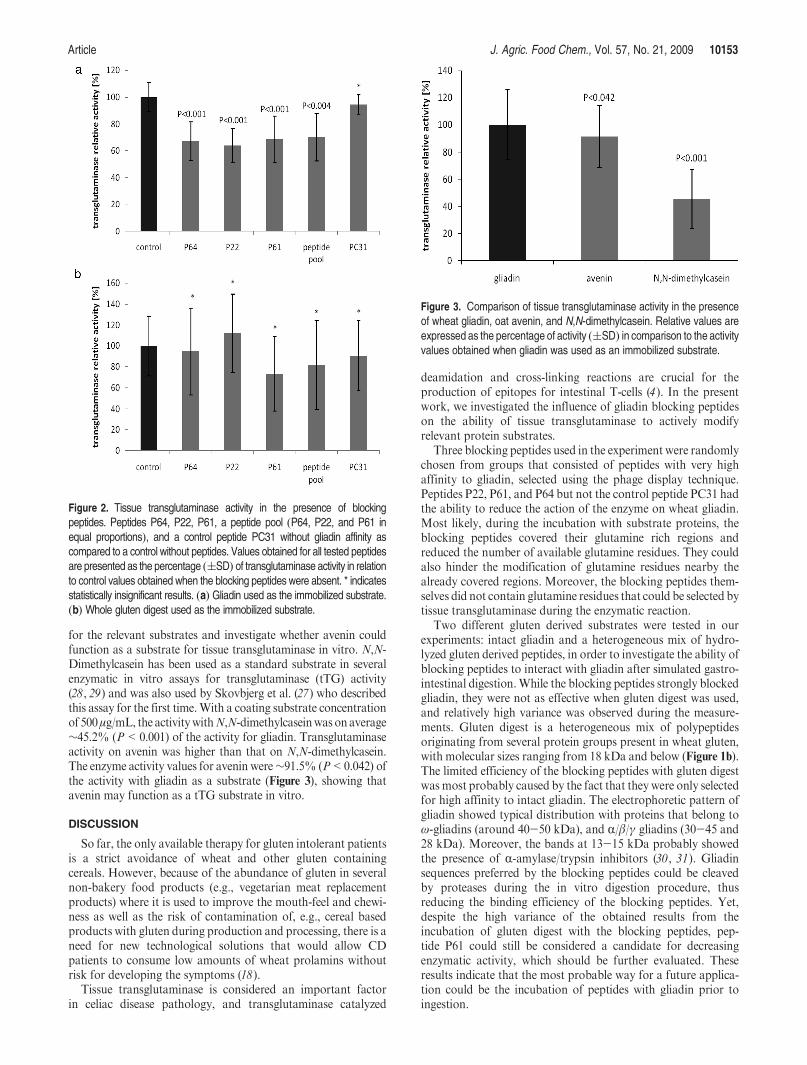
all cases, the tTG activity reduction compared with activity in theabsence of peptides ranged from ∼30 to 36.1% (Figure 2a). Thehighest reduction of enzymatic activity was observed for peptideP22 with ∼36% (P < 0.001); followed by ∼33% (P < 0.001)reduction for peptide P64; ∼31.4% (P < 0.001) reduction forpeptide P61; and ∼30% (P< 0.004) for the peptide pool. Theresults differed when in vitro digested gluten was used as a coatedtTG substrate (Figure 2b). Levels of the measured tTG activityreduction were between∼5.5-28.4%. The highest activity reduc-tion, ∼28%, was observed for peptide P61, ∼20.1% for thepeptide pool, and ∼5.5% for peptide P64. Peptide P22 enhancedthe tTG activity by ∼9.4%. However, because of the high levelof variance within the groups, these results were statisticallyinsignificant at the 95% significance level. The control peptidePC31 did not cause any significant changes in tTG activity witheither gliadin (P<0.11) or gluten digest (P<0.59) as substrates,which demonstrated the absence of unspecific blocking by thepeptide without gliadin affinity.
Substrate Specificity in the tTG Assay. Gliadin, avenin, andN,N-dimethylcasein were used to examine the enzyme specificity
Figure 1. Electrophoretic image of extracted wheat (gliadins) and oat(avenins) prolamins, and whole gluten digest used in the transglutaminaseactivity assay. (a) Lane 1, MWS; lane 2, gliadins; lane 3, avenins (b). Lane1, MWS; lane 2, whole gluten digest.
Article J. Agric. Food Chem., Vol. 57, No. 21, 2009 10153
for the relevant substrates and investigate whether avenin couldfunction as a substrate for tissue transglutaminase in vitro. N,N-Dimethylcasein has been used as a standard substrate in severalenzymatic in vitro assays for transglutaminase (tTG) activity(28, 29) and was also used by Skovbjerg et al. (27) who describedthis assay for the first time.With a coating substrate concentrationof 500μg/mL, the activitywithN,N-dimethylcaseinwas onaverage∼45.2% (P<0.001) of the activity for gliadin. Transglutaminaseactivity on avenin was higher than that on N,N-dimethylcasein.The enzyme activity values for avenin were∼91.5% (P<0.042) ofthe activity with gliadin as a substrate (Figure 3), showing thatavenin may function as a tTG substrate in vitro.
DISCUSSION
So far, the only available therapy for gluten intolerant patientsis a strict avoidance of wheat and other gluten containingcereals. However, because of the abundance of gluten in severalnon-bakery food products (e.g., vegetarian meat replacementproducts) where it is used to improve the mouth-feel and chewi-ness as well as the risk of contamination of, e.g., cereal basedproducts with gluten during production and processing, there is aneed for new technological solutions that would allow CDpatients to consume low amounts of wheat prolamins withoutrisk for developing the symptoms (18).
Tissue transglutaminase is considered an important factorin celiac disease pathology, and transglutaminase catalyzed
deamidation and cross-linking reactions are crucial for theproduction of epitopes for intestinal T-cells (4). In the presentwork, we investigated the influence of gliadin blocking peptideson the ability of tissue transglutaminase to actively modifyrelevant protein substrates.
Three blocking peptides used in the experiment were randomlychosen from groups that consisted of peptides with very highaffinity to gliadin, selected using the phage display technique.Peptides P22, P61, and P64 but not the control peptide PC31 hadthe ability to reduce the action of the enzyme on wheat gliadin.Most likely, during the incubation with substrate proteins, theblocking peptides covered their glutamine rich regions andreduced the number of available glutamine residues. They couldalso hinder the modification of glutamine residues nearby thealready covered regions. Moreover, the blocking peptides them-selves did not contain glutamine residues that could be selected bytissue transglutaminase during the enzymatic reaction.
Two different gluten derived substrates were tested in ourexperiments: intact gliadin and a heterogeneous mix of hydro-lyzed gluten derived peptides, in order to investigate the ability ofblocking peptides to interact with gliadin after simulated gastro-intestinal digestion.While the blocking peptides strongly blockedgliadin, they were not as effective when gluten digest was used,and relatively high variance was observed during the measure-ments. Gluten digest is a heterogeneous mix of polypeptidesoriginating from several protein groups present in wheat gluten,with molecular sizes ranging from 18 kDa and below (Figure 1b).The limited efficiency of the blocking peptides with gluten digestwas most probably caused by the fact that they were only selectedfor high affinity to intact gliadin. The electrophoretic pattern ofgliadin showed typical distribution with proteins that belong toω-gliadins (around 40-50 kDa), and R/β/γ gliadins (30-45 and28 kDa). Moreover, the bands at 13-15 kDa probably showedthe presence of R-amylase/trypsin inhibitors (30, 31). Gliadinsequences preferred by the blocking peptides could be cleavedby proteases during the in vitro digestion procedure, thusreducing the binding efficiency of the blocking peptides. Yet,despite the high variance of the obtained results from theincubation of gluten digest with the blocking peptides, pep-tide P61 could still be considered a candidate for decreasingenzymatic activity, which should be further evaluated. Theseresults indicate that the most probable way for a future applica-tion could be the incubation of peptides with gliadin prior toingestion.
Figure 2. Tissue transglutaminase activity in the presence of blockingpeptides. Peptides P64, P22, P61, a peptide pool (P64, P22, and P61 inequal proportions), and a control peptide PC31 without gliadin affinity ascompared to a control without peptides. Values obtained for all tested peptidesare presented as the percentage ((SD) of transglutaminase activity in relationto control values obtained when the blocking peptides were absent. * indicatesstatistically insignificant results. (a) Gliadin used as the immobilized substrate.(b) Whole gluten digest used as the immobilized substrate.
Figure 3. Comparison of tissue transglutaminase activity in the presenceof wheat gliadin, oat avenin, and N,N-dimethylcasein. Relative values areexpressed as the percentage of activity ((SD) in comparison to the activityvalues obtained when gliadin was used as an immobilized substrate.

10154 J. Agric. Food Chem., Vol. 57, No. 21, 2009 Hoffmann et al.
Currently, the role of oats in the gluten-free diet is rathercontroversial. Contradicting reports have been published statingeither the safety of oats during patient trials or the induction ofinflammatory reaction when oats are consumed in gluten-freediet (32, 33). Oats are recognized as safe for gluten intolerantpatients in some countries; however, they tend to be contami-nated by wheat both in the field and in milling, which may beresponsible for the development of symptoms (34,35). However,Arentz-Hansen et al. (36) recently isolated oat-specific T-cellsfrom some CD patients, showing that avenin is a recognizedsubstrate for the immune system cells for at least some patients. Itwas, however, not known towhat extent avenin could function asa substrate for tTG. Our data from substrate specificity experi-ments are in general agreement with those of Skovbjerg (27)showing that wheat gliadin is a better substrate for tissuetransglutaminase than N,N-dimethylcasein. Our results suggestthat avenin may be processed similarly to gliadin during in vitroassays with tTG. This is most probably due to the slightdifferences in the amount of glutamine residues in these twoproteins. Gliadin is particularly rich in this amino acid. Gluta-mine contributes to approximately 37.1% of the total amino acidcontent in this protein, whereas in avenin, it contributes to(34.1% (7). Although the results are interesting, they are notsufficient to draw any conclusions regarding the role of avenin inCD, but they could serve as the starting part for furtherinvestigations.
Transglutaminase catalyzed deamidation of gluten peptidesplays a crucial role in CD pathology; therefore, preventing thisevent could be of importance in possible future therapies. Therehave been several attempts to develop therapies based on tissuetransglutaminase. So far, only the enzyme inhibitor approach hasbeen tested in a number of studies. Among others, cystamine wasused during human transglutaminase 2 (TG2) treatment ofgliadin. This inhibitor binds covalently to TG2 and preventsit from catalyzing gliadin deamidation. TG2 inhibition resultsin a reduced proliferation of T-cells derived from intestinalbiopsies from CD patients during incubation with gliadin (37).Moreover, synthetic compounds, for example, dihydroisoxasolederivatives have shown to be efficient active-site inhibitorsof TG2 (38). During oral application in mice, these com-pounds were shown to have good bioavailability and wereable to inhibit TG2 in the small intestine. Their short serumhalf-life prevented the distribution to other tissues. However,the detoxification of proteins without inhibiting tissue transglu-taminase could also be considered a possible solution. In ourstudy, we showed that the gliadin blocking peptides had thispotential.
To our knowledge, this is the first study that uses a blockingpeptide approach to reduce the processing of gliadin by tissuetransglutaminase. The results show that these peptides have apotential for gluten detoxification and could be evaluated as analternative in designing new food products for gluten intolerantpatients provided that both peptides and their complexes withgliadin are stable during the passage through the GI tract, whichwill be further investigated in future work.
ABBREVIATIONS USED
T-20, Tween-20; PBS, phosphate buffered saline; P2000, poly-propylene glycol; MWS, molecular weight standard; tTG, tissuetransglutaminase.
ACKNOWLEDGMENT
We thank Annette Almgren for her participation in thetransglutaminase assay experiments.
LITERATURE CITED
(1) Fasano, A.; Berti, I.; Gerarduzzi, T.; Not, T.; Colletti, R. B.; Drago,S.; Elitsur, Y.; Green, P. H.; Guandalini, S.; Hill, I. D.; Pietzak, M.;Ventura, A.; Thorpe, M.; Kryszak, D.; Fornaroli, F.; Wasserman,S. S.; Murray, J. A.; Horvath, K. Prevalence of celiac disease in at-risk and not-at-risk groups in the United States: a large multicenterstudy. Arch. Intern. Med. 2003, 163, 286–92.
(2) van Heel, D. A.; West, J. Recent advances in coeliac disease. Gut2006, 55, 1037–46.
(3) Sollid, L. M. Coeliac disease: dissecting a complex inflammatorydisorder. Nat. Rev. Immunol. 2002, 2, 647–55.
(4) Dieterich, W.; Esslinger, B.; Schuppan, D. Pathomechanisms inceliac disease. Int. Arch. Allergy Immunol. 2003, 132, 98–108.
(5) Fasano,A.;Catassi,C.Current approaches todiagnosis and treatment ofceliac disease: an evolving spectrum.Gastroenterology 2001, 120, 636–51.
(6) Maki, M.; Collin, P. Coeliac disease. Lancet 1997, 349, 1755–9.(7) Belitz, H.-D.; Grosch, W.; Schieberle, P. Cereals and Cereal Pro-
ducts. In Food Chemistry, 3rd revised ed.; Springer-Verlag: Berlin,Germany, 2004; pp 681-692.
(8) Stern, M.; Ciclitira, P. J.; Van Eckert, R.; Feighery, C.; Janssen,F. W.; Mendez, E.; Mothes, T.; Troncone, R.; Wieser, H. Analysisand clinical effects of gluten in coeliac disease. Eur. J. Gastroent.Hepat. 2001, 13, 741–747.
(9) Hausch, F.; Shan, L.; Santiago, N. A.; Gray, G. M.; Khosla, C.Intestinal digestive resistance of immunodominant gliadin peptides.Am. J. Physiol. Gastrointest. Liver Physiol. 2002, 283, G996–G1003.
(10) Shan, L.; Molberg, O.; Parrot, I.; Hausch, F.; Filiz, F.; Gray, G. M.;Sollid, L. M.; Khosla, C. Structural basis for gluten intolerance inceliac sprue. Science 2002, 297, 2275–9.
(11) Ichinose, A.; Bottenus, R. E.; Davie, E. W. Structure of transgluta-minases. J. Biol. Chem. 1990, 265, 13411–4.
(12) Fleckenstein, B.; Molberg, O.; Qiao, S. W.; Schmid, D. G.;von der Mulbe, F.; Elgstoen, K.; Jung, G.; Sollid, L. M. Gliadin Tcell epitope selection by tissue transglutaminase in celiac disease.Role of enzyme specificity and pH influence on the transamidationversus deamidation process. J. Biol. Chem. 2002, 277, 34109–16.
(13) Folk, J. E.; Chung, S. I. Transglutaminases.Methods Enzymol. 1985,113, 358–75.
(14) Dieterich, W.; Ehnis, T.; Bauer, M.; Donner, P.; Volta, U.; Riecken,E. O.; Schuppan, D. Identification of tissue transglutaminase as theautoantigen of celiac disease. Nat. Med. 1997, 3, 797–801.
(15) Maki, M. Tissue transglutaminase as the autoantigen of coeliacdisease. Gut 1997, 41, 565–6.
(16) Sj::ostr
::om, H.; Lundin, K. E. A.; Molberg, O.; K
::orner, R.; Mcadam,
S. N.; Anthonsen, D.; Quarsten, H.; Noren, O.; Roepstorff, P.;Thorsby, E.; Sollid, L. M. Identification of a gliadin T-cell epitope incoeliac disease: general importance of gliadin deamidation forintestinal T-cell recognition. Scand. J. Immunol. 1998, 48, 111–115.
(17) Szabolcs, M.; Sipka, S.; Csorba, S. In vitro cross-linking of gluteninto high-molecular-weight polymers with transglutaminase. ActaPaediatr. Hung. 1987, 28, 215–27.
(18) Day, L.; Augustin, M. A.; Batey, I. L.; Wrigley, C. W. Wheat-glutenuses and industry needs. Trends Food Sci. Technol. 2006, 17, 82–90.
(19) Thompson, T.; Mendez, E. Commercial assays to assess glutencontent of gluten-free foods: why they are not created equal.J. Am. Diet. Assoc. 2008, 108, 1682–1687.
(20) Mimouni, B.; Azanza, J.-L.; Raymond, J. Wheat flour proteins:isolation and functionality of gliadin and HMW-glutenin enrichedfractions. J. Sci. Food Agric. 1998, 78, 423–428.
(21) Osborne, T. B. The Proteins of theWheat Kernel; Carnegie Institution:Washington DC, 1907.
(22) Weiss, W. C. V.; G::org, A. Electrophoretic characterization of wheat
grain allergens from different cultivars involved in bakers’ asthma.Electrophoresis 1993, 14, 805–816.
(23) Lending, C. R.; Chesnut, R. S.; Shaw, K. L.; Larkins, B. A.Immunolocalization of avenin and globulin storage proteinsin developing endosperm ofAvena sativaL.Planta 1989, 178, 315–324.
(24) Kim, S. I.; Charbonnier, L.; Mosse, J. Heterogeneity of avenin, theoat prolamin: fractionation, molecular weight and amino acidcomposition. Biochim. Biophys. Acta 1978, 537, 22–30.
Article J. Agric. Food Chem., Vol. 57, No. 21, 2009 10155
(25) Matuschek, E.; Towo, E.; Svanberg, U. Oxidation of polyphenols inphytate-reduced high-tannin cereals: effect on different phenolicgroups and on in vitro accessible iron. J. Agric. Food Chem. 2001,49, 5630–8.
(26) Laemmli, U. K. Cleavage of structural proteins during the assemblyof the head of bacteriophage T4. Nature 1970, 227, 680–5.
(27) Skovbjerg, H.; Noren, O.; Anthonsen, D.; Moller, J.; Sjostrom, H.Gliadin is a good substrate of several transglutaminases: possibleimplication in the pathogenesis of coeliac disease. Scand. J. Gastro-enterol. 2002, 37, 812–7.
(28) Jeon, W. M.; Lee, K. N.; Birckbichler, P. J.; Conway, E.; Patterson,M. K., Jr. Colorimetric assay for cellular transglutaminase. Anal.Biochem. 1989, 182, 170–175.
(29) Slaughter, T. F.; Achyuthan, K. E.; Lai, T. S.; Greenberg, C. S. Amicrotiter plate transglutaminase assay utilizing 5- (biotinamido)-pentylamine as substrate. Anal. Biochem. 1992, 205, 166–171.
(30) Gomez, L.; Martin, E.; Hern�andez, D.; S�anchez-Monge, R.; Barber,D.; del Pozo, V.; de Andres, B.; Armentia, A.; Lahoz, C.; Salced, G.;Palomino, F. Members of the [alpha]-amylase inhibitors familyfrom wheat endosperm are major allergens associated with baker’sasthma. FEBS Lett. 1990, 261, 85–88.
(31) Wieser, H. Chemistry of gluten proteins. Food Microbiol. 2007, 24,115–119.
(32) Janatuinen, E.K.;Kemppainen, T.A.; Julkunen,R. J.; Kosma,V.M.;Maki, M.; Heikkinen, M.; Uusitupa, M. I. No harm from five yearingestion of oats in coeliac disease. Gut 2002, 50, 332–5.
(33) Lundin, K. E.; Nilsen, E. M.; Scott, H. G.; Loberg, E. M.; Gjoen,A.; Bratlie, J.; Skar, V.; Mendez, E.; Lovik, A.; Kett, K.Oats induced villous atrophy in coeliac disease. Gut 2003, 52,1649–52.
(34) Thompson, T. Do oats belong in a gluten-free diet? J. Am. Diet.Assoc. 1997, 97, 1413–1416.
(35) Thompson, T. Gluten contamination of commercial oat products inthe United States. N. Engl. J. Med. 2004, 351, 2021–2.
(36) Arentz-Hansen, H.; Fleckenstein, B.; Molberg, O.; Scott, H.;Koning, F.; Jung, G.; Roepstorff, P.; Lundin, K. E.; Sollid, L. M.The molecular basis for oat intolerance in patients with celiacdisease. PLoS. Med. 2004, 1 (1), e1.
(37) Molberg, O.; McAdam, S. N.; Lundin, K. E.; Kristiansen, C.;Arentz-Hansen, H.; Kett, K.; Sollid, L.M. T cells from celiac diseaselesions recognize gliadin epitopes deamidated in situ by endogenoustissue transglutaminase. Eur. J. Immunol. 2001, 31, 1317–23.
(38) Choi, K.; Siegel, M.; Piper, J. L.; Yuan, L.; Cho, E.; Strnad, P.;Omary, B.; Rich, K. M.; Khosla, C. Chemistry and biology ofdihydroisoxazole derivatives: selective inhibitors of human transglu-taminase 2. Chem. Biol. 2005, 12, 469–475.
Received July 1, 2009. Revisedmanuscript received September 12, 2009.
Accepted September 20, 2009. The study was financed by The Swedish
Research Council for Environment, Agricultural Sciences and Spatial
Planning, no. 222-2004-2705.
Copyright © 2022 FDOKUMEN