Blocking Peptides Decrease Tissue Transglutaminase Processing of Gliadin in Vitro
Upload
independentCategory
view
0download
0
ARTICLE IN PRESS
0142-9612/$ - se
doi:10.1016/j.bi
�Correspondfax: +44121 35
E-mail addr
Biomaterials 26 (2005) 6518–6529
www.elsevier.com/locate/biomaterials
The cellular response to transglutaminase-cross-linked collagen
David Y.S. Chaua, Russell J. Collighanb, Elisabetta A.M. Verderioa,Victoria L. Addyc, Martin Griffinb,�
aSchool of Biomedical and Natural Sciences, The Nottingham Trent University, Clifton, Nottingham, NG11 8NS, UKbSchool of Life and Health Sciences, Aston University, Aston Triangle, Birmingham, B4 7ET, UK
cBLC Leather Technology Centre Ltd., Leather Trade House, Kings Park Road, Moulton Park, Northampton, NN3 6JD, UK
Received 7 January 2005; accepted 7 April 2005
Available online 31 May 2005
Abstract
Collagen, type I, is a highly abundant natural protein material which has been cross-linked by a variety of methods including
chemical agents, physical heating and UV irradiation with the aim of enhancing its physical characteristics such as mechanical
strength, thermal stability, resistance to proteolytic breakdown, thus increasing its overall biocompatibility. However, in view of the
toxicity of residual cross-linking agents, or impracticability at large scales, it would be more useful if the collagen could be cross-
linked by a milder, efficient and more practical means by using enzymes as biological catalysts.
We demonstrate that on treating native collagen type I (from bovine skin) with both tissue transglutaminase (TG2; tTG) and
microbial transglutaminase (mTG; Streptoverticillium mobaraense) leads to an enhancement in cell attachment, spreading and
proliferation of human osteoblasts (HOB) and human foreskin dermal fibroblasts (HFDF) when compared to culture on native
collagen. The transglutaminase-treated collagen substrates also showed a greater resistance to cell-mediated endogenous protease
degradation than the native collagen. In addition, the HOB cells were shown to differentiate at a faster rate than on native collagen
when assessed by measurement of alkaline phosphatase activity and osteopontin expression.
r 2005 Elsevier Ltd. All rights reserved.
Keywords: Bioactivity; Biocompatibility; Collagen; Fibroblasts; Osteoblasts; Tissue engineering
1. Introduction
Collagen is a very popular biomaterial due to itsbiocompatibility, i.e. the ability to support cell adhesionand proliferation. It is also biodegradable and onlyweakly antigenic—able to persist in the body withoutdeveloping a foreign body response that could lead to itspremature rejection [1]. The replacement of skin withartificial collagen–GAG matrices has been investigatedsince the early 1980s and is now in clinical use [2,3]. Theprimary reason for the usefulness of collagen inbiomedical applications is that collagen can form fibreswith extra strength and stability through its self-
e front matter r 2005 Elsevier Ltd. All rights reserved.
omaterials.2005.04.017
ing author. Tel.: +44121 204 3942;
9 0578.
ess: [email protected] (M. Griffin).
aggregation and in vivo cross-linking [4]. Unfortunately,collagen, like many natural polymers, once extractedfrom its original source and then reprocessed, suffersfrom weak mechanical properties, thermal instabilityand ease of proteolytic breakdown. To overcome theseproblems, collagen has been cross-linked by a variety ofagents—a subject of much recent research to findmethods of preventing rapid absorption by the body[4]. However, these methods suffer from the problemthat the residual catalysts, initiators and unreacted orpartially reacted cross-linking agents used can be toxicor cause inflammatory responses if not fully removed or,simply, not cost-effective or practical at the large scale[5–7]. As a consequence, research continues to findalternative methods to stabilise collagen which arenatural, milder, efficient and more practical.
Transglutaminases (EC 2.3.2.13) are a group of enzymesthat can catalyse several types of post-translational
ARTICLE IN PRESSD.Y.S. Chau et al. / Biomaterials 26 (2005) 6518–6529 6519
modifications to proteins. The most important of thesereactions results in the cross-linking of peptides or proteinsto form multimers via a e(g-glutamyl)lysine linkage usingthe side chains of lysine and glutamine residues. Transglu-taminases are also able to covalently attach primary aminecontaining compounds to peptide bound glutamine,facilitating modification of the physical, chemical andbiological properties of proteins [8]. For these reasons,transglutaminases have been utilised by the commercialsector in many different processes and have attractedmuch attention from the research community [9]. Micro-bial transglutaminase has been used to cross-link gelatinmatrices to further increase their strength [10] and, also, toincorporate cell adhesion factors within the gel matrix,resulting in an enhancement of cell proliferation [11].
Interestingly, a novel component of the cell/tissueresponse to cell damage and stress is tissue transgluta-minase (tTG), a Ca2+-dependent mammalian form ofthe enzyme, which modulates cell–matrix interactions,tissue stability and a variety of other cell functions[12,13]. The entire tissue repair process is regulated bythe interaction of cells with the surrounding extracel-lular matrix (ECM), ensuring cell adhesion, survival andproliferation [14,15]. To date, the cross-linking functionof tTG in the ECM leading to ECM stabilisation/remodelling has been identified in a number ofbiological processes important for tissue repair [12]: inaddition, at least three of the nine genes so farcharacterised are thought to be naturally involved inthe wound healing response process [see review, 16].
The aim of this study was to investigate the use of thetwo different transglutaminases; the mammalian (tTG;TG2; TG-2; isolated from guinea pig liver) and themicrobial enzyme (mTG; isolated from Streptoverticil-
lium mobaraense) in the modification of collagen type Iwith the view to investigate potential application as abiocompatible natural polymer for use in soft and hardtissue repair.
2. Materials and methods
All water used was deionised using an Elgastat System 2
water purifier (ELGA Ltd., UK) and a Milli-Q water purifier
(Millipore Waters, UK). All chemicals were purchased from
Sigma-Aldrich, Poole, UK, unless otherwise stated. Sterile
preparation of stock solutions and chemicals were performed
either by filtration through a 0.22 mm Whatmann sterile filter
and/or autoclaving at 121 1C at 1 bar for 1 h.
2.1. Cell culture
Human osteoblast (HOB) cells, isolated from explants of
trabecular bone dissected from femoral heads following
orthopaedic surgery as previously described [17] were kindly
supplied by Professor S. Downes and Dr. S. Anderson (School
of Biomedical Sciences, University of Nottingham) and used
during this investigation. Human foreskin dermal fibroblast
(HFDF) cells isolated from human neonatal foreskin were also
used. Both cell lines were used during their low-passage
number, ranging from 11 to 15 passages. Cell lines were
cultured and maintained, in vitro, as monolayers in T-flasks
using DMEM, supplemented with 10% heat-inactivated (56 1C
for 1 h) FCS, 1% non-essential amino acids and 2mM L-
glutamine. Periodic additions of 1% penicillin–streptomycin
were used to avoid bacterial contamination. Flasks were kept
in a humidified-atmosphere incubator at 37 1C and with 5%
CO2. Cells were routinely passaged and never allowed to reach
greater than 90% confluency at any one time. For detachment,
standard trypsinisation was performed using 0.25% (w/v)
trypsin/2mM EDTA solution in PBS solution.
2.2. Cell viability and proliferation
Cell counts and viability estimations were performed using
the standard trypan blue exclusion technique by means of a
0.22 mm sterile filtered 0.5% (w/v) trypan blue solution and a
haemocytometer. Non-viable cells stained blue due to the loss
of their membrane integrity and, hence, allowed the passage of
dye into the cell. Viable cells remained colourless.
Cell proliferation and viability were also measured using the
CellTiter AQ One Solution Cell ProliferationTM assay kit
(Promega, Southampton, UK. Cat no. G3580). Assays were
performed, with reduced lighting, simply by the addition of
20 ml of CellTiter AQ reagent into the relevant samples in
100 ml of culture medium. These samples were then incubated
in a humidified-atmosphere incubator at 37 1C and with 5%
CO2 for 90min before the absorbance was read at 490 nm
using a SpectraFluors plate reader.
2.3. Attachment and spreading
Cells were seeded on the relevant substrate at a density of
625 cells/mm2. After allowing cells to attach and spread, they
were fixed in 3.7% (w/v) paraformaldehyde, permeabilised by
the addition of 0.1% (v/v) Triton X-100 in PBS, before
staining with May-Grunwald (0.25% (w/v) in methanol) and
Giemsa stains (0.4% (w/v) in methanol, diluted 1:50 with
water). Cells were then viewed at � 400 magnification using an
Olympus CK2 microscope. Three separate fixed-size random
fields per sample were photographed with an Olympus DP10
digital camera. Pictures were analysed using Scion ImageTM
software (Scion Corporation, Maryland, USA). Spread cells
were distinguished and characterised based upon the presence
of a clear halo of cytoplasm surrounding their nucleus as
previously described [18].
2.4. Alkaline phosphatase (ALP) activity
The ALP Optimized Alkaline Phosphatase EC 3.1.3.1
Colorimetric Tests kit (obtained from Sigma-Aldrich, Poole,
UK. Cat no. DG1245-K) was used to quantify the ALP
activity. Alkaline phosphatase hydrolyses p-nitrophenyl phos-
phate to p-nitrophenol and inorganic phosphate. The hydro-
lysis occurs at alkaline pH and the p-nitrophenol formed
shows an absorbance maximum at 405 nm. The rate of increase
in absorbance at 405 nm is directly proportional to ALP
ARTICLE IN PRESSD.Y.S. Chau et al. / Biomaterials 26 (2005) 6518–65296520
activity in the sample. Samples were treated according to the
manufacturers’ instructions and analysed using a Beckmann
DU530 UV/Vis spectrophotometer.
2.5. Osteopontin (OPN) concentration
The OPN ELISA kit (obtained from CalBiochem, UK. Cat
no. 499262) was used to quantify the concentration of OPN in
the samples. The kit uses a polyclonal antibody to human
OPN immobilised on a micro-titre plate to bind to the human
OPN. The measured absorbance (450 nm) is directly propor-
tional to the concentration of human OPN. Samples were
treated according to the manufacturers’ instructions and
analysed using a SpectraFluors plate reader.
2.6. Transglutaminase
tTG was isolated and purified from guinea pig livers using a
combination of anion exchange, gel filtration and affinity
chromatography as previously described [19]. Commercial
samples of TG were also used during this investigation: tTG
from guinea pig liver (Sigma-Aldrich, Poole, UK. Cat no.
T5398) and microbial transglutaminase, mTG (Ajinomoto
Corporation Inc., Japan), isolated from Streptoverticillium
mobaraense, as the commercially available product, ActivaTM
WM. This required further purification steps to remove the
maltodextrin ingredient: briefly, the ActivaTM WM was
dissolved in ice-cold 20mM phosphate buffer, 2mM EDTA
pH 6.0 and filtered, before being loaded onto a 100ml SP-
Sepharose FF column overnight at a continuous flow rate of
5ml/min. The column was then washed and proteins eluted, at
the same flow rate, with a 0–1000mM gradient of NaCl in
20mM phosphate buffer, 2mM EDTA pH 6.0. Fractions were
assayed for protein using the Bio-Rad DC protein assay (Bio-
Rad Laboratories, Hertfordshire, UK. Cat no. 500-0120).
Fractions containing mTG were pooled, aliquoted, freeze
dried and stored at �70 1C. Before immediate use, tTG was
pre-treated in 2mM DTT in 50mM Tris buffer (pH 7.4) for
10min at room temperature to activate any oxidised enzyme,
before addition to a final buffered solution containing 5mM
CaCl2 and, a minimum of 1mM DTT in Tris buffer. Typical
activities for the transglutaminases used in this investigation
were as follows: tTG: 11500–13000U/mg and mTG:
16000–25000U/mg.
2.7. Transglutaminase activity
The incorporation of [14C]-putrescine into N,N0-dimethyl-
casein, as described previously [20], was used to assay for TG
activity and monitor the effects of the inhibitors. Unit of
transglutaminase activity is 1 nmol of putrescine incorporated
per hour.
2.8. Collagen
Commercial calf skin collagen type I (Sigma-Aldrich, Poole,
UK. Cat no. C9791) was used during this investigation. Native
collagen samples were solubilised in 0.2M acetic acid (Fisher
Scientific, Loughborough, UK. Cat no. A/0400/PB17) at 4 1C
with constant stirring for 24 h before use. Neutralisation of the
collagen mixture was performed using a [5:3:2] ratio of
[collagen: 2� DMEM: 0.2M NaOH buffer] respectively to a
final of pH 7.2. Tissue culture plastic was then covered using
this collagen mix (recommended at 6–10 mg/cm2) before being
placed into a humidified-atmosphere incubator for 12 h to
allow gelation to occur. In general, 50ml of the collagen mix
was added to each well of a 96-well plate. Plates were used
within 48 h of the collagen matrix formation.
2.9. Modified collagen by transglutaminase
Neutralised collagen mixture was subjected to treatment
with both tTG and mTG. Samples of the neutralised collagen,
as described above, were treated with 50–1000 mg/ml of tTG, in
a reaction mix consisting of 2mM DTT and 5mM CaCl2in 10mM Tris buffer (pH 7.4). Microbial enzyme was added in
10mM Tris buffer (pH 7.4). Stock solutions of: 2mg/ml tTG
and mTG, 1M DTT and 1M CaCl2 were used to minimise total
volume changes. The enzymes were always added last to the
collagen-reaction mix to minimise any self-imposed cross-
linking. Controls using 10mM EDTA (to block tTG activity)
and an active-site directed inhibitor, R281 (a synthetic CBZ-
glutaminyl-glycine analogue; 500 mM), were also included in
each assay. For 96-well plates, 50ml of the pre-treated collagen
mixture was added to each well before being placed into a
humidified-atmosphere incubator, at 37 1C and with 5% CO2,
for 8 h. On removal, wells were washed twice with sterile
distilled water and used immediately.
2.10. Determination of e-(g-glutamyl)lysine cross-link
Cross-linked and native samples of collagen were proteoly-
tically digested as previously described [21] which included an
initial digestion with microbial collagenase (Clostridiopeptidase
A.; 1mg/ml, Sigma-Aldrich, Poole, UK. Cat no. C9891) prior
to the addition of further proteases. After digestion, samples
were freeze dried and then resuspended in 0.1M HCl and
sonicated for 2min to aid dispersion. An aliquot (90 ml) wasmixed with 110 ml of loading buffer (0.2 M lithium citrate, 0.1%
phenol pH 2.2) and loaded onto a Dionex DC-4A resin
column 0.5 cm� 20 cm using a Pharmacia Alpha Plus amino
acid analyser. Derivatisation was performed post-column
using o-phthaldialdehyde (0.8M boric acid, 0.78M potassium
hydroxide, 600mg/ml o-phthaldialdehyde, 0.5% (v/v) metha-
nol, 0.75% (v/v) 2-mercaptoethanol, 0.35% (v/v) Brij 35) and
the absorbance was measured at 450 nm. Dipeptide was
determined by addition of known amounts of e(g-glutamyl)ly-
sine to the sample and comparing peak areas.
2.11. Coomassie blue staining assay of cell cultures
The capacity of both the HOB and HFDF cells to degrade
type I collagen was assessed as previously described [22].
Briefly, native and TG pre-treated collagen samples gels were
plated out at 50 ml per well of a 96-well plate. Hundred
microlitres of 2� 104 cells/ml, cultured in complete media, was
then added to the wells in triplicates. Plates were then kept in a
humidified-atmosphere incubator for the relevant time
point(s). After incubation, cells were removed from the
collagen matrix by addition of 0.5% (w/v) sodium
ARTICLE IN PRESSD.Y.S. Chau et al. / Biomaterials 26 (2005) 6518–6529 6521
deoxycholate in 10mM Tris–HCl. A rinse with distilled water
was performed before the collagen samples were stained with a
0.1% (w/v) Coomassie Brilliant blue stain solution (50% (v/v)
methanol; 10% (v/v) acetic acid; 40% (v/v) dH2O). Samples
were allowed to stain for 5min before a further rinse with
distilled water. Unstained areas, which appeared lighter blue,
gave an indication of collagen degradation by cells. Two
separate fixed-size random fields per triplicate samples were
photographed using an Olympus CK2 microscope and DP10
digital camera.
2.12. Protein concentration
The total protein content of the collagen samples was
determined by the Lowry method [23] using the Bio-Rad DC
protein assay kit (Bio-Rad Laboratories, Hertfordshire, UK.
Cat no. 500-0120).
2.13. Collagenase degradation of matrices following cell culture
Collagen substrates were subjected to digestive treatment
with 100 ml of a 1mg/ml microbial collagenase solution
(Clostridium histolyticum, Sigma-Aldrich, Poole, UK. Cat no.
C9891) followed by 100 ml 0.25% (w/v) trypsin/2mM EDTA
solution in PBS solution for 24 h at 37 1C. Samples were
washed twice with PBS followed by a wash with distilled water
before the enzymatic digestion treatment.
2.14. Zymography
Gelatin and collagen zymography were carried out as
previously described [24] with the following adaptations:
resolving gels were mixed with the following components, in
order: 1ml of 5mg/ml of type I collagen solution (Sigma
C9791) in 20mM acetic acid (for collagen zymography)/1ml of
5mg/ml porcine gelatin (Sigma G2625) in H2O (for gelatin
zymography), 3.1ml H2O, 2.5ml of 1.5M Tris–HCl pH 8.8,
3.33ml of 29% acrylamide/1% N,N0-methylene bisacrylamide,
50ml of 10% ammonium persulphate, 10 ml of N,N,N0,N0-
tetramethylethylenediamine (TEMED). SDS was found to
cause precipitation of the collagen and so was not added to the
resolving gel. Stacking gels were poured in the usual way, i.e.
0.65ml of 29% acrylamide/1% N,N0-methylene bisacrylamide,
3ml H2O, 1.25ml 0.5M Tris–HCl pH 6.8, 50 ml of 10% SDS,
25ml of 10% ammonium persulphate, 5 ml of TEMED.
Samples containing matrix metalloproteinases (MMPs)
were diluted 1:1 with loading buffer (1M Tris–HCl pH 6.8,
50% glycerol, 0.4% bromophenol blue) and electrophoresed at
100V in standard Laemmli running buffer (24mM Tris–HCl,
192mM glycine, 3.47mM SDS, pH 8.3), avoiding overheating
(approx. 4–5 h). After electrophoresis, gels were washed twice,
with shaking, for 30min each in 200ml of 2.5% Triton X-100,
to remove SDS and recover MMP activity. The gels were then
placed in digestion buffer (100mM Tris–HCl, 5mM CaCl2,
0.005% Brij-35, 1mM ZnCl2, 0.001% NaN3, pH 8) for 16–48 h
at 37 1C. Gels were stained with 0.2% Coomassie Brilliant blue
R-250 in 50% ethanol, 10% acetic acid for 2 h and destained
by microwaving for 15min (full power 850W) in three changes
of deionised H2O.
2.15. Statistical analysis of data
Differences between datasets (shown as mean7SD) were
determined by the Student’s t-test at a significance level of
po0:05.
3. Results
3.1. Cross-linking of collagen by microbial and tissue
transglutaminases
Native collagen (type I) was treated with both tTGand mTG, separately, in order to catalyse the formationof e-(g-glutamyl)lysine cross-linking. The extent ofcross-linking for each of the TG treatments is shownin Table 1. Treatment of collagen with increasingconcentrations of TG led to a corresponding increasein the amount of e-(g-glutamyl)lysine bonds present—with up to 1mol of cross-link per mole of collagenmonomer. Treatment with mTG gave a much greaterincrease (almost two-fold) in the amount of isopeptideformed for the equivalent protein concentration oftransglutaminase used. However, the increased specificactivity of the mTG probably accounts for thedifferences noted.
3.2. Resistance of native and cross-linked collagen to cell-
mediated degradation
Collagen treated with 50 mg/ml TG showed a greaterresistance to cell-mediated degradation as compared tothe native collagen, when HOB cells and HFDF wereseeded onto the collagen matrices and incubated for72 h. Following removal of cells, visual comparison ofthe Coomassie blue stained matrices and measurementof the residual collagen indicated the mTG-treatedcollagen to be more resistant than tTG-treated collagen(Table 2).
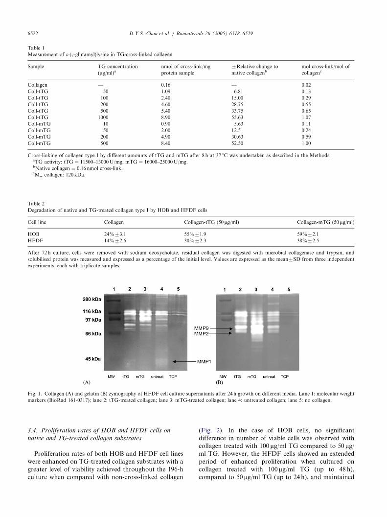
3.3. Matrix metalloproteinases secreted by HFDF cells
grown on transglutaminase collagen matrices
Following growth on type I collagen, fibroblastsshowed an induction of a wide array of collagenases andgelatinases when compared with growth on tissueculture plastic-ware alone (Fig. 1). After growth ontransglutaminase cross-linked type I collagen, theinduction of active MMP1 (45 kDa), Fig. 1A, is muchless pronounced compared to growth on native collagen,whereas the induction of active MMP2 (66 kDa) andMMP9 (86 kDa), Fig. 1B, was increased particularlywhen the cells were grown on collagen cross-linkedby tTG.
ARTICLE IN PRESS
Table 1
Measurement of e-(g-glutamyl)lysine in TG-cross-linked collagen
Sample TG concentration
(mg/ml)anmol of cross-link/mg
protein sample
7Relative change to
native collagenbmol cross-link/mol of
collagenc
Collagen — 0.16 — 0.02
Coll-tTG 50 1.09 6.81 0.13
Coll-tTG 100 2.40 15.00 0.29
Coll-tTG 200 4.60 28.75 0.55
Coll-tTG 500 5.40 33.75 0.65
Coll-tTG 1000 8.90 55.63 1.07
Coll-mTG 10 0.90 5.63 0.11
Coll-mTG 50 2.00 12.5 0.24
Coll-mTG 200 4.90 30.63 0.59
Coll-mTG 500 8.40 52.50 1.00
Cross-linking of collagen type I by different amounts of tTG and mTG after 8 h at 37 1C was undertaken as described in the Methods.aTG activity: tTG ¼ 11500–13000U/mg; mTG ¼ 16000–25000U/mg.bNative collagen ¼ 0.16 nmol cross-link.cMw collagen: 120 kDa.
Table 2
Degradation of native and TG-treated collagen type I by HOB and HFDF cells
Cell line Collagen Collagen-tTG (50mg/ml) Collagen-mTG (50 mg/ml)
HOB 24%73.1 55%71.9 59%72.1
HFDF 14%72.6 30%72.3 38%72.5
After 72 h culture, cells were removed with sodium deoxycholate, residual collagen was digested with microbial collagenase and trypsin, and
solubilised protein was measured and expressed as a percentage of the initial level. Values are expressed as the mean7SD from three independent
experiments, each with triplicate samples.
Fig. 1. Collagen (A) and gelatin (B) zymography of HFDF cell culture supernatants after 24 h growth on different media. Lane 1: molecular weight
markers (BioRad 161-0317); lane 2: tTG-treated collagen; lane 3: mTG-treated collagen; lane 4: untreated collagen; lane 5: no collagen.
D.Y.S. Chau et al. / Biomaterials 26 (2005) 6518–65296522
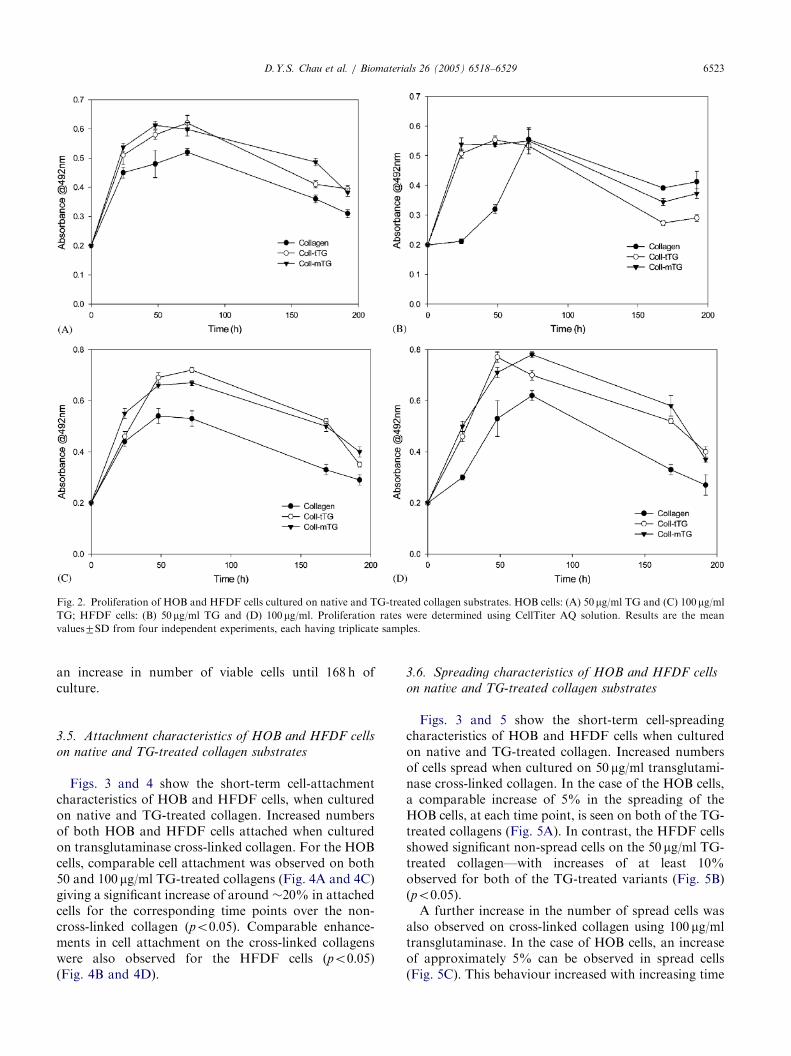
3.4. Proliferation rates of HOB and HFDF cells on
native and TG-treated collagen substrates
Proliferation rates of both HOB and HFDF cell lineswere enhanced on TG-treated collagen substrates with agreater level of viability achieved throughout the 196-hculture when compared with non-cross-linked collagen
(Fig. 2). In the case of HOB cells, no significantdifference in number of viable cells was observed withcollagen treated with 100 mg/ml TG compared to 50 mg/ml TG. However, the HFDF cells showed an extendedperiod of enhanced proliferation when cultured oncollagen treated with 100 mg/ml TG (up to 48 h),compared to 50 mg/ml TG (up to 24 h), and maintained
ARTICLE IN PRESS
Fig. 2. Proliferation of HOB and HFDF cells cultured on native and TG-treated collagen substrates. HOB cells: (A) 50 mg/ml TG and (C) 100mg/ml
TG; HFDF cells: (B) 50 mg/ml TG and (D) 100mg/ml. Proliferation rates were determined using CellTiter AQ solution. Results are the mean
values7SD from four independent experiments, each having triplicate samples.
D.Y.S. Chau et al. / Biomaterials 26 (2005) 6518–6529 6523
an increase in number of viable cells until 168 h ofculture.
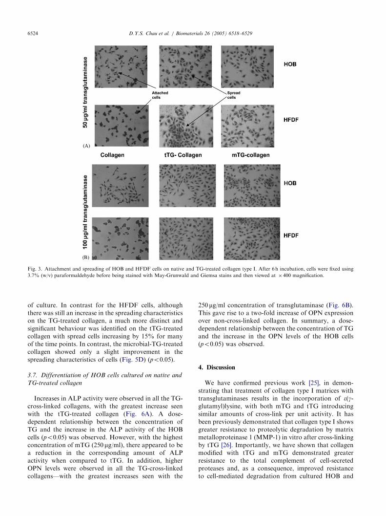
3.5. Attachment characteristics of HOB and HFDF cells
on native and TG-treated collagen substrates
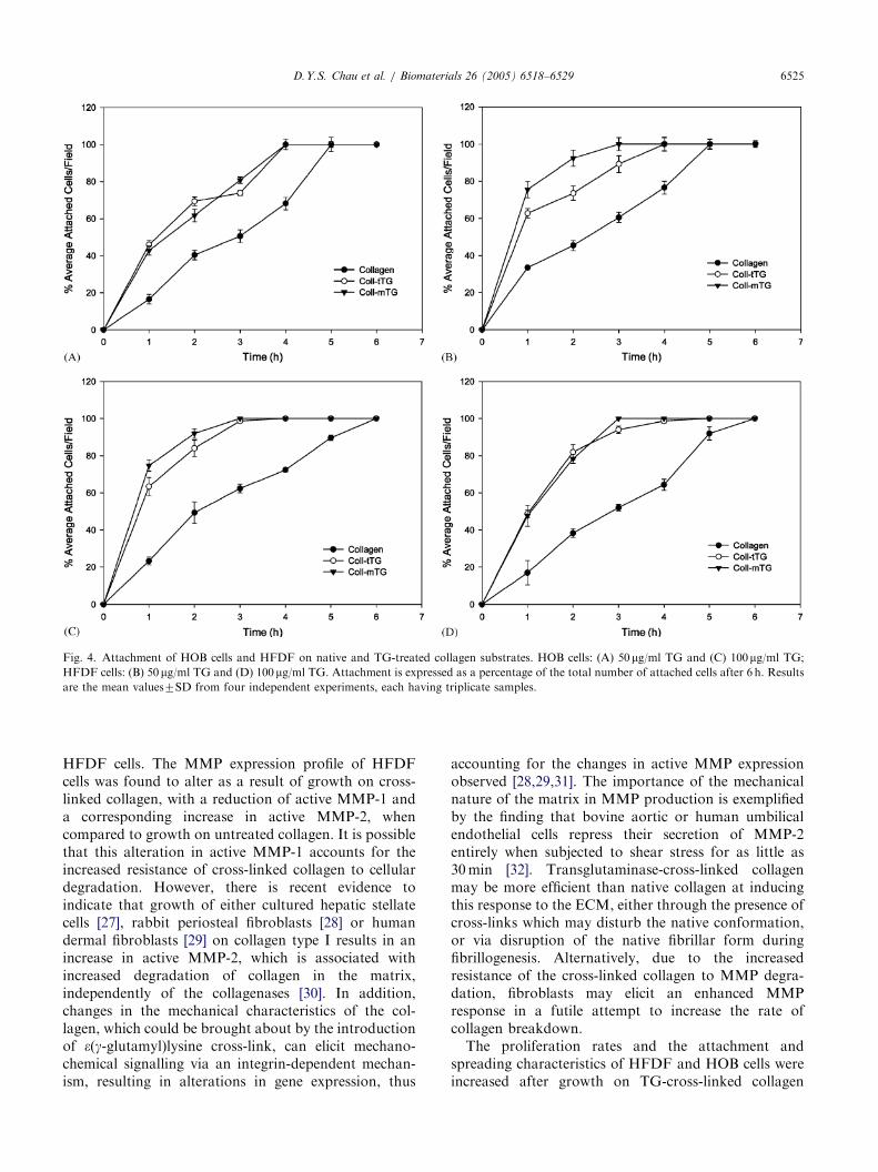
Figs. 3 and 4 show the short-term cell-attachmentcharacteristics of HOB and HFDF cells, when culturedon native and TG-treated collagen. Increased numbersof both HOB and HFDF cells attached when culturedon transglutaminase cross-linked collagen. For the HOBcells, comparable cell attachment was observed on both50 and 100 mg/ml TG-treated collagens (Fig. 4A and 4C)giving a significant increase of around �20% in attachedcells for the corresponding time points over the non-cross-linked collagen (po0:05). Comparable enhance-ments in cell attachment on the cross-linked collagenswere also observed for the HFDF cells (po0:05)(Fig. 4B and 4D).
3.6. Spreading characteristics of HOB and HFDF cells
on native and TG-treated collagen substrates
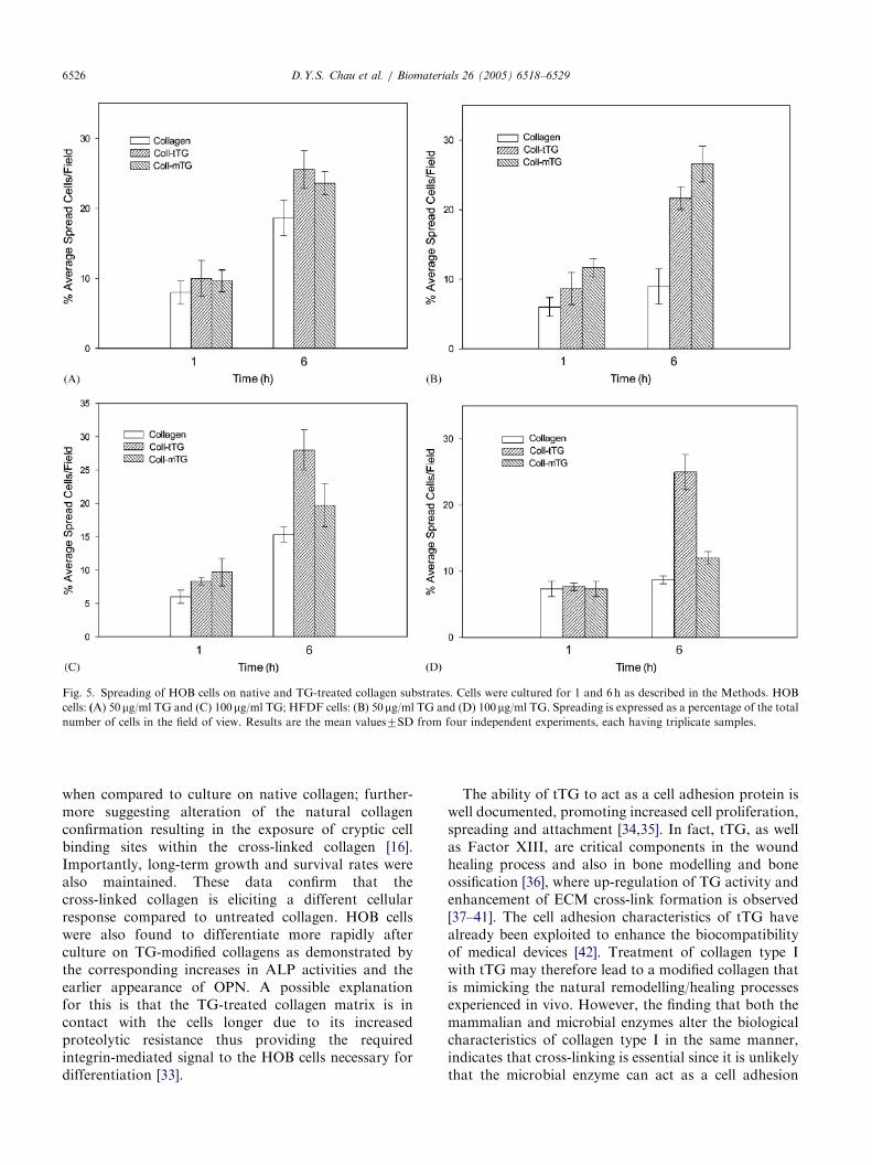
Figs. 3 and 5 show the short-term cell-spreadingcharacteristics of HOB and HFDF cells when culturedon native and TG-treated collagen. Increased numbersof cells spread when cultured on 50 mg/ml transglutami-nase cross-linked collagen. In the case of the HOB cells,a comparable increase of 5% in the spreading of theHOB cells, at each time point, is seen on both of the TG-treated collagens (Fig. 5A). In contrast, the HFDF cellsshowed significant non-spread cells on the 50 mg/ml TG-treated collagen—with increases of at least 10%observed for both of the TG-treated variants (Fig. 5B)(po0:05).
A further increase in the number of spread cells wasalso observed on cross-linked collagen using 100 mg/mltransglutaminase. In the case of HOB cells, an increaseof approximately 5% can be observed in spread cells(Fig. 5C). This behaviour increased with increasing time
ARTICLE IN PRESS
Fig. 3. Attachment and spreading of HOB and HFDF cells on native and TG-treated collagen type I. After 6 h incubation, cells were fixed using
3.7% (w/v) paraformaldehyde before being stained with May-Grunwald and Giemsa stains and then viewed at � 400 magnification.
D.Y.S. Chau et al. / Biomaterials 26 (2005) 6518–65296524
of culture. In contrast for the HFDF cells, althoughthere was still an increase in the spreading characteristicson the TG-treated collagen, a much more distinct andsignificant behaviour was identified on the tTG-treatedcollagen with spread cells increasing by 15% for manyof the time points. In contrast, the microbial-TG-treatedcollagen showed only a slight improvement in thespreading characteristics of cells (Fig. 5D) (po0:05).
3.7. Differentiation of HOB cells cultured on native and
TG-treated collagen
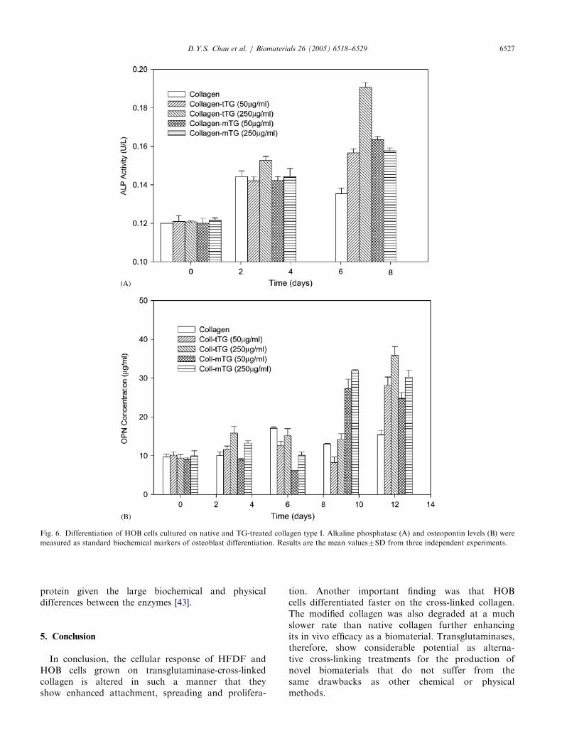
Increases in ALP activity were observed in all the TG-cross-linked collagens, with the greatest increase seenwith the tTG-treated collagen (Fig. 6A). A dose-dependent relationship between the concentration ofTG and the increase in the ALP activity of the HOBcells (po0:05) was observed. However, with the highestconcentration of mTG (250 mg/ml), there appeared to bea reduction in the corresponding amount of ALPactivity when compared to tTG. In addition, higherOPN levels were observed in all the TG-cross-linkedcollagens—with the greatest increases seen with the
250 mg/ml concentration of transglutaminase (Fig. 6B).This gave rise to a two-fold increase of OPN expressionover non-cross-linked collagen. In summary, a dose-dependent relationship between the concentration of TGand the increase in the OPN levels of the HOB cells(po0:05) was observed.
4. Discussion
We have confirmed previous work [25], in demon-strating that treatment of collagen type I matrices withtransglutaminases results in the incorporation of e(g-glutamyl)lysine, with both mTG and tTG introducingsimilar amounts of cross-link per unit activity. It hasbeen previously demonstrated that collagen type I showsgreater resistance to proteolytic degradation by matrixmetalloproteinase 1 (MMP-1) in vitro after cross-linkingby tTG [26]. Importantly, we have shown that collagenmodified with tTG and mTG demonstrated greaterresistance to the total complement of cell-secretedproteases and, as a consequence, improved resistanceto cell-mediated degradation from cultured HOB and
ARTICLE IN PRESS
Fig. 4. Attachment of HOB cells and HFDF on native and TG-treated collagen substrates. HOB cells: (A) 50mg/ml TG and (C) 100mg/ml TG;
HFDF cells: (B) 50 mg/ml TG and (D) 100mg/ml TG. Attachment is expressed as a percentage of the total number of attached cells after 6 h. Results
are the mean values7SD from four independent experiments, each having triplicate samples.
D.Y.S. Chau et al. / Biomaterials 26 (2005) 6518–6529 6525
HFDF cells. The MMP expression profile of HFDFcells was found to alter as a result of growth on cross-linked collagen, with a reduction of active MMP-1 anda corresponding increase in active MMP-2, whencompared to growth on untreated collagen. It is possiblethat this alteration in active MMP-1 accounts for theincreased resistance of cross-linked collagen to cellulardegradation. However, there is recent evidence toindicate that growth of either cultured hepatic stellatecells [27], rabbit periosteal fibroblasts [28] or humandermal fibroblasts [29] on collagen type I results in anincrease in active MMP-2, which is associated withincreased degradation of collagen in the matrix,independently of the collagenases [30]. In addition,changes in the mechanical characteristics of the col-lagen, which could be brought about by the introductionof e(g-glutamyl)lysine cross-link, can elicit mechano-chemical signalling via an integrin-dependent mechan-ism, resulting in alterations in gene expression, thus
accounting for the changes in active MMP expressionobserved [28,29,31]. The importance of the mechanicalnature of the matrix in MMP production is exemplifiedby the finding that bovine aortic or human umbilicalendothelial cells repress their secretion of MMP-2entirely when subjected to shear stress for as little as30min [32]. Transglutaminase-cross-linked collagenmay be more efficient than native collagen at inducingthis response to the ECM, either through the presence ofcross-links which may disturb the native conformation,or via disruption of the native fibrillar form duringfibrillogenesis. Alternatively, due to the increasedresistance of the cross-linked collagen to MMP degra-dation, fibroblasts may elicit an enhanced MMPresponse in a futile attempt to increase the rate ofcollagen breakdown.
The proliferation rates and the attachment andspreading characteristics of HFDF and HOB cells wereincreased after growth on TG-cross-linked collagen
ARTICLE IN PRESS
Fig. 5. Spreading of HOB cells on native and TG-treated collagen substrates. Cells were cultured for 1 and 6 h as described in the Methods. HOB
cells: (A) 50mg/ml TG and (C) 100mg/ml TG; HFDF cells: (B) 50mg/ml TG and (D) 100mg/ml TG. Spreading is expressed as a percentage of the total
number of cells in the field of view. Results are the mean values7SD from four independent experiments, each having triplicate samples.
D.Y.S. Chau et al. / Biomaterials 26 (2005) 6518–65296526
when compared to culture on native collagen; further-more suggesting alteration of the natural collagenconfirmation resulting in the exposure of cryptic cellbinding sites within the cross-linked collagen [16].Importantly, long-term growth and survival rates werealso maintained. These data confirm that thecross-linked collagen is eliciting a different cellularresponse compared to untreated collagen. HOB cellswere also found to differentiate more rapidly afterculture on TG-modified collagens as demonstrated bythe corresponding increases in ALP activities and theearlier appearance of OPN. A possible explanationfor this is that the TG-treated collagen matrix is incontact with the cells longer due to its increasedproteolytic resistance thus providing the requiredintegrin-mediated signal to the HOB cells necessary fordifferentiation [33].
The ability of tTG to act as a cell adhesion protein iswell documented, promoting increased cell proliferation,spreading and attachment [34,35]. In fact, tTG, as wellas Factor XIII, are critical components in the woundhealing process and also in bone modelling and boneossification [36], where up-regulation of TG activity andenhancement of ECM cross-link formation is observed[37–41]. The cell adhesion characteristics of tTG havealready been exploited to enhance the biocompatibilityof medical devices [42]. Treatment of collagen type Iwith tTG may therefore lead to a modified collagen thatis mimicking the natural remodelling/healing processesexperienced in vivo. However, the finding that both themammalian and microbial enzymes alter the biologicalcharacteristics of collagen type I in the same manner,indicates that cross-linking is essential since it is unlikelythat the microbial enzyme can act as a cell adhesion
ARTICLE IN PRESS
Fig. 6. Differentiation of HOB cells cultured on native and TG-treated collagen type I. Alkaline phosphatase (A) and osteopontin levels (B) were
measured as standard biochemical markers of osteoblast differentiation. Results are the mean values7SD from three independent experiments.
D.Y.S. Chau et al. / Biomaterials 26 (2005) 6518–6529 6527
protein given the large biochemical and physicaldifferences between the enzymes [43].
5. Conclusion
In conclusion, the cellular response of HFDF andHOB cells grown on transglutaminase-cross-linkedcollagen is altered in such a manner that theyshow enhanced attachment, spreading and prolifera-
tion. Another important finding was that HOBcells differentiated faster on the cross-linked collagen.The modified collagen was also degraded at a muchslower rate than native collagen further enhancingits in vivo efficacy as a biomaterial. Transglutaminases,therefore, show considerable potential as alterna-tive cross-linking treatments for the production ofnovel biomaterials that do not suffer from thesame drawbacks as other chemical or physicalmethods.
ARTICLE IN PRESSD.Y.S. Chau et al. / Biomaterials 26 (2005) 6518–65296528
Acknowledgements
The above work has been filed as a GB patentapplication and supported by grant number GR/521755/01 from the EPSRC and BLC Leather Technol-ogy Centre Ltd., Northampton, UK.
References
[1] Goo HC, Hwang YS, Choi YR, Cho HN, Suh H. Development of
collagenase-resistant collagen and its interaction with adult
human dermal fibroblasts. Biomaterials 2003;24(28):5099–113.
[2] Bell E, Ehrlich P, Buttle DJ, Nakatsuji T. Living tissue formed in
vitro and accepted as skin-equivalent of full thickness. Science
1981;221:1052.
[3] Burke JF, Yannas IV, Quimby WC, Bondoc CC, Jung WK.
Successful use of a physiologically acceptable artificial skin in the
treatment of extensive burn injury. Ann Surg 1981;194:413–48.
[4] Lee CH, Singla A, Lee Y. Biomedical applications of collagen. Int
J Pharm 2001;221(1–2):1–22.
[5] Matsuda S, Iwata H, Se N, Ikada Y. Bioadhesion of gelatin films
crosslinked with glutaraldehyde. J Biomed Mater Res 1999;45(1):
20–7.
[6] Ben-Slimane S, Guidoin R, Marceau D, Merhi Y, King MW,
Sigot-Luizard MF. In vivo evaluation of polyester arterial grafts
coated with albumin; the role and importance of cross-linking
agents. J Eur Surg Res 1988;20:18–28.
[7] Dunn MW, Stenzel KH, Rubin AL, Miyata T. Collagen implants
in the vitreous. Arch Ophthalmol 1969;82:840–4.
[8] Griffin M, Casadio R, Bergamini CM. Transglutaminases:
nature’s biological glues. J Biochem 2002;368:377–96.
[9] Collighan R, Cortez J, Griffin M. The biotechnological applica-
tions of transglutaminases. Minerva Biotechnol 2002;14(2):143–8.
[10] Broderick EP, O’Halloran DM, Rotchev YA, Griffin M,
Collighan RJ, Pandit AS. Enzymatic stabilisation of gelatin-
based scaffolds. J Biomed Mater Res Part B: Appl Biomater
2004;72B(1):37–42.
[11] Ito A, Mase A, Takizawa Y, Shinkai M, Honda H, Hata K, Ueda
M, Kobayashi T. Transglutaminase-mediated gelatin matrices
incorporating cell adhesion factors as a biomaterial for tissue
engineering. J Biosci Bioeng 2003;95(2):196–9.
[12] Aeschlimann D, Thommazy V. Protein crosslinking in assembly
and remodelling of extracellular matrices: the role of transgluta-
minases. Connect Tissue Res 2000;41:1–27.
[13] Griffin M, Casadio R, Bergamini CM. Transglutaminases:
nature’s biological glues. Biochem J 2002;368:377–96.
[14] Sechler JL, Schwarzbauer JE. Control of cell cycle progression
by fibronectin matrix architecture. J Biol Chem 1998;272:
25533–6.
[15] Singer AJ, Clark RA. Cutaneous wound healing. N Engl J Med
1999;341:738–46.
[16] Verderio E, Johnson T, Griffin M. Tissue transglutaminase in
normal and abnormal wound healing: review article. Amino Acids
2004;4:387–404.
[17] DiSilvio L. A novel application of two biomaterials for the
delivery of growth hormone, its effects on osteoblasts. PhD thesis,
University of London, UK, 1995.
[18] Jones RA, Nicholas B, Mian S, Davies PJA, Griffin M. Reduced
expression of tissue transglutaminase in a human endothelial cell
line leads to changes in cell spreading, cell adhesion and reduced
polymerisation of fibronectin. J Cell Sci 1997;110:2461–72.
[19] Leblanc A, Day N, Menard A, Keillor JW. Guinea pig liver
transglutaminase: a modified purification procedure affording
enzyme with superior activity in greater yield. Protein Expression
Purification 1999;17:89–95.
[20] Lorand L, Campbell-Wilkes LK, Cooperstein L. A filter paper
assay for transamidating enzymes using radioactive amine
substrates. Anal Biochem 1972;58:623–31.
[21] Griffin M, Wilson J. Detection of e-(g-glutamyl)lysine. Mol Cell
Biochem 1984;58:37–49.
[22] Holmbeck K, Bianco P, Caterina J, Yamada S, Kromer M,
Kuznetsov SA, Mankani M, Robey PG, Poole AR, Pidoux I,
Ward JM, Birkedal-Hansen H. MT1-MMP-deficient mice devel-
op dwarfism, osteopenia, arthritis and connective tissue disease
due to inadequate collagen turnover. Cell 1999;99:81–92.
[23] Lowry OH, Rosebrough NJ, Farr AL, Randall RJ. Protein
measurement with the folin phenol reagent. J Biol Chem 1951;
193:265–75.
[24] Herron GS, Banda MJ, Clark EJ, Gavrilovic J, Werb Z. Secretion
of metalloproteinases by stimulated capillary endothelial cells: II.
Expression of collagenase and stromelysin activities is regulated
by endogenous inhibitors. J Biol Chem 1986;261(6):2814–8.
[25] Collighan RJ, Li X, Parry J, Clara S, Griffin M. Transglutami-
nases as tanning agents for the leather industry. J Am Leather
Chem Assoc 2004;99(7):293–302.
[26] Johnson TS, Skill NJ, El Nahas AM, Oldroyd SD, Thomas GL,
Douthwaite JA, Haylor JL, Griffin M. Transglutaminase
transcription and antigen translocation in experimental renal
scarring. J Am Soc Nephrol 1999;10:2146–57.
[27] Wang D, Sato M, Li L, Miura M, Kojima N, Senoo H.
Stimulation of Pro-MMP-2 production and activation by native
form of extracellular type I collagen in cultured hepatic stellate
cells. Cell Struct Function 2003;28:505–13.
[28] Kessler D, Dethlefsen S, Haase I, Plomann M, Hirche F, Kreig T,
Eckes B. Fibroblasts in mechanically stressed collagen lattices
assume a ‘synthetic’ phenotype. J Biol Chem 2001;276(39):
36575–85.
[29] Zigrino P, Drescher C, Mauch C. Collagen-induced proMMP-2
activation by MT1-MMP in human dermal fibroblasts and the
possible role of alpha2beta integrins. Eur J Cell 2001;80(1):68–77.
[30] Kerkvliet EHM, Docherty AJP, Beertsen W, Everts V. Collagen
breakdown in soft connective tissue explants is associated with the
level of active gelatinase A (MMP-2) but not with collagenase.
Matrix Biol 1999;18:373–80.
[31] Ingber DE. Mechanobiology and diseases of mechanotransduc-
tion. Ann Med 2003;35(8):564–77.
[32] Yamane T, Yamaguchi N, Yoshida Y, Mitsumata M. Regulation
of the extracellular matrix production and degradation
of endothelial cells by shear stress. Int Congr Ser 2004;1262:
407–10.
[33] Mizuno M, Fujisawa R, Kuboki Y. Type I collagen-induced
osteoblastic differentiation of bone-marrow cells mediated by
collagen-alpha2beta1 integrin interaction. J Cell Physiol 2000;
184(2):207–13.
[34] Verderio E, Nicholas B, Gross S, Griffin M. Regulated expression
of tissue transglutaminase in Swiss 3T3 fibroblasts: effects on the
processing of fibronectin, cell attachment and cell death. Exp Cell
Res 1998;239:119–38.
[35] Verderio E, Coombes A, Jones RA, Li X, Heath D, Downes S,
Griffin M. Role of the cross-linking enzyme tissue transglutami-
nase in the biological recognition of synthetic biodegradable
polymers. J Biomed Mater Res 2000;54:294–304.
[36] Upchurch HF, Conway E, Patterson Jr MK, Maxwell MD.
Localisation of cellular transglutaminase on extracellular matrix
after wounding. Characteristics of the matrix bound enzyme.
J Cell Physiol 2001;149:375–82.
[37] Lorand L, Graham RM. Transglutaminases: crosslinking en-
zymes with pleiotropic functions. Nat Rev Mol Cell Biol 2003;4:
40–56.

ARTICLE IN PRESSD.Y.S. Chau et al. / Biomaterials 26 (2005) 6518–6529 6529
[38] Werner S, Grose R. Regulation of wound healing by growth
factors and cytokines. Physiol Rev 2003;83:835–70.
[39] Davis GE, Bayless KJ, Davis MJ, Meininger GA. Regulation
of tissue injury responses by the exposure of matricryptic sites
within extracellular matrix molecules. Am J Pathol 2000;56:
489–98.
[40] Inada R, Matsuki M, Yamada K, Morishima Y, Shen SC,
Kuramoto N, Yasuno H, Takahashi K, Miyachi Y, Yamanishi K.
Facilitated wound healing by activation of the Transglutaminase
1 gene. Am J Pathol 2000;157:1875–82.
[41] Haroon ZA, Hettasch JM, Lai TS, Dewhirst MW, Greenberg CS.
Tissue transglutaminase is expressed, active, and directly involved
in rat dermal wound healing and angiogenesis. FASEB J 1999;13:
1787–95.
[42] Heath DJ, Christian P, Griffin M. Involvement of tissue
transglutaminase in the stabilisation of biomaterial/tissue inter-
faces important in medical devices. Biomaterials 2002;23:1519–26.
[43] Motoki M, Okiyama A, Nonaka M, Tanaka H, Uchio R,
Matsuura A, Ando H, Umeda K. Transglutaminase, US Patent
No. 5156956, 1992.
Copyright © 2022 FDOKUMEN