Expression of unconventional myosin genes during neuronal development in zebrafish
10
Expression of unconventional myosin genes during neuronal development in zebrafish Vinoth Sittaramane a,b , Anand Chandrasekhar a,b,c, * a Division of Biological Sciences, Room 340D Bond Life Sciences Center, 1201 E. Rollins Street, University of Missouri, Columbia, MO 65211-7310, USA b Interdisciplinary Neuroscience Program, University of Missouri, Columbia, MO 65211, USA c Genetics Area Program, University of Missouri, Columbia, MO 65211, USA Received 25 September 2007; received in revised form 27 October 2007; accepted 30 October 2007 Available online 6 November 2007 Abstract Neuronal migration and growth cone motility are essential aspects of the development and maturation of the nervous system. These cellular events result from dynamic changes in the organization and function of the cytoskeleton, in part due to the activity of cytoskel- etal motor proteins such as myosins. Although specific myosins such as Myo2 (conventional or muscle myosin), Myo1, and Myo5 have been well characterized for roles in cell motility, the roles of the majority of unconventional (other than Myo2) myosins in cell motility events have not been investigated. To address this issue, we have undertaken an analysis of unconventional myosins in zebrafish, a pre- mier model for studying cellular and growth cone motility in the vertebrate nervous system. We describe the characterization and expres- sion patterns of several members of the unconventional myosin gene family. Based on available genomic sequence data, we identified 18 unconventional myosin- and 4 Myo2-related genes in the zebrafish genome in addition to previously characterized myosin (1, 2, 3, 5, 6, 7) genes. Phylogenetic analyses indicate that these genes can be grouped into existing classifications for unconventional myosins from mouse and man. In situ hybridization analyses using EST probes for 18 of the 22 identified genes indicate that 11/18 genes are expressed in a restricted fashion in the zebrafish embryo. Specific myosins are expressed in particular neuronal or neuroepithelial cell types in the developing zebrafish nervous system, spanning the periods of neuronal differentiation and migration, and of growth cone guidance and motility. Ó 2007 Elsevier B.V. All rights reserved. Keywords: Cytoskeleton; Unconventional myosin; Neuronal migration; Axon guidance; Growth cone motility; Zebrafish; Commissure; Spinal cord; Motor neuron; Neural crest; Somite; Ear; Eye; Morphogenesis; In situ hybridization; Phylogenetic tree; Hindbrain; Forebrain; Midbrain; Cranial muscles During embryogenesis, neuronal cell bodies and growth cones migrate extensively to establish precisely connected networks with target tissues. The functional properties of neural networks underlying physiology and behavior are critically dependent upon accurate positioning of neuronal cell bodies, and their axonal and dendritic processes. Not surprisingly, a number of brain disorders result from the loss of or aberrant migration of neurons and defective axon guidance (Copp and Harding, 1999; Gleeson and Walsh, 2000; Oster and Sretavan, 2003; ten Donkelaar et al., 2004). A large number of extracellular cues and their recep- tors have been intensively studied, and demonstrated to play essential roles in regulating the migration of neuronal growth cones and cell bodies (Chilton, 2006). Furthermore, the biochemical and molecular interactions between the signal transduction cascades initiated by these guidance cues and the cytoskeletal machinery that controls dynamic cellular and growth cone behaviors are being elucidated (Guan and Rao, 2003; Kalil and Dent, 2005). Nevertheless, the roles of specific components of the actin and microtu- bule cytoskeletons in generating particular responses to extracellular cues remain obscure. We are interested in 1567-133X/$ - see front matter Ó 2007 Elsevier B.V. All rights reserved. doi:10.1016/j.gep.2007.10.010 * Corresponding author. Address: Division of Biological Sciences, Room 340D Bond Life Sciences Center, 1201 E. Rollins Street, University of Missouri, Columbia, MO 65211-7310, USA. Tel.: +1 573 882 5166; fax: +1 573 884 9676. E-mail address: [email protected] (A. Chandrasekhar). www.elsevier.com/locate/gep Gene Expression Patterns 8 (2008) 161–170
-
Upload
georgiasouthern -
Category
Documents
-
view
0 -
download
0
Transcript of Expression of unconventional myosin genes during neuronal development in zebrafish

www.elsevier.com/locate/gep
Gene Expression Patterns 8 (2008) 161–170
Expression of unconventional myosin genes duringneuronal development in zebrafish
Vinoth Sittaramane a,b, Anand Chandrasekhar a,b,c,*
a Division of Biological Sciences, Room 340D Bond Life Sciences Center, 1201 E. Rollins Street, University of Missouri, Columbia, MO 65211-7310, USAb Interdisciplinary Neuroscience Program, University of Missouri, Columbia, MO 65211, USA
c Genetics Area Program, University of Missouri, Columbia, MO 65211, USA
Received 25 September 2007; received in revised form 27 October 2007; accepted 30 October 2007Available online 6 November 2007
Abstract
Neuronal migration and growth cone motility are essential aspects of the development and maturation of the nervous system. Thesecellular events result from dynamic changes in the organization and function of the cytoskeleton, in part due to the activity of cytoskel-etal motor proteins such as myosins. Although specific myosins such as Myo2 (conventional or muscle myosin), Myo1, and Myo5 havebeen well characterized for roles in cell motility, the roles of the majority of unconventional (other than Myo2) myosins in cell motilityevents have not been investigated. To address this issue, we have undertaken an analysis of unconventional myosins in zebrafish, a pre-mier model for studying cellular and growth cone motility in the vertebrate nervous system. We describe the characterization and expres-sion patterns of several members of the unconventional myosin gene family. Based on available genomic sequence data, we identified 18unconventional myosin- and 4 Myo2-related genes in the zebrafish genome in addition to previously characterized myosin (1, 2, 3, 5, 6, 7)genes. Phylogenetic analyses indicate that these genes can be grouped into existing classifications for unconventional myosins frommouse and man. In situ hybridization analyses using EST probes for 18 of the 22 identified genes indicate that 11/18 genes are expressedin a restricted fashion in the zebrafish embryo. Specific myosins are expressed in particular neuronal or neuroepithelial cell types in thedeveloping zebrafish nervous system, spanning the periods of neuronal differentiation and migration, and of growth cone guidance andmotility.� 2007 Elsevier B.V. All rights reserved.
Keywords: Cytoskeleton; Unconventional myosin; Neuronal migration; Axon guidance; Growth cone motility; Zebrafish; Commissure; Spinal cord;Motor neuron; Neural crest; Somite; Ear; Eye; Morphogenesis; In situ hybridization; Phylogenetic tree; Hindbrain; Forebrain; Midbrain; Cranial muscles
During embryogenesis, neuronal cell bodies and growthcones migrate extensively to establish precisely connectednetworks with target tissues. The functional properties ofneural networks underlying physiology and behavior arecritically dependent upon accurate positioning of neuronalcell bodies, and their axonal and dendritic processes. Notsurprisingly, a number of brain disorders result from theloss of or aberrant migration of neurons and defective axon
1567-133X/$ - see front matter � 2007 Elsevier B.V. All rights reserved.
doi:10.1016/j.gep.2007.10.010
* Corresponding author. Address: Division of Biological Sciences,Room 340D Bond Life Sciences Center, 1201 E. Rollins Street, Universityof Missouri, Columbia, MO 65211-7310, USA. Tel.: +1 573 882 5166; fax:+1 573 884 9676.
E-mail address: [email protected] (A. Chandrasekhar).
guidance (Copp and Harding, 1999; Gleeson and Walsh,2000; Oster and Sretavan, 2003; ten Donkelaar et al.,2004). A large number of extracellular cues and their recep-tors have been intensively studied, and demonstrated toplay essential roles in regulating the migration of neuronalgrowth cones and cell bodies (Chilton, 2006). Furthermore,the biochemical and molecular interactions between thesignal transduction cascades initiated by these guidancecues and the cytoskeletal machinery that controls dynamiccellular and growth cone behaviors are being elucidated(Guan and Rao, 2003; Kalil and Dent, 2005). Nevertheless,the roles of specific components of the actin and microtu-bule cytoskeletons in generating particular responses toextracellular cues remain obscure. We are interested in
162 V. Sittaramane, A. Chandrasekhar / Gene Expression Patterns 8 (2008) 161–170
characterizing the functions of the unconventional, non-muscle myosins in neuronal migration and axon guidance,and in elucidating whether these activities are regulated byguidance cues in the developing nervous system.
Unconventional non-muscle myosins are actin-bindingmotor proteins that lack the tail domain of conventionalmuscle myosin (myosin 2), and function in numerousprocesses including cell migration in lower eukaryotes,intracellular motility and trafficking, and sensory trans-duction (Libby and Steel, 2000; Tuxworth and Titus,2000; Wu et al., 2000; Soldati, 2003; Hirokawa andTakemura, 2003). Furthermore, unconventional myosinshave been localized to neuronal growth cones and regu-late growth cone motility in vitro (Wang et al., 1996;Evans et al., 1997; Suter et al., 2000; Diefenbach et al.,2002; Sousa et al., 2006). Phylogenetic analysis of thecatalytic head domain (containing the actin- and ATP-binding sites) of all available myosin heavy chainsequences shows that there are at least 18 families ofmyosins, including conventional (muscle) Myo2, severalmyosins found only in fungi or other lower eukaryotes(Myosins 4, 11, 12, 13, 14, and 17), and the plant-specificMyo8 (Hodge and Cope, 2000; Sellers, 2000; Berg et al.,2001; Reddy and Day, 2001; Volkmann et al., 2003). Inaddition to differences in the catalytic head domain,unconventional myosins belonging to various families
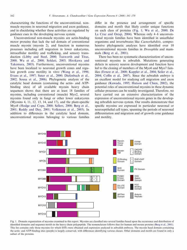
Fig. 1. Domain organization of myosins examined in this report. Myosins are cidentified domains and sequence motifs in the heavy chain polypeptide. The noThis list contains only those myosins for which ESTs were obtained and exprethe actin- and ATP-binding sites (purple) is largely conserved, with differencessubset of the proteins.
differ in the presence and arrangement of specificdomains and motifs that likely confer unique functionson each class of proteins (Fig. 1; Wu et al., 2000; DeLa Cruz and Ostap, 2004). Whereas only 4–6 unconven-tional myosin families have been identified in unicellularorganisms and invertebrates like Caenorhabditis, compre-hensive phylogenetic analyses have identified over 10unconventional myosin families in Drosophila and mam-mals (Berg et al., 2001).
There has been no systematic characterization of uncon-ventional myosins in zebrafish. Mutations generatingdefects in sensory neuron development and function haveled to the cloning of members of the Myo6 and Myo7 fam-ilies (Ernest et al., 2000; Kappler et al., 2004; Seiler et al.,2004; Coffin et al., 2007). Since the zebrafish embryo isan excellent model for studying cell migration and axonguidance (Kuwada, 1995; Hutson and Chien, 2002), thepotential roles of unconventional myosins in these dynamiccellular processes can be readily investigated. Therefore, wehave carried out an extensive characterization of theexpression of unconventional myosin genes in the develop-ing zebrafish nervous system. Our results demonstrate thatspecific myosins are expressed in particular neuronal orneuroepithelial cell types, spanning the periods of neuronaldifferentiation and migration and of growth cone guidanceand motility.
lassified into several families based upon the occurrence and distribution ofmenclature follows that for human and mouse proteins (Berg et al., 2001).ssion analyzed in zebrafish embryos. The myosin head domain containingidentifying various classes. Other domains and motifs are found in only a

V. Sittaramane, A. Chandrasekhar / Gene Expression Patterns 8 (2008) 161–170 163
1. Results and discussion
1.1. Phylogenetic analysis of zebrafish unconventional
myosins
Exhaustive search of the zebrafish genome assembly(Zv6, March 2006 and Zv7, July 2007) identified a totalof 22 myosin genes representing classes 1, 2, 5, 9, 10,15, 16, and 18. Zebrafish myosins 3, 6, and 7 wereexcluded from analyses because they have been describedpreviously, whereas other myosin classes (4, 8, 11, 12, 13,14, and 17) have been described only in fungi, other lowereukaryotes, or plants (Berg et al., 2001). In order to groupthe newly identified zebrafish (Danio rerio, Dr) myosinsinto appropriate classes, we constructed a phylogenetictree with human (Homo sapiens, Hs) and mouse (Mus
musculus, Mm) orthologs using amino acid sequences ofthe highly conserved core motor domains of various myo-sin classes, equivalent to residues 88–780 of chicken skel-etal myosin II (Hodge and Cope, 2000; Berg et al., 2001).Alignment of core domains was performed in Megalignusing ClustalW method (Reddy and Day, 2001; see Sec-tion 2 for additional details). A rectangular tree for thealigned sequences rooted to Myo1 (Fig. 2) was generatedby maximum parsimony method (Jiang and Ramachan-dran, 2004; see Section 2). The tree identifies the presenceof at least two genes each in the Myo1b, Myo1e, Myo5,Myo9a, Myo15, and Myo18 classes and three genes inthe Myo10 class in the available zebrafish genome. Ourstudy also identifies four conventional myo2 genes corre-sponding to different heavy chain genes as shown in thetree (Fig. 2). While most zebrafish myosins identified inthis study group with one of the classes of human andmouse myosins with >95% bootstrap value, the coremotor domains of zebrafish Myo9a1, Myo9a2, andMyo10b group with Myo16 and Myo18 with weak sup-port (51% bootstrap value; Fig. 2). However, when theentire polypeptide sequences are used for the phylogeneticanalysis, these genes group with their Myo9a and Myo10orthologs (100% bootstrap value; cladogram not shown),respectively, due to the presence of class specific domains(Fig. 1).
1.2. Expression analysis of zebrafish myosins
We carried out a detailed analysis of the expressionpatterns of various myosins for which ESTs were avail-able in 2004 (Table 1). This includes all zebrafish genesdescribed in Figs. 1 and 2, except myo1b1, myo1e2,myo5b, and myo15b, for which no ESTs were available,and myo3a (Thisse and Thisse, 2005), myo6 and myo7
whose expression patterns were previously described (Ern-est et al., 2000; Seiler et al., 2004; Kappler et al., 2004).We mostly examined expression patterns in embryos fixedat 18, 24, 30, and 36 hpf. Expression at earlier stages wasnot systematically examined since we are primarily inter-ested in potential roles for these molecules in neuronal
migration and axon guidance, and older ages were exam-ined as needed. The myosin genes exhibited restricted pat-terns of expression in various tissues. In the tissuesexamined, myo5a, myo10a, myo1b1 were expressed at allages tested; myo1e1 was expressed only between 30 and36 hpf; myo10b and myo18a were strongly expressedbetween 18 and 24 hpf, and weaker by 30 hpf; myo15a
was expressed only between 12 and 18 hpf; myo2hc1 wasexpressed strongly between 15 and 18 hpf, and weakerby 24–30 hpf; smyhc1, myo2hc2, and myo2hc9 wereexpressed in similar patterns between 18 and 36 hpf, withmyo2hc2 and smyhc1 expressed at least until 72 and120 hpf, respectively.
1.2.1. Expression in hindbrain segments
Two myosins are transiently expressed in a segmentedmanner in specific rhombomeres. At 18 hpf, myo10b isstrongly expressed in r5, and more weakly in r2, r3, andr4, and at the midbrain–hindbrain boundary (Fig. 3A).At 12 hpf, myo15a is expressed in r3, r5, and at the mid-brain–hindbrain boundary (Fig. 3B), with weaker expres-sion at 18 hpf (data not shown). Thus, these genes areexpressed in cells exhibiting sorting behaviors to set upwell-defined rhombomere boundaries (Cooke et al.,2005). From 30 to 36 hpf, myo1e1 is expressed in a smallpatch of mesendodermal cells located at the somite 2–3boundary (Fig. 3C), either the developing pancreas (Argen-ton et al., 1999; Stafford and Prince, 2002) or the proneph-ros (Drummond et al., 1998).
1.2.2. Expression in hindbrain neurons
Three myosins are expressed in specific neuronal popu-lations in the hindbrain. At 18 hpf, myo1b2 is expressedin a subset of the cranial sensory ganglia, as well as in asmall group of unidentified neurons in r2 (Fig. 3D). Anal-ysis of myo1b2 expression in the hedgehog pathway mutantdetour (gli1�/�; Vanderlaan et al., 2005) indicates thatthese cells are not cranial motor neurons (data not shown).At 36 hpf, myo1b2 is expressed by a small number of cellswithin each rhombomere, with strong expression in cellsflanking the boundaries of rhombomeres 5 and 6(Fig. 3G). At 18 hpf, myo5a is expressed in a subset ofthe cranial ganglia, and in differentiating neurons in everyrhombomere (Fig. 3E), similar to expression of the neuro-genesis marker huC (Bingham et al., 2003). By 24 hpf,myo5a expression expands into a continuous column, asadditional neurons differentiate in these regions(Fig. 3H). At 18 hpf, myo10a is expressed in the trigeminalganglion, and is also expressed at high levels in the lateralline ganglia by 30 hpf (Fig. 3F and I). In addition, myo10a
is expressed weakly by trigeminal and facial branchiomotorneurons from 18 to 30 hpf, and by other differentiatingneurons by 30 hpf (Fig. 3I). The expression of these myo-sins in cranial ganglia and in differentiating neurons coin-cides with axonogenesis or soma migration in theseneuronal populations (Metcalfe et al., 1990; Higashijimaet al., 2000).

Fig. 2. Phylogenetic relationship of unconventional myosin family members between zebrafish (Danio rerio), mouse (Mus musculus), and man (Homo
sapiens). Rectangular tree rooted to Myo1, with bootstrap values for family members that were examined in this report (see Section 2 for details). Myosinsfor which ESTs were characterized are identified by underlines. Those myosins among these that were expressed in zebrafish embryos (15–36 hpf) areidentified by bold type (Table 1). Zebrafish myosins outside these two categories but included in this phylogenetic tree were identified solely from databasesearches. Abbreviations: CaA, cardiac myosin A; Peri, perinatal skeletal myosin (Myhc8); Smyhc, slow myosin heavy chain.
164 V. Sittaramane, A. Chandrasekhar / Gene Expression Patterns 8 (2008) 161–170

Table 1Zebrafish myosin genes characterized in this report
Gene Ensembl ID Chromosome ESTs (source)*,% Length of ORF % Identity to Gene expressed
Human Mouse
myo1b2 ENSDARG00000024694 9 MPMGp609P206Q8 (B) MPMGp609G1826Q8 (B) A1G (A) Partial (90%) 81.4 81.5 Yesmyo1e1 ENSDARG00000036179 7 MPMGp609L0725Q8 (B) MPMGp609L0281Q8 (B) MPMGp637H137Q2
(B) IMAGp998P1112400Q3 (B) IMAGp998G1514300Q3 (B)Full-length 76.3 76.3 Yes
smyhc1 ENSDARG00000071430 24 MPMGp609B1310Q8 (B) Full-length 84.8 84.6 Yesmyo2hc1 (myhz1) ENSDARG00000067990 5 MPMGp609P1624Q8 (B) Full-length 80.8 82.1 Yesmyo2hc2 (myhz2) ENSDARG00000012944 5 MPMGp609M1825Q8 (B) Full-length 80.7 82.1 Yesmyo2hc9 (myhz9) ENSDARG00000001014 3 IMAGp998D0714289Q3 (B) Full-length 84.3 81.9 Yes
myo5a ENSDARG00000061635 18 129-E08-02 (S) Full-length 79.9 79.9 Yes
myo6a ENSDARG00000044016 20 IMAGp998H1712050Q3 (B) Full-length 80.9 80.6 No
myo9a1 ENSDARG00000019585 7 46-E08-02 (S) Partial (90%) 56.7 56.3 Nomyo9a2 ENSDARG00000063639 25 IMAGp998N0912391Q3 (B) Partial (80%) 54.8 54.3 No
myo9b ENSDARG00000062159 10 MPMGp609G0210Q8 (B) Full-length 57.3 58.1 No
myo10a ENSDARG00000062580 6 IMAGp998M2314313Q (B) Partial (98%) 61.6 62.1 Yesmyo10b ENSDARG00000017004 2 MPMGp609N1624Q8 (B) MPMGp609C1781Q8 (B) 88-A02-02 (S) Partial (84%) 68.3 67.7 Yesmyo10c ENSDARG00000063138 9 112-H11-02 (S) Full-length 62.1 62.6 No
myo15a ENSDARG00000059709 3 62-G10-02 (S) Full-length 62.2 61.2 Yes
myo16 ENSDARG00000004512 9 86-H05-02 (S) 100-C07-02 (S) Full-length 59.8 57.8 No
myo18a ENSDARG00000061862 15 DKFZp717N112Q2 (B) Full-length 78 76.8 Yesmyo18b ENSDARG00000061225 10 54-D04-02 (S) 98-C07-02 (S) Partial (87%) 73.7 73.3 No
* Source, B: RZPD, Berlin; S: Dr. Jinrong Peng, IMCB, Singapore; A: Research Genetics (Invitrogen), Alabama.% For genes with multiple ESTs, all ESTs were tested, and generated similar in situ results.
V.
Sitta
ram
an
e,A
.C
ha
nd
rasek
ha
r/
Gen
eE
xp
ression
Pa
tterns
8(
20
08
)1
61
–1
70
165

Fig. 3. Myosins exhibiting restricted expression patterns in the hindbrain. All panels show dorsal views of the hindbrain with anterior to the left. Asterisksmark the otic vesicle in each panel. (A and B) myo10b is expressed at varying levels in rhombomeres 2–5 (A), and myo15a is expressed in r3 and r5 (B), withboth expressed at the midbrain–hindbrain boundary (mhb). (C) myo1e1 is weakly but specifically expressed in a mesendodermal domain spanning the 2ndand 3rd somites (arrowhead). (D and G) At 18 hpf, myo1b2 is expressed in sensory ganglia (tg, ag) and a cluster of cells in r2 (arrowhead, D). By 36 hpf(G), there is prominent expression in cells (arrowheads) at the boundaries of r5 and r6. (E and H) At 18 hpf, myo5a is expressed in sensory ganglia, and indifferentiating neurons (arrowheads, E) within every rhombomere. By 24 hpf (H), expressing cells form a continuous column along the neural tube(arrowheads). (F and I) At 18 hpf, myo10a is expressed in the sensory ganglia (tg), and in the branchiomotor neurons in r2 and r5 (arrowheads, F). By30 hpf (I), expression persists in the sensory ganglia and migrating motor neurons (arrowhead), and has expanded to large clusters of cells in the anteriorhindbrain. tg, trigeminal ganglion; ag/pg, anterior and posterior lateral line ganglion. Scale bar, 75 lm.
166 V. Sittaramane, A. Chandrasekhar / Gene Expression Patterns 8 (2008) 161–170
1.2.3. Expression in the forebrain and midbrain
Two myosins, myo5a and myo10a, are expressed in spe-cific populations of forebrain and midbrain neurons(Fig. 4). In the forebrain, myo5a- and myo10a-expressingcells from 18 to 30 hpf (Fig. 4A–C; data not shown) appearto correspond to the telencephalic and diencephalic neu-rons that generate the anterior and post-optic commis-sures, respectively (Chitnis and Kuwada, 1990; Wilsonet al., 1990). The myo5a- and myo10a-expressing cells inthe midbrain are likely the nucleus of the medial longitudi-nal fasicle (nucMLF; Chitnis and Kuwada, 1990; Fig. 4A–C), and this was confirmed by anti-tubulin antibody label-ing of the MLF in embryos processed for myo10a in situs(Fig. 4D). Myosin gene expression in telencephalic andnucMLF neurons from 18 to 30 hpf coincides with the per-iod of growth cone motility and axon guidance in theseneurons. Myo2hc1 is strongly expressed at the midbrain–hindbrain boundary at 18 hpf, and becomes restricted toa narrow domain in the caudal optic tectum by 32 hpf(Fig. 4E and F).
1.2.4. Expression in the otic vesicle
In addition to myo6b and myo7a, whose expression pat-terns have been previously described (Ernest et al., 2000;Seiler et al., 2004; Kappler et al., 2004), myo10b andmyo2hc1 are also expressed in the otic vesicle. However,their expression is not restricted to the sensory neuronsof the epithelium as for myo6b and myo7a. At 18 hpf,
myo2hc1 transcripts are localized to the apical regions ofcells throughout the otic epithelium (Fig. 5B and D),whereas myo10b transcripts are localized to apical regionsof cells only in the rostroventral epithelium (Fig. 5A andC; see also Fig. 3A). These genes are expressed in the oticvesicle from 15 to 24 hpf, during morphogenesis of the ves-icle (Kimmel et al., 1995).
1.2.5. Expression in the eye
Three myosins are expressed in the eye, two in the devel-oping lens (myo1b2, myo10a), and one in the retina(myo2hc1). Interestingly, myo1b2 is expressed by cells onlyin the medial half of the developing lens (Fig. 6A and B),whereas myo10a is expressed only in the dorsal half ofthe developing lens (Fig. 6C; see also Fig. 4C). These local-ized patterns may reflect the progression of differentiationof lens fibers, or the inability of transcripts to distributeuniformly within differentiating fibers. At 18 hpf, myo2hc1
is strongly expressed in the medial layer of the optic cup(Fig. 4E) during the period of eye morphogenesis (Liet al., 2000), and becomes restricted to the ganglion celllayer by 32 hpf (Figs. 4F and 6D), when retinal axons areextending into the brain (Hutson and Chien, 2002).
1.2.6. Expression in the spinal cord and trunk
As expected, myo2hc2 is expressed strongly in all mus-cles (Figs. 7A and 8), and our probe labels expressing tissuemore effectively than previously described (Peng et al.,

Fig. 4. Myosin expression in the forebrain and midbrain. All panels showdorsal views of the head with anterior to the left. (A and B) At 18 hpf,myo5a is expressed in nascent neurons (arrowhead, A) in the forebrain,and in the nucMLF neurons (arrow) in the midbrain. By 24 hpf (B),forebrain expression has refined into distinguishable clusters representingthe neurons of the anterior and post-optic commissures (arrowheads, B),and is maintained in the nucMLF neurons (arrow). (C) At 30 hpf, myo10a
is expressed in the neurons of the nucMLF (arrow), and of the anteriorand post-optic commissures (arrowheads), and in the lens (see Fig. 6). (D)Double-labeling shows that tubulin antibody-labeled axons (arrowhead)of the MLF arise from the cluster of myo10a-expressing neurons (arrow),corresponding to the nucMLF. (E and F) At 18 hpf, myo2hc1 is expressedin the medial layer of the optic cup (arrowheads), and at the midbrain–hindbrain boundary (arrow). By 32 hpf (F), expression is restricted to theretinal ganglion layer (see Fig. 6), and to the caudal edge of the developingoptic tectum (arrow). Scale bar, 25 lm (D), 50 lm (A–F).
ig. 5. Myosin expression in the developing ear. Upper panels showorsal views, with anterior at top. Lower panels show cross-sectionsdorsal up) at the levels indicated in the upper panels. (A and C) myo10b isxpressed in epithelial cells (arrowhead) lining the anterior third (A), andhe ventral half (C) of the otic vesicle. (B and D) myo2hc1 is expressed inhe epithelial cells of the otic vesicle (arrowheads), adjacent to theindbrain. Asterisks (C and D) indicate the notochord. Scale bar, 30 lm.
Fig. 6. Myosin expression in the eye. (A and B) myo1b2 is expressed in thedifferentiating lens fibers (arrowhead, A) located in the medial half of thelens (B, dorsal view). (C) myo10a is expressed in a subset of lens cells at24 hpf, and at 30 hpf (see Fig. 4C). (D) myo2hc1 is expressed in retinalganglion cells (arrowhead) adjacent to the lens. Asterisks (A and C)indicate the choroid fissure. Scale bar (A, B, and D), 40 lm. Scale bar (C),30 lm.
V. Sittaramane, A. Chandrasekhar / Gene Expression Patterns 8 (2008) 161–170 167
2002). Interestingly, only one other myosin among 18tested was expressed in the presumptive musculature. At18 hpf, myo18a is expressed throughout the caudal (new-born) somites, and continues to be expressed strongly incells at the boundaries between the rostral (older) somites(Fig. 7B). At 18 hpf, myo1b2 is expressed in the superficialcells of the somites (Fig. 7C and D), and weakly expressedin a few cells in the spinal cord, likely neurons. At 18 hpf,myo5a is expressed by several presumptive neuronsthroughout the spinal cord, except in the most dorsal andventral regions (Fig. 7E and F). By 30 hpf, myo5a-express-ing cells are located mostly in the dorsal half of the neuraltube, with several putative migrating neural crest cells incharacteristic locations beside the somites (Fig. 7G andH). At 18 hpf, myo10a is expressed by cells in the dorsalspinal cord in putative neural crest cells (Fig. 7I and J).These results indicate that specific myosins are expressedin neurons and neural crest cells during the period of cellmigration and axon guidance.
Fd(etth
1.2.7. Expression of myo2hc2 in cranial muscles
Previous work (Peng et al., 2002) noted weak myo2hc2
(myhz2) expression in two cranial muscles. We found thatmyo2hc2 is expressed robustly in all cranial muscles(Fig. 8). By comparing the expression pattern to that ofanti-myosin antibody labeling of cranial muscles (Schilling
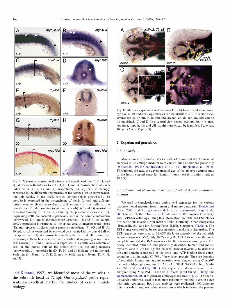
Fig. 8. Myosin2 expression in head muscles. (A) In a dorsal view, someeye (so, sr, lr) and jaw (lap) muscles can be identified. (B) In a side view,several eye (so, sr, mr, io, ir, am) and jaw (ah, ao, do, lap) muscles can bedistinguished. (C and D) In a ventral view, several eye (am, io, ir, lr, mr),jaw (ima, imp, ih, hh) and gill (tv, sh) muscles can be identified. Scale bar,100 lm (A–C), 50 lm (D).
Fig. 7. Myosin expression in the trunk and spinal cord. (A–C, E, G, andI) Side views with anterior to left. (D, F, H, and J) Cross-sections at levelsindicated in (C, E, G, and I), respectively. (A) myo2hc2 is stronglyexpressed in the differentiating muscles of the somites (white arrowheads),but only weakly in the newly formed somites (black arrowhead). (B)myo18a is expressed in the mesenchyme of newly formed and differen-tiating somites (black arrowhead), and strongly in the cells at theboundaries of older somites (white arrowheads). (C and D) myo1b2 isexpressed broadly in the trunk, excluding the presomitic mesoderm (C).Expressing cells are located superficially within the somitic mesoderm(arrowhead, D), and in the notochord (asterisk). (E and F) At 18 hpf,myo5a expression is restricted to the spinal cord at anterior trunk levels(E), and represents differentiating neurons (arrowhead, F). (G and H) At30 hpf, myo5a is expressed by scattered cells located in the dorsal half ofthe spinal cord (G). A cross-section in the anterior trunk (H) shows thatexpressing cells include neurons (arrowhead) and migrating neural crestcells (arrows). (I and J) myo10a is expressed in a continuous column ofcells in the dorsal half of the spinal cord (I), including neurons(arrowheads, J). Asterisks in (D, F, H, and J) indicate the notochord.Scale bar (I), 50 lm (A–C, E, G, and I). Scale bar (J), 50 lm (D, F, H,and J).
168 V. Sittaramane, A. Chandrasekhar / Gene Expression Patterns 8 (2008) 161–170
and Kimmel, 1997), we identified most of the muscles inthe zebrafish head at 72 hpf. Our myo2hc2 probe repre-sents an excellent marker for studies of cranial musclebiology.
2. Experimental procedures
2.1. Animals
Maintenance of zebrafish stocks, and collection and development ofembryos in E3 embryo medium were carried out as described previously(Westerfield, 1995; Chandrasekhar et al., 1997; Bingham et al., 2002).Throughout the text, the developmental age of the embryos correspondsto the hours elapsed since fertilization (hours post-fertilization, hpf, at28.5 �C).
2.2. Cloning and phylogenetic analysis of zebrafish unconventional
myosins
We used the nucleotide and amino acid sequences for the variousunconventional myosins from human and mouse databases (Hodge andCope, 2000, and http://www.mrc-lmb.cam.ac.uk/myosin/; Berg et al.,2001) to search the zebrafish EST databases at Washington Universityand RZPDfor orthologs. Using this information, we obtained EST clonesfor the various myosins from RZPD (Berlin, Germany), Open Biosystems(Huntsville, AL) and Dr. Jinrong Peng (IMCB, Singapore) (Table 1). TheEST clones were verified by sequencing prior to making in situ probes. TheEST sequences were used to BLAST the latest assembly of the zebrafishgenomic sequence (Zv7, July 2007) using BLASTN to retrieve the mostcomplete annotated cDNA sequences for the various myosin genes. Thenewly identified zebrafish and previously described human and mousemyosins were BLASTed against chicken skeletal myosin 2 to delineatethe core domain (composed of the actin- and ATP-binding sites) corre-sponding to amino acids 88–780 of the chicken protein. The core domainsof zebrafish, human and mouse myosins were aligned using ClustalWmethod in Megalign program of LASERGENE (DNASTAR Inc., Madi-son, WI; Reddy and Day, 2001). The aligned core domains were furtheranalyzed using Mac PAUP 4.0 b10 (http://paup.csit.fsu.edu/; Jiang andRamachandran, 2004) to generate a phylogenetic tree (Fig. 2). The heuris-tic search option was used in maximum parsimony method to create a treewith strict consensus. Bootstrap analyses were replicated 1000 times toobtain a robust support value at each node which indicates the percent

V. Sittaramane, A. Chandrasekhar / Gene Expression Patterns 8 (2008) 161–170 169
times that the grouping is supported out of 1000 trials (Hodge and Cope,2000; Sellers, 2000). Bootstrap values of greater than 70 indicate a stronglysupported node with 95% probability (Hills and Bull, 1993).
2.3. Histological procedures
The EST clones were verified by sequencing prior to probe synthesis.Synthesis of the digoxygenin-labeled probes, and whole-mount in situhybridization was carried out as described previously (Bingham et al.,2003). Given the large number of clones and time points, embryos of dif-ferent ages were pooled for the hybridization step following proteasetreatment. Whole-mount immunohistochemistry (tubulin and GFP anti-bodies) was performed as described previously (Vanderlaan et al.,2005). For combined in situ and antibody labeling, embryos were firstprocessed for in situs before antibody labeling. Embryos were deyolked,mounted in 70% glycerol, and examined with an Olympus BX60 micro-scope. At least 10 embryos were examined for each gene and time point.Images were acquired using an Optronics camera and Scion Imaging Soft-ware on a MacOS computer equipped with an LG3 frame grabber.Images were processed in Adobe Photoshop to adjust brightness and con-trast only.
Acknowledgements
We thank Jinrong Peng (IMCB, Singapore) for provid-ing several myosin ESTs used in our analysis, Tom Schil-ling (UC, Irvine) for help with muscle identification, andChris Pires and Robert Snyder (University of Missouri)for help with the phylogenetic analysis. We thank GaryVanderlaan for help in the early stages of the project,and Moe Baccam, Amy Foerstel, and other members ofour laboratory for excellent fish care. This work was sup-ported by an Interdisciplinary Neuroscience Program Fel-lowship from the University of Missouri GraduateSchool to V.S., and grants from the University of MissouriSystem (RB07-03) and NIH (NS40449) to A.C.
References
Argenton, F., Zecchin, E., Bortolussi, M., 1999. Early appearance ofpancreatic hormone-expressing cells in the zebrafish embryo. Mech.Dev. 87, 217–221.
Berg, J.S., Powell, B.C., Cheney, R.E., 2001. A millennial myosin census.Mol. Biol. Cell 12, 780–794.
Bingham, S., Chaudhari, S., Vanderlaan, G., Itoh, M., Chitnis, A.,Chandrasekhar, A., 2003. Neurogenic phenotype of mind bombmutants leads to severe patterning defects in the zebrafish hindbrain.Dev. Dyn. 228, 451–463.
Bingham, S., Higashijima, S., Okamoto, H., Chandrasekhar, A., 2002.The Zebrafish trilobite gene is essential for tangential migration ofbranchiomotor neurons. Dev. Biol. 242, 149–160.
Chandrasekhar, A., Moens, C.B., Warren Jr., J.T., Kimmel, C.B.,Kuwada, J.Y., 1997. Development of branchiomotor neurons inzebrafish. Development 124, 2633–2644.
Chilton, J.K., 2006. Molecular mechanisms of axon guidance. Dev. Biol.292, 13–24.
Chitnis, A.B., Kuwada, J.Y., 1990. Axonogenesis in the brain of zebrafishembryos. J. Neurosci. 10, 1892–1905.
Coffin, A.B., Dabdoub, A., Kelley, M.W., Popper, A.N., 2007. Myosin VIand VIIa distribution among inner ear epithelia in diverse fishes. Hear.Res. 224, 15–26.
Cooke, J.E., Kemp, H.A., Moens, C.B., 2005. EphA4 is required for celladhesion and rhombomere-boundary formation in the zebrafish. Curr.Biol. 15, 536–542.
Copp, A.J., Harding, B.N., 1999. Neuronal migration disorders inhumans and in mouse models – an overview. Epilepsy Res. 36,133–141.
De La Cruz, E.M., Ostap, E.M., 2004. Relating biochemistry and functionin the myosin superfamily. Curr. Opin. Cell Biol. 16, 61–67.
Diefenbach, T.J., Latham, V.M., Yimlamai, D., Liu, C.A., Herman, I.M.,Jay, D.G., 2002. Myosin 1c and myosin IIB serve opposing roles inlamellipodial dynamics of the neuronal growth cone. J. Cell Biol. 158,1207–1217.
Drummond, I.A., Majumdar, A., Hentschel, H., Elger, M., Solnica-Krezel, L., Schier, A.F., Neuhauss, S.C., Stemple, D.L., Zwartkruis,F., Rangini, Z., Driever, W., Fishman, M.C., 1998. Early developmentof the zebrafish pronephros and analysis of mutations affectingpronephric function. Development 125, 4655–4667.
Ernest, S., Rauch, G.J., Haffter, P., Geisler, R., Petit, C., Nicolson,T., 2000. Mariner is defective in myosin VIIA: a zebrafishmodel for human hereditary deafness. Hum. Mol. Genet. 9,2189–2196.
Evans, L.L., Hammer, J., Bridgman, P.C., 1997. Subcellular localizationof myosin V in nerve growth cones and outgrowth from dilute-lethalneurons. J. Cell Sci. 110, 439–449.
Gleeson, J.G., Walsh, C.A., 2000. Neuronal migration disorders: fromgenetic diseases to developmental mechanisms. Trends Neurosci. 23,352–359.
Guan, K.L., Rao, Y., 2003. Signalling mechanisms mediating neuronalresponses to guidance cues. Nat. Rev. Neurosci. 4, 941–956.
Higashijima, S., Hotta, Y., Okamoto, H., 2000. Visualization of cranialmotor neurons in live transgenic zebrafish expressing green fluorescentprotein under the control of the islet-1 promoter/enhancer. J.Neurosci. 20, 206–218.
Hirokawa, N., Takemura, R., 2003. Biochemical and molecular charac-terization of diseases linked to motor proteins. Trends Biochem. Sci.28, 558–565.
Hills, D.M., Bull, J.J., 1993. An empirical-test of bootstrapping as amethod for assessing confidence in phylogenetic analysis. Syst. Biol.42, 182–192.
Hodge, T., Cope, M.J., 2000. A myosin family tree. J. Cell Sci. 113, 3353–3354.
Hutson, L.D., Chien, C.B., 2002. Wiring the zebrafish: axon guidance andsynaptogenesis. Curr. Opin. Neurobiol. 12, 87–92.
Jiang, S., Ramachandran, S., 2004. Identification and molecular charac-terization of myosin gene family in Oryza sativa genome. Plant CellPhysiol. 45, 590–599.
Kalil, K., Dent, E.W., 2005. Touch and go: guidance cues signal to thegrowth cone cytoskeleton. Curr. Opin. Neurobiol. 15, 521–526.
Kappler, J.A., Starr, C.J., Chan, D.K., Kollmar, R., Hudspeth, A.J., 2004.A nonsense mutation in the gene encoding a zebrafish myosin VIisoform causes defects in hair-cell mechanotransduction. Proc. Natl.Acad. Sci. USA 101, 13056–13061.
Kimmel, C.B., Ballard, W.W., Kimmel, S.R., Ullmann, B., Schilling, T.F.,1995. Stages of embryonic development of the zebrafish. Dev. Dyn.203, 253–310.
Kuwada, J.Y., 1995. Development of the zebrafish nervous system: geneticanalysis and manipulation. Curr. Opin. Neurobiol. 5, 50–54.
Li, Z., Joseph, N.M., Easter Jr., S.S., 2000. The morphogenesis of thezebrafish eye, including a fate map of the optic vesicle. Dev. Dyn. 218,175–188.
Libby, R.T., Steel, K.P., 2000. The roles of unconventional myosins inhearing and deafness. Essays Biochem. 35, 159–174.
Metcalfe, W.K., Myers, P.Z., Trevarrow, B., Bass, M.B., Kimmel, C.B.,1990. Primary neurons that express the L2/HNK-1 carbohydrateduring early development in the zebrafish. Development 110, 491–504.
Oster, S.F., Sretavan, D.W., 2003. Connecting the eye to the brain: themolecular basis of ganglion cell axon guidance. Br. J. Ophthalmol. 87,639–645.
Peng, M.Y., Wen, H.J., Shih, L.J., Kuo, C.M., Hwang, S.P., 2002.Myosin heavy chain expression in cranial, pectoral fin, and tail

170 V. Sittaramane, A. Chandrasekhar / Gene Expression Patterns 8 (2008) 161–170
muscle regions of zebrafish embryos. Mol. Reprod. Dev. 63,422–429.
Reddy, A.S., Day, I.S., 2001. Analysis of the myosins encoded in therecently completed Arabidopsis thaliana genome sequence. GenomeBiol. 2, RESEARCH0024.
Schilling, T.F., Kimmel, C.B., 1997. Musculoskeletal patterning in thepharyngeal segments of the zebrafish embryo. Development 124, 2945–2960.
Seiler, C., Ben-David, O., Sidi, S., Hendrich, O., Rusch, A., Burnside, B.,Avraham, K.B., Nicolson, T., 2004. Myosin VI is required forstructural integrity of the apical surface of sensory hair cells inzebrafish. Dev. Biol. 272, 328–338.
Sellers, J.R., 2000. Myosins: a diverse superfamily. Biochim. Biophys.Acta 1496, 3–22.
Soldati, T., 2003. Unconventional myosins, actin dynamics and endocy-tosis: a menage a trois. Traffic 4, 358–366.
Sousa, A.D., Berg, J.S., Robertson, B.W., Meeker, R.B., Cheney, R.E.,2006. Myo10 in brain: developmental regulation, identification of aheadless isoform and dynamics in neurons. J. Cell Sci. 119, 184–194.
Stafford, D., Prince, V.E., 2002. Retinoic acid signaling is required for acritical early step in zebrafish pancreatic development. Curr. Biol. 12,1215–1220.
Suter, D.M., Espindola, F.S., Lin, C.H., Forscher, P., Mooseker, M.S.,2000. Localization of unconventional myosins V and VI in neuronalgrowth cones. J. Neurobiol. 42, 370–382.
ten Donkelaar, H.J., Lammens, M., Wesseling, P., Hori, A., Keyser, A.,Rotteveel, J., 2004. Development and malformations of the humanpyramidal tract. J. Neurol. 251, 1429–1442.
Thisse, C., Thisse, B., 2005. High Throughput Expression Analysis of ZF-Models Consortium Clones. ZFIN Direct Data Submission (http://zfin.org).
Tuxworth, R.I., Titus, M.A., 2000. Unconventional myosins: anchors inthe membrane traffic relay. Traffic 1, 11–18.
Vanderlaan, G., Tyurina, O.V., Karlstrom, R.O., Chandrasekhar, A.,2005. Gli function is essential for motor neuron induction in zebrafish.Dev. Biol. 282, 550–570.
Volkmann, D., Mori, T., Tirlapur, U.K., Konig, K., Fujiwara, T.,Kendrick-Jones, J., Baluska, F., 2003. Unconventional myosins of theplant-specific class VIII: endocytosis, cytokinesis, plasmodesmata/pit-fields, and cell-to-cell coupling. Cell Biol. Int. 27, 289–291.
Wang, F.S., Wolenski, J.S., Cheney, R.E., Mooseker, M.S., Jay, D.G.,1996. Function of myosin-V in filopodial extension of neuronal growthcones. Science 273, 660–663.
Westerfield, M., 1995. The Zebrafish Book. University of Oregon, Eugene,OR.
Wilson, S.W., Ross, L.S., Parrett, T., Easter, S.J., 1990. The developmentof a simple scaffold of axon tracts in the brain of the embryoniczebrafish, Brachydanio rerio. Development 108, 121–145.
Wu, X., Jung, G., Hammer 3rd, J.A., 2000. Functions of unconventionalmyosins. Curr. Opin. Cell Biol. 12, 42–51.