Control of Myosin motor activity and Actin filament translation ...

A New Role for Myosin II in Vesicle FissionJuan A. Flores., Santiago Balseiro-Gomez., Jose M. Cabeza, Jorge Acosta, Pilar Ramirez-Ponce, Eva Ales*
Dpto. Fisiologıa Medica y Biofısica, Facultad de Medicina, Universidad de Sevilla, Seville, Spain
Abstract
An endocytic vesicle is formed from a flat plasma membrane patch by a sequential process of invagination, bud formationand fission. The scission step requires the formation of a tubular membrane neck (the fission pore) that connects theendocytic vesicle with the plasma membrane. Progress in vesicle fission can be measured by the formation and closure ofthe fission pore. Live-cell imaging and sensitive biophysical measurements have provided various glimpses into thestructure and behaviour of the fission pore. In the present study, the role of non-muscle myosin II (NM-2) in vesicle fissionwas tested by analyzing the kinetics of the fission pore with perforated-patch clamp capacitance measurements to detectsingle vesicle endocytosis with millisecond time resolution in peritoneal mast cells. Blebbistatin, a specific inhibitor of NM-2,dramatically increased the duration of the fission pore and also prevented closure during large endocytic events. Using thefluorescent markers FM1-43 and pHrodo Green dextran, we found that NM-2 inhibition greatly arrested vesicle fission in alate phase of the scission event when the pore reached a final diameter of , 5 nm. Our results indicate that loss of theATPase activity of myosin II drastically reduces the efficiency of membrane scission by making vesicle closure incompleteand suggest that NM-2 might be especially relevant in vesicle fission during compound endocytosis.
Citation: Flores JA, Balseiro-Gomez S, Cabeza JM, Acosta J, Ramirez-Ponce P, et al. (2014) A New Role for Myosin II in Vesicle Fission. PLOS ONE 9(6): e100757.doi:10.1371/journal.pone.0100757
Editor: Ludger Johannes, Institut Curie, France
Received February 5, 2014; Accepted May 28, 2014; Published June 24, 2014
Copyright: � 2014 Flores et al. This is an open-access article distributed under the terms of the Creative Commons Attribution License, which permitsunrestricted use, distribution, and reproduction in any medium, provided the original author and source are credited.
Funding: This work was supported by the Ministerio de Ciencia e Innovacion ISCIII and Fondos FEDER Grant PI11/00257 (to E. A.). The funders had no role instudy design, data collection and analysis, decision to publish, or preparation of the manuscript.
Competing Interests: The authors have declared that no competing interests exist.
* Email: [email protected]
. These authors contributed equally to this work.
Introduction
Mast cells are specialized cells that respond to inflammatory
signals by secreting large amounts of a wide range of inflammatory
products. Some products, such as histamine, proteases and
proteoglycans, are stored in cytoplasmic secretory vesicles and
can be released by exocytosis upon stimulation, ensuring an
immediate and maximal biological effect [1]. Early ultrastructural
and electrophysiological studies showed that mast cell degranula-
tion involves compound exocytosis, which, in addition to the
fusion of vesicles at the plasma membrane, implicates the fusion of
vesicles in either a multivesicular or sequential manner to allow the
formation of degranulation channels [2,3]. After secretory vesicles
fuse with the plasma membrane, membrane retrieval must occur
to maintain a constant cell size and facilitate the reuse of vesicular
membrane components. Exocytosis in mast cells is followed by
several forms of compensatory endocytosis, including kiss-and-run
endocytosis and compound endocytosis, a mechanism by which
the compound cavity, formed by the cumulative fusion of many
secretory vesicles, is retrieved in a single membrane fission event
[4]. In these modes of exo-endocytosis, the fused vesicles are not
obliged to flatten, allowing the vesicles to be retrieved largely
intact. These mechanisms do not require the effort of the
invaginating membrane to form a deeply invaginated bud but
do require the constriction and fission of the endocytic tubular
neck to separate the vesicle from the plasma membrane and the
inward movement of the vesicle into the cytosol [5,6].
The mechanism by which vesicles separate from the plasma
membrane is called membrane scission, and this process requires
the large guanosine triphosphate hydrolase (GTPase) dynamin,
curvature sensing/inducing N-terminal helix-containing Bin/
Amphiphysin/Rvs (N-BAR) domain proteins and regulation by
the actin cytoskeleton [7]. Accumulating evidence indicates a
major role for actin in scission. Disruption of actin polymerization
causes an increase in the number of endocytic vesicles that are
unable to fully separate from the plasma membrane [8,9],
suggesting that actin polymerization is important for vesicle fission
during endocytosis by providing direct mechanical forces. The
formation of actin plumes at the constricted neck of the budding
vesicle might provide the necessary force for pushing the bud
deeper into the cytoplasm and may increase the strain on the stalk
until it severs. However, a recent study demonstrated that actin
polymerization does not provide direct mechanical forces for
vesicle fission by analyzing the kinetics of the endocytic tubular
membrane neck with capacitance measurements [10]. Other
recent findings have indicated the involvement of actin-binding
motor proteins in endocytosis, as in exocytosis. F-actin/myosin II
interactions govern vesicle transport and are essential to ensure
‘‘normal’’ vesicular fusion kinetics, ending in full collapse [11–14].
F-actin/myosin II may influence fusion by exerting mechanical
tension over the entire vesicle, or alternatively, this tension may
only affect a hypothetical cytoskeletal scaffold required for pore
dilation. By contrast, F-actin and NM-2 (or other myosin classes)
molecules may directly affect the proteins that are responsible for
the formation and expansion of the fusion pore [15]. Because NM-
2 has emerged as a regulator of the exocytic fusion pore, NM-2
could be playing an unsuspected role in fission pore closure.
Indeed, the depletion of NM-2 inhibits the scission of tubular
carrier precursors that are positive for Rab6 [16], and the loss of
PLOS ONE | www.plosone.org 1 June 2014 | Volume 9 | Issue 6 | e100757

NM-2 function causes defects in synaptic vesicle retrieval in
response to stimulation [17,18].
Rapid endocytosis can be distinguished kinetically from slower
clathrin-mediated endocytosis using capacitance measurements
[19] and by imaging techniques that assess the ability of a vesicle
to release or uptake fluorescent luminal probes [20–22]. In the
present study, we investigated the role of NM-2 in endocytosis by
perforated-patch capacitance measurements and the activity-
dependent markers FM1-43 and pHrodo Green dextran. By
inducing the loss of NM-2 through pharmacologic inhibition, we
found a new role for NM-2 in vesicle fission that predominantly
affects the compound endocytic mode.
Results
NM-2 inhibition affects individual fusion and fissionevents
The kinetics of fusion and fission of single vesicles were
determined by perforated-patch capacitance measurements.
Figures 1A and B show typical recordings from mast cells under
control conditions and NM-2 inhibition, respectively. The cells
were incubated in an extracellular solution containing blebbistatin
(5 mM) for , 10 min before the pipette was sealed onto the plasma
membrane. Blebbistatin is a NM-2 ATPase inhibitor that has been
found to bind to non-muscle myosin heavy chain IIA. Blebbistatin
blocks the myosin heads by forming a complex with low affinity for
actin [23,24]. The upward and downward capacitance steps,
which are associated with exocytosis and endocytosis, respectively,
displayed slower kinetics in the cells treated with blebbistatin. To
characterize the average fusion kinetics, we analyzed the rise time
(RT) and upward-slope (Su) for each upward step. Blebbistatin-
treated cells showed an increased rise time (RT = 63.166.2 ms;
n = 582; 19 cells) (Figure 1C) with a reduced upward-slope
(Su = 450616 fF/s) (Figure 1D) compared to control cells
(RT = 45.962.8 ms; Su = 696631 fF/s; n = 497; 23 cells; p,
0.001), suggesting slower individual release events. Similarly, the
kinetics of fission were analyzed by calculating the decay time (DT)
and downward-slope (Sd). The decay time of the downward steps
under NM-2 inhibition was also increased (DT = 7936220 ms;
n = 237) (Figure 1E) compared to the control cells (DT = 300643
ms; n = 293; p,0.001). However, the downward-slope was not
significantly different between the groups (Sd = 198616 fF/s in
control cells; Sd = 161611 fF/s in blebbistatin-treated cells)
(Figure 1F).
The number of exocytic events observed in the perforated-patch
recording and induced by stimulating with compound 48/80 was
similar in control cells (63.164.5%) and cells under NM-2
inhibition (68.365.4%). However, the number of flicker events
was reduced in blebbistatin-treated cells (12.161.8%) compared to
control cells (25.664%) (Figure 1G). The number of endocytic
events was not significantly different between the two groups.
However, the downward steps increased by 83% when the
inhibitor was present (11.362.5% versus 20.764.5%).
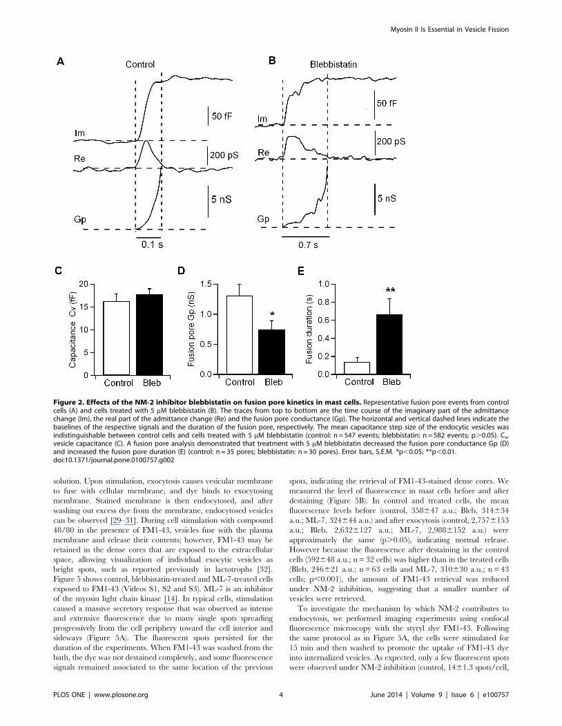
For a small subset of individual exocytic events, the increase in
the imaginary (Im) trace was associated with a detectable change
in the real (Re) trace, reflecting the detection of a narrow fusion
pore (Figure 2A), which was characterized by the fusion pore
conductance (Gp) and duration. From the Im and Re traces, the
time courses of Gp were calculated. We defined fusion pore
expansion as full when Gp was . 2 nS. We then examined the
fusion pore kinetics of exocytic events in cells treated with 5 mM
blebbistatin. Our results showed that cells treated with this
inhibitor displayed no changes in the capacitance step size of the
exocytic events (16.361.6 fF; n = 547 in 23 control cells and
17.761.7 fF; n = 582 events in 19 cells inhibited with blebbistatin)
(Figure 2C). As in control cells, the fusion pore conductance in
blebbistatin-treated cells grew above 2 nS (Figure 2B), indicating
that the fusion pore is opened [25,26] and the vesicle fuses fully
with the plasma membrane. However, the fusion pore Gp was
reduced in blebbistatin-treated cells (0.7560.15 nS; n = 30 pores)
compared to control cells (1.360.2 nS; n = 35 pores) (Figure 2D),
while the fusion pore duration was shorter in control cells
(0.1460.05 s) than in blebbistatin-treated cells (0.6760.17 s)
(Figure 2E), indicating a requirement for NM-2 in fusion pore
dynamics. Consistent with these results, the overexpression of an
nonphosphorylatable form of NM-2 in chromaffin cells has shown
that this protein is an important factor in the rapid kinetics of pore
expansion [12].
Given the capability of this molecular motor to act on fusion
pore expansion [12,13,27], we tested whether NM-2 has a direct
role in vesicle fission during endocytosis by examining the fission
pore kinetics of endocytic events in cells inhibited with blebbistatin
(Figure 3). Typically, the fission pore closure manifests itself as a
transient increase in the real part (Re) of the membrane
admittance on a time scale of 100–3000 ms and is associated
with a time-resolved decrease in the imaginary part (Im)
(Figure 3A). Under blebbistatin inhibition, the durations of the
transients in the Re trace were usually longer than 3000 ms
(Figure 3B), and long transients that increased gradually without
returning to the baseline were frequent (54% of pores) (Figure 3C).
The example shown in Figure 3C illustrates the unsuccessful
formation of an endocytic vesicle. A downward step is accompa-
nied by a change in the Re trace; however, instead of being
transient and vanishing when the vesicle membrane is excised
from the plasma membrane and the fission pore is completely
closed, the Re deflection reaches and remains at a stationary value
when the capacitance step drops to the minimum level. The
resulting pore conductance reveals the formation of a large vesicle
that does not close completely and whose capacitance size remains
unknown.
The mean step size of the downward steps was 35.562.9 fF
(n = 124 events) in control cells and 27.762.6 fF (n = 148 events) in
blebbistatin-treated cells, suggesting a significant change in vesicle
size (p,0.05). The distributions of the endocytic capacitance step
size and vesicle diameter were also different between control
(Figure 3D) and blebbistatin conditions (Figure 3E), indicating a
reduction in the number of large, but not small, endocytic events
in cells lacking NM-2 function. Thus, the effect of blebbistatin on
endocytosis is due to a decrease in the number of large events.
The dynamics of fission pore closure reflect the molecular events
leading to fission. Fission pore dynamics were quantified for large
endocytic vesicles by measuring the lifetime of each fission pore
and the fission pore conductance. Our results showed that the
duration of fission pore closure was longer in cells lacking NM-2
function (control, 1.060.2 s, n = 46 events; Bleb, 14.764.5 ms,
n = 26 events; p,0.001), indicating slow fission pore closure
(Figure 3F). To define better the effects of blebbistatin on fission
pore properties, we plotted the frequency distribution of Gp values
measured during fission pore closure. The frequency distribution
shows a large peak around 300–400 pS, indicating that the fission
pore keeps longer in a partly opened state in blebbistatin-treated
cells (Figures 3G and H). Figure 4 shows typical traces of the
fission pore conductance (Gp), fission pore diameter (Dp) and
fission pore resistance (Rp) of fission events in control and
blebbistatin-treated cells. In control cells, fission pore closure was
more rapid, and most fission events closed rapidly (,1 s). A typical
fission pore is analyzed in Figure 4A. This event lasted , 1 s, and
analysis of the Gp revealed that it can be best fit to a double
Myosin II Is Essential in Vesicle Fission
PLOS ONE | www.plosone.org 2 June 2014 | Volume 9 | Issue 6 | e100757

exponential, reflecting that the decrease in Gp occurs in phases. In
the initial phase, the conductance decreases rapidly from 12 to 2
nS for approximately 40 ms, and then a slower decrease with an
overall slope of approximately 1 pS/ms occurs. Finally, the Gp
decreases more steeply. We used Gp to estimate the fission pore
diameter (Dp) with the assumption that all conductance decreases
are due to changes in diameter (Figure 4C). The initial Dp (,15 nm) decreased to a final Dp of 3.8 nm during the first phase of
Gp decay. Second, the Dp decreased very slowly and approached
zero. The time–course of the fission pore resistance (Rp)
(Figure 4E) reveals a linear trend increase in the second phase
that suggests elongation of the pore at a constant speed. Finally, a
sudden increase in the resistance indicates fission pore closure.
The model that best explains the time-course of Gp has been
previously defined by three stages: 1) a rapid reduction in
diameter, 2) a relatively prolonged time period where the pore
grows in length and 3) a final rapid closure phase [28]. However,
in cells treated with blebbistatin, we observed a significant number
of fission pores (14 of 26 pores) that exhibited longer lifetimes
without the last phase of final closure, namely uncompleted fission
pores. A fission pore from a blebbistatin-treated cell is shown in
Figures 4B, D and F. This event lasted 15.9 s and shows a slow and
steady increase in fission pore resistance, but the abrupt final
increase that is observed in control pores was absent here. In
addition, the pore diameter decreased but remained at a final size
of 3.2 nm.
We further analyzed the effect of blebbistatin by examining
which phase of the fission pore closure kinetics is affected. The rate
of pore closure during the first phase was 37.669.4 GV/s (n = 46)
in control cells. This rate was significantly reduced in cells
inhibited by blebbistatin (16.465.1 GV/s; n = 26; p,0.05)
(Figure 4G). During the second phase, the Rp in control cells
can be fitted with a straight line and an average slope of
9.862.2 GV/s (Figure 4H). This rate is significantly faster than
the rate observed with blebbistatin (0.660.2 GV/s; p,0.001).
Together, these data suggest that blebbistatin has a predominant
effect on the fission pore lifetime and determines the final
membrane excision.
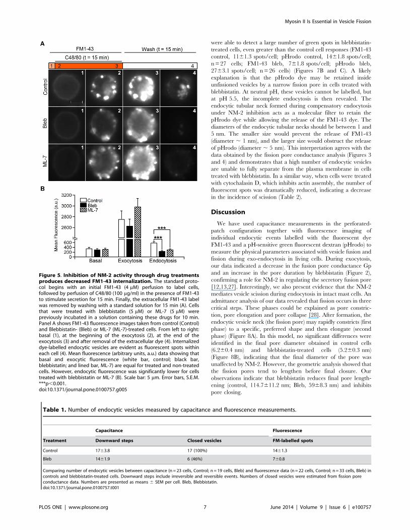
NM-2 inhibition blocks vesicle membrane recapturefollowing exocytosis
Previously, we established that mast cell vesicles stain with the
fluorescent dye FM1-43 [4]. FM1-43 and similar styryl dyes have
proven useful as probes for membrane trafficking because they
reversibly stain membranes, are impermeable to membranes, and
are more fluorescent when bound to membranes than when in
Figure 1. Inhibition of NM-2 activity through acute blebbistatin treatment slows the kinetics of exocytosis and endocytosis.Representative traces of capacitance steps from a control (A) and blebbistatin-treated cell (B). Re and Im represent the real and imaginary parts,respectively, of the admittance change. The steps labelled 1–4 at higher magnification show single downward steps, upwards steps and capacitanceflickers (arrows). The kinetic parameters of the capacitance steps demonstrate that blebbistatin treatment affects the rise time (C) and upward-slope(D) to prolong the duration of the exocytic event. Blebbistatin also produces a increase in the decay time (E) without significantly affecting thedownward-slope (F). The proportion of capacitance flickers (reversible fusion) decreased in blebbistatin-treated cells versus control cells (G) (p,0.05).Error bars, S.E.M. ***p,0.01.doi:10.1371/journal.pone.0100757.g001
Myosin II Is Essential in Vesicle Fission
PLOS ONE | www.plosone.org 3 June 2014 | Volume 9 | Issue 6 | e100757
solution. Upon stimulation, exocytosis causes vesicular membrane
to fuse with cellular membrane, and dye binds to exocytosing
membrane. Stained membrane is then endocytosed, and after
washing out excess dye from the membrane, endocytosed vesicles
can be observed [29–31]. During cell stimulation with compound
48/80 in the presence of FM1-43, vesicles fuse with the plasma
membrane and release their contents; however, FM1-43 may be
retained in the dense cores that are exposed to the extracellular
space, allowing visualization of individual exocytic vesicles as
bright spots, such as reported previously in lactotrophs [32].
Figure 5 shows control, blebbistatin-treated and ML-7-treated cells
exposed to FM1-43 (Videos S1, S2 and S3). ML-7 is an inhibitor
of the myosin light chain kinase [14]. In typical cells, stimulation
caused a massive secretory response that was observed as intense
and extensive fluorescence due to many single spots spreading
progressively from the cell periphery toward the cell interior and
sideways (Figure 5A). The fluorescent spots persisted for the
duration of the experiments. When FM1-43 was washed from the
bath, the dye was not destained completely, and some fluorescence
signals remained associated to the same location of the previous
spots, indicating the retrieval of FM1-43-stained dense cores. We
measured the level of fluorescence in mast cells before and after
destaining (Figure 5B). In control and treated cells, the mean
fluorescence levels before (control, 358647 a.u.; Bleb, 314634
a.u.; ML-7, 324644 a.u.) and after exocytosis (control, 2,7576153
a.u.; Bleb, 2,6326127 a.u.; ML-7, 2,9886152 a.u.) were
approximately the same (p.0.05), indicating normal release.
However because the fluorescence after destaining in the control
cells (592648 a.u.; n = 32 cells) was higher than in the treated cells
(Bleb, 246621 a.u.; n = 63 cells and ML-7, 310630 a.u.; n = 43
cells; p,0.001), the amount of FM1-43 retrieval was reduced
under NM-2 inhibition, suggesting that a smaller number of
vesicles were retrieved.
To investigate the mechanism by which NM-2 contributes to
endocytosis, we performed imaging experiments using confocal
fluorescence microscopy with the styryl dye FM1-43. Following
the same protocol as in Figure 5A, the cells were stimulated for
15 min and then washed to promote the uptake of FM1-43 dye
into internalized vesicles. As expected, only a few fluorescent spots
were observed under NM-2 inhibition (control, 1461.3 spots/cell,
Figure 2. Effects of the NM-2 inhibitor blebbistatin on fusion pore kinetics in mast cells. Representative fusion pore events from controlcells (A) and cells treated with 5 mM blebbistatin (B). The traces from top to bottom are the time course of the imaginary part of the admittancechange (Im), the real part of the admittance change (Re) and the fusion pore conductance (Gp). The horizontal and vertical dashed lines indicate thebaselines of the respective signals and the duration of the fusion pore, respectively. The mean capacitance step size of the endocytic vesicles wasindistinguishable between control cells and cells treated with 5 mM blebbistatin (control: n = 547 events; blebbistatin: n = 582 events; p.0.05). Cv,vesicle capacitance (C). A fusion pore analysis demonstrated that treatment with 5 mM blebbistatin decreased the fusion pore conductance Gp (D)and increased the fusion pore duration (E) (control: n = 35 pores; blebbistatin: n = 30 pores). Error bars, S.E.M. *p,0.05; **p,0.01.doi:10.1371/journal.pone.0100757.g002
Myosin II Is Essential in Vesicle Fission
PLOS ONE | www.plosone.org 4 June 2014 | Volume 9 | Issue 6 | e100757

n = 22 cells and Bleb, 760.8 spots/cell, n = 33 cells; p,0.001)
(Figures 6A and C). This decrease in the number of endocytosis
using FM1-43 in cells treated with blebbistatin don’t agree with
capacitance data. Capacitance measurements allow to detect the
formation of an endocytic vesicle as a downward step (Figure 1).
Therefore, the number of capacitance downward steps usually
reflects the number of endocytic events. However, the analysis of
the fission pore conductance that reveals the dynamics of
individual fission pore closures indicates that a 54% of resolved
fission pores in the presence of blebbistatin were unclosed
(Figure 3). According to this proportion, capacitance and
fluorescence observations agree very closely (Table 1).
To estimate the size of the internalized membrane, we next
measured the area of the spots at different single sections obtained
in the Z plane. In control cells, a large number of fluorescent spots
showed an increase by two, three or greater fold of the area of a
single spot (Figure 6B), indicating the compound endocytosis of
several vesicles by a single fission event. The size of the fluorescent
spot area varied but was smaller in treated cells than in control
cells (control, 1.2860.13 mm2, 257 spots, n = 22 cells; Bleb,
0.9860.07 mm2, 237 spots, n = 29 cells; p,0.05) (Figure 6D).
The distribution of the spot area showed that large endocytic spots
greater than 2 mm2 were almost non-existent in blebbistatin-
treated cells (Figure 6E and 6F). Interestingly, these data are
consistent with the distribution of the downward steps as measured
by capacitance (Figure 3D and E). Together, these results suggest
that the inhibition of endocytosis by blebbistatin primarily affects
the mode of compound endocytosis.
Blebbistatin hinders complete vesicle closureWe next investigated the incomplete fission events using FM1-
43 and pHrodo Green dextran simultaneously. pHrodo Green is a
10,000 MW dextran-conjugated, pH-sensitive form of rhodamine
that exhibits green fluorescence after endocytosis into the acidic
interior of vesicles. We stimulated the cell preparation with
Figure 3. Effects of the NM-2 inhibitor blebbistatin on fission pore kinetics in mast cells. Representative fission pore events from controlcells (A) and cells treated with 5 mM blebbistatin (B, C). The traces from top to bottom are the time course of the imaginary part of the admittancechange (Im), the real part of the admittance change (Re) and the fission pore conductance (Gp). The horizontal and vertical dashed lines indicate thebaselines of the respective signals and the duration of the fission pore, respectively. Two types of individual endocytic events were detected usingperforated-patch capacitance measurements under NM-2 inhibition: a long event with detectable membrane scission (B) and an endocytic eventwithout detectable pore closure, i.e., when a vesicle tries to undergo fission without closing completely (C). The change in the real trace during thistype of event cannot be the result of an incorrect phase setting as demonstrated by the separation of Im and Re during a pure capacitance increaseof 100 fF that was applied for online phase calibration (start of Im trace; Cal). The distribution of vesicle diameters were derived from endocyticcapacitance step sizes from control cells (D, white) and blebbistatin-treated cells (E, black). Large downward steps associated with the retrieval oflarge areas of membrane (greater than one vesicle) were largely abolished (control: n = 124 events; blebbistatin: n = 148 events). Blebbistatinproduced an increase in the fission pore duration compared to control cells (F). Frequency distribution of Gp values during fission pore closure ofendocytic events from control cells (G, white) and blebbistatin-treated cells (H, black). Error bars, S.E.M. ***p,0.001.doi:10.1371/journal.pone.0100757.g003
Myosin II Is Essential in Vesicle Fission
PLOS ONE | www.plosone.org 5 June 2014 | Volume 9 | Issue 6 | e100757

compound 48/80 for 15 min in the presence of FM1-43 and
pHrodo. After washing with standard solution (pH = 7.25), bright
spots reflecting internalized vesicles were observed in red and
green images and overlapped very closely (Figure 7B). As
expected, the number of fluorescent spots with both FM1-43
and pHrodo at pH 7.25 in the presence of blebbistatin was
scarce (FM1-43 control, 1461.3 spots/cell; pHrodo control,
1262.2 spots/cell, n = 22 cells; FM1-43 bleb, 760.8 spots/cell;
pHrodo bleb, 660.8 spots/cell, n = 33 cells). However, when we
used an acidic extracellular solution after removing the dyes, we
Figure 4. Blebbistatin affects fission pore dynamics. The time–course of Gp showed three different kinetic phases in control cells (A). A doubleexponential function depicts the two initial phases (red dashed line). A more pronounced Gp decrease accounts for the third phase (not appreciablein this magnification). This last phase is not present in the fission pore from the cell treated with blebbistatin (B). Fission pore diameters (Dp) fromcontrol (C) and treated cells (D) were calculated from the conductance data with the assumption that all conductance decreases result from changesin the diameter (assuming a pore length of 15 nm, which corresponds to the thickness of two membrane bilayers). A time–course of fission poreresistance (Rp) was calculated from the conductance data (E and F). The second phase could be fitted to a straight line in both the control pore (E)and the pore inhibited with blebbistatin (F) (dashed line; Pearson’s correlation coefficients: 0.89 and 0.95, respectively). The sudden increase in poreresistance indicated that the final pore closure (third phase) was absent from the pores in most cells lacking NM-2 function. The pore closure ratesduring the first (G) and second (H) phases in control conditions were higher than blebbistatin-treated pores. Error bars, S.E.M. *p,0.05; ***p,0.001.doi:10.1371/journal.pone.0100757.g004
Myosin II Is Essential in Vesicle Fission
PLOS ONE | www.plosone.org 6 June 2014 | Volume 9 | Issue 6 | e100757
were able to detect a large number of green spots in blebbistatin-
treated cells, even greater than the control cell responses (FM1-43
control, 1161.3 spots/cell; pHrodo control, 1461.8 spots/cell;
n = 27 cells; FM1-43 bleb, 761.8 spots/cell; pHrodo bleb,
2763.1 spots/cell; n = 26 cells) (Figures 7B and C). A likely
explanation is that the pHrodo dye may be retained inside
unfissioned vesicles by a narrow fission pore in cells treated with
blebbistatin. At neutral pH, these vesicles cannot be labelled, but
at pH 5.5, the incomplete endocytosis is then revealed. The
endocytic tubular neck formed during compensatory endocytosis
under NM-2 inhibition acts as a molecular filter to retain the
pHrodo dye while allowing the release of the FM1-43 dye. The
diameters of the endocytic tubular necks should be between 1 and
5 nm. The smaller size would prevent the release of FM1-43
(diameter , 1 nm), and the larger size would obstruct the release
of pHrodo (diameter , 5 nm). This interpretation agrees with the
data obtained by the fission pore conductance analysis (Figures 3
and 4) and demonstrates that a high number of endocytic vesicles
are unable to fully separate from the plasma membrane in cells
treated with blebbistatin. In a similar way, when cells were treated
with cytochalasin D, which inhibits actin assembly, the number of
fluorescent spots was dramatically reduced, indicating a decrease
in the incidence of scission (Table 2).
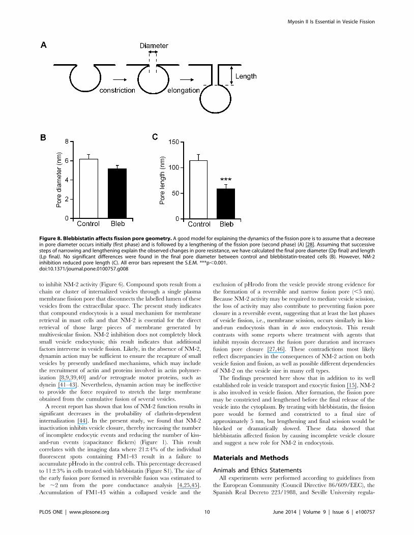
Discussion
We have used capacitance measurements in the perforated-
patch configuration together with fluorescence imaging of
individual endocytic events labelled with the fluorescent dye
FM1-43 and a pH-sensitive green fluorescent dextran (pHrodo) to
measure the physical parameters associated with vesicle fusion and
fission during exo-endocytosis in living cells. During exocytosis,
our data indicated a decrease in the fusion pore conductance Gp
and an increase in the pore duration by blebbistatin (Figure 2),
confirming a role for NM-2 in regulating the secretory fusion pore
[12,13,27]. Interestingly, we also present evidence that the NM-2
mediates vesicle scission during endocytosis in intact mast cells. An
admittance analysis of our data revealed that fission occurs in three
critical steps. These phases could be explained as pore constric-
tion, pore elongation and pore collapse [28]. After formation, the
endocytic vesicle neck (the fission pore) may rapidly constricts (first
phase) to a specific, preferred shape and then elongate (second
phase) (Figure 8A). In this model, no significant differences were
identified in the final pore diameter obtained in control cells
(6.260.4 nm) and blebbistatin-treated cells (5.260.3 nm)
(Figure 8B), indicating that the final diameter of the pore was
unaffected by NM-2. However, the geometric analysis showed that
the fission pores tend to lengthen before final closure. Our
observations indicate that blebbistatin reduces final pore length-
ening (control, 114.7611.2 nm; Bleb, 5968.3 nm) and inhibits
pore closing.
Figure 5. Inhibition of NM-2 activity through drug treatmentsproduces decreased FM1-43 internalization. The standard proto-col begins with an initial FM1-43 (4 mM) perfusion to label cells,followed by perfusion of C48/80 (100 mg/ml) in the presence of FM1-43to stimulate secretion for 15 min. Finally, the extracellular FM1-43 labelwas removed by washing with a standard solution for 15 min (A). Cellsthat were treated with blebbistatin (5 mM) or ML-7 (5 mM) werepreviously incubated in a solution containing these drugs for 10 min.Panel A shows FM1-43 fluorescence images taken from control (Control)and Blebbistatin- (Bleb) or ML-7 (ML-7)-treated cells. From left to right:basal (1), at the beginning of the exocytosis (2), at the end of theexocytosis (3) and after removal of the extracellular dye (4). Internalizeddye-labelled endocytic vesicles are evident as fluorescent spots withineach cell (4). Mean fluorescence (arbitrary units, a.u.) data showing thatbasal and exocytic fluorescence (white bar, control; black bar,blebbistatin; and lined bar, ML-7) are equal for treated and non-treatedcells. However, endocytic fluorescence was significantly lower for cellstreated with blebbistatin or ML-7 (B). Scale bar: 5 mm. Error bars, S.E.M.***p,0.001.doi:10.1371/journal.pone.0100757.g005
Table 1. Number of endocytic vesicles measured by capacitance and fluorescence measurements.
Capacitance Fluorescence
Treatment Downward steps Closed vesicles FM-labelled spots
Control 1763.8 17 (100%) 1461.3
Bleb 1461.9 6 (46%) 760.8
Comparing number of endocytic vesicles between capacitance (n = 23 cells, Control; n = 19 cells, Bleb) and fluorescence data (n = 22 cells, Control; n = 33 cells, Bleb) incontrols and blebbistatin-treated cells. Downward steps include irreversible and reversible events. Numbers of closed vesicles were estimated from fission poreconductance data. Numbers are presented as means 6 SEM per cell. Bleb, Blebbistatin.doi:10.1371/journal.pone.0100757.t001
Myosin II Is Essential in Vesicle Fission
PLOS ONE | www.plosone.org 7 June 2014 | Volume 9 | Issue 6 | e100757

Some vesicles fused reversibly with the plasma membrane as
indicated by an upward (exocytic) capacitance deflection followed
by a downward (endocytic) event of similar magnitude (Figure 1).
These kiss-and-run events accounted for 25.6% of the total events
(black circles, Figure 1G). This value was reduced to 12.1% upon
exposure of the cells to blebbistatin. Concomitantly, the number of
downward steps was increased in the presence of blebbistatin
(11.3% versus 20.7%). Surprisingly, the number of FM1-43 and
pHrodo spots in cells exposed to the inhibitor at pH 7.2 was
strongly reduced (Figure 6), suggesting that endocytosis was
inhibited. This apparent discrepancy was resolved using acidic
solution to discover the presence of pHrodo trapped inside frozen
vesicles in a late step of endocytosis when the fission pore is formed
and constricted (Figure 7). At pH 5.5, we were able to observe an
even greater number of pHrodo spots in treated cells than in
control cells in agreement with the electrophysiological data and
Figure 6. Compound endocytosis is inhibited by blebbistatin. The left images show brightfield single confocal sections, while the rightimages show Z-stack projections of endocytic spots labelled with FM1-43 (A). The data are from control cells (Control) and cells treated withblebbistatin (Bleb) (5 mM). In panel B, the upper pictures (1–4) show a sequence obtained gradually (at 2 mm separation) from the control cell shownin A. The lower pictures show an enlarged view of groups of endocytic vesicles (enclosed by a box in images 1–4) (B). A bar graph shows that thenumber of FM1-43 spots per cell in control (white) and blebbistatin conditions (black) was significantly different (C). The area of FM1-43 spots per cellwas significantly reduced by blebbistatin (black) compared to control cells (white) (D). Histograms of the spot area for control conditions (E, white)and blebbistatin treatment (F, black). Scale bars: 5 and 1 (insets) mm. Error bars, S.E.M. *p,0.05; ***p,0.001.doi:10.1371/journal.pone.0100757.g006
Table 2. Number of endocytic vesicles labelled with FM1-43 and pHrodo Green dyes.
FM1-43-labelled spots/cell pHrodo-labelled spots/cell
treatment pH 7.25 pH 5.5 pH 7.25 pH 5.5
Control 1461.3 1161.3 1262.2 1461.8
Bleb 760.8 761.8 660.8 2763.1
variation 250% (0.000) 237% (0.000) 250% (0.000) + 93% (0.001)
Cyto 460.9 460.9 160.3 1462.9
variation 271% (0.000) 264% (0.000) 292% (0.000) 0% (0.479)
Comparing control (22 cells at pH 7.25 and 27 cells at pH 5.5) to exposure to 5 mM Bleb (33 cells at pH 7.25 and 26 cells at pH 5.5) and to 4 mM Cyto (21 cells at pH 7.25and pH 5.5). The pairs of data sets were compared using a two-tailed U-Mann-Whitney rank-sum test, and the result is indicated, between brackets, next to the variationof the mean. Numbers are presented as means 6 SEM. Bleb, Blebbistatin; Cyto, Cytochalasin D.doi:10.1371/journal.pone.0100757.t002
Myosin II Is Essential in Vesicle Fission
PLOS ONE | www.plosone.org 8 June 2014 | Volume 9 | Issue 6 | e100757

likely reflecting a compensatory but unsuccessful cell mechanism
to retrieve membrane. The exclusion of FM1-43, which is
estimated to be , 1.1 nm in size [33], from most endocytic
vesicles and the accumulation of pHrodo Green dextran
(10,000 MW), which is estimated to be , 5 nm [34], in the
endocytic vesicles of cells treated with the inhibitor at pH 5.5
suggest that the final neck diameter of vesicles that fail to close fully
has an upper size limit of approximately 5 nm. This constricted
pore is the fluorescent correlate of the , 5.2 nm fission pore
dimensions from endocytic events as estimated by conductance
analysis (Figure 8). Because the final fission pore diameter seems
not to be changed by the loss of NM-2 (Figure 8B), the present
data support a role for NM-2 in the last stage of endocytosis to
disconnect the vesicle completely. After narrowing, the late vesicle
is pulled away from the cell surface, and the membranous neck
that still connects the vesicle lengthens. Afterward, the neck is
broken and the vesicle disconnects. Blebbistatin shortens the final
pore length (Figure 8C). Therefore, based on our results, we
propose a model that NM-2 is active at the site of vesicle fission
through the dynamic re-arrangement of filamentous actin
surrounding the vesicles. NM-2 is known to be required to
coordinate actin assembly with coat compression and promote the
retrieval of the exocytic granules [35]. It is possible that NM-2
exerts a tensional pressure in the F-actin network thereby affecting
membrane tension, fission pore constriction or/and lengthening
and finally fission of vesicle membrane. This action could exert a
force that drives the vesicle away from the plasma membrane,
increasing the strain on the constricted stalk until it severs. As a
matter of fact, in culture mast cells, endocytic vesicles use actin
polymerization to move into the citosol [36]. Alternatively, NM-2
might be able to influence the activity of the molecular machinery
of endocytosis. In this sense, an association has been demonstrated
between actin and dynamin [37]. On the other hand, when cells
were treated with cytochalasin D endocytosis was drastically
inhibited (71%). Nevertheless, when vesicles were subjected at
acidic pH, the number of pHrodo-positive spots quite agreed with
the number observed in control cells, revealing the involvement of
F-actin in vesicle scission, according to the action observed by
blebbistatin (Table 2). Actin polimerization plays a more general
role in clathrin-mediated endocytosis. By using latrunculin,
previous studies uncovered multiple aspects of clathrin-coated
structure dynamics, including a marked reduction of approxi-
mately 80% in the incidence of scission [8,9]. Curiously,
staurosporine has also been observed to inhibit the separation of
endocytic vesicles from the plasma membrane [38]. This dominant
effect of staurosporine resembles the effect observed here by
blebbistatin.
Furthermore, NM-2 may be especially relevant to mediate
fission in compound endocytosis. This mechanism involves the
recapture of the compound cavity that is formed by several fused
vesicles in a sequential mode [4]. This mechanism was indicated
by the analysis of capacitance measurements and FM1-43
fluorescence. The distribution of downward step sizes was shifted
to the smaller values in cells treated with blebbistatin, suggesting a
reduced number of large endocytic organelles (Figure 3). Addi-
tionally, a low number of compound FM1-43 spots were observed
Figure 7. NM-2 inhibition blocks membrane scission andpromotes unclosed vesicles through a narrow neck. Schematicof endocytic spots labelled with FM1-43 and pHrodo Green (A). Afteraddition, the dyes incorporate into exocytic vesicles, which fuse withthe membrane upon a stimulus (C48/80). If these vesicles areinternalized and acidified, the vesicles will be stained. Therefore, afterremoving the extracellular dyes, only fluorescent endocytic vesiclesremained. The addition of an extracellular acidic solution reveals thepresence of uncompleted fission pores. The images in panel B show Z-stack projections from control cells (Control) or cells treated with
blebbistatin (5 mM) (Bleb) at pH 7.25 or pH 5.5 after stimulation by C48/80 (100 mg/ml) (B). The stainings from left to right are: FM1-43 (red),pHrodo Green dextran (green) and a merge of FM1-43 and pHrodoGreen dextran. The mean number of FM1-43 (black) and pHrodo Greendextran (grey) spots per cell in control conditions at pH 7.25 or pH 5.5and after treatment with blebbistatin at pH 7.25 or pH 5.5 (C). Scale bar:5 mm. Error bars, S.E.M. ***p,0.001.doi:10.1371/journal.pone.0100757.g007
Myosin II Is Essential in Vesicle Fission
PLOS ONE | www.plosone.org 9 June 2014 | Volume 9 | Issue 6 | e100757
to inhibit NM-2 activity (Figure 6). Compound spots result from a
chain or cluster of internalized vesicles through a single plasma
membrane fission pore that disconnects the labelled lumen of these
vesicles from the extracellular space. The present study indicates
that compound endocytosis is a usual mechanism for membrane
retrieval in mast cells and that NM-2 is essential for the direct
retrieval of those large pieces of membrane generated by
multivesicular fission. NM-2 inhibition does not completely block
small vesicle endocytosis; this result indicates that additional
factors intervene in vesicle fission. Likely, in the absence of NM-2,
dynamin action may be sufficient to ensure the recapture of small
vesicles by presently undefined mechanisms, which may include
the recruitment of actin and proteins involved in actin polymer-
ization [8,9,39,40] and/or retrograde motor proteins, such as
dynein [41–43]. Nevertheless, dynamin action may be ineffective
to provide the force required to stretch the large membrane
obtained from the cumulative fusion of several vesicles.
A recent report has shown that loss of NM-2 function results in
significant decreases in the probability of clathrin-dependent
internalization [44]. In the present study, we found that NM-2
inactivation inhibits vesicle closure, thereby increasing the number
of incomplete endocytic events and reducing the number of kiss-
and-run events (capacitance flickers) (Figure 1). This result
correlates with the imaging data where 2164% of the individual
fluorescent spots containing FM1-43 result in a failure to
accumulate pHrodo in the control cells. This percentage decreased
to 1163% in cells treated with blebbistatin (Figure S1). The size of
the early fusion pore formed in reversible fusion was estimated to
be ,2 nm from the pore conductance analysis [4,25,45].
Accumulation of FM1-43 within a collapsed vesicle and the
exclusion of pHrodo from the vesicle provide strong evidence for
the formation of a reversible and narrow fusion pore (,5 nm).
Because NM-2 activity may be required to mediate vesicle scission,
the loss of activity may also contribute to preventing fusion pore
closure in a reversible event, suggesting that at least the last phases
of vesicle fission, i.e., membrane scission, occurs similarly in kiss-
and-run endocytosis than in de novo endocytosis. This result
contrasts with some reports where treatment with agents that
inhibit myosin decreases the fusion pore duration and increases
fusion pore closure [27,46]. These contradictions most likely
reflect discrepancies in the consequences of NM-2 action on both
vesicle fusion and fission, as well as possible different dependencies
of NM-2 on the vesicle size in many cell types.
The findings presented here show that in addition to its well
established role in vesicle transport and exocytic fusion [15], NM-2
is also involved in vesicle fission. After formation, the fission pore
may be constricted and lengthened before the final release of the
vesicle into the cytoplasm. By treating with blebbistatin, the fission
pore would be formed and constricted to a final size of
approximately 5 nm, but lengthening and final scission would be
blocked or dramatically slowed. These data showed that
blebbistatin affected fission by causing incomplete vesicle closure
and suggest a new role for NM-2 in endocytosis.
Materials and Methods
Animals and Ethics StatementsAll experiments were performed according to guidelines from
the European Community (Council Directive 86/609/EEC), the
Spanish Real Decreto 223/1988, and Seville University regula-
Figure 8. Blebbistatin affects fission pore geometry. A good model for explaining the dynamics of the fission pore is to assume that a decreasein pore diameter occurs initially (first phase) and is followed by a lengthening of the fission pore (second phase) (A) [28]. Assuming that successivesteps of narrowing and lengthening explain the observed changes in pore resistance, we have calculated the final pore diameter (Dp final) and length(Lp final). No significant differences were found in the final pore diameter between control and blebbistatin-treated cells (B). However, NM-2inhibition reduced pore length (C). All error bars represent the S.E.M. ***p,0.001.doi:10.1371/journal.pone.0100757.g008
Myosin II Is Essential in Vesicle Fission
PLOS ONE | www.plosone.org 10 June 2014 | Volume 9 | Issue 6 | e100757

tions on laboratory animal care. The experiments were approved
by the ethical committee of the University of Seville. Wild-type
C57BL/6 mice were housed at regulated temperature (2261uC) in
a 12 h light/dark cycle with ad libitum access to food and drink. All
efforts were made to minimize suffering. Mice were deeply
anesthetized with CO2 exposure before decapitation.
Cell preparation, reagents and materialsPeritoneal mast cells were prepared from 2- to 3-month-old
mice following a procedure described in detail elsewhere [2,47].
Briefly, cells were obtained by peritoneal lavage with a solution of
the following composition (in mM): 140 NaCl, 45 NaHCO3, 10
HEPES, 3 KCl, 2 MgCl2, 1 CaCl2, 0.4 HNa2PO4 and 6 glucose.
The cells were incubated at 37uC under 5% CO2 and 95% air
atmosphere until use (between 1 and 6 h after culture). The
external solution contained the following components (in mM):
140 NaCl, 10 HEPES, 3 KOH, 2 MgCl2 and 1 CaCl2. Glucose
was added to adjust the osmolarity to 310 mOsmol.kg21 (pH was
adjusted to 7.25 or 5.5). Cytochalasin D inhibits actin polymer-
ization. Stock solutions were prepared in dimethylsulfoxide at a
final concentration of 4 mM (cytochalasin D), 1 mM (blebbistatin)
and 10 mM 1-(5-Iodonaphthalene-1-sulfonyl)-1H-hexahydro-1,4-
diazepine hydrochloride (ML-7) and were stored at 220uC.
Compound 48/80 (C48/80) is a polymer that is known for its role
in activating mast cells [48]. Stock solutions were prepared in
water at a final concentration of 50 mg/ml and were stored at 2
20uC. All reagents and chemicals were obtained from Sigma-
Aldrich with some exceptions: FM1-43 (Invitrogen-Molecular
Probes, T35356) and pHrodo Green dextran (Invitrogen-Molec-
ular Probes, P35368). The pH-sensitive rhodamine-based pHrodo
Green dye has a pH-sensitive fluorescence emission that increases
in intensity with increasing acidity. At extracellular pH 7.25, its
fluorescence is diminished considerably. However, upon internal-
ization, the acidic environment (pH<5.5) of the vesicles elicits a
bright green fluorescent signal from this dextran conjugate. This
probe has been used to study autophagy [49] as well as to describe
the sites and kinetics of vesicle release [50]. FM1-43 (1 mM) and
pHrodo Green (0.5 mg/ml) stock solutions were aliquoted and
store at 220uC, protected from light.
Confocal microscopyIsolated mast cells were exposed to C48/80 (100 mg/ml) along
with FM1-43 (4 mM) and pHrodo Green (50 mg/ml) for 15 min.
Then, the cells were washed with an external solution (pH 7.25 or
pH 5.5). For treated preparations, the cells were previously
incubated with blebbistatin (5 mM) or cytochalasin D (4 mM) for
10 min at room temperature. Live cell confocal microscopy was
used to generate the imaging data. Images were captured using an
upright Olympus FV1000 confocal laser scanning microscope
equipped with three excitation laser lines (argon-krypton laser with
488, 561 and 633 nm excitation lines). Images were obtained using
a 60X water immersion objective (82 nm/image pixel, NA 1.2)
with similar conditions (laser intensities and photomultiplier
voltages). During image acquisition (t = 8 ms/pixel), an alternating
sequence of laser pulses at 561 and 488 nm wavelengths were used
to illuminate FM1-43- and pHrodo-loaded vesicles, respectively.
Fluorescence was filtered at 5802620 nm (FM1-43) and 5002
560 nm (pHrodo Green), according to their emission wavelengths.
All of our data are three-dimensional images reconstructed from
1 mm Z-stacks using ImageJ software (W. Rasband, National
Institutes of Health, Bethesda, MD). To determine the endocytic
spots, single slices from a Z-stack of each cell were analyzed. In this
manner, duplicated spots found in more than a single slice were
discarded. Endocytic spots were assumed to be simple (a single
vesicle) or compound (a group of vesicles). A reversible fusion was
defined when the spot only exhibited the FM1-43 label. The
reversible fusion was quantified by determining the number of
FM1-43-positive and pHrodo-negative spots with respect to the
total number of FM1-43-labeled spots (all endocytic events). For
detecting and measuring endocytic spots, we used HCImage
software (Hamamatsu Photonics) to perform quantitative analysis
on the raw images. The spots were selected based on threshold
brightness and a minimum spot area. All experiments were
performed at room temperature (22–24uC).
Epifluorescence microscopyEpifluorescence imaging was performed as described previously
[4]. Briefly, experiments were conducted using an Axiovert 200
inverted microscope equipped with a Hamamatsu ORCA-R2
camera. All images were acquired using a 63X oil immersion
objective (102 nm/image pixel, NA 1.3) and a standard filter set
(XF115-2; Omega Optical). Exposures lasted for 0.2 s, and images
were acquired at , 1 Hz. External solutions were exchanged by a
fast superfusion device that consisted of a multibarreled pipette.
The common outlet of the pipette was positioned 50–100 mm from
the cell. The standard and FM1-43 solutions were changed using a
pinch valve controller system (Warner Instruments) that was
manipulated by a manual switch. The flow rate (0.5–1 ml.min21)
was regulated by gravity to achieve a complete replacement of the
cell surroundings in less than 2 s. All experiments were performed
at room temperature (22–24uC). Cells that were treated with
blebbistatin (5 mM) or ML-7 (5 mM) were incubated at room
temperature in the presence of these reagents for 10 min
immediately before recording, according to the standard protocol
(Figure 5A).
Unprocessed images were analysed using HCImage software
(Hamamatsu Photonics). Each cell was manually selected, and the
average fluorescence was measured (basal, exocytosis and endo-
cytosis fluorescence). All values were corrected by subtracting the
background.
Electrophysiological recordingsElectrophysiological recordings in this study were performed in
the perforated-patch configuration. The perforated-patch pipette
solution contained (in mM): 135 Cs-glutamate, 10 HEPES, 9.5
NaCl and 0.5 tetraethylammonium chloride. The solution was
adjusted to pH 7.2 with CsOH. Pipettes with 2–5 MV resistance
were pulled from borosilicate glass capillary tubes, fire-polished
and partially coated with wax. Patched cells were allowed to
perforate to less than 30 MV series resistance prior to recording.
Perforated-patch capacitance was measured with a lock-in
amplifier (SR-830; Stanford Research Instruments). Cells that
were treated with blebbistatin (5 mM) were incubated for 10 min
immediately before the recording. The C-slow and G-series
potentiometers of an EPC-7 patch clamp amplifier (Heka
Electronics) were used to cancel out the incoming membrane
current to resolve small capacitance changes. We obtained a
calibration signal by unbalancing the C-slow potentiometer by 100
fF, which corresponds to a change of 10 mm2 (assuming a specific
membrane capacitance of 1 mF.cm22). We set the phase manually
at the beginning of each recording. The V-command was a 50 mV
sine wave (root mean square, 1 kHz). The data acquisition was
performed with a 16-bit A/D converter (6052-E; National
Instruments) with locally written software in Igor Pro (Wave-
metrics, Inc.). One data point was obtained every millisecond.
Myosin II Is Essential in Vesicle Fission
PLOS ONE | www.plosone.org 11 June 2014 | Volume 9 | Issue 6 | e100757

Data analysisThe X and Y outputs of the lock-in amplifier reflect the real
(Re) and imaginary (Im) conductance components. Online
phase calibration resulted in good separation of the Im and Re
conductance changes. The remaining small phase offsets were
corrected offline [51]. When the Re is constant, the capaci-
tance changes are reflected by Im/2pf, where f is the sine wave
frequency. However, when Re changes transiently upon a
capacitance step, which indicates a fission pore, the vesicle
capacitance (Cv) and pore conductance (Gp) were calculated as
described [52]. Data analysis was performed using macros for
Igor (Wavemetrics Inc.). We analyzed the step and rise/decay
time from each single exocytic/endocytic event (Figure 9).
When a step was detected, regression lines were fitted through
100 ms segments both before and after the putative step. The
noise about the regression lines and the step amplitude was
measured. Steps were accepted if the amplitude exceeded the
noise about the regression lines at least 3-fold. The duration of
the event was taken as the time between the onset and the end
of the capacitance step. The onset of the step was determined
by the decay of the signal from the baseline by 3xRMS (root-
mean-square) noise. The step endpoint was taken as the first
point with a value that intercepts the baseline. The capacitance
step size reflects the area of the fused and retrieved vesicles.
The rise time (RT) and decay time (DT) indicate the fusion and
fission pore lifetime, respectively, while the rate of membrane
fusion and fission are measured as the slope of the linear fit to
each upward and downward capacitance step, respectively.
Vesicle radii were calculated by assuming that a vesicle is
spherical and its specific membrane capacitance is 1 mF/cm2.
Pore conductance (Gp) was calculated from the Re and Im
parts of the admittance after baseline subtraction using the
following equation (1): Gp = (Re2 + Im2)/Re. Most of the
fusion and fission pores were reliably detected for steps sizes .
50 fF, because the unavoidable noise of the RC circuit formed
by access resistance and membrane capacitance [53,54]
requires large granules to obtain detectable signals. Individual
steps smaller than 50 fF were not accompanied by a clear
conductance change; however, exocytic/endocytic events
larger than 50 fF were frequently accompanied by a transient
change in conductance. The time constants (t) of the Gp
decreases were determined by fitting to a double exponential
function. Gp was used to estimate the fission pore diameter
[26,55] using the equation (2): Dp = (4rL/p)0.5Gp0.5. The
simplest model is a cylindrical pore where r, the bath solution
resistivity, is a constant (100 V?cm) and L, the length of the
pore, is commonly assumed to be a constant that is equivalent
to the thickness of two membrane bilayers (15 nm). Resistance
is directly proportional to the length of the pore and is also
inversely to the area of a cross-section of the pore. Therefore, if
the change in pore conductance were caused solely by an
increased in pore length, the elongation would be proportional
to an increase in pore resistance. The rate of fission pore
closure was approximated from the slope of the linear fit to the
resistance trace. Assuming that successive steps of narrowing
and lengthening explain the observed changes in pore
resistance, we have calculated the final pore diameter (Dp
final) and length (Lp final). For each fission event, we
converted the Gp trace into Dp and measured the pore size
at the end of the first phase before the vesicle starts to elongate
using equation (2). To find the pore length before scission, we
measured the value of Rp before the final steep rise and
calculated the pore length using equation (2).
StatisticsAll data are expressed as the mean 6 S.E.M. Statistical
significance was performed using a two-tailed, nonparametric
Mann-Whitney test (samples were considered significantly differ-
ent when p#0.05) with SPSS Statistics 22.0 software.
Supporting Information
Figure S1 Blebbistatin reduces the number of revers-ible fusion events. A schema of endocytic events labelled with
FM1-43 and pHrodo Green (A). After addition, the dyes
incorporate into exocytic vesicles, which fuse with the membrane
upon a stimulus (C48/80). If the vesicle performs a reversible
fusion, a narrow pore would be created (,5 nm) whereby only the
FM1-43 can diffuse (lower cartoon). However, an irreversible
fusion followed by a fission event would simultaneously exhibit
FM1-43 and pHrodo staining (upper cartoon). Panel B shows a Z-
stack projection obtained from a control cell. The left image
corresponds to an overlay of FM1-43 and pHrodo Green
fluorescence (merge). The upper panels show a fission event (1).
This spot is simultaneously labelled with FM1-43 (red) and
pHrodo Green (green). As a result, the overlap between the two
images results in yellow fluorescence (merge). The lower panels
show a reversible fusion event (2) where the fluorescent spot results
from FM1-43 staining (left) but not pHrodo labelling (middle).
Therefore, the spot does not exhibit yellow fluorescence (B). The
number of spots labelled with FM1-43 but not pHrodo Green
(reversible fusion) were quantified and are shown in the bar graph
(control: 2164%, n = 19 cells; Bleb: 1163%, n = 27 cells) (C).
Scale bars: 5 and 1 (insets) mm. Error bars, S.E.M. *p,0.05.
(TIF)
Video S1 Exocytosis and endocytosis dynamics of amast cell labelled with FM1-43. Scale bar: 5 mm.
(ZIP)
Figure 9. Capacitance parameters. The vertical distance at the stepposition defined the amplitude of DCm. The rise time (RT) or decay time(DT) shows the time required to reach the maximum or minimumamplitude, respectively, of the capacitance step, while the rate ofmembrane fusion and fission are measured as the slope of the linear fitto each upward (A) and downward (B) correspond to Su and Sd
respectively (red dashed lines).doi:10.1371/journal.pone.0100757.g009
Myosin II Is Essential in Vesicle Fission
PLOS ONE | www.plosone.org 12 June 2014 | Volume 9 | Issue 6 | e100757

Video S2 Exocytosis and endocytosis dynamics of amast cell treated with blebbistatin in the presence ofFM1-43. Scale bar: 5 mm.
(ZIP)
Video S3 Exocytosis and endocytosis dynamics of amast cell treated with ML-7 in the presence of FM1-43.Scale bar: 5 mm.
(ZIP)
Acknowledgments
We thank Dolores Gutierrez for technical assistance.
Author Contributions
Conceived and designed the experiments: PRP EA. Performed the
experiments: JAF SBG JMC JA PRP EA. Analyzed the data: JAF SBG
JMC JA EA. Wrote the paper: SBG EA.
References
1. Metcalfe DD, Baram D, Mekori YA (1997) Mast cells. Physiol Rev 77: 1033–79.
2. Alvarez de Toledo G, Fernandez JM (1990) Compound versus multigranular
exocytosis in peritoneal mast cells. J Gen Physiol 95: 397–409.
3. Rohlich P, Anderson P, Uvnas B (1971) Electron microscope observations on
compounds 48-80-induced degranulation in rat mast cells. Evidence for
sequential exocytosis of storage granules. J Cell Biol 51: 465–83.
4. Cabeza JM, Acosta J, Ales E (2013) Mechanisms of granule membrane
recapture following exocytosis in intact mast cells. J Biol Chem 288: 20293–305.
5. Kaksonen M, Toret CP, Drubin DG (2006) Harnessing actin dynamics for
clathrin-mediated endocytosis. Nat Rev Mol Cell Biol 7: 404–14.
6. Galletta BJ, Mooren OL, Cooper JA (2010) Actin dynamics and endocytosis in
yeast and mammals. Curr Opin Biotechnol 21: 604–10.
7. Taylor MJ, Lampe M, Merrifield CJ (2012) A feedback loop between dynamin
and actin recruitment during clathrin-mediated endocytosis. PLoS Biol 10:
e1001302
8. Merrifield CJ, Porrais D, Zenisek D (2005) Direct visualization of single
membrane scission events at clathrin-coated pits using a novel optical assay and
evanescent field microscopy. Microsc Microanal 11 Suppl 2: 240–1.
9. Yarar D, Waterman-Storer CM, Schmid SL (2005) A dynamic actin
cytoskeleton functions at multiple stages of clathrin-mediated endocytosis. Mol
Biol Cell 16: 964–75.
10. Yao LH, Rao Y, Bang C, Kurilova S, Varga K, et al. (2013) Actin
polymerization does not provide direct mechanical forces for vesicle fission
during clathrin-mediated endocytosis. J Neurosci 33: 15793–8.
11. Neco P, Giner D, Viniegra S, Borges R, Villarroel A, et al. (2004) New roles of
myosin II during vesicle transport and fusion in chromaffin cells. J Biol Chem
279: 27450–7.
12. Neco P, Fernandez-Peruchena C, Navas S, Gutierrez LM, de Toledo GA, et al.
(2008) Myosin II contributes to fusion pore expansion during exocytosis. J Biol
Chem 283: 10949–57.
13. Doreian BW, Fulop TG, Smith CB (2008) Myosin II activation and actin
reorganization regulate the mode of quantal exocytosis in mouse adrenal
chromaffin cells. J Neurosci 28: 4470–8.
14. Berberian K, Torres AJ, Fang Q, Kisler K, Lindau M (2009) F-actin and myosin
II accelerate catecholamine release from chromaffin granules. J Neurosci 29:
863–70.
15. Gutierrez LM (2012) New insights into the role of the cortical cytoskeleton in
exocytosis from neuroendocrine cells. Int Rev Cell Mol Biol 295: 109–37.
16. Miserey-Lenkei S, Chalancon G, Bardin S, Formstecher E, Goud B, et al. (2010)
Rab and actomyosin-dependent fission of transport vesicles at the Golgi
complex. Nat Cell Biol 12: 645–54.
17. Chandrasekar I, Huettner JE, Turney SG, Bridgman PC (2013) Myosin II
regulates activity dependent compensatory endocytosis at central synapses.
J Neurosci 33: 16131–45.
18. Yue HY, Xu J (2014) Myosin light chain kinase accelerates vesicle endocytosis at
the calyx of held synapse. J Neurosci 34: 295–304.
19. Artalejo CR, Elhamdani A, Palfrey HC (2002) Sustained stimulation shifts the
mechanism of endocytosis from dynamin-1-dependent rapid endocytosis to
clathrin- and dynamin-2-mediated slow endocytosis in chromaffin cells. Proc
Natl Acad Sci U S A 99: 6358–63.
20. Taraska JW, Perrais D, Ohara-Imaizumi M, Nagamatsu S, Almers W (2003)
Secretory granules are recaptured largely intact after stimulated exocytosis in
cultured endocrine cells. Proc Natl Acad Sci U S A 100: 2070–5.
21. Perrais D, Kleppe IC, Taraska JW, Almers W (2004) Recapture after exocytosis
causes differential retention of protein in granules of bovine chromaffin cells.
J Physiol 560: 413–28.
22. Fulop T, Radabaugh S, Smith C (2005) Activity-dependent differential
transmitter release in mouse adrenal chromaffin cells. J Neurosci 25: 7324–32.
23. Straight AF, Cheung A, Limouze J, Chen I, Westwood NJ, et al. (2003)
Dissecting temporal and spatial control of cytokinesis with a myosin II Inhibitor.
Science 299: 1743–7.
24. Kovacs M, Toth J, Hetenyi C, Malnasi-Csizmadia A, Sellers JR (2004)
Mechanism of blebbistatin inhibition of myosin II. J Biol Chem 279: 35557–63.
25. Breckenridge LJ, Almers W (1987) Currents through the fusion pore that forms
during exocytosis of a secretory vesicle. Nature 328: 814–7.
26. Spruce AE, Breckenridge LJ, Lee AK, Almers W (1990) Properties of the fusion
pore that forms during exocytosis of a mast cell secretory vesicle. Neuron 4: 643–
54.
27. Aoki R, Kitaguchi T, Oya M, Yanagihara Y, Sato M, et al. (2010) Duration of
fusion pore opening and the amount of hormone released are regulated bymyosin II during kiss-and-run exocytosis. Biochem J 429: 497–504.
28. Cabeza JM, Acosta J, Ales E (2010) Dynamics and regulation of endocytoticfission pores: role of calcium and dynamin. Traffic 11: 1579–90.
29. Angleson JK, Cochilla AJ, Kilic G, Nussinovitch I, Betz WJ (1999) Regulation of
dense core release from neuroendocrine cells revealed by imaging single exocytic
events. Nat Neurosci 2: 440–6.
30. Cochilla AJ, Angleson JK, Betz WJ (1999) Monitoring secretory membrane withFM1-43 fluorescence. Annu Rev Neurosci 22: 1–10.
31. Brumback AC, Lieber JL, Angleson JK, Betz WJ (2004) Using FM1-43 to study
neuropeptide granule dynamics and exocytosis. Methods 33: 287–94.
32. Cochilla AJ, Angleson JK, Betz WJ (2000) Differential regulation of granule-to-
granule and granule-to-plasma membrane fusion during secretion from ratpituitary lactotrophs. J Cell Biol 150: 839–48.
33. Wu Y, Ma L, Cheley S, Bayley H, Cui Q, et al. (2011) Permeation of styryl dyesthrough nanometer-scale pores in membranes. Biochemistry 50: 7493–502.
34. Babich V, Meli A, Knipe L, Dempster JE, Skehel P, et al. (2008) Selective release
of molecules from Weibel-Palade bodies during a lingering kiss. Blood 111:5282–90.
35. Yu HY, Bement WM (2007) Multiple myosins are required to coordinate actinassembly with coat compression during compensatory endocytosis. Mol Biol Cell
18: 4096–105.
36. Merrifield CJ, Moss SE, Ballestrem C, Imhof BA, Giese G, et al. (1999)Endocytic vesicles move at the tips of actin tails in cultured mast cells. Nat Cell
Biol 1: 72–4.
37. Gu C, Yaddanapudi S, Weins A, Osborn T, Reiser J, et al. (2010) Direct
dynamin-actin interactions regulate the actin cytoskeleton. Embo J 29: 3593–606.
38. Henkel AW, Meiri H, Horstmann H, Lindau M, Almers W (2000) Rhythmicopening and closing of vesicles during constitutive exo- and endocytosis in
chromaffin cells. Embo J 19: 84–93.
39. Benesch S, Polo S, Lai FP, Anderson KI, Stradal TE, et al. (2005) N-WASPdeficiency impairs EGF internalization and actin assembly at clathrin-coated
pits. J Cell Sci 118: 3103–15.
40. Merrifield CJ, Feldman ME, Wan L, Almers W (2002) Imaging actin and
dynamin recruitment during invagination of single clathrin-coated pits. Nat CellBiol 4: 691–8.
41. Spudich G, Chibalina MV, Au JS, Arden SD, Buss F, et al. (2007) Myosin VItargeting to clathrin-coated structures and dimerization is mediated by binding
to Disabled-2 and PtdIns(4,5)P2. Nat Cell Biol 9: 176–83.
42. Krendel M, Osterweil EK, Mooseker MS (2007) Myosin 1E interacts withsynaptojanin-1 and dynamin and is involved in endocytosis. FEBS Lett 581:
644–50.
43. Bananis E, Nath S, Gordon K, Satir P, Stockert RJ, et al. (2004) Microtubule-
dependent movement of late endocytic vesicles in vitro: requirements for Dyneinand Kinesin. Mol Biol Cell 15: 3688–97.
44. Chandrasekar I, Goeckeler ZM, Turney SG, Wang P, Wysolmerski RB, et al.
(2014) Nonmuscle Myosin II is a Critical Regulator of Clathrin Mediated
Endocytosis. Traffic.
45. Alvarez de Toledo G, Fernandez-Chacon R, Fernandez JM (1993) Release ofsecretory products during transient vesicle fusion. Nature 363: 554–8.
46. Bhat P, Thorn P (2009) Myosin 2 maintains an open exocytic fusion pore insecretory epithelial cells. Mol Biol Cell 20: 1795–803.
47. Monck JR, Alvarez de Toledo G, Fernandez JM (1990) Tension in secretory
granule membranes causes extensive membrane transfer through the exocytoticfusion pore. Proc Natl Acad Sci U S A 87: 7804–8.
48. Fawcett DW (1954) Cytological and pharmacological observations on the releaseof histamine by mast cells. J Exp Med 100: 217–24.
49. Kobayashi S, Kojidani T, Osakada H, Yamamoto A, Yoshimori T, et al. (2010)
Artificial induction of autophagy around polystyrene beads in nonphagocytic
cells. Autophagy 6: 36–45.
50. Chen M, Van Hook MJ, Zenisek D, Thoreson WB (2013) Properties of ribbonand non-ribbon release from rod photoreceptors revealed by visualizing
individual synaptic vesicles. J Neurosci 33: 2071–86.
51. Debus K, Lindau M (2000) Resolution of patch capacitance recordings and of
fusion pore conductances in small vesicles. Biophys J 78: 2983–97.
52. Lollike K, Lindau M (1999) Membrane capacitance techniques to monitorgranule exocytosis in neutrophils. J Immunol Methods 232: 111–20.
Myosin II Is Essential in Vesicle Fission
PLOS ONE | www.plosone.org 13 June 2014 | Volume 9 | Issue 6 | e100757

53. Neher E, Marty A (1982) Discrete changes of cell membrane capacitance
observed under conditions of enhanced secretion in bovine adrenal chromaffincells. Proc Natl Acad Sci U S A 79: 6712–6.
54. Lindau M, Neher E (1988) Patch-clamp techniques for time-resolved
capacitance measurements in single cells. Pflugers Arch 411: 137–46.
55. Rosenboom H, Lindau M (1994) Exo-endocytosis and closing of the fission pore
during endocytosis in single pituitary nerve terminals internally perfused with
high calcium concentrations. Proc Natl Acad Sci U S A 91: 5267–71.
Myosin II Is Essential in Vesicle Fission
PLOS ONE | www.plosone.org 14 June 2014 | Volume 9 | Issue 6 | e100757
Copyright © 2022 FDOKUMEN