Recruitment of Endophilin to Clathrin-Coated Pit Necks Is Required for Efficient Vesicle Uncoating...
25
Recruitment of endophilin to clathrin coated pit necks is required for efficient vesicle uncoating after fission Ira Milosevic *,1 , Silvia Giovedi *,1,2 , Xuelin Lou 1,# , Andrea Raimondi 1,§ , Chiara Collesi 1,4,† , Hongying Shen 1 , Summer Paradise 1 , Eileen O'Toole 3 , Shawn Ferguson 1 , Ottavio Cremona 4 , and Pietro De Camilli 1,• 1 Department of Cell Biology, Howard Hughes Medical Institute, Program in Cellular Neuroscience, Neurodegeneration and Repair; Kavli Institute for Neuroscience, Yale University School of Medicine, New Haven, CT 06519, USA 2 Department of Experimental Medicine, University of Genova, Italy 3 Boulder Laboratory for 3D Electron Microscopy of Cells, Department of Molecular, Cellular and Developmental Biology, University of Colorado, Boulder, CO 80309, USA 4 Università Vita–Salute San Raffaele and IFOM (Istituto FIRC di Oncologia Molecolare), Milano, Italy Abstract Endophilin is a membrane binding protein with curvature-generating/sensing properties that participates in clathrin-dependent endocytosis of synaptic vesicle membranes. Endophilin also binds the GTPase dynamin and the phosphoinositide phosphatase synaptojanin, and is thought to coordinate constriction of coated pits with membrane fission (via dynamin) and subsequent uncoating (via synaptojanin). We show that although synaptojanin is recruited by endophilin at bud necks before fission, the knockout of all three mouse endophilins results in the accumulation of clathrin-coated vesicles but not of clathrin-coated pits at synapses. The absence of endophilin impairs, but does not abolish synaptic transmission and results in perinatal lethality, while partial endophilin absence causes severe neurological defects, including epilepsy, and neurodegeneration. Our data supports a model in which endophilin recruitment to coated pit necks, due to its curvature-sensing properties, primes vesicle buds for subsequent uncoating after membrane fission, without being critically required for the fission reaction itself. Synaptic transmission relies on the fusion of synaptic vesicles (SVs) with the presynaptic plasma membrane (exocytosis) to release neurotransmitters. After exocytosis, excess plasma membrane resulting from the addition of SV membrane is rapidly internalized by compensatory endocytosis and used to generate new SVs. Proper nervous system function relies critically on the efficiency of this membrane recycling traffic. • Correspondence should be addressed: [email protected], 1-203-737-4461. * These authors contributed equally # Current address: Department of Neuroscience, University of Wisconsin, Madison, WI, USA § Department of Neuroscience and Brain Technology, IIT, Genova, Italy † Current address: ICGEB, Trieste, Italy Publisher's Disclaimer: This is a PDF file of an unedited manuscript that has been accepted for publication. As a service to our customers we are providing this early version of the manuscript. The manuscript will undergo copyediting, typesetting, and review of the resulting proof before it is published in its final citable form. Please note that during the production process errors may be discovered which could affect the content, and all legal disclaimers that apply to the journal pertain. NIH Public Access Author Manuscript Neuron. Author manuscript; available in PMC 2012 May 17. Published in final edited form as: Neuron. 2011 November 17; 72(4): 587–601. doi:10.1016/j.neuron.2011.08.029. NIH-PA Author Manuscript NIH-PA Author Manuscript NIH-PA Author Manuscript
-
Upload
independent -
Category
Documents
-
view
0 -
download
0
Transcript of Recruitment of Endophilin to Clathrin-Coated Pit Necks Is Required for Efficient Vesicle Uncoating...

Recruitment of endophilin to clathrin coated pit necks isrequired for efficient vesicle uncoating after fission
Ira Milosevic*,1, Silvia Giovedi*,1,2, Xuelin Lou1,#, Andrea Raimondi1,§, Chiara Collesi1,4,†,Hongying Shen1, Summer Paradise1, Eileen O'Toole3, Shawn Ferguson1, OttavioCremona4, and Pietro De Camilli1,•
1Department of Cell Biology, Howard Hughes Medical Institute, Program in CellularNeuroscience, Neurodegeneration and Repair; Kavli Institute for Neuroscience, Yale UniversitySchool of Medicine, New Haven, CT 06519, USA2Department of Experimental Medicine, University of Genova, Italy3Boulder Laboratory for 3D Electron Microscopy of Cells, Department of Molecular, Cellular andDevelopmental Biology, University of Colorado, Boulder, CO 80309, USA4Università Vita–Salute San Raffaele and IFOM (Istituto FIRC di Oncologia Molecolare), Milano,Italy
AbstractEndophilin is a membrane binding protein with curvature-generating/sensing properties thatparticipates in clathrin-dependent endocytosis of synaptic vesicle membranes. Endophilin alsobinds the GTPase dynamin and the phosphoinositide phosphatase synaptojanin, and is thought tocoordinate constriction of coated pits with membrane fission (via dynamin) and subsequentuncoating (via synaptojanin). We show that although synaptojanin is recruited by endophilin atbud necks before fission, the knockout of all three mouse endophilins results in the accumulationof clathrin-coated vesicles but not of clathrin-coated pits at synapses. The absence of endophilinimpairs, but does not abolish synaptic transmission and results in perinatal lethality, while partialendophilin absence causes severe neurological defects, including epilepsy, and neurodegeneration.Our data supports a model in which endophilin recruitment to coated pit necks, due to itscurvature-sensing properties, primes vesicle buds for subsequent uncoating after membranefission, without being critically required for the fission reaction itself.
Synaptic transmission relies on the fusion of synaptic vesicles (SVs) with the presynapticplasma membrane (exocytosis) to release neurotransmitters. After exocytosis, excess plasmamembrane resulting from the addition of SV membrane is rapidly internalized bycompensatory endocytosis and used to generate new SVs. Proper nervous system functionrelies critically on the efficiency of this membrane recycling traffic.
•Correspondence should be addressed: [email protected], 1-203-737-4461.*These authors contributed equally#Current address: Department of Neuroscience, University of Wisconsin, Madison, WI, USA§Department of Neuroscience and Brain Technology, IIT, Genova, Italy†Current address: ICGEB, Trieste, ItalyPublisher's Disclaimer: This is a PDF file of an unedited manuscript that has been accepted for publication. As a service to ourcustomers we are providing this early version of the manuscript. The manuscript will undergo copyediting, typesetting, and review ofthe resulting proof before it is published in its final citable form. Please note that during the production process errors may bediscovered which could affect the content, and all legal disclaimers that apply to the journal pertain.
NIH Public AccessAuthor ManuscriptNeuron. Author manuscript; available in PMC 2012 May 17.
Published in final edited form as:Neuron. 2011 November 17; 72(4): 587–601. doi:10.1016/j.neuron.2011.08.029.
NIH
-PA Author Manuscript
NIH
-PA Author Manuscript
NIH
-PA Author Manuscript

Clathrin-mediated endocytosis is a major pathway for SV recycling (Dittman and Ryan,2009; Heuser and Reese, 1973). In this process, nucleation and growth of the clathrin coathelps gather proteins to be internalized and generates/stabilizes the bilayer curvaturerequired for the formation of the endocytic bud. PI(4,5)P2, a phosphoinositide selectivelyenriched in the plasma membrane, plays a key role in the recruitment and assembly of theendocytic clathrin adaptors which, in turn, recruit and promote the assembly of clathrin (DiPaolo and De Camilli, 2006). After a deeply invaginated clathrin-coated pit (CCP) isgenerated, it undergoes fission with the help of the GTPase dynamin (Ferguson et al., 2007;Raimondi et al., 2011) and then rapidly loses its coat. The clathrin disassembly reaction ismediated by Hsc70 (an ATP-dependent chaperone) and auxilin (a J-domain containingcofactor for Hsc70)(Guan et al., 2010; Yim et al., 2010), while shedding of the adaptorsrequires degradation of PI(4,5)P2, via the action of PI(4,5)P2 phosphatases, primarilysynaptojanin (Cremona et al., 1999; Hayashi et al., 2008). These reactions are assisted by avariety of accessory factors, which prominently include members of the BAR domaincontaining protein superfamily (Frost et al., 2009; Peter et al., 2004).
BAR domains undergo dimerization to generate membrane-associated modules, which mosttypically have a crescent shape with a basic, membrane-binding surface at their convexsurface. These modules bind curved bilayers and function as curvature sensors and/orinducers (Antonny, 2006; Frost et al., 2009; Peter et al., 2004). An abundant endocytic BARdomain-containing protein is endophilin A (referred to henceforth as endophilin), which isconserved from yeast (Rvs167) to mammals, where it is encoded by three different genes[SH3GL2, SH3GL1 and SH3GL3 encoding endophilin 1, 2 and 3 respectively] (de Heuvelet al., 1997; Ringstad et al., 1997). The N-terminal BAR domain of endophilin is followed,after a short sequence, by a C-terminal SH3 domain whose major interactors in the nervoussystem are dynamin and synapotojanin. The three endophilins have different patterns ofexpression, but are all expressed in the brain, with endophilin 1 being the most abundantisoform (de Heuvel et al., 1997; Ringstad et al., 1997; Ringstad et al., 2001).
While endophilin has been extensively investigated, its precise function remains debated. Itsmolecular properties suggest a role in coordinating CCP neck constriction, via its BARdomain, with the recruitment of both dynamin (to mediate CCP fission form the plasmamembrane) and synaptojanin (to help in uncoating), via its SH3 domain. Indeed, endophilinis recruited to CCPs shortly before fission (Perera et al., 2006) and independently ofdynamin recruitment (Ferguson et al, 2009).
Microinjection experiments at the lamprey giant axon and genetic studies in Drosophila andC. elegans have explored endophilin functions at synapses. While initial experiments in thelamprey model had suggested both early and late actions of endophilin in clathrin-mediatedbudding (Ringstad et al., 1999), subsequent studies have indicated primarily late actions(Dickman et al., 2005; Gad et al., 2000; Schuske et al., 2003; Verstreken et al., 2002;Verstreken et al., 2003), consistent with the recruitment of endophilin at CCPs shortlybefore fission. An accumulation of clathrin coated vesicles (CCVs), reflecting a major rolein uncoating, was the predominant consequence of the disruption of endophilin function inthese studies, but a build-up of budding intermediates was also reported, consistent with arole of endophilin in fission (Gad et al., 2000; Schuske et al., 2003; Verstreken et al., 2002;Verstreken et al., 2003).
Evidence for a role of endophilin in fission also comes from its ability to co-assemble withdynamin in a tubular coat (Farsad et al., 2001; Sundborger et al., 2011) and by studies ofendophilin (Rvs167) in budding yeast (Kaksonen et al., 2005). Further interest in a potentialrole of endophilin in fission was elicited by the proposal that synaptojanin-dependentPI(4,5)P2 dephosphorylation is directly implicated in the fission reaction by generating a line
Milosevic et al. Page 2
Neuron. Author manuscript; available in PMC 2012 May 17.
NIH
-PA Author Manuscript
NIH
-PA Author Manuscript
NIH
-PA Author Manuscript
tension between the PI(4,5)P2-rich plasma membrane and a PI(4,5)P2-depleted deeplyinvaginated coated bud (Liu et al., 2009). Thus, endophilin could participate in fission via itsinteraction with both dynamin and synaptojanin. However, an essential action of endophilinand synaptojanin in fission contrasts with the prominent accumulation of CCVs but not ofCCPs in synaptojanin 1 KO mice (Cremona et al., 1999; Hayashi et al., 2008). Finally, andsurprisingly, a recent study suggested that the interactions of endophilin with dynamin andsynaptojanin are not required for the role of endophilin in endocytic SV recycling (Bai et al.,2010).
To help dissect the role of endophilin at synapses, in particular in SV fission and uncoating,we have carried out an analysis of the effects produced by deletion of all three endophilingenes in mice. The most striking change observed at synapses without endophilin is anaccumulation of CCVs without a change in the number of CCPs, supporting the idea that themajor function of endophilin is to couple fission to uncoating in partnership withsynaptojanin and Hsc70/auxilin. We also show that synaptojanin is recruited before fissionand independently of dynamin, suggesting that depletion of PI(4,5)P2 from the vesicle budmay precede fission. Collectively, these findings advance our understanding of the sequenceof events underlying SV recycling, and more generally the process of clathrin-mediatedendocytosis.
RESULTSSynaptojanin 1 is recruited to endocytic clathrin coated pits upstream of dynamin
The accumulation of endophilin on the tubular stalks of the arrested endocytic CCPs infibroblasts that lack dynamin [dynamin 1,2 double KO cells] proves that the recruitment ofendophilin to the pits occurs before fission and independently of dynamin (Ferguson et al.,2009). However, it is unknown whether synaptojanin 1, in particular the synaptojanin 1isoform that lacks clathrin and AP-2 binding sites (synaptojanin 1-145), is recruitedupstream of dynamin as well. To address this question, fluorescently tagged synaptojanin1-145, endophilin 2 and clathrin light chain (LC) were expressed in pair-wise combinationsin dynamin double KO cells, which were then imaged by confocal microscopy.
In control cells, both endophilin and synaptojanin 1-145 had a primarily cytosolicdistribution with only few transient puncta that coincided with a subpopulation of late stageCCPs (Perera et al., 2006)(Fig 1A and insets). By contrast, in cells without dynamin,numerous synaptojanin 1-145 bright spots were present (Fig 1B), that colocalized with theintense endophilin signal previously shown to represent the tubular stalks of arrested CCPs(Fig 1B and insets)(Ferguson et al., 2009). Accordingly, these spots appeared as shortelongated elements adjacent to clathrin spots (Fig 1C–D). Interestingly, the EGFP-taggedBAR domain-only of endophilin (aa 1–247) was also recruited to the base of the arrestedCCPs (Fig 1C), indicating that targeting of endophilin to endocytic CCPs does not requirethe SH3 domain. Most likely, the recruitment of the BAR domain to CCP stalks is due to itspropensity to bind PI(4,5)P2-rich, highly curved membranes (Antonny, 2006; Chang-Ileto etal., 2011; Cui et al., 2009; Ferguson et al., 2009; Frost et al., 2009; Madsen et al., 2010;Peter et al., 2004).
Synaptojanin 1-145 was no longer recruited to the arrested CCPs of dynamin KO cellsfollowing the siRNA dependent knock-down of endophilin 2, the major endophilin isoformin fibroblasts (Fig 1E–G). We conclude that synaptojanin 1-145 is recruited before fissionand that endophilin plays a primary role in its recruitment (see model in Fig 1D). Overall,these findings strongly support a scenario in which the actions of endophilin andsynaptojanin begin before fission. To gain new insight into the relation of endophilin tomembrane fission and vesicle uncoating we generated mice that lack endophilin.
Milosevic et al. Page 3
Neuron. Author manuscript; available in PMC 2012 May 17.
NIH
-PA Author Manuscript
NIH
-PA Author Manuscript
NIH
-PA Author Manuscript
Absence of endophilins causes perinatal lethalityThe endophilin 1 gene was inactivated by deleting exon 1 (Fig S1A). Surprisingly,endophilin 1 KO mice had a normal life span and no obvious phenotypic defects, suggestinga functional compensation by endophilin 2 and/or 3. Therefore we disrupted the two othergenes. Endophilin 2 KO mice were obtained by deleting all exons except exon 1 (Fig S1A).For endophilin 3, a conditional allele was first generated by floxing exon 1 (Fig S1A), andKO mice were subsequently obtained by mating endophilin 3 conditional KO mice to β-actin-Cre mice. Both endophilin 2 and endophilin 3 single KO mice appeared normal andfertile.
The single KO mice were bred to each other to generate double and triple KO mice. Whileendophilin 1,3 and endophilin 2,3 double KO mice lived to adulthood (Fig S1B), endophilin1,2 double KO mice (henceforth DKOs) appeared normal at birth, but approximately 30% ofthe them died within 24 h (71/246 animals from 35 litters)(Fig 2A, 2C). The remaining micesurvived up to 3 weeks, but had a compromised growth curve (Fig 2C–D) and majorneurological defects, as revealed by poor motor coordination (Movie S1 and see below) andspontaneous epileptic seizures (Movie S2). Endophilin triple KO (TKO) mice were bornaccording to Mendelian ratio (75/315 animals from 56 litters born to E1−/−E2+/−E3−/−
parents), but were distinguishable from their littermates immediately after birth due to theirslightly smaller size, breathing problems and lack of milk in their stomachs (Fig 2B, 2D).They died immediately or within a few hours after birth (Fig 2C). Mice with a single WTendophilin 2 allele (E1−/−E2+/−E3−/−) lived to adulthood, but had severe epileptic seizures(Fig S1C, Movie S3).
Lack of expression of the respective endophilin isoform in each of the mutant genotypes wasconfirmed by western blot analysis of brain homogenates with isoform-specific antibodiesand a pan-endophilin antibody (Fig 2E). These results were further validated by westernblotting of material that had been affinity purified from newborn brain extracts by a highaffinity ligand for all three endophilins: the proline-rich domain (PRD) of synaptojanin1-145 (Fig 2E). Since we were interested in the basic functions of endophilin in nerve cells,we focused our subsequent studies on neurons of TKO mice, although we also carried outselected experiments on endophilin 1,2 DKOs.
No abnormalities were observed in the newborn TKO brain upon gross histologicalexamination. Immunoblot studies of TKO brain homogenates did not show significantchanges relative to wild-type (WT) in the levels of clathrin-coat proteins and other endocyticproteins (clathrin, α-adaptin, AP-180, dynamin, amphiphysin, SNX9, auxilin, Hsc70), withthe exception of syndapin/pacsin and synaptojanin, whose levels were decreased (Fig 2F).Significant reductions were also observed for intrinsic (synaptobrevin 2, synaptophysin,synaptotagmin 1, vGLUT1 and vGAT) and peripheral (synapsin 1, Rab3a and GAD65) SVproteins (Fig 2F). However, the post-synaptic protein PSD95 did not show any change,arguing against a reduction in the number of synapses.
Synaptic transmission is impaired but not abolished in neurons lacking endophilinThe occurrence of some movement in endophilin TKO newborn mice prior to their deathindicates that neurotransmission is not completely impaired. We therefore analyzed synaptictransmission in dissociated cortical neuronal cultures from these mice by whole-cell voltage-clamp recordings.
While TKO neurons in culture differentiated normally and appeared healthy, miniatureexcitatory postsynaptic currents (mEPSC) had strongly reduced frequency (over 2.5 times;Fig 3A), and a decreased amplitude (69.7% of control) that was not due to a smaller SV size(Fig S3A). Responses to a single stimulus revealed a reduction in EPSC peak amplitude in
Milosevic et al. Page 4
Neuron. Author manuscript; available in PMC 2012 May 17.
NIH
-PA Author Manuscript
NIH
-PA Author Manuscript
NIH
-PA Author Manuscript

TKO synapses relative to WT (Fig 3B). This change may reflect, at least in part, a reductionin SV number, as shown below in Fig 5. The property of cells without endophilin tomaintain secretion in response to a sustained stimulus was also compromised. The synapticdepression produced by a 30 AP/10 Hz stimulus was enhanced in TKO neurons (Fig 3C),although no difference was observed when same number of stimuli was delivered at 1 Hz.To monitor a potential defect in recovery, neurons were next subjected to a strong stimulus(300 AP at 20 Hz). Not only synaptic depression was enhanced, but the recovery was alsosignificantly delayed in TKOs (Fig 3D). Thus, the endophilins are not essential for synaptictransmission but are required for efficient and sustained evoked release consistent withstudies in invertebrates (Dickman et al., 2005; Schuske et al., 2003; Verstreken et al., 2003).
Slower post-stimulus endocytic reinternalization of SV proteins in endophilin TKOsynapses
To assess the potential occurrence of an endocytic delay, as expected if endophilin wasinvolved in CCP fission, we performed dynamic assays of endocytosis using asynaptopHluorin-based strategy (Sankaranarayanan and Ryan, 2000). In TKO cells, the timeconstant of endocytic recovery following a 10 Hz stimulus for 30 s was approximately 2.5-fold slower in TKO (71.4±15.8 s) than in WT (29.3±5.2 s)(Fig 4A). Given sufficient time,however, the signal recovered and synapses could sustain multiple rounds of exo/endocytosis. Similar results were obtained with vGLUT1-pHluorin, a chimera of thevesicular glutamate transporter vGLUT1 with pHluorin (Voglmaier et al., 2006)(26.6.5±6.7s in WT and 82.2±12 s in TKO)(Fig 4B). Thus, although the SH3 domains of endophilin 1and 3 interact with vGLUT1 (Voglmaier et al., 2006), the defect in the compensatoryendocytic recapture of this protein in endophilin TKO cells is not significantly more severethat the defect in the re-internalization of synaptobrevin.
In principle, the delayed post-stimulus recovery could be due to a delay in the acidificationof the newly formed vesicles. However, a brief exposure to acid medium during therecovery (Sankaranarayanan and Ryan, 2000) demonstrated that the pHluorin responsiblefor the increased signal remained cell surface exposed, thus suggesting a bona fide endocyticdelay (Fig 4F).
The slower kinetics of endocytosis in TKO neurons could be fully rescued by transfectionwith endophilin 1 (Fig 4C–E). In contrast, a mutant endophilin 1 construct that contains theBAR domain but that lacks the SH3 domain produced a limited rescue of the endocyticdefect, and primarily during the late phase of the recovery (Fig 4C–E). Since the BARdomain of endophilin alone is recruited to the CCP neck, a possible interpretation of thispartial rescue of endocytosis is a facilitatory/stabilizing effect of the overexpressed BARdomain on the vesicle neck.
Endophilin TKO synapses accumulate clathrin-coated vesicles but not clathrin-coated pitsTo gain direct insight into whether the endocytic delay observed in TKO cultures was due toa block in fission, electron microscopy (EM) was performed. TKO synapses revealed astrikingly different phenotype relative to controls: reduced number of SVs and a strongaccumulation of clathrin-coated vesicular profiles (Fig 5A–C). Surprisingly, noaccumulation of CCPs was observed. In sections of some nerve terminals, nearly the entirepool of SVs had been replaced by clathrin-coated profiles (Fig 5B). Quantification of EMmicrographs showed that the mean number of SVs per synapse was substantially lower(39.8%) in TKO than in controls, while the number of CCVs had increased more than 31times (Fig 5F–I). Similar, but less severe changes, were observed at synapses of DKOneurons (Fig 5F–I).
Milosevic et al. Page 5
Neuron. Author manuscript; available in PMC 2012 May 17.
NIH
-PA Author Manuscript
NIH
-PA Author Manuscript
NIH
-PA Author Manuscript

The abundance and overall organization of clathrin-coated vesicular profiles in endophilinTKO synapses was reminiscent of that observed in synaptojanin 1 and dynamin 1 KOcultures (Cremona et al., 1999; Ferguson et al., 2007; Hayashi et al., 2008; Raimondi et al.,2011). In all three types of mutant synapses, coated vesicular profiles were sparsely packedand spatially segregated from the tightly packed SV clusters that remained anchored to theactive zone, but were much smaller than in controls (Fig 5C–E). However, in dynamin KOsynapses, many coated profiles had tubular necks clearly visible in a single section. Incontrast, in both endophilin TKO and synaptojanin 1 KO synapses, such necks were notpresent and CCPs connected to the cell surface were extremely rare (Fig 5C–E), with nosignificant increase of CCPs in TKO relative to WT (Fig 5H). EM tomography confirmedthe dramatic difference between control and endophilin KO synapses (Fig 5J–L), anddemonstrated that, as in the case of synaptojanin 1 KO synapses, but in striking contrast withdynamin KO synapses (Ferguson et al., 2007; Hayashi et al., 2008; Raimondi et al., 2011),the overwhelming majority of coated profiles of endophilin TKO neurons were free CCVs(Fig 5L). Similar observations were made in tomograms of endophilin DKO synapses (Fig5K).
Further evidence for lack of connection of coated vesicular profiles to the plasma membranecame from incubation of TKO cultures on ice with an endocytic tracer, horseradishperoxidase-conjugated cholera toxin (CT-HRP)(Fig S3B–C). Coated profiles of endophilinTKO synapses were not accessible to the tracer (Fig S3), in contrast to their accessibility indynamin mutant synapses (Ferguson et al., 2007; Raimondi et al., 2011). However, when theincubation on ice was followed by a further incubation at 37°C for 1h, several CCVs ofTKO synapses were positive for the HRP reaction product, indicating their recent formationand thus participation in membrane recycling. Labeled vesicles were primarily CCVs in theTKO, but SVs in WT, consistent with delayed uncoating in endophilin TKO neurons.
In conclusion, SV recycling is heavily backed-up at the CCV stage in TKO synapses. Aplausible explanation for the discrepancy between the endocytic defect suggested by thepHluorin data and evidence for a post-fission (rather than fission) delay suggested by EM isthat availability of endocytic proteins involved in steps leading to fission may be ratelimiting due to their sequestration on CCVs. Such a scenario would be consistent with thereported accumulation of SV proteins at the plasma membrane when the function ofendophilin is impaired (Bai et al., 2010; Schuske et al., 2003), an observation that we havealso made in endophilin TKO synapses (Fig S2). Rate limiting availability of clathrin coatproteins is supported by fact that levels of such proteins were not increased in TKO neuronsin spite of the dramatic increase in CCV number. Consistent with this explanation, anendocytic delay based on a pHluorin assay, in spite of a selective accumulation of CCVs butnot of CCPs, was previously observed in studies of synaptojanin and auxilin KO synapses(Mani et al., 2007; Yim et al., 2010). It is also possible that the kinetic delay of endocytosisdetected by the pHluorin assay may not be sufficiently robust to reflect an accumulation ofCCPs. Regardless, EM data demonstrate that the key defect produced by the lack ofendophilin is impaired uncoating.
Redistribution of endocytic proteins in nerve terminals in the absence of endophilinImmunofluorescence analysis of the distribution of endocytic proteins in endophilin TKOcultures provided further support to the idea that a large fraction of such proteins issequestered on assembled coats, and that the endophilin KO and synaptojanin KOphenotypes are similar. No difference was observed in the immunoreactivity pattern for theactive zone marker Bassoon, indicating no overall difference in the formation, organizationor number of synapses (Fig 6A). However, as reported for dynamin 1 KO and synaptojanin1 KO neuronal cultures (Ferguson et al., 2007; Hayashi et al., 2008; Raimondi et al., 2011),the strong accumulation of clathrin-coated structures in nerve terminals (Fig 5) was reflected
Milosevic et al. Page 6
Neuron. Author manuscript; available in PMC 2012 May 17.
NIH
-PA Author Manuscript
NIH
-PA Author Manuscript
NIH
-PA Author Manuscript

by a stronger (relative to control) punctate synaptic immunoreactivity for endocytic clathrincoat components, namely clathrin itself (clathrin LC), α-adaptin (a subunit of AP-2) andAP180 (Fig 6A and B). Surprisingly, the two synaptic dynamins, dynamin 1 and 3 which areendophilin interactors, were also strongly clustered in both endophilin TKO andsynaptojanin 1 KO synapses (Fig 6A–B). In contrast, the localization of synaptojanin 1 inendophilin TKO neurons was more diffuse than in the control (Fig 6A–B). These findingssupport the idea that endophilin is more important for the recruitment of synaptojanin thanof dynamin to endocytic sites. Amphiphysin 1 and 2, which were also more clustered atendophilin TKO synapses, may participate in this recruitment (Fig 6A–B).
Rescue experiments to assess the specificity of endocytic protein clustering were performedby transfection of EGFP-clathrin LC (to selectively visualize clathrin in transfected cells)with or without Cherry-tagged endophilin constructs. Robust clustering of the clathrin signalwas detected in cultures transfected with EGFP-clathrin LC alone (Fig 6C and S4). Incontrast, the distribution of clathrin in cultures co-transfected with full length endophilin(see E1FL in Fig 6C) was diffuse and similar to the fluorescence observed in WT cultures(Fig 6C–D and S4). Importantly, when EGFP-clathrin LC was co-expressed with theendophilin BAR domain construct, no rescue was observed (Fig 6C–D and S4),demonstrating the importance of the SH3 domain for the rescue.
Additionally, the diffuse localization of synaptojanin 1 in endophilin TKO neurons (Fig 6A–B) could not be rescued by overexpression of the endophilin BAR construct (Fig S5). Thesedata are in agreement with our observations in fibroblasts (Fig 1E–G) and indicate that thesynaptojanin localization is dependent on interactions with endophilin's SH3 domain.
Auxilin was clustered both at endophilin TKO and synaptojanin 1 KO synapses. Thus, lackof clathrin uncoating does not result from impaired auxilin recruitment, but auxilin clearlydoes not achieve its function when recruited under these conditions. In contrast, auxilin didnot cluster in dynamin 1 KO synapses (Fig 6A–B), where the overwhelming majority ofclathrin-coated structures are pits, consistent with the previous report that auxilin is recruitedto the sites of clathrin-mediated endocytosis only after membrane fission (Massol et al.,2006).
Finally, the clustering of endocytic proteins in both endophilin TKOs and synaptojanin 1KO neurons was dependent on synaptic activity (Fig 6A), as in the case of dynamin KOsynapses (Ferguson et al., 2007; Raimondi et al., 2011). Overnight treatment with TTX tosilence activity drastically decreased clustering (Fig 6A), a change likely due a reduction ofexo-endocytosis and progression of accumulated coated structures to SVs.
Increased yield of clathrin coated vesicles upon subcellular fractionationA fraction enriched in CCVs was obtained from WT and endophilin TKO cultures (DIV21)(Girard et al., 2005)(Fig 6E). As expected from the defect in CCV uncoating revealed byEM, the recovery of clathrin and the AP-2 α-subunit in such fraction relative to the startinghomogenate was higher in TKO samples (Fig 6E, right panel). In contrast, the recovery of γ-adaptin, a subunit of the Golgi localized clathrin adaptor complex AP-1, was the same as incontrols, confirming a selective increase of endocytic CCVs. Importantly, the recovery ofauxilin and Hsc70 was also increased in the TKO samples confirming that defectiveuncoating is not due to deficient recruitment of these proteins to coats. The even higherincreased recovery of dynamin and amphiphysin was unexpected because these two proteinsare typically not enriched in CCV fractions (Fig 6E)(Blondeau et al., 2004). Such anincrease is consistent with the increased clustering of dynamin and amphiphysin in neuronalcultures seen by immunofluorescence and may reflect an overall defect in the shedding ofendocytic proteins due impaired synaptojanin recruitment and PI(4,5)P2 dephosphorylation.
Milosevic et al. Page 7
Neuron. Author manuscript; available in PMC 2012 May 17.
NIH
-PA Author Manuscript
NIH
-PA Author Manuscript
NIH
-PA Author Manuscript
Collectively, these results emphasize the importance of endophilin for clathrin coat sheddingin nerve terminals.
Neurodegeneration in endophilin 1,2 DKO miceAs discussed above (Fig 5), defects similar to those observed at TKO synapses were alsoobserved in endophilin 1,2 DKO neuron cultures, although they were less severe. Thesurvival up to 3 weeks of a subset of DKO mice (Fig 2) gave us the opportunity to explorethe impact of these defects on neurons in situ at a postnatal stage when synaptogenesis ismore advanced. As in neuronal cultures, the obvious difference from controls was theabundance of CCVs and the smaller size of the SV cluster (Fig 5M–N). At later times, signsof neurodegeneration appeared. These were investigated in greater detail in the cerebellum.
Hematoxylin and eosin (H&E) staining of the cerebellar cortex at P18 revealed numerousvacuolar spaces (reminiscent of spongiform neurodegeneration) randomly distributed withinthe granule cell layer (Fig 7A). EM analysis showed that these spaces contained membranesand cell debris (Fig 7B). Nearby mossy fiber terminals displayed the typical reduction in thenumber of SVs and an increase in CCV abundance (Fig 7C). Immunofluorescence stainingfor various neuronal markers demonstrated a striking change in the architecture of climbingfibers, as shown by double labeling with anti-vGLUT2 antibodies (markers of these fibers)and anti-IP3 receptor antibodies (markers of Purkinje cells)(Fig 7D). Climbing fibers ofDKO animals were thicker and shorter than in WT and only surrounded the proximalportion of the Purkinje cells' major dendrites. Even in this case, EM showed a reduction ofSV number and an increase in CCVs (Fig 7E). Overall, these observations demonstrate thatthe absence of endophilin 1 and 2 in the intact brain results in neurodegeneration.
DISCUSSIONThis study, which represents the first comprehensive genetic analysis of the mammalianendophilins, provides new insights into the sequence of events underlying the transitionfrom a CCP to an uncoated endocytic vesicle at neuronal synapses. Our results demonstratethat a key function of the endophilin family at mammalian synapses is to facilitate clathrinuncoating, thus strongly supporting the hypothesis that a major role of endophilin is torecruit the PI(4,5)P2 phosphatase synaptojanin to endocytic sites. These results emphasizethe scaffold function of endophilin, which binds the membrane via its BAR domain andinteracts with dynamin and synaptojanin via its SH3 domain. They demonstrate the muchgreater contribution of endophilin to vesicle uncoating than to membrane fission, suggestingthat their likely function in fission is greatly overlapping with that of other BAR proteinsthat also bind to CCP necks. We further show that endophilin 1, 2 and 3 have at leastpartially redundant roles, and that even in the absence of all three endophilinsneurotransmission and SV recycling is impaired but not abolished.
The perinatal lethality of TKO mice, and the severe neurological defects and short life spansof DKO mice, indicate that the collective actions of the endophilins become essential onlyafter birth, most likely because their absence impacts the proper network activity of thenervous system. Partially impaired endophilin function during postnatal life, as it occurs inthe endophilin DKO, results in early neurodegeneration. Interestingly, endophilin wasrecently reported to bind with high affinity to Parkin, one of the Parkinson's disease genes(Trempe et al., 2009), and also to Huntingtin and ataxin-2, two additional proteinsimplicated in neurodegenerative diseases (Ralser et al., 2005).
Milosevic et al. Page 8
Neuron. Author manuscript; available in PMC 2012 May 17.
NIH
-PA Author Manuscript
NIH
-PA Author Manuscript
NIH
-PA Author Manuscript

Endophilin, like synaptojanin, is primarily needed for uncoating during SV endocytosisEndophilin is recruited to CCPs prior to membrane fission (Ferguson et al., 2009; Perera etal., 2006). Yet, the major ultrastructural defect produced by the impairment of endophilin isa back-up of SV recycling traffic at the stage of CCVs, not an accumulation of CCPs, as itwould be predicted by a delay or block in the fission reaction. Thus, the phenotype ofendophilin TKO nerve terminals is very similar to that of synaptojanin 1 KOs (Cremona etal., 1999; Hayashi et al., 2008), and both phenotypes are strikingly different from that ofdynamin 1 KO synapses, which are characterized by an accumulation of CCPs instead ofCCVs (Ferguson et al., 2007; Hayashi et al., 2008; Raimondi et al., 2011). These findingsstress the importance of the partnership of endophilin with synaptojanin that had also beenreported in invertebrates. More specifically, similar phenotypes had been detected in fliesand worms harboring endophilin or synaptojanin mutations (Dickman et al., 2005; Schuskeet al., 2003; Verstreken et al., 2003). Additionally, injection of a peptide that block the SH3dependent interaction of endophilin in the lamprey giant axon had demonstrated a majordefect in uncoating, as expected if the recruitment of synaptojain was impaired (Gad et al.,2000).
While the studies mentioned above had also reported an accumulation of CCPs followingperturbation of endophilin function (Gad et al., 2000; Schuske et al., 2003; Verstreken et al.,2003), we did not detect such increase at endophilin TKO mouse synapses. Our findings areconsistent with the 10-fold greater affinity of the SH3 domain of endophilin for synaptojaninthan for dynamin (Trempe et al., 2009). They are also in agreement with the observation thatin a cell-free study involving brain cytosol and liposomes, the occlusion of endophilin's SH3domain with an inhibitory peptide nearly completely blocked the recruitment ofsynaptojanin to liposomes, but only had a modest effect on dynamin recruitment (Gad et al.,2000).
The concept that a major function of endophilin is to recruit synaptojanin contrasts with theconclusion of a recent study in C. elegans demonstrating that an exogenous endophilinconstruct lacking the SH3 domain is sufficient to rescue the viability and endocytic defect ofendophilin mutant worms (Bai et al., 2010). In principle, the functional link betweenendophilin and synaptojanin may not be mediated exclusively by their SH3-dependentinteraction. However, the evolutionary conservation of the SH3-dependent interaction fromlower organisms to mammals suggests its critical importance.. In fact, in our present study aBAR domain construct was targeted to the CCP necks, but did not rescue the clathrinaccumulation phenotype, and it only produced a partial rescue of compensatory endocytosisin the pHluorin-based assay. This partial rescue could be the result of heterodimerizationwith other BAR domain proteins or of a facilitating/stabilizing effect on the vesicle neck(the pre-fission intermediate). It is also possible that the BAR domain may promote someform of clathrin-independent endocytosis, considering that rescue experiments withexogenous proteins are likely to result in at least some degree of overexpression (Bai et al.,2010).
While our data and previous studies emphasize the major similarity of the defects producedby the absence of either endophilin or synaptojanin 1, one notable difference was observed.In contrast to what we have found here at endophilin TKO synapses, the amplitude ofmEPSCs was increased relative to control at synaptojanin 1 KO synapses. Interestingly, asimilar discrepancy was observed in Drosophila, where other properties of endophilin andsynaptojanin mutant synapses were similar (Dickman et al., 2005). Determining whether thisdiscrepancy is due to a different impact of the lack of endophilin and of synaptojanin onpostsynaptic functions is an interesting question for future investigations.
Milosevic et al. Page 9
Neuron. Author manuscript; available in PMC 2012 May 17.
NIH
-PA Author Manuscript
NIH
-PA Author Manuscript
NIH
-PA Author Manuscript
Endophilin and membrane curvatureEndophilin's bilayer deforming properties had suggested that it helps bend the membrane atCCPs, perhaps starting early in the process and then shaping their neck (Farsad et al., 2001;Gallop et al., 2006; Ringstad et al., 1999). However, imaging data have demonstrated thatendophilin is recruited only shortly before fission, when most of the curvature of the budand of its neck is already acquired (Ferguson et al., 2009; Perera et al., 2006).
Proteins suited to bind curved bilayers may function as curvature inducers or sensorsdepending on severalparameters, including their concentration, bilayer chemistry and avariety of regulatory mechanisms (Antonny, 2006). Both curvaturesensing and generatingproperties of endophilin were directly demonstrated (Chang-Ileto et al., 2011; Cui et al.,2009; Farsad et al., 2001; Madsen et al., 2010). Curvature sensing may predominate in theinitial recruitment of endophilin at CCP necks, although additional polymerization mayfacilitate curvature stabilization and neck elongation. Our observation that the endophilinBAR construct is targeted to the CCPs supports this possibility. Consistent with thisscenario, absence of the endophilin homologue Rvs167 in yeast leads to endocyticinvaginations that bounce back and forth and often do not proceed to fission, suggesting arole of Rvs167 in stabilizing a pre-formed invagination (Kaksonen et al., 2005). An action ofendophilin before fission, even if one of its main effects becomes manifested only afterfission, also agrees with the finding that a plasma membrane tethered endophilin-chimericconstruct rescued the absence of endophilin in worms (Bai et al., 2010).
The clathrin uncoating reactionThe role of Hsc70 and its co-chaperone auxilin in the disassembly of the clathrin lattice iswell established. However, Hsc70/auxilin recruitment cannot account for the shedding of theadaptors from the membrane, a process that requires PI(4,5)P2 dephosphorylation (Cremonaet al., 1999; Hayashi et al., 2008). It was proposed that the synaptojanin's phosphataseactivity triggers both adaptor shedding and auxilin recruitment (Guan et al., 2010), thusproviding an efficient coordination of the two events to promote uncoating. Since auxilinrecruitment follows dynamin-dependent fission (Massol et al., 2006), this scenario implies aselective action of synaptojanin after fission. However, recruitment of endophilin andsynaptojanin upstream of dynamin (this study) and presence of auxilin, but not synaptojaninand endophilin on CCVs (Blondeau et al., 2004), favors a dissociation of the two events.Furthermore, we have found that while auxilin is not clustered at dynamin KO synapses,confirming its post-fission recruitment, it is clustered in endophilin TKO and synaptojanin 1KO synapses. Thus, the function of synaptojanin is dispensable for auxilin recruitment,although it remains possible that it may be needed for its function. Perhaps, when auxilin isrecruited under these conditions, such as by interactions with clathrin and AP-2, its catalyticdomain is not engaged at the membrane and thus is not active. Furthermore, even if auxilinand Hsc70 were able to disassemble the clathrin lattice on CCVs of synaptojanin KO andendophilin TKO synapses, persistence of PI(4,5)P2, and thus of the adaptors, on the vesicleswould result in continuous clathrin reassembly. It is the shedding of the adaptors that makesclathrin disassembly an irreversible process. The presence of these two proteins at thevesicle neck implies that they may prime the vesicle for uncoating before fission occurs.
A putative model of clathrin-coated vesicle fission and uncoating at synapsesThis study, along with our results on dynamin (Ferguson et al., 2007; Raimondi et al., 2011)and synaptojanin (Cremona et al., 1999; Hayashi et al., 2008), suggests the followingsequence of events (Fig 8). Assembly and maturation of endocytic CCPs is independent ofendophilin, which is recruited only to the highly curved bud neck, due to its curvature-sensing properties. Such recruitment may be amplified in a feed-forward mechanism by theproperty of endophilin to stabilize curvature and to assemble in a polymeric tubular coat via
Milosevic et al. Page 10
Neuron. Author manuscript; available in PMC 2012 May 17.
NIH
-PA Author Manuscript
NIH
-PA Author Manuscript
NIH
-PA Author Manuscript

its BAR domain. However, the dynamin-endophilin interaction is not required for therecruitment of either endophilin (Ferguson et al., 2009) or dynamin [(Gad et al., 2000) andthis study]. Since endophilin can inhibit dynamin's GTP activity (Farsad et al., 2001), it maybe part of a check-point mechanism to ensure that dynamin acts only at the optimal time.
In contrast, the recruitment of synaptojanin by endophilin at the vesicle stalk is important forthe fate of the vesicles after fission. PI(4,5)P2 dephosphorylation by synaptojanin may resultin a PI(4,5)P2-depleted compartment only above the vesicle neck due to the presence aroundthe neck of a collar (comprising endophilin, other BAR proteins and possibly dynamin) thatprevents PI(4,5)P2 diffusion from the surrounding PI(4,5)P2-rich plasma membrane.Conversely, depletion of PI(4,5)P2 under the clathrin lattice covering the bud is notsufficient to promote uncoating prior to fission since the recruitment of auxilin/Hsc70 todisassemble the clathrin lattice requires fission, and the assembled cage cannot be releasedfrom the membrane because of its narrow neck. In turn, persistence of the clathrin lattice onthe deeply invaginated bud, in spite of the loss of PI(4,5)P2, prevents adaptor shedding dueto the multiple interaction of the adaptors with both cargo and clathrin (Traub, 2009). Ourmodel is consistent with the suggestion that PI(4,5)P2 dephosphorylation also plays a role infission because the generation of a boundary between a PI(4,5)P2-rich and a PI(4,5)P2-poorenvironment could generate a line tension that assists the action of dynamin in this process(Chang-Ileto et al., 2011; Liu et al., 2009). However, since SV endocytosis proceeds beyondfission even when the synaptojanin function is defective, the role of the line tension infission, while very attractive, requires further experimental evidence. Likewise, otherfunctions of endophilin, in particular non-endocytic functions like regulation of SV releaseprobability (Weston et al., 2011), need to be investigated. It will also be interesting todetermine whether the endophilin-synaptojanin partnership is as important in non-neuronalcells as it is at synapses. The endophilin KO mice that we have generated represent powerfultools for these studies.
EXPERIMENTAL PROCEDURESReagents and gene targeting strategies
Unless otherwise stated, all chemical were purchased from Sigma. SupplementalInformation lists the antibodies used (Table S1) and describes gene targeting (Fig S1A) andcloning strategies.
Cell cultureConditional dynamin 1,2 double KO mouse fibroblasts also expressing Cre-ER were treatedwith tamoxifen to induce recombination of the floxed dynamin 1 and 2 genes (dynamin KOcells)(Ferguson et al., 2009). RNAi-based KD of endophilin 2 in these cells was performedas in (Ferguson et al., 2009). EGFP-synaptojanin 1–145 (Perera et al., 2006), endophilin 2-Ruby and mRFP-clathrin LCa were expressed in these cells.
Primary cortical cultures were prepared as previously described from P0 brains (Ferguson etal., 2007). All constructs used in neurons were expressed, following AMAXA (Lonza,Basel, Switzerland) based transfection, under the control of the chicken-β-actin promoter toallow for long-term and even expression.
Biochemical proceduresFor GST pull-downs, Triton X-100 extracts from WT and endophilin mutant P0 brains (~3mg) were affinity-purified onto a GST fusion of synaptojanin 1's PRD domain (aa 1042–1310, 300 γg). CCVs enriched fractions were prepared from 21 DIV neuronal cultures (seeabove) grown on poly-L-lysine coated dishes (~4×10⁶/dish). 9–10 dishes for each genotype
Milosevic et al. Page 11
Neuron. Author manuscript; available in PMC 2012 May 17.
NIH
-PA Author Manuscript
NIH
-PA Author Manuscript
NIH
-PA Author Manuscript

were homogenized in 0.1M 2-(N-morpholino)ethanesulfonic acid, 1 mM EGTA, 0.5mMMgCl2, protease inhibitors (Roche), pH 6.5. The lysate was then processed as in Girard et al.(2005). SDS-PGE and western blotting were carried out by standard procedure.
Fluorescence imagingImmunofluorescence of frozen brain sections and cultured neurons (DIV 14–24) was carriedas described (Ferguson et al., 2007; Ringstad et al., 2001). Fluorescent puncta werequantified as in (Hayashi et al., 2008). Data are presented as number of puncta per 100 μm2
and are normalized to controls. At least 10 images from 3–6 experiments were analyzed foreach genotype, and the t-test was used for the statistics. Live mouse fibroblasts were imagedusing a Perkin Elmer Ultraview spinning-disk confocal microscope with 100× CFI PlanApoVC objective.
ElectrophysiologyCortical neurons were plated at a density of 50000–75000/cm2 and examined at 20–22°C atDIV 10–14. Whole-cell patch clamp recordings were obtained using a double EPC-10amplifier (HEKA Elektronik, Germany) and an Olympus BX51 microscope. Seriesresistance (Rs) was 3–5 MΩ and was compensated by 50–70% during recording. The pipettesolution contained (in mM) 137 K-Gluconate, 10 NaCl, 10 HEPES, 5 Na2-phosphocreatine,0.2 EGTA, 4 Mg2+ATP, and 0.3 Na+GTP, pH 7.3. The extracellular solution contained (inmM): 122 NaCl, 2.5 KCl, 2 CaCl2, 1 MgCl2, 10 glucose, 20 HEPES, 20 μM bicuculin and 2μM strychnine, pH 7.3. 1 μM TTX and 50 μM DAP5 were included in the above solutionfor mEPSC recordings. EPSCs were elicited by an extracellular stimulation electrode set at~200 μm away from the recorded soma, the output of stimulation was controlled by anisolated pulse stimulator (Model 2100, AM Systems) and synchronized by Pulse software(HEKA). The holding potential was −70 mV for all the experiments without correction ofliquid-junction potential. Data were analyzed with Igor Pro 5.04.
Optical imaging of vesicle dynamicsImaging of neurons expressing synaptopHluorin or vGLUT1-pHluorin (Voglmaier et al.,2006) under the chicken-β-actin promoter was performed 13–20 days after platingessentially as described (Mani et al., 2007; Sankaranarayanan and Ryan, 2000). Neuronswere subjected to electrical field stimulation at 10 Hz using a Chamlide stimulation chamber(LCI, Seoul, Korea) and imaged at RT in Tyrode solution containing (in mM) 119 NaCl, 2.5KCl, 2 CaCl2, 2 MgCl2, 25 HEPES (pH 7.4), 30 glucose, 10 μM CNQX and 50 μM APVusing a Nikon Eclipse Ti-E microscope with a 60× Apo (1.49 NA) objective and a EMCCDiXon 897 (Andor Technologies) camera. The average fluorescence of at least 48 fluorescentsynaptic boutons was monitored over time and used to generate traces of the fluorescencesignal by a custom written macro using Igor Pro 5.04. Data were normalized to the pre-stimulation fluorescence and fluorescence in ammonium chloride, and re-normalized to themaximum fluorescence after stimulation. Post-stimulation time constants were determinedby fitting the post-stimulation fluorescent decay as described in (Sankaranarayanan andRyan, 2000). For rescue experiments, rat endophilin 1 or endophilin 1 BAR (1–290) fused tomRFP (see Supplemental Information) were co-transfected with the pHluorins at the time ofplating.
Electron microscopy and tomographyEM and EM tomography were carried out as described (Hayashi et al., 2008)(see alsoSupplemental Information). Quantitative analysis of SVs and clathrin coated structures wasperformed under blind experimental conditions using ITEM (Soft Imaging System,
Milosevic et al. Page 12
Neuron. Author manuscript; available in PMC 2012 May 17.
NIH
-PA Author Manuscript
NIH
-PA Author Manuscript
NIH
-PA Author Manuscript

Skillman, NJ). Data from 6 experiments were quantified and the t-test was used for thestatistical analysis.
Supplementary MaterialRefer to Web version on PubMed Central for supplementary material.
AcknowledgmentsWe thank L. Li, L. Lucast and F. Wilson for superb technical assistance, M. Messa for help with CCV purification,J. Baskin for discussion, G. Bertoni & R. Brescia (IIT, Genova, Italy) for help with tomography. We are grateful toL. Johnson and C. Zeiss (Yale Mice Research Pathology Facility) for histological analysis and to T. Nottoli (YaleCancer Center Animal Genomics Shared Resource) for gene targeting. This work was supported in part by grantsfrom the G. Harold and Leila Y. Mathers Charitable Foundation, the NIH (DK45735, DA018343 and NS36251),the W.M. Keck Foundation and a NARSAD Distinguished Investigator Award to PDC, grants from PRIN2008 toOC and SG, grants from Cariplo, Telethon and AIRC to OC, a pilot grant from the Yale DERC to XL, grantRR-000592 from the National Center for Research Resources of the NIH to A. Hoenger, and EMBO and EpilepsyFoundation fellowships to IM.
REFERENCESAntonny B. Membrane deformation by protein coats. Curr Opin Cell Biol. 2006; 18:386–394.
[PubMed: 16782321]Bai J, Hu Z, Dittman JS, Pym EC, Kaplan JM. Endophilin functions as a membrane-bending molecule
and is delivered to endocytic zones by exocytosis. Cell. 2010; 143:430–441. [PubMed: 21029864]Blondeau F, Ritter B, Allaire PD, Wasiak S, Girard M, Hussain NK, Angers A, Legendre-Guillemin
V, Roy L, Boismenu D, et al. Tandem MS analysis of brain clathrin-coated vesicles reveals theircritical involvement in synaptic vesicle recycling. Proc Natl Acad Sci U S A. 2004; 101:3833–3838.[PubMed: 15007177]
Chang-Ileto B, Frere SG, Chan RB, Voronov SV, Roux A, Di Paolo G. Synaptojanin 1-mediatedPI(4,5)P2 hydrolysis is modulated by membrane curvature and facilitates membrane fission. DevCell. 2011; 20:206–218. [PubMed: 21316588]
Cremona O, Di Paolo G, Wenk MR, Luthi A, Kim WT, Takei K, Daniell L, Nemoto Y, Shears SB,Flavell RA, et al. Essential role of phosphoinositide metabolism in synaptic vesicle recycling. Cell.1999; 99:179–188. [PubMed: 10535736]
Cui H, Ayton GS, Voth GA. Membrane binding by the endophilin N-BAR domain. Biophys J. 2009;97:2746–2753. [PubMed: 19917228]
de Heuvel E, Bell AW, Ramjaun AR, Wong K, Sossin WS, McPherson PS. Identification of the majorsynaptojanin-binding proteins in brain. J Biol Chem. 1997; 272:8710–8716. [PubMed: 9079704]
Di Paolo G, De Camilli P. Phosphoinositides in cell regulation and membrane dynamics. Nature. 2006;443:651–657. [PubMed: 17035995]
Dickman DK, Horne JA, Meinertzhagen IA, Schwarz TL. A slowed classical pathway rather than kiss-and-run mediates endocytosis at synapses lacking synaptojanin and endophilin. Cell. 2005;123:521–533. [PubMed: 16269341]
Dittman J, Ryan TA. Molecular circuitry of endocytosis at nerve terminals. Annu Rev Cell Dev Biol.2009; 25:133–160. [PubMed: 19575674]
Farsad K, Ringstad N, Takei K, Floyd SR, Rose K, De Camilli P. Generation of high curvaturemembranes mediated by direct endophilin bilayer interactions. J Cell Biol. 2001; 155:193–200.[PubMed: 11604418]
Ferguson SM, Brasnjo G, Hayashi M, Wolfel M, Collesi C, Giovedi S, Raimondi A, Gong LW, ArielP, Paradise S, et al. A selective activity-dependent requirement for dynamin 1 in synaptic vesicleendocytosis. Science. 2007; 316:570–574. [PubMed: 17463283]
Ferguson SM, Raimondi A, Paradise S, Shen H, Mesaki K, Ferguson A, Destaing O, Ko G, Takasaki J,Cremona O, et al. Coordinated actions of actin and BAR proteins upstream of dynamin atendocytic clathrin-coated pits. Dev Cell. 2009; 17:811–822. [PubMed: 20059951]
Milosevic et al. Page 13
Neuron. Author manuscript; available in PMC 2012 May 17.
NIH
-PA Author Manuscript
NIH
-PA Author Manuscript
NIH
-PA Author Manuscript

Frost A, Unger VM, De Camilli P. The BAR domain superfamily: membrane-moldingmacromolecules. Cell. 2009; 137:191–196. [PubMed: 19379681]
Gad H, Ringstad N, Low P, Kjaerulff O, Gustafsson J, Wenk M, Di Paolo G, Nemoto Y, Crun J,Ellisman MH, et al. Fission and uncoating of synaptic clathrin-coated vesicles are perturbed bydisruption of interactions with the SH3 domain of endophilin. Neuron. 2000; 27:301–312.[PubMed: 10985350]
Gallop JL, Jao CC, Kent HM, Butler PJ, Evans PR, Langen R, McMahon HT. Mechanism ofendophilin N-BAR domain-mediated membrane curvature. EMBO J. 2006; 25:2898–2910.[PubMed: 16763559]
Girard M, Allaire PD, Blondeau F, McPherson PS. Isolation of clathrin-coated vesicles by differentialand density gradient centrifugation. Curr Protoc Cell Biol. 2005; Chapter 3(Unit 3):13. [PubMed:18228473]
Guan R, Han D, Harrison SC, Kirchhausen T. Structure of the PTEN-like region of auxilin, a detectorof clathrin-coated vesicle budding. Structure. 2010; 18:1191–1198. [PubMed: 20826345]
Hayashi M, Raimondi A, O'Toole E, Paradise S, Collesi C, Cremona O, Ferguson SM, De Camilli P.Cell- and stimulus-dependent heterogeneity of synaptic vesicle endocytic recycling mechanismsrevealed by studies of dynamin 1-null neurons. Proc Natl Acad Sci U S A. 2008; 105:2175–2180.[PubMed: 18250322]
Heuser JE, Reese TS. Evidence for recycling of synaptic vesicle membrane during transmitter releaseat the frog neuromuscular junction. J Cell Biol. 1973; 57:315–344. [PubMed: 4348786]
Kaksonen M, Toret CP, Drubin DG. A modular design for the clathrin- and actin-mediatedendocytosis machinery. Cell. 2005; 123:305–320. [PubMed: 16239147]
Liu J, Sun Y, Drubin DG, Oster GF. The mechanochemistry of endocytosis. PLoS Biol. 2009;7:e1000204. [PubMed: 19787029]
Madsen KL, Bhatia VK, Gether U, Stamou D. BAR domains, amphipathic helices and membrane-anchored proteins use the same mechanism to sense membrane curvature. FEBS Lett. 2010;584:1848–1855. [PubMed: 20122931]
Mani M, Lee SY, Lucast L, Cremona O, Di Paolo G, De Camilli P, Ryan TA. The dual phosphataseactivity of synaptojanin1 is required for both efficient synaptic vesicle endocytosis andreavailability at nerve terminals. Neuron. 2007; 56:1004–1018. [PubMed: 18093523]
Massol RH, Boll W, Griffin AM, Kirchhausen T. A burst of auxilin recruitment determines the onsetof clathrin-coated vesicle uncoating. Proc Natl Acad Sci U S A. 2006; 103:10265–10270.[PubMed: 16798879]
Perera RM, Zoncu R, Lucast L, De Camilli P, Toomre D. Two synaptojanin 1 isoforms are recruited toclathrin-coated pits at different stages. Proc Natl Acad Sci U S A. 2006; 103:19332–19337.[PubMed: 17158794]
Peter BJ, Kent HM, Mills IG, Vallis Y, Butler PJ, Evans PR, McMahon HT. BAR domains as sensorsof membrane curvature: the amphiphysin BAR structure. Science. 2004; 303:495–499. [PubMed:14645856]
Raimondi A, Ferguson SM, Lou X, Armbruster M, Paradise S, Giovedi S, Messa M, Kono N,Takasaki J, Cappello V, et al. Overlapping role of dynamin isoforms in synaptic vesicleendocytosis. Neuron. 2011; 70:1100–1114. [PubMed: 21689597]
Ralser M, Nonhoff U, Albrecht M, Lengauer T, Wanker EE, Lehrach H, Krobitsch S. Ataxin-2 andhuntingtin interact with endophilin-A complexes to function in plastin-associated pathways. HumMol Genet. 2005; 14:2893–2909. [PubMed: 16115810]
Ringstad N, Gad H, Low P, Di Paolo G, Brodin L, Shupliakov O, De Camilli P. Endophilin/SH3p4 isrequired for the transition from early to late stages in clathrin-mediated synaptic vesicleendocytosis. Neuron. 1999; 24:143–154. [PubMed: 10677033]
Ringstad N, Nemoto Y, De Camilli P. The SH3p4/Sh3p8/SH3p13 protein family: binding partners forsynaptojanin and dynamin via a Grb2-like Src homology 3 domain. Proc Natl Acad Sci U S A.1997; 94:8569–8574. [PubMed: 9238017]
Ringstad N, Nemoto Y, De Camilli P. Differential expression of endophilin 1 and 2 dimers at centralnervous system synapses. J Biol Chem. 2001; 276:40424–40430. [PubMed: 11518713]
Milosevic et al. Page 14
Neuron. Author manuscript; available in PMC 2012 May 17.
NIH
-PA Author Manuscript
NIH
-PA Author Manuscript
NIH
-PA Author Manuscript

Sankaranarayanan S, Ryan TA. Real-time measurements of vesicle-SNARE recycling in synapses ofthe central nervous system. Nat Cell Biol. 2000; 2:197–204. [PubMed: 10783237]
Schuske KR, Richmond JE, Matthies DS, Davis WS, Runz S, Rube DA, van der Bliek AM, JorgensenEM. Endophilin is required for synaptic vesicle endocytosis by localizing synaptojanin. Neuron.2003; 40:749–762. [PubMed: 14622579]
Sundborger A, Soderblom C, Vorontsova O, Evergren E, Hinshaw JE, Shupliakov O. An endophilin-dynamin complex promotes budding of clathrin-coated vesicles during synaptic vesicle recycling.J Cell Sci. 2011; 124:133–143. [PubMed: 21172823]
Traub LM. Tickets to ride: selecting cargo for clathrin-regulated internalization. Nat Rev Mol CellBiol. 2009; 10:583–596. [PubMed: 19696796]
Trempe JF, Chen CX, Grenier K, Camacho EM, Kozlov G, McPherson PS, Gehring K, Fon EA. SH3domains from a subset of BAR proteins define a Ubl-binding domain and implicate parkin insynaptic ubiquitination. Mol Cell. 2009; 36:1034–1047. [PubMed: 20064468]
Verstreken P, Kjaerulff O, Lloyd TE, Atkinson R, Zhou Y, Meinertzhagen IA, Bellen HJ. Endophilinmutations block clathrin-mediated endocytosis but not neurotransmitter release. Cell. 2002;109:101–112. [PubMed: 11955450]
Verstreken P, Koh TW, Schulze KL, Zhai RG, Hiesinger PR, Zhou Y, Mehta SQ, Cao Y, Roos J,Bellen HJ. Synaptojanin is recruited by endophilin to promote synaptic vesicle uncoating. Neuron.2003; 40:733–748. [PubMed: 14622578]
Voglmaier SM, Kam K, Yang H, Fortin DL, Hua Z, Nicoll RA, Edwards RH. Distinct endocyticpathways control the rate and extent of synaptic vesicle protein recycling. Neuron. 2006; 51:71–84. [PubMed: 16815333]
Weston MC, Nehring RB, Wojcik SM, Rosenmund C. Interplay between VGLUT isoforms andendophilin A1 regulates neurotransmitter release and short-term plasticity. Neuron. 2011;69:1147–1159. [PubMed: 21435559]
Yim YI, Sun T, Wu LG, Raimondi A, De Camilli P, Eisenberg E, Greene LE. Endocytosis andclathrin-uncoating defects at synapses of auxilin knockout mice. Proc Natl Acad Sci U S A. 2010;107:4412–4417. [PubMed: 20160091]
Milosevic et al. Page 15
Neuron. Author manuscript; available in PMC 2012 May 17.
NIH
-PA Author Manuscript
NIH
-PA Author Manuscript
NIH
-PA Author Manuscript

Figure 1. The recruitment of synaptojanin 1-145 at endocytic pits is dynamin independent, butrequires endophilinControl fibroblasts and fibroblasts that lack expression of dynamin 1 and 2 (dynamin KO)were transfected with fluorescent proteins and examined by spinning disk confocalmicroscopy.(A) and (B) Endophilin 2-Ruby and EGFP-synaptojanin 1 precisely colocalize in bothcontrol and dynamin KO cells (magnified in the inset). The few fluorescent spots in controlcells represent very late pits (Perera et al., 2006) while the numerous puncta in dynamin KOcells represent arrested CCPs. Bar=10γm.(C) Both C-terminally EGFP-tagged full length (FL) endophilin 2 and endophilin 2 BARdomain (1–247) are recruited to the arrested CCPs in dynamin KO cells also expressingmRFP-clathrin LC. Bar=5γm(D) Model of endophilin and synaptojanin localization at the necks of the CCPs in cells withand without dynamin.(E–G) Dynamin DKO cells were transfected with synaptojanin 1-145-EGFP and clathrinLC-mRFP after siRNA-dependent KD of endophilin 2 (the major isoform in fibroblasts). (E)The synaptojanin 1–145 puncta visible in control siRNA treated cells (reflecting protein onthe tubular necks of the pits) are drastically reduced after endophilin 2 KD, whoseeffectiveness is demonstrated by the western blot of (F). 96.0%±1.5% of the synaptojaninspots were directly adjacent to clathrin puncta (n=194 synaptojanin spots from 5 cells).Bar=5γm.
Milosevic et al. Page 16
Neuron. Author manuscript; available in PMC 2012 May 17.
NIH
-PA Author Manuscript
NIH
-PA Author Manuscript
NIH
-PA Author Manuscript
(G) Quantification of the synaptojanin puncta density in the two conditions (8 cells/condition, p=0.0002).
Milosevic et al. Page 17
Neuron. Author manuscript; available in PMC 2012 May 17.
NIH
-PA Author Manuscript
NIH
-PA Author Manuscript
NIH
-PA Author Manuscript

Figure 2. Absence of endophilin causes perinatal lethality(A) Endophilin 1,2 double KO (DKO) mouse and control littermate heterozygous forendophilin 1 and 2 at P18. Note the smaller size of the DKO mouse.(B) Endophilin triple KO (TKO) newborn mouse and control littermate WT for endophilin2. Note absence of milk (arrows) in the stomach of the TKO.(C) Survival curves of endophilin TKOs, endophilin 1,2 DKOs and control mice derivedfrom the same litters.(D) Weight at birth and growth curves of endophilin mutant mice. Note the early arrest ofgrowth of endophilin 1,2 DKOs, but the steady growth of control littermates. Mean±SEM ofmice from at least 14 litters.(E) Immunoblot analysis of brain homogenates (left) and GST pull-downs (right) from WTand endophilin KO mice with anti-endophilin isoform-specific antibodies and anti-pan-endophilin antibody. Endophilin 1 and 3 have the same electrophoretic motility (arrow),while endophilin 2 has a slower motility (arrowhead). The relevant endophilins are absent inthe corresponding KO material. Note (left panel) that the absence of endophilin 1 has agreater impact than the absence of endophilin 3 on the intensity of the pan-endophilin band(arrow), indicating the greater abundance of endophilin 1 in brain. There is no difference inthe expression of the remaining endophilins in the corresponding KOs. Right panel: blots ofGST pull-downs from WT, DKO and TKO brain extracts using synaptojanin 1's PRDdomain as a bait.
Milosevic et al. Page 18
Neuron. Author manuscript; available in PMC 2012 May 17.
NIH
-PA Author Manuscript
NIH
-PA Author Manuscript
NIH
-PA Author Manuscript

(F) Histogram showing levels of synaptic proteins, as detected by quantitativeimmunoblotting, in brain extracts of TKO newborn mice normalized to WT. The TKOextracts show reduced levels of many SV proteins. Bars=mean±SEM (N≥4, *p<0.05, t-test).
Milosevic et al. Page 19
Neuron. Author manuscript; available in PMC 2012 May 17.
NIH
-PA Author Manuscript
NIH
-PA Author Manuscript
NIH
-PA Author Manuscript

Figure 3. Synaptic transmission is impaired but not abolished in endophilin TKO neurons(A) Reduced frequency of spontaneous mEPSCs in TKO neurons (*p=0.007, t-test). Theamplitude is also slightly reduced (*p=0.007, t-test).(B) The amplitudes of EPSCs in response to a single action potential (AP) are smaller in theTKO (N=20) than in control (N=19) neurons (*p=0.0027, two-tail wilcoson test).(C) Synaptic depression in response to 30 APs delivered at low (1 Hz) and high (10 Hz)frequency. Left panels show examples of individual recordings (in the 1Hz recordings, APtraces are superimposed). Right panels, which show control (N=19) and TKO (N=20)responses normalized to the first EPSC, demonstrate a difference only at the 20 Hzstimulation.(D) Decline of EPSC peak amplitude in response to a longer stimulation (300 AP deliveredat 20 Hz; upper panels) and subsequent recovery upon interruption of the stimulus asdetected by a 0.2 Hz test stimuli (lower panels). Depression occurred faster and recoveredslower in TKO (N=17) relative to WT (N=17) neurons. EPSCs were normalized to theEPSC peak amplitude of the train.
Milosevic et al. Page 20
Neuron. Author manuscript; available in PMC 2012 May 17.
NIH
-PA Author Manuscript
NIH
-PA Author Manuscript
NIH
-PA Author Manuscript

Figure 4. Compensatory endocytic recovery is slower in endophilin TKO neurons(A–B) The average time-course of the endocytic recovery after a 10 Hz 300 AP stimulus, asmeasured by synaptopHluorin (N=14 for WT, 13 for TKO) and vGLUT1-pHluorin (N=16for both WT and TKO) is slower in TKO than in WT. Arrows mark the start of the stimuli.Values shown are mean±SEM.(C–D) Overexpression of full length endophilin 1-mRFP rescues the slower endocyticrecovery of synaptopHluorin and vGLUT1-pHluorin signals, respectively (green traces). Incontrast, an endophilin BAR construct (aa1-290)-mRFP shows only a partial rescue of theendocytic defect, which is more prominent during the late phase of the recovery (bluetraces). The green and blue traces from the rescue experiments are superimposed on therecoveries shown in (A) and (B) to allow a direct comparison of the kinetics of recovery.(E) Average t1/2 recovery times for the conditions shown in (A–D). *p<0.05, t-test, errorbars=SEM.(F) The delayed recovery from the increase of pHluorin fluorescence is not due to a defect inreacidification of newly endocytosed vesicles. Neurons expressing vGLUT1-pHluorin werebriefly exposed to pulses of extracellular acid solution (pH 5.5) at the times indicated. Inboth WT (N=8) and TKO (N=8), the fluorescence quenched consistently to the same levelsbefore and after the stimulus, arguing against a lag in the acidification of a newlyinternalized vesicle pool.
Milosevic et al. Page 21
Neuron. Author manuscript; available in PMC 2012 May 17.
NIH
-PA Author Manuscript
NIH
-PA Author Manuscript
NIH
-PA Author Manuscript
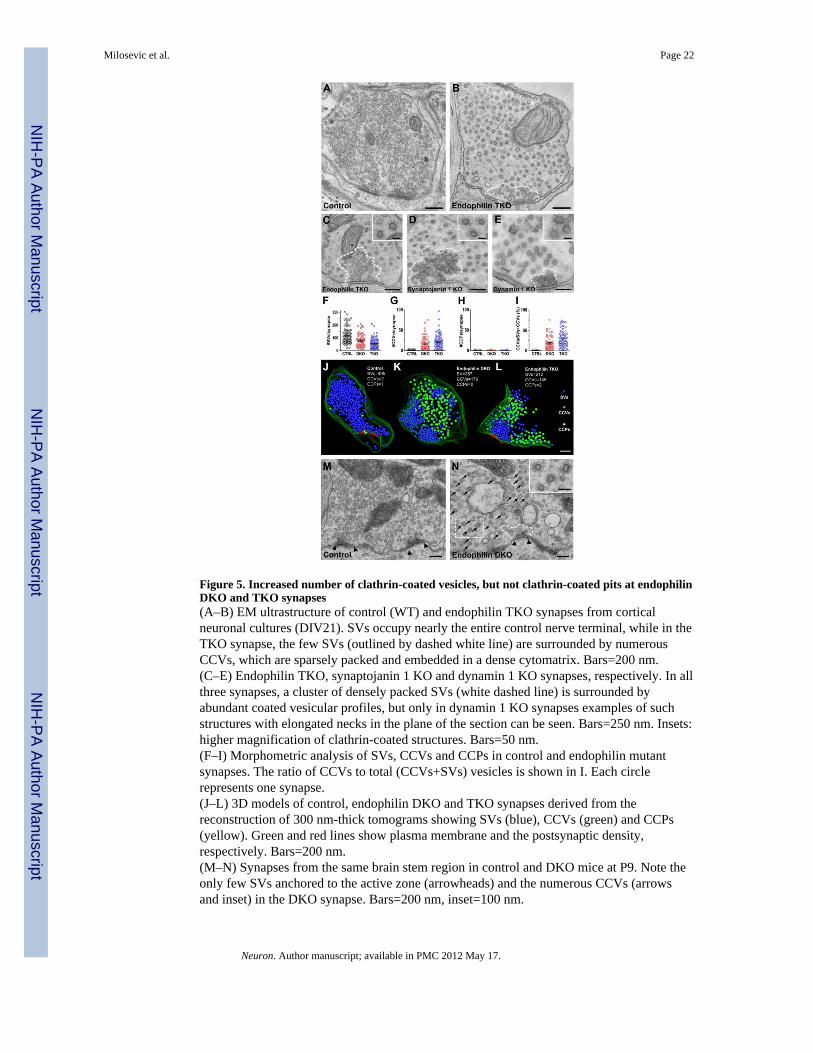
Figure 5. Increased number of clathrin-coated vesicles, but not clathrin-coated pits at endophilinDKO and TKO synapses(A–B) EM ultrastructure of control (WT) and endophilin TKO synapses from corticalneuronal cultures (DIV21). SVs occupy nearly the entire control nerve terminal, while in theTKO synapse, the few SVs (outlined by dashed white line) are surrounded by numerousCCVs, which are sparsely packed and embedded in a dense cytomatrix. Bars=200 nm.(C–E) Endophilin TKO, synaptojanin 1 KO and dynamin 1 KO synapses, respectively. In allthree synapses, a cluster of densely packed SVs (white dashed line) is surrounded byabundant coated vesicular profiles, but only in dynamin 1 KO synapses examples of suchstructures with elongated necks in the plane of the section can be seen. Bars=250 nm. Insets:higher magnification of clathrin-coated structures. Bars=50 nm.(F–I) Morphometric analysis of SVs, CCVs and CCPs in control and endophilin mutantsynapses. The ratio of CCVs to total (CCVs+SVs) vesicles is shown in I. Each circlerepresents one synapse.(J–L) 3D models of control, endophilin DKO and TKO synapses derived from thereconstruction of 300 nm-thick tomograms showing SVs (blue), CCVs (green) and CCPs(yellow). Green and red lines show plasma membrane and the postsynaptic density,respectively. Bars=200 nm.(M–N) Synapses from the same brain stem region in control and DKO mice at P9. Note theonly few SVs anchored to the active zone (arrowheads) and the numerous CCVs (arrowsand inset) in the DKO synapse. Bars=200 nm, inset=100 nm.
Milosevic et al. Page 22
Neuron. Author manuscript; available in PMC 2012 May 17.
NIH
-PA Author Manuscript
NIH
-PA Author Manuscript
NIH
-PA Author Manuscript

Figure 6. Redistribution of endocytic proteins at endophilin TKO synapses(A) Immunofluorescence of the proteins indicated at left in cortical neuronal cultures (DIV18–24) from control, dynamin 1 KO, synaptojanin 1 KO and endophilin TKO mice.Clathrin, α–adaptin and amphiphysin 1 are predominantly diffuse in control neurons, butclustered in all three mutant genotypes. Dynamin 1 and auxilin are clustered in synaptojaninand endophilin mutant synapses, but auxilin is not clustered in dynamin 1 KO. Synaptojaninis slightly more diffuse in the absence of endophilin. The punctate Bassoonimmunofluorescence is similar in all genotypes. Following treatment with TTX (1 μM, 14–18 h) to silence neuronal activity, clathrin was no longer clustered. Bar=15 μm.(B) Quantification of the clustering of immunoreactivity. The y-axis represents the foldincrease of fluorescence puncta in mutant synapses normalized to controls. *p<0.05, t-test.Bars=mean±SEM.(C) Fluorescence analysis of EGFP-clathrin LC in WT and endophilin TKO neurons (DIV18–24). Clathrin is predominantly diffuse in control neurons, but clustered in endophilinTKOs. Clustering was rescued by expression of endophilin 1 FL-Cherry, but not of theendophilin 1 BAR-Cherry construct.(D) Quantification of the clustering of the clathrin fluorescence in the four conditions shownin (C). The y-axis represents the fold increase of fluorescence puncta in mutant synapsesnormalized to WT. Bars=mean±SEM, n.s. not significant, *p<.05, t-test.(E) Left: Western blot analysis of starting lysates and CCV enriched fractions obtained fromWT and endophilin TKO primary cultures (DIV 21). Right: Levels of proteins in the CCVenriched fractions from endophilin TKO cultures normalized to the WT values. Bars=mean±SEM, *p<0.05, t-test.
Milosevic et al. Page 23
Neuron. Author manuscript; available in PMC 2012 May 17.
NIH
-PA Author Manuscript
NIH
-PA Author Manuscript
NIH
-PA Author Manuscript

Figure 7. Neurodegeneration in endophilin 1, 2 DKO mice (P18)(A) H&E stained cerebellar cortex of control (E1+/−E2+/−E3+/+) and endophilin DKO mice.Unstained vacuolar structures, suggesting a form of spongiform neurodegeneration, arepresent in DKO granule cell layer. Bars=100 μm.(B) EM of the DKO granule cell layer showing a vacuolar structure filled with membranousdebris. Bars=1 μm.(C) EM of mossy fiber synapses in control and DKO cerebella. Note in the DKO the lownumber of SVs and the presence of sparsely packed CCVs (higher magnification in theinset). Bars=500 nm, inset 50 nm.(D) Morphology of climbing fibers (top row), as revealed by immunofluorescence forvGLUT2, in control and DKO mice. Sections were counterstained for the IP3 receptor, aPurkinje cell marker (middle row). Note in the overlay image that climbing fibers in theDKOs are swollen and do not extend beyond the most proximal portion of Purkinje celldendrites. Bars=20 μm.(E) EM of climbing fiber synapses. Note the low number of SVs and the presence ofsparsely packed CCVs (higher magnification in the inset) in the DKOs. Bars=250 nm, inset50 nm.
Milosevic et al. Page 24
Neuron. Author manuscript; available in PMC 2012 May 17.
NIH
-PA Author Manuscript
NIH
-PA Author Manuscript
NIH
-PA Author Manuscript

Figure 8. Putative model of clathrin-coated vesicle fission and uncoating at synapsesAssembly and early maturation of endocytic CCPs is independent of endophilin. Endophilinis recruited only to the neck of late stage pits. The dynamin-endophilin interaction mayregulate dynamin function, but it is dispensable for dynamin recruitment and for fission. Incontrast, the synaptojanin-endophilin interaction is critically important for the fate of thevesicle after fission. Loss of PI(4,5)P2 on the bud may start before fission and be restrictedto the bud due to the presence of a collar comprising endophilin, other BAR proteins anddynamin (see Discussion). Auxilin recruitment and uncoating are triggered only afterfission.
Milosevic et al. Page 25
Neuron. Author manuscript; available in PMC 2012 May 17.
NIH
-PA Author Manuscript
NIH
-PA Author Manuscript
NIH
-PA Author Manuscript