Clathrin adaptor AP1B controls adenovirus infectivity of epithelial cells

Cell, Vol. 115, 37–48, October 3, 2003, Copyright 2003 by Cell Press
Formation of an Endophilin-Ca2�
Channel Complex Is Criticalfor Clathrin-Mediated Synaptic Vesicle Endocytosis
Camilli, 2000). It displays lysophosphatidic acid acyl-transferase activity at its N terminus (Schmidt et al.,1999). Its C-terminal Src-homology-3 (SH3) domain se-lectively interacts with other endocytic proteins, suchas dynamin, and may serve to recruit these proteins to
Yuan Chen,1 Lunbin Deng,2
Yuka Maeno-Hikichi,1 Meizan Lai,1
Shaohua Chang,1 Gong Chen,2
and Ji-fang Zhang1,*1Department of PharmacologyUniversity of Pennsylvania School of Medicine the nerve terminal (Ringstad et al., 1997; Schmidt et al.,
1999). Studies have shown that the clathrin-mediated3620 Hamilton WalkPhiladelphia, Pennsylvania 19104 endocytosis machinery, particularly endophilin, is en-
riched in the release zone, even in unstimulated nerve2 Department of BiologyPennsylvania State University terminals (Gonzalez-Gaitan and Jackle, 1997; Guichet
et al., 2002; Ringstad et al., 2001; Roos and Kelly, 1999;University Park, Pennsylvania 16802Teng and Wilkinson, 2000). It remains unclear how theuniversal clathrin-mediated endocytosis machinery canbe selectively targeted to release zones, where it isSummaryneeded after SV exocytosis.
Compared with exocytosis, the exact role of Ca2� in SVA tight balance between synaptic vesicle exocytosisand endocytosis is fundamental to maintaining synap- endocytosis is not well understood. Mounting evidence
suggests that Ca2� is involved in regulating the SV endo-tic structure and function. Calcium influx through volt-age-gated Ca2� channels is crucial in regulating synap- cytosis process. For instance, retrieval of synaptic mem-
brane at the Drosophila neuromuscular junction is Ca2�tic vesicle exocytosis. However, much less is knownabout how Ca2� regulates vesicle endocytosis or how dependent (Kuromi and Kidokoro, 2002; Ramaswami et
al., 1994). Elevated Ca2� levels activate a rapid mode ofthe endocytic machinery becomes enriched at thenerve terminal. We report here a direct interaction endocytosis in hippocampal neurons, cochlear inner
hair cells, retinal bipolar cells, and chromaffin cellsbetween voltage-gated Ca2� channels and endophilin,a key regulator of clathrin-mediated synaptic vesicle (Beutner et al., 2001; Griesinger et al., 2002; Klingauf et
al., 1998; Neher and Zucker, 1993; Neves et al., 2001;endocytosis. Formation of the endophlin-Ca2� channelcomplex is Ca2� dependent. The primary Ca2� binding Sankaranarayanan and Ryan, 2001). It remains unclear
as to how and where Ca2� could exert its regulatory ef-domain resides within endophilin and regulates bothendophilin-Ca2� channel and endophilin-dynamin com- fects.
Through their interaction with the SNARE protein com-plexes. Introduction into hippocampal neurons of adominant-negative endophilin construct, which con- plex, voltage-gated Ca2� channels (VGCCs) are an inte-
gral part of the SV release machinery (Catterall, 1999).stitutively binds to Ca2� channels, significantly reducesendocytosis-mediated uptake of FM 4-64 dye without Ca2� influx through VGCCs plays a key role in regulating
fast SV exocytosis (Augustine, 2001; Catterall, 1999;abolishing exocytosis. These results suggest an im-portant role for Ca2� channels in coordinating synaptic Gundelfinger et al., 2003; Rizo and Sudhof, 2002). Bind-
ing of Ca2� to the C2 domain in synaptotagmin servesvesicle recycling by directly coupling to both exocy-totic and endocytic machineries. as the Ca2� sensor to regulate exocytosis (Augustine,
2001; Fernandez-Chacon et al., 2001; Yoshihara et al.,2003). At the nerve terminal, the exocytotic and endo-Introductioncytic processes are tightly coupled and highly coordi-nated (Gundelfinger et al., 2003). But, the exact mecha-Clathrin-mediated endocytosis is one of the primary
mechanisms by which eukaryotic cells internalize nutri- nisms for such coupling and coordination are poorlyunderstood.ents, antigens, and growth factors and recycle receptors
and vesicles (Conner and Schmid, 2003; Di Fiore and De Here, we report that VGCCs are also an important partCamilli, 2001). At the nerve terminal, clathrin-mediated of the SV endocytic machinery. VGCCs and endophilinendocytosis plays a crucial role in synaptic vesicle (SV) form a macromolecular complex in neurons, and forma-recycling (Brodin et al., 2000; Higgins and McMahon, tion of this complex is Ca2� dependent. Binding of2002; Murthy and De Camilli, 2003; Takei and Haucke, endophilin to Ca2� channels reaches a maximum at2001). Many components of the clathrin-mediated endo- 100–300 nM Ca2�, equivalent to resting Ca2� levels incytosis machinery have been identified, including neurons. When Ca2� exceeds 1 �M, the complex disso-dynamin, endophilin, and synaptojanin (Huttner and ciates. Through mutagenesis, we have identified the pri-Schmidt, 2002; Slepnev and De Camilli, 2000). Endophi- mary Ca2� binding domain for regulating formation oflin has been implicated in several stages of clathrin- both endophilin-Ca2� channel and endophilin-dynaminmediated SV endocytosis, from generating membrane complexes. Finally, transfection into hippocampal neu-curvature, an early step, to later events such as vesicle rons of a dominant-negative construct, which displacesfission and uncoating (Gad et al., 2000; Huttner and the endogenous endophilin from binding to Ca2� chan-Schmidt, 2002; Ringstad et al., 1999; Slepnev and De nels, significantly reduces uptake of fluorescent dye FM
4-64. The reduction seems to be due primarily to im-paired endocytosis. Our results suggest an important*Correspondence: [email protected]
Cell38
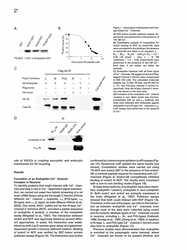
Figure 1. Association of Endophilin with Volt-age-Gated Ca2� Channels
(A) GST-fusion protein pulldown assays. Ex-periments were performed in the presence of100 nM Ca2�.(B) Quantitative analysis of competition forendo2 binding to NCF by endo2-D8. Datawere normalized to the binding in the absenceof endo2-D8 and fitted to an equation: y �
Bmin � (Bmax � Bmin)/(1 � (x/IC50)n). IC50 � 4.2 �
0.48 �M (mean � sem, n � 4). Hillcoefficient � 1.4 � 0.08. Experiments wereperformed in the presence of 300 nM Ca2�.Error bars, if not visible, are within thesymbols.(C) Endophilin interacts with all three typesof Ca2� channels. HA-tagged endo2 and Flag-tagged channel C termini were coexpressedin HEK 293 cells. The calculated molecularweights are 73 kDa, 69 kDa, and 68 kDa forL-, N-, and P/Q-type channel C termini re-spectively. Only the N-type channel C termi-nus was shown in the input lane.(D) Formation of the endophilin-Ca2� channelcomplex in vivo. Brain lysate was immuno-precipitated with an anti-�1B antibody andblots were detected with antibodies againstendophilin II and N-type Ca2� channels (�1B).CoIP assays were performed in the absenceof Ca2�.
role of VGCCs in coupling exocytotic and endocytic confirmed by coimmunoprecipitation (coIP) assays (Fig-ure 1C). Reciprocal coIP yielded the same results (notmachineries for SV recycling.shown). Competition studies were carried out using35S-NCF and endo2-GST in the presence of free endo2-
Results D8, a minimal peptide required for interacting with Ca2�
channels (Figure 3). Endo2-D8 competitively inhibitedFormation of an Endophilin-Ca2� Channel binding of endo2 to NCF. The results were consistentComplex in Neurons with a one-to-one binding model (Figure 1B).To identify proteins that might interact with Ca2� chan- At least three isoforms of endophilin have been identi-nels and play a role in Ca2�-dependent signal transduc- fied, endophilin I (endo1), endophilin II, and endophilintion, we carried out yeast two-hybrid screening of a rat III. Both endo1 and endo2 are primarily expressed inbrain cDNA library using the full length C termini of three the brain (Ringstad et al., 1997). Pulldown assaysdifferent Ca2� channel �1-subunits, �1A (P/Q-type), �1B showed that both could interact with NCF (Figure 1A).(N-type), and �1C (L-type), as baits (Maeno-Hikichi et al., Therefore, in the rest of the paper, we refer to the interac-2003). One clone, N427, pulled out by the N-type Ca2�
tion as between endophilin and Ca2� channels, evenchannel C terminus (NCF), encoded a partial sequence though most of the data were collected using endo2of endophilin II (endo2), a member of the endophilin and its mutants. Multiple types of Ca2� channels coexistfamily (Ringstad et al., 1997). The interaction between in neurons, including L-, N-, and P/Q-types (Catterall,endo2 and NCF was rigorously tested by several differ- 1998; Dunlap et al., 1995; Zhang et al., 1993). In additionent approaches. In yeast, the interaction was estab- to N-type, L- and P/Q-type Ca2� channel C termini couldlished by both LacZ reporter gene assay and galactose- also bind to endophilin (Figure 1C).dependent growth in leucine-deficient medium. Binding Previous studies have demonstrated that endophilinof endo2 to NCF was verified by GST-fusion protein is enriched at the presynaptic nerve terminal, where
Ca2� channels are known to be present (Huttner andpulldown assays (Figure 1A). The interaction was further
Ca2� Channels in Synaptic Vesicle Endocytosis39
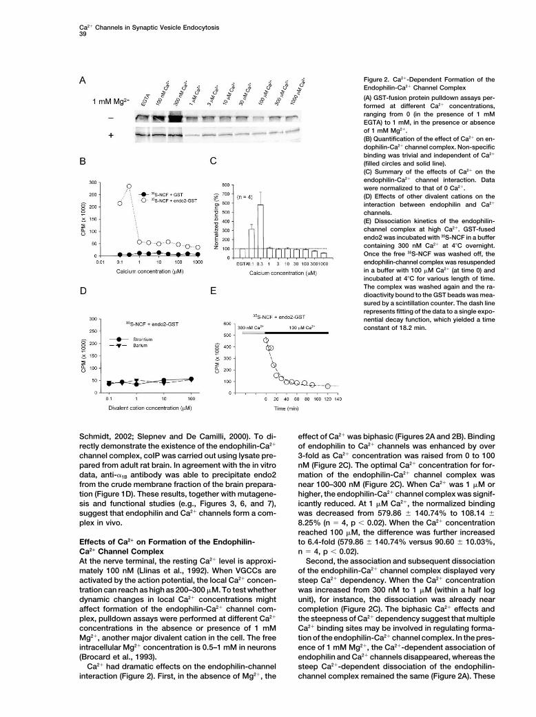
Figure 2. Ca2�-Dependent Formation of theEndophilin-Ca2� Channel Complex
(A) GST-fusion protein pulldown assays per-formed at different Ca2� concentrations,ranging from 0 (in the presence of 1 mMEGTA) to 1 mM, in the presence or absenceof 1 mM Mg2�.(B) Quantification of the effect of Ca2� on en-dophilin-Ca2� channel complex. Non-specificbinding was trivial and independent of Ca2�
(filled circles and solid line).(C) Summary of the effects of Ca2� on theendophilin-Ca2� channel interaction. Datawere normalized to that of 0 Ca2�.(D) Effects of other divalent cations on theinteraction between endophilin and Ca2�
channels.(E) Dissociation kinetics of the endophilin-channel complex at high Ca2�. GST-fusedendo2 was incubated with 35S-NCF in a buffercontaining 300 nM Ca2� at 4�C overnight.Once the free 35S-NCF was washed off, theendophilin-channel complex was resuspendedin a buffer with 100 �M Ca2� (at time 0) andincubated at 4�C for various length of time.The complex was washed again and the ra-dioactivity bound to the GST beads was mea-sured by a scintillation counter. The dash linerepresents fitting of the data to a single expo-nential decay function, which yielded a timeconstant of 18.2 min.
Schmidt, 2002; Slepnev and De Camilli, 2000). To di- effect of Ca2� was biphasic (Figures 2A and 2B). Bindingof endophilin to Ca2� channels was enhanced by overrectly demonstrate the existence of the endophilin-Ca2�
channel complex, coIP was carried out using lysate pre- 3-fold as Ca2� concentration was raised from 0 to 100nM (Figure 2C). The optimal Ca2� concentration for for-pared from adult rat brain. In agreement with the in vitro
data, anti-�1B antibody was able to precipitate endo2 mation of the endophilin-Ca2� channel complex wasnear 100–300 nM (Figure 2C). When Ca2� was 1 �M orfrom the crude membrane fraction of the brain prepara-
tion (Figure 1D). These results, together with mutagene- higher, the endophilin-Ca2� channel complex was signif-icantly reduced. At 1 �M Ca2�, the normalized bindingsis and functional studies (e.g., Figures 3, 6, and 7),
suggest that endophilin and Ca2� channels form a com- was decreased from 579.86 � 140.74% to 108.14 �8.25% (n � 4, p � 0.02). When the Ca2� concentrationplex in vivo.reached 100 �M, the difference was further increasedto 6.4-fold (579.86 � 140.74% versus 90.60 � 10.03%,Effects of Ca2� on Formation of the Endophilin-
Ca2� Channel Complex n � 4, p � 0.02).Second, the association and subsequent dissociationAt the nerve terminal, the resting Ca2� level is approxi-
mately 100 nM (Llinas et al., 1992). When VGCCs are of the endophilin-Ca2� channel complex displayed verysteep Ca2� dependency. When the Ca2� concentrationactivated by the action potential, the local Ca2� concen-
tration can reach as high as 200–300 �M. To test whether was increased from 300 nM to 1 �M (within a half logunit), for instance, the dissociation was already neardynamic changes in local Ca2� concentrations might
affect formation of the endophilin-Ca2� channel com- completion (Figure 2C). The biphasic Ca2� effects andthe steepness of Ca2� dependency suggest that multipleplex, pulldown assays were performed at different Ca2�
concentrations in the absence or presence of 1 mM Ca2� binding sites may be involved in regulating forma-tion of the endophilin-Ca2� channel complex. In the pres-Mg2�, another major divalent cation in the cell. The free
intracellular Mg2� concentration is 0.5–1 mM in neurons ence of 1 mM Mg2�, the Ca2�-dependent association ofendophilin and Ca2� channels disappeared, whereas the(Brocard et al., 1993).
Ca2� had dramatic effects on the endophilin-channel steep Ca2�-dependent dissociation of the endophilin-channel complex remained the same (Figure 2A). Theseinteraction (Figure 2). First, in the absence of Mg2�, the

Cell40
data suggest that there are at least two distinct Ca2�
binding sites, one of which is also sensitive to Mg2�.Both endo1 and endo2 exhibited similar Ca2�-depen-dent interactions with Ca2� channels (not shown).
Pulldown assays were performed in the presence ofstrontium or barium instead of Ca2� to further testwhether other divalent cations elicited effects similarto Ca2�. Neither Sr2� nor Ba2� affected the interactionbetween endophilin and NCF (Figure 2D), suggestingthat Ca2� plays a major role in regulating the formationof the endophilin-Ca2� channel complex.
We next tested whether the endophilin-channel com-plex, once formed, could disassemble at high Ca2� lev-els. The endophilin-channel complex, formed at 300 nMCa2�, was incubated in a buffer with 100 �M Ca2� forvarious periods of time at 4�C. As expected, once inthe high Ca2� solution, the endophilin-channel complexstarted to dissociate with time, following a single expo-nential decay function (Figure 2E). This data suggeststhat formation of the endophilin-channel complex isreadily reversible at high Ca2� concentrations.
Ca2� Channel Binding Domain on EndophilinEndophilin interacts with its partner proteins throughthe C-terminal SH3 domain (Huttner and Schmidt, 2000;Slepnev and De Camilli, 2000). To delineate the Ca2�
channel binding domain on endophilin, we generated aseries of endophilin deletion mutants (Figure 3A). Theisolated SH3 domain (D6) failed to pull down 35S-NCF(Figure 3A), indicating that the SH3 domain itself did notinteract directly with Ca2� channels. The first 120 aminoacid residues (D3) also did not bind to the channel C ter-minus.
Analysis of the mutants revealed a pattern. Mutantslacking the SH3 domain exhibited much stronger associ- Figure 3. Identification of the Ca2� Channel Binding Domain in En-ation with the Ca2� channel C terminus compared with dophilinthose with an intact SH3 domain (Figures 3A and 3B). (A) Effects of deletion mutations on the interaction between endophi-For instance, removing the SH3 domain from D1 caused lin and the Ca2� channel C terminus. Schematic representation of
the wild-type endophilin II and deletion mutants. Numbers repre-a 3-fold increase in binding for D5, from 56,820 � 2,846sented the starting and ending amino acids for a particular construct.(n � 3) to 167,055 � 16,830 (n � 6, p � 0.01). The(B) Deletion of the SH3 domain increases binding of endophilinincreased affinity between SH3-less mutants and NCFmutants to Ca2� channels.
was observed at all Ca2� concentrations tested (Figure (C) SH3-less mutants display increased binding to Ca2� channels3C). Thus, while the SH3 domain itself did not directly at all Ca2� levels.contribute to the formation of the endophilin-Ca2� chan-nel complex, the presence of the SH3 domain appeared
EF-hand deleted. Removing this EF-hand did not affectto hinder the interaction between endophilin and Ca2�
the Ca2�-dependent interaction (Figure 4A).channels. Amino acid residues 200–219 were crucial forSince endophilin mutants with or without the SH3 do-the formation of the endophilin-Ca2� channel complex.
main showed distinct binding to NCF (Figure 3), we ex-Inclusion of this segment led to a 7-fold increase in theamined whether these two groups of mutants exhibitedendophilin-Ca2� channel complex (Figures 3A and 3B).differential sensitivities to Ca2� in their interactions withThis segment is highly conserved among the three en-Ca2� channels. The effects of Ca2� were compared be-dophilin isoforms (Ringstad et al., 1997).tween three pairs of constructs, including WT endo2versus D4, D1 versus D5, and D8 versus D7 (Figure3A). All three pairs displayed the same patterns in theirCa2�-Dependent Blockade of the Endophilin-
Channel Interaction by the SH3 Domain binding to NCF at different Ca2� levels. Those with theSH3 domain exhibited robust Ca2�-dependent interac-Two well-defined Ca2� binding domains, the C2 domain
and EF-hand, are known to participate in many Ca2�- tions, in sharp contrast to those without the SH3 domain(Figure 4B). For example, when the Ca2� level was in-dependent signaling processes. Although endophilin
does not contain any known Ca2� binding domains, the creased from 300 nM to 10 �M, the normalized bindingof D8 to NCF was significantly reduced, from 1,568.8 �Ca2� channel C terminus has an EF-hand (Babitch,
1990). To determine if this EF-hand was responsible for 264.0% to 90.9 � 7.8% (n � 3, p � 0.01, Figure 4C).D7, on the other hand, remained bound to the channelthe Ca2�-dependent formation of the endophilin-chan-
nel complex, a mutant was generated with the entire C terminus even at 1 mM Ca2� (Figures 4B and 4C).

Ca2� Channels in Synaptic Vesicle Endocytosis41
Figure 4. The SH3 Domain Blocks the Interaction between Endophilin and Ca2� Channels
(A) An EF-hand in the Ca2� channel C terminus is not the primary Ca2� binding site for regulating the endophilin-Ca2� channel complex.(B) Deletion mutants lacking the SH3 domain become insensitive to Ca2� in their interactions with Ca2� channels.(C) Quantitative comparison between deletion mutants D7 and D8 in their interaction with 35S-NCF at different Ca2� concentrations. Data werenormalized to that at 0 Ca2�.(D) Quantitative comparison between deletion mutants D5 and D7. Compared to D7, D5 displayed a small, but significant, degree of Ca2�-dependent dissociation.
There were some subtle differences among these PRD) as their partner modules (Pawson, 1995). In addi-tion to the SH3 domain, all endophilins have a PRDSH3-less mutants in their Ca2�-dependent binding to(Sparks et al., 1996), located within the Ca2� channelNCF. For instance, D5 exhibited some degree of Ca2�-binding domain (amino acid residues 257–264 in endo2,dependent dissociation from NCF (Figure 4D). As theFigure 5B). To test whether this PRD directly interactedCa2� level was raised from 300 nM to 10 �M, the normal-with the SH3 domain, a PRD peptide was synthesizedized binding was decreased from 195.4 � 26.5% toand included in the pulldown assay buffer at a final107.7 � 6.1% (n � 6, p � 0.01). D7, on the other hand, didconcentration of 10 �M. In the presence of this peptide,not show any significant Ca2�-dependent dissociation.the endophilin-channel interaction became insensitiveWhile the SH3 domain itself did not bind directly toto Ca2� (Figure 5B), as though the SH3 domain wereCa2� channels (Figure 3A), the presence of the SH3 do-deleted (c.f. Figure 4B). A control peptide with prolinesmain weakened the interaction between endophilin andreplaced by alanines had no effect (Figure 5B).Ca2� channels at Ca2� concentrations over 1 �M. One
To directly demonstrate that the SH3 domain inter-possibility is that at high Ca2� levels, the SH3 domainacted with the PRD, pulldown assays were carried outmight interact with another domain within endophilin,using the GST-fused SH3 domain (D6, Figure 3A) andconsequently preventing the formation of the endophi-the GFP-tagged SH3-less mutants expressed in HEKlin-Ca2� channel complex. The prediction from such a293 cells. At low Ca2� levels (0–300 nM), binding of thehypothesis was that the interaction between the SH3-SH3 domain to D4 was weak (Figure 5C). However, whenless mutants and NCF should become sensitive to Ca2�
the Ca2� level was raised to above 1 �M, there was ain the presence of a separate SH3 domain peptide. Con-robust interaction between the SH3 domain and GFP-sistent with this prediction, interaction between 35S-NCFtagged D4, suggesting that the affinity of the PRD forand D7 became Ca2� dependent in the presence of athe SH3 domain is increased by Ca2�. This is oppositepoly-histidine-tagged SH3 domain peptide (8 �M, Figureto the effects of Ca2� on the endophilin-Ca2� channel5A). The results suggest that the SH3 domain blocksinteraction (c.f. Figure 2A). Similar results were observedthe formation of endophilin-Ca2� channel complex whenbetween the SH3 domain and GFP-tagged D7 (notthe Ca2� level is over 1 �M.shown). It remains to be determined whether the interac-tion between the PRD and the SH3 domain is intramolec-
Molecular Nature of the Primary Ca2� ular or intermolecular, although studies have shown thatBinding Domain such an interaction can be intramolecular (Sicheri et al.,SH3 domains serve as molecular modules for protein- 1997; Xu et al., 1997).
How does Ca2� alter the affinity of the PRD for theprotein interactions, with proline-rich domains (PxxP,

Cell42
Figure 5. Ca2�-Dependent Change in the Af-finity of the PRD for the SH3 Domain withinEndophilin
(A) SH3 domain peptide (final concentration 8�M) restores the Ca2�-dependent interactionbetween SH3-less endophilin mutants andthe Ca2� channel C terminus.(B) Effects of the PRD peptide on the interac-tion between endophilin and NCF. A diagramdepicts the location of the PRD within theendophilin molecule. Pulldown assays wereperformed in the presence of a synthetic pep-tide, PRD (WT), encompassing the prolinerich domain: PRREFKPRPQEPFELG. A pep-tide with prolines replaced by alanines, PRD(P → A), was used as the control.(C) Ca2�-dependent interaction between theSH3 domain and SH3-less endophilin mu-tants. An anti-GFP antibody was used for im-munoblotting.(D) An E264A point mutation eliminates theCa2�-dependent interaction between endophi-lin and the Ca2� channel C terminus.(E) Dose-response relationship for Ca2� to in-crease binding of the PRD and the SH3 do-main. Pulldown assays were carried out using35S-labeled D5 or D5 (E264A) and GST-fusedSH3 domain (D6). Equal amounts of D5 (WT)or D5 (E264A) were used. The radioactivitybound to GST beads was measured by a scin-tillation counter. All data were normalized tothat of wild-type D5 at 10 �M Ca2�. For wild-type D5, data were fitted to a dose-responseequation: y � Bmin � (Bmax � Bmin)*xn/(xn � Kdn),which yielded a Kd for Ca2� of 425.2 � 21.3nM and a Hill coefficient of 2.3 � 0.09 (mean �
sem). Error bars, if not seen, are within thesymbols. We did not fit the data of D5 (E264A),since the binding might not reach saturationat this Ca2� range.(F) Dynamin-endophilin interaction is alsosensitive to Ca2�. All results shown in thisfigure are representatives of at least threeseparate experiments.
SH3 domain? Depending on their orientations, proline- required to form a high-affinity binding site for Ca2�.Examples include the Ca2� selectivity filter in Ca2� chan-rich domains can be divided into two classes, class I
and class II (Mayer, 2001; Pawson, 1995). The PRD in nels, where four glutamate residues in the Ca2� channelpore region form a high-affinity Ca2� binding site withendo2 resembles that of class II, which bears the signa-
ture sequence of PxxPx�, where � denotes a basic the Kd for Ca2� of 1 �M (Yang et al., 1993). We hypothe-sized that E264 might be part of a Ca2� binding site andresidue, typically an arginine (Mayer, 2001; Tong et al.,
2002). This basic residue is thought to determine the that binding of Ca2� to E264 could increase the affinityof the PRD for the SH3 domain in endophilin.affinity and specificity of a PRD to its SH3 domain by
binding to a negatively charged pocket on the SH3 do- To test this hypothesis, a point mutation was gener-ated in which glutamate 264 was replaced with alaninemain (Nguyen et al., 1998; Pawson, 1995). The PRD in
endo2, however, deviates from this consensus se- (endo2 E264A). Binding of the E264A mutant to NCFbecame insensitive to Ca2� (Figure 5D), as though thequence in that it has a negatively charged glutamate
residue in this key position: PRPQEPFE (E264). Further- SH3 domain had been deleted (c.f. Figure 4B). The re-sults suggested that E264 is part of the Ca2� bindingmore, there are additional negative charges in the imme-
diate vicinity of the core PxxP sequence. Such multiple site(s). The importance of E264 was further evaluatedby pulldown assays using 35S-labeled D5 (WT) or D5negative charges are rarely seen in other class II proline-
rich domains (Tong et al., 2002). For a typical class II (E264A) and the GST-fused SH3 domain. As expected,binding of D5 to the SH3 domain increased with Ca2�PRD, introduction of a negative charge immediately after
the PxxP core sequence abolishes its affinity for the (Figure 5E). The increase was over 6-fold when Ca2�
was increased from 0 to 10 �M. Fitting the data with aSH3 domain (Zhao et al., 2000).On the other hand, glutamate residues are known to dose-response curve yielded the Kd for Ca2� of 425.2 �
21.3 nM (n � 3) and the Hill coefficient of 2.3 � 0.09,play a key role in forming Ca2� binding domains, suchas the EF-hand. Often, multiple glutamate residues are indicating that at least two Ca2� ions were associated

Ca2� Channels in Synaptic Vesicle Endocytosis43
with endophilin. The Kd for Ca2� and steep Ca2�-depen- apsin, a presynaptic protein (Figure 6A), and by electro-physiology recordings (Figure 7).dent association of the PRD and the SH3 domain re-
We hypothesized that interaction of endophilin withsulted in the steep dissociation of the endophilin-chan-Ca2� channels could recruit endophilin to the releasenel complex when Ca2� rose from 300 nM to 1 �M (Figurezone. A simple prediction is that overexpression of D52). In sharp contrast, the E264A mutation severely ham-(E264A) would displace the endogenous endophilin atpered binding of the mutant D5 to the SH3 domain,the release zone, thus impairing the clathrin-mediatedparticularly at high Ca2� levels. This led to the failure ofSV endocytosis. To test this prediction, we performedthe mutant endophilin to dissociate from the channel atFM 4-64 dye uptake experiments on cultured hippocam-high Ca2� levels (Figure 5D). These findings suggest thatpal neurons transfected with GFP, endo2-GFP, or D5-Ca2� can bind to endophilin directly and that E264 playsGFP (E264A). FM 4-64 is a derivative of FM 1-43, aa critical role in Ca2�-dependent increase in the affinityfluorescent dye which becomes trapped in SVs afterbetween the PRD and the SH3 domain. This glutamateendocytosis (Betz and Bewick, 1993). FM dye wasresidue is conserved in both endo1 and endo2 (Ringstadloaded into synaptic vesicles by depolarizing neuronset al., 1997). Since multiple glutamate residues are re-with a 40 mM K� solution. For neurons transfected withquired for formation of a high-affinity Ca2� binding siteGFP or GFP-tagged endophilin, uptake of FM 4-64 (red(e.g.Yang et al., 1993), it remains to be investigatedpuncta) was found throughout the GFP-labeled axonswhether other glutamate residues contribute to the for-(Figure 6B). In contrast, neurons transfected with D5mation of the Ca2� binding site(s) in endophilin.(E264A) often displayed long segments of axons devoidEndophilin interacts with other endocytic proteins,of FM 4-64 staining. Quantitative analysis showed thatsuch as dynamin, through its SH3 domain (Huttner andthe intensity of FM 4-64 staining was significantly re-Schmidt, 2002; Slepnev and De Camilli, 2000). The effectduced (Figure 6C), indicating that SV endocytosis pro-of Ca2� on the interaction between the PRD and the SH3cess might be impaired as a result of overexpression ofdomain within endophilin led us to investigate whetherD5 (E264A).the interaction of endophilin and dynamin was also sen-
Electrophysiology recordings were performed to in-sitive to Ca2�. Similar to its effects on the endophilin-vestigate whether reduction of the FM4-64 uptake bychannel interaction, Ca2� exerted the steep biphasicD5 (E264A) resulted from impaired endocytosis or theeffects on the endophilin-dynamin complex in the ab-arrest of exocytosis. Evoked excitatory postsynapticsence of Mg2�. In the presence of 1 mM Mg2�, the en-currents (EPSCs) were recorded, by dual whole-cell re-dophilin-dynamin complex only displayed Ca2�-depen-cordings, from neurons whose presynaptic neuronsdent dissociation (Figure 5F).were transfected with wild-type endophilin or D5(E264A). Neurons transfected with either construct werecapable of releasing neurotransmitters in response toEndophilin-Ca2� Channel Complex Is Requiredbrief depolarizing pulses (Figures 7A and 7B). At lowfor Clathrin-Mediated SV Endocytosisfrequency stimulations (0.1 Hz), the EPSC amplitude inAs a first step toward understanding the importance ofD5 (E264A)-transfected neurons was 500.1 � 154.6 pAthe endophilin-Ca2� channel complex in SV endocytosis,(n � 20), which was slightly reduced but not significantlywe sought to determine whether introduction of a domi-different from that of neurons transfected with WT en-nant-negative endophilin construct into hippocampaldophilin (818.2 � 221.6 pA, n � 12; p 0.2; Figure 7E).neurons could affect SV endocytosis. Based on bio-The results suggested that D5 does not significantlychemical studies, we elected to use GFP-tagged D5affect SV exocytosis. Ablation of endophilin in Drosoph-(E264A) as the dominant-negative construct (Figure 3A).ila yielded similar results (Verstreken et al., 2002). Analy-Because of N- and C-terminal deletions, this constructsis of spontaneous miniature EPSCs (mEPSCs) showeddoes not have the acyltransferase activity and will notthat transfection of D5 (E264A) did not alter either the
interact with other endocytic proteins (Farsad et al.,mEPSC amplitude (23.2 � 2.1 pA for D5, n � 11; 24.5 �
2001; Guichet et al., 2002; Schmidt et al., 1999; Slepnev1.4 for WT, n � 9; p 0.6) or the mEPSC frequency
and De Camilli, 2000). Other than its interaction with Ca2�
(1.34 � 0.28 for D5, n � 11; 1.88 � 0.44 for WT, n � 9;channels (this study), the middle portion of endophilin is p 0.3; Figures 7C, 7D, 7F, and 7G). These resultsnot known to interact with any other proteins directly or indicated that D5 (E264A) did not change the postsynap-indirectly involved in synaptic transmission. D5 dis- tic receptor properties.played a much higher affinity for Ca2� channels and the The effects of D5 (E264A) were further evaluated bybinding was insensitive to Ca2� (Figures 3 and 4). Hence, high frequency stimulations to produce short-term syn-the mutant should bind selectively to Ca2� channels. In aptic depression as a result of depletion of the readilyaddition, the E264A mutation eliminated the possibility releasable pool of synaptic vesicles (RRP). If the primaryof its interaction with endogenous endophilin through effect of D5 (E264A) was to impair SV endocytosis asthe PRD (Figure 5E). Preliminary tests showed that ex- suggested by the imaging studies (Figure 6), we wouldpression of GFP-tagged D5 (E264A) was much better expect that at high frequency stimulations, short-termthan D7 (E264A) in hippocampal neurons. Thus, intro- synaptic depression would become more profound dueduction of D5 (E264A) into hippocampal neurons should to impaired endocytosis in neurons transfected with D5compete with the endogenous endophilin for associa- (E264A). To test this hypothesis, transfected neuronstion with Ca2� channels but would not carry out any were repetitively stimulated at 5 Hz (100 stimuli). Asbiological functions. Transient expression of D5 (E264A) expected, the EPSC amplitude in neurons transfectedin hippocampal neurons did not affect formation of func- with D5 (E264A) (n � 7) decreased quickly and reached
a significantly lower plateau (�20% of the control) com-tional synapses, as revealed by immunostaining of syn-

Cell44
Figure 6. A Dominant-Negative EndophilinConstruct Reduces Uptake of FM4-64 in Pre-synaptic Boutons
(A) Immunostaining of a presynaptic protein,synapsin, in D5 (E264A)-transfected neurons.Scale bar � 25 �m.(B) Effect of D5 (E264A) on FM 4-64 uptake.Scale bar � 25 �m.(C) Summary of FM 4-64 uptake for neuronstransfected with GFP, endo2-GFP, and D5-GFP (E264A). Numbers in parentheses repre-sent that of transfected neurons of at leastthree batches of transfection. One way ANOVAwas used for group comparison among threedifferent conditions.
pared to that in neurons transfected with WT endophilin Discussion(n � 5, p � 0.001 for the last 5 points, Figure 7H), consis-tent with previous observations when SV endocytosis Our results show a direct interaction between VGCCswas impaired (Shupliakov et al., 1997; Verstreken et and the endocytic machinery. We propose that throughal., 2002). interactions with endophilin, VGCCs serve as an anchor
When given multiple train stimulations, there was a to recruit the clathrin-mediated endocytosis machinerysteady decrease in the average EPSC amplitude evoked to the release zone where SV endocytosis is to takeat 0.1 Hz during the interval between train stimulations place. Such a mechanism is reminiscent of the interac-(Figure 7I). Again, neurons transfected with D5 (E264A) tion between VGCCs and the exocytotic machinery (Cat-(n � 6) exhibited an accelerated decrease in the EPSC terall, 1999) and is consistent with observations that theamplitude compared to that in neurons transfected with clathrin-mediated endocytosis machinery is enriched inWT endophilin (n � 5) (Figure 7I, p � 0.05 after the the release zone (Gonzalez-Gaitan and Jackle, 1997;2nd and 3rd train, and p � 0.002 after 4th train). The Ringstad et al., 2001; Roos and Kelly, 1999; Teng anddifference in EPSC amplitudes after train stimulations Wilkinson, 2000). Thus, as with exocytosis, VGCCs aresuggested that replenishment of the RRP is hindered in also an integral part of the SV endocytic machinery.the D5-transfected neurons, most likely as a conse- While the interaction between VGCCs and the exocytotic
machinery, such as syntaxin, occurs at the II-III loop ofquence of impaired SV endocytosis. Overall, results fromimaging and electrophysiology demonstrated that the the Ca2� channel �1 subunit (Sheng et al., 1996), the
interaction with the clathrin-mediated endocytosis ma-effects of the dominant-negative construct were primar-ily on SV endocytosis, consistent with recent reports chinery is through the channel C terminus (current
study). Thus, through these interactions, Ca2� channelswhich showed that ablation of endophilin in Drosophilaimpaired clathrin-mediated vesicle endocytosis (Gui- bring both exocytotic and endocytic machineries in
close proximity, providing a potential mechanism forchet et al., 2002; Verstreken et al., 2002). We concludethat the endophilin-Ca2� channel complex was critical spatial coordination of vesicular exocytosis and endocy-
tosis (Gundelfinger et al., 2003).for clathrin-mediated SV endocytosis.

Ca2� Channels in Synaptic Vesicle Endocytosis45
Figure 7. Quantitative Analysis of the Effectsof WT and a Dominant-Negative EndophilinConstruct on Basal and Sustained Neuro-transmitter Release
(A and B) Representative traces of the aver-age EPSCs evoked from a presynaptic neu-ron transfected with endo2-GFP (A) or D5-GFP (E264A) (B).(C and D) Typical traces showing mEPSCs inan endo2-GFP transfected neuron (C) and aD5-GFP (E264A) transfected neuron (D).(E) D5 (E264A) has no significant effect on theEPSC amplitude. EPSCs were recorded fromneurons transfected with WT endophilin (n �
12) or D5 (E264A) (n � 20, p 0.2).(F and G) D5 (E264A) does not change themEPSC amplitude (p 0.6) or frequency(p 0.3).(H) Effects of D5 (E264A) on short-term syn-aptic depression. Normalized EPSC ampli-tude during the first train stimulation (5 Hz,100 stimuli) recorded from neurons trans-fected with WT endophilin (n � 5) or D5(E264A) (n � 7). p � 0.001 for the last fivepoints.(I) Effects of multiple train stimulations on theEPSC amplitude. Multiple train stimulations(5 Hz, 100 stimuli) were applied with a 3-mininterval, during which the presynaptic neuronwas stimulated at 0.1 Hz. EPSCs recordedduring the interval from WT endophilin (n �
5) or D5 (E264A) (n � 6) transfected neuronswere averaged and normalized to that ob-tained before stimulations. *, p � 0.05; **,p � 0.002.
It is well established that Ca2� influx through VGCCs VGCCs may serve as a signal for temporal coordinationof vesicular exocytosis and endocytosis (Gundelfingeris crucial for release of neurotransmitters at the nerve
terminal (Augustine, 2001; Catterall, 1999; Gundelfinger et al., 2003).We have demonstrated a role for the SH3 domain inet al., 2003; Rizo and Sudhof, 2002). Through their inter-
action with syntaxin, VGCCs become part of the SNARE regulating the interaction between endophilin and itspartner proteins, particularly the Ca2�-dependent disso-protein complex for exocytosis. Syntaxin-Ca2� channel
interaction is Ca2�-dependent, presumably responsible ciation of endophilin-Ca2� channel and endophilin-dynamin complexes. The affinity of the PRD to the SH3for Ca2�-dependent docking of SVs to the release zone
(Sheng et al., 1996). Binding of Ca2� to synaptotagmin domain within endophilin is determined by Ca2�. Whenthe Ca2� level is low, the PRD is not bound to the SH3serves as a trigger for neurotransmitter release (Fernan-
dez-Chacon et al., 2001). The data in this study provide domain, allowing binding of endophilin to Ca2� channelsand/or dynamin. When the Ca2� concentration rises toa plausible mechanism by which Ca2� may regulate SV
endocytosis (Beutner et al., 2001; Griesinger et al., 2002; above 1 �M, binding of Ca2� to E264 in the PRD signifi-cantly increases the affinity of the PRD for the SH3Klingauf et al., 1998; Kuromi and Kidokoro, 2002; Neher
and Zucker, 1993; Neves et al., 2001; Ramaswami et al., domain. This, in turn, blocks the access of Ca2� channelsand dynamin to endophilin. Thus we propose that this1994; Sankaranarayanan and Ryan, 2001). Formation
of the endophilin-Ca2� channel complex as well as the Ca2�-dependent change in the affinity of the PRD forthe SH3 domain within endophilin serves as the primaryendophilin-dynamin complex is Ca2�-dependent (Fig-
ures 2 and 5). In particular, Ca2�-dependent dissociation Ca2� sensor for the formation of the endophilin-Ca2�
channel and endophilin-dynamin complexes. In addi-of these complexes occurs at the Ca2� concentrationrange when Ca2� channels are activated by action po- tion, our results provide an example that the affinity
of the PRD for its SH3 domain can be regulated bytentials during triggered exocytosis, allowing Ca2� toregulate SV endocytosis at the beginning of the clathrin- physiological factors targeting at amino acid residues
in the vicinity of the core PxxP signature sequence.mediated endocytosis process. Thus, Ca2� influx through

Cell46
Figure 8. A Molecular Model for Ca2�-Dependent Interaction between Endophilin and Ca2� Channels
The model depicts the interaction between the PRD and its SH3 domain within endophilin as intramolecular, but it is also possible that theinteraction could be intermolecular.
30 (Qiagen) for poly-histidine-tagged (6His) proteins. GST-fusionWe propose that endophilin has two distinct modesproteins and 6His proteins were produced in E. coli. Glutathioneor conformations, an open mode and a closed modeagarose beads (Pierce) were used to purify GST-fused proteins. Ni-(Figure 8). Under resting Ca2� levels (100–300 nM), en-NTA columns (Qiagen) were used for purification of 6His proteins.
dophilin is in its open mode due to the low affinity of To express the recombinant proteins in mammalian cells, cDNAthe PRD for the SH3 domain. In this open mode, endo- constructs were subcloned into the following vectors: HA-tagged
pcDNA 3, FLAG-tagged pcDNA 3, or pEGFP (Clontech). The calciumphilin is able to interact with both Ca2� channels andphosphate method was used for transfection in HEK 293 cells ordynamin and the interactions serve to recruit the endo-hippocampal neurons.cytic machinery into the nerve terminal. When local Ca2�
levels rise to over 1 �M as a consequence of activationGST-Fusion Protein Pulldown Assaysof VGCCs, binding of Ca2� to E264 in the PRD switchesPulldown assays were carried out as previously described (Maeno-endophilin to the closed mode, when the PRD locks ontoHikichi et al., 2003). Briefly, 35S-labeled proteins were synthesizedthe SH3 domain. In this closed mode, endophilin is nousing an in vitro protein translation kit (Promega). The assay buffer
longer available for binding to either Ca2� channels or contained 100 mM NaCl, 20 mM TrisHCl, and 5% glycerol (pH 7.0)dynamin, which would presumably allow the liberated along with 1% Triton X-100 and a cocktail of protease inhibitorsendophilin and dynamin to become actively involved in (phenylmethylsulfonyl fluoride, 1 �M; pepstatin, 1 �g/ml; leupeptin,
1 �g/ml; aprotinin, 1 �g/ml; and benzamidine, 0.1 mg/ml). Approxi-endocytosis immediately after SV exocytosis. By couplingmately 5 �g of GST-fused protein were incubated with 2 �l oftightly to both the exocytotic and endocytic machiner-35S-labeled protein with gentle rocking. The length of incubationies, voltage-gated Ca2� channels are thus uniquely posi-varied from 2 hr to overnight. All pulldown assays were performed
tioned to coordinate the SV recycling process. at 4�C. The results were analyzed by SDS-PAGE and autoradiogra-phy or by a scintillation counter. Typically, 0.5 �l of 35S-labeled
Experimental Procedures protein was used as input on the gel. Values of CPM (count perminute) were expressed as mean � SEM. To minimize the variability
Yeast Two-Hybrid Screening of radioactivity between experiments, CPM values were normalizedDetails of screening can be found elsewhere (Maeno-Hikichi et al., to that obtained in the presence of 1 mM EGTA for quantification2003). The C terminus of �1A (CaV2.1, amino acid residues 1819– of the Ca2� effects. Non-specific binding was insignificant and inde-2422; accession number X57477), �1B (CaV2.2, 1707–2339; M94172), pendent of Ca2� (e.g., Figures 1A and 2B). A computer programor �1C (CaV1.2, 1505–2171; X15539) subunit was used as bait to was used to prepare Ca2� solutions at low concentrations (http://screen a rat brain cDNA library (OriGene Inc.). Approximately 7 www.stanford.edu/cpatton/maxc.html).106 to 1 108 independent clones were screened for each bait. Atotal of over 200 clones were sequenced out of approximately 2,000positive clones. Coimmunoprecipitation and Immunoblotting
Coimmunoprecipitation of endophilin and calcium channels wasperformed using adult rat cortical extracts as previously describedConstruction and Expression of Recombinant Proteins
The PCR-based method was used to generate all of the constructs (Maeno-Hikichi et al., 2003). Briefly, the soluble membrane fractionof adult rat cortical extracts was incubated with anti-�1B antibodyused in this study. Point mutations were introduced with the Quik-
Change kit (Stratagene). All constructs were verified by sequencing. (Alomone Labs) conjugated protein G sepharose beads over nightat 4�C. The IP buffer contained (in mM): NaCl, 137; KCl, 2.7; Na2HPO4,To generate recombinant proteins, cDNA constructs were sub-
cloned into pGEX-4T-1 (Pharmacia) for GST-fused proteins or pQE- 4.3; KH2PO4, 1.4; EGTA, 5; and EDTA 5 (pH 7.5). Immune complexes

Ca2� Channels in Synaptic Vesicle Endocytosis47
were resolved by SDS-PAGE, and analyzed by immunoblotting with Referencesantibodies against endophilin II (Santa Cruz) and �1B.
Augustine, G.J. (2001). How does calcium trigger neurotransmitterrelease? Curr. Opin. Neurobiol. 11, 320–326.Hippocampal Neuron CultureBabitch, J. (1990). Channel hands. Nature 346, 321–322.Hippocampal neurons were cultured in microislands from postnatal
1- to 2-day-old Sprague-Dawley rats (Bekkers and Stevens, 1991). Bekkers, J.M., and Stevens, C.F. (1991). Excitatory and inhibitoryThe culture medium contained MEM, 5% fetal bovine serum, B-27, autaptic currents in isolated hippocampal neurons maintained inan additional 20 mM glucose, and 25 unit/ml penicillin/streptomycin cell culture. Proc. Natl. Acad. Sci. USA 88, 7834–7838.supplied with 5% CO2. Typically, cultured neurons were transfected
Betz, W.J., and Bewick, G.S. (1993). Optical monitoring of transmitterwith different constructs on 10 DIV (days in vitro) and physiology
release and synaptic vesicle recycling at the frog neuromuscularexperiments were performed on 12 DIV.
junction. J. Physiol. 460, 287–309.
Beutner, D., Voets, T., Neher, E., and Moser, T. (2001). CalciumImmunocytochemistry dependence of exocytosis and endocytosis at the cochlear innerHippocampal neurons were fixed with 4% paraformaldehyde and hair cell afferent synapse. Neuron 29, 681–690.4% sucrose in PBS for 10 min, permeabilized with 0.25% Triton
Brocard, J.B., Rajdev, S., and Reynolds, I.J. (1993). Glutamate-X-100 in PBS for 5 min, and quenched with 0.1 M glycine in PBS.induced increases in intracellular free Mg2� in cultured cortical neu-An anti-synapsin I antibody (1:400, Oncogene) was used for immu-rons. Neuron 11, 751–757.nostaining. An alexa 568-conjugated secondary antibody (MolecularBrodin, L., Low, P., and Shupliakov, O. (2000). Sequential steps inProbes) was used for visualization. Fluorescent images were col-clathrin-mediated synaptic vesicle endocytosis. Curr. Opin. Neuro-lected with a Nikon Eclipse TE200 inverted microscope.biol. 10, 312–320.
Catterall, W.A. (1998). Structure and function of neuronal Ca2� chan-Electrophysiology Recordings and FM 4-64 Labelingnels and their role in neurotransmitter release. Cell Calcium 24,Evoked EPSCs were obtained on cultured hippocampal neurons307–323.using dual whole-cell recordings by stimulating transfected presyn-
aptic neurons with brief depolarizing pulses (�70 mV to 0 mV, 1.5 Catterall, W.A. (1999). Interactions of presynaptic Ca2� channels andms). Spontaneous miniature EPSCs were recorded from transfected SNARE proteins in neurotransmitter release. Ann. N Y Acad. Sci.neurons. A MultiClamp 700A amplifier (dual channels) and pClamp 8 868, 144–159.software (Axon Instrument, CA) were used for data acquisition and Conner, S.D., and Schmid, S.L. (2003). Regulated portals of entryanalysis. mEPSCs were analyzed using MiniAnalysis software (Sy- into the cell. Nature 422, 37–44.naptosoft Inc.). Current traces were digitized at 10 kHz and low-
Di Fiore, P.P., and De Camilli, P. (2001). Endocytosis and signaling.pass filtered at 1 kHz. The extracellular bath solution contained (inan inseparable partnership. Cell 106, 1–4.mM): NaCl, 128; KCl, 5; CaCl2, 2; MgCl2, 1; glucose, 30; and HEPES,Dunlap, K., Luebke, J.I., and Turner, T.J. (1995). Exocytotic Ca2�25 (pH 7.3). The pipette solution contained (in mM): KCl, 147; Na2-channels in mammalian central neurons. Trends Neurosci. 18, 89–98.phosphocreatine, 5; EGTA, 2; MgATP, 2; Na2GTP, 0.3; and HEPES,
10 (pH 7.2). Farsad, K., Ringstad, N., Takei, K., Floyd, S.R., Rose, K., and DeImaging experiments were performed on a Zeiss inverted micro- Camilli, P. (2001). Generation of high curvature membranes medi-
scope (Axiovert 200) equipped with an ORCA-ER CCD camera (Ha- ated by direct endophilin bilayer interactions. J. Cell Biol. 155,mamatsu, Japan) and DG-5 fast wavelength switcher (Sutter Inc., 193–200.CA). Fluorescence images were acquired and analyzed with Simple Fernandez-Chacon, R., Konigstorfer, A., Gerber, S.H., Garcia, J.,PCI imaging software (Compix Inc., Pittsburg), and processed with Matos, M.F., Stevens, C.F., Brose, N., Rizo, J., Rosenmund, C.,Photoshop. GFP and FM 4-64 fluorescence signals were obtained and Sudhof, T.C. (2001). Synaptotagmin I functions as a calciumusing band-pass filters from Chroma (Oregon). Neurons were regulator of release probability. Nature 410, 41–49.stained in 10 �M FM 4-64 and 40 mM potassium solution for 2 min.
Gad, H., Ringstad, N., Low, P., Kjaerulff, O., Gustafsson, J., Wenk,After collecting FM staining images, neurons were subjected toM., Di Paolo, G., Nemoto, Y., Crun, J., Ellisman, M.H., et al. (2000).destaining in 40 mM potassium solution for 6 min. Images obtainedFission and uncoating of synaptic clathrin-coated vesicles are per-after destaining were used for background subtraction. The intensityturbed by disruption of interactions with the SH3 domain of endophi-of FM 4-64 fluorescence was quantified by measuring the sum oflin. Neuron 27, 301–312.FM fluorescence intensity on subtracted images (staining minusGonzalez-Gaitan, M., and Jackle, H. (1997). Role of Drosophiladestaining) along an axon segment delineated by GFP signal. On�-adaptin in presynaptic vesicle recycling. Cell 88, 767–776.average, the total length of axon segments measured was 478.6
�m per neuron. Arbitrary units (au) were calculated by dividing the Griesinger, C.B., Richards, C.D., and Ashmore, J.F. (2002). FM1–43sum of the FM fluorescence intensity by the surface area of the Reveals Membrane Recycling in Adult Inner Hair Cells of the Mam-measured axon segment. malian Cochlea. J. Neurosci. 22, 3939–3952.
Data were expressed as mean � SEM. Guichet, A., Wucherpfennig, T., Dudu, V., Etter, S., Wilsch-Brauniger,M., Hellwig, A., Gonzalez-Gaitan, M., Huttner, W.B., and Schmidt,
Acknowledgments A.A. (2002). Essential role of endophilin A in synaptic vesicle buddingat the Drosophila neuromuscular junction. EMBO J. 21, 1661–1672.
We thank I. Levitan, K. Foskett, C. Deutsch, J. Field, J. Eberwine, Gundelfinger, E.D., Kessels, M.M., and Qualmann, B. (2003). Tempo-J. Meinkoth, and R. Pittman for stimulating discussions and helpful ral and spatial coordination of exocytosis and endocytosis. Nat.suggestions, M. Lemmon and M. Marks for the human dynamin I Rev. Mol. Cell Biol. 4, 127–139.construct, R. Pittman for the use of the Nikon Eclipse TE200 inverted
Higgins, M.K., and McMahon, H.T. (2002). Snap-shots of clathrin-microscope, and M. Jiang for technical help. This work was sup-mediated endocytosis. Trends Biochem. Sci. 27, 257–263.ported by grants from the National Institutes of Health (NS39355,Huttner, W.B., and Schmidt, A. (2000). Lipids, lipid modification andJ.F.Z.), the American Heart Association (J.F.Z.), the National Alliancelipid-protein interaction in membrane budding and fission–insightsfor Research on Schizophrenia and Depression (Y.M.H.), and thefrom the roles of endophilin A1 and synaptophysin in synaptic vesi-Pennsylvania State University Life Science Consortium Innovativecle endocytosis. Curr. Opin. Neurobiol. 10, 543–551.Biotechnology Research Fund (G.C.).Huttner, W.B., and Schmidt, A.A. (2002). Membrane curvature: acase of endofeelin’ em leader. Trends Cell Biol. 12, 155–158.Received: May 5, 2003
Revised: August 27, 2003 Klingauf, J., Kavalali, E.T., and Tsien, R.W. (1998). Kinetics and regu-lation of fast endocytosis at hippocampal synapses. Nature 394,Accepted: August 29, 2003
Published: October 2, 2003 581–585.

Cell48
Kuromi, H., and Kidokoro, Y. (2002). Selective replenishment of two Teng, H., and Wilkinson, R.S. (2000). Clathrin-mediated endocytosisnear active zones in snake motor boutons. J. Neurosci. 20, 7986–vesicle pools depends on the source of Ca2� at the Drosophila
synapse. Neuron 35, 333–343. 7993.
Tong, A.H.Y., Drees, B., Nardelli, G., Bader, G.D., Brannetti, B., Cas-Llinas, R., Sugimori, M., and Silver, R.B. (1992). Microdomains oftagnoli, L., Evangelista, M., Ferracuti, S., Nelson, B., Paoluzi, S., ethigh calcium concentration in a presynaptic terminal. Scienceal. (2002). A combined experimental and computational strategy to256, 677–679.define protein interaction networks for peptide recognition modules.Maeno-Hikichi, Y., Chang, S., Matsumura, K., Lai, M., Lin, H., Naka-Science 295, 321–324.gawa, N., Kuroda, S., and Zhang, J.F. (2003). A PKC�-ENH-channelVerstreken, P., Kjaerulff, O., Lloyd, T.E., Atkinson, R., Zhou, Y., Mein-complex specifically modulates N-type Ca2� channels. Nat. Neu-ertzhagen, I.A., and Bellen, H.J. (2002). Endophilin mutations blockrosci. 6, 468–475.clathrin-mediated endocytosis but not neurotransmitter release. CellMayer, B.J. (2001). SH3 domains: complexity in moderation. J. Cell109, 101–112.Sci. 114, 1253–1263.Xu, W., Harrison, S.C., and Eck, M.J. (1997). Three-dimensionalMurthy, V.N., and De Camilli, P. (2003). Cell biology of the presynap-structure of the tyrosine kinase c-Src. Nature 385, 595–602.tic terminal. Annu. Rev. Neurosci. 26, 701–728.Yang, J., Ellinor, P.T., Sather, W.A., Zhang, J.F., and Tsien, R.W.Neher, E., and Zucker, R.S. (1993). Multiple calcium-dependent pro-(1993). Molecular determinants of Ca2� selectivity and ion perme-cesses related to secretion in bovine chromaffin cells. Neuron 10,ation in L-type Ca2� channels. Nature 366, 158–161.21–30.Yoshihara, M., Adolfsen, B., and Littleton, J.T. (2003). Is synaptotag-Neves, G., Gomis, A., and Lagnado, L. (2001). Calcium influx selectsmin the calcium sensor? Curr. Opin. Neurobiol. 13, 315–323.the fast mode of endocytosis in the synaptic terminal of retinalZhang, J.F., Randall, A.D., Ellinor, P.T., Horne, W.A., Sather, W.A.,bipolar cells. Proc. Natl. Acad. Sci. USA 98, 15282–15287.Tanabe, T., Schwarz, T.L., and Tsien, R.W. (1993). Distinctive phar-Nguyen, J.T., Turck, C.W., Cohen, F.E., Zuckermann, R.N., and Lim,macology and kinetics of cloned neuronal Ca2� channels and theirW.A. (1998). Exploiting the basis of proline recognition by SH3 andpossible counterparts in mammalian CNS neurons. Neuropharma-WW domains: design of N-substituted inhibitors. Science 282, 2088–cology 32, 1075–1088.2092.Zhao, Z.-S., Manser, E., and Lim, L. (2000). Interaction between PAK
Pawson, T. (1995). Protein modules and signalling networks. Natureand Nck: a Template for Nck Targets and Role of PAK Autophos-
373, 573–580.phorylation. Mol. Cell. Biol. 20, 3906–3917.
Ramaswami, M., Krishnan, K.S., and Kelly, R.B. (1994). Intermedi-ates in synaptic vesicle recycling revealed by optical imaging ofDrosophila neuromuscular junctions. Neuron 13, 363–375.
Ringstad, N., Nemoto, Y., and De Camilli, P. (1997). The SH3p4/Sh3p8/SH3p13 protein family: binding partners for synaptojanin anddynamin via a Grb2-like Src homology 3 domain. Proc. Natl. Acad.Sci. USA 94, 8569–8574.
Ringstad, N., Gad, H., Low, P., Di Paolo, G., Brodin, L., Shupliakov,O., and De Camilli, P. (1999). Endophilin/SH3p4 is required for thetransition from early to late stages in clathrin-mediated synapticvesicle endocytosis. Neuron 24, 143–154.
Ringstad, N., Nemoto, Y., and De Camilli, P. (2001). Differential ex-pression of endophilin 1 and 2 dimers at central nervous systemsynapses. J. Biol. Chem. 276, 40424–40430.
Rizo, J., and Sudhof, T.C. (2002). SNARES and Munc18 in synapticvesicle fusion. Nat. Rev. Neurosci. 3, 641–653.
Roos, J., and Kelly, R.B. (1999). The endocytic machinery in nerveterminals surrounds sites of exocytosis. Curr. Biol. 9, 1411–1414.
Sankaranarayanan, S., and Ryan, T.A. (2001). Calcium acceleratesendocytosis of vSNAREs at hippocampal synapses. Nat. Neurosci.4, 129–136.
Schmidt, A., Wolde, M., Thiele, C., Fest, W., Kratzin, H., Podtelejni-kov, A.V., Witke, W., Huttner, W.B., and Soling, H.D. (1999). Endophi-lin I mediates synaptic vesicle formation by transfer of arachidonateto lysophosphatidic acid. Nature 401, 133–141.
Sheng, Z.H., Rettig, J., Cook, T., and Catterall, W.A. (1996). Calcium-dependent interaction of N-type calcium channels with the synapticcore complex. Nature 379, 451–454.
Shupliakov, O., Low, P., Grabs, D., Gad, H., Chen, H., David, C.,Takei, K., De Camilli, P., and Brodin, L. (1997). Synaptic vesicleendocytosis impaired by disruption of dynamin-SH3 domain interac-tions. Science 276, 259–263.
Sicheri, F., Moarefi, I., and Kuriyan, J. (1997). Crystal structure ofthe Src family tyrosine kinase Hck. Nature 385, 602–609.
Slepnev, V.I., and De Camilli, P. (2000). Accessory factors in clathrin-dependent synaptic vesicle endocytosis. Nat. Rev. Neurosci. 1,161–172.
Sparks, A.B., Hoffman, N.G., McConnell, S.J., Fowlkes, D.M., andKay, B.K. (1996). Cloning of ligand targets: systematic isolation ofSH3 domain-containing proteins. Nat. Biotechnol. 14, 741–744.
Takei, K., and Haucke, V. (2001). Clathrin-mediated endocytosis:membrane factors pull the trigger. Trends Cell Biol. 11, 385–391.
Copyright © 2022 FDOKUMEN