Interaction Of The Parkin UBL Domain With SH3-containing Proteins Involved In Synaptic Vesicle...
14
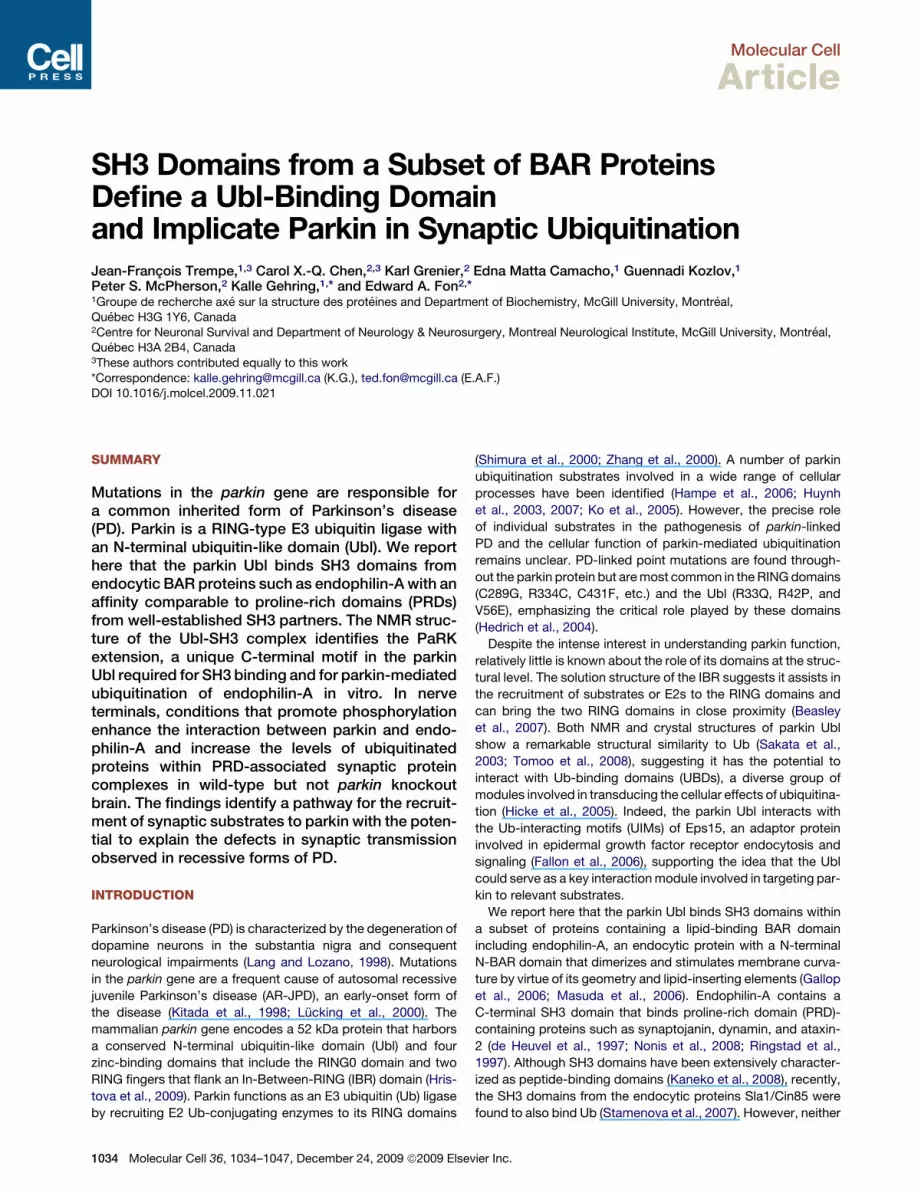
Molecular Cell Article SH3 Domains from a Subset of BAR Proteins Define a Ubl-Binding Domain and Implicate Parkin in Synaptic Ubiquitination Jean-Franc ¸ ois Trempe, 1,3 Carol X.-Q. Chen, 2,3 Karl Grenier, 2 Edna Matta Camacho, 1 Guennadi Kozlov, 1 Peter S. McPherson, 2 Kalle Gehring, 1, * and Edward A. Fon 2, * 1 Groupe de recherche axe ´ sur la structure des prote ´ ines and Department of Biochemistry, McGill University, Montre ´ al, Que ´ bec H3G 1Y6, Canada 2 Centre for Neuronal Survival and Department of Neurology & Neurosurgery, Montreal Neurological Institute, McGill University, Montre ´ al, Que ´ bec H3A 2B4, Canada 3 These authors contributed equally to this work *Correspondence: [email protected] (K.G.), [email protected] (E.A.F.) DOI 10.1016/j.molcel.2009.11.021 SUMMARY Mutations in the parkin gene are responsible for a common inherited form of Parkinson’s disease (PD). Parkin is a RING-type E3 ubiquitin ligase with an N-terminal ubiquitin-like domain (Ubl). We report here that the parkin Ubl binds SH3 domains from endocytic BAR proteins such as endophilin-A with an affinity comparable to proline-rich domains (PRDs) from well-established SH3 partners. The NMR struc- ture of the Ubl-SH3 complex identifies the PaRK extension, a unique C-terminal motif in the parkin Ubl required for SH3 binding and for parkin-mediated ubiquitination of endophilin-A in vitro. In nerve terminals, conditions that promote phosphorylation enhance the interaction between parkin and endo- philin-A and increase the levels of ubiquitinated proteins within PRD-associated synaptic protein complexes in wild-type but not parkin knockout brain. The findings identify a pathway for the recruit- ment of synaptic substrates to parkin with the poten- tial to explain the defects in synaptic transmission observed in recessive forms of PD. INTRODUCTION Parkinson’s disease (PD) is characterized by the degeneration of dopamine neurons in the substantia nigra and consequent neurological impairments (Lang and Lozano, 1998). Mutations in the parkin gene are a frequent cause of autosomal recessive juvenile Parkinson’s disease (AR-JPD), an early-onset form of the disease (Kitada et al., 1998; Lu ¨ cking et al., 2000). The mammalian parkin gene encodes a 52 kDa protein that harbors a conserved N-terminal ubiquitin-like domain (Ubl) and four zinc-binding domains that include the RING0 domain and two RING fingers that flank an In-Between-RING (IBR) domain (Hris- tova et al., 2009). Parkin functions as an E3 ubiquitin (Ub) ligase by recruiting E2 Ub-conjugating enzymes to its RING domains (Shimura et al., 2000; Zhang et al., 2000). A number of parkin ubiquitination substrates involved in a wide range of cellular processes have been identified (Hampe et al., 2006; Huynh et al., 2003, 2007; Ko et al., 2005). However, the precise role of individual substrates in the pathogenesis of parkin-linked PD and the cellular function of parkin-mediated ubiquitination remains unclear. PD-linked point mutations are found through- out the parkin protein but are most common in the RING domains (C289G, R334C, C431F, etc.) and the Ubl (R33Q, R42P, and V56E), emphasizing the critical role played by these domains (Hedrich et al., 2004). Despite the intense interest in understanding parkin function, relatively little is known about the role of its domains at the struc- tural level. The solution structure of the IBR suggests it assists in the recruitment of substrates or E2s to the RING domains and can bring the two RING domains in close proximity (Beasley et al., 2007). Both NMR and crystal structures of parkin Ubl show a remarkable structural similarity to Ub (Sakata et al., 2003; Tomoo et al., 2008), suggesting it has the potential to interact with Ub-binding domains (UBDs), a diverse group of modules involved in transducing the cellular effects of ubiquitina- tion (Hicke et al., 2005). Indeed, the parkin Ubl interacts with the Ub-interacting motifs (UIMs) of Eps15, an adaptor protein involved in epidermal growth factor receptor endocytosis and signaling (Fallon et al., 2006), supporting the idea that the Ubl could serve as a key interaction module involved in targeting par- kin to relevant substrates. We report here that the parkin Ubl binds SH3 domains within a subset of proteins containing a lipid-binding BAR domain including endophilin-A, an endocytic protein with a N-terminal N-BAR domain that dimerizes and stimulates membrane curva- ture by virtue of its geometry and lipid-inserting elements (Gallop et al., 2006; Masuda et al., 2006). Endophilin-A contains a C-terminal SH3 domain that binds proline-rich domain (PRD)- containing proteins such as synaptojanin, dynamin, and ataxin- 2(de Heuvel et al., 1997; Nonis et al., 2008; Ringstad et al., 1997). Although SH3 domains have been extensively character- ized as peptide-binding domains (Kaneko et al., 2008), recently, the SH3 domains from the endocytic proteins Sla1/Cin85 were found to also bind Ub (Stamenova et al., 2007). However, neither 1034 Molecular Cell 36, 1034–1047, December 24, 2009 ª2009 Elsevier Inc.
-
Upload
independent -
Category
Documents
-
view
0 -
download
0
Transcript of Interaction Of The Parkin UBL Domain With SH3-containing Proteins Involved In Synaptic Vesicle...
Molecular Cell
Article
SH3 Domains from a Subset of BAR ProteinsDefine a Ubl-Binding Domainand Implicate Parkin in Synaptic UbiquitinationJean-Francois Trempe,1,3 Carol X.-Q. Chen,2,3 Karl Grenier,2 Edna Matta Camacho,1 Guennadi Kozlov,1
Peter S. McPherson,2 Kalle Gehring,1,* and Edward A. Fon2,*1Groupe de recherche axe sur la structure des proteines and Department of Biochemistry, McGill University, Montreal,
Quebec H3G 1Y6, Canada2Centre for Neuronal Survival and Department of Neurology & Neurosurgery, Montreal Neurological Institute, McGill University, Montreal,
Quebec H3A 2B4, Canada3These authors contributed equally to this work
*Correspondence: [email protected] (K.G.), [email protected] (E.A.F.)DOI 10.1016/j.molcel.2009.11.021
SUMMARY
Mutations in the parkin gene are responsible fora common inherited form of Parkinson’s disease(PD). Parkin is a RING-type E3 ubiquitin ligase withan N-terminal ubiquitin-like domain (Ubl). We reporthere that the parkin Ubl binds SH3 domains fromendocytic BAR proteins such as endophilin-A with anaffinity comparable to proline-rich domains (PRDs)from well-established SH3 partners. The NMR struc-ture of the Ubl-SH3 complex identifies the PaRKextension, a unique C-terminal motif in the parkinUbl required for SH3 binding and for parkin-mediatedubiquitination of endophilin-A in vitro. In nerveterminals, conditions that promote phosphorylationenhance the interaction between parkin and endo-philin-A and increase the levels of ubiquitinatedproteins within PRD-associated synaptic proteincomplexes in wild-type but not parkin knockoutbrain. The findings identify a pathway for the recruit-ment of synaptic substrates to parkin with the poten-tial to explain the defects in synaptic transmissionobserved in recessive forms of PD.
INTRODUCTION
Parkinson’s disease (PD) is characterized by the degeneration of
dopamine neurons in the substantia nigra and consequent
neurological impairments (Lang and Lozano, 1998). Mutations
in the parkin gene are a frequent cause of autosomal recessive
juvenile Parkinson’s disease (AR-JPD), an early-onset form of
the disease (Kitada et al., 1998; Lucking et al., 2000). The
mammalian parkin gene encodes a 52 kDa protein that harbors
a conserved N-terminal ubiquitin-like domain (Ubl) and four
zinc-binding domains that include the RING0 domain and two
RING fingers that flank an In-Between-RING (IBR) domain (Hris-
tova et al., 2009). Parkin functions as an E3 ubiquitin (Ub) ligase
by recruiting E2 Ub-conjugating enzymes to its RING domains
1034 Molecular Cell 36, 1034–1047, December 24, 2009 ª2009 Elsev
(Shimura et al., 2000; Zhang et al., 2000). A number of parkin
ubiquitination substrates involved in a wide range of cellular
processes have been identified (Hampe et al., 2006; Huynh
et al., 2003, 2007; Ko et al., 2005). However, the precise role
of individual substrates in the pathogenesis of parkin-linked
PD and the cellular function of parkin-mediated ubiquitination
remains unclear. PD-linked point mutations are found through-
out the parkin protein but are most common in the RING domains
(C289G, R334C, C431F, etc.) and the Ubl (R33Q, R42P, and
V56E), emphasizing the critical role played by these domains
(Hedrich et al., 2004).
Despite the intense interest in understanding parkin function,
relatively little is known about the role of its domains at the struc-
tural level. The solution structure of the IBR suggests it assists in
the recruitment of substrates or E2s to the RING domains and
can bring the two RING domains in close proximity (Beasley
et al., 2007). Both NMR and crystal structures of parkin Ubl
show a remarkable structural similarity to Ub (Sakata et al.,
2003; Tomoo et al., 2008), suggesting it has the potential to
interact with Ub-binding domains (UBDs), a diverse group of
modules involved in transducing the cellular effects of ubiquitina-
tion (Hicke et al., 2005). Indeed, the parkin Ubl interacts with
the Ub-interacting motifs (UIMs) of Eps15, an adaptor protein
involved in epidermal growth factor receptor endocytosis and
signaling (Fallon et al., 2006), supporting the idea that the Ubl
could serve as a key interaction module involved in targeting par-
kin to relevant substrates.
We report here that the parkin Ubl binds SH3 domains within
a subset of proteins containing a lipid-binding BAR domain
including endophilin-A, an endocytic protein with a N-terminal
N-BAR domain that dimerizes and stimulates membrane curva-
ture by virtue of its geometry and lipid-inserting elements (Gallop
et al., 2006; Masuda et al., 2006). Endophilin-A contains a
C-terminal SH3 domain that binds proline-rich domain (PRD)-
containing proteins such as synaptojanin, dynamin, and ataxin-
2 (de Heuvel et al., 1997; Nonis et al., 2008; Ringstad et al.,
1997). Although SH3 domains have been extensively character-
ized as peptide-binding domains (Kaneko et al., 2008), recently,
the SH3 domains from the endocytic proteins Sla1/Cin85 were
found to also bind Ub (Stamenova et al., 2007). However, neither
ier Inc.

Figure 1. Parkin Interacts with Endophilin-A
(A) Mass spectrometry identification of parkin Ubl ligands. GST-parkin-Ubl (1–108) and GST bound to glutathione-Sepharose resin were incubated with mouse
brain lysate, washed, and resolved on SDS-PAGE. Ubl-specific bands were excised, digested with trypsin, and identified using mass spectrometry and the
program MASCOT.
(B) Brain endophilin-A1 interacts specifically with the parkin Ubl. The indicated GST fusion proteins were immobilized on glutathione-Sepharose beads, incubated
with mouse brain lysate, washed, eluted with loading buffer, loaded on SDS-PAGE, and revealed by immunoblotting with an endophilin-A1 antibody (above) and
by Ponceau staining (below).
(C) Parkin and endophilin-A1 interact in cells. HEK293 cells were transfected with GFP-endophilin-A1 and either pcDNA or Flag-parkin. The cell lysates were
immunoprecipitated with anti-Flag or mouse serum, and the bound products were resolved on SDS-PAGE and probed with an endophilin-A1 antibody.
(D) Endogenous parkin and endophilin-A1 interact in brain. Mouse brain synaptic lysates were prepared from wild-type and parkin KO mice and immunoprecip-
itated with preimmune serum or an endophilin-A1 antibody. The bound products were resolved on SDS-PAGE and probed with parkin and endophilin-A1
antibodies.
Molecular Cell
Parkin Ubl Interacts with Endophilin-A SH3
the cellular function of the SH3 interaction with Ub nor its
capacity to bind Ubl domains in addition to Ub is known. Here,
we demonstrate that the endophilin-A1 SH3 domain binds the
parkin Ubl domain with an affinity comparable to the PRD of syn-
aptojanin. The structural basis for the highly selective Ubl-SH3
interaction lies in a basic C-terminal extension unique to the par-
kin Ubl. Finally, we show that conditions that promote phosphor-
ylation drive the interaction between endogenous parkin and
endophilin-A in synaptosomes, which in turn leads to an increase
in the levels of ubiquitinated synaptic proteins in WT but not in
parkin knockout (KO) mice. Together, our findings define the
SH3 domain as a parkin Ubl-binding partner and implicate
BAR-SH3 proteins in parkin-mediated synaptic ubiquitination.
RESULTS
Parkin Interacts with Endophilin-AIn an effort to identify parkin Ubl-binding proteins, we used
a GST-Ubl fusion protein to carry out affinity chromatography
in mouse brain lysates. Specific proteins that bound to GST-
Ubl but not GST or GST-Ub were identified using mass spec-
trometry. In addition to the previously established parkin Ubl
Molecular
ligand Eps15 (Fallon et al., 2006), we identified endophilin-A1,
-A2, and -A3 (Figures 1A and S1). The interaction with endophi-
lin-A1 was confirmed by immunoblotting (Figure 1B). No binding
was detected with Ub or the Ubl of Plic1 (another protein with an
N-terminal Ubl domain), indicating that the interaction with par-
kin is specific. Parkin was immunoprecipitated with endophilin-
A1 in transfected HEK293 cells (Figure 1C), demonstrating that
the full-length proteins interact in cells. Moreover, coimmuno-
precipitation from synaptic brain lysates indicates that endoge-
nous parkin and endophilin-A1 interact in vivo (Figure 1D).
SH3 Domains from a Subset of BAR Proteins Definea Parkin Ubl-Binding DomainThe association of the parkin Ubl with endophilin-A isoforms was
surprising, as the latter do not contain a classic UBD. To deter-
mine the domain that mediates the interaction with the parkin
Ubl, endophilin-A1 deletions were used to pull down HA-tagged
full-length or DUbl-parkin expressed in HEK293 cells (Figure 2A).
The results showed that the endophilin SH3 domain is both
necessary and sufficient for the interaction with parkin.
The extent of parkin Ubl selectivity toward different SH3
domains was investigated by testing a panel of recombinant
Cell 36, 1034–1047, December 24, 2009 ª2009 Elsevier Inc. 1035

Figure 2. The Parkin Ubl Interacts with a Subset of SH3 Domains Found in BAR-Containing Proteins
(A) The SH3 and Ubl domains mediate the interaction between endophilin-A1 and parkin. HEK293 cells were transfected with HA-parkin, HA-parkin-DUbl, or
a control vector (pcDNA). The cell lysates were incubated with the indicated GST-endophilin-A1 constructs, and the bound products were immunoblotted
with an anti-HA antibody (above) or stained with Ponceau (below). The band in lane 1 is a spillover from lane 2 (see Figure 5C for similar control).
(B) The parkin Ubl interacts with a subset of SH3 domains from BAR-domain proteins. Glutathione-Sepharose-immobilized GST or GST-SH3 proteins (lower
panel) were incubated with the parkin Ubl (1–76, top) or Ub (middle). The washed products were eluted with loading buffer, resolved by SDS-PAGE, and stained
with Coomassie blue.
(C) Domain organization of BAR-SH3 proteins (top) that interact with the parkin Ubl domain (bottom). CLAP is a clathrin/AP-2-interacting domain.
Molecular Cell
Parkin Ubl Interacts with Endophilin-A SH3
GST-SH3 fusion proteins for their capacity to bind the parkin Ubl
or Ub (Figures 2B and S2). The three isoforms of endophilin-A all
bound the parkin Ubl, although the endophilin-A2 interaction
was weaker. The SH3 domains from syndapin-2/PACSIN-2
and amphiphysin-2 also bound the parkin Ubl in vitro as well
as in HEK293 cells (Figure S2C). A striking common link among
SH3 proteins that bind the parkin Ubl is that they all contain
a BAR domain (Figure 2C) and are involved in vesicle trafficking.
In contrast, the parkin Ubl did not bind the SH3 domain from
endophilin-B1, an endophilin-A homolog involved in mitochon-
drial membrane dynamics (Karbowski et al., 2004), nor did it
bind the SH3 domains from Grb2, STAM, intersectin-1, and
CIN85, which do not have a BAR domain. Ub has been reported
to bind the SH3 domains of yeast Sla1 and its vertebrate ortholog
CIN85, as well as amphiphysin-1 and -2 (Stamenova et al., 2007).
However, no SH3 domains bound to Ub under the stringent
conditions used to test Ubl binding. Thus, the SH3 domains
from a subset of BAR-domain proteins involved in vesicle traf-
ficking define a parkin-specific Ubl-binding domain.
1036 Molecular Cell 36, 1034–1047, December 24, 2009 ª2009 Elsev
The Parkin Ubl Domain Binds to the PRD-BindingSurface of the Endophilin-A1 SH3 DomainSH3 domains are known to bind peptide ligands through the
surface formed by invariant proline and tryptophan side chains
(Kaneko et al., 2008). Structural biology techniques were there-
fore employed to assess binding of the parkin Ubl and different
PRDs to the endophilin-A1 SH3 domain. The crystal structure
of the free SH3 domain was determined at 1.4 A resolution
(Figure S3 and Table S1), and its NMR 15N-1H HSQC spectrum
was assigned. Addition of parkin Ubl to 15N-labeled endophi-
lin-A1 SH3 induced large and specific NMR chemical shift
perturbations on the peptide-binding surface of the SH3 domain
(Figures 3A and S4). The perturbations were in the fast-interme-
diate exchange regime and enabled us to calculate a dissociation
constant of 13 ± 4 mM (Figures 3B and S5). This affinity is signif-
icantly stronger than that of Ub toward the SH3 domains of Sla1
and CIN85 SH3-C, which have KD (dissociation constant) values
of 40 and 171 mM, respectively (Bezsonova et al., 2008; Stame-
nova et al., 2007). Interestingly, a similar titration experiment
ier Inc.
Molecular Cell
Parkin Ubl Interacts with Endophilin-A SH3
carried out with Ub induced negligible chemical shift perturba-
tions, which confirmed the absence of binding observed in
pull-down assays (Figure S6).
A PRD peptide derived from the synaptojanin E20 binding site
(PPARPAPPQRPPPPS1113–1127) induced chemical shift pertur-
bations on the same surface of endophilin-A1 SH3 that binds
the Ubl domain (Figure 3A) and bound with a similar affinity
(Figures 3B and S5), suggesting they compete for binding. This
was directly demonstrated by addition of synaptojanin E20 PRD
to 15N-labeled parkin Ubl bound to endophilin-A1 SH3 domain
(Figure S5F). A large molar excess of PRD peptide was needed
to completely displace the Ubl domain, since their affinities for
the SH3 domain are similar. A dynamin PRD peptide (RRAPAVP-
PARP783–793) induced chemical shift perturbations similar to the
E20 PRD, but the interaction had a much weaker affinity (Figures
3B and S5). Thus, the SH3 domain binds the Ubl and PRDs via
a common site, with similar affinities.
The C Terminus of the Parkin Ubl Confers SH3-BindingSelectivityThe selectivity of the parkin Ubl toward certain SH3 domains
prompted us to determine the structure of the Ubl-SH3 complex.
A model of the rat parkin Ubl was derived from the crystal struc-
ture of murine parkin Ubl (Tomoo et al., 2008) and docked on the
SH3 crystal structure using 24 NMR intermolecular NOEs
(Figure S7) and 119 15N-1H residual dipolar couplings (RDCs)
(Figure S8). Chemical shift perturbations (Figure 3C) and hetero-
nuclear NOE measurements (Figures 3D and S9) suggested that
the flexible C-terminal tail of the parkin Ubl (72–76) was involved
in binding and became structured upon complex formation. The
conformation of the C-terminal tail was therefore calculated de
novo and incorporated in the structure calculation, which
converged to yield an ensemble of structures with a 0.65 A back-
bone root-mean-square deviation (rmsd) from the lowest energy
structure (Table 1 and Figure S10A). The hydrophobic patch
formed by Ile44, Gly47, His68, and Val70 of the Ubl interacts
with the peptide-binding surface of the SH3 domain (Fig-
ure 3E). The alignment of the two domains is reminiscent of
that observed in the Sla1- and Cin85-SH3-Ub complexes (Bez-
sonova et al., 2008; He et al., 2007), but the SH3:Ubl complex
shows a considerable shift in the position of the two domains
(Figure S10B). The side chain of Ile44 is located between the
side chains of Tyr299 and Tyr343 in the SH3 domain, which
moves the C terminus of the Ubl closer to the center of the
peptide-binding surface of the SH3 domain (Figure 3F). The
C-terminal tail of the Ubl adopts an extended b strand conforma-
tion that mediates interactions with the SH3 domain that are
similar to PRD:SH3 interactions. Notably, the Ubl residue
Pro73 packs between the invariant SH3 residues Pro340 and
Trp327, in a manner similar to the second proline in a PXXP motif
bound to the related Grb2 SH3 domain (Wittekind et al., 1997). In
this specific tail conformation, the side chain of Arg75 is well
positioned to interact with the acidic loop formed by Asp324
and Glu325 in the SH3 domain (Figure 3F).
The C-terminal stretch (73–76) of the Ubl is unique to mamma-
lian parkin and forms the consensus sequence PxRK, which we
refer to as the PaRK extension (Figure 4A). The role of the PaRK
extension in binding SH3 domains was tested using several
Molecular
constructs, including a truncation mutant lacking the two
C-terminal PaRK extension residues (1–74), a chimera in which
residues 73–76 (PQRK) were replaced with the corresponding
residues in Ub (LRGG), and two single-site mutants of parkin
1–76 (R75G and K76G). The C-terminal truncation, the chimera,
and the R75G mutation completely abolished binding to GST-
endophilin-A1 SH3 (Figure 4B), whereas the K76G mutant
retained some affinity for the SH3 domain. The PaRK extension
is essential and specific for SH3 binding, as the R75G mutant
was still able to bind the tandem UIMs from Eps15 (Fallon
et al., 2006) (Figure 4C).
Additional parkin Ubl mutants were produced to test the
contribution of other residues to SH3 binding. Asn8 is located
in the b1-b2 loop that contacts Asn342 in the SH3 domain
(Figure 3F); mutation to alanine or leucine (the residue in Ub)
abolished binding to endogenous endophilin-A1 in brain lysate
(Figure 4D). The PD-linked mutant R42P, which was shown to
unfold the Ubl (Safadi and Shaw, 2007), was also unable to
bind endophilin-A1. The I44A and K48A mutations also disrupted
the interaction, in agreement with the contacts observed in the
structure of the complex (Figure 3F). Ile44 and Lys48 are
conserved in Ub and therefore do not constitute selectivity deter-
minants, as opposed to Asn8 and the PaRK extension, which are
unique to parkin (Figure 4A).
A Unique Glutamate Residue within Endophilin-A SH3Domains Confers Parkin Ubl-Binding SpecificityAlignment of SH3 domain sequences (Figure 5A) and the NMR
solution structure (Figures 3E and 3F) led us to test specificity
determinants in the endophilin-A SH3 domain using single-site
mutagenesis and in vitro pull-down assays. Three glutamic acid
residues (Glu302, Glu304, and Glu325) conserved in Ubl-binding
SH3 domains (Figure 5A) and located in regions that display large
chemical shift perturbations (Figure 3A) were mutated to alanine.
Strikingly, only the E304A mutation abrogated the interaction
(Figure 5B). The three endophilin-A isoforms are the only verte-
brate SH3 proteins to have an acidic residue at this position
(Figure 5A). In the solution structure, the side chain of Glu304 is
in proximity to that of the conserved Ubl residue Arg72 and
thus could form a salt bridge with the latter. Syndapin-2 and
amphiphysin-2 have polar residues and show weaker binding,
whereas endophilin-B1 and amphiphysin-1 have alanine and
show no binding. Despite the shared Ubl- and PRD-binding
surface on the SH3 (Figure 3A), the E304A mutation, which abol-
ishes Ubl binding (Figure 5B), did not significantly reduce the
affinity for the synaptojanin E20 PRD (Figure 3B). Glu304 is
therefore a crucial selectivity determinant for Ubl binding.
Other residues were also found to contribute to the interaction.
The mutation D324A, which disrupts an electrostatic interaction
with the parkin-specific Arg75, reduced the interaction. The
mutations N342A and Y343F reduced binding, likely through
loss of a polar interaction with the Ubl. Finally, we also tested
the binding of full-length and DUbl parkin expressed in
HEK293 cells to endophilin-A1 SH3 mutants that showed
reduced affinity toward the Ubl, and an identical pattern of
binding was observed (Figure 5C). Together, our findings
demonstrate that the endophilin-A1 residues Glu304, Asp324,
Asn342, and Tyr343 constitute critical SH3 selectivity
Cell 36, 1034–1047, December 24, 2009 ª2009 Elsevier Inc. 1037
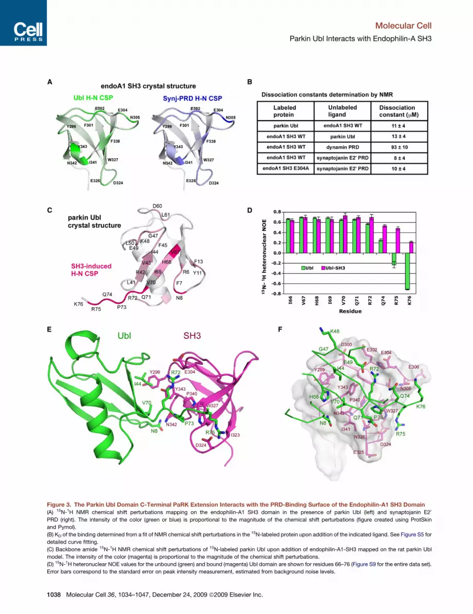
Figure 3. The Parkin Ubl Domain C-Terminal PaRK Extension Interacts with the PRD-Binding Surface of the Endophilin-A1 SH3 Domain
(A) 15N-1H NMR chemical shift perturbations mapping on the endophilin-A1 SH3 domain in the presence of parkin Ubl (left) and synaptojanin E20
PRD (right). The intensity of the color (green or blue) is proportional to the magnitude of the chemical shift perturbations (figure created using ProtSkin
and Pymol).
(B) KD of the binding determined from a fit of NMR chemical shift perturbations in the 15N-labeled protein upon addition of the indicated ligand. See Figure S5 for
detailed curve fitting.
(C) Backbone amide 15N-1H NMR chemical shift perturbations of 15N-labeled parkin Ubl upon addition of endophilin-A1-SH3 mapped on the rat parkin Ubl
model. The intensity of the color (magenta) is proportional to the magnitude of the chemical shift perturbations.
(D) 15N-1H heteronuclear NOE values for the unbound (green) and bound (magenta) Ubl domain are shown for residues 66–76 (Figure S9 for the entire data set).
Error bars correspond to the standard error on peak intensity measurement, estimated from background noise levels.
Molecular Cell
Parkin Ubl Interacts with Endophilin-A SH3
1038 Molecular Cell 36, 1034–1047, December 24, 2009 ª2009 Elsevier Inc.
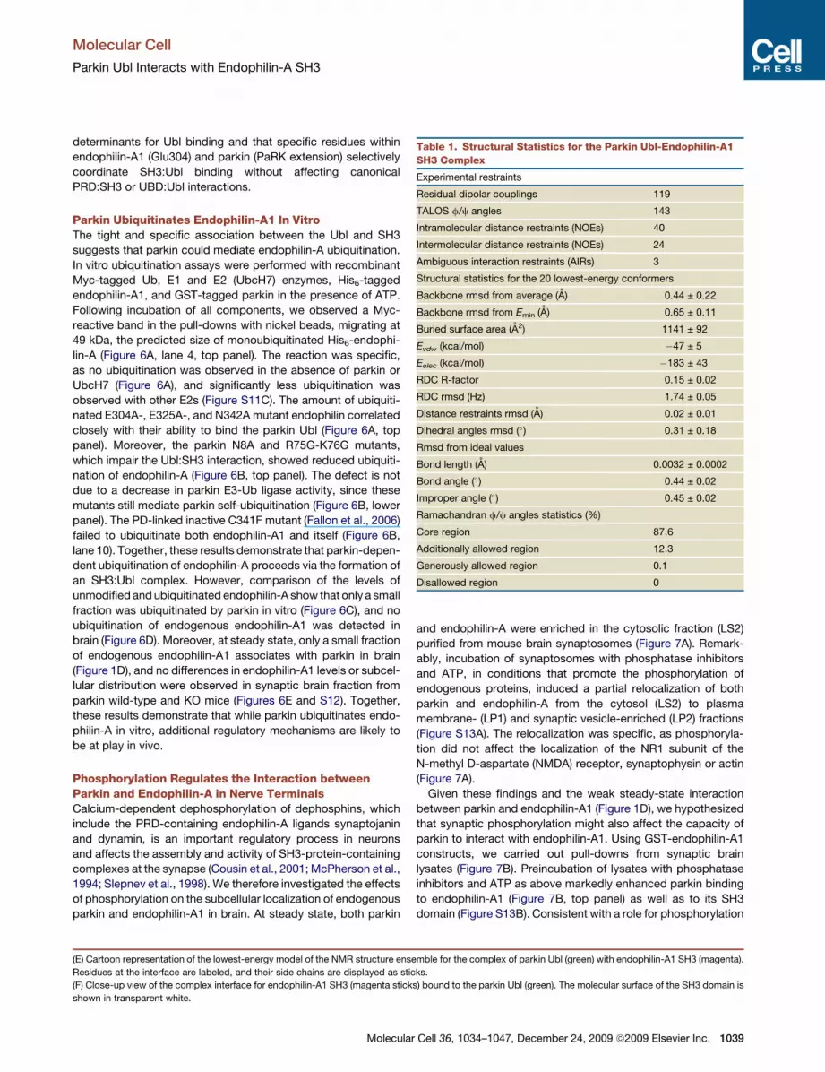
Table 1. Structural Statistics for the Parkin Ubl-Endophilin-A1
SH3 Complex
Experimental restraints
Residual dipolar couplings 119
TALOS f/c angles 143
Intramolecular distance restraints (NOEs) 40
Intermolecular distance restraints (NOEs) 24
Ambiguous interaction restraints (AIRs) 3
Structural statistics for the 20 lowest-energy conformers
Backbone rmsd from average (A) 0.44 ± 0.22
Backbone rmsd from Emin (A) 0.65 ± 0.11
Buried surface area (A2) 1141 ± 92
Evdw (kcal/mol) �47 ± 5
Eelec (kcal/mol) �183 ± 43
RDC R-factor 0.15 ± 0.02
RDC rmsd (Hz) 1.74 ± 0.05
Distance restraints rmsd (A) 0.02 ± 0.01
Dihedral angles rmsd (�) 0.31 ± 0.18
Rmsd from ideal values
Bond length (A) 0.0032 ± 0.0002
Bond angle (�) 0.44 ± 0.02
Improper angle (�) 0.45 ± 0.02
Ramachandran f/c angles statistics (%)
Core region 87.6
Additionally allowed region 12.3
Generously allowed region 0.1
Disallowed region 0
Molecular Cell
Parkin Ubl Interacts with Endophilin-A SH3
determinants for Ubl binding and that specific residues within
endophilin-A1 (Glu304) and parkin (PaRK extension) selectively
coordinate SH3:Ubl binding without affecting canonical
PRD:SH3 or UBD:Ubl interactions.
Parkin Ubiquitinates Endophilin-A1 In VitroThe tight and specific association between the Ubl and SH3
suggests that parkin could mediate endophilin-A ubiquitination.
In vitro ubiquitination assays were performed with recombinant
Myc-tagged Ub, E1 and E2 (UbcH7) enzymes, His6-tagged
endophilin-A1, and GST-tagged parkin in the presence of ATP.
Following incubation of all components, we observed a Myc-
reactive band in the pull-downs with nickel beads, migrating at
49 kDa, the predicted size of monoubiquitinated His6-endophi-
lin-A (Figure 6A, lane 4, top panel). The reaction was specific,
as no ubiquitination was observed in the absence of parkin or
UbcH7 (Figure 6A), and significantly less ubiquitination was
observed with other E2s (Figure S11C). The amount of ubiquiti-
nated E304A-, E325A-, and N342A mutant endophilin correlated
closely with their ability to bind the parkin Ubl (Figure 6A, top
panel). Moreover, the parkin N8A and R75G-K76G mutants,
which impair the Ubl:SH3 interaction, showed reduced ubiquiti-
nation of endophilin-A (Figure 6B, top panel). The defect is not
due to a decrease in parkin E3-Ub ligase activity, since these
mutants still mediate parkin self-ubiquitination (Figure 6B, lower
panel). The PD-linked inactive C341F mutant (Fallon et al., 2006)
failed to ubiquitinate both endophilin-A1 and itself (Figure 6B,
lane 10). Together, these results demonstrate that parkin-depen-
dent ubiquitination of endophilin-A proceeds via the formation of
an SH3:Ubl complex. However, comparison of the levels of
unmodified and ubiquitinated endophilin-A show that only a small
fraction was ubiquitinated by parkin in vitro (Figure 6C), and no
ubiquitination of endogenous endophilin-A1 was detected in
brain (Figure 6D). Moreover, at steady state, only a small fraction
of endogenous endophilin-A1 associates with parkin in brain
(Figure 1D), and no differences in endophilin-A1 levels or subcel-
lular distribution were observed in synaptic brain fraction from
parkin wild-type and KO mice (Figures 6E and S12). Together,
these results demonstrate that while parkin ubiquitinates endo-
philin-A in vitro, additional regulatory mechanisms are likely to
be at play in vivo.
Phosphorylation Regulates the Interaction betweenParkin and Endophilin-A in Nerve TerminalsCalcium-dependent dephosphorylation of dephosphins, which
include the PRD-containing endophilin-A ligands synaptojanin
and dynamin, is an important regulatory process in neurons
and affects the assembly and activity of SH3-protein-containing
complexes at the synapse (Cousin et al., 2001; McPherson et al.,
1994; Slepnev et al., 1998). We therefore investigated the effects
of phosphorylation on the subcellular localization of endogenous
parkin and endophilin-A1 in brain. At steady state, both parkin
(E) Cartoon representation of the lowest-energy model of the NMR structure ense
Residues at the interface are labeled, and their side chains are displayed as stic
(F) Close-up view of the complex interface for endophilin-A1 SH3 (magenta stick
shown in transparent white.
Molecular
and endophilin-A were enriched in the cytosolic fraction (LS2)
purified from mouse brain synaptosomes (Figure 7A). Remark-
ably, incubation of synaptosomes with phosphatase inhibitors
and ATP, in conditions that promote the phosphorylation of
endogenous proteins, induced a partial relocalization of both
parkin and endophilin-A from the cytosol (LS2) to plasma
membrane- (LP1) and synaptic vesicle-enriched (LP2) fractions
(Figure S13A). The relocalization was specific, as phosphoryla-
tion did not affect the localization of the NR1 subunit of the
N-methyl D-aspartate (NMDA) receptor, synaptophysin or actin
(Figure 7A).
Given these findings and the weak steady-state interaction
between parkin and endophilin-A1 (Figure 1D), we hypothesized
that synaptic phosphorylation might also affect the capacity of
parkin to interact with endophilin-A1. Using GST-endophilin-A1
constructs, we carried out pull-downs from synaptic brain
lysates (Figure 7B). Preincubation of lysates with phosphatase
inhibitors and ATP as above markedly enhanced parkin binding
to endophilin-A1 (Figure 7B, top panel) as well as to its SH3
domain (Figure S13B). Consistent with a role for phosphorylation
mble for the complex of parkin Ubl (green) with endophilin-A1 SH3 (magenta).
ks.
s) bound to the parkin Ubl (green). The molecular surface of the SH3 domain is
Cell 36, 1034–1047, December 24, 2009 ª2009 Elsevier Inc. 1039
Figure 4. The Parkin Ubl C-Terminal PaRK Extension Is Required for Binding SH3 Domains
(A) Sequence alignment of the parkin Ubl domains from rat (RN, Rattus norvegicus), mouse (MM, Mus musculus), human (HS, Homo sapiens), chimp (PT, Pan
troglodytes), bovine (BT, Bos taurus), chicken (GG, Gallus gallus), and fly (DM, Drosophila melanogaster), as well as Ub and the human plic1 Ubl. The secondary
structure elements of parkin Ubl are shown above the alignment. Boxed residues are unique to mammalian parkin and required for parkin to interact with the
endophilin-A1 SH3.
(B) The PaRK extension of the parkin Ubl is required for binding the endophilin-A1 SH3 domain. GST-endophilin-A1 SH3 (left panel) or GST (middle panel) was
immobilized on glutathione-Sepharose beads and incubated with the parkin Ubl (1–76) wild-type or mutants (PQRK73–76 to LRGG73–76, R75G, and K76G), a dele-
tion (1–74), or Ub (Ub). The procedure is the same as in Figure 3B.
(C) Arg75 of parkin is required for binding SH3 domains but not the Eps15 UIMs. The procedure is the same as in Figure 4B.
(D) Identification of parkin Ubl residues mediating interaction with endophilin-A1. GST-parkin Ubl mutants were incubated with mouse brain lysate, and retained
proteins were detected by an endophilin-A1 antibody.
Molecular Cell
Parkin Ubl Interacts with Endophilin-A SH3
per se, which requires ATP hydrolysis, the enhancement was
blocked by the nonhydrolyzable ATP analog AMP-PNP (Fig-
ure 7C). However, the effect did not appear to be mediated by
kinases previously implicated in parkin phosphorylation and
function (Rubio de la Torre et al., 2009; Yamamoto et al.,
2005), as inhibitors of Cdk5, caseine kinase I, and protein kinase
C all failed to block the enhancement (Figure S13C). Preincuba-
tion with phosphatase inhibitors and ATP did not affect the inter-
action between endophilin-A1 and its main PRD-containing
ligand synaptojanin (Figure 7B, middle panel), suggesting that
phosphorylation stimulates parkin-endophilin-A binding directly
rather than by decreasing competition of PRD proteins for SH3
binding.
To test whether phosphorylation enhanced the interaction
between endogenous endophilin-A1 and parkin in synapto-
somes, we used a GST fusion protein encoding a PRD peptide
from synaptojanin E20 site (1111–1129) to purify endophilin-con-
taining complexes from mouse brain synaptosomes. This PRD
1040 Molecular Cell 36, 1034–1047, December 24, 2009 ª2009 Elsev
peptide has been shown previously to efficiently pull down endo-
philin-A from brain (Gad et al., 2000). Under basal conditions,
parkin could not be detected in the PRD-purified complexes
despite the efficient recruitment of endophilin-A1 (Figure 7D,
lane 3). Strikingly, preincubation of synaptosomes with phos-
phatase inhibitors and ATP drove parkin into endophilin-A-con-
taining complexes (Figure 7D, lane 4). Experiments with purified
proteins confirmed that parkin is recruited to the PRD indirectly
by endophilin-A1 and does not bind the PRD directly (Fig-
ure 7E). As the parkin Ubl and synaptojanin PRD bind the
same site on endophilin-A1’s single SH3 domain (Figure 3A),
the mechanism is likely to involve endophilin-A1 dimerization
via its N-BAR domain. Endophilin-A1 dimers can simultaneously
accommodate PRD and Ubl binding via their two SH3 domains.
Interestingly, the endophilin-A1 N-BAR domain binds mem-
branes as dimers, but is predominantly monomeric in the cytosol
(Gallop et al., 2006). Thus, the relocalization of endophilin-A1
from the cytosol to synaptic membrane compartments that we
ier Inc.

Figure 5. Selectivity for the Parkin Ubl Is Determined by a Set of Polar Residues on the PRD-Binding Surface of the SH3 Domain
(A) Sequence alignment of SH3 domains from proteins involved in endocytosis. All sequences are from Rattus norvegicus except the budding yeast sla1 SH3-3
domain. Residues critical for the interaction with the parkin Ubl are boxed. Secondary structure elements and characteristics loops are labeled above the
alignment.
(B) Identification of specificity determinants in the endophilin-A1 SH3 domain required for parkin Ubl-binding. GST or endophilin-A1 GST-SH3 mutants were
immobilized on glutathione-Sepharose beads and incubated with the parkin Ubl (1–76). The procedure is the same as in Figure 3B.
(C) HEK293 cells were transfected with HA-parkin, HA-parkin-DUbl, or a control vector (pcDNA). The cell lysates were incubated with GST-endophilin-A1 SH3
mutants immobilized on glutathione-Sepharose beads. The bound products were immunoblotted with anti-HA antibodies.
Molecular Cell
Parkin Ubl Interacts with Endophilin-A SH3
observe in response to phosphorylation (Figure 7A) may promote
endophilin-A1 dimerization, which would allow endophilin-A-
bound parkin to be indirectly recruited by GST-PRD (Fig-
ure 7D). Taken together, these experiments suggest that phos-
phorylation promotes the interaction of endogenous parkin
with endophilin-A in synaptic membrane fractions.
Parkin Ubiquitinates PRD-Associated Proteins in NerveTerminalsSynaptic activity has also been shown to decrease ubiquitination
in nerve terminals and correlates with dephosphorylation of
synaptic proteins (Chen et al., 2003). We thus sought to test
whether the converse was true, specifically that phosphorylation
could increase protein ubiquitination within synaptic endophilin
complexes. Immunoblotting of GST-PRD-purified endophilin-
A1 complexes with an antibody against Ub revealed a marked
increase in the levels of Ub-protein conjugates copurified from
synaptosomes incubated with phosphatase inhibitors (Fig-
ure 7D, lower panel). Importantly, this increase was attenuated
in synaptosomes prepared from parkin KO mice (Figure 7D,
lane 8), indicating that ubiquitination of proteins within syn-
Molecular
aptic endophilin-A complexes is largely parkin dependent.
A long exposure of the endophilin-A1 blot showed that these
ubiquitination products are probably not ubiquitinated forms
of endophilin-A1 (Figure S13D). These results are consistent
with a model whereby phosphorylation targets endophilin-A to
membrane fractions in nerve terminals, where it dimerizes and
recruits parkin to promote the ubiquitination of synaptic proteins
(Figure 7F).
DISCUSSION
SH3 Domains: A Ubl-Binding DomainMost known SH3-binding partners contain small peptide motifs
such as PRDs, but there have been several recent reports
describing weak interactions with Ub (Kang et al., 2008; Stame-
nova et al., 2007). We show here that a Ubl domain can bind
specific SH3 proteins with an affinity comparable to PRDs.
Most parkin Ubl residues interacting with the endophilin-A1
SH3 domain are identical to those found in Ub. Two exceptions
are Asn8 and Arg75, which are conserved in all mammalian
Cell 36, 1034–1047, December 24, 2009 ª2009 Elsevier Inc. 1041

Figure 6. Parkin-Mediated Ubiquitination of Endophilin-A
(A) Endophilin-A1 ubiquitination levels correlate with the capacity of the mutant SH3 domains to bind the parkin Ubl. Ubiquitination reactions were performed in
the presence of E1, ATP, MgCl2, and the components labeled with (+). GST-parkin was immobilized on glutathione-Sepharose resin (bottom panel), and His6-
endophilin-A1 (supernatant of GST pull-down) was immobilized on Ni-NTA agarose (top panel). The products were resolved on SDS-PAGE and probed with an
anti-Myc antibody.
(B) Endophilin-A1 ubiquitination was reduced by the N8A and RK/GG Ubl mutations that reduce SH3 binding as well as by the C341F ligase-inactive, PD-linked
RING mutant (Fallon et al., 2006). Ubiquitination reactions were as above.
(C) Parkin weakly monoubiquitinates endophilin-A1 in vitro. Ubiquitination reactions were performed as described above. The products were probed with an anti-
His antibody. The band indicated by an asterisk (*) represents a degradation product of endophilin-A1.
(D) Endophilin-A1 is not significantly ubiquitinated in synaptosomes. Mouse brain synaptosomes prepared from wild-type and parkin KO mice were resolved on
SDS-PAGE and probed against endophilin-A1.
(E) Distribution of endophilin-A1 in subsynaptic fractions prepared from parkin WT and parkin KO mouse brain lysate. Equal amounts (50 mg) of proteins were
loaded in each lane and probed against endophilin-A1, parkin, NR1 (plasma membrane marker), synaptophysin (synaptic vesicle marker), synuclein (cytosol
marker), and actin (general marker).
Molecular Cell
Parkin Ubl Interacts with Endophilin-A SH3
parkin Ubls and absent in Ub and other Ubls. Both residues are
essential for SH3 binding and unique specificity determinants.
Ub was shown to bind the third SH3 domain of CIN85 as well
as the SH3 domains from amphiphysin-1 and amphiphysin-2
(Stamenova et al., 2007). In our binding assays, we did not detect
an interaction between Ub and any of the SH3 domains (Figures
1042 Molecular Cell 36, 1034–1047, December 24, 2009 ª2009 Elsev
2B and S2). The high cellular levels of free Ub may compensate
for the weak affinity of SH3:Ub interactions; indeed, other UBDs
also bind weakly to Ub, with affinities rarely below 20 mM (Hicke
et al., 2005). One of the key Ub-binding determinants in the Sla1/
CIN85 SH3 domain was a phenylalanine residue located at
a position equivalent to Tyr343 in endophilin-A1 (Bezsonova
ier Inc.

Molecular Cell
Parkin Ubl Interacts with Endophilin-A SH3
et al., 2008; Stamenova et al., 2007). Mutation of this phenylala-
nine to tyrosine in Sla1/CIN85 SH3 abolished binding to Ub, and
all Ub-interacting SH3 domains were found to have a phenylala-
nine at this position. Interestingly, we observed the reverse situ-
ation for parkin Ubl binding: the Y343F mutant reduces binding.
The presence of Asp324, although not unique to Ubl-binding
SH3 domains, is essential, as it provides an anchor point for re-
cruiting Arg75 in the parkin Ubl C-terminal PaRK extension. Simi-
larly, Asn342 is not unique to endophilin-A isoforms, but it does
contribute to Ubl binding. Interestingly, this asparagine corre-
lates with the ability of the two brain-enriched endophilin-A iso-
forms (A1 and A3) to bind the parkin Ubl better than endophi-
lin-A2, which has a serine at this position. This observation
suggests a neuronal role for the interaction between parkin
and endophilin-A.
Effects of Phosphorylation on Parkin-SH3 Interactionsand Synaptic UbiquitinationIn brain, relatively little parkin binds endophilin-A at steady state
(Figure 1D). We find that conditions that favor protein phosphor-
ylation in synaptosomes promote parkin and endophilin-A traf-
ficking from the cytosol to membrane compartments and mark-
edly enhance their association. One possible explanation is that
endophilin-A is not available for parkin binding at steady state
because it is constitutively bound to PRD proteins such as
synaptojanin and dynamin. These PRD proteins, collectively
referred to as dephosphins, are believed to trigger synaptic
vesicle endocytosis upon dephosphorylation by calcineurin,
a calcium-activated phosphatase (Cousin et al., 2001; Slepnev
et al., 1998). Antagonistic phosphorylation of dephosphins by
kinases such as Cdk5 is essential for the recycling of synaptic
vesicles (Tan et al., 2003). Upon phosphorylation, dephosphins
release their SH3 partners, making them available for parkin
binding. Indeed, a phosphomimetic mutation in the vicinity of
synaptojanin’s PRD was shown to decrease its affinity for endo-
philin-A1 (Lee et al., 2004). Surprisingly, we found that synapto-
janin binding to GST-endophilin-A1 was not affected by the
addition of ATP and PPI (Figure 7B), suggesting that other mech-
anisms could be at play. For instance, phosphorylation could
activate parkin (or inactivate a parkin inhibitor) and make the
Ubl available for binding. In agreement with this model, we found
that recombinant GST-Ubl can efficiently recruit endogenous
brain endophilin-A1 in the absence of PPI and ATP (Figure 1B).
Taken together, these observations imply that phosphorylation
would release parkin or one of its interacting partners and
make the Ubl available for binding. Cdk5, a candidate regulatory
kinase, has been shown to phosphorylate parkin, possibly
affecting its interactions, activity, and cellular localization (Avra-
ham et al., 2007; Rubio de la Torre et al., 2009; Yamamoto et al.,
2005). However, Cdk5 inhibitors did not inhibit binding to
recombinant GST-endophilin-A in our assays (Figure S13C),
suggesting that other kinases may be involved.
Beyond the phosphorylation dependence of the parkin-endo-
philin-A interaction, we observed that phosphorylation promotes
the parkin-dependent ubiquitination of PRD-associated proteins
in nerve terminals (Figure 7D). The wide smear of protein-Ub
conjugates observed in nerve terminals appears to arise from
proteins other than endophilin-A1. What are these in vivo
Molecular
substrates of parkin? Parkin has been shown previously to ubiq-
uitinate synaptic vesicle-associated proteins such as CDCrel-1
(Zhang et al., 2000) and synaptotagmin (Huynh et al., 2003).
Dimers of endophilin-A and other BAR-SH3 proteins may act
as platforms for the ubiquitination of synaptic proteins by simul-
taneously recruiting parkin and PRD-containing proteins (Fig-
ure 7F). For instance, ataxin-2 has recently been shown to be
a substrate for parkin (Huynh et al., 2007), and the ataxin-2
PRD binds selectively to the SH3 domains of endophilin-A1
and -A3 (Nonis et al., 2008), as is the case for parkin. The assign-
ment of one SH3 domain to the parkin Ubl and the other to a PRD
may not be stochastic and could be governed by additional
factors. Moreover, a parkin substrate need not be recruited
through the other SH3 domain in an endophilin-A dimer and
could be recruited to endophilin-A by another, yet unknown
mechanism. At present, our data cannot discriminate between
these alternatives. Future work will therefore aim at identifying
synaptic parkin substrates that depend on the association of
parkin with endophilin-A as well as the factors involved in
substrate recruitment.
Implication of Parkin-Mediated Ubiquitinationin Synaptic Transmission and PDAn important clue toward understanding parkin’s role comes
from its ability to interact with brain-enriched SH3 proteins that
harbor BAR domains. The N-BAR domains of endophilin-A and
amphiphysin form homodimers that can induce curvature in lipid
membrane (Gallop et al., 2006; Masuda et al., 2006; Peter et al.,
2004) and are thought to regulate endocytic vesicles by recruit-
ing enzymes at various stages of the vesicle’s formation and traf-
ficking (Dawson et al., 2006; Simpson et al., 1999). For example,
in vivo studies have shown that the primary role of endophilin-A is
to recruit synaptojanin, a phosphatidylinositol-4,5-biphosphate
phosphatase (McPherson et al., 1996), to endocytic vesicles
for clathrin uncoating (Schuske et al., 2003; Verstreken et al.,
2003). The syndapin/PACSIN proteins have an F-BAR domain
composed of an Fes-CIP homology and BAR domain (Halbach
et al., 2007) that may serve a scaffolding function by bridging
actin organization to vesicle endocytosis and trafficking (Ang-
gono et al., 2006; Qualmann and Kelly, 2000). How could this
be relevant to PD? One of the most consistent and intriguing
findings associated with both dominant (Tong et al., 2009) and
recessive (Nakamura and Edwards, 2007) forms of PD, including
those due to parkin mutations (Kitada et al., 2009), has been
defects in synaptic transmission, possibly related to altered syn-
aptic vesicle endocytosis, recycling, or release. Yet the molec-
ular mechanisms involved have remained completely unknown.
Thus, by linking parkin to endophilin-A, a protein at the heart of
synaptic vesicle endocytosis and recycling, our findings provide
a molecular link between recessive PD genes and defects in
synaptic transmission.
EXPERIMENTAL PROCEDURES
Antibodies, Reagents, Plasmids, and Recombinant Proteins
Antibodies, reagents, plasmids, and recombinant protein expression and puri-
fication are described in the Supplemental Experimental Procedures.
Cell 36, 1034–1047, December 24, 2009 ª2009 Elsevier Inc. 1043

Figure 7. Phosphorylation Regulates Parkin and Endophilin-A in Nerve Terminals
(A) Phosphorylation-dependent relocalization of endogenous parkin and endophilin-A1 to the synaptic plasma membranes and vesicles. Crude mouse brain
synaptosomes (P2) were incubated for 15 min at 37�C with or without phosphatase inhibitors (PPI) and 2 mM ATP. The synaptosomes were then fractionated
by centrifugation to yield LP1 (plasma membrane), LP2 (vesicle), and LS2 (cytosol). Equal amounts of proteins (50 mg) were resolved on SDS-PAGE and probed
with the antibodies marked on the right.
(B) Phosphorylation-dependent binding of endogenous brain parkin to the endophilin-A1 SH3 domain. GST fusion proteins were immobilized on glutathione-
Sepharose beads and incubated with mouse brain LS1 that was preincubated for 15 min at 37�C with (+) or without (�) PPI in the presence of 2 mM ATP.
Molecular Cell
Parkin Ubl Interacts with Endophilin-A SH3
1044 Molecular Cell 36, 1034–1047, December 24, 2009 ª2009 Elsevier Inc.

Molecular Cell
Parkin Ubl Interacts with Endophilin-A SH3
Mouse Brain Synaptosome Preparation
Whole mouse brain was fractionated by differential centrifugation, as previ-
ously described (Fallon et al., 2002; Huttner et al., 1983). The brain was homog-
enized in 0.32 M sucrose, 10 mM HEPES (pH 7.4) supplemented with protease
inhibitors (0.5 mg/ml leupeptin, 0.5 mg/ml aprotinin, 100 mg/ml benzamidine,
20 mg/ml PMSF). The homogenate was centrifuged for 10 min at 1,0003 g,
and the supernatant (S1) was collected and centrifuged again for 15 min at
12,0003 g to produce a synaptic pellet (P2). P2 was resuspended in the orig-
inal volume of buffer and centrifuged for 15 min at 13,0003 g to produce the
P20 pellet. The soft, white component of P20 was used as the crude synapto-
some fraction. Purified synaptosomes were either kept on ice or incubated
for 15 min at 37�C in control buffer (132 mM NaCl, 4.8 mM KCL, 2.4 mM
MgSO4, 10 mM glucose, 1.1 mM CaCl2, 0.1 mM EGTA, 10 mM HEPES-
NaOH [pH 7.4]), either with or without protein phosphatase inhibitors (1 mM
okadaic acid, 0.5 mM cyclosporin A, 50 mM NaF, 2 mM Na3VO4) and 2 mM
ATP. To further fractionate synaptosomes into subsynaptic components, P20
was resuspended in nine volumes of water and disrupted in a glass-teflon
homogenizer (three strokes). The water was adjusted to 10 mM HEPES, and
the sample was centrifuged for 20 min at 33,0003 g to yield the synaptic
plasma membrane-enriched pellet (LP1). The supernatant (LS1) was centri-
fuged for 2 hr at 260,0003 g to yield the synaptic vesicle-enriched pellet
(LP2) and synaptic cytosol-enriched supernatant (LS2).
Immunoprecipitation and Pull-Down Assays
Mouse brain S1 fractions were incubated with 1% Triton X-100 for 30 min at
4�C, followed by centrifugation for 30 min at 260,0003 g to yield soluble S2
fractions, which were used for GST-Ubl pull-downs (Figures 1A, 1B, and 4D).
Mass spectrometry protein identification was performed at the Genome
Quebec Proteomics platform (Figure 1A). LS1 fractions were used for GST-
endophilin-A1 pull-down (Figures 7B and 7C). For the GST-PRD (synaptoja-
nin-1 VAPPARPAPPQRPPPPSGA1111–1129) pull-down (Figure 7D), purified
synaptosomes were prepared from crude synaptosomes by an additional
Percoll gradient purification, as described (Dunkley et al., 1986). After incu-
bation with or without phosphatase inhibitors and ATP (see above), the
synaptosomes were centrifuged at 140,0003 g for 10 min, and the pellets
were lysed in binding buffer: 50 mM Tris-HCl (pH 7.4), 20 mM NaCl, 1 mM
DTT, 0.1 mM EDTA, 0.5% Triton X-100, plus protease inhibitors with or
without protein phosphatase inhibitors and ATP, as described above. Frac-
tions were incubated overnight at 4�C with equivalent amounts of GST fusion
protein, immobilized on glutathione-Sepharose beads. The in vitro GST-PRD
pull-down (Figure 7E) was performed as described above using purified
MBP-parkin and His-endophilin-A1. In all cases, the beads were rinsed
four times in binding buffer, and bound proteins were eluted in SDS sample
buffer at 65�C. Samples were subjected to SDS-PAGE followed by immuno-
blotting.
HEK293 cells were maintained at 37�C and 5% CO2 in DMEM supplemented
with 10% FBS (heat inactivated), 2 mM glutamine, 100 U/ml penicillin, 100 mg/
ml streptomycin. For pull-downs (Figures 2A and 5C) and immunoprecipita-
tions (Figure 1C), HEK293 cells were transfected with the indicated plasmids
using Lipofectamine 2000 (Invitrogen). Cells were lysed 48 hr posttransfection
in 50 mM Tris-HCl (pH 7.4), 150 mM NaCl, 0.5% Triton X-100, plus protease
inhibitors, for 30 min on ice. Lysates were cleared by centrifugation at
14,0003 g for 10 min. For pull-downs, the supernatant was incubated at
The washed products were eluted with loading buffer, loaded on SDS-PAGE, an
stained with Ponceau to show loading (lower panel).
(C) ATP hydrolysis is required for parkin binding to GST-endophilin-A1. The proce
(D) Phosphorylation- and parkin-dependent ubiquitination of endophilin-A1-asso
from wild-type or parkin KO mouse brain and incubated for 15 min at 37�C with (
with GST or GST-PRD (synaptojanin 1111–1129); the bound products were resolv
kin (middle), or Ub (bottom).
(E) The association of parkin with GST-PRD is mediated by endophilin-A1 in vitro
with GST or GST-PRD. Bound products were resolved on SDS-PAGE and probe
(F) Schematic model for the recruitment of the parkin E3 Ub-ligase to BAR-SH3-co
a dimer and induces curvature in lipid vesicles. It has two SH3 domains that can
substrates close to parkin.
Molecular
4�C overnight with GST fusion proteins immobilized on glutathione-Sepharose
beads in buffer (50 mM Tris-HCl, 20 mM NaCl, 1 mM DTT, 0.1 mM EDTA, 0.5%
Triton X-100 [pH 7.4]) with protease inhibitors (described above), washed three
times with buffer, and eluted with SDS sample buffer. For immunoprecipita-
tions, the supernatant was incubated for 2 hr at 4�C with Anti-Flag M2 affinity
gel (Sigma, F2426), washed four times with lysis buffer, and eluted with SDS
sample buffer. For coimmunoprecipitation of endogenous proteins (Figure 1D),
2 mg of LS1 fractions were incubated with goat anti-endophilin-A1 antibody
overnight at 4�C. The mixture was incubated with Protein A Sepharose beads
for 4 hr, followed by washing four times with lysis buffer and eluting in SDS
sample buffer. Samples were subjected to SDS-PAGE, followed by immuno-
blotting with the indicated antibodies.
In vitro GST pull-down assays with recombinant proteins (Figures 2B, 4B,
4C, and 5B) were performed using 60 mg of GST fusion proteins and 15 ml of
glutathione-Sepharose resin. The GST-bound resin was incubated for
15 min with 12 mg of Ubl or Ub (25 ml at 50 mM) in HBS-I buffer (HBS with
0.02% [v/v] Igepal 630). The resin was washed twice with 1 ml of HBS-I buffer
for 30 s and eluted with 15 ml of SDS-PAGE loading buffer. The products were
resolved using SDS-PAGE and stained with Coomassie blue.
NMR Spectroscopy
All data sets in H2O were acquired at 30�C on a 600 MHz Bruker NMR spec-
trometer equipped with a triple-resonance (1H, 13C, 15N) cryoprobe, whereas
experiments in D2O were acquired at 30�C on a 800 MHz Varian spectrometer
equipped with a triple-resonance room temperature probe. Backbone assign-
ments were performed on both free and ligand-bound 15N,13C-labeled pro-
teins using standard triple-resonance NMR methods. SH3 and Ubl titrations
were performed by recording HSQC spectra on 0.3 mM 15N-labeled protein
in 5% D2O, to which unlabeled protein was added. PRD titrations were done
with 0.4 mM 15N-labeled SH3 (WT or E304A) in 5% D2O, to which concentrated
PRD peptides (10 mM) were added. Dissociation constants were estimated by
least-square minimization of the mean-squared chemical shifts difference
calculated from a chemical equilibrium equation. RDCs were measured using
stretched polyacrylamide gels according to a published procedure (Chou
et al., 2001). 13C-edited HSQC-NOESY and HCCH-COSY experiments with
and without 13C decoupling were recorded on samples containing 1.2 mM13C-labeled SH3 or 0.8 mM 13C-labeled Ubl mixed with an equimolar concen-
tration of unlabeled ligand, lyophilized and resuspended in D2O. Additional
details can be found in the Supplemental Experimental Procedures.
Structure Calculation
The endophilin-A1 SH3 domain crystal structure determination and homology
modeling of the rat parkin Ubl are described in the Supplemental Experimental
Procedures. Data-driven docking of the SH3 and Ubl structures was per-
formed with the program HADDOCK (Dominguez et al., 2003) using RDCs,
NOEs, ambiguous interaction restraints (AIRs), and backbone dihedral angles
(Tables 1 and S2 for details). Ubl residues 7–13 and 71–76 were defined as fully
flexible. High-temperature rigid body docking, starting from randomized orien-
tations, was run to generate 1000 structures. Structures were sorted accord-
ing to an intermolecular energy scoring term equal to the sum of Evdw, Eelec,
Edist, Edih, and Esani. The 200 lowest-energy structures were selected for semi-
flexible simulated annealing and explicit water refinement. The 20 lowest
energy structures were selected for the final ensemble of structures (Table 1).
d immunoblotted against parkin (top panel) or synaptojanin (middle panel) or
dure is the same as in Figure 7B but with PPI-only and PPI/AMP-PNP controls.
ciated proteins in mouse brain synaptosomes. Synaptosomes were prepared
+) or without (�) PPI and 2 mM ATP. Synaptosomes were lysed and incubated
ed on SDS-PAGE and probed with antibodies against endophilin-A1 (top), par-
. Recombinant MBP-parkin and/or His-tagged endophilin-A1 were incubated
d with antibodies against parkin (top) and endophilin-A1 (bottom).
ntaining proteins such as endophilin-A. The BAR domain binds membranes as
each interact with the Ubl and/or PRD motif, thus bringing potential synaptic
Cell 36, 1034–1047, December 24, 2009 ª2009 Elsevier Inc. 1045

Molecular Cell
Parkin Ubl Interacts with Endophilin-A SH3
In Vitro Ubiquitination Assays
Reactions were carried out in a total volume of 100 ml of ubiquitination buffer
(50 mM Tris-HCl [pH 7.5], 5 mM MgCl2, 1 mM DTT, 100 mM NaCl) containing
90 nM E1 enzyme, 4 mM ATP, 0.4 mM Myc-tagged Ub, 0.04 mg/ml UbcH7,
0.04 mg/ml wild-type or mutant glutathione-Sepharose-bound GST-parkin,
and 0.04 mg/ml of wild-type or mutant His6-tagged endophilin-A1. The reac-
tions were incubated for 2 hr at 37�C, followed by centrifugation at 30003 g.
The pellet, containing GST-parkin, was washed once with ubiquitination
buffer, boiled 10 min in SDS sample buffer, and immunoblotted for anti-Myc.
The supernatant was incubated at 4�C overnight in 1 ml Ni-NTA binding buffer
(50 mM NaH2PO4, 300 mM NaCl, 10 mM imidazole [pH 8.0]) containing 20 ml
Ni-NTA beads. The Ni-NTA beads were washed once with the Ni-NTA binding
buffer, boiled 10 min in SDS sample buffer, and immunoblotted with anti-His or
anti-Myc. Blots were imaged with the Odyssey Infrared Imaging System
according to the manufacturer’s instructions.
ACCESSION NUMBERS
Atomic coordinates, structure factors and NMR data have been deposited with
the Protein Data Bank under accession numbers 3IQL and 2KNB for the endo-
philin-A1 SH3 crystal structure and SH3:Ubl complex, respectively.
SUPPLEMENTAL DATA
Supplemental Data include Supplemental Experimental Procedures, Supple-
mental References, 13 figures, and two tables and can be found online at
http://www.cell.com/molecular-cell/supplemental/S1097-2765(09)00862-4.
ACKNOWLEDGMENTS
We thank colleagues who generously provided material used in this study:
Fiona Bedford for the GST-Plic1 Ubl construct, Morag Park for the Grb2
SH3 domains, Ivan Dikic for the CIN85 SH3 domains, and Markus Plomann
for endophilin-B1b and syndapin-2/PACSIN-2 constructs. We acknowledge
the CHESS synchrotron radiation facility (Cornell University) for experimental
time allocation and technical support. Thanks to Tara Sprules (Quebec/
Eastern Canada High Field NMR facility) for the recording of NMR spectra.
This work was supported by the Canadian Institutes of Health Research
(fellowships to J.-F.T. and K. Grenier; grant to E.A.F.; grant #81277 to K. Gehr-
ing), the Canadian Foundation for Innovation (K. Gehring), the R.H. Tomlinson
Fellowship program (J.-F.T.), and the Fonds de la Recherche en Sante du
Quebec (K. Gehring, K. Grenier, and E.A.F.).
Received: April 21, 2009
Revised: August 26, 2009
Accepted: November 6, 2009
Published: December 24, 2009
REFERENCES
Anggono, V., Smillie, K.J., Graham, M.E., Valova, V.A., Cousin, M.A., and Rob-
inson, P.J. (2006). Syndapin I is the phosphorylation-regulated dynamin I
partner in synaptic vesicle endocytosis. Nat. Neurosci. 9, 752–760.
Avraham, E., Rott, R., Liani, E., Szargel, R., and Engelender, S. (2007). Phos-
phorylation of Parkin by the cyclin-dependent kinase 5 at the linker region
modulates its ubiquitin-ligase activity and aggregation. J. Biol. Chem. 282,
12842–12850.
Beasley, S.A., Hristova, V.A., and Shaw, G.S. (2007). Structure of the Parkin
in-between-ring domain provides insights for E3-ligase dysfunction in auto-
somal recessive Parkinson’s disease. Proc. Natl. Acad. Sci. USA 104, 3095–
3100.
Bezsonova, I., Bruce, M.C., Wiesner, S., Lin, H., Rotin, D., and Forman-Kay,
J.D. (2008). Interactions between the three CIN85 SH3 domains and ubiquitin:
implications for CIN85 ubiquitination. Biochemistry 47, 8937–8949.
1046 Molecular Cell 36, 1034–1047, December 24, 2009 ª2009 Elsev
Chen, H., Polo, S., Di Fiore, P.P., and De Camilli, P.V. (2003). Rapid Ca2+-
dependent decrease of protein ubiquitination at synapses. Proc. Natl. Acad.
Sci. USA 100, 14908–14913.
Chou, J.J., Gaemers, S., Howder, B., Louis, J.M., and Bax, A. (2001). A simple
apparatus for generating stretched polyacrylamide gels, yielding uniform
alignment of proteins and detergent micelles. J. Biomol. NMR 21, 377–382.
Cousin, M.A., Tan, T.C., and Robinson, P.J. (2001). Protein phosphorylation is
required for endocytosis in nerve terminals: potential role for the dephosphins
dynamin I and synaptojanin, but not AP180 or amphiphysin. J. Neurochem. 76,
105–116.
Dawson, J.C., Legg, J.A., and Machesky, L.M. (2006). Bar domain proteins:
a role in tubulation, scission and actin assembly in clathrin-mediated endocy-
tosis. Trends Cell Biol. 16, 493–498.
de Heuvel, E., Bell, A.W., Ramjaun, A.R., Wong, K., Sossin, W.S., and McPher-
son, P.S. (1997). Identification of the major synaptojanin-binding proteins in
brain. J. Biol. Chem. 272, 8710–8716.
Dominguez, C., Boelens, R., and Bonvin, A.M. (2003). HADDOCK: a protein-
protein docking approach based on biochemical or biophysical information.
J. Am. Chem. Soc. 125, 1731–1737.
Dunkley, P.R., Jarvie, P.E., Heath, J.W., Kidd, G.J., and Rostas, J.A. (1986).
A rapid method for isolation of synaptosomes on Percoll gradients. Brain
Res. 372, 115–129.
Fallon, L., Moreau, F., Croft, B.G., Labib, N., Gu, W.J., and Fon, E.A. (2002).
Parkin and CASK/LIN-2 associate via a PDZ-mediated interaction and are
co-localized in lipid rafts and postsynaptic densities in brain. J. Biol. Chem.
277, 486–491.
Fallon, L., Belanger, C.M., Corera, A.T., Kontogiannea, M., Regan-Klapisz, E.,
Moreau, F., Voortman, J., Haber, M., Rouleau, G., Thorarinsdottir, T., et al.
(2006). A regulated interaction with the UIM protein Eps15 implicates parkin
in EGF receptor trafficking and PI(3)K-Akt signalling. Nat. Cell Biol. 8, 834–842.
Gad, H., Ringstad, N., Low, P., Kjaerulff, O., Gustafsson, J., Wenk, M., Di
Paolo, G., Nemoto, Y., Crun, J., Ellisman, M.H., et al. (2000). Fission and
uncoating of synaptic clathrin-coated vesicles are perturbed by disruption of
interactions with the SH3 domain of endophilin. Neuron 27, 301–312.
Gallop, J.L., Jao, C.C., Kent, H.M., Butler, P.J., Evans, P.R., Langen, R., and
McMahon, H.T. (2006). Mechanism of endophilin N-BAR domain-mediated
membrane curvature. EMBO J. 25, 2898–2910.
Halbach, A., Morgelin, M., Baumgarten, M., Milbrandt, M., Paulsson, M., and
Plomann, M. (2007). PACSIN 1 forms tetramers via its N-terminal F-BAR
domain. FEBS J. 274, 773–782.
Hampe, C., Ardila-Osorio, H., Fournier, M., Brice, A., and Corti, O. (2006).
Biochemical analysis of Parkinson’s disease-causing variants of Parkin, an
E3 ubiquitin-protein ligase with monoubiquitylation capacity. Hum. Mol.
Genet. 15, 2059–2075.
He, Y., Hicke, L., and Radhakrishnan, I. (2007). Structural basis for ubiquitin
recognition by SH3 domains. J. Mol. Biol. 373, 190–196.
Hedrich, K., Eskelson, C., Wilmot, B., Marder, K., Harris, J., Garrels, J., Meija-
Santana, H., Vieregge, P., Jacobs, H., Bressman, S.B., et al. (2004). Distribu-
tion, type, and origin of Parkin mutations: review and case studies. Mov.
Disord. 19, 1146–1157.
Hicke, L., Schubert, H.L., and Hill, C.P. (2005). Ubiquitin-binding domains. Nat.
Rev. Mol. Cell Biol. 6, 610–621.
Hristova, V.A., Beasley, S.A., Rylett, R.J., and Shaw, G.S. (2009). Identification
of a novel Zn2+-binding domain in the autosomal recessive juvenile Parkinson-
related E3 ligase parkin. J. Biol. Chem. 284, 14978–14986.
Huttner, W.B., Schiebler, W., Greengard, P., and De Camilli, P. (1983). Synap-
sin I (protein I), a nerve terminal-specific phosphoprotein. III. Its association
with synaptic vesicles studied in a highly purified synaptic vesicle preparation.
J. Cell Biol. 96, 1374–1388.
Huynh, D.P., Scoles, D.R., Nguyen, D., and Pulst, S.M. (2003). The autosomal
recessive juvenile Parkinson disease gene product, parkin, interacts with and
ubiquitinates synaptotagmin XI. Hum. Mol. Genet. 12, 2587–2597.
ier Inc.

Molecular Cell
Parkin Ubl Interacts with Endophilin-A SH3
Huynh, D.P., Nguyen, D.T., Pulst-Korenberg, J.B., Brice, A., and Pulst, S.M.
(2007). Parkin is an E3 ubiquitin-ligase for normal and mutant ataxin-2 and
prevents ataxin-2-induced cell death. Exp. Neurol. 203, 531–541.
Kaneko, T., Li, L., and Li, S.S. (2008). The SH3 domain—a family of versatile
peptide- and protein-recognition module. Front. Biosci. 13, 4938–4952.
Kang, J., Kang, S., Kwon, H.N., He, W., and Park, S. (2008). Distinct interac-
tions between ubiquitin and the SH3 domains involved in immune signaling.
Biochim. Biophys. Acta 1784, 1335–1341.
Karbowski, M., Jeong, S.Y., and Youle, R.J. (2004). Endophilin B1 is required
for the maintenance of mitochondrial morphology. J. Cell Biol. 166, 1027–
1039.
Kitada, T., Asakawa, S., Hattori, N., Matsumine, H., Yamamura, Y., Minosh-
ima, S., Yokochi, M., Mizuno, Y., and Shimizu, N. (1998). Mutations in the
parkin gene cause autosomal recessive juvenile parkinsonism. Nature 392,
605–608.
Kitada, T., Pisani, A., Karouani, M., Haburcak, M., Martella, G., Tscherter, A.,
Platania, P., Wu, B., Pothos, E.N., and Shen, J. (2009). Impaired dopamine
release and synaptic plasticity in the striatum of parkin-/- mice. J. Neurochem.
110, 613–621.
Ko, H.S., von Coelln, R., Sriram, S.R., Kim, S.W., Chung, K.K., Pletnikova, O.,
Troncoso, J., Johnson, B., Saffary, R., Goh, E.L., et al. (2005). Accumulation of
the authentic parkin substrate aminoacyl-tRNA synthetase cofactor, p38/
JTV-1, leads to catecholaminergic cell death. J. Neurosci. 25, 7968–7978.
Lang, A.E., and Lozano, A.M. (1998). Parkinson’s disease. First of two parts.
N. Engl. J. Med. 339, 1044–1053.
Lee, S.Y., Wenk, M.R., Kim, Y., Nairn, A.C., and De Camilli, P. (2004). Regula-
tion of synaptojanin 1 by cyclin-dependent kinase 5 at synapses. Proc. Natl.
Acad. Sci. USA 101, 546–551.
Lucking, C.B., Durr, A., Bonifati, V., Vaughan, J., De Michele, G., Gasser, T.,
Harhangi, B.S., Meco, G., Denefle, P., Wood, N.W., et al; French Parkinson’s
Disease Genetics Study Group; European Consortium on Genetic Suscepti-
bility in Parkinson’s Disease. (2000). Association between early-onset Parkin-
son’s disease and mutations in the parkin gene. N. Engl. J. Med. 342, 1560–
1567.
Masuda, M., Takeda, S., Sone, M., Ohki, T., Mori, H., Kamioka, Y., and Mochi-
zuki, N. (2006). Endophilin BAR domain drives membrane curvature by two
newly identified structure-based mechanisms. EMBO J. 25, 2889–2897.
McPherson, P.S., Takei, K., Schmid, S.L., and De Camilli, P. (1994). p145,
a major Grb2-binding protein in brain, is co-localized with dynamin in nerve
terminals where it undergoes activity-dependent dephosphorylation. J. Biol.
Chem. 269, 30132–30139.
McPherson, P.S., Garcia, E.P., Slepnev, V.I., David, C., Zhang, X., Grabs, D.,
Sossin, W.S., Bauerfeind, R., Nemoto, Y., and De Camilli, P. (1996). A presyn-
aptic inositol-5-phosphatase. Nature 379, 353–357.
Nakamura, K., and Edwards, R.H. (2007). Physiology versus pathology in Par-
kinson’s disease. Proc. Natl. Acad. Sci. USA 104, 11867–11868.
Nonis, D., Schmidt, M.H., van de Loo, S., Eich, F., Dikic, I., Nowock, J., and
Auburger, G. (2008). Ataxin-2 associates with the endocytosis complex and
affects EGF receptor trafficking. Cell. Signal. 20, 1725–1739.
Peter, B.J., Kent, H.M., Mills, I.G., Vallis, Y., Butler, P.J., Evans, P.R., and
McMahon, H.T. (2004). BAR domains as sensors of membrane curvature:
the amphiphysin BAR structure. Science 303, 495–499.
Qualmann, B., and Kelly, R.B. (2000). Syndapin isoforms participate in
receptor-mediated endocytosis and actin organization. J. Cell Biol. 148,
1047–1062.
Molecular
Ringstad, N., Nemoto, Y., and De Camilli, P. (1997). The SH3p4/Sh3p8/
SH3p13 protein family: binding partners for synaptojanin and dynamin via
a Grb2-like Src homology 3 domain. Proc. Natl. Acad. Sci. USA 94, 8569–
8574.
Rubio de la Torre, E., Luzon-Toro, B., Forte-Lago, I., Minguez-Castellanos, A.,
Ferrer, I., and Hilfiker, S. (2009). Combined kinase inhibition modulates parkin
inactivation. Hum. Mol. Genet. 18, 809–823.
Safadi, S.S., and Shaw, G.S. (2007). A disease state mutation unfolds the par-
kin ubiquitin-like domain. Biochemistry 46, 14162–14169.
Sakata, E., Yamaguchi, Y., Kurimoto, E., Kikuchi, J., Yokoyama, S., Yamada,
S., Kawahara, H., Yokosawa, H., Hattori, N., Mizuno, Y., et al. (2003). Parkin
binds the Rpn10 subunit of 26S proteasomes through its ubiquitin-like domain.
EMBO Rep. 4, 301–306.
Schuske, K.R., Richmond, J.E., Matthies, D.S., Davis, W.S., Runz, S., Rube,
D.A., van der Bliek, A.M., and Jorgensen, E.M. (2003). Endophilin is required
for synaptic vesicle endocytosis by localizing synaptojanin. Neuron 40,
749–762.
Shimura, H., Hattori, N., Kubo, S., Mizuno, Y., Asakawa, S., Minoshima, S.,
Shimizu, N., Iwai, K., Chiba, T., Tanaka, K., and Suzuki, T. (2000). Familial Par-
kinson disease gene product, parkin, is a ubiquitin-protein ligase. Nat. Genet.
25, 302–305.
Simpson, F., Hussain, N.K., Qualmann, B., Kelly, R.B., Kay, B.K., McPherson,
P.S., and Schmid, S.L. (1999). SH3-domain-containing proteins function at
distinct steps in clathrin-coated vesicle formation. Nat. Cell Biol. 1, 119–124.
Slepnev, V.I., Ochoa, G.C., Butler, M.H., Grabs, D., and De Camilli, P. (1998).
Role of phosphorylation in regulation of the assembly of endocytic coat
complexes. Science 281, 821–824.
Stamenova, S.D., French, M.E., He, Y., Francis, S.A., Kramer, Z.B., and Hicke,
L. (2007). Ubiquitin binds to and regulates a subset of SH3 domains. Mol. Cell
25, 273–284.
Tan, T.C., Valova, V.A., Malladi, C.S., Graham, M.E., Berven, L.A., Jupp, O.J.,
Hansra, G., McClure, S.J., Sarcevic, B., Boadle, R.A., et al. (2003). Cdk5 is
essential for synaptic vesicle endocytosis. Nat. Cell Biol. 5, 701–710.
Tomoo, K., Mukai, Y., In, Y., Miyagawa, H., Kitamura, K., Yamano, A., Shindo,
H., and Ishida, T. (2008). Crystal structure and molecular dynamics simulation
of ubiquitin-like domain of murine parkin. Biochim. Biophys. Acta 1784, 1059–
1067.
Tong, Y., Pisani, A., Martella, G., Karouani, M., Yamaguchi, H., Pothos, E.N.,
and Shen, J. (2009). R1441C mutation in LRRK2 impairs dopaminergic neuro-
transmission in mice. Proc. Natl. Acad. Sci. USA 106, 14622–14627.
Verstreken, P., Koh, T.W., Schulze, K.L., Zhai, R.G., Hiesinger, P.R., Zhou, Y.,
Mehta, S.Q., Cao, Y., Roos, J., and Bellen, H.J. (2003). Synaptojanin is
recruited by endophilin to promote synaptic vesicle uncoating. Neuron 40,
733–748.
Wittekind, M., Mapelli, C., Lee, V., Goldfarb, V., Friedrichs, M.S., Meyers, C.A.,
and Mueller, L. (1997). Solution structure of the Grb2 N-terminal SH3 domain
complexed with a ten-residue peptide derived from SOS: direct refinement
against NOEs, J-couplings and 1H and 13C chemical shifts. J. Mol. Biol.
267, 933–952.
Yamamoto, A., Friedlein, A., Imai, Y., Takahashi, R., Kahle, P.J., and Haass, C.
(2005). Parkin phosphorylation and modulation of its E3 ubiquitin ligase
activity. J. Biol. Chem. 280, 3390–3399.
Zhang, Y., Gao, J., Chung, K.K., Huang, H., Dawson, V.L., and Dawson, T.M.
(2000). Parkin functions as an E2-dependent ubiquitin- protein ligase and
promotes the degradation of the synaptic vesicle-associated protein,
CDCrel-1. Proc. Natl. Acad. Sci. USA 97, 13354–13359.
Cell 36, 1034–1047, December 24, 2009 ª2009 Elsevier Inc. 1047


![Expert - Jurnal - Univ. Bandar Lampung [UBL]](https://static.fdokumen.com/doc/165x107/6312278fd43a2591a9054707/expert-jurnal-univ-bandar-lampung-ubl.jpg)


![Untitled - Jurnal - Univ. Bandar Lampung [UBL]](https://static.fdokumen.com/doc/165x107/63150beec72bc2f2dd04947c/untitled-jurnal-univ-bandar-lampung-ubl.jpg)