Posttranslational regulation of NF-YA modulates NF-Y transcriptional activity
Binding of the Proline-Rich Segment of Myelin Basic Protein to SH3 Domains:Spectroscopic, Microarray, and Modeling Studies of Ligand Conformation and
Effects of Posttranslational Modifications†
Eugenia Polverini,‡ Godha Rangaraj,§ David S. Libich,| Joan M. Boggs,§ and George Harauz*,|
Dipartimento di Fisica and CNISM, UniVersita di Parma, V. le Usberti, 7/A, 43100 Parma, Italy, Department of MolecularStructure and Function, Research Institute, Hospital for Sick Children, 555 UniVersity AVenue,
Toronto, Ontario M5G 1X8, Canada, Department of Laboratory Medicine and Pathobiology, UniVersity of Toronto,Toronto, Ontario M5G 1L5, Canada, and Department of Molecular and Cellular Biology, and Biophysics Interdepartmental
Group, UniVersity of Guelph, 50 Stone Road East, Guelph, Ontario N1G 2W1, Canada
ReceiVed July 6, 2007; ReVised Manuscript ReceiVed September 27, 2007
ABSTRACT: Myelin basic protein (MBP) is a multifunctional protein involved in maintaining the stabilityand integrity of the myelin sheath by a variety of interactions with membranes and with cytoskeletal andother proteins. A central segment of MBP is highly conserved in mammals and consists of a membranesurface-associated amphipathicR-helix, immediately followed by a proline-rich segment that we hypothesizeis an SH3 ligand. We show by circular dichroic spectroscopy that this proline-rich segment forms apolyproline type II helix in vitro under physiological conditions and that phosphorylation at a constituentthreonyl residue has a stabilizing effect on its conformation. Using SH3 domain microarrays, we observethat the unmodified recombinant murine 18.5 kDa MBP isoform (rmC1 component) binds the followingSH3 domains: Yes1> PSD95> cortactin) PexD) Abl ) Fyn ) c-Src) Itk in order of decreasingaffinity. A quasi-deiminated form of the protein (rmC8) binds the SH3 domains Yes1> Fyn > cortactin) c-Src> PexD) Abl. Phosphorylation of rmC1 at 1-2 threonines within the proline-rich segment bymitogen-activated protein kinase in vitro has no effect on the binding specificity to the SH3 domains onthe array. An SH3 domain of chicken Fyn is also demonstrated to bind to lipid membrane-associated C1,phosphorylated C1, and rmC8. Molecular docking simulations of the interaction of the putative SH3ligand of classic MBP with the human Fyn SH3 domain indicate that the strength of the interaction is ofthe same order of magnitude as with calmodulin and that the molecular recognition and association ismediated by some weak CH‚‚‚π interactions between the ligand prolyl residues and the aromatic ones ofthe SH3 binding site. One such interaction is well-conserved and involves the stacking of an MBP-peptide prolyl and an SH3 domain tryptophanyl residue, as in most other SH3-ligand complexes. Lysyland arginyl residues in the peptide canonically interact via salt bridges and cation-π interactions withnegatively charged and aromatic residues in the SH3 domain binding site. Posttranslational modifications(phosphorylation or methylation) of the ligand cause noticeable shifts in the conformation of the flexiblepeptide and its side chains but do not predict any major inhibition of the binding beyond somewhat lessfavorable interactions for peptides with phosphorylated seryl or threonyl residues.
The myelin basic protein (MBP)1 family arises from thegenetic unit called Golli (genes of the oligodendrocytelineage) via use of several different transcription sites (tss1,tss2, and tss3), alternative splicing, and regulation by acombinatorial network of control sequences (1). The classic(from tss3) 18.5 kDa MBP isoform (Figure 1) is one of themajor protein components of human and bovine central
nervous system myelin (14 kDa in mice) and maintains thetight multilamellar packing of the sheath (2, 3). The Golli-
† This work was supported by the Istituto Nazionale per la Fisicadella Materia (INFM), the Canadian Institutes of Health Research (MOP6506 to J.M.B. and MOP 74468 to G.H.), and the Natural Sciencesand Engineering Research Council of Canada (Discovery GrantRG121541 to G.H.). D.S.L. was the recipient of an Ontario GraduateScholarship.
* Author to whom correspondence should be addressed: phone 519-824-4120, ext. 52535; fax 519-837-1802; e-mail [email protected].
‡ Universitadi Parma.§ Hospital for Sick Children and University of Toronto.| University of Guelph.
1 Abbreviations: ADM, asymmetrically dimethylated; ADT, Au-toDockTools; bC1, bovine myelin basic protein charge isomer C1; ds,docking simulation; Golli, genes of the oligodendrocyte lineage; GSK-3â, glycogen synthase kinase 3â; GST, glutathione-S-transferase; HRP,horseradish peroxidase; MAGUK, membrane-associated guanylatekinase; MAP, microtubule-associated protein; MAPK, mitogen-activatedprotein kinase; MBP, myelin basic protein; MLV, multilamellarvesicle; MM, monomethylated; NRTK, nonreceptor tyrosine kinase;PC, phosphatidylcholine; PDB, Protein Data Bank; PG, phosphatidyl-glycerol; Ph-bC1, phosphorylated bC1; Ph-rmC1, phosphorylatedrmC1; PLC, phospholipase C; PPII, polyproline type II; PhS, phos-phorylated serine; PhT, phosphorylated threonine; PSD95, presynapticdensity protein 95; rmC1, recombinant murine myelin basic proteinC1 component; rmC8, recombinant murine myelin basic protein C8component; RMSD, root mean square deviation; SDM, symmetric-ally dimethylated; SH3, Src homology domain 3; ZO-1, zonulaoccludens 1.
267Biochemistry2008,47, 267-282
10.1021/bi701336n CCC: $40.75 © 2008 American Chemical SocietyPublished on Web 12/08/2007
MBPs from tss1 may be involved in regulation of neuro-genesis and myelination and are implicated in remyelinationin the human neurodegenerative disease multiple sclerosis(1, 4). Posttranslational modifications of MBP generateconsiderable further microheterogeneity. Three major modi-fications of classic MBP of interest are deimination, phos-phorylation, and methylation, whose levels change duringboth myelin development and the pathogenesis of multiplesclerosis (5-8). The various posttranslational modificationsof MBP have been shown to affect its targeting to micro-domains in myelin in vivo, as well as to modify its (local)conformation and modulate its interactions with lipid mem-branes and proteins such as calmodulin, actin, and tubulin(2, 3, 7, 9-19).
Why this extensive posttranslational modification? It hasbeen postulated that “multisite modification on a proteinconstitutes a complex regulatory program that resembles a‘dynamic molecular barcode’ and transduces information to
and from signaling pathways” (20), an idea recently reviewedfurther elsewhere (21). Moreover, all known forms of MBPfall into the “intrinsically disordered” or “conformationallyadaptable” class, which comprises many signaling molecules(22-25). Such proteins have a large intermolecular interfaceand a relatively high net charge, being thus designed tointeract with a plethora of ligands, often acting as hubs ininteraction networks (26-28). Phosphorylation is also aleitmotif in the functions of such proteins (29, 30). Theextensive diversity of the MBP family, both genetic andposttranslational, and its conformational adaptability suggestthat MBP is more than just a membrane adhesive in thecentral nervous system; it may modulate the local physico-chemical properties of the myelin membrane and the as-sembly of the underlying cytoskeleton (2, 3, 11, 13, 15-17,31-33). In this sense, MBP has many physicochemical andphenomenological similarities to other intrinsically unstruc-tured, cytoskeletal assembly proteins such as MARCKS,
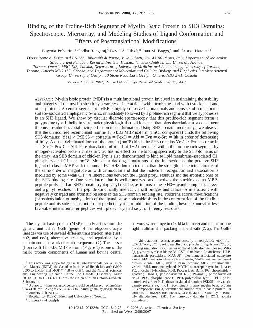
FIGURE 1: Amino acid sequences of the “classic” murine MBP splice isoforms, ranging in size from 14 to 21.5 kDa. The classic exons aredenoted by Roman numerals. The numbers at the beginning of each line for the 18.5 kDa isoform sequence are used for numbering. Sincethe N-terminal methionyl residue is cleaved in these proteins, it is numbered zero. The putative SH3 domain binding site is shown inboldface type. The arginyl/lysyl residues in the rmC1 isoform substituted by glutamine to yield the quasi-deiminated rmC8 variant areshown in boldface type and underlined. The peptide used for circular dichroism is outlined by a box.
268 Biochemistry, Vol. 47, No. 1, 2008 Polverini et al.

tau, and microtubule-associated protein 2c (MAP-2c) (34-36).
However, the known interactions of MBP with the myelinmembrane and with the proteins calmodulin, actin, andtubulin may represent an indirect (passive) signaling role interms of modifying a local milieu. A direct interaction ofMBP with signaling molecules would indicate an active rolein myelin signaling. In Figure 1, we see that there is apotential SH3 domain target (TPRTP) in the classic MBPprotein sequence, as has been previously noted (37, 38). TheSH3 domains are small conserved protein modules about 60residues in size (39-46) that interact with relatively lowaffinity with a diverse range of proline-rich ligands (41, 46-55), which are minimally an XP-x-XP consensus sequencethat forms a left-handed polyproline type II (PPII) helix (56-58). These SH3 domains are found in a large number ofintracellular signaling proteins, including the nonreceptortyrosine kinases (NRTKs). The NRTK Fyn is of particularinterest because it participates directly in MBP gene expres-sion and in compact myelin sheath formation in the centralnervous system (59-64). In lipid rafts isolated from myelin,there is a colocalization of Fyn and Lyn with Golli and classicMBP isoforms (14, 65). In late myelination and in maturemyelin, there is a partitioning of MBP and phospho-Thr95-MBP (murine 18.5 kDa sequence numbering) into lipid rafts(14, 66).
Here, we demonstrate by circular dichroic (CD) spectros-copy that the putative SH3 ligand of MBP forms a polypro-line type II (PPII) helix in vitro. Using protein microarrayand sedimentation assays, we show that full-length MBPbinds various SH3 domains from several proteins and thatmembrane-associated MBP can also bind an SH3 domain.The tertiary structures of several SH3 domains, somecomplexed with ligands, are known from X-ray crystal-lography and NMR spectroscopy (67-70). We have thusbeen able to investigate the details of SH3 domain interac-tions with the putative target in MBP by molecular modelingand docking simulations in silico, as we have recentlypublished for calmodulin and its various targets in MBP (71).The effects of posttranslational modifications (viz., phos-phorylation, deimination, and methylation) were investigatedboth experimentally and by modeling.
MATERIALS AND METHODS
Circular Dichroism.Two 18-residue peptides encompass-ing the classic polyproline segment of MBP (outlinedsegment in Figure 1) were purchased from AnaSpec Inc. (SanJose, CA); the manufacturer denotes them as MBP(89-105)and MBP(89-98T-106). It should be noted that this manu-facturer’s designation is based on the human 18.5 kDasequence numbering that includes the N-terminal methionylresidue as 1, even though it is actually cleaved posttransla-tionally (72, 73), and that the numbering used in the literaturealways starts with the alanyl residue as 1 (2, 3), whichconvention we shall follow here. The peptide sequence isFFKNIVTPRTPPPSQGKG and is identical in all mammals(2), corresponding to murine 18.5 kDa residues F86-G103(cf. Figure 1). The second peptide was phosphorylated onresidue T98 (human sequence, corresponding to residue T95in the mouse protein.
CD spectroscopy was performed on a Jasco J-810 spec-tropolarimeter (Japan Scientific, Tokyo, Japan), equipped
with a recirculating water bath. Samples were of 0.3 mLvolume, in a 0.1 cm path-length quartz cuvette. The samplesanalyzed were of each peptide (0.3 mg/mL) in aqueoussolution (10 mM Na phosphate and 10 mM KCl, pH 6.5),or the full-length recombinant murine 18.5 kDa protein(rmC1) (73), at 0.2 mg/mL in 100 mM KCl, pH 6.5).Measurements were taken at a 100 nm/min rate, at 0.1 nmintervals, over a range of 190-250 nm. Measurements wererecorded at temperatures ranging from 5 to 65°C in 5 °Cincrements. At each temperature, four successive scans wererecorded, the sample blank was subtracted, and the scanswere averaged. The data averaging and smoothing (by useof an inverse square algorithm) operations were accomplishedwith the SigmaPlot (SPSS, Chicago, IL) computer program.
Microarray and Sedimentation Assays: (A) Materials.Theleast modified, most highly positively charged 18.5 kDaisoform of MBP, bC1, was purified from bovine brain MBPas described (74). Expression constructs containing cDNAfor murine 18.5 kDa MBP (rmC1) and quasi-deiminatedMBP (rmC8) in pET22b(+) vectors (Novagen), were trans-formed into and expressed inEscherichia coli BL21-CodonPlus(DE3)-RP (Stratagene) (73, 75). Recombinantmurine 18.5 kDa MBP rmC1 is unmodified, save for anLEH6 tag (73). The variant rmC8 was generated from rmC1by sequential site-directed mutations (first R25Q, then R33Q,K119Q, R127Q, R157Q, and finally R168Q, murine se-quence numbering) via the QuikChange protocol (Stratagene,La Jolla, CA) as described previously (75). Both recombinantvariants were purified via nickel-chelation chromatographywith Ni2+-NTA agarose beads (Qiagen, Mississauga, ON).The proteins bC1 and rmC1 were phosphorylated withrecombinant p42 MAPK (New England Biolabs) at Thr94and Thr97 (bovine sequence) and at Thr92 and Thr95(murine sequence) as described (17), yielding Ph-bC1 andPh-rmC1, respectively. Electrophoresis on alkaline tube gels(17) indicated that rmC1 yielded mostly singly phosphory-lated protein, whereas bC1 yielded mostly doubly phospho-rylated protein. The SH3 domain of Fyn, residues 85-142,containing a FLAG epitope and a His6 tag at the C-terminusand a short N-terminal tail, was a kind gift from Dr. AlanDavidson, University of Toronto, and was prepared andpurified as described (76).
EggL-R-phosphatidylcholine (PC) andL-R-phosphatidyl-glycerol (PG; prepared from egg PC) were purchased fromAvanti Polar Lipids, Inc. (Alabaster, AL). [3H]Cholesterolwas from Amersham (Baie d’Urfe, QC, Canada).
(B) Preparation of Large Multilamellar Vesicles.Aliquotsof chloroform/methanol (2:1) solutions of the lipids werecombined to give a PC/PG mole ratio of 8.5:1.5. [3H]-Cholesterol was added to give a specific activity of 100 000cpm/10µmol of lipid. The solvent was evaporated under astream of nitrogen with the tube maintained at roomtemperature in a water bath, and the lipid film was evacuatedin a lyophilizer for 2 h. The dry lipid film was dissolved in1-2 mL of benzene, frozen, and lyophilized overnight. Thelipid (2 mg) was hydrated in 1 mL of 5 mM Tris-HCl buffercontaining 0.2 mM CaCl2, 50 mM KCl, 2 mM MgCl2, 1mM ATP, and 0.5 mM dithiothreitol at pH 7.5 (buffer A).Multilamellar vesicles (MLVs) were prepared by freeze-thawing five times in a dry ice/acetone bath followed by a40 °C water bath, and dispersing the lipid by vigorous vortexmixing.
MBP-SH3 Domain Interactions Biochemistry, Vol. 47, No. 1, 2008269
(C) Fyn SH3 Domain Binding to Liposome-AssociatedMBP Charge Isomers.The MBP isomers bC1 (natural bovine18.5 kDa C1 component), Ph-bC1, and rmC8 were dissolvedin distilled water at a concentration of 2 mg/mL, and theFyn SH3 domain was supplied in 50 mM phosphate buffercontaining 100 mM NaCl at pH 7.0 at a concentration of1.74 mg/mL. An aliquot of each MBP sample (50µg) wasadded to an aliquot of MLVs (500µg) and mixed gently,and the mixture was incubated for 10 min at room temper-ature. Aliquots of the Fyn SH3 domain were added (givinginitial molar ratios of Fyn SH3 domain to bC1 of 2.2-6.5and to Ph-bC1 and rmC8 of 2.2), and the sample was madeup to a total volume of 0.5 mL in buffer A and was incubatedfor a further 1 h at room temperature. A control samplewithout MBP was prepared similarly. The samples werecentrifuged at 11800g for 15 min at 4°C in an Eppendorfbench centrifuge. The supernatant was removed; the pelletwas washed gently by addition of 0.4 mL of buffer, withoutmixing, and was then resuspended in a fresh aliquot of 0.4mL of buffer. Aliquots of the supernatant and resuspendedpellet were taken for counting of [3H]cholesterol, for proteinassay by the Peterson method (77), and for gel electrophore-sis on 10% Bis-Tris NuPage gels (Invitrogen, Mississauga,ON, Canada) along with standards of bC1 and Fyn SH3.Coomassie blue-stained gels were analyzed with a UVPimage analyzer (UVP, Upland, CA), and band areas werecompared to those of the standards in order to quantitate theamount of each protein in the sample. Band densities werewithin the linear range.
(D) Binding of rmC1, Ph-rmC1, and rmC8 to SH3Domains on a Microarray.The binding of rmC1, phospho-rylated rmC1 (Ph-rmC1), and rmC8 to a TransSignal SH3domain array I (Panomics, Redwood City, CA) was deter-mined by incubation of the membrane filters with 15µg/mL rmC1, Ph-rmC1, or rmC8, in the blocking bufferprovided in the kit according to the manufacturer’s instruc-tions. After washing, bound protein was detected by incuba-tion with HRP-conjugated anti-His antibody, provided in thekit.
Molecular Modeling: (A) Software.For docking simula-tions, the peptides were constructed as described below byuse of the program SYBYL version 7.0 (SYBYL, TriposAssociated Inc., St. Louis, MO; cf. ref78), and energy-minimized by use of the Tripos force field (79). Suchcomputations were performed on an SGI Octane R12000running IRIX 6.5 (Silicon Graphics Inc., Mountain View,CA). The lengths of the peptides were kept as small aspossible, while the requisite binding characteristics wereretained, in order to minimize calculation time.
(B) Choice of SH3 Domain Structure.The choice of theSH3 domain structure to use for the docking simulations wasmade on the basis of several considerations. Because the FynNRTK plays a major role in central nervous system myeli-nation and contains an SH3 domain, we sought a convenientstructure for the SH3 domain among the crystal structuresavailable for Fyn (all from human Fyn) in the Protein DataBank (80). After a careful analysis and comparison, we chosethe crystal structure 1shf, chain A, because it was completeand had the best resolution, 1.9 Å (67). The torsion angle ofthe side chain of Tyr91 (a residue that is usually involvedin the interaction with the ligand) of 1shf was modified
according to the conformation that it has in the other crystalstructures of human Fyn.
(C) Building of MBP Peptide.The potential SH3 target inthe classic MBP isoforms [murine 18.5 kDa MBP(T92-P96)) TPRTP] (Figure 1) presents the known core motif XP-x-XP and is of interest because of the various serine andthreonine phosphorylation sites and the arginine methylationsite in its vicinity, as will be discussed below. Here, wemodeled the structure of an opportune segment (T1′ P2′ R3′
T4′ P5′ P6′ P7′ S8′ Q9′ G10′ K11′ G12′ R13′; residues T92-R104in the murine 18.5 kDa MBP sequence), following the typicalFyn SH3 ligand characteristics. With the SYBYL software,we built the backbone of the residues from T1′ to P7′ in aPPII conformation (æ ) -78° andæ ) +149°). This choicewas driven by the reported structural characteristics of theSH3 domain ligands (40, 81), deduced from the experimen-tally resolved structures, and is in agreement with our CDresults. The torsion angles of the side chains of the T1′-P7′
segment were left free to rotate in the docking simulations.The residues from S8′ to R13′ were temporarily built in anextended conformation; however, backbone and side-chaintorsion angles were completely free to rotate in the dockingsimulations. The overall conformation of the constructedpeptide was at first relaxed by a moderate energy minimiza-tion.
(D) Posttranslational Modifications of the Ligands.Post-translational modifications (phosphorylation and methylation)were performed in silico, building on our previous experiencewith MBP-calmodulin modeling (71). First, the unmodifiedligand was docked, and the best structure obtained from thissimulation was then modified. The modified ligands werethen docked to the SH3 domain and left free to change theirconformation and position with the same degrees of freedomof the unmodified peptide. The following six modificationswere individually applied: the residue S8′ was modified toPhS (phosphorylated serine), the residues T1′ and T4′ weremodified to PhT (phosphorylated threonine), and finally theresidue R13′ was monomethylated (MM), asymmetricallydimethylated (ADM), and symmetrically dimethylated (SDM).
The PhS residue is included in the nonstandard amino acidmolecular library of the SYBYL program; therefore, theattribution to each atom of its partial charges was madeaccording to Kollman (82), as is usual for a peptidic ligand.The T1′, T4′, and R13′ modifications were constructed, alwayswith the SYBYL software, and the calculation of partialcharge for the atoms of modified peptides was madeaccording to Gasteiger (83), as is usual for a generic ligand.To check if the different charge attributions could lead todifferent results, we first performed two docking simulationson the unmodified MBP peptide, with both the Kollman andGasteiger partial charges sets.
Molecular Docking Simulations.Simulations of the inter-action of MBP unmodifed and modified peptides with theSH3 domain were performed with the Autodock3 softwarepackage (84) with the aid of the AutoDockTools (ADT)interface, running on a PC cluster (AMD Opteron 64 bit).For the docking calculations, the Lamarckian genetic algo-rithm (84) was used.
In all the simulations, the backbone torsion angles of thePPII region (residues T1′-P7′) were kept constant (althoughthe side chains were flexible), whereas the flanking regionwas completely flexible (both the backbone and the side-
270 Biochemistry, Vol. 47, No. 1, 2008 Polverini et al.
chain torsion angles could vary). In this way, during theconformational search, not only were all all of the torsionangles free to rotate, but also the whole peptide could changeits orientation and explore the space around the binding site,defined by the grid dimensions.
Preliminary Test Simulation and Docking ParametersSetting.A preliminary test simulation was performed on thestructure 1azg (70), which is a minimized average NMRstructure of the Fyn SH3 domain complexed with a Pro-rich peptide (PPRPLPVAPGSSKT, residues P91-T104)from subunit P85 of PI3 kinase. The SH3 domain structureand the PI3 peptide initial coordinates were extracted fromthe original 1azg PDB file and some docking tests wereperformed between them, in order to check if the rightcomplex was correctly identified by the docking algorithmand to set the best simulation parameters for obtaining acorrect result. This step permitted the use of such parametersfor a similar ligand (the MBP peptide) with the same highnumber of degrees of freedom, docked into the analogousbinding site of the 1shf crystal structure of the SH3 domain.Similar to the MBP peptides, the PI3 ligand torsion angleswere left flexible with the exception of the backbone torsionangles of the first nine residues, which were in a PPIIconformation. The choice of the 1azg ligand peptide for thedocking test was made because the ligand peptide is part ofa natural protein, PI3 kinase, and because it is 14 residueslong, just one amino acid longer than our MBP peptide;therefore, the degrees of freedom are comparable.
On the basis of the good results obtained with this testpeptide, the following docking parameters were chosen forall the other simulations with the MBP peptide: the numberof individuals in the population was 300, the maximumnumber of energy evaluations was 25 000 000, and thenumber of runs was 200. For the other docking parameters,the default settings were used. The docking simulations thatwere run subsequently between the unmodified and modifiedMBP peptides and the SH3 domain are denotedds1-ds6and are summarized in Table 1.
For ds1a and ds1b, a grid with a spacing of 0.5 Å anddimensions of 80, 78, and 74 points (in thex, y, and zdirections, respectively) was initially used. Then the bestconformation obtained was optimized with a grid spacingof 0.375 and with (x, y, z) dimensions of 100, 86, and 94points. The latter grid parameters were used for all thesubsequent simulations with the modified peptides.
For each simulation, the resulting complexes were clus-tered with a threshold of 3 Å rmsd (root mean squared
deviation) for ligand structure similarity. Then, we comparedthe structure with the absolute lowest docked energy andthe structure with the lowest docked energy in the morepopulated cluster to choose the best conformation for theanalyis.
Structures were examined with the ADT interface inAutodock (84), SwissPdbViewer (85), and RasMol (86)software packages. The “contact residues” of the SH3 domainwith the ligand were assigned by means of the FirstGlancein Jmol program (at http://molvis.sdsc.edu/fgij/index.htm,copyright 2005, 2006 by Eric Martz). The CH‚‚‚π interac-tions were identified on the basis of geometric criteria (87).All figures were drawn with the ViewerLite 5.0 (AccelrysSoftware Inc., San Diego, CA) software package.
RESULTS AND DISCUSSION
MBP Possesses a PutatiVe SH3 Domain Ligand.Theclassic MBP isoforms all possess a highly conserved, putativeSH3 ligand: T92-P93-R94-T95-P96 (murine 18.5 kDasequence numbering, unless otherwise noted) (Figure 1). Thismotif is encoded by classic exon IV and is contained in allsix known classic MBP isoforms, as well as in the Golli-MBP isoform J37 (1, 88). The murine Golli-MBP isoformsJ37 (88) and BG21 (89) contain an additional potential SH3target [Golli(H80-P84) ) HPADP], but we focused hereon the classic site because the various posttranslationalmodifications within it and nearby have been better docu-mented. The segment immediately preceding this motif,P82VVHFFKNIVTP93, is an immunodominant epitope ofMBP and forms an amphipathic, surface-embedded,R-helix(90, 91). The residues P93 (one of the two prolyl residuesflanking theR-helix) and S99 are exposed to the cytoplasm(90, 92), suggesting that the putative SH3 ligand should beoriented outside the membrane where it would be accessibleto other modifying or recognition proteins.
Within the immunodominant epitope (P82-P93), the onlyresidue modified is T92, which is a secondary mitogen-activated protein kinase (MAPK) site (93). The residue T95within the putative SH3 ligand is a primary MAPK andglycogen synthase kinase 3â (GSK-3â) site (94, 95), andMBP modified here is associated with lipid rafts (putativesignaling platforms) in mature myelin (14, 19, 66). It hasbeen suggested that interaction of proteins such as tau withan SH3-domain-containing protein might direct them to lipidrafts (35). Residue R94 is a potential secondary deiminationsite (albeit not in rmC8, which contains primary deiminationsites), whereas S99 is phosphorylated by protein kinase C(2, 5). Slightly further downstream, R104 is either unmodifed,monomethylated (ω-NG-monomethyl-Arg), or symmetricallydimethylated (ω-NG,N′G-dimethyl-Arg) (2, 5). Most proteinshave an asymmetric dimethylarginine, so MBP is unusualin that it has asymmetricdimethylarginine. Regardless, insome cases, the methyl group causes steric hindrance ininteractions with SH3-domain-containing and other proteins(96-99), and in MBP it is thought to interfere withdeimination by peptidylarginine deiminase (7, 100). Thus,methylation and phosphorylation of MBP, especially in thevicinity of the putative SH3 ligand, might affect its localconformation (9, 10) and binding to SH3 domains, as is thecase for other proteins such as tau and other microtubule-associated proteins (99, 101, 102).
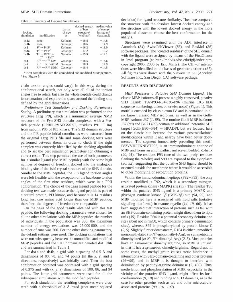
Table 1: Summary of Docking Simulations
dockingsimulation modification
partialcharge
set
docked energyof best
structurea(kcal/mol)
median valueof energyhistogramb
(kcal/mol)
ds1a none Kollman -18.8 -14.8ds1b none Gasteiger -18.6 -14.9ds2 S8′ f PhS8′ Kollman -16.2 -11.0ds3a T4′ f PhT4′ Gasteiger -17.2 -13.2ds3b T1′ f PhT1′ +
T4′ f PhT4′Gasteiger -15.9 -12.1
ds4 R13′ f R13′-MM Gasteiger -18.5 -14.6ds5 R13′ f R13′-ADM Gasteiger -18.3 -14.9ds6 R13′ f R13′-SDM Gasteiger -18.4 -14.5
a Best complexes with the unmodified and modified MBP peptides.b See Figure 5.
MBP-SH3 Domain Interactions Biochemistry, Vol. 47, No. 1, 2008271
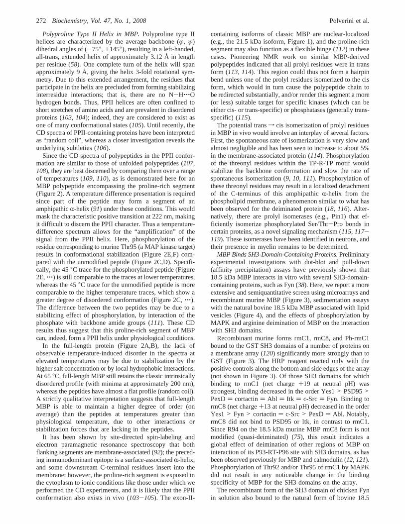
Polyproline Type II Helix in MBP.Polyproline type IIhelices are characterized by the average backbone (æ, ψ)dihedral angles of (-75°, +145°), resulting in a left-handed,all-trans, extended helix of approximately 3.12 Å in lengthper residue (58). One complete turn of the helix will spanapproximately 9 Å, giving the helix 3-fold rotational sym-metry. Due to this extended arrangement, the residues thatparticipate in the helix are precluded from forming stabilizinginterresidue interactions; that is, there are no N-H‚‚‚Ohydrogen bonds. Thus, PPII helices are often confined toshort stretches of amino acids and are prevalent in disorderedproteins (103, 104); indeed, they are considered to exist asone of many conformational states (105). Until recently, theCD spectra of PPII-containing proteins have been interpretedas “random coil”, whereas a closer investigation reveals theunderlying subtleties (106).
Since the CD spectra of polypeptides in the PPII confor-mation are similar to those of unfolded polypeptides (107,108), they are best discerned by comparing them over a rangeof temperatures (109, 110), as is demonstrated here for anMBP polypeptide encompassing the proline-rich segment(Figure 2). A temperature difference presentation is requiredsince part of the peptide may form a segment of anamphipathicR-helix (91) under these conditions. This wouldmask the characteristic positive transition at 222 nm, makingit difficult to discern the PPII character. Thus a temperature-difference spectrum allows for the “amplification” of thesignal from the PPII helix. Here, phosphorylation of theresidue corresponding to murine Thr95 (a MAP kinase target)results in conformational stabilization (Figure 2E,F) com-pared with the unmodified peptide (Figure 2C,D). Specifi-cally, the 45°C trace for the phosphorylated peptide (Figure2E,‚‚‚) is still comparable to the traces at lower temperatures,whereas the 45°C trace for the unmodified peptide is morecomparable to the higher temperature traces, which show agreater degree of disordered conformation (Figure 2C,‚‚‚).The difference between the two peptides may be due to astabilizing effect of phosphorylation, by interaction of thephosphate with backbone amide groups (111). These CDresults thus suggest that this proline-rich segment of MBPcan, indeed, form a PPII helix under physiological conditions.
In the full-length protein (Figure 2A,B), the lack ofobservable temperature-induced disorder in the spectra atelevated temperatures may be due to stabilization by thehigher salt concentration or by local hydrophobic interactions.At 65 °C, full-length MBP still retains the classic intrinsicallydisordered profile (with minima at approximately 200 nm),whereas the peptides have almost a flat profile (random coil).A strictly qualitative interpretation suggests that full-lengthMBP is able to maintain a higher degree of order (onaverage) than the peptides at temperatures greater thanphysiological temperature, due to other interactions orstabilization forces that are lacking in the peptides.
It has been shown by site-directed spin-labeling andelectron paramagnetic resonance spectroscopy that bothflanking segments are membrane-associated (92); the preced-ing immunodominant epitope is a surface-associatedR-helix,and some downstream C-terminal residues insert into themembrane; however, the proline-rich segment is exposed inthe cytoplasm to ionic conditions like those under which weperformed the CD experiments, and it is likely that the PPIIconformation also exists in vivo (103-105). The exon-II-
containing isoforms of classic MBP are nuclear-localized(e.g., the 21.5 kDa isoform, Figure 1), and the proline-richsegment may also function as a flexible hinge (112) in thesecases. Pioneering NMR work on similar MBP-derivedpolypeptides indicated that all prolyl residues were in transform (113, 114). This region could thus not form a hairpinbend unless one of the prolyl residues isomerized to the cisform, which would in turn cause the polypeptide chain tobe redirected substantially, and/or render this segment a more(or less) suitable target for specific kinases (which can beeither cis- or trans-specific) or phosphatases (generally trans-specific) (115).
The potential transf cis isomerization of prolyl residuesin MBP in vivo would involve an interplay of several factors.First, the spontaneous rate of isomerization is very slow andalmost negligible and has been seen to increase to about 5%in the membrane-associated protein (114). Phosphorylationof the threonyl residues within the TP-R-TP motif wouldstabilize the backbone conformation and slow the rate ofspontaneous isomerization (9, 10, 111). Phosphorylation ofthese threonyl residues may result in a localized detachmentof the C-terminus of this amphipathicR-helix from thephospholipid membrane, a phenomenon similar to what hasbeen observed for the deiminated protein (18, 116). Alter-natively, there are prolyl isomerases (e.g., Pin1) that ef-ficiently isomerize phosphorylated Ser/Thr-Pro bonds incertain proteins, as a novel signaling mechanism (115, 117-119). These isomerases have been identified in neurons, andtheir presence in myelin remains to be determined.
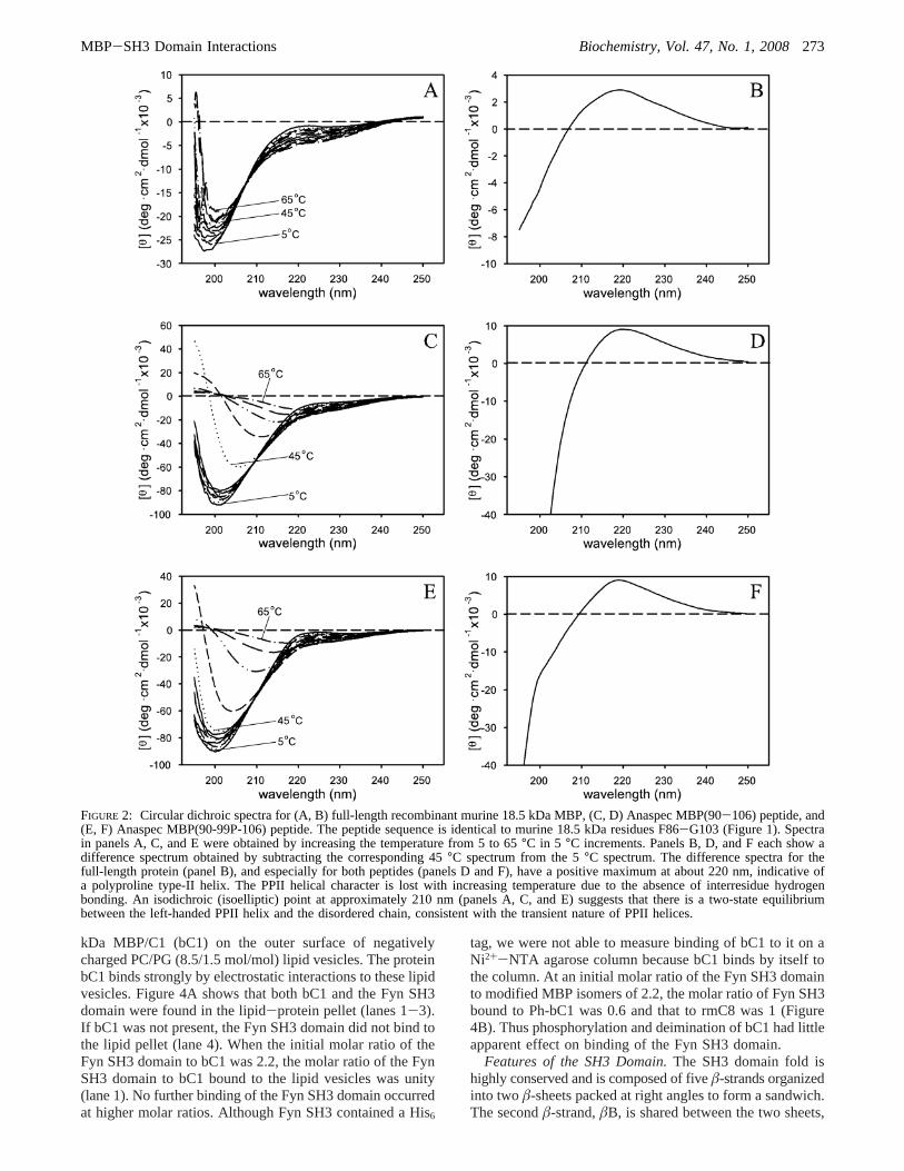
MBP Binds SH3-Domain-Containing Proteins.Preliminaryexperimental investigations with dot-blot and pull-down(affinity precipitation) assays have previously shown that18.5 kDa MBP interacts in vitro with several SH3-domain-containing proteins, such as Fyn (38). Here, we report a moreextensive and semiquantitative screen using microarrays andrecombinant murine MBP (Figure 3), sedimentation assayswith the natural bovine 18.5 kDa MBP associated with lipidvesicles (Figure 4), and the effects of phosphorylation byMAPK and arginine deimination of MBP on the interactionwith SH3 domains.
Recombinant murine forms rmC1, rmC8, and Ph-rmC1bound to the GST SH3 domains of a number of proteins ona membrane array (120) significantly more strongly than toGST (Figure 3). The HRP reagent reacted only with thepositive controls along the bottom and side edges of the array(not shown in Figure 3). Of those SH3 domains for whichbinding to rmC1 (net charge+19 at neutral pH) wasstrongest, binding decreased in the order Yes1> PSD95>PexD) cortactin) Abl ) Itk ) c-Src) Fyn. Binding tormC8 (net charge+13 at neutral pH) decreased in the orderYes1> Fyn > cortactin) c-Src> PexD) Abl. Notably,rmC8 did not bind to PSD95 or Itk, in contrast to rmC1.Since R94 on the 18.5 kDa murine MBP rmC8 form is notmodified (quasi-deiminated) (75), this result indicates aglobal effect of deimination of other regions of MBP oninteraction of its P93-RT-P96 site with SH3 domains, as hasbeen observed previously for MBP and calmodulin (12, 121).Phosphorylation of Thr92 and/or Thr95 of rmC1 by MAPKdid not result in any noticeable change in the bindingspecificity of MBP for the SH3 domains on the array.
The recombinant form of the SH3 domain of chicken Fynin solution also bound to the natural form of bovine 18.5
272 Biochemistry, Vol. 47, No. 1, 2008 Polverini et al.
kDa MBP/C1 (bC1) on the outer surface of negativelycharged PC/PG (8.5/1.5 mol/mol) lipid vesicles. The proteinbC1 binds strongly by electrostatic interactions to these lipidvesicles. Figure 4A shows that both bC1 and the Fyn SH3domain were found in the lipid-protein pellet (lanes 1-3).If bC1 was not present, the Fyn SH3 domain did not bind tothe lipid pellet (lane 4). When the initial molar ratio of theFyn SH3 domain to bC1 was 2.2, the molar ratio of the FynSH3 domain to bC1 bound to the lipid vesicles was unity(lane 1). No further binding of the Fyn SH3 domain occurredat higher molar ratios. Although Fyn SH3 contained a His6
tag, we were not able to measure binding of bC1 to it on aNi2+-NTA agarose column because bC1 binds by itself tothe column. At an initial molar ratio of the Fyn SH3 domainto modified MBP isomers of 2.2, the molar ratio of Fyn SH3bound to Ph-bC1 was 0.6 and that to rmC8 was 1 (Figure4B). Thus phosphorylation and deimination of bC1 had littleapparent effect on binding of the Fyn SH3 domain.
Features of the SH3 Domain.The SH3 domain fold ishighly conserved and is composed of fiveâ-strands organizedinto two â-sheets packed at right angles to form a sandwich.The secondâ-strand,âB, is shared between the two sheets,
FIGURE 2: Circular dichroic spectra for (A, B) full-length recombinant murine 18.5 kDa MBP, (C, D) Anaspec MBP(90-106) peptide, and(E, F) Anaspec MBP(90-99P-106) peptide. The peptide sequence is identical to murine 18.5 kDa residues F86-G103 (Figure 1). Spectrain panels A, C, and E were obtained by increasing the temperature from 5 to 65°C in 5 °C increments. Panels B, D, and F each show adifference spectrum obtained by subtracting the corresponding 45°C spectrum from the 5°C spectrum. The difference spectra for thefull-length protein (panel B), and especially for both peptides (panels D and F), have a positive maximum at about 220 nm, indicative ofa polyproline type-II helix. The PPII helical character is lost with increasing temperature due to the absence of interresidue hydrogenbonding. An isodichroic (isoelliptic) point at approximately 210 nm (panels A, C, and E) suggests that there is a two-state equilibriumbetween the left-handed PPII helix and the disordered chain, consistent with the transient nature of PPII helices.
MBP-SH3 Domain Interactions Biochemistry, Vol. 47, No. 1, 2008273
possessing a kink that changes the direction of the polypep-tide. One side of the sandwich is rather hydrophobic andconstitutes the ligand-binding surface. It is delimited on one
side by an RT loop between theâA andâB strands (residuesGlu94-Ser102, following human Fyn SH3 domain number-ing), and on the other side by a small loop between theâB
FIGURE 3: Recombinant murine 18.5 kDa isoforms rmC1 and rmC8 bound to a subset of SH3 domains. A Panomics Array I of GST-SH3domain fusion proteins was probed with His6-tagged rmC1 and rmC8 and HRP-conjugated anti-His antibody. (A) Configuration of proteinarray. Each GST-SH3 domain was spotted in duplicate. (B) Binding of rmC1, rmC8, and Ph-rmC1 to the microarray as detected on filmvia chemiluminescence. Binding to GST alone was much weaker. The same solution of rmC1 as used for phosphorylation was used forpanel B. Representative arrays are shown. The SH3 domains on the array were amphiphysin, Discs large homologue 2 or MAGUK P55subfamily member 2 (Dig2), VAV proto-oncogene SH3 domain 1 (VAV-D1),â-lymphocyte-specific protein tyrosine kinase (BLK), humanT-lymphocyte-specific protein tyrosine kinase p56 LCK (LCK), 55 kDa erythrocyte membrane protein (EMP55), cytoplasmic protein NCK1SH3 domain 3 (NCK1-D3), Abelson tyrosine kinase (Abl), nonerythrocytic spectrinR-chain (SPCN), cellular Gardner-Rasheed felinesarcoma virus protein (FGR), PAK-interacting exchange factor-â (Y124), phospholipase Cγ-1 (PLCr), Src substrate cortactin, proto-oncogenetyrosine protein kinase Fyn (SLK), peroxisomal membrane protein PEX13 (PEXD), retinoblastoma protein-interacting zinc finger (Riz),mixed-lineage kinase 3 (MLPK3), nebulin, tyrosine-protein kinase BTK (BTK), phosphatidylinositol 3-kinase regulatoryâ-subunit (PI3â),Yamaguchi sarcoma virus oncogene homologue 1 (Yes1), cellular Rous sarcoma viral oncogene homologue (c-Src), Ras GTPase-activatingprotein 1 (RasGAP), intersectin 1 SH3 domain 1 (ITSN-D1), Abelson-related protein (Abl2), Fyn-binding protein SH3 domain 1 (FYB-D1), presynaptic density protein 95 (PSD95), intersectin 1 SH3 domain 2 (ITSN-D2), erythrocytic spectrinR-chain (SJHUA), hematopoieticcell kinase (Hck),Fguanine nucleotide exchange factor 5 (Tim), tyrosine protein kinase TXK (TXK), interleukin-2-inducible T-cell kinase(Itk), VAV-2 protein SH3 domain 2 (VAV2-D2), hematopoietic-specific protein (HS1), glutathione S-transferase (GST), avian sarcomavirus CT10 oncogene homologue SH3 domain 2 (CRK-D2), neutrophil cytosol factor 2 SH3 domain 1 (NOF2-D1), and signal-transducingadaptor molecule (Stam).
274 Biochemistry, Vol. 47, No. 1, 2008 Polverini et al.

and âC strands (called the n-Src insertion point loop,residues Ser114-Trp119) and by a five-residue-long 310
helix (Ser135-Tyr137), which separates theâD and âEstrands.
Numerous aromatic amino acids are present in the bindingsurface, viz., Tyr132, Trp119, Tyr137, Tyr91, and Tyr93.Cesareni et al. (81) identified hydrophobic pockets formedby the residues AL(YF)D(YF) (89-93), WW (119-120),and PXNY (134-137), although residues Ala89, Leu90, andTrp120 are quite far from the binding site. The first of thesebinding pockets in the SH3 domain is lined by negativelycharged residuessGlu98, Asp99, and Asp100 in the RT loop;Glu116, Asp118, and Glu121 in the n-SRC loop; and Glu129in the âD strand before the 310 helixsand can host apositively charged side chain flanking the core motif in theligand (see below).
The SH3 domain structure that we chose to use for thedocking simulations was the 1shf Fyn structure, for reasonsdescribed above. Even though the 1shf structure was withoutany complexed ligand peptide, the comparison with otherknown SH3-peptide complexes confirmed that the presenceof the ligand did not involve structural changes in the SH3
domain, as is generally the case (46). [It has recently beennoted that ligand-binding actually stabilizes the SH3 domainstructure (122).]
Features of SH3 Ligands.Canonical SH3 ligands usuallycontain two identical XP dipeptides, separated by a scaf-folding residue (often a proline) (40-42, 46, 50, 81). Thetwo XP moieties in the core motif (XP-x-XP) occupy twoof the hydrophobic pockets (see above), and a stackinginteraction between one of the Pro residues and the Trp ofthe binding pocket is well-conserved. In fact, the prolineresidues of the ligand seem to be implicated in having a rolein molecular association employing C-H‚‚‚π interactionswith aromatic residues at the binding site (123), of whichthe above-mentioned Pro-Trp stacking is well-conserved.Moreover, the interaction with the ligand is both hydrophobic(in particular between the conserved aromatic residues ofthe binding site of the SH3 domain and the aliphatic sidechains of the ligand), and also mediated by hydrogen bonds(in particular those from the aromatic residues, as well asAsn136, of the binding site and the backbone oxygens ofthe ligand, which are characteristic) (40, 69). The third SH3domain binding pocket, lined by negatively charged residues,can host a positively charged side chain flanking the coremotif, as indicated before. Usually, the positively chargedresidue interacts with the acidic cluster on the RT loop. The7-9 amino acid core region of the ligand binds to SH3receptors in a left-handed PPII helical conformation (40, 81),in either of two opposite orientations depending on theposition of a positively charged residue in the peptidesequence.
In Fyn and some other SH3 domains, the residue Arg/Ile96 (human Fyn numbering) in the RT loop seems to bethe key for the selectivity and high affinity of the binding(40). Actually, in the analyzed PDB structures (see Table 2below), this residue does not make any contacts directly withthe core motif but only with the rest of the protein linked toit, if present [e.g., the Nef protein in PDB entries 1avz and1efn (124, 125)]. It has been reported that the flankingpeptide region usually enhances the affinity and selectivityof ligand binding; therefore, the whole structure of the boundprotein is important and not only that of the ligand peptide.There are, however, many exceptions to the general bindingmode.
Docking Simulations of MBP Peptides to SH3 Domains.In order to set the correct parameters for a docking simulationwith such a high number of degrees of freedom and todetermine if the docking algorithm could find the favoredbinding complex, a preliminary test simulation was per-formed with the Fyn SH3 domain structure and the initialcoordinates of the PI3 ligand, extracted from the 1azg PDBstructure (see Materials and Methods). The family of 25NMR structures deposited into the related file 1a0n (fromwhich 1azg was obtained by averaging and minimization),demonstrate the flexibility in solution of the differentstructural regions of this type of SH3 domain ligand. Thiscomparison reveals that the C-terminal region of the peptideis very flexible and can assume very different spatialorientations, whereas the N-terminal residues are in a PPIIhelix and remain in the same position in all the structures.
The docking test simulation recovered the correct complexconformation and not only allowed us to set the best dockingparameters for subsequent runs, as indicated under Materials
FIGURE 4: (A) Fyn SH3 domain bound to PC/PG (8.5/1.5 mol/mol) lipid vesicles in the presence of the natural bovine 18.5 kDaMBP isomer bC1 (lanes 1-3) but not in its absence (lane 4). Thelipid-protein pellet sedimented at 11800g and was resuspendedand run on the gel. Lane 5 is blank. Protein standards are 1.75µgof Fyn SH3 domain (lane 6) and 2µg of bC1 (lane 7). The moleratio of Fyn SH3 to bC1 added to the vesicles was 2.2 (lane 1), 4.3(lane 2), and 6.5 (lane 3). The mole ratio of Fyn SH3 to bC1recovered in the pellet was determined by densitometry andcomparison to the standards. (B) Less Fyn SH3 domain bound toPC/PG (8.7/1.3 mol/mol) lipid vesicles in the presence of Ph-bC1(lane 2) than bC1 (lane 1). Fyn SH3 domain bound similarly tothe lipid vesicles in the presence of rmC8 (lane 3) as bC1. Theinitial mole ratio of Fyn SH3 domain to MBP added to the vesicleswas 2.2 in all cases. Fyn SH3 domain did not bind to the lipidvesicles in the absence of MBP (lane 4). Equal amounts of lipidpellet were run on the gel for lanes 1-4. Less Ph-bC1 and rmC8bound to the lipid vesicles under these conditions than bC1. Lane5 is blank. Protein standards are 0.9µg of Fyn SH3 domain (lane6) and 1.0µg of bC1 (lane 7). The mole ratio of Fyn SH3 to MBPcharge isomer recovered in the pellet was determined by densito-metry and comparison to the standards.
MBP-SH3 Domain Interactions Biochemistry, Vol. 47, No. 1, 2008275
and Methods, but also supported the choice to keep the PPII(T1′-P7′) backbone torsion angles of the ligand constant inthe following simulations (see also Materials and Methods).This choice was in agreement with our CD results on theMBP peptide, which were consistent with an N-terminalstable PPII helix, particularly after phosphorylation.
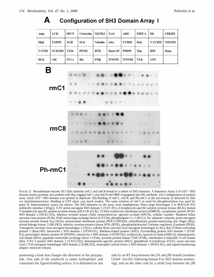
In Table 2 are summarized the “contact residues” of theSH3 domain with the ligand, analyzed for the best structureobtained from each simulation. The docked energies, andthe energy value around which each energy histogram (Figure5) is centered, are summarized in Table 1.
Docking Simulationds1: Unmodified Peptide.Comparisonof the results of the two docking simulations performed onthe unmodifed MBP peptide, to which either Kollman orGasteiger partial charges were assigned, showed that theyconverged to the same best complex conformation, with thesame very favorable docked energy (-18.8/-18.6 kcal/mol,respectively, Table 1), slightly better than the one calculatedfor the MBP-calmodulin interaction (71). The dissociationconstant (Kd) of MBP with calmodulin has been experimen-tally measured to be in the micromolar range (12, 121).Generally, the interactions of SH3 domains with proteinscontaining proline-rich sequences are also weak, withexperimentally measured dissociation constants in this range,enabling rapid association and dissociation (41); therefore,we can surmise that they are of a similar order of magnitudealso for interaction of SH3 domains with MBP.
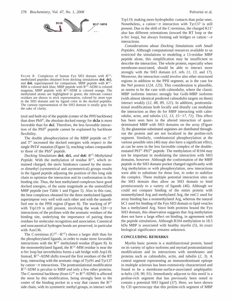
The PPII helix of the MBP peptide lies in the canonicalbinding pocket, and the flexible backbone region at theC-terminus is bent close to the SH3 domain surface (Figure6). Analysis of the contact residues (Table 2) showed thatthe positively charged amino acids in the ligand “canonically”interact with the acidic residues that line the binding pocket.In particular, R13′ makes a salt bridge with Asp118 in the
n-Src loop and interacts with the residues of the 310 helix;the other arginine, R3′, that lies just inside the core motifinteracts with the negatively charged residues in the RT loop,making a salt bridge with Asp99. The third positive residuein the MBP peptide, K11′, makes a cation-π interaction withTrp119 and interacts also with the 310 helix region. ResidueP5′ in MBP peptide is stacked with the aromatic ring ofTrp119 in the SH3 domain, forming a C-H‚‚‚π noncanonicalhydrogen bond, a characteristic interaction of one of the twoSH3 domain hydrophobic binding sites (81). The prolineresidues of the ligand in SH3 complexes are implicated inmolecular association, employing these interactions withconserved aromatic residues at the binding site (123). Here,P7′ also forms a C-H‚‚‚π interaction with Trp 119 and P5′
with Y132, as was similarly found for the Abl tyrosine kinase(123). Such aromatic residues (along with Asn136) formcanonical and noncanonical (i.e., by means of their CHgroup) hydrogen bonds with the backbone oxygens of theligand.
Interestingly, the SH3 domain residue Arg96 is also incontact with the MBP peptide, in particular with P6′ and S8′,but not by the terminal positively charged side chain of Arg,that, as in almost all the known SH3 domain structures, isturned outward.
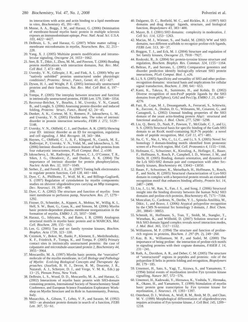
Docking Simulationds2 and ds3: Phosphorylated Pep-tides.Docking simulationsds2 andds3 were performed toinvestigate the effects of three kinds of phosphorylation onthe MBP peptide: on residue S8′, on residue T4′, and on bothresidues T1′ and T4′. The best complex conformationsresulting from the docking of the three phosphorylatedpeptides superimposed well with each other and with thatof the unmodified peptide. The PPII region of the ligandlies in the same site, and although the C-termini show somedifferences in their backbone and side-chain torsion angles,
Table 2: Residues of the SH3 Domain That Made Contacts with the Liganda
ds1a,b ds2 ds3a ds3b ds4 ds5 ds6
canonical Fynb unmodified MBP MBP PhS8′ MBP PhT4′ MBP PhT1′ + PhT4′ MBP R13′-MM MBP R13′-ADM MBP R13′-SDM
RT loopY91 Y91 Y91 Y91 Y91 Y91
D92 D92 D92 D92 D92 D92Y93 Y93 Y93 Y93 Y93 Y93 Y93 Y93E94c E94 E94 E94 E94 E94 E94 E94A95c A95 A95 A95 A95 A95 A95[R,I]96 R96 R96 R96 R96 R96 R96 R96T97 T97 T97 T97 T97 T97 T97 T97E98c
D99c D99 D99 D99 D99 D99 D99 D99D100 D100 D100 D100 D100 D100 D100 D100
nSrc loopE116 E116 E116 E116 E116 E116 E116 E116G117 G117D118 D118 D118 D118 D118 D118W119 W119 W119 W119 W119 W119 W119 W119
âD + 310 helixY132 Y132 Y132 Y132 Y132 Y132 Y132
P134 P134 P134 P134 P134 P134 P134S135 S135 S135 S135
N136 N136 N136 N136 N136 N136 N136 N136Y137 Y137 Y137 Y137 Y137 Y137 Y137 Y137
a Analyzed with the tool “FirstGlance in Jmol”. The majority of the residues are in common with the canonical Fyn SH3 domain; the exceptions(D92, Y132, and S135) are shown in italic type. In all the complexes, stacking of Trp119 in the SH3 domain binding site with Pro5′ in the peptideis always present.b Extrapolated from the PDB structures of Fyn SH3.c This group of residues was involved in the interaction only in the complexeswhere the ligand was the whole protein, i.e., Nef protein from human immunodeficiency virus 1 (PDB entries 1avz and 1efn).
276 Biochemistry, Vol. 47, No. 1, 2008 Polverini et al.
they remain oriented in the same direction, forming a bundleof structures occupying the same space (Figure 7).
The contact residues of the peptides in the SH3 domainare listed in Table 2. In comparison with the unmodifiedpeptide, the main preserved interactions in all the complexesare the R3′-Asp99 salt bridge, the C-H‚‚‚π Pro-aromaticinteractions (in particular the stacking of P5′ with Trp119),and the characteristic H-bonds between the ligand backboneoxygens and the residues Asn136 and Tyr137. The R13′
residue maintains the salt bridge with Asp118 in all thecomplexes except that with the double-phosphorylated pep-tide, in which it is substituted with a cation-π interactionwith Tyr91 and Tyr137 in the RT loop. The K11′ residue isthe more variable one, changing the orientation of its longside chain from the 310 helix to the RT loop, always formingcation-π interactions with the conserved aromatic residuesof the binding site (Trp119, Tyr91, and Tyr137).
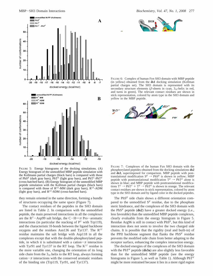
The PhS8′ side chain shows a different orientation com-pared to the unmodified S8′ residue, due to the phosphatesteric hindrance, and the complexes of the SH3 domain withthe PhS8′ peptide (ds2) have a greater docked energy (i.e.,less favorable) than the unmodified MBP peptide complexes,clearly evaluable from the energy histogram in Figure 5.Residue Arg96 is still in contact with PhS8′, but this kind ofinteraction does not seem to involve the two charged sidechains. It is possible that the rigidity (real and built-in) ofthe PPII backbone segment that flanks the PhS8′ residueprevents the modified side chain from better adapting to thereceptor surface, enhancing the complex interaction energy.
The docked energies of the complexes of the SH3 domainwith the PhT4′ peptide (ds3a) are also slightly less favorablethan for the unmodified MBP peptide (see the energyhistograms in Figure 5, as well as Table 1). Although PhT4′
might be more restrained because it lies in a more rigid region
FIGURE 5: Energy histograms of the docking simulations. (A)Energy histogram of the unmodified MBP peptide simulation withthe Kollmann partial charges (black bars) is compared with thoseof PhS8′ (dark gray bars), PhT4′ (light gray bars), and PhT1′-PhT4′
(cross-hatched bars). (B) Energy histogram of the unmodified MBPpeptide simulation with the Kollman partial charges (black bars)is compared with those of R13′-MM (dark gray bars), R13′-ADM(light gray bars), and R13′-SDM (cross-hatched bars).
FIGURE 6: Complex of human Fyn SH3 domain with MBP peptide(in yellow) obtained from theds1 docking simulation (Kollmanpartial charges set). The SH3 domain is represented with itssecondary structure elements (â-sheets in cyan, 310-helix in red,and turns in green). The relevant contact residues are shown instick representation, colored by atom type in the SH3 domain andyellow in the MBP peptide.
FIGURE 7: Complexes of the human Fyn SH3 domain with thephosphorylated peptides obtained from the docking simulationsds2and ds3, superimposed for comparison. MBP peptide with post-translational modification S8′ f PhS8′ is shown in yellow; MBPpeptide with posttranslational modification T4′ f PhT4′ alone isshown in blue; and MBP peptide with posttranslational modifica-tions T1′ f PhT1′ + T4′ f PhT4′ is shown in orange. The relevantcontact residues are shown in stick representation, colored by atomtype in the SH3 domain and by ligand color in the docked peptides.
MBP-SH3 Domain Interactions Biochemistry, Vol. 47, No. 1, 2008277
(real and built-in) of the peptide (center of the PPII backbone)than does PhS8′, the absolute docked energy fords3ais morefavorable than fords2. Therefore, the less favorable interac-tion of the PhS8′ peptide cannot be explained by backboneflexibility.
The double phosphorylation of the MBP peptide on T1′
and T4′ increased the docked energies with respect to thesingle PhT4′ mutation (Figure 5), reaching values comparableto those of the PhS8′ peptide.
Docking Simulationsds4, ds5, and ds6: R13′-MethylatedPeptide.With the methylation of residue R13′, which re-mained charged, the steric hindrance caused by the mono-or dimethyl (symmetrical and asymmetrical) groups resultsin the ligand peptide adjusting the position of this long sidechain to optimize the interaction and its conformation in thebinding site. Thus, the best methylated complexes have gooddocked energies, of the same magnitude as the unmodifiedMBP peptide (see Table 1 and Figure 5). Also in this case,the best complexes obtained for the three methylated ligandssuperimpose very well with each other and with the unmodi-fied one in the PPII region (Figure 8). The stacking of P5′
with Trp119 is still present, involving the weak CH‚‚‚πinteractions of the prolines with the aromatic residues of thebinding site, underlying the importance of pairing theseresidues for molecular recognition and association. Canonicaland noncanonical hydrogen bonds are preserved, in particularwith Asn136.
The C-terminus (G10′-R13′) shows a larger shift than forthe phosphorylated ligands, in order to make more favorableinteractions with the R13′ methylated residue (Figure 8). Inthe monomethylated ligand, the R13′-MM residue is near then-Src loop but nevertheless forms a salt bridge with Asp118.Instead, R13′-ADM shifts toward the first residues of the RTloop, interacting with the aromatic rings of Tyr91 and Tyr137by cation-π interactions. The posttranslational modificationR13′-SDM is peculiar to MBP and only a few other proteins.The C-terminal backbone (from G10′ to R13′-SDM) is affectedthe most by this modification, being strongly bent to thecenter of the binding pocket in a way that causes the R13′
side chain, with its symmetric methyl groups, to interact with
Trp119, making more hydrophobic contacts than polar ones.Nonetheless, a cation-π interaction with Tyr137 is stillpresent. Due to the shift of the C-terminus, the charged K11′also has different orientations (toward the RT loop or then-Src loop), but always forming salt bridges or cation--πinteractions.
Considerations about Docking Simulations with SmallPeptides.Although computational resources available to usrestricted the simulations to modeling a 13-residue MBPpeptide alone, this simplification may be insufficient todescribe the interaction. The whole protein, especially whenmembrane-associated, should be able to interact morestrongly with the SH3 domain (cf. refs11, 13, and 17).Moreover, the interaction could involve also other structuredregions in addition to the PPII segment, as is the case forthe Nef protein (124, 125). This consideration is plausible,as seems to be the case with calmodulin, where the classicMBP isoforms interact strongly but Golli-MBP isoforms(with almost identical predicted calmodulin targets on them)interact weakly (12, 88, 89, 121). In addition, posttransla-tional modifications both locally and distally can modulatethe interaction as they do for MBP interacting with calm-odulin, actin, and tubulin (12, 13, 15-17, 71). This effecthas been seen here in the altered interaction of quasi-deiminated MBP with SH3 domains on the array (Figure3); the glutamine-substituted arginines are distributed through-out the protein and are not localized in the proline-richsegment. Similarly, combinatorial phosphorylation at thevarious possible sites (46) may also have a significant effect,as can be seen in the less favorable complex of the double-mutated PhT1′-PhT4′ peptide. The methylation of R104 maynot be important in modulating the interaction with SH3domains, however. Although the conformation of the MBPpeptide in the SH3 domain pocket changed significantly withArg methylation or with phosphorylation, new interactionswere able to substitute for those lost, in order to stabilizethe complex. These multiple potential interaction sites onthe SH3 domain thus allow an SH3 domain to bindpromiscuously to a variety of ligands (46). Although wecould not compare binding of the entire protein withnonmethylated Arg and methylated Arg, the rmC1 used forarray binding has a nonmethylated Arg, whereas the naturalbC1 used for binding of the Fyn SH3 domain to lipid vesicleshas a methylated Arg. Since both proteins bound the FynSH3 domain, this observation suggests that Arg methylationdoes not have a large effect on binding, in agreement withthe peptide simulations. Although R104 methylation in 18.5kDa MBP is associated with healthy myelin (5), its exactbiological significance remains unknown.
CONCLUDING REMARKS
Myelin basic protein is a multifunctional protein, basedon its variety of splice isoforms and myriad posttranslationalmodifications and its interactions with membranes andproteins such as calmodulin, actin, and tubulin (2, 3). Acentral segment representing an immunodominant epitopein multiple sclerosis has been extensively characterized andfound to be a membrane-surface-associated amphipathicR-helix (18, 90, 91). Immediately adjacent to this motif is aproline-rich segment that has previously been noted tocontain a potential SH3 ligand (37). Here, we have shownby CD spectroscopy that this proline-rich segment of MBP
FIGURE 8: Complexes of human Fyn SH3 domain with R13′-methylated peptides obtained from docking simulationsds4, ds5,and ds6, superimposed for comparison. MBP peptide with R13′-MM is colored dark blue; MBP peptide with R13′-ADM is coloredmagenta; MBP peptide with R13′-SDM is colored orange. Themethylated atoms are highlighted in green; the relevant contactresidues are shown in stick representation, colored by atom typein the SH3 domain and by ligand color in the docked peptides.The cartoon representation of the SH3 domain is totally gray forthe sake of clarity.
278 Biochemistry, Vol. 47, No. 1, 2008 Polverini et al.

forms a PPII helix in vitro under physiological conditionsand that phosphorylation at a constituent threonyl residuehas a stabilizing effect on its conformation.
Previous preliminary experimental investigations haveshown that 18.5 kDa MBP interacts with SH3-domain-containing proteins in vitro (38), which we have exploredhere more comprehensively using SH3 domain microarrays.We have shown here that the minimally modified 18.5 kDarecombinant murine MBP isoform rmC1 bound to the SH3domains of the Src tyrosine kinases Yes1, Fyn, and c-Src,the kinases Abl, Itk, and PSD95, and the actin-bindingprotein cortactin. Several Src kinases, including Fyn and Src,and cortactin are present in oligodendrocytes and/or myelin(63, 126). PSD95 (presynaptic density protein 95), a mem-brane-associated guanylate kinase (MAGUK), is not knownto be present in oligodendrocytes. However, the tight junctionprotein ZO-1, also a MAGUK, is present in oligodendrocytesand myelin tight junctions (127, 128), and its SH3 domainhas 57% similarity to that of PSD-95 (129). We have alsodemonstrated that the Fyn SH3 domain can bind to membrane-associated MBP. It is known that MBP binds to cytoskeletalproteins (2, 3, 11, 13, 15-17) and is required to transmitextracellular signals, provided by anti-GalC antibody, to theoligodendrocyte cytoskeleton (130). Binding of MBP to theSH3 domains of several proteins supports a role for MBP insignal transduction and membrane-cytoskeleton associa-tions.
Spin-labeled P93C and S99C, in membrane-associatedrmC1 and rmC8, was exposed to the aqueous phase (90, 92),suggesting that the entire Pro-rich region of both isomersshould be accessible to enzymes and other proteins, includingthose with SH3 domains, even when MBP is associated withthe membrane. Indeed, when bound to a lipid membrane,MBP was cleaved by trypsin at R94-T95 and R104-G105,although two other tryptic sites closer to the N-terminus,R22-H23 and R62-T63, were protected (131). The presentresults, which show that this region of MBP binds Fyn tothe vesicle surface, support the conclusion that the Pro-richregion is accessible on the bilayer surface and suggest thatMBP could bind Fyn and other SH3-domain-containingproteins to the oligodendrocyte and/or myelin membrane.
In order to gain further structural insight, we haveinvestigated via molecular docking simulations the interactionof the putative SH3 ligand of classic MBP with an SH3-domain-containing protein. The results suggested that thestrength of the interaction would be of the same order ofmagnitude as with calmodulin (71), from which we canhypothesize that the dissociation constants (Kd) are in thesame micromolar range. Thus, there would be rapid associa-tion and dissociation, an important feature for signaling hubs.Molecular recognition and association could be mediated bythe weak CH‚‚‚π interactions of the ligand prolyl residueswith the aromatic residues in the binding site, in particularby the stacking of a proline with the tryptophanyl residue inthe n-Src loop (here Trp119), that is characteristic of thiskind of interaction and was identified in particular for SH3domain binding (123). Lysyl and arginyl residues in thepeptide interacted via salt bridges and cation-π interactionswith negatively charged and aromatic residues in the SH3domain. Posttranslational modification (phosphorylation ormethylation) of the ligand did not cause any major inhibitionof the binding beyond a somewhat less favorable interaction,
especially for the S8′ phosphorylated and T1′-T4′ double-phosphorylated peptides.
ACKNOWLEDGMENT
We are grateful to Dr. Alan Davidson (Toronto) for theFyn SH3 domain and to Professor Paolo Cavatorta (Parma)for helpful discussions.
REFERENCES
1. Campagnoni, A. T. and Campagnoni, C. (2004) Myelin basicprotein gene, inMyelin Biology and Disorders(Lazzarini, R. A.,Griffin, J. W., Lassman, H., Nave, K.-A., Miller, R. H., and Trapp,B. D., Eds.) pp 387-400, Elsevier Academic Press, San Diego,CA.
2. Harauz, G., Ishiyama, N., Hill, C. M. D., Bates, I. R., Libich, D.S., and Fare`s, C. (2004) Myelin basic protein - diverse confor-mational states of an intrinsically unstructured protein and its rolesin myelin assembly and multiple sclerosis,Micron 35, 503-542.
3. Boggs, J. M. (2006) Myelin basic protein: a multifunctionalprotein,Cell Mol. Life Sci. 63, 1945-1961.
4. Jacobs, E. C., Pribyl, T. M., Kampf, K., Campagnoni, C., Colwell,C. S., Reyes, S. D., Martin, M., Handley, V., Hiltner, T. D.,Readhead, C., Jacobs, R. E., Messing, A., Fisher, R. S., andCampagnoni, A. T. (2005) Region-specific myelin pathology inmice lacking the golli products of the myelin basic protein gene,J. Neurosci. 25, 7004-7013.
5. Kim, J. K., Mastronardi, F. G., Wood, D. D., Lubman, D. M.,Zand, R., and Moscarello, M. A. (2003) Multiple sclerosis: animportant role for post-translational modifications of myelin basicprotein in pathogenesis,Mol. Cell. Proteomics 2, 453-462.
6. Mastronardi, F. G., and Moscarello, M. A. (2005) Moleculesaffecting myelin stability: a novel hypothesis regarding thepathogenesis of multiple sclerosis,J. Neurosci. Res. 80, 301-308.
7. Harauz, G., and Musse, A. A. (2007) A tale of two citrullines -structural and functional aspects of myelin basic protein deimi-nation in health and disease,Neurochem. Res. 32, 137-158.
8. Moscarello, M. A., Mastronardi, F. G., and Wood, D. D. (2007)The role of citrullinated proteins suggests a novel mechanism inthe pathogenesis of multiple sclerosis,Neurochem. Res. 32, 251-256.
9. Ramwani, J. J., Epand, R. M., and Moscarello, M. A. (1989)Secondary structure of charge isomers of myelin basic proteinbefore and after phosphorylation,Biochemistry 28, 6538-6543.
10. Deibler, G. E., Stone, A. L., and Kies, M. W. (1990) Role ofphosphorylation in conformational adaptability of bovine myelinbasic protein,Proteins: Struct., Funct., Genet. 7, 32-40.
11. Boggs, J. M., and Rangaraj, G. (2000) Interaction of lipid-boundmyelin basic protein with actin filaments and calmodulin,Bio-chemistry 39, 7799-7806.
12. Libich, D. S., Hill, C. M. D., Bates, I. R., Hallett, F. R., Armstrong,S., Siemiarczuk, A., and Harauz, G. (2003) Interaction of the 18.5-kD isoform of myelin basic protein with Ca2+-calmodulin: effectsof deimination assessed by intrinsic Trp fluorescence spectroscopy,dynamic light scattering, and circular dichroism,Protein Sci. 12,1507-1521.
13. Boggs, J. M., Rangaraj, G., Hill, C. M., Bates, I. R., Heng, Y.M., and Harauz, G. (2005) Effect of arginine loss in myelin basicprotein, as occurs in its deiminated charge isoform, on mediationof actin polymerization and actin binding to a lipid membrane invitro, Biochemistry 44, 3524-3534.
14. DeBruin, L. S., Haines, J. D., Wellhauser, L. A., Radeva, G.,Schonmann, V., Bienzle, D., and Harauz, G. (2005) Develop-mental partitioning of myelin basic protein into membranemicrodomains,J. Neurosci. Res. 80, 211-225.
15. Hill, C. M. D., and Harauz, G. (2005) Charge effects modulateactin assembly by classic myelin basic protein isoforms,Biochem.Biophys. Res. Commun. 329, 362-369.
16. Hill, C. M. D., Libich, D. S., and Harauz, G. (2005) Assembly oftubulin by classic myelin basic protein isoforms and regulationby post-translational modification,Biochemistry 44, 16672-16683.
17. Boggs, J. M., Rangaraj, G., Gao, W., and Heng, Y. M. (2006)Effect of phosphorylation of myelin basic protein by MAPK on
MBP-SH3 Domain Interactions Biochemistry, Vol. 47, No. 1, 2008279
its interactions with actin and actin binding to a lipid membranein vitro, Biochemistry 45, 391-401.
18. Musse, A. A., Boggs, J. M., and Harauz, G. (2006) Deiminationof membrane-bound myelin basic protein in multiple sclerosisexposes an immunodominant epitope,Proc. Natl. Acad. Sci. U.S.A.103, 4422-4427.
19. DeBruin, L. S., and Harauz, G. (2007) White matter rafting -membrane microdomains in myelin,Neurochem. Res. 32, 213-228.
20. Yang, X. J. (2005) Multisite protein modification and intramo-lecular signaling,Oncogene 24, 1653-1662.
21. Seet, B. T., Dikic, I., Zhou, M. M., and Pawson, T. (2006) Readingprotein modifications with interaction domains,Nat. ReV. Mol.Cell Biol. 7, 473-483.
22. Uversky, V. N., Gillespie, J. R., and Fink, A. L. (2000) Why are“natively unfolded” proteins unstructured under physiologicconditions?,Proteins: Struct., Funct., Genet. 41, 415-427.
23. Dyson, H. J., and Wright, P. E. (2005) Intrinsically unstructuredproteins and their functions,Nat. ReV. Mol. Cell Biol. 6, 197-208.
24. Tompa, P. (2005) The interplay between structure and functionin intrinsically unstructured proteins,FEBS Lett. 579, 3346-3354.
25. Receveur-Bre´chot, V., Bourhis, J. M., Uversky, V. N., Canard,B., and Longhi, S. (2006) Assessing protein disorder and inducedfolding, Proteins: Struct., Funct., Bioinf. 62, 24-45.
26. Dunker, A. K., Cortese, M. S., Romero, P., Iakoucheva, L. M.,and Uversky, V. N. (2005) Flexible nets. The roles of intrinsicdisorder in protein interaction networks,FEBS J. 272, 5129-5148.
27. Uversky, V. N., Oldfield, C. J., and Dunker, A. K. (2005) Showingyour ID: intrinsic disorder as an ID for recognition, regulationand cell signaling,J. Mol. Recognit. 18, 343-384.
28. Haynes, C., Oldfield, C. J., Ji, F., Klitgord, N., Cusick, M. E.,Radivojac, P., Uversky, V. N., Vidal, M., and Iakoucheva, L. M.(2006) Intrinsic disorder is a common feature of hub proteins fromfour eukaryotic interactomes,PLoS Comput. Biol. 2, e100.
29. Iakoucheva, L. M., Radivojac, P., Brown, C. J., O’Connor, T. R.,Sikes, J. G., Obradovic, Z., and Dunker, A. K. (2004) Theimportance of intrinsic disorder for protein phosphorylation,Nucleic Acids Res. 32, 1037-1049.
30. Serber, Z., and Ferrell, J. E., Jr. (2007) Tuning bulk electrostaticsto regulate protein function,Cell 128, 441-444.
31. Dyer, C. A., Phillbotte, T., Wolf, M. K., and Billings-Gagliardi,S. (1997) Regulation of cytoskeleton by myelin components:studies on shiverer oligodendrocytes carrying anMbp transgene,DeV. Neurosci. 19, 395-409.
32. Dyer, C. A. (2002) The structure and function of myelin: frominert membrane to perfusion pump,Neurochem. Res. 27, 1279-1292.
33. Fitzner, D., Schneider, A., Kippert, A., Mobius, W., Willig, K. I.,Hell, S. W., Bunt, G., Gaus, K., and Simons, M. (2006) Myelinbasic protein-dependent plasma membrane reorganization in theformation of myelin,EMBO J. 25, 5037-5048.
34. Harauz, G., Ishiyama, N., and Bates, I. R. (2000) Analogousstructural motifs in myelin basic protein and in MARCKS,Mol.Cell. Biochem. 209, 155-163.
35. Lee, G. (2005) Tau and src family tyrosine kinases,Biochim.Biophys. Acta 1739, 323-330.
36. Csizmok, V., Bokor, M., Banki, P., Klement, E., Medzihradszky,K. F., Friedrich, P., Tompa, K., and Tompa, P. (2005) Primarycontact sites in intrinsically unstructured proteins: the case ofcalpastatin and microtubule-associated protein 2,Biochemistry 44,3955-3964.
37. Moscarello, M. A. (1997) Myelin basic protein, the “executive”molecule of the myelin membrane, inCell Biology and Pathologyof Myelin: EVolVing Biological Concepts and Therapeutic Ap-proaches.(Juurlink, B. H. J., Devon, R. M., Doucette, J. R.,Nazarali, A. J., Schreyer, D. J., and Verge, V. M. K., Eds.) pp13-25, Plenum Press, New York.
38. DeBruin, L. S., Wood, D. D., Moscarello, M. A., and Harauz, G.(2002) Interactions of myelin basic protein with SH3-domaincontaining proteins, International Society of Neurochemistry SmallConference, and European Science Foundation Exploratory Work-shop on Myelin Structure and its Role in Autoimmunity, Potenza,Italy.
39. Musacchio, A., Gibson, T., Lehto, V. P., and Saraste, M. (1992)SH3 - an abundant protein domain in search of a function,FEBSLett. 307, 55-61.
40. Dalgarno, D. C., Botfield, M. C., and Rickles, R. J. (1997) SH3domains and drug design: ligands, structure, and biologicalfunction,Biopolymers 43, 383-400.
41. Mayer, B. J. (2001) SH3 domains: complexity in moderation,J.Cell Sci. 114, 1253-1263.
42. Macias, M. J., Wiesner, S., and Sudol, M. (2002) WW and SH3domains, two different scaffolds to recognize proline-rich ligands,FEBS Lett. 513, 30-37.
43. Boggon, T. J., and Eck, M. J. (2004) Structure and regulation ofSrc family kinases,Oncogene 23, 7918-7927.
44. Roskoski, R., Jr. (2004) Src protein-tyrosine kinase structure andregulation,Biochem. Biophys. Res. Commun. 324, 1155-1164.
45. Beltrao, P., and Serrano, L. (2005) Comparative genomics anddisorder prediction identify biologically relevant SH3 proteininteractions,PLoS Comput. Biol. 1, e26.
46. Li, S. S. (2005) Specificity and versatility of SH3 and other proline-recognition domains: structural basis and implications for cellularsignal transduction,Biochem. J. 390, 641-653.
47. Kami, K., Takeya, R., Sumimoto, H., and Kohda, D. (2002)Diverse recognition of non-PxxP peptide ligands by the SH3domains from p67(phox), Grb2 and Pex13p,EMBO J. 21, 4268-4276.
48. Fazi, B., Cope, M. J., Douangamath, A., Ferracuti, S., Schirwitz,K., Zucconi, A., Drubin, D. G., Wilmanns, M., Cesareni, G., andCastagnoli, L. (2002) Unusual binding properties of the SH3domain of the yeast actin-binding protein Abp1: structural andfunctional analysis,J. Biol. Chem. 277, 5290-5298.
49. Liu, Q., Berry, D., Nash, P., Pawson, T., McGlade, C. J., and Li,S. S. (2003) Structural basis for specific binding of the Gads SH3domain to an RxxK motif-containing SLP-76 peptide: a novelmode of peptide recognition,Mol. Cell 11, 471-481.
50. Jia, C. Y., Nie, J., Wu, C., Li, C., and Li, S. S. (2005) Novel Srchomology 3 domain-binding motifs identified from proteomicscreen of a Pro-rich region,Mol. Cell. Proteomics 4, 1155-1166.
51. Hofmann, G., Schweimer, K., Kiessling, A., Hofinger, E., Bauer,F., Hoffmann, S., Rosch, P., Campbell, I. D., Werner, J. M., andSticht, H. (2005) Binding, domain orientation, and dynamics ofthe Lck SH3-SH2 domain pair and comparison with other Src-family kinases,Biochemistry 44, 13043-13050.
52. Bauer, F., Schweimer, K., Meiselbach, H., Hoffmann, S., Rosch,P., and Sticht, H. (2005) Structural characterization of Lyn-SH3domain in complex with a herpesviral protein reveals an extendedrecognition motif that enhances binding affinity,Protein Sci. 14,2487-2498.
53. Liu, J., Li, M., Ran, X., Fan, J. S., and Song, J. (2006) Structuralinsight into the binding diversity between the human Nck2 SH3domains and proline-rich proteins,Biochemistry 45, 7171-7184.
54. Moncalian, G., Cardenes, N., Deribe, Y. L., Spinola-Amilibia, M.,Dikic, I., and Bravo, J. (2006) Atypical polyproline recognitionby the CMS N-terminal Src homology 3 domain,J. Biol. Chem.281, 38845-38853.
55. Schmidt, H., Hoffmann, S., Tran, T., Stoldt, M., Stangler, T.,Wiesehan, K., and Willbold, D. (2007) Solution structure of aHck SH3 domain ligand complex reveals novel interaction modes,J. Mol. Biol. 365, 1517-1532.
56. Williamson, M. P. (1994) The structure and function of proline-rich regions in proteins,Biochem. J. 297(Pt. 2), 249-260.
57. Kay, B. K., Williamson, M. P., and Sudol, M. (2000) Theimportance of being proline: the interaction of proline-rich motifsin signaling proteins with their cognate domains,FASEB J. 14,231-241.
58. Rath, A., Davidson, A. R., and Deber, C. M. (2005) The structureof “unstructured” regions in peptides and proteins: role of thepolyproline II helix in protein folding and recognition,Biopolymers80, 179-185.
59. Umemori, H., Sato, S., Yagi, T., Aizawa, S., and Yamamoto, T.(1994) Initial events of myelination involve Fyn tyrosine kinasesignalling,Nature 367, 572-576.
60. Umemori, H., Kadowaki, Y., Hirosawa, K., Yoshida, Y., Hironaka,K., Okano, H., and Yamamoto, T. (1999) Stimulation of myelinbasic protein gene transcription by Fyn tyrosine kinase formyelination,J. Neurosci. 19, 1393-1397.
61. Osterhout, D. J., Wolven, A., Wolf, R. M., Resh, M. D., and Chao,M. V. (1999) Morphological differentiation of oligodendrocytesrequires activation of Fyn tyrosine kinase,J. Cell Biol. 145, 1209-1218.
280 Biochemistry, Vol. 47, No. 1, 2008 Polverini et al.

62. Seiwa, C., Sugiyama, I., Yagi, T., Iguchi, T., and Asou, H. (2000)Fyn tyrosine kinase participates in the compact myelin sheathformation in the central nervous system,Neurosci. Res. 37, 21-31.
63. Sperber, B. R., Boyle-Walsh, E. A., Engleka, M. J., Gadue, P.,Peterson, A. C., Stein, P. L., Scherer, S. S., and McMorris, F. A.(2001) A unique role for Fyn in CNS myelination,J. Neurosci.21, 2039-2047.
64. Seiwa, C., Yamamoto, M., Tanaka, K., Fukutake, M., Ueki, T.,Takeda, S., Sakai, R., Ishige, A., Watanabe, K., Akita, M., Yagi,T., Tanaka, K., and Asou, H. (2007) Restoration of FcRgamma/Fyn signaling repairs central nervous system demyelination,J.Neurosci. Res. 85, 954-966.
65. Kramer, E. M., Klein, C., Koch, T., Boytinck, M., and Trotter, J.(1999) Compartmentation of Fyn kinase with glycosylphosphati-dylinositol-anchored molecules in oligodendrocytes facilitateskinase activation during myelination,J. Biol. Chem. 274, 29042-29049.
66. DeBruin, L. S., Haines, J. D., Bienzle, D., and Harauz, G. (2006)Partitioning of myelin basic protein into membrane microdomainsin a spontaneously demyelinating mouse model for multiplesclerosis,Biochem. Cell Biol. 84, 993-1005.
67. Noble, M. E., Musacchio, A., Saraste, M., Courtneidge, S. A.,and Wierenga, R. K. (1993) Crystal structure of the SH3 domainin human Fyn; comparison of the three-dimensional structures ofSH3 domains in tyrosine kinases and spectrin,EMBO J. 12, 2617-2624.
68. Musacchio, A., Saraste, M., and Wilmanns, M. (1994) High-resolution crystal structures of tyrosine kinase SH3 domainscomplexed with proline-rich peptides,Nat. Struct. Biol. 1, 546-551.
69. Yu, H., Chen, J. K., Feng, S., Dalgarno, D. C., Brauer, A. W.,and Schreiber, S. L. (1994) Structural basis for the binding ofproline-rich peptides to SH3 domains,Cell 76, 933-945.
70. Renzoni, D. A., Pugh, D. J., Siligardi, G., Das, P., Morton, C. J.,Rossi, C., Waterfield, M. D., Campbell, I. D., and Ladbury, J. E.(1996) Structural and thermodynamic characterization of theinteraction of the SH3 domain from Fyn with the proline-richbinding site on the p85 subunit of PI3-kinase,Biochemistry 35,15646-15653.
71. Polverini, E., Boggs, J. M., Bates, I. R., Harauz, G., and Cavatorta,P. (2004) Electron paramagnetic resonance spectroscopy andmolecular modelling of the interaction of myelin basic protein(MBP) with calmodulin (CaM)-diversity and conformationaladaptability of MBP CaM-targets,J. Struct. Biol. 148, 353-369.
72. Sherman, F., Stewart, J. W., and Tsunasawa, S. (1985) Methionineor not methionine at the beginning of a protein,Bioessays 3, 27-31.
73. Bates, I. R., Matharu, P., Ishiyama, N., Rochon, D., Wood, D.D., Polverini, E., Moscarello, M. A., Viner, N. J., and Harauz, G.(2000) Characterization of a recombinant murine 18.5-kDa myelinbasic protein,Protein Expression Purif. 20, 285-299.
74. Cheifetz, S., Moscarello, M. A., and Deber, C. M. (1984) NMRinvestigation of the charge isomers of bovine myelin basic protein,Arch. Biochem. Biophys. 233, 151-160.
75. Bates, I. R., Libich, D. S., Wood, D. D., Moscarello, M. A., andHarauz, G. (2002) An Arg/Lysf Gln mutant of recombinantmurine myelin basic protein as a mimic of the deiminated formimplicated in multiple sclerosis,Protein Expression Purif. 25,330-341.
76. Maxwell, K. L., and Davidson, A. R. (1998) Mutagenesis of aburied polar interaction in an SH3 domain: sequence conservationprovides the best prediction of stability effects,Biochemistry 37,16172-16182.
77. Peterson, G. L. (1977) A simplification of the protein assay methodof Lowry et al. which is more generally applicable,Anal. Biochem.83, 346-356.
78. Molnar, L., Vago, I., and Feher, A. (2003) Construction of a Linuxbased chemical and biological information system,Mol. DiVersity7, 61-67.
79. Clark, M., Cramer, R. D., and Vanopdenbosch, N. (1989)Validation of the general-purpose Tripos 5.2 force-field,J.Comput. Chem. 10, 982-1012.
80. Berman, H. M., Westbrook, J., Feng, Z., Gilliland, G., Bhat, T.N., Weissig, H., Shindyalov, I. N., and Bourne, P. E. (2000) TheProtein Data Bank,Nucleic Acids Res. 28, 235-242.
81. Cesareni, G., Panni, S., Nardelli, G., and Castagnoli, L. (2002)Can we infer peptide recognition specificity mediated by SH3domains?,FEBS Lett. 513, 38-44.
82. Weiner, S. J., Kollman, P. A., Case, D. A., Chandra-Singh, U.,Ghio, C., Alagona, G., Profeta, S., Jr., and Weiner, P. (1984) Anew force field for molecular mechanical stimulation of nucleicacids and proteins,J. Am. Chem. Soc. 106, 765-784.
83. Gasteiger, J., and Marsili, M. (1980) Iterative partial equalisationof orbital electronegativity - a rapid access to atomic charges,Tetrahedron 36, 3219-3228.
84. Morris, G. M., Goodsell, D. S., Halliday, R. S., Huey, R., Hart,W. E., Below, R. K., and Olson, A. J. (1998) Automated dockingusing a Lamarckian genetic algorithm and an empirical bindingfree energy function,J. Comput. Chem. 19, 1639-1662.
85. Guex, N., and Peitsch, M. C. (1997) SWISS-MODEL and theSwiss-PdbViewer: an environment for comparative protein mod-eling, Electrophoresis 18, 2714-2723.
86. Sayle, R. A. and Milner-White, E. J. (1995) RASMOL: biomo-lecular graphics for all,Trends Biochem. Sci. 20, 374.
87. Babu, M. M. (2003) NCI: A server to identify non-canonicalinteractions in protein structures,Nucleic Acids Res. 31, 3345-3348.
88. Kaur, J., Libich, D. S., Campagnoni, C. W., Wood, D. D.,Moscarello, M. A., Campagnoni, A. T., and Harauz, G. (2003)Expression and properties of the recombinant murine Golli-myelinbasic protein isoform J37,J. Neurosci. Res. 71, 777-784.
89. Bamm, V. V., Ahmed, M. A., Ladizhansky, V., and Harauz, G.(2007) Purification and spectroscopic characterization of therecombinant BG21 isoform of murine golli myelin basic protein,J. Neurosci. Res. 85, 272-284.
90. Bates, I. R., Feix, J. B., Boggs, J. M., and Harauz, G. (2004) Animmunodominant epitope of myelin basic protein is an amphip-athic alpha-helix,J. Biol. Chem. 279, 5757-5764.
91. Fare`s, C., Libich, D. S., and Harauz, G. (2006) Solution NMRstructure of an immunodominant epitope of myelin basic protein.Conformational dependence on environment of an intrinsicallyunstructured protein,FEBS J. 273, 601-614.
92. Bates, I. R., Boggs, J. M., Feix, J. B., and Harauz, G. (2003)Membrane-anchoring and charge effects in the interaction ofmyelin basic protein with lipid bilayers studied by site-directedspin labeling,J. Biol. Chem. 278, 29041-29047.
93. Hirschberg, D., Radmark, O., Jornvall, H., and Bergman, T. (2003)Thr94 in bovine myelin basic protein is a second phosphorylationsite for 42-kDa mitogen-activated protein kinase (ERK2),J.Protein Chem. 22, 177-181.
94. Erickson, A. K., Payne, D. M., Martino, P. A., Rossomando, A.J., Shabanowitz, J., Weber, M. J., Hunt, D. F., and Sturgill, T.W. (1990) Identification by mass spectrometry of threonine 97 inbovine myelin basic protein as a specific phosphorylation site formitogen-activated protein kinase,J. Biol. Chem. 265, 19728-19735.
95. Yu, J. S., and Yang, S. D. (1994) Protein kinase FA/glycogensynthase kinase-3 predominantly phosphorylates the in vivo siteThr97-Pro in brain myelin basic protein: evidence for Thr-Proand Ser-Arg-X-X-Ser as consensus sequence motifs,J. Neuro-chem. 62, 1596-1603.
96. Bedford, M. T., Frankel, A., Yaffe, M. B., Clarke, S., Leder, P.,and Richard, S. (2000) Arginine methylation inhibits the bindingof proline-rich ligands to Src homology 3, but not WW, domains,J. Biol. Chem. 275, 16030-16036.
97. Bedford, M. T., and Richard, S. (2005) Arginine methylation: anemerging regulator of protein function,Mol. Cell 18, 263-272.
98. Boisvert, F. M., Chenard, C. A., and Richard, S. (2005) Proteininterfaces in signaling regulated by arginine methylation,Sci.STKE 2005, re2.
99. Ostareck-Lederer, A., Ostareck, D. H., Rucknagel, K. P., Schier-horn, A., Moritz, B., Huttelmaier, S., Flach, N., Handoko, L., andWahle, E. (2006) Asymmetric arginine dimethylation of hetero-geneous nuclear ribonucleoprotein K by protein-arginine meth-yltransferase 1 inhibits its interaction with c-Src,J. Biol. Chem.281, 11115-11125.
100. Raijmakers, R., Zendman, A. J., Egberts, W. V., Vossenaar, E.R., Raats, J., Soede-Huijbregts, C., Rutjes, F. P., van Veelen, P.A., Drijfhout, J. W., and Pruijn, G. J. (2007) Methylation ofarginine residues interferes with citrullination by peptidylargininedeiminasesin Vitro, J. Mol. Biol. 367, 1118-1129.
101. Bhaskar, K., Yen, S. H., and Lee, G. (2005) Disease relatedmodifications in tau affect the interaction between Fyn and tau,J. Biol. Chem. 280, 35119-35125.
102. Sa´nchez, C., Tompa, P., Szucs, K., Friedrich, P., and Avila, J.(1996) Phosphorylation and dephosphorylation in the proline-rich
MBP-SH3 Domain Interactions Biochemistry, Vol. 47, No. 1, 2008281
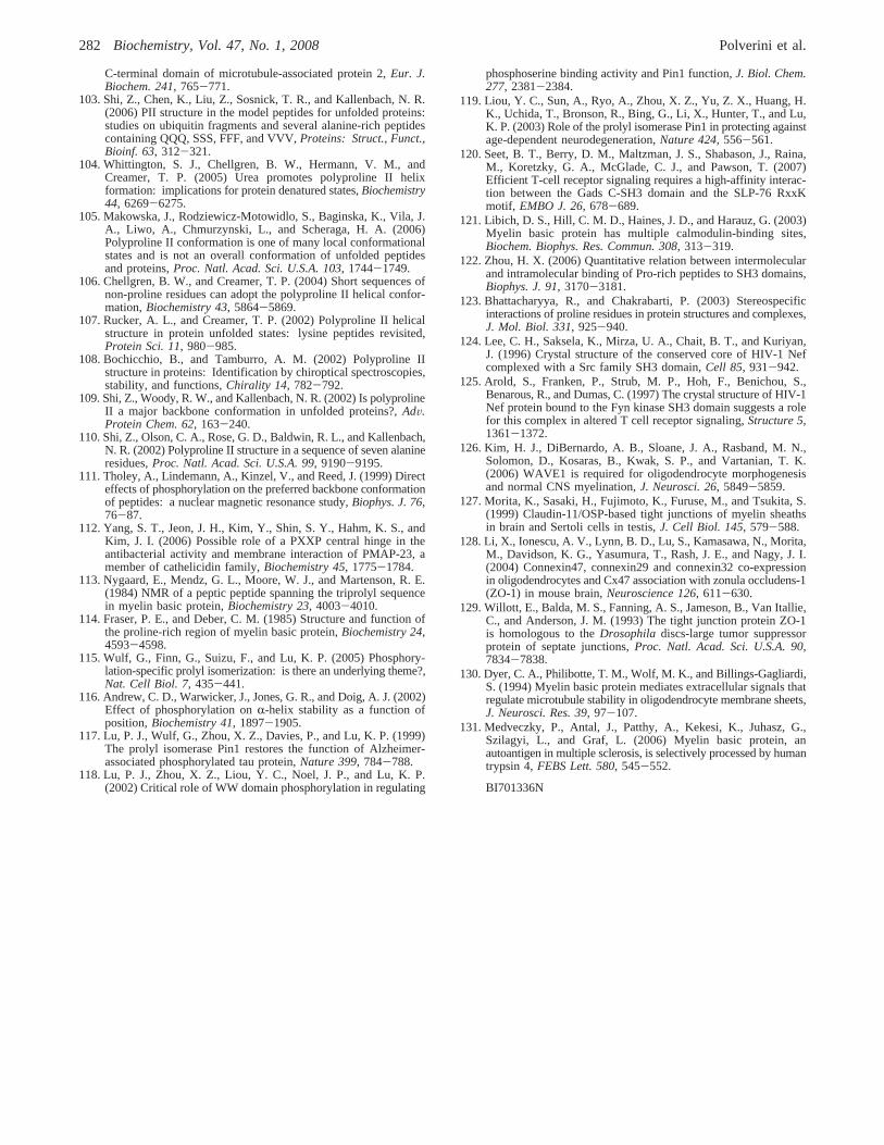
C-terminal domain of microtubule-associated protein 2,Eur. J.Biochem. 241, 765-771.
103. Shi, Z., Chen, K., Liu, Z., Sosnick, T. R., and Kallenbach, N. R.(2006) PII structure in the model peptides for unfolded proteins:studies on ubiquitin fragments and several alanine-rich peptidescontaining QQQ, SSS, FFF, and VVV,Proteins: Struct., Funct.,Bioinf. 63, 312-321.
104. Whittington, S. J., Chellgren, B. W., Hermann, V. M., andCreamer, T. P. (2005) Urea promotes polyproline II helixformation: implications for protein denatured states,Biochemistry44, 6269-6275.
105. Makowska, J., Rodziewicz-Motowidlo, S., Baginska, K., Vila, J.A., Liwo, A., Chmurzynski, L., and Scheraga, H. A. (2006)Polyproline II conformation is one of many local conformationalstates and is not an overall conformation of unfolded peptidesand proteins,Proc. Natl. Acad. Sci. U.S.A. 103, 1744-1749.
106. Chellgren, B. W., and Creamer, T. P. (2004) Short sequences ofnon-proline residues can adopt the polyproline II helical confor-mation,Biochemistry 43, 5864-5869.
107. Rucker, A. L., and Creamer, T. P. (2002) Polyproline II helicalstructure in protein unfolded states: lysine peptides revisited,Protein Sci. 11, 980-985.
108. Bochicchio, B., and Tamburro, A. M. (2002) Polyproline IIstructure in proteins: Identification by chiroptical spectroscopies,stability, and functions,Chirality 14, 782-792.
109. Shi, Z., Woody, R. W., and Kallenbach, N. R. (2002) Is polyprolineII a major backbone conformation in unfolded proteins?,AdV.Protein Chem. 62, 163-240.
110. Shi, Z., Olson, C. A., Rose, G. D., Baldwin, R. L., and Kallenbach,N. R. (2002) Polyproline II structure in a sequence of seven alanineresidues,Proc. Natl. Acad. Sci. U.S.A. 99, 9190-9195.
111. Tholey, A., Lindemann, A., Kinzel, V., and Reed, J. (1999) Directeffects of phosphorylation on the preferred backbone conformationof peptides: a nuclear magnetic resonance study,Biophys. J. 76,76-87.
112. Yang, S. T., Jeon, J. H., Kim, Y., Shin, S. Y., Hahm, K. S., andKim, J. I. (2006) Possible role of a PXXP central hinge in theantibacterial activity and membrane interaction of PMAP-23, amember of cathelicidin family,Biochemistry 45, 1775-1784.
113. Nygaard, E., Mendz, G. L., Moore, W. J., and Martenson, R. E.(1984) NMR of a peptic peptide spanning the triprolyl sequencein myelin basic protein,Biochemistry 23, 4003-4010.
114. Fraser, P. E., and Deber, C. M. (1985) Structure and function ofthe proline-rich region of myelin basic protein,Biochemistry 24,4593-4598.
115. Wulf, G., Finn, G., Suizu, F., and Lu, K. P. (2005) Phosphory-lation-specific prolyl isomerization: is there an underlying theme?,Nat. Cell Biol. 7, 435-441.
116. Andrew, C. D., Warwicker, J., Jones, G. R., and Doig, A. J. (2002)Effect of phosphorylation onR-helix stability as a function ofposition,Biochemistry 41, 1897-1905.
117. Lu, P. J., Wulf, G., Zhou, X. Z., Davies, P., and Lu, K. P. (1999)The prolyl isomerase Pin1 restores the function of Alzheimer-associated phosphorylated tau protein,Nature 399, 784-788.
118. Lu, P. J., Zhou, X. Z., Liou, Y. C., Noel, J. P., and Lu, K. P.(2002) Critical role of WW domain phosphorylation in regulating
phosphoserine binding activity and Pin1 function,J. Biol. Chem.277, 2381-2384.
119. Liou, Y. C., Sun, A., Ryo, A., Zhou, X. Z., Yu, Z. X., Huang, H.K., Uchida, T., Bronson, R., Bing, G., Li, X., Hunter, T., and Lu,K. P. (2003) Role of the prolyl isomerase Pin1 in protecting againstage-dependent neurodegeneration,Nature 424, 556-561.
120. Seet, B. T., Berry, D. M., Maltzman, J. S., Shabason, J., Raina,M., Koretzky, G. A., McGlade, C. J., and Pawson, T. (2007)Efficient T-cell receptor signaling requires a high-affinity interac-tion between the Gads C-SH3 domain and the SLP-76 RxxKmotif, EMBO J. 26, 678-689.
121. Libich, D. S., Hill, C. M. D., Haines, J. D., and Harauz, G. (2003)Myelin basic protein has multiple calmodulin-binding sites,Biochem. Biophys. Res. Commun. 308, 313-319.
122. Zhou, H. X. (2006) Quantitative relation between intermolecularand intramolecular binding of Pro-rich peptides to SH3 domains,Biophys. J. 91, 3170-3181.
123. Bhattacharyya, R., and Chakrabarti, P. (2003) Stereospecificinteractions of proline residues in protein structures and complexes,J. Mol. Biol. 331, 925-940.
124. Lee, C. H., Saksela, K., Mirza, U. A., Chait, B. T., and Kuriyan,J. (1996) Crystal structure of the conserved core of HIV-1 Nefcomplexed with a Src family SH3 domain,Cell 85, 931-942.
125. Arold, S., Franken, P., Strub, M. P., Hoh, F., Benichou, S.,Benarous, R., and Dumas, C. (1997) The crystal structure of HIV-1Nef protein bound to the Fyn kinase SH3 domain suggests a rolefor this complex in altered T cell receptor signaling,Structure 5,1361-1372.
126. Kim, H. J., DiBernardo, A. B., Sloane, J. A., Rasband, M. N.,Solomon, D., Kosaras, B., Kwak, S. P., and Vartanian, T. K.(2006) WAVE1 is required for oligodendrocyte morphogenesisand normal CNS myelination,J. Neurosci. 26, 5849-5859.
127. Morita, K., Sasaki, H., Fujimoto, K., Furuse, M., and Tsukita, S.(1999) Claudin-11/OSP-based tight junctions of myelin sheathsin brain and Sertoli cells in testis,J. Cell Biol. 145, 579-588.
128. Li, X., Ionescu, A. V., Lynn, B. D., Lu, S., Kamasawa, N., Morita,M., Davidson, K. G., Yasumura, T., Rash, J. E., and Nagy, J. I.(2004) Connexin47, connexin29 and connexin32 co-expressionin oligodendrocytes and Cx47 association with zonula occludens-1(ZO-1) in mouse brain,Neuroscience 126, 611-630.
129. Willott, E., Balda, M. S., Fanning, A. S., Jameson, B., Van Itallie,C., and Anderson, J. M. (1993) The tight junction protein ZO-1is homologous to theDrosophila discs-large tumor suppressorprotein of septate junctions,Proc. Natl. Acad. Sci. U.S.A. 90,7834-7838.
130. Dyer, C. A., Philibotte, T. M., Wolf, M. K., and Billings-Gagliardi,S. (1994) Myelin basic protein mediates extracellular signals thatregulate microtubule stability in oligodendrocyte membrane sheets,J. Neurosci. Res. 39, 97-107.
131. Medveczky, P., Antal, J., Patthy, A., Kekesi, K., Juhasz, G.,Szilagyi, L., and Graf, L. (2006) Myelin basic protein, anautoantigen in multiple sclerosis, is selectively processed by humantrypsin 4,FEBS Lett. 580, 545-552.
BI701336N
282 Biochemistry, Vol. 47, No. 1, 2008 Polverini et al.
Copyright © 2022 FDOKUMEN