Posttranslational Modifications of Proteins by Nitric Oxide: A New Tool of Metabolome Regulation
11
Posttranslational Modifications of Proteins in the Pathobiology of Medically Relevant Fungi Michelle D. Leach and Alistair J. P. Brown School of Medical Sciences, University of Aberdeen, Institute of Medical Sciences, Foresterhill, Aberdeen, United Kingdom Posttranslational modifications of proteins drive a wide variety of cellular processes in eukaryotes, regulating cell growth and division as well as adaptive and developmental processes. With regard to the fungal kingdom, most information about post- translational modifications has been generated through studies of the model yeasts Saccharomyces cerevisiae and Schizosaccha- romyces pombe, where, for example, the roles of protein phosphorylation, glycosylation, acetylation, ubiquitination, sumoyla- tion, and neddylation have been dissected. More recently, information has begun to emerge for the medically important fungal pathogens Candida albicans, Aspergillus fumigatus, and Cryptococcus neoformans, highlighting the relevance of posttransla- tional modifications for virulence. We review the available literature on protein modifications in fungal pathogens, focusing in particular upon the reversible peptide modifications sumoylation, ubiquitination, and neddylation. T he biological exploration of fungal pathogens, many of which lack an exploitable sexual cycle, has often focused upon pro- tein/gene expression levels. Indeed, changes in protein (or tran- script) abundance have provided valuable clues about the cellular processes that contribute to fungal pathogenesis. However, mea- surements of protein (or transcript) abundances alone are not sufficient to understand the regulation of such processes, because the activities of many eukaryotic proteins are modulated at post- translational levels. Posttranslational modifications are covalent processing events that often change the properties of a protein, for example, by proteolytic cleavage or by the addition of a modifying group to one or more amino acid residues (Table 1). Some posttransla- tional modifications make major structural contributions to their target proteins. For example, the glycosylation of cell wall manno- proteins fundamentally influences the shape of the fungal cell sur- face and hence the interactions of fungal pathogens with their hosts (86, 91, 100). Other regulatory modifications determine the activity state of specific proteins, their stability, their localization, and/or their interactions with other proteins. In signal transduc- tion pathways, for example, protein kinase cascades are activated and inactivated by the reversible addition and removal of phos- phate groups to specific residues in signaling proteins (22). More- over, the posttranslational marking of cyclins by ubiquitination targets them for destruction during defined phases of the cell cycle (170). For cellular processes to be regulated with requisite accuracy, posttranslational modification must be executed with exquisite specificity with regard to the choice of protein target, the selection of the residues to be modified, and the nature of the modifying group. Posttranslational modification mechanisms and their reg- ulatory significance have been studied extensively in the model yeast Saccharomyces cerevisiae, and more recent data have high- lighted the contributions of specific regulatory modifications to fungal pathogenicity. The main focus of this review is the revers- ible peptide modifications ubiquitination, neddylation, and su- moylation, whose contribution to fungal pathogenicity has been a topic of recent study. To place these modifications in context, we begin with a brief overview of posttranslational modifications and their impact upon fungal virulence (Table 1). POSTTRANSLATIONAL MODIFICATIONS AND VIRULENCE The fungal cell wall is the first point of contact between the host and pathogen and therefore plays an important role in the adher- ence to and colonization of host tissues and in host recognition. Numerous cell surface proteins are posttranslationally modified in fungi, and disruption of these modifications can significantly attenuate virulence. For example, cell wall and secreted manno- proteins are highly glycosylated in S. cerevisiae and Candida albi- cans (6, 113) (Table 1). The importance of N-glycosylation for fungal pathogenesis has been confirmed, for example, by disrup- tion of the 1,6-mannosyltransferase Och1 (6). Och1 initiates a distinct branch on the N-glycan core, providing the platform for subsequent addition of the extensively mannosylated outer chains. C. albicans och1 mutants display major cell wall defects and attenuated virulence in a murine model of infection (6). O-Mannosylation is initiated in the endoplasmic reticulum (ER) by the family of Dol-P-Man:protein O-mannosyltransferases (PMTs) (100). The characterization of pmt mutants has clearly demonstrated the essentiality of protein O-mannosylation in fungi. Tanner and coworkers showed that particular combina- tions of triple pmt mutants are lethal in S. cerevisiae (e.g., pmt1 pmt2 pmt4 or pmt2 pmt3 pmt4) (44). In pathogenic fungi, O-linked mannosylation is required for virulence. Loss of Pmt1, Pmt2, Pmt4, Pmt5, or Pmt6 attenuates the virulence of C. albicans (136, 148, 165, 166). Furthermore, loss of PMT4 or PMT1 in Cryp- tococcus neoformans attenuates the virulence of this pathogen both in murine models of disseminated cryptococcal virulence (124) and in murine inhalation models of cryptococcosis (178). Asper- gillus species also retain a highly conserved PMT gene family, and single pmt2 and double pmt1 pmt4 deletions are lethal in Aspergil- lus fumigatus (115). The importance of N- and O-glycosylation in pathogenic fungi can be largely attributed to their key roles in the Published ahead of print 9 December 2011 Address correspondence to Michelle D. Leach, [email protected]. Copyright © 2012, American Society for Microbiology. All Rights Reserved. doi:10.1128/EC.05238-11 The authors have paid a fee to allow immediate free access to this article. MINIREVIEW 98 ec.asm.org 1535-9778/12/$12.00 Eukaryotic Cell p. 98 –108
-
Upload
independent -
Category
Documents
-
view
1 -
download
0
Transcript of Posttranslational Modifications of Proteins by Nitric Oxide: A New Tool of Metabolome Regulation
Posttranslational Modifications of Proteins in the Pathobiology ofMedically Relevant Fungi
Michelle D. Leach and Alistair J. P. Brown
School of Medical Sciences, University of Aberdeen, Institute of Medical Sciences, Foresterhill, Aberdeen, United Kingdom
Posttranslational modifications of proteins drive a wide variety of cellular processes in eukaryotes, regulating cell growth anddivision as well as adaptive and developmental processes. With regard to the fungal kingdom, most information about post-translational modifications has been generated through studies of the model yeasts Saccharomyces cerevisiae and Schizosaccha-romyces pombe, where, for example, the roles of protein phosphorylation, glycosylation, acetylation, ubiquitination, sumoyla-tion, and neddylation have been dissected. More recently, information has begun to emerge for the medically important fungalpathogens Candida albicans, Aspergillus fumigatus, and Cryptococcus neoformans, highlighting the relevance of posttransla-tional modifications for virulence. We review the available literature on protein modifications in fungal pathogens, focusing inparticular upon the reversible peptide modifications sumoylation, ubiquitination, and neddylation.
The biological exploration of fungal pathogens, many of whichlack an exploitable sexual cycle, has often focused upon pro-
tein/gene expression levels. Indeed, changes in protein (or tran-script) abundance have provided valuable clues about the cellularprocesses that contribute to fungal pathogenesis. However, mea-surements of protein (or transcript) abundances alone are notsufficient to understand the regulation of such processes, becausethe activities of many eukaryotic proteins are modulated at post-translational levels.
Posttranslational modifications are covalent processing eventsthat often change the properties of a protein, for example, byproteolytic cleavage or by the addition of a modifying group toone or more amino acid residues (Table 1). Some posttransla-tional modifications make major structural contributions to theirtarget proteins. For example, the glycosylation of cell wall manno-proteins fundamentally influences the shape of the fungal cell sur-face and hence the interactions of fungal pathogens with theirhosts (86, 91, 100). Other regulatory modifications determine theactivity state of specific proteins, their stability, their localization,and/or their interactions with other proteins. In signal transduc-tion pathways, for example, protein kinase cascades are activatedand inactivated by the reversible addition and removal of phos-phate groups to specific residues in signaling proteins (22). More-over, the posttranslational marking of cyclins by ubiquitinationtargets them for destruction during defined phases of the cell cycle(170).
For cellular processes to be regulated with requisite accuracy,posttranslational modification must be executed with exquisitespecificity with regard to the choice of protein target, the selectionof the residues to be modified, and the nature of the modifyinggroup. Posttranslational modification mechanisms and their reg-ulatory significance have been studied extensively in the modelyeast Saccharomyces cerevisiae, and more recent data have high-lighted the contributions of specific regulatory modifications tofungal pathogenicity. The main focus of this review is the revers-ible peptide modifications ubiquitination, neddylation, and su-moylation, whose contribution to fungal pathogenicity has been atopic of recent study. To place these modifications in context, webegin with a brief overview of posttranslational modifications andtheir impact upon fungal virulence (Table 1).
POSTTRANSLATIONAL MODIFICATIONS AND VIRULENCE
The fungal cell wall is the first point of contact between the hostand pathogen and therefore plays an important role in the adher-ence to and colonization of host tissues and in host recognition.Numerous cell surface proteins are posttranslationally modifiedin fungi, and disruption of these modifications can significantlyattenuate virulence. For example, cell wall and secreted manno-proteins are highly glycosylated in S. cerevisiae and Candida albi-cans (6, 113) (Table 1). The importance of N-glycosylation forfungal pathogenesis has been confirmed, for example, by disrup-tion of the �1,6-mannosyltransferase Och1 (6). Och1 initiates adistinct branch on the N-glycan core, providing the platform forsubsequent addition of the extensively mannosylated outerchains. C. albicans och1 mutants display major cell wall defects andattenuated virulence in a murine model of infection (6).
O-Mannosylation is initiated in the endoplasmic reticulum(ER) by the family of Dol-P-Man:protein O-mannosyltransferases(PMTs) (100). The characterization of pmt mutants has clearlydemonstrated the essentiality of protein O-mannosylation infungi. Tanner and coworkers showed that particular combina-tions of triple pmt mutants are lethal in S. cerevisiae (e.g., pmt1pmt2 pmt4 or pmt2 pmt3 pmt4) (44). In pathogenic fungi,O-linked mannosylation is required for virulence. Loss of Pmt1,Pmt2, Pmt4, Pmt5, or Pmt6 attenuates the virulence of C. albicans(136, 148, 165, 166). Furthermore, loss of PMT4 or PMT1 in Cryp-tococcus neoformans attenuates the virulence of this pathogen bothin murine models of disseminated cryptococcal virulence (124)and in murine inhalation models of cryptococcosis (178). Asper-gillus species also retain a highly conserved PMT gene family, andsingle pmt2 and double pmt1 pmt4 deletions are lethal in Aspergil-lus fumigatus (115). The importance of N- and O-glycosylation inpathogenic fungi can be largely attributed to their key roles in the
Published ahead of print 9 December 2011
Address correspondence to Michelle D. Leach, [email protected].
Copyright © 2012, American Society for Microbiology. All Rights Reserved.
doi:10.1128/EC.05238-11
The authors have paid a fee to allow immediate free access to this article.
MINIREVIEW
98 ec.asm.org 1535-9778/12/$12.00 Eukaryotic Cell p. 98–108
construction and maintenance of a robust cell wall, an essentialstructure in fungi.
Glycosylphosphatidylinositol (GPI) anchoring of proteins onthe cell surface also plays a role in fungal virulence (Table 1). For
example, the GPI-anchored proteins Phr1, Phr2, and Utr2 areinvolved in C. albicans cell wall biosynthesis and modeling (142).Also, Sod5 combats macrophage-induced oxidative stress (38),the phospholipase B Plb5 promotes the colonization of organs in
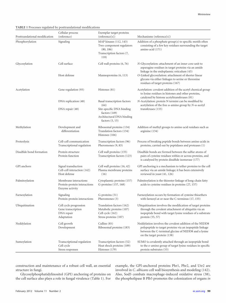
TABLE 1 Processes regulated by posttranslational modifications
Posttranslational modificationCellular process(reference)
Exemplar target proteins(reference[s]) Mechanisms (reference[s])
Phosphorylation Signaling MAP kinases (112, 143) Addition of a phosphate group(s) to specific motifs oftenconsisting of a few key residues surrounding the targetamino acid (171)
Two-component regulators(80, 186)
Transcription factors (7,118)
Glycosylation Cell surface Cell wall proteins (6, 76) N-Glycosylation: attachment of an inner core unit toasparagine residues in target proteins via an amidelinkage in the endoplasmic reticulum (43)
Host defense Mannoproteins (6, 113) O-Linked glycosylation: attachment of shorter linearglycans via either linkages to serine or threonineresidues of target proteins (167)
Acetylation Gene regulation (93) Histones (81) Acetylation: covalent addition of the acetyl chemical groupto lysine residues in histones and other proteins,catalyzed by histone acetyltransferases (81)
DNA replication (48) Basal transcription factors(64)
N-Acetylation: protein N termini can be modified byacetylation of the free �-amino group by N-�-acetyltransferases (133)DNA repair (48) Site-specific DNA binding
factors (149)Architectural DNA binding
factors (5, 15)
Methylation Development anddifferentiation
Ribosomal proteins (134) Addition of methyl groups to amino acid residues such asarginine (134)Translation factors (134)
Histones (104)
Proteolysis Cell-cell communication Transcription factors (96) Process of breaking peptide bonds between amino acids inproteins, carried out by peptidases and proteases (1)Transcriptional regulation Pheromones (9, 83)
Disulfide bond formation Protein structure Cell wall proteins (135) Disulfide bonds are formed between the sulfur atoms ofpairs of cysteine residues within or across proteins, andis catalyzed by protein disulfide isomerase (177)
Protein function Transcription factors (123)
GPI anchors Signal transduction Cell wall proteins (16, 42) GPI anchoring is a mechanism to tether proteins to the cellsurface via an amide linkage; it has been extensivelyreviewed in yeast (41, 126)
Cell-cell interaction (142) Plasma membrane proteins(16)Host defense
Palmitoylation Membrane interactions Cytoplasmic proteins (157) Palmitoylation is the thioester linkage of long-chain fattyacids to cysteine residues in proteins (27, 157)Protein-protein interactions G-proteins (157, 168)
Enzyme activity
Farnesylation Signaling G-proteins (51) Farnesylation occurs by formation of cysteine thioetherswith farnesyl at or near the C-terminus (17, 155)Protein-protein interactions Pheromones (3)
Ubiquitination Cell cycle progression Translation factors (162) Ubiquitination involves the modification of target proteinsthrough the covalent attachment of ubiquitin via anisopeptide bond with target lysine residues of a substrateprotein (55, 57)
Gene transcription Metabolic proteins (107)DNA repair Cell cycle (162)Adaptation Stress proteins (107)
Neddylation Cell growth Cullins (85) Neddylation involves the covalent addition of the NEDD8polypeptide to target proteins via an isopeptide linkagebetween the C-terminal glycine of NEDD8 and a lysineon the target protein (138)
Development Ribosomal proteins (183)
Sumoylation Transcriptional regulation Transcription factors (52) SUMO is covalently attached through an isopeptide bondto the �-amino group of target lysine residues in specificprotein substrates (53)
Cell cycle Heat shock proteins (189)Stress responses Histones (179)
Minireview
February 2012 Volume 11 Number 2 ec.asm.org 99
vivo (164), and ALS protein family members and Hwp1 enhanceadherence to host tissues (60, 154).
Stress responses and environmental adaptation are of particu-lar significance for fungal pathogens, as they must mount effectiveresponses to counteract the defenses of their host and adapt toavailable nutrients in diverse host niches. This adaptation is fre-quently achieved through posttranslational modifications of sig-naling proteins (Table 1). For example, Ras GTPases are highlyconserved signaling proteins that play central roles in key physio-logical processes, such as growth, morphology, and survival. Ras1in C. albicans signals through the cyclic AMP (cAMP) or Cek1mitogen-activated protein (MAP) kinase signaling cascades (30)initiating hyphal growth, a key virulence trait of C. albicans. Ras1localization to the plasma membrane, which is critical for activa-tion, is controlled by farnesylation and palmitoylation in C. albi-cans (132). In C. neoformans, palmitoylation is required for Ras1localization, which is necessary for morphogenesis (120). Addi-tionally, protein phosphorylation, a ubiquitous and reversiblemodification that is crucial for the regulation of multiple cellularevents (153), regulates MAP kinase and two-component signalingcascades in fungal pathogens (80, 186). Also, protein phosphory-lation via cAMP-protein kinase A signaling contributes to fungalvirulence through regulation of yeast-hypha morphogenesis andstress resistance in C. albicans (13, 14, 45) and mating, capsuleformation, and melanin production in C. neoformans (2). Notsurprisingly, protein phosphorylation has been studied exten-sively, and its role in yeast biology and fungal pathogenicity hasbeen reviewed elsewhere (7, 112, 143).
Signaling pathways often culminate in the phosphorylation ofspecific transcription factors that drive the changes in gene expres-sion patterns that underpin the corresponding adaptive or devel-opmental process. For example, the heat shock transcription fac-tor Hsf1, which controls thermal adaptation in C. albicans andother eukaryotic cells, is phosphorylated in response to heat shock(118). Hsf1 phosphorylation is required for adaptation to thermalinsults and also for the virulence of C. albicans (119). However, theactivity of transcription factors can be modified by other types ofposttranslational modification. For instance, the activity of thepH-responsive transcription factor Rim101, which is necessaryfor in vivo pathogenesis in C. albicans (25), is modulated by theproteolytic removal of 100 carboxy-terminal residues (96).
Accurate genome organization, gene regulation, DNA replica-tion, and DNA repair are essential for growth and pathogenicity.These processes are frequently controlled by histone acetylation, aposttranslational modification that plays a major role in the mod-ulation of high-order chromatin structures. In the yeasts S. cerevi-siae and Schizosaccharomyces pombe, newly synthesized histoneH3 molecules are heavily acetylated on lysine 56 (H3K56) (72,106, 181). This occurs through the fungus-specific histone acetyl-transferase (HAT) enzyme, Rtt109, and the histone chaperoneAsf1 (28, 50, 139, 169). Loss of Rtt109 or Asf1 in S. cerevisiae causesdelayed cell cycle progression (28) and spontaneous DNA damage(32, 106), and blocked acetylation at H3K56 results in similarphenotypes (106, 139). Therefore, the acetylation of H3K56 ap-pears to be a particularly important posttranslational modifica-tion for fungal growth.
Recent studies have shown that Rtt109 (and presumablyH3K56 acetylation) is required for C. albicans pathogenicity. C.albicans rtt109 mutants are relatively sensitive to genotoxic agents,highly susceptible to macrophage killing, and less virulent than the
wild type in a mouse model of systemic infection (102). Interest-ingly, the increased sensitivity to macrophages is dependent on thehost’s ability to produce reactive oxygen species (ROS), indicatingthat acetylation also contributes to stress adaptation. Histone(de)acetylation also contributes to morphogenetic regulation andphenotypic switching in C. albicans, both of which promote viru-lence (56, 159).
Histone acetylation has also been shown to contribute to thepathogenicity of C. neoformans. In S. cerevisiae the histone acetyl-transferase Gcn5 is involved in the regulation of transcriptionalresponses to various environmental stresses, including high tem-perature, osmotic stress, nutrient deprivation, and oxidative dam-age (61). Similarly, C. neoformans gcn5 mutants show defects ingrowth at high temperatures, sensitivity to oxidative stress andFK506 (a calcineurin inhibitor), and defects in capsule attachmentto the cell surface (125). Stress adaptation and capsule formationpromote C. neoformans pathogenesis, and therefore it is not sur-prising that the gcn5 mutants are avirulent in animal models ofcryptococcosis (125).
Protein methylation also contributes to a range of cellular pro-cesses, including protein transport, transcriptional regulation,and signaling (Table 1). For example, protein arginine methyl-ation by Hmt1 has been shown to promote the nuclear export oftarget proteins in C. albicans (108). However, the impact of pro-tein methylation upon fungal pathogenicity remains to be tested.
The reversible addition to proteins of specific peptide moieties,such as ubiquitin, NEDD, and SUMO, has been shown to influ-ence fungal virulence (Table 1). These posttranslational modifica-tions are discussed in the next sections.
REVERSIBLE PEPTIDE MODIFICATIONSUbiquitination. Ubiquitination involves the posttranslationalmodification of target proteins through the covalent attachmentof ubiquitin (Ub), a highly conserved 76-amino-acid protein thathas the unusual property of forming a stable chemical bond withother proteins. The carboxyl group of the carboxy-terminal gly-cine of ubiquitin forms an isopeptide bond with the �-aminogroup of target lysine residues, or occasionally with the aminogroup at the amino terminus of a substrate protein (55, 57). Ubiq-uitin conjugation is achieved via a three-step reaction catalyzed bythree enzymes: E1, a ubiquitin-activating enzyme; E2, a ubiquitin-conjugating enzyme; and E3, a ubiquitin ligase. Briefly, the acti-vation of ubiquitin by E1 is followed by the conjugation of ubiq-uitin to an E2 enzyme. Finally, ubiquitin is transferred to theprotein substrate or target by an E3 ubiquitin ligase (184; see ref-erence 166 for a review). Ubiquitination is reversible, like the pro-cess of (de)phosphorylation. The cleavage of ubiquitin from sub-strates is carried out by specific deubiquitinating enzymes (58).
Ubiquitination can modulate the activity of target proteins invarious ways. These include the proteasome-mediated degrada-tion of some target proteins and the stabilization of othersthrough deubiquitination (110). Ubiquitination can also influ-ence the activity of some proteins, or their cellular localization.Ubiquitin modifications take the form of monoubiquitin (attach-ment of a single ubiquitin moiety), multiple monoubiquitination(modification of several target lysines with a single ubiquitin), orpolyubiquitination (attachment of four or more ubiquitin moi-eties) (175). The different types of ubiquitin conjugates controldifferent cellular processes. Polyubiquitination generally targetsthe substrates for degradation, whereas the addition of fewer ubiq-
Minireview
100 ec.asm.org Eukaryotic Cell

uitin molecules can alter the substrate protein function or targetthe substrate protein to the endosome (185). Proteins involved inmany cellular processes are targets for ubiquitination, in manycases their ubiquitination acting as a signal for recognition by anubiquitin-binding protein. Therefore, ubiquitination contributesto the regulation of numerous cellular processes, including cellcycle progression, gene transcription, DNA repair, and inflamma-tion (49, 55, 116, 131).
In S. cerevisiae, ubiquitin is encoded by a multigene family ofnatural gene fusions (UBI1, UBI2, UBI3, and UBI4). UBI1, UBI2,and UBI3 encode hybrid proteins in which ubiquitin is fused tounrelated amino acid sequences (128). UBI1 and UBI2 encodeidentical 52-residue polypeptide tails, which are components ofthe large ribosomal subunit. In contrast, UBI3 encodes a different76-residue tail that corresponds to the S34 protein of the smallribosomal subunit (33, 128). The fourth ubiquitin gene in S.cerevisiae, UBI4, encodes a polyubiquitin precursor protein com-prising five tandem repeats of ubiquitin in a spacerless head-to-tail arrangement (128).
All four ubiquitin genes are expressed in exponentially growingS. cerevisiae cells, but the expression of UBI1 and UBI2 is repressedin stationary-phase cells. Mutants lacking the ubiquitin-hybridgenes display slow-growth phenotypes, particularly in the case ofthe ubi3 deletion. UBI1 and UBI2 encode functionally redundantproteins that execute an essential function, as a ubi1 ubi2 doublemutant is not viable (33). The UBI4 gene is strongly induced bystarvation, high temperatures, and oxidative stress (18, 35), andoverexpression of UBI4 restores heat shock sensitivity in the ab-sence of the enzyme serine palmitoyltransferase, required for re-sistance to heat shock (40). Therefore, UBI4 is thought to pro-vide ubiquitin under stress conditions (18, 34, 35, 128). Theroles of ubiquitin in fungal pathogens have been less well char-acterized than those in model yeasts, but recent data stronglyimplicate polyubiquitin in growth, metabolism, and stress re-sponses (Fig. 1).
(i) Ubiquitination and stress. Two ubiquitin-encoding geneshave been identified in C. albicans, C. neoformans, and Aspergillusnidulans. These are UBI3 and UBI4 in C. albicans and UBI1 and
FIG 1 Roles of ubiquitination and sumoylation. Polyubiquitinated proteins are targeted to the proteasome for degradation via E1, E2, and E3 enzymes. Dubs(deubiquitination enzymes) reverse this process. Ubiquitin targets a number of proteins involved in different cellular processes (blue lines). Sumoylationcontributes to similar processes (red lines) but modifies proteins by attaching SUMO via E1, E2, and E3 enzymes, and this modification generally activates aprotein. Sumoylation is reversible by ULP proteases. These modifications are often triggered by specific environmental stimuli, and the cellular processes theyregulate contribute to fungal pathogenicity.
Minireview
February 2012 Volume 11 Number 2 ec.asm.org 101
UBI4 in C. neoformans and A. nidulans. As yet, these C. neoformansgenes have not been characterized in any detail: they have beenisolated (158), but they have yet to be dissected functionally. Nev-ertheless, the involvement of the ubiquitination system in stressadaptation has been examined in C. neoformans, with twoubiquitin-conjugating genes, UBC6-2 and UBC8, being impli-cated in oxidative stress responses (77). Furthermore, the expres-sion of the F-box protein Fbp1 is regulated by glucose in C. neo-formans, highlighting a potential role for this SCF (for “Skp1/Cdc53/F-box protein”) complex (and hence ubiquitination) inglucose sensing by this pathogen. (F-box proteins are exchange-able subunits that promote the capacity of SCF to specifically tar-get protein substrates for ubiquitination and degradation.) Signif-icantly, a C. neoformans fbp1 mutant is unable to generate sporesand displays attenuated virulence in a murine intranasal-inhalation model, highlighting the key role of the SCF E3 ubiqui-tin ligase-mediated pathway in these processes (99).
In the model filamentous fungus A. nidulans, the regulation ofthe polyubiquitin-encoding ubi1 and ubi4 transcripts has beencharacterized, with specific focus on stress. Both genes are inducedupon heat stress, but only the ubi1 gene is induced by peroxidestress (121), suggesting that these ubiquitin genes play differentialroles in stress adaptation. Similarly, recent work on C. albicans hashighlighted differential roles for the ubiquitin-encoding genesUBI3 and UBI4 in stress adaptation. The polyubiquitin gene UBI4is induced in response to numerous stress responses, includingheat, peroxide, and cell wall stresses (88). This contrasts with aubiquitin hybrid protein, Ubi3, which is involved in ribosomebiogenesis and which is coordinately regulated with componentsof the translational apparatus (145, 147).
A number of stress-related proteins have been identified asubiquitination targets under stress conditions in C. albicans.These include Pst2 (an oxidative-stress response protein), which isubiquitinated in response to peroxide stress, and Ssb1 (a memberof the Hsp70 chaperone family), which is ubiquitinated in re-sponse to both peroxide and heat stress (88). During heat stress,chaperone proteins are upregulated to prevent accumulation ofaggregated proteins and to promote the refolding of unfolded pro-teins (47, 130). Increasing ubiquitin-dependent degradation cansuppress this normally essential requirement for heat shock pro-tein induction (40). This would seem to imply that an essentialaspect of stress adaptation is the removal of misfolded proteins,either by refolding or through ubiquitin-mediated degradation.
Heat shock proteins such as Hsp70 and Hsp90 have been im-plicated in the maintenance of the ubiquitin-proteasome system,specifically, the 26S proteasome in S. cerevisiae (62, 63). Further-more, Ssa1 (another Hsp70 family member) has been linked toprotein quality control, directing cytoplasmically mislocalizedproteins for proteasomal degradation (109, 129). More recently, arole for Ssa1 in regulating ubiquitination and degradation of cor-rectly folded gluconeogenic enzymes was elucidated, demonstrat-ing that the Hsp70 chaperone machinery is involved in regulatorydegradation of metabolic enzymes (70). Indeed, a positive feed-back loop may exist, whereby ubiquitination of stress-related pro-teins might activate them to work with the polyubiquitin pathwayin the degradation of aggregated proteins, whose inherent toxicitywould be detrimental to the cell. A second hypothesis may be thatchaperone proteins are merely targeted for degradation after aninitial upregulation during temperature stress. These ideas are notmutually exclusive. It would be interesting to test whether such a
feedback loop, involving the regulation of ubiquitination by ubiq-uitinated proteins, exists in fungi.
(ii) Ubiquitination and metabolism. Both metabolic diversityand efficiency are required for microorganisms to successfully ex-ploit the dynamically changing microenvironments they occupy.This flexibility can be achieved through gene regulation but alsothrough posttranslational modifications such as ubiquitination.Indeed, ubiquitination has been shown to be required for the sur-vival of S. cerevisiae under starvation conditions (176). Also, anumber of specific proteins are degraded via the proteasome dur-ing metabolic transitions, for example, when cells switch fromgluconeogenic to glycolytic metabolism. Fructose-1,6-bisphosphatase(FBPase) (150, 151), Std1 (74, 114), and the regulatory proteinMth1 (37) are rapidly degraded following glucose addition to S.cerevisiae cells. Interestingly, when glucose is abundant, Mth1 istargeted for proteasomal degradation by Grr1 (an F-box protein)(36, 37). What is more, cells lacking Grr1 have several metabolicdefects, including sensitivity to nitrogen starvation (36).
A number of metabolic proteins have been identified as ubiq-uitination targets in C. albicans, including proteins involved inglycolysis and/or gluconeogenesis (Eno1, Fba1, and Pgk1), thepentose phosphate pathway (Tkl1), fatty acid metabolism (Pdx3),acetate utilization (Asc2), and glycerol synthesis (Gpd2) (88). Fur-thermore, a lack of polyubiquitin (Ubi4) reduces the ability of C.albicans to survive under starvation conditions. Strikingly, C. al-bicans ubi4 mutants rapidly lost viability when cells were starvedfor nitrogen in the presence of excess carbon, suggesting thatubiquitin-mediated protein degradation is required for nitrogenrecycling under these conditions. Furthermore, specific C. albi-cans proteins are rapidly degraded in response to glucose (173),although the exact mechanisms by which this occurs are yet to berevealed. Interestingly, the ubiquitin ligase gene, UBC8, whoseorthologue is involved in glucose-accelerated protein degradationin S. cerevisiae (140, 152), is conserved in C. albicans (122). Fur-thermore, UBC8 is the only ubiquitin-related gene that is upregu-lated at the transcriptional level following glucose exposure in C.albicans (144). Therefore, analogous ubiquitin-dependent mech-anisms probably contribute to metabolic adaptation in S. cerevi-siae and C. albicans and may exist in other fungal pathogens.
(iii) Ubiquitination, growth, and morphogenesis. Roles for ubiq-uitination in morphogenesis and other developmental processeshave been reported for both C. albicans and A. nidulans. Interest-ingly, A. nidulans GrrA (which is closely related to S. cerevisiaeGrr1) plays a role in meiosis and sexual spore formation. DeletinggrrA inhibits the formation of mature ascospores due to a block inmeiosis (79). In C. albicans, the deletion of UBI4 leads to a heter-ogeneous mixture of yeast and hyphal cells (88, 146) whereby thehyphal cells display an apparent mitotic arrest (88). The exactmechanisms by which this occurs are currently unknown. How-ever, it is well known that cellular morphogenesis is intimatelylinked with cell cycle regulation in many eukaryotes, particularlyin budding yeasts (92). Also, ubiquitin-mediated protein degra-dation plays a central role in cell cycle control, with cyclin degra-dation being tightly regulated by ubiquitin-dependent proteolysis(78, 188). The multiprotein ubiquitin ligase SCF plays an impor-tant role in cell cycle control by degrading key proteins involved incell cycle regulation in S. cerevisiae and other eukaryotes (20, 78,188). In S. cerevisiae, the F-box protein Cdc4 (SCFCdc4) is requiredfor degradation of the cyclin-dependent kinase inhibitors Sic1(117) and Far1 (12), while its orthologue in C. albicans has a role in
Minireview
102 ec.asm.org Eukaryotic Cell
regulating morphogenesis (4). Additionally, SCFGrr1 targets theG1 cyclins Cln1 and Cln2 for degradation in S. cerevisiae (10, 75);similarly, deletion of GRR1 stabilizes two G1 cyclins, Ccn1 andCln3, with the mutant growing constitutively as pseudohyphae(97). Also, the ubiquitin-conjugating (E2) enzyme Rad6 has beenshown to downregulate hyphal development in C. albicans (90).
Clearly, the ubiquitination pathway contributes to the regula-tion of diverse cellular processes in fungal pathogens, includingcell division and developmental processes as well as stress andmetabolic adaptation. Given the fundamental importance of theseprocesses, it is hardly surprising that ubiquitination contributessignificantly to fungal virulence (88). However, the exact mecha-nisms by which ubiquitination regulates these cellular processesand hence fungal pathogenesis needs further dissection.
Sumoylation. Like ubiquitination, sumoylation is a highly con-served, reversible posttranslational modification that involves theconjugation of a polypeptide to target proteins. The SUMO (smallubiquitin-like modifier) polypeptide is expressed in all eukaryotes(29, 39, 54). All yeasts and invertebrates studied to date contain asingle SUMO gene, whereas vertebrates contain three SUMOgenes, SUMO-1, SUMO-2, and SUMO-3 (68, 71, 103). Plantscontain additional SUMO genes, with eight such genes havingbeen found in Arabidopsis (82).
SUMO is covalently attached through an isopeptide bond tothe �-amino group of target lysine residues in specific proteinsubstrates. This occurs via a three-step enzymatic pathway that isroughly analogous to the ubiquitination pathway (46, 111).Briefly, after proteolytic maturation, the S. cerevisiae SUMO pro-tein, Smt3, is activated in an ATP-dependent fashion by a SUMO-activating enzyme, E1, through the formation of a thioester bondbetween its carboxy terminus and the E1-activating complex,Uba2/Aos1. Activated SUMO is then transferred to the E2-conjugating enzyme, Ubc9. SUMO is then transferred from Ubc9to the substrate protein with the assistance of one of the severalSUMO protein ligases (Siz1 and Siz2 in S. cerevisiae), termed E3s.Ultimately, Ubc9 catalyzes the covalent link between the carboxy-terminal glycine in Smt3 and a lysine residue present in the su-moylation consensus sequence of the target protein.
Ubc9 contributes to substrate recognition and Smt3 ligation(8), whereas E3 ligases play important roles in enhancing substrateidentification and specificity (111). Many target lysine residues liein the short consensus sequence �KXE, where � is a large hydro-phobic amino acid (generally isoleucine, leucine, or valine), K isthe lysine residue that is modified, X is any residue, and E is glu-tamic acid. Sumoylation is a reversible modification, and removalof SUMO is carried out by enzymes of the Ulp family that specif-ically cleave at the carboxy terminus of SUMO (94, 95).
In S. cerevisiae, the single SUMO gene, SMT3, is essential forviability. In the fission yeast, S. pombe, cells lacking the SUMOgene pmt3 are barely viable and have defects in genome mainte-nance (163). In S. cerevisiae, sumoylation regulates a variety ofcellular processes, including septin ring formation (66, 67), sisterchromatid cohesion (11, 160), DNA repair (65, 172), and tran-scriptional regulation (156). Unlike polyubiquitination, whichgenerally targets proteins for degradation, sumoylation generallyexerts its regulatory effects upon target proteins by modulatingprotein-protein interactions and protein localization. In somecases, sumoylation antagonizes ubiquitin conjugation, therebypromoting protein stabilization (26, 46). Recent studies have re-vealed that there is cross talk between sumoylation and ubiquiti-
nation, whereby sumoylation can acts as a target for SUMO-directed ubiquitin ligases (137, 161, 182).
Virtually nothing is known about sumoylation in the patho-genic fungi. To date, there are only two studies of sumoylation inC. albicans and A. nidulans, with no reports for C. neoformans.Interestingly, the SUMO gene is not essential in C. albicans or A.nidulans, but mutants that lack SUMO exhibit impaired growth.In A. nidulans, sumO� cells display reduced conidiation and in-creased sensitivity to the DNA-damaging agent methyl methane-sulfonate (MMS) and to the DNA synthesis inhibitor hydroxyurea(HU) (180). sumO� cells also display self-sterility, suggesting thatsumoylation of key targets is required for the development of vi-able meiotic progeny.
The inactivation of SMT3 in C. albicans generates a heteroge-neous population of slowly growing, enlarged, elongated,pseudohypha-like cells (87). This suggested defects in cell cycleprogression, and this was confirmed when DAPI (4=,6=-diamidino-2-phenylindole) staining revealed many multinucleateC. albicans smt3 cells (87). The possibility that septin sumoylationis required for normal septation might provide an explanation forthis phenotype. S. cerevisiae mutants with defects in any one of theseptin genes CDC3, CDC10, CDC11, and CDC12 commonly formbranched, highly elongated, multinucleate cellular clusters (23,31, 101). These mutants are unable to organize the bud neck fila-ments, which have essential roles in cytokinesis (23, 31, 101). In-terestingly, the S. cerevisiae septins Cdc3, Cdc11, and Shs1/Sep7contain sumoylation motifs. When these motifs were mutated,septin ring formation was lost, and there was also a loss of budneck-associated SUMO (66). Septin sumoylation is dependent onthe E3 enzyme Siz1 (67). Similar mechanisms may also operate inC. albicans. Although attempts to identify septin sumoylationhave proved unsuccessful to date (105), C. albicans cdc11 mutantsdisplay cell cycle defects similar to those of smt3 mutants (174).Furthermore, C. albicans Cdc11 contains three type I (�-K-X-E)consensus sumoylation sites, and Cdc3 contains two type I andone type II consensus sumoylation sites (141). To our knowledge,no one has tested the effects of mutating all sumoylation sites inCdc11, although sumoylation appears to regulate a number of cellcycle-related proteins, and hence a number of cell cycle pathways.For example, Tub1, Cct7, and Mlc1 have been identified as su-moylation targets, suggesting that this posttranslational modifica-tion modulates tubulin and actin assembly, which could also ac-count for the elongated, multinucleate cell phenotype of C.albicans smt3 cells (87). It is conceivable that sumoylation of otherbud neck proteins is required for normal morphogenesis.
C. albicans smt3 mutants also display susceptibility to a widerange of stresses, including temperature, oxidative, and cell wallstresses (87). Given the severe growth defects of these mutants,this was not surprising. Nevertheless, a number of S. cerevisiaeproteins are sumoylated in response to stress (189), although theexact mechanisms by which sumoylation contributes to stress ad-aptation and/or survival are largely unknown. Similarly, the chap-erones Hsp104 and Hsp60 were identified in a proteomics screenfor stress-dependent sumoylation targets in C. albicans. Mutationof the consensus sumoylation site in Hsp104 caused cells to be-come more susceptible to thermal insults (87). Therefore, sumoy-lation of Hsp104 is important for thermal resistance, possibly bymodulating Hsp104 activity.
An interesting phenotype was observed when the consensussumoylation site in the mitochondrial chaperone Hsp60 was mu-
Minireview
February 2012 Volume 11 Number 2 ec.asm.org 103

tated. A significant proportion of these cells grew in an elongated,pseudohypha-like fashion (87), suggesting a link between mito-chondrial activity and filamentous growth in C. albicans. TheseHSP60K324R cells were also temperature sensitive, but only whencells were stressed with a respiratory inhibitor (87). Additionalsumoylation targets may contribute to thermal resistance as anumber of other C. albicans proteins appear to be sumoylated inresponse to stress (87). Indeed, sumoylation of HSF1 in mamma-lian cells is strongly induced by heat shock, which results in in-creased DNA binding and activity (59). Interestingly, HsfA2 su-moylation in Arabidopsis represses its transcriptional activity,suggesting that sumoylation can also negatively regulate proteinsin response to stress (21).
Evidently, sumoylation contributes significantly to the controlof growth, cell division, and stress adaptation, all of which are vitalfor fungal virulence (Fig. 1). Yet sumoylation is an understudiedtopic, especially in fungal pathogens. The mechanisms by whichthe sumoylation of specific target proteins control these cellularprocesses and hence fungal virulence remain to be elucidated.
Neddylation. Neddylation involves the covalent addition of theNEDD8 polypeptide (Rub1 in S. cerevisiae) to a small number oftarget proteins via an isopeptide linkage between the carboxy-terminal glycine (Gly)76 of NEDD8 and a lysine on the targetprotein (138). The mechanisms of protein neddylation are closelyrelated to those involved in ubiquitination. The first identifiedtarget of Rub1 in S. cerevisiae was Cdc53 (84), which is a memberof the cullin family of proteins (molecular scaffolds for ubiquitinligases). Right across the eukaryotic kingdom from yeasts to hu-mans, the cullin family has three to six members, suggesting theimportance of neddylation in regulating cullin function. Indeed,all yeast cullins, including S. cerevisiae Cul3, Cdc53, and Rtt101(85) and S. pombe: Pcu1, Pcu3, and Pcu4 (127, 187), are neddy-lated on a conserved lysine in their carboxy-terminal domain invivo. This neddylation positively regulates E3 ligase activity andthe assembly of Skp1/cullin/F-box (SCF) complexes (24, 84, 98).Defective neddylation causes severe defects in cell growth and de-velopment, ranging from cell cycle arrest in S. pombe to arrest inembryonic development for Caenorhabditis elegans (69).
Neddylation also contributes to fungal pathogenicity. C. neo-formans rub1 mutants exhibit severe survival defects when grownin cerebral spinal fluid, as well as attenuated virulence in C. elegansand a rabbit model of cryptococcal meningitis (89). Neddylationappears to be important for facilitating the attachment of ubiqui-tin E2 to the E3 ligase, allowing for efficient proteasome targetingof proteins (73). Therefore, this severe virulence defect could bedue to defective protein turnover and adaptation to potentiallyhost environments. A RUB1 orthologue has been identified in C.albicans. Functional analysis of this gene is likely to confirm theimportance of neddylation for fungal pathogenicity.
CONCLUDING REMARKS
A diverse range of posttranslational modifications regulate theactivity and abundance of specific proteins in fungal pathogens,thereby controlling key cellular processes that contribute to thevirulence of these organisms. While protein phosphorylation hasreceived considerable attention through studies of signal trans-duction and transcriptional regulation, other functionally impor-tant posttranslational modifications (such as ubiquitination, su-moylation, and neddylation) have been largely ignored. It isevident that these processes contribute significantly to the growth
and adaptation of fungal pathogens. The extent to which there iscross talk between posttranslational modifications is not known,but precedents clearly exist. For example, the phosphorylation ofsome target proteins is known to be a prerequisite for ubiquitina-tion and subsequent substrate degradation (19). Although thistopic has merely been touched upon in this review, it is clearly ofhigh relevance and warrants further study. Therefore, further ef-forts are required to define the mechanisms by which these post-translational modifications contribute to disease establishmentand progression.
ACKNOWLEDGMENTS
M.L. was supported by a Carnegie/Caledonian Scholarship from The Car-negie Trust. A.J.P.B. is supported by grants from the UK Biotechnologyand Biological Research Council (BB/D009308/1; BB/F00513X/1), theWellcome Trust (080088), and the European Commission (FINSysB,PITN-GA-2008-214004; STRIFE, ERC-2009-AdG-249793).
REFERENCES1. Achstetter T, Wolf DH. 1985. Proteinases, proteolysis and biological
control in the yeast Saccharomyces cerevisiae. Yeast 1:139 –157.2. Alspaugh JA, Perfect JR, Heitman J. 1997. Cryptococcus neoformans
mating and virulence are regulated by the G-protein alpha subunit GPA1and cAMP. Genes Dev. 11:3206 –3217.
3. Anderegg RJ, Betz R, Carr SA, Crabb JW, Duntze W. 1988. Structureof Saccharomyces cerevisiae mating hormone a-factor. Identification ofS-farnesyl cysteine as a structural component. J. Biol. Chem. 263:18236 –18240.
4. Atir-Lande A, Gildor T, Kornitzer D. 2005. Role for the SCFCDC4
ubiquitin ligase in Candida albicans morphogenesis. Mol. Biol. Cell 16:2772–2785.
5. Bannister AJ, Miska EA. 2000. Regulation of gene expression by tran-scription factor acetylation. Cell. Mol. Life Sci. 57:1184 –1192.
6. Bates S, et al. 2006. Outer chain N-glycans are required for cell wallintegrity and virulence of Candida albicans. J. Biol. Chem. 281:90 –98.
7. Beltrao P, et al. 2009. Evolution of phosphoregulation: comparison ofphosphorylation patterns across yeast species. PLoS Biol. 7:e1000134.
8. Bencsath KP, Podgorski MS, Pagala VR, Slaughter CA, Schulman BA.2002. Identification of a multifunctional binding site on Ubc9p requiredfor Smt3p conjugation. J. Biol. Chem. 277:47938 – 47945.
9. Bennett RJ, Uhl MA, Miller MG, Johnson AD. 2003. Identification andcharacterization of a Candida albicans mating pheromone. Mol. Cell.Biol. 23:8189 – 8201.
10. Berset C, et al. 2002. Transferable domain in the G1 cyclin Cln2 suffi-cient to switch degradation of Sic1 from the E3 ubiquitin ligase SCF-(Cdc4) to SCF(Grr1). Mol. Cell. Biol. 22:4463– 4476.
11. Biggins S, Bhalla N, Chang A, Smith DL, Murray AW. 2001. Genesinvolved in sister chromatid separation and segregation in the buddingyeast Saccharomyces cerevisiae. Genetics 159:453– 470.
12. Blondel M, et al. 2000. Nuclear-specific degradation of Far1 is con-trolled by the localization of the F-box protein Cdc4. EMBO J. 19:6085–6097.
13. Bockmuhl DP, Krishnamurthy S, Gerads M, Sonneborn A, Ernst JF.2001. Distinct and redundant roles of the two protein kinase A isoformsTpk1p and Tpk2p in morphogenesis and growth of Candida albicans.Mol. Microbiol. 42:1243–1257.
14. Brown AJ, Gow NA. 1999. Regulatory networks controlling Candidaalbicans morphogenesis. Trends Microbiol. 7:333–338.
15. Bustin M. 1999. Regulation of DNA-dependent activities by the func-tional motifs of the high-mobility-group chromosomal proteins. Mol.Cell. Biol. 19:5237–5246.
16. Caro LHP, et al. 1997. In silico identification of glycosyl-phosphatidylinositol-anchored plasma-membrane and cell wall proteinsof Saccharomyces cerevisiae. Yeast 13:1477–1489.
17. Casey PJ, Seabra MC. 1996. Protein prenyltransferases. J. Biol. Chem.271:5289 –5292.
18. Cheng L, Watt R, Piper PW. 1994. Polyubiquitin gene expressioncontributes to oxidative stress resistance in respiratory yeast (Saccharo-myces cerevisiae). Mol. Gen. Genet. 243:358 –362.
Minireview
104 ec.asm.org Eukaryotic Cell

19. Ciechanover A, Orian A, Schwartz AL. 2000. Ubiquitin-mediated pro-teolysis: biological regulation via destruction. Bioessays 22:442– 451.
20. Clarke DJ. 2002. Proteolysis and the cell cycle. Cell Cycle 1:233–234.21. Cohen-Peer R, Schuster S, Meiri D, Breiman A, Avni A. 2010. Sumoy-
lation of Arabidopsis heat shock factor A2 (HsfA2) modifies its activityduring acquired thermotolerance. Plant Mol. Biol. 74:33– 45.
22. Cohen P. 2000. The regulation of protein function by multisite phos-phorylation—a 25 year update. Trends Biochem. Sci. 25:596 – 601.
23. Cooper JA, Kiehart DP. 1996. Septins may form a ubiquitous family ofcytoskeletal filaments. J. Cell Biol. 134:1345–1348.
24. Cope GA, et al. 2002. Role of predicted metalloprotease motif of Jab1/Csn5 in cleavage of Nedd8 from Cul1. Science 298:608 – 611.
25. Davis D, Edwards JE, Mitchell AP, Ibrahim AS. 2000. Candida albicansRIM101 pH response pathway is required for host-pathogen interac-tions. Infect. Immun. 68:5953–5959.
26. Desterro JM, Rodriguez MS, Hay RT. 1998. SUMO-1 modification ofIkBa inhibits NF-kB activation. Mol. Cell 2:233–239.
27. Dietrich LEP, Ungermann C. 2004. On the mechanism of protein pal-mitoylation. EMBO Rep. 5:1053–1057.
28. Driscoll R, Hudson A, Jackson SP. 2007. Yeast Rtt109 promotes ge-nome stability by acetylating histone H3 on lysine 56. Science 315:649 –652.
29. Epps JL, Tanda S. 1998. The Drosophila semushi mutation blocks nu-clear import of bicoid during embryogenesis. Curr. Biol. 8:1277–1280.
30. Feng Q, Summers E, Guo B, Fink G. 1999. Ras signaling is required forserum-induced hyphal differentiation in Candida albicans. J. Bacteriol.181:6339 – 6346.
31. Field CM, Kellogg D. 1999. Septins: cytoskeletal polymers or signallingGTPases? Trends Cell Biol. 9:387–394.
32. Fillingham J, et al. 2008. Chaperone control of the activity and specific-ity of the histone H3 acetyltransferase Rtt109. Mol. Cell. Biol. 28:4342–4353.
33. Finley D, Bartel B, Varshavsky A. 1989. The tails of ubiquitin precur-sors are ribosomal proteins whose fusion to ubiquitin facilitates ribo-some biogenesis. Nature 338:394 – 401.
34. Finley D, Chau V. 1991. Ubiquitination. Annu. Rev. Cell Biol. 7:25– 69.35. Finley D, Ozkaynak E, Varshavsky A. 1987. The yeast polyubiquitin
gene is essential for resistance to high temperatures, starvation, and otherstresses. Cell 48:1035–1046.
36. Flick JS, Johnston M. 1991. GRR1 of Saccharomyces cerevisiae is requiredfor glucose repression and encodes a protein with leucine-rich repeats.Mol. Cell. Biol. 11:5101–5112.
37. Flick KM, et al. 2003. Grr1-dependent inactivation of Mth1 mediatesglucose-induced dissociation of Rgt1 from HXT gene promoters. Mol.Biol. Cell 14:3230 –3241.
38. Fradin C, et al. 2005. Granulocytes govern the transcriptional response,morphology and proliferation of Candida albicans in human blood. Mol.Microbiol. 56:397– 415.
39. Fraser AG, et al. 2000. Functional genomic analysis of C. elegans chro-mosome I by systematic RNA interference. Nature 408:325–330.
40. Friant S, Meier KD, Riezman H. 2003. Increased ubiquitin-dependentdegradation can replace the essential requirement for heat shock proteininduction. EMBO J. 22:3783–3791.
41. Fujita M, Jigami Y. 2008. Lipid remodeling of GPI-anchored proteinsand its function. Biochim. Biophys. Acta 1780:410 – 420.
42. Garcia-Sanchez S, et al. 2005. Global roles of Ssn6 in Tup1- and Nrg1-dependent gene regulation in the fungal pathogen, Candida albicans.Mol. Biol. Cell 16:2913–2925.
43. Gavel Y, von Heijne G. 1990. Sequence differences between glycosylatedand non-glycosylated Asn-X-Thr/Ser acceptor sites: implications forprotein engineering. Protein Eng. 3:433– 442.
44. Gentzsch M, Tanner W. 1996. The PMT gene family: proteinO-glycosylation in Saccharomyces cerevisiae is vital. EMBO J. 15:5752–5759.
45. Giacometti R, Kronberg F, Biondi RM, Passeron S. 2009. Catalyticisoforms Tpk1 and Tpk2 of Candida albicans PKA have non-redundantroles in stress response and glycogen storage. Yeast 26:273–285.
46. Gill G. 2004. SUMO and ubiquitin in the nucleus: different functions,similar mechanisms? Genes Dev. 18:2046 –2059.
47. Glover JR, Lindquist S. 1998. Hsp104, Hsp70, and Hsp40: a novelchaperone system that rescues previously aggregated proteins. Cell 94:73– 82.
48. Groth A, Rocha W, Verreault A, Almouzni G. 2007. Chromatin chal-lenges during DNA replication and repair. Cell 128:721–733.
49. Haglund K, et al. 2003. Multiple monoubiquitination of RTKs is suffi-cient for their endocytosis and degradation. Nat. Cell Biol. 5:461– 466.
50. Han J, et al. 2007. Rtt109 acetylates histone H3 lysine 56 and functionsin DNA replication. Science 315:653– 655.
51. Hancock JF, Magee AI, Childs JE, Marshall CJ. 1989. All ras proteinsare polyisoprenylated but only some are palmitoylated. Cell 57:1167–1177.
52. Hannich JT, et al. 2005. Defining the SUMO-modified proteome bymultiple approaches in Saccharomyces cerevisiae. J. Biol. Chem. 280:4102– 4110.
53. Hay RT. 2005. SUMO: a history of modification. Mol. Cell 18:1–12.54. Hayashi T, et al. 2002. Ubc9 is essential for viability of higher eukaryotic
cells. Exp. Cell Res. 280:212–221.55. Hershko A, Ciechanover A. 1998. The ubiquitin system. Annu. Rev.
Biochem. 67:425– 479.56. Hnisz D, Majer O, Frohner IE, Komnenovic V, Kuchler K. 2010. The
Set3/Hos2 histone deacetylase complex attenuates cAMP/PKA signalingto regulate morphogenesis and virulence of Candida albicans. PLoS Pat-hog. 6:e1000889.
57. Hochstrasser M. 2006. Lingering mysteries of ubiquitin-chain assembly.Cell 124:27–34.
58. Hochstrasser M, et al. 1995. The DOA pathway: studies on the functionsand mechanisms of ubiquitin-dependent protein degradation in theyeast Saccharomyces cerevisiae. Cold Spring Harbor Symp. Quant. Biol.60:503–513.
59. Hong Y, et al. 2001. Regulation of heat shock transcription factor 1 bystress-induced SUMO-1 modification. J. Biol. Chem. 276:40263– 40267.
60. Hoyer LL. 2001. The ALS gene family of Candida albicans. Trends Mi-crobiol. 9:176 –180.
61. Huisinga KL, Pugh BF. 2004. A genome-wide housekeeping role forTFIID and a highly regulated stress-related role for SAGA in Saccharo-myces cerevisiae. Mol. Cell 13:573–585.
62. Imai J, Maruya M, Yashiroda H, Yahara I, Tanaka K. 2003. Themolecular chaperone Hsp90 plays a role in the assembly and mainte-nance of the 26S proteasome. EMBO J. 22:3557–3567.
63. Imai J, Yashiroda H, Maruya M, Yahara I, Tanaka K. 2003. Protea-somes and molecular chaperones: cellular machinery responsible forfolding and destruction of unfolded proteins. Cell Cycle 2:585–590.
64. Imhof A, et al. 1997. Acetylation of general transcription factors byhistone acetyltransferases. Curr. Biol. 7:689 – 692.
65. Johnson ES. 2004. Protein modification by SUMO. Annu. Rev.Biochem. 73:355–382.
66. Johnson ES, Blobel G. 1999. Cell cycle-regulated attachment of theubiquitin-related protein SUMO to the yeast septins. J. Cell Biol. 147:981–994.
67. Johnson ES, Gupta AA. 2001. An E3-like factor that promotes SUMOconjugation to the yeast septins. Cell 106:735–744.
68. Johnson ES, Schwienhorst I, Dohmen RJ, Blobel G. 1997. Theubiquitin-like protein Smt3p is activated for conjugation to other pro-teins by an Aos1p/Uba2p heterodimer. EMBO J. 16:5509 –5519.
69. Jones D, Candido EP. 2000. The NED-8 conjugating system in Caeno-rhabditis elegans is required for embryogenesis and terminal differentia-tion of the hypodermis. Dev. Biol. 226:152–165.
70. Juretschke J, Menssen R, Sickmann A, Wolf DH. 2010. The Hsp70chaperone Ssa1 is essential for catabolite induced degradation of thegluconeogenic enzyme fructose-1,6-bisphosphatase. Biochem. Biophys.Res. Commun. 397:447– 452.
71. Kamitani T, Kito K, Nguyen HP, Fukuda-Kamitani T, Yeh ET. 1998.Characterization of a second member of the sentrin family of ubiquitin-like proteins. J. Biol. Chem. 273:11349 –11353.
72. Kaplan T, et al. 2008. Cell cycle- and chaperone-mediated regulation ofH3K56ac incorporation in yeast. PLoS Genet. 4:e1000270.
73. Kawakami T, et al. 2001. NEDD8 recruits E2-ubiquitin to SCF E3 ligase.EMBO J. 20:4003– 4012.
74. Kim J-H, Brachet V, Moriya H, Johnston M. 2006. Integration oftranscriptional and posttranslational regulation in a glucose signal trans-duction pathway in Saccharomyces cerevisiae. Eukaryot. Cell 5:167–173.
75. Kishi T, Yamao F. 1998. An essential function of Grr1 for the degrada-tion of Cln2 is to act as a binding core that links Cln2 to Skp1. J. Cell Sci.111(Pt. 24):3655–3661.
Minireview
February 2012 Volume 11 Number 2 ec.asm.org 105
76. Klis FM, de Groot P, Hellingwerf K. 2001. Molecular organization ofthe cell wall of Candida albicans. Med. Mycol. 39(Suppl. 1):1– 8.
77. Ko YJ, et al. 2009. Remodeling of global transcription patterns of Cryp-tococcus neoformans genes mediated by the stress-activated HOG signal-ing pathways. Eukaryot. Cell 8:1197–1217.
78. Koepp DM, Harper JW, Elledge SJ. 1999. How the cyclin became acyclin: regulated proteolysis in the cell cycle. Cell 97:431– 434.
79. Krappmann S, et al. 2006. The Aspergillus nidulans F-box protein GrrAlinks SCF activity to meiosis. Mol. Microbiol. 61:76 – 88.
80. Kruppa M, Calderone R. 2006. Two-component signal transduction inhuman fungal pathogens. FEMS Yeast Res. 6:149 –159.
81. Kurdistani SK, Grunstein M. 2003. Histone acetylation and deacetyla-tion in yeast. Nat. Rev. Mol. Cell Biol. 4:276 –284.
82. Kurepa J, et al. 2003. The small ubiquitin-like modifier (SUMO) proteinmodification system in Arabidopsis. J. Biol. Chem. 278:6862– 6872.
83. Kurjan J, Herskowitz I. 1982. Structure of a yeast pheromone gene(MFa): a putative a-factor precursor contains four tandem copies ofmature a-factor. Cell 30:933–943.
84. Lammer D, et al. 1998. Modification of yeast Cdc53p by the ubiquitin-related protein rub1p affects function of the SCFCdc4 complex. GenesDev. 12:914 –926.
85. Laplaza JM, Bostick M, Scholes DT, Curcio MJ, Callis J. 2004. Saccha-romyces cerevisiae ubiquitin-like protein Rub1 conjugates to cullin pro-teins Rtt101 and Cul3 in vivo. Biochem. J. 377:459 – 467.
86. Latgé J-P. 2010. Tasting the fungal cell wall. Cell. Microbiol. 12:863– 872.87. Leach MD, Stead DA, Argo E, Brown AJ. 2011. Identification of
sumoylation targets, combined with inactivation of SMT3, reveals theimpact of sumoylation upon growth, morphology, and stress resistancein the pathogen Candida albicans. Mol. Biol. Cell 22:687–702.
88. Leach MD, Stead DA, Argo E, MacCallum DM, Brown AJ. 2011.Molecular and proteomic analyses highlight the importance of ubiquiti-nation for the stress resistance, metabolic adaptation, morphogeneticregulation and virulence of Candida albicans. Mol. Microbiol. 79:1574 –1593.
89. Lee A, et al. 2010. Survival defects of Cryptococcus neoformans mutantsexposed to human cerebrospinal fluid result in attenuated virulence in anexperimental model of meningitis. Infect. Immun. 78:4213– 4225.
90. Leng P, Sudbery PE, Brown AJP. 2000. Rad6p represses yeast-hyphamorphogenesis in the human fungal pathogen Candida albicans. Mol.Microbiol. 35:1264 –1275.
91. Lengeler K, Tielker D, Ernst J. 2008. Protein-O-mannosyltransferasesin virulence and development. Cell. Mol. Life Sci. 65:528 –544.
92. Lew DJ, Reed SI. 1993. Morphogenesis in the yeast cell cycle: regulationby Cdc28 and cyclins. J. Cell Biol. 120:1305–1320.
93. Li B, Carey M, Workman JL. 2007. The role of chromatin duringtranscription. Cell 128:707–719.
94. Li SJ, Hochstrasser M. 1999. A new protease required for cell-cycleprogression in yeast. Nature 398:246 –251.
95. Li SJ, Hochstrasser M. 2003. The Ulp1 SUMO isopeptidase: distinctdomains required for viability, nuclear envelope localization, and sub-strate specificity. J. Cell Biol. 160:1069 –1081.
96. Li W, Mitchell AP. 1997. Proteolytic activation of Rim1p, a positiveregulator of yeast sporulation and invasive growth. Genetics 145:63–73.
97. Li WJ, et al. 2006. The F-box protein Grr1 regulates the stability of Ccn1,Cln3 and Hof1 and cell morphogenesis in Candida albicans. Mol. Micro-biol. 62:212–226.
98. Liakopoulos D, Doenges G, Matuschewski K, Jentsch S. 1998. A novelprotein modification pathway related to the ubiquitin system. EMBO J.17:2208 –2214.
99. Liu T-B, et al. 2011. The F-box protein Fbp1 regulates sexual reproduc-tion and virulence in Cryptococcus neoformans. Eukaryot. Cell 10:791–802.
100. Lommel M, Strahl S. 2009. Protein O-mannosylation: conserved frombacteria to humans. Glycobiology 19:816 – 828.
101. Longtine MS, et al. 1996. The septins: roles in cytokinesis and otherprocesses. Curr. Opin. Cell Biol. 8:106 –119.
102. Lopes da Rosa J, Boyartchuk VL, Zhu LJ, Kaufman PD. 2010. Histoneacetyltransferase Rtt109 is required for Candida albicans pathogenesis.Proc. Natl. Acad. Sci. U. S. A. 107:1594 –1599.
103. Mahajan R, Delphin C, Guan T, Gerace L, Melchior F. 1997. A smallubiquitin-related polypeptide involved in targeting RanGAP1 to nuclearpore complex protein RanBP2. Cell 88:97–107.
104. Martin C, Zhang Y. 2005. The diverse functions of histone lysine meth-ylation. Nat. Rev. Mol. Cell Biol. 6:838 – 849.
105. Martin SW, Konopka JB. 2004. SUMO modification of septin-interacting proteins in Candida albicans. J. Biol. Chem. 279:40861–40867.
106. Masumoto H, Hawke D, Kobayashi R, Verreault A. 2005. A role forcell-cycle-regulated histone H3 lysine 56 acetylation in the DNA damageresponse. Nature 436:294 –298.
107. Mayor T, Lipford JR, Graumann J, Smith GT, Deshaies RJ. 2005.Analysis of polyubiquitin conjugates reveals that the Rpn10 substratereceptor contributes to the turnover of multiple proteasome targets. Mol.Cell. Proteomics 4:741–751.
108. McBride AE, et al. 2007. Protein arginine methylation in Candida albi-cans: role in nuclear transport. Eukaryot. Cell 6:1119 –1129.
109. McClellan AJ, Scott MD, Frydman J. 2005. Folding and quality controlof the VHL tumor suppressor proceed through distinct chaperone path-ways. Cell 121:739 –748.
110. Mei Y, Hahn AA, Hu S, Yang X. 2011. The USP19 deubiquitinaseregulates the stability of c-IAP1 and c-IAP2. J. Biol. Chem.
111. Melchior F, Schergaut M, Pichler A. 2003. SUMO: ligases, isopeptidasesand nuclear pores. Trends Biochem. Sci. 28:612– 618.
112. Molina M, Cid VJ, Martín H. 2010. Fine regulation of Saccharomycescerevisiae MAPK pathways by posttranslational modifications. Yeast 27:503–511.
113. Mora-Montes HM, et al. 2010. A multifunctional mannosyltransferasefamily in Candida albicans determines cell wall mannan structure andhost-fungus interactions. J. Biol. Chem. 285:12087–12095.
114. Moriya H, Johnston M. 2004. Glucose sensing and signaling in Saccha-romyces cerevisiae through the Rgt2 glucose sensor and casein kinase I.Proc. Natl. Acad. Sci. U. S. A. 101:1572–1577.
115. Mouyna I, et al. 2010. Members of protein O-mannosyltransferase fam-ily in Aspergillus fumigatus differentially affect growth, morphogenesisand viability. Mol. Microbiol. 76:1205–1221.
116. Mukhopadhyay D, Riezman H. 2007. Proteasome-independent func-tions of ubiquitin in endocytosis and signaling. Science 315:201–205.
117. Nash P, et al. 2001. Multisite phosphorylation of a CDK inhibitor sets athreshold for the onset of DNA replication. Nature 414:514 –521.
118. Nicholls S, Leach MD, Priest CL, Brown AJ. 2009. Role of the heatshock transcription factor, Hsf1, in a major fungal pathogen that isobligately associated with warm-blooded animals. Mol. Microbiol.74:844 – 861.
119. Nicholls S, et al. 2011. Activation of the heat shock transcription factorHsf1 is essential for the full virulence of the fungal pathogen Candidaalbicans. Fungal Genet. Biol. 48:297–305.
120. Nichols CB, Ferreyra J, Ballou ER, Alspaugh JA. 2009. Subcellularlocalization directs signaling specificity of the Cryptococcus neoformansRas1 protein. Eukaryot. Cell 8:181–189.
121. Noventa-Jordão MA, do Nascimento AM, Goldman MHS, TerenziHF, Goldman GH. 2000. Molecular characterization of ubiquitin genesfrom Aspergillus nidulans: mRNA expression on different stress andgrowth conditions. Biochim. Biophys. Acta 1490:237–244.
122. Oh J, et al. 2010. Gene annotation and drug target discovery in Candidaalbicans with a tagged transposon mutant collection. PLoS Pathog.6:e1001140.
123. Okazaki S, Tachibana T, Naganuma A, Mano N, Kuge S. 2007.Multistep disulfide bond formation in Yap1 Is required for sensing andtransduction of H2O2 stress signal. Mol. Cell 27:675– 688.
124. Olson GM, Fox DS, Wang P, Alspaugh JA, Buchanan KL. 2007. Roleof protein O-mannosyltransferase Pmt4 in the morphogenesis and viru-lence of Cryptococcus neoformans. Eukaryot. Cell 6:222–234.
125. O’Meara TR, Hay C, Price MS, Giles S, Alspaugh JA. 2010. Cryptococ-cus neoformans histone acetyltransferase Gcn5 regulates fungal adapta-tion to the host. Eukaryot. Cell 9:1193–1202.
126. Orlean P, Menon AK. 2007. Thematic review series: lipid posttransla-tional modifications. GPI anchoring of protein in yeast and mammaliancells, or: how we learned to stop worrying and love glycophospholipids. J.Lipid Res. 48:993–1011.
127. Osaka F, et al. 2000. Covalent modifier NEDD8 is essential for SCFubiquitin-ligase in fission yeast. EMBO J. 19:3475–3484.
128. Ozkaynak E, Finley D, Solomon MJ, Varshavsky A. 1987. The yeastubiquitin genes: a family of natural gene fusions. EMBO J. 6:1429 –1439.
129. Park SH, et al. 2007. The cytoplasmic Hsp70 chaperone machinerysubjects misfolded and endoplasmic reticulum import-incompetent
Minireview
106 ec.asm.org Eukaryotic Cell

proteins to degradation via the ubiquitin-proteasome system. Mol. Biol.Cell 18:153–165.
130. Parsell DA, et al. 1993. The role of heat-shock proteins in thermotoler-ance Phil. Trans. R. Soc. Lond. B Biol. Sci. 339:279 –286.
131. Pickart CM. 2001. Ubiquitin enters the new millennium. Mol. Cell8:499 –504.
132. Piispanen AE, et al. 2011. Roles of Ras1 membrane localization duringCandida albicans hyphal growth and farnesol response. Eukaryot. Cell10:1473–1484.
133. Polevoda B, Sherman F. 2000. N�-terminal acetylation of eukaryoticproteins. J. Biol. Chem. 275:36479 –36482.
134. Polevoda B, Sherman F. 2007. Methylation of proteins involved intranslation. Mol. Microbiol. 65:590 – 606.
135. Popolo L, et al. 2008. Disulfide bond structure and domain organizationof yeast �(1,3)-glucanosyltransferases involved in cell wall biogenesis. J.Biol. Chem. 283:18553–18565.
136. Prill SK, et al. 2005. PMT family of Candida albicans: five protein man-nosyltransferase isoforms affect growth, morphogenesis and antifungalresistance. Mol. Microbiol. 55:546 –560.
137. Prudden J, et al. 2007. SUMO-targeted ubiquitin ligases in genomestability. EMBO J. 26:4089 – 4101.
138. Rabut G, Peter M. 2008. Function and regulation of protein neddyla-tion. ‘Protein modifications: beyond the usual suspects’ review series.EMBO Rep. 9:969 –976.
139. Recht J, et al. 2006. Histone chaperone Asf1 is required for histone H3lysine 56 acetylation, a modification associated with S phase in mitosisand meiosis. Proc. Natl. Acad. Sci. U. S. A. 103:6988 – 6993.
140. Regelmann J, et al. 2003. Catabolite degradation of fructose-1,6-bisphosphatase in the yeast Saccharomyces cerevisiae: a genome-widescreen identifies eight novel GID genes and indicates the existence of twodegradation pathways. Mol. Biol. Cell 14:1652–1663.
141. Ren J, et al. 2009. Systematic study of protein sumoylation: developmentof a site-specific predictor of SUMOsp 2.0. Proteomics 9:3409 –3412.
142. Richard ML, Plaine A. 2007. Comprehensive analysis of glycosylphos-phatidylinositol-anchored proteins in Candida albicans. Eukaryot. Cell6:119–133.
143. Rispail N, et al. 2009. Comparative genomics of MAP kinase andcalcium-calcineurin signalling components in plant and human patho-genic fungi. Fungal Genet. Biol. 46:287–298.
144. Rodaki A, et al. 2009. Glucose promotes stress resistance in the fungalpathogen Candida albicans. Mol. Biol. Cell 20:4845– 4855.
145. Roig P, Gozalbo D. 2002. The Candida albicans UBI3 gene encoding ahybrid ubiquitin fusion protein involved in ribosome biogenesis is essen-tial for growth. FEMS Yeast Res. 2:25–30.
146. Roig P, Gozalbo D. 2003. Depletion of polyubiquitin encoded by theUBI4 gene confers pleiotropic phenotype to Candida albicans cells. Fun-gal Genet. Biol. 39:70 – 81.
147. Roig P, Martínez JP, Gil ML, Gozalbo D. 2000. Molecular cloning andcharacterization of the Candida albicans UBI3 gene coding for aubiquitin-hybrid protein. Yeast 16:1413–1419.
148. Rouabhia M, et al. 2005. Virulence of the fungal pathogen Candidaalbicans requires the five isoforms of protein mannosyltransferases. In-fect. Immun. 73:4571– 4580.
149. Sartorelli V, et al. 1999. Acetylation of MyoD directed by PCAF isnecessary for the execution of the muscle program. Mol. Cell 4:725–734.
150. Schork SM, Bee G, Thumm M, Wolf DH. 1994. Catabolite inactivationof fructose-1,6-bisphosphatase in yeast is mediated by the proteasome.FEBS Lett. 349:270 –274.
151. Schork SM, Thumm M, Wolf DH. 1995. Catabolite inactivation offructose-1,6-bisphosphatase of Saccharomyces cerevisiae. Degradationoccurs via the ubiquitin pathway. J. Biol. Chem. 270:26446 –26450.
152. Schule T, Rose M, Entian KD, Thumm M, Wolf DH. 2000. Ubc8pfunctions in catabolite degradation of fructose-1, 6-bisphosphatase inyeast. EMBO J. 19:2161–2167.
153. Seet BT, Dikic I, Zhou MM, Pawson T. 2006. Reading protein modi-fications with interaction domains. Nat. Rev. Mol. Cell Biol. 7:473– 483.
154. Sharkey LL, McNemar MD, Saporito-Irwin SM, Sypherd PS, FonziWA. 1999. HWP1 functions in the morphological development of Can-dida albicans downstream of EFG1, TUP1, and RBF1. J. Bacteriol. 181:5273–5279.
155. Sinensky M. 2000. Recent advances in the study of prenylated proteins.Biochim. Biophys. Acta 1484:93–106.
156. Smolen GA, Vassileva MT, Wells J, Matunis MJ, Haber DA. 2004.
SUMO-1 modification of the Wilms’ tumor suppressor WT1. CancerRes. 64:7846 –7851.
157. Smotrys JE, Linder ME. 2004. Palmitoylation of intracellular signalingproteins: regulation and function. Annu. Rev. Biochem. 73:559 –587.
158. Spitzer ED, Spitzer SG. 1995. Structure of the ubiquitin-encoding genesof Cryptococcus neoformans. Gene 161:113–117.
159. Srikantha T, Tsai L, Daniels K, Klar AJS, Soll DR. 2001. The histonedeacetylase genes HDA1 and RPD3 play distinct roles in regulation ofhigh-frequency phenotypic switching in Candida albicans. J. Bacteriol.183:4614 – 4625.
160. Stead K, et al. 2003. Pds5p regulates the maintenance of sister chromatidcohesion and is sumoylated to promote the dissolution of cohesion. J.Cell Biol. 163:729 –741.
161. Sun H, Leverson JD, Hunter T. 2007. Conserved function of RNF4family proteins in eukaryotes: targeting a ubiquitin ligase to SUMOylatedproteins. EMBO J. 26:4102– 4112.
162. Tagwerker C, et al. 2006. A tandem affinity tag for two-step purificationunder fully denaturing conditions. Mol. Cell Proteomics 5:737–748.
163. Tanaka K, et al. 1999. Characterization of a fission yeast SUMO-1 ho-mologue, Pmt3p, required for multiple nuclear events, including thecontrol of telomere length and chromosome segregation. Mol. Cell. Biol.19:8660 – 8672.
164. Theiss S, et al. 2006. Inactivation of the phospholipase B gene PLB5 inwild-type Candida albicans reduces cell-associated phospholipase A2 ac-tivity and attenuates virulence. Int. J. Med. Microbiol. 296:405– 420.
165. Timpel C, Strahl-Bolsinger S, Ziegelbauer K, Ernst JF. 1998. Multiplefunctions of Pmt1p-mediated protein O-mannosylation in the fungalpathogen Candida albicans. J. Biol. Chem. 273:20837–20846.
166. Timpel C, Zink S, Strahl-Bolsinger S, Schroppel K, Ernst J. 2000.Morphogenesis, adhesive properties, and antifungal resistance dependon the Pmt6 protein mannosyltransferase in the fungal pathogen Can-dida albicans. J. Bacteriol. 182:3063–3071.
167. Tootle TL, Rebay I. 2005. Posttranslational modifications influencetranscription factor activity: a view from the ETS superfamily. Bioessays27:285–298.
168. Torrecilla I, Tobin AB. 2006. Co-ordinated covalent modification ofG-protein coupled receptors. Curr. Pharm. Des. 12:1797–1808.
169. Tsubota T, et al. 2007. Histone H3–K56 acetylation is catalyzed byhistone chaperone-dependent complexes. Mol. Cell 25:703–712.
170. Tyers M, Jorgensen P. 2000. Proteolysis and the cell cycle: with thisRING I do thee destroy. Curr. Opin. Genet. Dev. 10:54 – 64.
171. Ubersax JA, Ferrell JE, Jr. 2007. Mechanisms of specificity in proteinphosphorylation. Nat. Rev. Mol. Cell Biol. 8:530 –541.
172. Ulrich HD. 2004. How to activate a damage-tolerant polymerase: con-sequences of PCNA modifications by ubiquitin and SUMO. Cell Cycle3:15–18.
173. Vieira N, et al. 2010. Functional specialization and differential regula-tion of short-chain carboxylic acid transporters in the pathogen Candidaalbicans. Mol. Microbiol. 75:1337–1354.
174. Warenda AJ, Konopka JB. 2002. Septin function in Candida albicansmorphogenesis. Mol. Biol. Cell 13:2732–2746.
175. Weissman AM. 2001. Themes and variations on ubiquitylation. Nat.Rev. Mol. Cell Biol. 2:169 –178.
176. Werner-Washburne M, Braun E, Johnston GC, Singer RA. 1993.Stationary phase in the yeast Saccharomyces cerevisiae. Microbiol. Rev.57:383– 401.
177. Wilkinson B, Gilbert HF. 2004. Protein disulfide isomerase. Biochim.Biophys. Acta 1699:35– 44.
178. Willger SD, Ernst JF, Alspaugh JA, Lengeler KB. 2009. Characteriza-tion of the PMT gene family in Cryptococcus neoformans. PLoS One4:e6321.
179. Wohlschlegel JA, Johnson ES, Reed SI, Yates JR, III. 2004. Globalanalysis of protein sumoylation in Saccharomyces cerevisiae. J. Biol.Chem. 279:45662– 45668.
180. Wong KH, et al. 2008. Sumoylation in Aspergillus nidulans: sumO inac-tivation, overexpression and live-cell imaging. Fungal Genet. Biol. 45:728 –737.
181. Xhemalce B, et al. 2007. Regulation of histone H3 lysine 56 acetylationin Schizosaccharomyces pombe. J. Biol. Chem. 282:15040 –15047.
182. Xie Y, et al. 2007. The yeast Hex3-Slx8 heterodimer is a ubiquitin ligasestimulated by substrate sumoylation. J. Biol. Chem. 282:34176 –34184.
183. Xirodimas DP, et al. 2008. Ribosomal proteins are targets for theNEDD8 pathway. EMBO Rep. 9:280 –286.
Minireview
February 2012 Volume 11 Number 2 ec.asm.org 107

184. Ye Y, Rape M. 2009. Building ubiquitin chains: E2 enzymes at work. Nat.Rev. Mol. Cell Biol. 10:755–764.
185. Zhang H-G, Wang J, Yang X, Hsu H-C, Mountz JD. 2004. Regulationof apoptosis proteins in cancer cells by ubiquitin. Oncogene 23:2009 –2015.
186. Zhao X, Mehrabi R, Xu JR. 2007. Mitogen-activated protein kinasepathways and fungal pathogenesis. Eukaryot. Cell 6:1701–1714.
187. Zhou C, et al. 2001. The fission yeast COP9/signalosome is involved incullin modification by ubiquitin-related Ned8p. BMC Biochem. 2:7.
188. Zhou P, Howley PM. 1998. Ubiquitination and degradation of thesubstrate recognition subunits of SCF ubiquitin-protein ligases. Mol.Cell 2:571–580.
189. Zhou W, Ryan JJ, Zhou H. 2004. Global analyses of sumoylated proteinsin Saccharomyces cerevisiae. J. Biol. Chem. 279:32262–32268.
Minireview
108 ec.asm.org Eukaryotic Cell