Acetyl analogs of combretastatin A-4: Synthesis and biological studies

Reversible Post-Translational Carboxylation Modulates TheEnzymatic Activity Of N-Acetyl-L-Ornithine Transcarbamylase†
Yongdong Li1,2, Xiaolin Yu1, Jeremy Ho1, David Fushman3, Norma M. Allewell3, MendelTuchman1, and Dashuang Shi1,‡
1Research Center for Genetic Medicine and Department of Integrative Systems Biology,Children’s National Medical Center, The George Washington University, Washington, DC 20010,USA.2Key Laboratory of Organo-Pharmaceutical Chemistry, Jiangxi Province, Gannan NormalUniversity, Ganzhou 341000, China.3Department of Chemistry and Biochemistry, College of Chemical and Life Sciences, University ofMaryland, College Park, MD 20742, USA.
AbstractN-acetyl-L-ornithine transcarbamylase (AOTCase), rather than ornithine transcarbamylase(OTCase), is the essential carbamylase enzyme in the arginine biosynthesis of several plant andhuman pathogens. The specificity of this unique enzyme provides a potential target for controllingthe spread of these pathogens. Recently, several crystal structures of AOTCase from Xanthomonascampestris (xc) have been determined. In these structures, an unexplained electron density at thetip of Lys302 side-chain was observed. Using 13C NMR spectroscopy, we show herein thatLys302 is post-translationally carboxylated. The structure of wild-type AOTCase complexed withthe bisubstrate analogue, Nδ-(phosphonoacetyl)-Nα-acetyl-L-ornithine (PALAO), indicates that thecarboxyl group on Lys302 forms a strong hydrogen bonding network with surrounding active siteresidues, Lys252, Ser253, His293, and Glu92 from the adjacent subunit either directly or via awater molecule. Furthermore, the carboxyl group is involved in binding N-acetyl-L-ornithine via awater molecule. Activity assays with the wild-type enzyme and several mutants demonstrate thatthe post translational modification of lysine 302 has an important role in catalysis.
Post-translational modification of the ε-amino group of lysine residues in proteins is acommon mechanism used by organisms to regulate protein functions including DNA-proteininteractions, subcellular localization, transcriptional activity, and protein stability andactivity (1). Lysine residues can be modified by the addition of functional groups to becomeacetylated, methylated, carbamylated or carboxylated. The role of histone lysine acetylationand methylation in affecting chromatin structure and gene expression has been wellestablished for more than a decade (2). However, the biological roles for lysinecarbamylation and carboxylation have rarely been investigated.
†This work was supported by Public Health Service grants DK-47870 (MT) and DK-067935 (DS) from the National Institute ofDiabetes, Digestive and Kidney Diseases. JH was supported by a Scholarship from the Doug and Lynn Parsons Family Foundation.The Cornell High Energy Synchrotron Source (CHESS) is supported by the National Science Foundation under award DMR 0225180and the Macromolecular Diffraction Facility at CHESS (MacCHESS) is supported by award RR-01646 from the National Institutes ofHealth, through its National Center for Research Resources.‡Corresponding author. [email protected]. Phone: 202-476-5817. Fax: 202-476-6014.SUPPORTING INFORMATION AVAILABLEFigure S1. Structure and hydrogen bonding network around residue 302 for previously determined AOTCase structures. This materialis available free of charge via the Internet at http://pubs.acs.org.
NIH Public AccessAuthor ManuscriptBiochemistry. Author manuscript; available in PMC 2011 August 17.
Published in final edited form as:Biochemistry. 2010 August 17; 49(32): 6887–6895. doi:10.1021/bi1007386.
NIH
-PA Author Manuscript
NIH
-PA Author Manuscript
NIH
-PA Author Manuscript

In vivo, lysine acetylation and methylation are usually carried out by acetyltransferase andmethyltransferase enzymes, respectively (3). In addition, some proteins such as hemoglobinand human serum albumin can be acetylated non-enzymatically by chemicals such asaspirin, methyl acetyl phosphate, and other acetylating agents such as acetyl-CoA (4–8).Lysine can also be methylated by small chemicals in vitro, and this has routinely been usedas a rescue method for protein crystallization (9). Lysine carbamylation and lysinecarboxylation have only been achieved by using chemicals and no enzyme has yet beenfound to catalyze these modifications. Lysine carbamylation was one of the earliest post-translational modification of proteins to be elucidated when it was identified as a product ofreversible denaturation-renaturation studies of proteins with urea (10,11). Thiscarbamylation, which produces homocitrulline, has also been detected in uremic patients(12) and in patients with elevated plasma and/or urinary lysine levels (13). In contrast, lysinecarboxylation is not as commonly reported, but has been identified in a number of proteinsvia crystal structure determinations. In most of these proteins, the carboxyl groups ofmodified lysines are involved in bridging two metal ions that play a structural role in theactive site. In several other proteins, however, a direct role for a carboxylated lysine in thecatalytic mechanism has been reported (14–16).
N-acetyl-L-ornithine transcarbamylase (AOTCase, EC 2.1.3.9) was recently discovered tobe part of a novel arginine biosynthesis pathway in plant pathogens of theXanthomonadaceae family such as Xylella and Xanthomonas (17–19). These pathogensattack a variety of economically important crops including citrus fruits, cotton, tomatoes,and rice (20,21). Genome sequence analyses showed that an AOTCase-like gene is alsopresent in some human pathogens such as Stenotrophomonas maltophilia and members ofthe genus Bacteroides (22). In the case of Bacteroides fragilis, this gene was later confirmedto encode another novel transcarbamylase, N-succinyl-L-ornithine transcarbamylase(SOTCase, EC 2.1.3.11) (23). Crystal structures of both AOTCase and SOTCase bound withsubstrate or substrate analogues have recently been determined (17,18,23). An extendeddensity at the side-chain tip of Lys302 in AOTCase was observed suggesting a post-translational modification. Since Lys302 is located within the active site of AOTCase and isnot present in SOTCase, it was proposed as one of three key signature residues todistinguish the two carbamylases (22). Here, we demonstrate that Lys302 is post-translationally modified by carboxylation and that this change affects the catalytic functionof the enzyme.
MATERIALS AND METHODSMaterials
All chemicals were purchased from Sigma Chemical Company unless otherwise specified.ANOR was purchased from Indofine Chemical Co., Inc. N-acetyl-L-citrulline was customsynthesized and purified by Chiral Quest Company. PALAO (>95% purity) was synthesizedby IMI TAMI Institute of Research and Development Ltd. (19). xcAOTCase was preparedand purified as previously described (18). Mutants K302A (primer: 5’-CTGCGTCGCAACGTCGCGGCTACTGATGCGGTG-3’), K302E (primer:5’-CTGCGTCGCAACGTCGAGGCTACTGATGCGGTG-3’) and K302R (primer: 5’-CTGCGTCGCAACGTCAGGGCTACTGATGCGGTG-3’) were generated by site-directedmutagenesis using the “Quik Change” mutagenesis kit (Stratagene) according to themanufacturer’s protocol. The correct mutants were confirmed by DNA sequencing.Recombinant mutant proteins were expressed and purified in the same manner as the wild-type enzyme.
Li et al. Page 2
Biochemistry. Author manuscript; available in PMC 2011 August 17.
NIH
-PA Author Manuscript
NIH
-PA Author Manuscript
NIH
-PA Author Manuscript

Activity assayThe modified colorimetric assay method, which detects the formation of the ureido groupduring the transcarbamylation reaction (24), was used to measure enzyme activity. CP andAORN concentration were kept constant as 4.0 mM. After an incubation of 5 minutes, thereaction was stopped by the addition of 1 ml of color reagent, as described previously (19).A set of tubes containing known amounts of N-acetyl-L-citrulline was included with eachrack of enzyme assays to produce a standard curve for calculation of the enzyme specificactivity.
Mass spectrometric analysisIn order to identify the post-translational modification, mass spectrometric analysis wascarried out on a 4700 ABI TOF/TOF mass spectrometer (Applier Biosystems) based on themethod described previously (25). In brief, 10 µg of native protein were digested overnightat 310 K using trypsin in 50 mM ammonium bicarbonate pH 7.4. After desalting using aC18 ZipTip micropipette tip, the resulting peptides were eluted in 10 µl of acetonitrile/0.1%TFA [70:30(v:v)]. The sample was mixed with matrix solution and spotted on a MALDIplate to be submitted to the mass spectrometric analysis.
Chemical rescue experimentsThe assay in the presence of various selected chemical was conducted as described above.The stock solutions of small chemicals were titrated to the pH of the assay with KOH orHCl.
13C NMR experimentsThe wild-type and K302A mutant protein of AOTCase (~10 mg) was precipitated bydegassed buffer (pH 4.5) containing 25 mM sodium acetate. After centrifugation, theprecipitate was re-dissolved by adding a buffer containing 20 mM NaH13CO3, 100 mM TrisHCl (pH 8.0) and 50 mM NaCl. Before NMR experiments, 40 µl D2O was added to 500 µlprotein sample. The 13C NMR spectra were collected on a Bruker Avance 600 spectrometer(operating at 14.1 T) equipped with a direct 13C-detection probe at 298 K. The experimentalsettings and processing parameters for the wild-type protein and K302A variant wereidentical. 512 transients were collected with 4K time domain points and a spectral width of3019 Hz centered at 160 ppm. The spectra were processed using exponential multiplicationwith the line broadening factor set to 3Hz. The similarity of protein concentration in bothsamples was verified by 1H NMR (not shown).
Crystallization, data collection and processingPALAO-bound wild-type and mutant AOTCase crystals were grown using the hanging-dropvapor diffusion method, with conditions similar to those used to produce native and ligand-complexed AOTCase crystals (18,23). 2.0 µl of ~10 mg/ml solution of AOTCase weremixed with 1.6 µl of reservoir solution and 0.4 µl PALAO solution (~0.01 M). The reservoirsolution contained 20% (w/v) PEG 3350, 0.2 M lithium sulfate and 0.1 M bis-Tris, pH 6.0.Diffraction data for the PALAO-bound crystal were collected at 100 K at the F1 beam lineof the Cornell High Energy Synchrotron Source. Data sets for the PALAO-bound mutantAOTCase crystals were obtained using a Rigaku anode x-ray generator in the MolecularStructure Section of the National Institute of Health. All data were processed usingHKL2000 package (26) and reduced using the program TRUNCATE in the CCP4 suite (27).Data collection parameters are listed in Table 1.
Li et al. Page 3
Biochemistry. Author manuscript; available in PMC 2011 August 17.
NIH
-PA Author Manuscript
NIH
-PA Author Manuscript
NIH
-PA Author Manuscript

Structure solution and refinementMolecular replacement was used for phase determination of the PALAO-bound wild-typeand mutant AOTCase structures. The coordinates of AOTCase (PDB code: 3KZO) afterremoval of ligands or water molecules were used for phase determination. Upon rigid-bodyrefinement, electron density corresponding to the ligands could be clearly visualized. Theligands were built into the map using the graphics program O (28). Refinements werecarried out using molecular annealing, energy minimization and restrained B factorrefinement with the program CNS1.1 (29). During refinements, 5% of the reflections atvarious resolutions were randomly selected to set aside to calculate Rfree to monitor theprogress of refinement (30). After every cycle of refinement, the model was manuallyadjusted using the program O (28). Water molecules were automatically assigned using theprogram WATERPICK of CNS. Model quality was checked using the programPROCHECK (31) to ensure good stereochemistry for all three models. The final refinementstatistics are listed in Table 1.
Figures 1 was drawn using the programs Pymol (http://www.pymol.org). Figure 3 wasdrawn using ChemDraw 8.0. The coordinates have been deposited with the RCSB PDB asentries 3M4J, 3M4N, 3M5C and 3M5D.
RESULTSLys302 in AOTCase is carboxylated
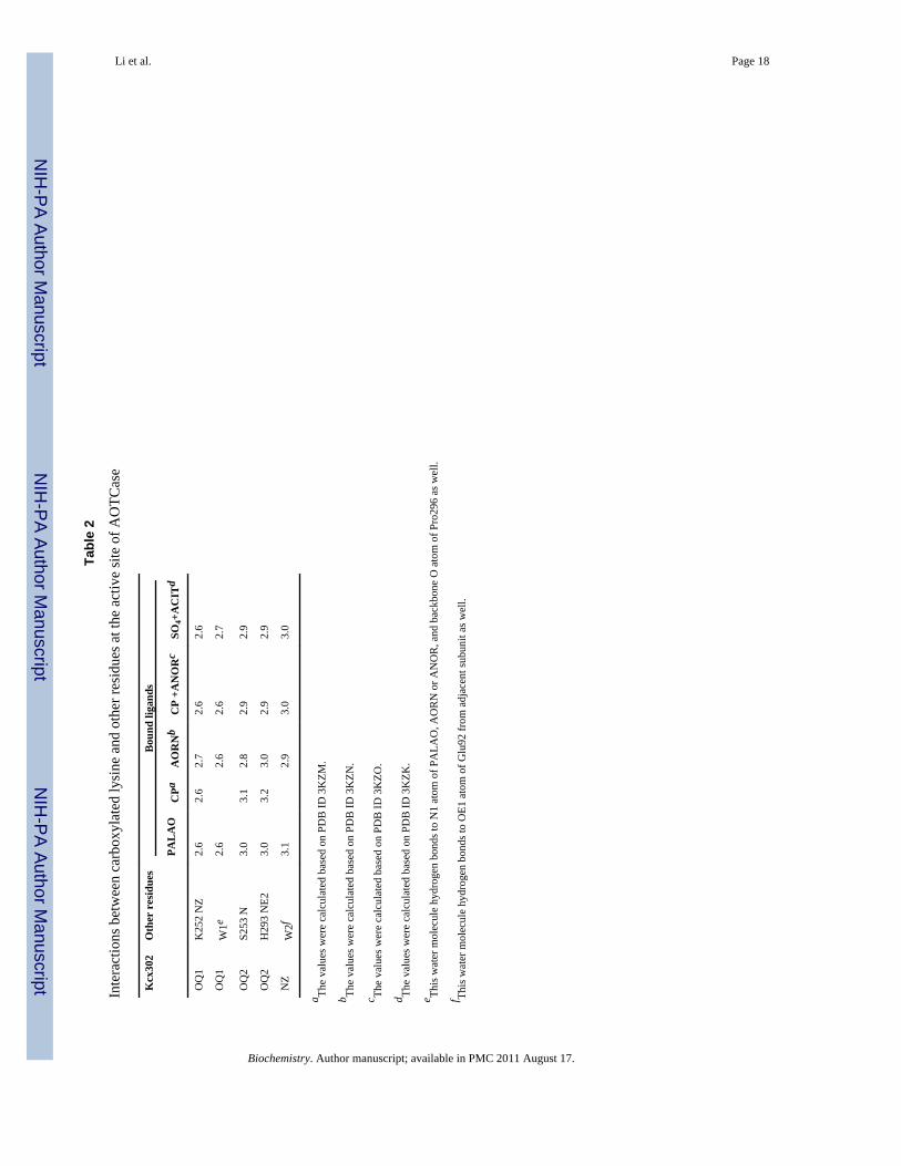
To investigate the nature of the modification of Lys302 and how it affects catalytic activity,we revisited all AOTCase structures. In the PALAO-bound AOTCase structure, the electrondensity map clearly indicates that Lys302 is post-translationally modified (Figure 1A). Thetype of modification can include methylation, acetylation, carbamylation, and carboxylation.The shape of the electron density can been used to distinguish methyl groups from largerfunctional groups, but it is difficult to distinguish between acetyl, carbamyl, and carboxylgroups, all of which have three non-hydrogen atoms in a plane. Given the hydrogen bondingnetwork with surrounding residues (Lys252, Ser253 and His293, Table 2), a carboxylatedmodification is the most likely choice for the modification of Lys302 in AOTCase. Toexclude that the modification’s identity represents chemically stable moieties (methyl,acetyl, carbamyl), we analyzed trypsin digested fragments of purified AOTCase by TOF-TOF mass spectroscopy. As expected, only a peptide fragment with an unmodified Lys302was observed, consistent with the lability of the carboxylic group in acidic solutions. At lowpH, the carboxyl group is spontaneously released as carbon dioxide (14, 32), in contrast toother modified groups that are stably bound and can be observed by mass spectrometryanalysis after proteolysis (33).
The putative carboxyl group on the modified Lys302 forms direct hydrogen bonds withmain-chain or side-chain nitrogen atoms of Lys252, Ser253 and His293 (Figure 1A andTable 2). Among these, Lys252 is involved in direct hydrogen bonding to the carboxylgroup of the AORN moiety of PALAO, and His293 forms a strong hydrogen bond with themain-chain nitrogen atom of Leu295 in the conserved His293-Cys294-Leu295-Pro206(HCLP) motif. The hydrogen bonding network between the carboxyl group of modifiedLys302, His293 and the main-chain nitrogen atom of Leu295 is reminiscent of the similarhydrogen bonding network, Glu310-His302-Leu304 and Glu299-Leu272-Leu274, found inhuman and E. coli OTCase, respectively (34, 35). These three residues are conserved in allOTCase sequences, and the interactions between them are important for maintaining theHCLP motif in a specific conformation to orientate their main-chain oxygen atoms towardsthe active site. In all known transcarbamylase structures, a leucine residue corresponding toLeu295 is in an energetically unfavorable conformation and the peptide bond between this
Li et al. Page 4
Biochemistry. Author manuscript; available in PMC 2011 August 17.
NIH
-PA Author Manuscript
NIH
-PA Author Manuscript
NIH
-PA Author Manuscript

leucine and Pro296 is in the cis conformation. In addition to the direct hydrogen bondinginteraction above, the carboxyl group on the modified Lys302 interacts with the α-aminonitrogen atom of the AORN moiety of PALAO and Glu92 from the adjacent subunit viawater molecules.
When we revisited all previously determined AOTCase structures (see supplementaryFigure S1) we found: (1) Lys302 was carboxylated in the absence of substrate binding, butsubstrate binding immobilizes the side-chain of Lys302 further by hydrogen bondinginteraction via water molecules. (2) Water-mediated hydrogen bonding promotesinteractions of carboxylated Lys302 with Glu92 from the adjacent subunit and the α-aminonitrogen atom of AORN. (3) Similarly to AOTCase, in the structure of SOTCase E92Z (Z =Ala, Ser, Pro, Val), mutant with N-succinyl-L-norvaline bound (22) the carboxylated Lys302hydrogen bonds to the α-amino nitrogen atom and the succinyl carboxyl group of N-succinyl-L-norvaline via water molecules (Figure S1).
To obtain direct, independent evidence for the carboxylation of Lys302, 13C NMRexperiments were carried out with both wild-type protein and the K302A mutant. Asobserved for other proteins with carboxylated lysine (36,37), the strong 13C NMR signal at164 ppm characteristic of a carboxyl group was clearly detectable in AOTCase wild-typeprotein labeled by 13C-bicarbonate, in contrast to the K302A mutant where the signal wasweak (Figure 2). Since there are 17 other lysine residues in the protein, the weak signal seenfor the K302A mutant might be caused by the adventitious carboxylation of another lysinewith reduced pKa, as has been observed for the K392A mutant of the sensor domain of theBlaR protein (38).
Functional and structural studies of Lys302 mutantsTo investigate the effect of lysine carboxylation on enzyme activity, Lys302 was mutated toalanine, glutamate or arginine. Each of these variants was expressed in E. coli and gavesimilar yields. Enzymatic assays demonstrated a significant decrease in enzymatic activity inall three mutants, reflecting the functional importance of Lys302 (Table 3). The level ofenzymatic activity for the wild-type (WT) and three mutants was WT > K302A > K302E ≫K302R. To determine the structural basis of these results, the WT and mutant enzymesbound with PALAO were crystallized and their structures were determined at 1.8–2.2 Åresolution. Only the K302R mutation had and appreciable effect on the structure of theprotein. Since K302 is located near the AORN binding site, the mutations would weakenAORN binding to the active site.
In the structure of the K302A mutant, three additional water molecules (labeled as w3, w4and w5 in Figure 1B) replace the carboxylated lysine. The two water molecules (labeled w1and w2 in Figure 1A–1D) that mediate the hydrogen bonding interaction of carboxylatedLys302 with PALAO and Glu92 from the adjacent subunit are also found in the K302Amutant structure. Furthermore, these water molecules maintain a similar hydrogen-bondingnetwork to the wild-type enzyme. These results might explain why the K302A mutantretains significant catalytic activity (Table 3). To investigate whether adding short-chaincarboxylic acids to the K302A mutant increases its activity as other enzymes (14, 15, 39,40), the activity of the K302A mutant was measured in the presence of high formate andacetate concentration (0.5 M). Surprisingly, the activity of the K302A mutant was notsignificantly improved. The crystal structure of the K302A mutant soaking with thecrystallization buffer in the presence of 0.5 M acetate was also determined (not shown) andit was observed that the same five water molecules were present in the cavity that replacedthe side chain of the carboxylated lysine. This, the acetate’s inability to replace the watermolecules in the crystal structure, is consistent with the unchanged activity assay results.
Li et al. Page 5
Biochemistry. Author manuscript; available in PMC 2011 August 17.
NIH
-PA Author Manuscript
NIH
-PA Author Manuscript
NIH
-PA Author Manuscript

The side-chain of Glu302 in the K302E mutant structure is well defined and anchored byhydrogen bonding interaction with the main-chain nitrogen atom of Arg298 and weaklyhydrogen bonded to the main-chain nitrogen atom of Ser253 (Figure 1C). Two of threeadditional water molecules (w4 and w5) observed in the K302A mutant structure occupiedthe same position as the carboxyl oxygen atoms of Glu302 and form a similar hydrogen-bonding network. Relative to the PALAO-bound wild-type structure, there is only one morewater molecule (w3) at the position of the carboxyl group of the carboxylated Lys302. Thiswater molecule mediates a hydrogen bonding interaction between Glu302 and Lys252. Twocommon water molecules (w1 and w2) that interact with PALAO and Glu92 from theadjacent subunit, respectively, were also identified in the K302E structure.
Our observation that the K302E mutant had lower enzymatic activity than that of the K302Amutant (Table 3) was surprising since the carboxyl group of the glutamate could conceivablyfunction similarly to a carboxylated lysine. The explanation may be that, in the K302Amutant, the hydrogen bonding network is well maintained by water molecules in the cavitythat replaces the carboxylated lysine. In particular, w3 is optimally located for stronghydrogen bonding to w1 (2.7 Å), which in turn binds AORN. The distances between w1 andthe carboxyl oxygen of carboxylated Lys302 in all wild-type crystal structures are within2.4–2.7 Å, but the distance between w1 and w2 in the K302E structure is significantlygreater (3.2 Å). The weaker hydrogen bonding interaction may be a reason for lowerenzymatic activity of the K302E mutant.
In contrast to the K302A and K302E structures, the K302R structure shows a much largerreduction in enzyme activity relative to the wild-type enzyme. The electron density for theside-chain of Arg302 is weak and the temperature factor of its side-chain is 54.4 Å2,significantly higher than those of carboxylated Lys302 (44.7 Å2) and Glu302 (33.4 Å2),implying greater flexibility. Furthermore, the side-chain of Arg302 is oriented differentlyfrom the carboxyl group of carboxylated Lys302 and pushes the nearby residues His180,Pro181 and Lys182 outwards about 1.0 Å (Figure 1D). However, the water moleculesinvolved in hydrogen bonding to the α-amino nitrogen atom of PALAO (w1) and the side-chains of Lys252 (w3) and Glu92 (w2) from the adjacent subunit are conserved. Consistentwith the K302E structure, the distance between w1 and w2 is even greater (3.4 Å) than inthe WT structure and the hydrogen bonding interaction between w2 and w3 is no longerobserved. Thus, the almost undetectable enzymatic activity of the K302R mutant probablyresults from the changes at its active site, including the weakened hydrogen bondingnetwork involved in substrate binding.
DISCUSSIONSeveral lines of evidence clearly indicate that Lys302 in AOTCase is carboxylated. First, theextra electron density indicates that the side-chain of Lys302 is modified. Second, thehydrogen bonding environment of Lys302 for hydrogen bonding interactions is compatiblewith a carboxyl group, but not for a positively charged lysine side-chain. Third, themodification is labile at low pH, since mass spectroscopy of samples prepared at low pHindicated that Lys302 was no longer modified. Fourth, the clear presence of theindicative 13C NMR signal at 164 ppm for wild-type protein and its absence in the K302Amutant confirms carboxylation of Lys302.
It is well known that lysine carboxylation is non-enzymatic and reversible, while other post-translational modifications such as methylation, acetylation, and carbamylation areirreversible and detectable by mass spectroscopy. Furthermore, lysine methylation andacetylation usually require an enzyme-catalyzed reaction in vivo (41). Therefore, it isunlikely that such lysine modifications will be observed in recombinant proteins
Li et al. Page 6
Biochemistry. Author manuscript; available in PMC 2011 August 17.
NIH
-PA Author Manuscript
NIH
-PA Author Manuscript
NIH
-PA Author Manuscript
overexpressed in a foreign host (e.g. E. coli). Lysine methylation can be achieved by usingspecial chemicals in vitro, but these chemicals are not present in vivo. Lysine carbamylationand carboxylation use completely different mechanisms to form functionally differentgroups (Figure 3). Carbamylation can be achieved by cyanate produced frommyeloperoxidase-catalyzed oxidation of thiocyanate, an anion abundant in plasma andincreased in smokers, or from urea in the plasma. Lysine carboxylation, on the other hand,occurs readily in aqueous solution in the presence of carbon dioxide at a basic pH (32,42).Even though carbamylation and carboxylation use very different mechanisms, the two areconfused in the literatures. Lysine carbamylation (or carbamoylation) is referred to inseveral publications (15,32,42–44), when the actual reaction is in fact carboxylation.
The activity of the K302A mutant is almost half of that of the wild-type enzyme raising thequestion of why AOTCase retains a lysine in this position. Perhaps this lysine wasmaintained through evolution to distinguish AOTCase from SOTCase which uses N-succinyl-L-ornithine (SORN) rather than AORN (22), and OTCase which uses L-ornithine.An alternative explanation may be found in the very low activity of the K302R mutant. Theside-chain of arginine has a positive charge while carboxylated lysine has a negative charge.The side chain of unmodified lysine is usually located in a similar position as that ofarginine, as observed in the structure of UV damage endonuclease (14). It would beexpected that the activity of AOTCase with an uncarboxylated lysine would be as low as theK302R mutant’s. It could further be surmised that, the respective organisms need to usecarboxylation as a switch to turn “on” or “off” the arginine biosynthetic pathway. It has beenwell known that rubulose-1,5-bisphosphate carboxylase/oxygenase (Rubisco) in plant cellsuses the carboxylation on Lys201 as a switch to turn the enzyme “on” during the day and“off” at night by removing the carboxyl group (45,46). Carbon dioxide and bicarbonate havebeen found to play an important biological role in modulating several biological processesincluding photosynthetic carbon fixation (47), pH homeostasis (48), carbon metabolism(49), activation of virulence in pathogenic organisms (50), sperm maturation (51),stimulation of mammalian G-protein-responsive adenylyl cyclase (52), and as an alarmonein Drosophila (53,54). Whether or not carboxylation of a key lysine in their related proteinsis used as an underlying regulatory mechanism should be investigated further.
There are 197 structures with carboxylated lysine residue (modified residue indicated asKcx) in the Protein Data Bank (PDB). If structures with 90% identity are counted only once,there are still 52 unique structures remaining in this pool (Table 4). These proteins includehydantoinase (40,55), folylpolyglutamate synthase (43), UV damage endonuclease (14),OXA10, OXA-1 class D β-lactamase (38,56,57), urease (42), phosphotriesterase (58),dihydroorotase (59), dihydropyrimidinase (60), organophosphate hydrolase (61) and MurEand MurD ligases (44,62). In most of these proteins, the carboxylated lysine bridges twometal ions, similar to the role of glutamate or aspartate in proteins with two metal-bindingsites (26 structures among 52). However, the urease apoenzyme can be activated in vitroonly in the presence of carbon dioxide prior to nickel binding (63), suggesting that thecarboxylated lysine may have other structural roles beyond binding metals. In some proteinssuch as β-lactamase, UV damage endonuclease, Rubisco, MurD and MurE ligase and BlaRsignal transducer protein, a carboxylated lysine plays an essential catalytic role. Moreinterestingly, in three structures (PDB ID: 1HL9, 1PU6 and 2UYN for fucosiadase, 3-methyladenine DNA glycosylase and TdcF protein of unknown function, respectively), thecarboxylated lysines are located near the surface of proteins, presumably playing primarily astructure stabilizing role (64–66). Since the carboxyl group is labile at acidic pH, but easilyformed in the presence of carbon dioxide at basic pH, the number of proteins with lysinecarboxylation must be underestimated. Furthermore, the carboxylated lysine must be fixedin place by metal ions (either one or two) or hydrogen bonding with other protein residues(at least one). Therefore, any detection method involving denaturing the proteins will result
Li et al. Page 7
Biochemistry. Author manuscript; available in PMC 2011 August 17.
NIH
-PA Author Manuscript
NIH
-PA Author Manuscript
NIH
-PA Author Manuscript

in release of the carboxyl group. With current technology, 13C NMR (38) andcrystallography are the only methods that can detect this modification. However, thesemethods are not amenable to high-throughput investigations. The majority (49 out of 52structures in the PDB) of known lysine carboxylation modifications were found to belocated at or near the active site, probably because these sites receive the most attention.Revisiting the structures in PDB with more attention to surface lysines might reveal morestructures with carboxylated lysines.
In conclusion, we have shown that Lys302 in AOTCase is post-translationally modified bycarboxylation and that this modification may be functionally important for enzymaticactivity. Lysine carboxylation is likely to be a more common event than currentlyappreciated and may play a critical role in enzymatic activity and protein stability.
Supplementary MaterialRefer to Web version on PubMed Central for supplementary material.
Abbreviations
ACIT N-acteyl-L-citrulline
ANOR N-acetyl-L-norvaline
AORN N-acetyl-L-Ornithine
AOTCase N-acetyl-L-ornithine transcarbamylase
ATCase aspartate transcarbamlyase
OTCase ornithine transcarbamylase
CP carbamyl phosphate
ORN L-ornithine
PALAO Nδ-(phosphonacetyl)-Nα-acetyl-L-ornithine
SORN N-succinyl-L-ornithine
WT wild-type
xc Xanthomonas campestris
AcknowledgmentsWe thank Dr. David Davies for facilitating our use of the diffraction equipment in the Molecular Structure Sectionof the National Institute of Health and Dr. Fred Dyda for help in data collection and processing, and Dr. Yui-FaiLam in the University of Maryland for help in setting up NMR measurements.
REFERENCES1. Close P, Creppe C, Gillard M, Ladang A, Chapelle JP, Nguyen L, Chariot A. The emerging role of
lysine acetylation of non-nuclear proteins. Cell Mol Life Sci. 2010; 67:1255–1264. [PubMed:20082207]
2. Geiman TM, Robertson KD. Chromatin remodeling, histone modifications, and DNA methylation-how does it all fit together? J Cell Biochem. 2002; 87:117–125. [PubMed: 12244565]
3. An W. Histone acetylation and methylation: combinatorial players for transcriptional regulation.Subcell Biochem. 2007; 41:351–369. [PubMed: 17484136]
Li et al. Page 8
Biochemistry. Author manuscript; available in PMC 2011 August 17.
NIH
-PA Author Manuscript
NIH
-PA Author Manuscript
NIH
-PA Author Manuscript

4. Yang F, Bian C, Zhu L, Zhao G, Huang Z, Huang M. Effect of human serum albumin on drugmetabolism: structural evidence of esterase activity of human serum albumin. J Struct Biol. 2007;157:348–355. [PubMed: 17067818]
5. Xu AS, Labotka RJ, London RE. Acetylation of human hemoglobin by methyl acetylphosphate.Evidence of broad regio-selectivity revealed by NMR studies. J Biol Chem. 1999; 274:26629–26632. [PubMed: 10480863]
6. Ueno H, Pospischil MA, Manning JM. Methyl acetyl phosphate as a covalent probe for anion-binding sites in human and bovine hemoglobins. J Biol Chem. 1989; 264:12344–12351. [PubMed:2745446]
7. Stavropoulos P, Nagy V, Blobel G, Hoelz A. Molecular basis for the autoregulation of the proteinacetyl transferase Rtt109. Proc Natl Acad Sci U S A. 2008; 105:12236–12241. [PubMed:18719104]
8. Bewley MC, Graziano V, Jiang J, Matz E, Studier FW, Pegg AE, Coleman CS, Flanagan JM.Structures of wild-type and mutant human spermidine/spermine N1-acetyltransferase, a potentialtherapeutic drug target. Proc Natl Acad Sci U S A. 2006; 103:2063–2068. [PubMed: 16455797]
9. Walter TS, Meier C, Assenberg R, Au KF, Ren J, Verma A, Nettleship JE, Owens RJ, Stuart DI,Grimes JM. Lysine methylation as a routine rescue strategy for protein crystallization. Structure.2006; 14:1617–1622. [PubMed: 17098187]
10. Stark GRSW, Moore S. Reactions of the cyanante present in aqueous urea with amino acids andproteins. J Biol Chem. 1960; 235:3177–3181.
11. Bobb D, Hofstee BH. Gel isoelectric focusing for following the successive carbamylations ofamino groups in chymotrypsinogen A. Anal Biochem. 1971; 40:209–217. [PubMed: 5550146]
12. Kraus LM, Kraus AP Jr. Carbamoylation of amino acids and proteins in uremia. Kidney Int Suppl.2001; 78:S102–S107. [PubMed: 11168993]
13. Al-Dirbashi OY, Al-Hassnan ZN, Rashed MS. Determination of homocitrulline in urine of patientswith HHH syndrome by liquid chromatography tandem mass spectrometry. Anal Bioanal Chem.2006; 386:2013–2017. [PubMed: 17053917]
14. Meulenbroek EM, Paspaleva K, Thomassen EA, Abrahams JP, Goosen N, Pannu NS. Involvementof a carboxylated lysine in UV damage endonuclease. Protein Sci. 2009; 18:549–558. [PubMed:19241382]
15. Dementin S, Bouhss A, Auger G, Parquet C, Mengin-Lecreulx D, Dideberg O, van Heijenoort J,Blanot D. Evidence of a functional requirement for a carbamoylated lysine residue in MurD, MurEand MurF synthetases as established by chemical rescue experiments. Eur J Biochem. 2001;268:5800–5807. [PubMed: 11722566]
16. Cha J, Mobashery S. Lysine N(zeta)-decarboxylation in the BlaR1 protein from Staphylococcusaureus at the root of its function as an antibiotic sensor. J Am Chem Soc. 2007; 129:3834–3835.[PubMed: 17343387]
17. Shi D, Yu X, Roth L, Morizono H, Tuchman M, Allewell NM. Structures of N-acetylornithinetranscarbamoylase from Xanthomonas campestris complexed with substrates and substrate analogsimply mechanisms for substrate binding and catalysis. Proteins. 2006; 64:532–542. [PubMed:16741992]
18. Shi D, Morizono H, Yu X, Roth L, Caldovic L, Allewell NM, Malamy MH, Tuchman M. Crystalstructure of N-acetylornithine transcarbamylase from Xanthomonas campestris: a novel enzyme ina new arginine biosynthetic pathway found in several eubacteria. J Biol Chem. 2005; 280:14366–14369. [PubMed: 15731101]
19. Morizono H, Cabrera-Luque J, Shi D, Gallegos R, Yamaguchi S, Yu X, Allewell NM, MalamyMH, Tuchman M. Acetylornithine transcarbamylase: a novel enzyme in arginine biosynthesis. JBacteriol. 2006; 188:2974–2982. [PubMed: 16585758]
20. da Silva FR, Vettore AL, Kemper EL, Leite A, Arruda P. Fastidian gum: the Xylella fastidiosaexopolysaccharide possibly involved in bacterial pathogenicity. FEMS Microbiol Lett. 2001;203:165–171. [PubMed: 11583843]
21. da Silva AC, Ferro JA, Reinach FC, Farah CS, Furlan LR, Quaggio RB, Monteiro-Vitorello CB,Van Sluys MA, Almeida NF, Alves LM, do Amaral AM, Bertolini MC, Camargo LE, CamarotteG, Cannavan F, Cardozo J, Chambergo F, Ciapina LP, Cicarelli RM, Coutinho LL, Cursino-Santos
Li et al. Page 9
Biochemistry. Author manuscript; available in PMC 2011 August 17.
NIH
-PA Author Manuscript
NIH
-PA Author Manuscript
NIH
-PA Author Manuscript

JR, El-Dorry H, Faria JB, Ferreira AJ, Ferreira RC, Ferro MI, Formighieri EF, Franco MC,Greggio CC, Gruber A, Katsuyama AM, Kishi LT, Leite RP, Lemos EG, Lemos MV, Locali EC,Machado MA, Madeira AM, Martinez-Rossi NM, Martins EC, Meidanis J, Menck CF, MiyakiCY, Moon DH, Moreira LM, Novo MT, Okura VK, Oliveira MC, Oliveira VR, Pereira HA, RossiA, Sena JA, Silva C, de Souza RF, Spinola LA, Takita MA, Tamura RE, Teixeira EC, Tezza RI,Trindade dos Santos M, Truffi D, Tsai SM, White FF, Setubal JC, Kitajima JP. Comparison of thegenomes of two Xanthomonas pathogens with differing host specificities. Nature. 2002; 417:459–463. [PubMed: 12024217]
22. Shi D, Yu X, Cabrera-Luque J, Chen TY, Roth L, Morizono H, Allewell NM, Tuchman M. Asingle mutation in the active site swaps the substrate specificity of N-acetyl-L-ornithinetranscarbamylase and N-succinyl-L-ornithine transcarbamylase. Protein Sci. 2007; 16:1689–1699.[PubMed: 17600144]
23. Shi D, Morizono H, Cabrera-Luque J, Yu X, Roth L, Malamy MH, Allewell NM, Tuchman M.Structure and catalytic mechanism of a novel N-succinyl-L-ornithine transcarbamylase in argininebiosynthesis of Bacteroides fragilis. J Biol Chem. 2006; 281:20623–20631. [PubMed: 16704984]
24. Pastra-Landis SC, Foote J, Kantrowitz ER. An improved colorimetric assay for aspartate andornithine transcarbamylases. Anal Biochem. 1981; 118:358–363. [PubMed: 7337232]
25. Shi D, Yu X, Roth L, Morizono H, Hathout Y, Allewell NM, Tuchman M. Expression,purification, crystallization and preliminary X-ray crystallographic studies of a novelacetylcitrulline deacetylase from Xanthomonas campestris. Acta Crystallogr Sect F Struct BiolCryst Commun. 2005; 61:676–679.
26. Otwinowski Z, Minor W. Processing of X-ray Diffraction Data Collected in Oscillation Mode.Methods in Enzymology. 1997; 276:307–326.
27. The CCP4 suite: programs for protein crystallography. Acta Crystallogr D Biol Crystallogr. 1994;50:760–763. [PubMed: 15299374]
28. Jones TA, Zou JY, Cowan SW, Kjeldgaard M. Improved methods for building protein models inelectron density maps and the location of errors in these models. Acta Crystallogr A. 1991; 47(Pt2):110–119. [PubMed: 2025413]
29. Brunger AT, Adams PD, Clore GM, DeLano WL, Gros P, Grosse-Kunstleve RW, Jiang JS,Kuszewski J, Nilges M, Pannu NS, Read RJ, Rice LM, Simonson T, Warren GL. Crystallography& NMR system: A new software suite for macromolecular structure determination. ActaCrystallogr D Biol Crystallogr. 1998; 54:905–921. [PubMed: 9757107]
30. Brunger AT. Free R value: a novel statistical quantity for assessing the accuracy of crystalstructures. Nature. 1992; 355:472–475. [PubMed: 18481394]
31. Laskowski RA, MacArthur MW, Moss DS, Thornton JM. PROCHECK: a program to check thestereochemical quality of protein structures. J Appl Crystallogr. 1993; 26:283–291.
32. Golemi D, Maveyraud L, Vakulenko S, Samama JP, Mobashery S. Critical involvement of acarbamylated lysine in catalytic function of class D beta-lactamases. Proc Natl Acad Sci U S A.2001; 98:14280–14285. [PubMed: 11724923]
33. Lapko VN, Smith DL, Smith JB. In vivo carbamylation and acetylation of water-soluble humanlens alphaB-crystallin lysine 92. Protein Sci. 2001; 10:1130–1136. [PubMed: 11369851]
34. Shi D, Morizono H, Ha Y, Aoyagi M, Tuchman M, Allewell NM. 1.85-A resolution crystalstructure of human ornithine transcarbamoylase complexed with N-phosphonacetyl-L-ornithine.Catalytic mechanism and correlation with inherited deficiency. J Biol Chem. 1998; 273:34247–34254. [PubMed: 9852088]
35. Langley DB, Templeton MD, Fields BA, Mitchell RE, Collyer CA. Mechanism of inactivation ofornithine transcarbamoylase by Ndelta -(N'-Sulfodiaminophosphinyl)-L-ornithine, a true transitionstate analogue? Crystal structure and implications for catalytic mechanism. J Biol Chem. 2000;275:20012–20019. [PubMed: 10747936]
36. Cha J, Vakulenko SB, Mobashery S. Characterization of the beta-lactam antibiotic sensor domainof the MecR1 signal sensor/transducer protein from methicillin-resistant Staphylococcus aureus.Biochemistry. 2007; 46:7822–7831. [PubMed: 17550272]
Li et al. Page 10
Biochemistry. Author manuscript; available in PMC 2011 August 17.
NIH
-PA Author Manuscript
NIH
-PA Author Manuscript
NIH
-PA Author Manuscript

37. O'Leary MH, Jaworski RJ, Hartman FC. C nuclear magnetic resonance study of the CO(2)activation of ribulosebisphosphate carboxylase from Rhodospirillum rubrum. Proc Natl Acad SciU S A. 1979; 76:673–675. [PubMed: 16592618]
38. Golemi-Kotra D, Cha JY, Meroueh SO, Vakulenko SB, Mobashery S. Resistance to beta-lactamantibiotics and its mediation by the sensor domain of the transmembrane BlaR signaling pathwayin Staphylococcus aureus. J Biol Chem. 2003; 278:18419–18425. [PubMed: 12591921]
39. Schneider KD, Bethel CR, Distler AM, Hujer AM, Bonomo RA, Leonard DA. Mutation of theactive site carboxy-lysine (K70) of OXA-1 beta-lactamase results in a deacylation-deficientenzyme. Biochemistry. 2009; 48:6136–6145. [PubMed: 19485421]
40. Huang CY, Hsu CC, Chen MC, Yang YS. Effect of metal binding and posttranslational lysinecarboxylation on the activity of recombinant hydantoinase. J Biol Inorg Chem. 2009; 14:111–121.[PubMed: 18781344]
41. Zhang Q, Wang Y. High mobility group proteins and their post-translational modifications.Biochim Biophys Acta. 2008; 1784:1159–1166. [PubMed: 18513496]
42. Jabri E, Carr MB, Hausinger RP, Karplus PA. The crystal structure of urease from Klebsiellaaerogenes. Science. 1995; 268:998–1004. [PubMed: 7754395]
43. Young PG, Smith CA, Metcalf P, Baker EN. Structures of Mycobacterium tuberculosisfolylpolyglutamate synthase complexed with ADP and AMPPCP. Acta Crystallogr D BiolCrystallogr D. 2008; 64:745–753.
44. Gordon E, Flouret B, Chantalat L, van Heijenoort J, Mengin-Lecreulx D, Dideberg O. Crystalstructure of UDP-N-acetylmuramoyl-L-alanyl-D-glutamate: meso-diaminopimelate ligase fromEscherichia coli. J Biol Chem. 2001; 276:10999–11006. [PubMed: 11124264]
45. Taylor TC, Andersson I. Structure of a product complex of spinach ribulose-1,5-bisphosphatecarboxylase/oxygenase. Biochemistry. 1997; 36:4041–4046. [PubMed: 9092835]
46. Jensen R. Activation of Rubisco controls CO(2) assimilation in light: a perspective on itsdiscovery. Photosynth Res. 2004; 82:187–193. [PubMed: 16151874]
47. Falkowski PG. Photosynthesis: the paradox of carbon dioxide efflux. Curr Biol. 1997; 7:R637–R639. [PubMed: 9368746]
48. Roos A, Boron WF. Intracellular pH. Physiol Rev. 1981; 61:296–434. [PubMed: 7012859]49. Smith KS, Ferry JG. Prokaryotic carbonic anhydrases. FEMS Microbiol Rev. 2000; 24:335–366.
[PubMed: 10978542]50. Bahn YS, Muhlschlegel FA. CO2 sensing in fungi and beyond. Curr Opin Microbiol. 2006; 9:572–
578. [PubMed: 17045514]51. Esposito G, Jaiswal BS, Xie F, Krajnc-Franken MA, Robben TJ, Strik AM, Kuil C, Philipsen RL,
van Duin M, Conti M, Gossen JA. Mice deficient for soluble adenylyl cyclase are infertile becauseof a severe sperm-motility defect. Proc Natl Acad Sci U S A. 2004; 101:2993–2998. [PubMed:14976244]
52. Townsend PD, Holliday PM, Fenyk S, Hess KC, Gray MA, Hodgson DR, Cann MJ. Stimulation ofmammalian G-protein-responsive adenylyl cyclases by carbon dioxide. J Biol Chem. 2009;284:784–791. [PubMed: 19008230]
53. Kwon JY, Dahanukar A, Weiss LA, Carlson JR. The molecular basis of CO2 reception inDrosophila. Proc Natl Acad Sci U S A. 2007; 104:3574–3578. [PubMed: 17360684]
54. Jones WD, Cayirlioglu P, Kadow IG, Vosshall LB. Two chemosensory receptors together mediatecarbon dioxide detection in Drosophila. Nature. 2007; 445:86–90. [PubMed: 17167414]
55. Xu Z, Liu Y, Yang Y, Jiang W, Arnold E, Ding J. Crystal structure of D-Hydantoinase fromBurkholderia pickettii at a resolution of 2.7 Angstroms: insights into the molecular basis ofenzyme thermostability. J Bacteriol. 2003; 185:4038–4049. [PubMed: 12837777]
56. Sun T, Nukaga M, Mayama K, Braswell EH, Knox JR. Comparison of beta-lactamases of classesA and D: 1.5-A crystallographic structure of the class D OXA-1 oxacillinase. Protein Sci. 2003;12:82–91. [PubMed: 12493831]
57. Maveyraud L, Golemi D, Kotra LP, Tranier S, Vakulenko S, Mobashery S, Samama JP. Insightsinto class D beta-lactamases are revealed by the crystal structure of the OXA10 enzyme fromPseudomonas aeruginosa. Structure. 2000; 8:1289–1298. [PubMed: 11188693]
Li et al. Page 11
Biochemistry. Author manuscript; available in PMC 2011 August 17.
NIH
-PA Author Manuscript
NIH
-PA Author Manuscript
NIH
-PA Author Manuscript

58. Benning MM, Kuo JM, Raushel FM, Holden HM. Three-dimensional structure of the binuclearmetal center of phosphotriesterase. Biochemistry. 1995; 34:7973–7978. [PubMed: 7794910]
59. Thoden JB, Phillips GN Jr, Neal TM, Raushel FM, Holden HM. Molecular structure ofdihydroorotase: a paradigm for catalysis through the use of a binuclear metal center. Biochemistry.2001; 40:6989–6997. [PubMed: 11401542]
60. Abendroth J, Niefind K, Schomburg D. X-ray structure of a dihydropyrimidinase from Thermussp. at 1.3 A resolution. J Mol Biol. 2002; 320:143–156. [PubMed: 12079340]
61. Yang H, Carr PD, McLoughlin SY, Liu JW, Horne I, Qiu X, Jeffries CM, Russell RJ, OakeshottJG, Ollis DL. Evolution of an organophosphate-degrading enzyme: a comparison of natural anddirected evolution. Protein Eng. 2003; 16:135–145. [PubMed: 12676982]
62. Bertrand JA, Auger G, Martin L, Fanchon E, Blanot D, Le Beller D, van Heijenoort J, Dideberg O.Determination of the MurD mechanism through crystallographic analysis of enzyme complexes. JMol Biol. 1999; 289:579–590. [PubMed: 10356330]
63. Park IS, Hausinger RP. Requirement of carbon dioxide for in vitro assembly of the urease nickelmetallocenter. Science. 1995; 267:1156–1158. [PubMed: 7855593]
64. Sulzenbacher G, Bignon C, Nishimura T, Tarling CA, Withers SG, Henrissat B, Bourne Y. Crystalstructure of Thermotoga maritima alpha-L-fucosidase. Insights into the catalytic mechanism andthe molecular basis for fucosidosis. J Biol Chem. 2004; 279:13119–13128. [PubMed: 14715651]
65. Eichman BF, O'Rourke EJ, Radicella JP, Ellenberger T. Crystal structures of 3-methyladenineDNA glycosylase MagIII and the recognition of alkylated bases. Embo J. 2003; 22:4898–4909.[PubMed: 14517230]
66. Burman JD, Stevenson CE, Sawers RG, Lawson DM. The crystal structure of Escherichia coliTdcF, a member of the highly conserved YjgF/YER057c/UK114 family. BMC Struct Biol. 2007;7:30. [PubMed: 17506874]
Li et al. Page 12
Biochemistry. Author manuscript; available in PMC 2011 August 17.
NIH
-PA Author Manuscript
NIH
-PA Author Manuscript
NIH
-PA Author Manuscript

Li et al. Page 13
Biochemistry. Author manuscript; available in PMC 2011 August 17.
NIH
-PA Author Manuscript
NIH
-PA Author Manuscript
NIH
-PA Author Manuscript

Figure 1.Stereo view of the structure and hydrogen bonding network surrounding residue 302. A,PALAO bound wild-type AOTCase, B, PALAO bound K302A AOTCase C, PALAO boundK302E AOTCase, D, PALAO bound K302R AOTCase. Contours of the electron densitymaps (2Fo-Fc) around PALAO, residue 302 and water molecules are shown as a brown cageat 1.0σ. The final refined positions of the ligands and surrounding protein residues arerepresented as colored sticks. The predicted hydrogen bonding interactions are in pinkdashed lines. The water molecules are represented as pink balls. The carbon of PALAO,residue 302 and other protein residues are shown in pink, light blue and green sticks,respectively.
Li et al. Page 14
Biochemistry. Author manuscript; available in PMC 2011 August 17.
NIH
-PA Author Manuscript
NIH
-PA Author Manuscript
NIH
-PA Author Manuscript

Figure 2.13C NMR spectra of wild-type (upper panel) and K302A mutant (lower panel) AOTCase (1mM). Experiments were performed in 100 mM Tris HCl, 50 mM NaCl, 7% D2O, pH 8.0,supplemented with 20 mM NaH13CO3. The position of the resonance attributed tocarboxylated lysine in the enzyme is around 164 ppm.
Li et al. Page 15
Biochemistry. Author manuscript; available in PMC 2011 August 17.
NIH
-PA Author Manuscript
NIH
-PA Author Manuscript
NIH
-PA Author Manuscript

Figure 3.Chemical structure of carbamylated vs. carboxylated lysine.
Li et al. Page 16
Biochemistry. Author manuscript; available in PMC 2011 August 17.
NIH
-PA Author Manuscript
NIH
-PA Author Manuscript
NIH
-PA Author Manuscript

NIH
-PA Author Manuscript
NIH
-PA Author Manuscript
NIH
-PA Author Manuscript
Li et al. Page 17
Table 1
Data collection and refinement statistics
Dataset PALAO K302A K302E K302R
Space group I213 I213 I213 I213
Resolution (Å) 2.2 1.9 1.85 2.2
Unit-cell parameters (Å) a = b = c =128.88 a = b = c =128.92 a = b = c =129.29 a = b = c =127.39
Measurements 219,475 305,757 390,128 246,817
Unique reflections 18,269 (1,832) a 28,236 (1,365) 30,622 (1,456) 17,635 (879)
Redundancy 12.0 (11.8) 10.8(5.4) 12.8 (5.4) 14.0 (13.1)
Completeness (%) 99.8 (100.0) 100.0 (100.0) 99.7 (95.1) 100.0 (100.0)
<I/σ (I)> 15.0 (4.9) 16.4 (2.3) 19.8 (2.8) 8.7 (3.7)
Rmerg b 7.4 (48.4) 6.5(64.9) 5.2 (55.3) 9.8 (79.1)
Wilson B (Å2) 30.4 27.6 28.6 21.9
Refinement
Resolution range (Å) 50.0-2.2 50-1.9 50-1.85 50-2.2
No. of protein atoms 2620 2613 2617 2619
No. of water atoms 90 219 193 146
No. of hetero atoms 24 24 24 24
Rmsd of bond lengths (Å) 0.006 0.005 0.005 0.005
Rmsd of bond angle (°) 1.1 1.2 1.2 1.2
Rwork (%)c 20.0 19.8 20.0 18.9
Rfree (%)d 24.3 23.2 23.2 22.2
Average B factor (Å2) 41.7 32.2 32.3 35.3
aFigures in brackets apply to the highest-resolution shell.
bRmerg = ΣhΣi|I(h,i)-<I(h)>|/∑hΣiI(h,i), where I(h,i) is the intensity of the ith observation of reflection h, and < I(h)> is the average intensity of
redundant measurements of reflection h.
cRwork= Σh‖Fobs| – |Fcalc‖/Σh|Fobs|.
dRfree = Σh‖Fobs| – |Fcalc‖/Σh|Fobs| for 5% of the reserved reflections.
Biochemistry. Author manuscript; available in PMC 2011 August 17.
NIH
-PA Author Manuscript
NIH
-PA Author Manuscript
NIH
-PA Author Manuscript
Li et al. Page 18
Tabl
e 2
Inte
ract
ions
bet
wee
n ca
rbox
ylat
ed ly
sine
and
oth
er re
sidu
es a
t the
act
ive
site
of A
OTC
ase
Kcx
302
Oth
er r
esid
ues
Bou
nd li
gand
s
PAL
AO
CPa
AO
RN
bC
P +A
NO
Rc
SO4+
AC
ITd
OQ
1K
252
NZ
2.6
2.6
2.7
2.6
2.6
OQ
1W
1e2.
62.
62.
62.
7
OQ
2S2
53 N
3.0
3.1
2.8
2.9
2.9
OQ
2H
293
NE2
3.0
3.2
3.0
2.9
2.9
NZ
W2f
3.1
2.9
3.0
3.0
a The
valu
es w
ere
calc
ulat
ed b
ased
on
PDB
ID 3
KZM
.
b The
valu
es w
ere
calc
ulat
ed b
ased
on
PDB
ID 3
KZN
.
c The
valu
es w
ere
calc
ulat
ed b
ased
on
PDB
ID 3
KZO
.
d The
valu
es w
ere
calc
ulat
ed b
ased
on
PDB
ID 3
KZK
.
e This
wat
er m
olec
ule
hydr
ogen
bon
ds to
N1
atom
of P
ALA
O, A
OR
N o
r AN
OR
, and
bac
kbon
e O
ato
m o
f Pro
296
as w
ell.
f This
wat
er m
olec
ule
hydr
ogen
bon
ds to
OE1
ato
m o
f Glu
92 fr
om a
djac
ent s
ubun
it as
wel
l.
Biochemistry. Author manuscript; available in PMC 2011 August 17.

NIH
-PA Author Manuscript
NIH
-PA Author Manuscript
NIH
-PA Author Manuscript
Li et al. Page 19
Table 3
Specific activity of wild-type and mutant AOTCase in the presence of acids (0.5M).
Compounds added Specific activity(µmol/min/mg)
Wild-type K302A K302E K302R
None 43.4 ± 0.4a 23.0 ± 0.5 7.1 ± 0.1 0.059±0.01
Formate 44.1 ± 1.2 26.4 ± 0.6 6.7 ± 0.2 0.093±0.01
Acetate 48.5 ± 1.1 21.2 ± 0.8 6.6 ± 0.5 0.104±0.03
aThe Mean ± S.D. are shown (n = 3).
Biochemistry. Author manuscript; available in PMC 2011 August 17.

NIH
-PA Author Manuscript
NIH
-PA Author Manuscript
NIH
-PA Author Manuscript
Li et al. Page 20
Table 4
Protein structures with lysine carboxylation modification
PDB ID Enzyme name Residue Organism source Funciton
2OGJ Dihydroorotase 175 A.tumefaciens Bridging two Zn(II)
2Z26 Dihydroorotase 102 E.coli Bridging two Zn(II)
3JZE Dihydroorotase 103 S.enterica Bridging two Zn(II)
2GWN Dihydroorotase 149 P. gingivalis Bridging two Zn(II)
3F4C Organophosphorus hydrolase 243 G. stearothermophilus Bridging two Co(II)
3ICJ Metal-dependent hydrolase 294 P. furiosus Bridging two Zn(II)
3GTX Organophosphorus hydrolase 243 D. radiodurans Bridging two Co(II)
2QPX Metal-dependent hydrolase 166 L. casei Bridging two Zn(II)
2FTW Dihydropyrimidinase 158 D. discoideum Bridging two Zn(II)
2FVK Dihydropyrimidinase 167 S. kluyveri Bridging two Zn(II)
3DC8 Dihydropyrimidinase 147 S. meliloti Bridging two Zn(II)
3GNH L-Lys/Arg carboxypeptidase 211 C. crescentus cb15 Bridging two Zn(II)
3DUG Arginine carboxypeptidase 182 Unidentified Bridging two Zn(II)
2VC7 Phosphotriesterase 137 S. solfataricus Bridging two Co(II)
2R1N Metallophosphotriesterases 169 A. tumefaciens Bridging two Co(II)
2OB3 Phosphotriesterase 169 B. diminuta Bridging two Zn(II)
3E74 Allantoinase 146 E. coli Bridging two Fe(III)
1EJX Urease 217 K. aerogenes Bridging two Ni(II)
1E9Z Urease 219 H. pylori Bridging two Ni(II)
4UBP Urease 220 B. pasteurii Bridging two Ni(II)
1ONW Isoaspartyl dipeptidase 162 E. coli Bridging two Zn(II)
1K1D D-hydanroinase 150 G. stearothermophilus Bridging two Zn(II)
1GKR L-hydanroinase 147 A. aurescens Bridging two Zn(II)
1GKP D-hydanroinase 150 Thermus sp. Bridging two Zn(II)
1NFG D-hydantoinase 148 R. pickettii Bridging two Zn(II)
2ICS Adenine deaminase 154 E. faecalis Bridging two Zn(II)
1RQB Transcarboxylase 184 P. freudenreichii Binding one Co(II)
2QF7 Pyruvate carboxylase 718 R. etli Binding one Zn(II)
3BG3 Pyruvate carboxylase 741 H. sapiens Binding one Mn(II)
2OEM Rubisco-like protein 173 G. kaustophilus Binding one Mg(II)
1WDD Rubisco 201 O. sativa Binding one Mg(II)
1GK8 Rubisco 201 C. reinhardtii Binding one Mg(II)
1BWV Rubisco 201 G. partita Binding one Mg(II)
2WTZ ATP-dependent MurE ligase 262 M. tuberculosis Binding one Mg(II)
2JFG MurD ligase 198 E. coli Catalytic role?
1E8C MurE ligase 224 E. coli Catalytic role?
1JBW Folypolyglutamate synthetase 185 L. casei Catalytic role?
1W78 FolC bifunctional protein 188 E. coli Binding one Mg(II)
3HBR OXA-48 β-lactamase 73 K. pneumoniae Catalytic role
Biochemistry. Author manuscript; available in PMC 2011 August 17.

NIH
-PA Author Manuscript
NIH
-PA Author Manuscript
NIH
-PA Author Manuscript
Li et al. Page 21
PDB ID Enzyme name Residue Organism source Funciton
3ISG Class D β-lactamase 70 E. coli Catalytic role
2P9V AmpC beta-lactamase 315 E. coli Catalytic role
1K55 OXA-10 β-lactamase 70 P. aeruginosa Catalytic role
1K38 β-lactamase OXA-2 70 S. typhimurium Catalytic role
1XQL Alanine racemase 129 G. stearothermophilus Binding substrate?
1VFS Alanine racemase 129 S. lavendulae Binding substrate?
1RCQ Alanine racemase 122 P. aeruginosa Binding substrate?
2J6V UV damage endonuclease 229 T. thermophilus Catalytic role
1H01 Cell division protein kinase 2 33 H. sapiens Catalytic role?
2UYN Protein TdcF A58 E. coli Structural role?
1HL9 Fucosidase 338 T. maritime Structural role?
1PU6 3-methyladenine DNA glycosylase 205 H. pylori Structural role?
Biochemistry. Author manuscript; available in PMC 2011 August 17.
Copyright © 2022 FDOKUMEN