Transfer of the human NaI symporter gene enhances iodide uptake in hepatoma cells
Upload
independentCategory
view
0download
0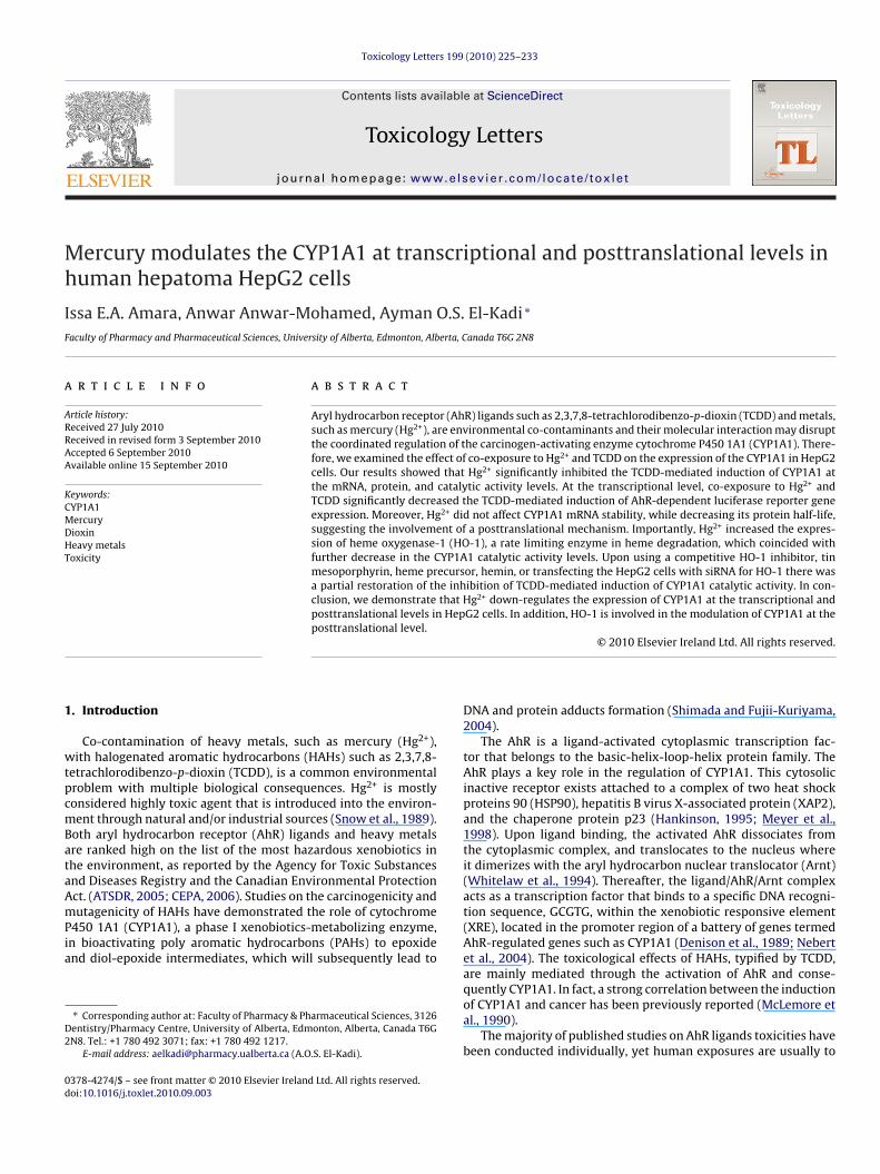
Mh
IF
a
ARRAA
KCMDHT
1
wtpcmBataAmPia
D2
0d
Toxicology Letters 199 (2010) 225–233
Contents lists available at ScienceDirect
Toxicology Letters
journa l homepage: www.e lsev ier .com/ locate / tox le t
ercury modulates the CYP1A1 at transcriptional and posttranslational levels inuman hepatoma HepG2 cells
ssa E.A. Amara, Anwar Anwar-Mohamed, Ayman O.S. El-Kadi ∗
aculty of Pharmacy and Pharmaceutical Sciences, University of Alberta, Edmonton, Alberta, Canada T6G 2N8
r t i c l e i n f o
rticle history:eceived 27 July 2010eceived in revised form 3 September 2010ccepted 6 September 2010vailable online 15 September 2010
eywords:YP1A1ercuryioxin
a b s t r a c t
Aryl hydrocarbon receptor (AhR) ligands such as 2,3,7,8-tetrachlorodibenzo-p-dioxin (TCDD) and metals,such as mercury (Hg2+), are environmental co-contaminants and their molecular interaction may disruptthe coordinated regulation of the carcinogen-activating enzyme cytochrome P450 1A1 (CYP1A1). There-fore, we examined the effect of co-exposure to Hg2+ and TCDD on the expression of the CYP1A1 in HepG2cells. Our results showed that Hg2+ significantly inhibited the TCDD-mediated induction of CYP1A1 atthe mRNA, protein, and catalytic activity levels. At the transcriptional level, co-exposure to Hg2+ andTCDD significantly decreased the TCDD-mediated induction of AhR-dependent luciferase reporter geneexpression. Moreover, Hg2+ did not affect CYP1A1 mRNA stability, while decreasing its protein half-life,suggesting the involvement of a posttranslational mechanism. Importantly, Hg2+ increased the expres-
eavy metalsoxicity
sion of heme oxygenase-1 (HO-1), a rate limiting enzyme in heme degradation, which coincided withfurther decrease in the CYP1A1 catalytic activity levels. Upon using a competitive HO-1 inhibitor, tinmesoporphyrin, heme precursor, hemin, or transfecting the HepG2 cells with siRNA for HO-1 there wasa partial restoration of the inhibition of TCDD-mediated induction of CYP1A1 catalytic activity. In con-clusion, we demonstrate that Hg2+ down-regulates the expression of CYP1A1 at the transcriptional andposttranslational levels in HepG2 cells. In addition, HO-1 is involved in the modulation of CYP1A1 at theposttranslational level.
. Introduction
Co-contamination of heavy metals, such as mercury (Hg2+),ith halogenated aromatic hydrocarbons (HAHs) such as 2,3,7,8-
etrachlorodibenzo-p-dioxin (TCDD), is a common environmentalroblem with multiple biological consequences. Hg2+ is mostlyonsidered highly toxic agent that is introduced into the environ-ent through natural and/or industrial sources (Snow et al., 1989).
oth aryl hydrocarbon receptor (AhR) ligands and heavy metalsre ranked high on the list of the most hazardous xenobiotics inhe environment, as reported by the Agency for Toxic Substancesnd Diseases Registry and the Canadian Environmental Protectionct. (ATSDR, 2005; CEPA, 2006). Studies on the carcinogenicity and
utagenicity of HAHs have demonstrated the role of cytochrome450 1A1 (CYP1A1), a phase I xenobiotics-metabolizing enzyme,n bioactivating poly aromatic hydrocarbons (PAHs) to epoxidend diol-epoxide intermediates, which will subsequently lead to
∗ Corresponding author at: Faculty of Pharmacy & Pharmaceutical Sciences, 3126entistry/Pharmacy Centre, University of Alberta, Edmonton, Alberta, Canada T6GN8. Tel.: +1 780 492 3071; fax: +1 780 492 1217.
E-mail address: [email protected] (A.O.S. El-Kadi).
378-4274/$ – see front matter © 2010 Elsevier Ireland Ltd. All rights reserved.oi:10.1016/j.toxlet.2010.09.003
© 2010 Elsevier Ireland Ltd. All rights reserved.
DNA and protein adducts formation (Shimada and Fujii-Kuriyama,2004).
The AhR is a ligand-activated cytoplasmic transcription fac-tor that belongs to the basic-helix-loop-helix protein family. TheAhR plays a key role in the regulation of CYP1A1. This cytosolicinactive receptor exists attached to a complex of two heat shockproteins 90 (HSP90), hepatitis B virus X-associated protein (XAP2),and the chaperone protein p23 (Hankinson, 1995; Meyer et al.,1998). Upon ligand binding, the activated AhR dissociates fromthe cytoplasmic complex, and translocates to the nucleus whereit dimerizes with the aryl hydrocarbon nuclear translocator (Arnt)(Whitelaw et al., 1994). Thereafter, the ligand/AhR/Arnt complexacts as a transcription factor that binds to a specific DNA recogni-tion sequence, GCGTG, within the xenobiotic responsive element(XRE), located in the promoter region of a battery of genes termedAhR-regulated genes such as CYP1A1 (Denison et al., 1989; Nebertet al., 2004). The toxicological effects of HAHs, typified by TCDD,are mainly mediated through the activation of AhR and conse-
quently CYP1A1. In fact, a strong correlation between the inductionof CYP1A1 and cancer has been previously reported (McLemore etal., 1990).The majority of published studies on AhR ligands toxicities havebeen conducted individually, yet human exposures are usually to

2 ogy Le
mtiaaapmvaawGt
seEsmm
epmdmsg
2
2
hs((waHHCtDHfbam(s
2
D11ec
2
(8dht
26 I.E.A. Amara et al. / Toxicol
ixtures of these ligands and metals such as Hg2+. Hg2+ is a metalhat is widely used in the foundry, mining, and manufacturingndustries and is a component in a number of electrical instrumentsnd medical products such as thermometers, thermostats, dentalmalgams, switches, and batteries (Gochfeld, 2003). Among met-ls, Hg2+ is unique in that it is found in the environment in severalhysical and chemical forms. At room temperature, elemental (oretallic) Hg2+ exists as a liquid (Zalups, 2000). As a result of its high
apor pressure, this form of Hg2+ is released into the environments Hg2+ vapor. Hg also exists in three different oxidation states suchs Hg0, which exists in metallic form as vapor, and Hg+ or Hg2+,hich can form stable organic compound such as methylmercury.enerally, the route and efficiency of exposure depends mainly on
he oxidation state of Hg (Ercal et al., 2001; Tezel et al., 2001).Previous reports from our laboratory and others have demon-
trated that Hg2+ alters the expression of the carcinogen-activatingnzyme CYP1A1, at different signaling pathway levels (Korashy andl-Kadi, 2004; Vakharia et al., 2001). Therefore, the objective of thistudy was to determine the possible effects of Hg2+ on the TCDD-ediated induction of CYP1A1, and to investigate the underlyingolecular mechanisms involved in this pathway.We provide here the evidence that Hg2+ modulates the
xpression of CYP1A1 through affecting its transcriptional andosttranslational levels. The inhibitory effect of Hg2+ on the TCDD-ediated induction of CYP1A1 catalytic activity might be in part
ue to its effect on HO-1, which will subsequently lead to the for-ation of a hollow functionless CYP1A1 protein. Furthermore, Hg2+
ignificantly alters the expression of CYP1A1 protein stability, sug-esting posttranslational down-regulation of CYP1A1 by Hg2+.
. Materials and methods
.1. Materials
3-(4,5-Dimethylthiazol-2-yl)-2,5-diphenyltetrazolium bromide (MTT), cyclo-eximide (CHX), 7-ethoxyresorufin, fluorescamine, anti-goat IgG peroxidaseecondary antibody, hemin, protease inhibitor cocktail, and mercuric chlorideHgCl2) were purchased from Sigma Chemical Co. (St. Louis, MO). Tin mesoporphyrinSnMP), was purchased from Frontier Scientific Inc. (Logan, UT). TCDD, >99% pure,as purchased from Cambridge Isotope Laboratories (Woburn, MA). TRIzol reagent
nd Lipofectamine 2000 reagents were purchased from Invitrogen (San Diego, CA).igh-Capacity cDNA Reverse Transcription Kit, SYBR® Green PCR Master Mix, humanmox1 (HO-1) validated siRNA was purchased from Applied Biosystems (Foster City,A). INTERFERin siRNA transfecting reagent was purchased from Polyplus transfec-ion (Illkirch, France). Actinomycin-D (Act-D) was purchased from Calbiochem (Saniego, CA). Chemiluminescence Western blotting detection reagents were from GEealthcare Life Sciences (Piscataway, NJ). Nitrocellulose membrane was purchased
rom Bio-Rad Laboratories (Hercules, CA). CYP1A1 mouse polyclonal primary anti-ody, GAPDH rabbit polyclonal antibody, and anti-rabbit IgG peroxidase secondaryntibody were purchased from Santa Cruz Biotechnology, Inc. (Santa Cruz, CA). Anti-ouse IgG peroxidase secondary antibody was purchased from R&D Systems, Inc.
Minneapolis, MN). Luciferase assay reagents were obtained from Promega (Madi-on, WI). All other chemicals were purchased from Fisher Scientific (Toronto, ON).
.2. Cell culture
HepG2 cell line, ATCC number HB-8065 (Manassas, VA), were maintained inulbecco’s modified Eagle’s medium (DMEM) with phenol red, supplemented with0% heat-inactivated fetal bovine serum, 20 �M l-glutamine, 50 �g/ml amikacin,00 IU/ml penicillin, 10 �g/ml streptomycin, 25 ng/ml amphotericin B, 0.1 mM non-ssential amino acids, and vitamin supplement solution. Cells were grown in 75-cm2
ell culture flasks at 37 ◦C in a 5% CO2 humidified incubator.
.3. Chemical treatments
Cells were treated in serum free medium with various concentrations of Hg2+
2.5–10 �M) in the absence and presence of 1 nM TCDD, and/or 5 �M SnMP and0 �M hemin as described in figure legends. TCDD and SnMP were dissolved inimethylsulfoxide (DMSO) and maintained in DMSO at −20 ◦C until use. Hg2+ andemin (10 mM stocks) were prepared freshly in double de-ionized water. In allreatments, the DMSO concentration did not exceed 0.05% (v/v).
tters 199 (2010) 225–233
2.4. Effect of Hg2+ on cell viability
The effect of Hg2+ on cell viability was determined using the MTT assay asdescribed previously (Anwar-Mohamed and El-Kadi, 2009). MTT assay measures theconversion of MTT to formazan in living cells via mitochondrial enzymes of viablecells. In brief, HepG2 cells were seeded onto 96-well microtiter cell culture plates andincubated for 24 h at 37 ◦C in a 5% CO2 humidified incubator. Cells were treated withvarious concentrations of Hg2+ (2.5–50 �M) in the absence and presence of 1 nMTCDD. After 24 h incubation, the medium was removed and replaced with cell cul-ture medium containing 1.2 mM MTT dissolved in phosphate buffered saline (PBS)(pH 7.4). After 2 h of incubation, the formed crystals were dissolved in isopropanol.The intensity of the color in each well was measured at a wavelength of 550 nmusing the Bio-Tek EL 312e microplate reader (Bio-Tek Instruments, Winooski, VT).
2.5. RNA extraction and quantitative real-time PCR
After incubation with the test compounds for the specified time periods, totalcellular RNA was isolated using TRIzol reagent, according to manufacturer’s instruc-tions (Invitrogen), and quantified by measuring the absorbance at 260 nm. Forreverse transcription-polymerase chain reaction (RT-PCR), first-strand cDNA wassynthesized from 1.0 �g of total RNA using the High-Capacity cDNA Reverse Tran-scription Kit with random primers. Real-time PCR reactions were performed onan ABI 7500 real-time PCR system (Applied Biosystems), using SYBR® Green PCRMaster Mix (Applied Biosystems). The amplification reactions were performed asfollows: 10 min at 95 ◦C, and 40 cycles of 94 ◦C for 15 s and 60 ◦C for 1 min. Primersand probes for human CYP1A1 were: Forward primer 5′-CTA TCT GGG CTG TGG GCAA-3′ , reverse primer 5′-CTG GCT CAA GCA CAA CTT GG-3′ . Heme oxygenase-1 (HO-1): forward primer 5′-ATG GCC TCC CTG TAC CAC ATC-3′ , reverse primer 5′-TGT TGCGCT CAA TCT CCT CCT-3′ and for �-actin: forward primer 5′-CTG GCA CCC AGG ACAATG-3′ , reverse primer 5′-GCC GAT CCA CAC GGA GTA-3′ were purchased from Inte-grated DNA technologies (IDT, Coralville, IA). The fold change in the level of CYP1A1or HO-1 (target genes) between treated and untreated cells, corrected by the level of�-actin, was determined using the following equation: fold change = 2−�(�Ct), where�Ct = Ct(target) − Ct(�-actin) and �(�Ct) = �Ct(treated) − �Ct(untreated).
2.6. Protein extraction and Western blot analysis
Twenty-four hours after incubation with the test compounds, cells were col-lected in lysis buffer containing 50 mM HEPES, 0.5 M sodium chloride, 1.5 mMmagnesium chloride, 1 mM EDTA, 10% (v/v) glycerol, 1% Triton X-100, and 5 �l/mlof protease inhibitor cocktail. The cell homogenates were obtained by incubatingthe cell lysates on ice for 1 h, with intermittent vortexing every 10 min, followedby centrifugation at 12,000 × g for 10 min at 4 ◦C. Proteins (50 �g) were resolvedby denaturing gel electrophoresis, as described previously (Elbekai and El-Kadi,2004). Briefly, the cell homogenates were dissolved in 1X sample buffer, boiled for5 min, separated by 10% SDS-PAGE and electrophoretically transferred to a nitro-cellulose membrane. Protein blots were blocked for 24 h at 4 ◦C in blocking buffercontaining 5% skim milk powder, 2% bovine serum albumin and 0.05% (v/v) Tween-20 in tris-buffered saline solution (TBS; 0.15 M sodium chloride, 3 mM potassiumchloride, 25 mM Tris–base). After blocking, the blots were incubated with a pri-mary polyclonal mouse anti-rat CYP1A1 antibody for 2 h at room temperature,and primary polyclonal rabbit anti-human GAPDH antibody for overnight at 4 ◦Cin TBS containing 0.05% (v/v) Tween-20 and 0.02% sodium azide. Incubation witha peroxidase-conjugated anti-mouse IgG secondary antibody for CYP1A1 and anti-rabbit for GAPDH was carried out in blocking buffer for 1 h at room temperature.The bands were visualized with the enhanced chemiluminescence method accord-ing to manufacturer’s instructions (GE Healthcare Life Sciences, Piscataway, NJ). Theintensity of CYP1A1 protein bands was quantified, relative to the signals obtainedfor GAPDH protein, using ImageJ software.
2.7. Determination of CYP1A1 enzymatic activity
CYP1A1-dependent 7-ethoxyresorufin O-deethylase (EROD) was performedon intact living cells using 7-ethoxyresorufin as previously described (Anwar-Mohamed et al., 2008). Enzymatic activity was normalized for cellular proteincontent, which was determined using a modified fluorescent assay (Lorenzen andKennedy, 1993).
2.8. Transient transfection and luciferase assay
HepG2 cells were plated onto 12-well cell culture plates. Each well of cellswas transfected with 1.6 �g of XRE-driven luciferase reporter plasmid pGudLuc 6.1,generously provided by Dr. M.S. Denison (University of California, Davies), usinglipofectamine 2000 reagent according to manufacturer’s instructions (Invitrogen).
Luciferase assay was performed according to manufacturer’s instructions (Promega)as described previously (Elbekai and El-Kadi, 2007). Briefly, after incubation withtest compounds for 24 h, cells were washed with PBS and a 200 �l of 1× lysisbuffer was added into each well with continuous shaking for at least 20 min, thenthe content of each well was collected separately in 1.5 ml microcentrifuge tubes.Luciferase activities were analyzed in 100-�l cell extracts with the Luciferase Assay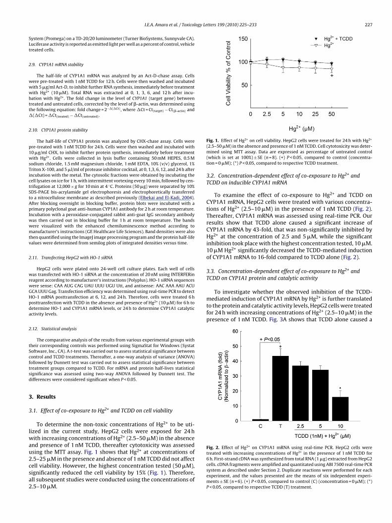
ogy Letters 199 (2010) 225–233 227
SLt
2
wwwbtt�
2
p1wsTictStApIwwmtv
2
wrwGHpda
2
tScftsd
3
3
lwau2csa2
2+ 2+
To investigate whether the observed inhibition of the TCDD-mediated induction of CYP1A1 mRNA by Hg2+ is further translatedto the protein and catalytic activity levels, HepG2 cells were treatedfor 24 h with increasing concentrations of Hg2+ (2.5–10 �M) in thepresence of 1 nM TCDD. Fig. 3A shows that TCDD alone caused a
Fig. 2. Effect of Hg2+ on CYP1A1 mRNA using real-time PCR. HepG2 cells weretreated with increasing concentrations of Hg2+ in the presence of 1 nM TCDD for
I.E.A. Amara et al. / Toxicol
ystem (Promega) on a TD-20/20 luminometer (Turner BioSystems, Sunnyvale CA).uciferase activity is reported as emitted light per well as a percent of control, vehiclereated cells.
.9. CYP1A1 mRNA stability
The half-life of CYP1A1 mRNA was analyzed by an Act-D-chase assay. Cellsere pre-treated with 1 nM TCDD for 12 h. Cells were then washed and incubatedith 5 �g/ml Act-D, to inhibit further RNA synthesis, immediately before treatmentith Hg2+ (10 �M). Total RNA was extracted at 0, 1, 3, 6, and 12 h after incu-
ation with Hg2+. The fold change in the level of CYP1A1 (target gene) betweenreated and untreated cells, corrected by the level of �-actin, was determined usinghe following equation: fold change = 2−�(�Ct), where �Ct = Ct(target) − Ct(�-actin) and
(�Ct) = �Ct(treated) − �Ct(untreated).
.10. CYP1A1 protein stability
The half-life of CYP1A1 protein was analyzed by CHX-chase assay. Cells werere-treated with 1 nM TCDD for 24 h. Cells were then washed and incubated with0 �g/ml CHX, to inhibit further protein synthesis, immediately before treatmentith Hg2+. Cells were collected in lysis buffer containing 50 mM HEPES, 0.5 M
odium chloride, 1.5 mM magnesium chloride, 1 mM EDTA, 10% (v/v) glycerol, 1%riton X-100, and 5 �l/ml of protease inhibitor cocktail, at 0, 1,3, 6, 12, and 24 h afterncubation with the metal. The cytosolic fractions were obtained by incubating theell lysates on ice for 1 h, with intermittent vortexing every 10 min, followed by cen-rifugation at 12,000 × g for 10 min at 4 ◦C. Proteins (50 �g) were separated by 10%DS-PAGE bis-acrylamide gel electrophoresis and electrophoretically transferredo a nitrocellulose membrane as described previously (Elbekai and El-Kadi, 2004).fter blocking overnight in blocking buffer, protein blots were incubated with arimary polyclonal goat anti-human CYP1A1 antibody for 2 h at room temperature.
ncubation with a peroxidase-conjugated rabbit anti-goat IgG secondary antibodyas then carried out in blocking buffer for 1 h at room temperature. The bandsere visualized with the enhanced chemiluminescence method according toanufacturer’s instructions (GE Healthcare Life Sciences). Band densities were also
hen quantified using the ImageJ image processing program and the protein half-lifealues were determined from semilog plots of integrated densities versus time.
.11. Transfecting HepG2 with HO-1 siRNA
HepG2 cells were plated onto 24-well cell culture plates. Each well of cellsas transfected with HO-1 siRNA at the concentration of 20 nM using INTERFERin
eagent according to manufacturer’s instructions (Polyplus). HO-1 siRNA sequencesere sense: CAA AUG CAG UAU UUU UGU Utt, and antisense: AAC AAA AAU ACUCA UUU Gag. Transfection efficiency was determined using real-time PCR to detectO-1 mRNA posttransfection at 6, 12, and 24 h. Therefore, cells were treated 6 hosttransfection with TCDD in the absence and presence of Hg2+ (10 �M) for 6 h toetermine HO-1 and CYP1A1 mRNA levels, or 24 h to determine CYP1A1 catalyticctivity levels.
.12. Statistical analysis
The comparative analysis of the results from various experimental groups withheir corresponding controls was performed using SigmaStat for Windows (Systatoftware, Inc., CA). A t-test was carried out to assess statistical significance betweenontrol and TCDD treatments. Thereafter, a one-way analysis of variance (ANOVA)ollowed by Dunnett test was carried out to assess statistical significance betweenreatment groups compared to TCDD. For mRNA and protein half-lives statisticalignificance was assessed using two-way ANOVA followed by Dunnett test. Theifferences were considered significant when P < 0.05.
. Results
.1. Effect of co-exposure to Hg2+ and TCDD on cell viability
To determine the non-toxic concentrations of Hg2+ to be uti-ized in the current study, HepG2 cells were exposed for 24 h
ith increasing concentrations of Hg2+ (2.5–50 �M) in the absencend presence of 1 nM TCDD, thereafter cytotoxicity was assessedsing the MTT assay. Fig. 1 shows that Hg2+ at concentrations of
.5–25 �M in the presence and absence of 1 nM TCDD did not affectell viability. However, the highest concentration tested (50 �M),ignificantly reduced the cell viability by 15% (Fig. 1). Therefore,ll subsequent studies were conducted using the concentrations of.5–10 �M.Fig. 1. Effect of Hg on cell viability. HepG2 cells were treated for 24 h with Hg(2.5–50 �M) in the absence and presence of 1 nM TCDD. Cell cytotoxicity was deter-mined using MTT assay. Data are expressed as percentage of untreated control(which is set at 100%) ± SE (n = 8). (+) P < 0.05, compared to control (concentra-tion = 0 �M); (*) P < 0.05, compared to respective TCDD treatment.
3.2. Concentration-dependent effect of co-exposure to Hg2+ andTCDD on inducible CYP1A1 mRNA
To examine the effect of co-exposure to Hg2+ and TCDD onCYP1A1 mRNA, HepG2 cells were treated with various concentra-tions of Hg2+ (2.5–10 �M) in the presence of 1 nM TCDD (Fig. 2).Thereafter, CYP1A1 mRNA was assessed using real-time PCR. Ourresults show that TCDD alone caused a significant increase ofCYP1A1 mRNA by 43-fold, that was non-significantly inhibited byHg2+ at the concentration of 2.5 and 5 �M, while the significantinhibition took place with the highest concentration tested, 10 �M.10 �M Hg2+ significantly decreased the TCDD-mediated inductionof CYP1A1 mRNA to 16-fold compared to TCDD alone (Fig. 2).
3.3. Concentration-dependent effect of co-exposure to Hg2+ andTCDD on CYP1A1 protein and catalytic activity
6 h. First-strand cDNA was synthesized from total RNA (1 �g) extracted from HepG2cells. cDNA fragments were amplified and quantitated using ABI 7500 real-time PCRsystem as described under Section 2. Duplicate reactions were performed for eachexperiment, and the values presented are the means of six independent experi-ments ± SE (n = 6). (+) P < 0.05, compared to control (C) (concentration = 0 �M); (*)P < 0.05, compared to respective TCDD (T) treatment.

228 I.E.A. Amara et al. / Toxicology Letters 199 (2010) 225–233
Fig. 3. Effect of Hg2+ on CYP1A1 protein and EROD activity. HepG2 cells were treatedfor 24 h with increasing concentrations of Hg2+ in the presence of 1 nM TCDD. (A)Protein (50 �g) was separated on a 10% SDS-PAGE and transferred to nitrocellulosemembrane. Protein blots were then blocked overnight at 4 ◦C and then incubatedwith a primary CYP1A1 antibody for 2 h at room temperature, followed by 1 h incu-bation with secondary antibody at room temperature. CYP1A1 protein was detectedusing the enhanced chemiluminescence method. The intensity of bands was nor-malized to GAPDH signals, which was used as loading control. One of the threerepresentative experiments is shown. (B) EROD activity was measured in intact liv-ios(
2CctsHc,r
3
H
Fig. 4. Effect of Hg2+ on luciferase activity. HepG2 cells were transiently transfectedwith the XRE-luciferase transporter plasmid pGudLuc 6.1. Cells were treated withvehicle, TCDD (1 nM), Hg2+ (10 �M), TCDD (1 nM) + Hg2+ (10 �M) for 24 h. Cells were
ng cells treated with increasing concentrations of Hg2+, in the absence and presencef 1 nM TCDD for 24 h. CYP1A1 activity was measured using 7-ethoxyresorufin as aubstrate. Values are presented as mean ± SE (n = 6). (+) P < 0.05, compared to controlC); (*) P < 0.05, compared to respective TCDD (T) treatment.
4-fold increase in CYP1A1 protein level. In agreement with theYP1A1 mRNA results, Hg2+ at the concentration of 10 �M signifi-antly decreased the TCDD-mediated induction of CYP1A1 proteino ∼14-fold compared to TCDD alone. In addition, TCDD aloneignificantly increased CYP1A1 activity by approximately 11-fold.owever, Hg2+ decreased the TCDD-mediated induction of CYP1A1atalytic activity level in a concentration-dependent manner to 7.5-5- and 2-fold with Hg2+ concentrations of 2.5, 5, and 10, �M,espectively, compared to control (Fig. 3B).
.4. Transcriptional inhibition of CYP1A1 gene by Hg2+
In order to understand whether or not the inhibitory effect ofg2+ on TCDD-mediated induction of CYP1A1 mRNA is actually due
lysed and luciferase activity was measured according to manufacturer’s instruc-tion. Luciferase activity is reported as relative light unit. Values are presented asmean ± SE (n = 6). (+) P < 0.05, compared to control (C); (*) P < 0.05, compared torespective TCDD (T) treatment.
to a transcriptional mechanism, HepG2 cells were transiently trans-fected with the XRE-driven luciferase reporter gene. Luciferaseactivity results showed that 10 �M Hg2+ alone inhibited the con-stitutive expression of the luciferase activity to 0.7-fold comparedto control (Fig. 4). On the other hand, 1 nM TCDD alone causeda significant increase in the luciferase activity by 60-fold com-pared to control. Interestingly, co-treatment with Hg2+ and TCDDsignificantly decreased the TCDD-mediated induction of luciferaseactivity to 34-fold compared to TCDD alone (Fig. 4).
3.5. Posttranscriptional modification of CYP1A1 mRNA by Hg2+
Although a transcriptional mechanism is involved in the Hg2+-mediated down-regulation of the TCDD-mediated induction ofCYP1A1 mRNA levels, there was still a possibility that a post-transcriptional mechanism might be involved. The level of mRNAexpression is not only a function of the transcription rate, but is alsodependent on the elimination rate, through processing or degrada-tion. If Hg2+ decreases CYP1A1 mRNA via decreasing its stability, adecrease in half-life would be expected to take place. To examinethe effect of Hg2+ on the CYP1A1 mRNA stability, we performedthe Act-D chase experiment on intact viable HepG2 cells. Fig. 5shows that CYP1A1 mRNA decayed with a half-life of 5.57 ± 0.99 h.Furthermore, Hg2+ did not significantly alter the CYP1A1 mRNAhalf-life which was 7.14 ± 0.95 h (Fig. 5).
3.6. Posttranslational modification of CYP1A1 protein by Hg2+
The fact that Hg2+ inhibited the TCDD-mediated induction ofCYP1A1 catalytic activity much more than what is observed at themRNA levels raised the question of whether Hg2+ could modifyCYP1A1 protein stability. Therefore, we measured the effect of Hg2+
on CYP1A1 protein half-life using CHX-chase experiment. Fig. 6shows that CYP1A1 protein induced by TCDD degraded with a half-life of 10.78 ± 0.61 h. Interestingly, Hg2+ significantly decreasedthe stability of CYP1A1 protein which degraded with a half-life of4.75 ± 0.81 h (Fig. 6).
2+
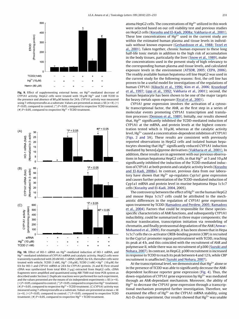
3.7. Effect of co-exposure to Hg and TCDD on HO-1 mRNAThe inverse relation between HO-1 expression and CYP1A1activity directed us to probe the role of Hg2+ in inhibiting theTCDD-mediated induction of CYP1A1 at the catalytic activity level.

I.E.A. Amara et al. / Toxicology Letters 199 (2010) 225–233 229
Fig. 5. Effect of Hg2+ on CYP1A1 mRNA half-life using real-time PCR. HepG2 cellswere grown to 90% confluence in six-well cell culture plates, and then treated with1 nM TCDD for 12 h. The cells were then washed and incubated in a fresh media con-taining 10 �M Hg2+ plus 5 �g/ml Act-D, a RNA synthesis inhibitor. First-strand cDNAwas synthesized from total RNA (1 �g) extracted from HepG2 cells. cDNA fragmentswere amplified and quantitated using ABI 7500 real-time PCR system as describedunder Materials and Methods. Duplicate reactions were performed for each exper-iment, and the values presented are the means of six independent experiments.mRNA decay curves were analyzed individually, and the half-life was estimatedfrom the slope of a straight line fitted by linear regression analysis (r2 ≥ 0.81) toa semilog plot of mRNA amount, expressed as a percent of treatment at time = 0 h(e
Tlwtumicpi
3pH
Ti
Fig. 6. Effect of Hg2+ on the CYP1A1 protein half-life. HepG2 cells were grown to 90%confluence in six-well cell culture plates. Thereafter, the cells were treated with 1 nMTCDD for 24 h. Cells were washed and incubated in fresh media containing 10 �MHg2+ plus 10 �g/ml CHX, a protein translation inhibitor. Total cellular protein wasextracted at the designated time points after the addition of CHX. Protein (50 �g)was separated by 10% SDS-PAGE and transferred to a nitrocellulose membrane. Theintensities of CYP1A1 protein bands were normalized to GAPDH signals, which wereused as loading controls. All protein decay curves were analyzed individually. Thehalf-life was estimated from the slope of a straight line fitted by linear regression
reversed the Hg2+-mediated decrease of CYP1A1 activity to reach
maximum, 100%) level, versus time. The half-lives obtained from six independentxperiments were then used to calculate the mean half-life (mean ± SE, n = 6).
herefore, we examined the effect of Hg2+ on HO-1 mRNA, a rate-imiting enzyme of heme degradation. For this purpose, HepG2 cells
ere treated with increasing concentrations of Hg2+ (2.5–10 �M) inhe presence of 1 nM TCDD. Thereafter, HO-1 mRNA was measuredsing real-time PCR. Fig. 7 shows that TCDD alone did not alter HO-1RNA level. Whereas, co-exposure to TCDD and Hg2+ significantly
ncreased the HO-1 mRNA level by 4.5-, 8-, and, 22-fold with con-entrations of 2.5, 5, and 10 �M, respectively. Thus, HO-1 might bearticipating in the Hg2+-mediated decrease of the TCDD-mediated
nduction of CYP1A1 at the catalytic activity (Fig. 7).
.8. The effect of SnMP as a competitive inhibitor of HO-1 on theosttranslational modification of CYP1A1 catalytic activity byg2+
To confirm the role of HO-1 in Hg2+-mediated decrease of theCDD-mediated induction of CYP1A1 catalytic activity, we exam-ned the effect of HO-1 inhibitor, SnMP, on the decrease of CYP1A1
analysis to a semilog plot of protein amount, expressed as a percentage of treatmentat time = 0 h (maximum, 100%) level, versus time. The half-lives obtained from threeindependent experiments were then used to calculate the mean half-life (mean ± SE,n = 3). (*) P < 0.05 compared with TCDD.
catalytic activity-mediated by Hg2+. For this purpose HepG2 cellswere co-exposed to 10 �M Hg2+ and 1 nM TCDD in the presenceand absence of 5 �M SnMP. SnMP alone or in the presence ofTCDD or Hg2+ plus TCDD did not affect CYP1A1 mRNA levels atall treatments, thus eliminating the possibility that SnMP reversesthe inhibitory effect of Hg2+ on CYP1A1 catalytic activity throughaffecting its mRNA levels (Fig. 8A). Similarly, SnMP alone or inthe presence of TCDD did not alter the CYP1A1 catalytic activity.TCDD alone increased the CYP1A1 catalytic activity by ∼11-fold.On the other hand, Hg2+ at the concentration of 10 �M decreasedthe TCDD-mediated induction of CYP1A1 catalytic activity to ∼5-fold compared to control (Fig. 8B). Intriguingly, SnMP partially
∼9-fold compared to control. In spite of being successful in par-tially reversing the Hg2+-mediated decrease of CYP1A1 activitythrough inhibiting HO-1, SnMP was unable to completely restorethe CYP1A1 activity.

230 I.E.A. Amara et al. / Toxicology Letters 199 (2010) 225–233
Fig. 7. Effect of Hg2+ on HO-1 mRNA. HepG2 cells were treated for 6 h with increasingconcentrations of Hg2+ in the presence of 1 nM TCDD. First-strand cDNA was syn-thesized from total RNA (1 �g) extracted from HepG2 cells. cDNA fragments wereamplified and quantitated using ABI 7500 real-time PCR system as described underSpct
3C
wcesid(
3C
aoiogtCftOmhatHstwsnCH
p(
Fig. 8. Effect of SnMP on CYP1A1 mRNA and catalytic activity levels in the pres-ence of Hg2+. HepG2 cells were treated with 10 �M of Hg2+ and 1 nM TCDD in thepresence and absence of 5 �M SnMP for 6 h for CYP1A1 mRNA, and for 24 h forCYP1A1 catalytic activity. (A) First-strand cDNA was synthesized from total RNA(1 �g) extracted from HepG2 cells. cDNA fragments were amplified and quantitatedusing ABI 7500 real-time PCR system as described under Section 2. Duplicate reac-tions were performed for each experiment, and the values presented are the meansof six independent experiments ± SE (n = 6). (+) P < 0.05, compared to control; (*)
ection 2. Duplicate reactions were performed for each experiment, and the valuesresented are the means of six independent experiments ± SE (n = 6). (+) P < 0.05,ompared with control (C); (*) P < 0.05, compared with the respective TCDD (T)reatment.
.9. The effect of exogenous heme on Hg2+-mediated decrease ofYP1A1 activity
In an attempt to examine whether the presence of external hemeill restore the Hg2+-mediated decrease of CYP1A1 activity, HepG2
ells were co-exposed to 10 �M Hg2+ and 1 nM TCDD in the pres-nce and absence of 80 �M hemin, a precursor of heme. Our resultshowed that hemin alone did not affect CYP1A1 activity. Interest-ngly, the addition of hemin partially restored the Hg2+-mediatedecrease of CYP1A1 activity by 2-fold compared to Hg2+ plus TCDDFig. 9).
.10. The effect of HO-1 siRNA on Hg2+-mediated inhibition ofYP1A1 catalytic activity
Despite using selective pharmacological inhibitors such as SnMPnd heme precursors like hemin to inhibit HO-1 activity, it wasf importance to confirm our hypothesis that the Hg2+-mediatedncrease in HO-1 is in part responsible for the down-regulationf CYP1A1 at the catalytic activity level. Therefore, we took aenetic approach to confirm whether or not HO-1 is involved inhe Hg2+-mediated decrease of the TCDD-mediated induction ofYP1A1 catalytic activity. For this purpose, HepG2 cells were trans-
ected with human HO-1 siRNA for 6 h, and then the cells werereated with 10 �M Hg2+ in the presence and absence of 1 nM TCDD.ur results showed that HO-1 siRNA significantly decreased HO-1RNA by 0.8-fold as compared to control (Fig. 10A). On the other
and, Hg2+ was able to increase HO-1 mRNA levels, in the absencend presence of TCDD, to reach 25-fold compared to control. Whenhe cells were transfected with HO-1 siRNA, and then treated withg2+ in the presence or absence of TCDD there was a statistically
ignificant decrease in HO-1 mRNA to reach ∼4.5-fold comparedo control (Fig. 10A). To test the selectivity of the siRNA for HO-1,e determined the CYP1A1 mRNA levels in cells transfected with
iRNA for HO-1. Fig. 10B shows that CYP1A1 mRNA levels wereot altered by the HO-1 siRNA. Thus, the observed effects on the
YP1A1 catalytic activity levels are solely through knocking-downO-1.Looking at CYP1A1 catalytic activity, Hg2+ alone or in theresence of HO-1 siRNA did not affect CYP1A1 catalytic activityFig. 10C). TCDD alone increased the CYP1A1 catalytic activity by
P < 0.05, compared to respective TCDD treatment. (B) CYP1A1 activity was measuredusing 7-ethoxyresorufin as a substrate. Values are presented as mean ± SE (n = 8). (+)P < 0.05, compared to control; (*) P < 0.05, compared to respective TCDD treatment;(#) P < 0.05, compared to respective Hg2+ + TCDD treatment.
21-fold, whereas Hg2+ significantly decreased the TCDD-mediatedinduction of CYP1A1 catalytic activity to reach 6-fold compared tocontrol. Interestingly, when HepG2 cells were transfected with HO-1 siRNA and then co-exposed to Hg2+ and TCDD, Hg2+ decreasedthe TCDD-induced catalytic activity to reach 17-fold, compared tocontrol, and was unable to maintain the same inhibitory effect onCYP1A1 catalytic activity when compared to non-transfected cells(Fig. 10C).
4. Discussion
Although environmental co-exposures involve both AhR-ligands, typified by TCDD, and heavy metals, typified by Hg2+, thecurrent methods for assessing the potential toxicological conse-
quences are often assuming the additivity of responses. However,this might not be the case, because there is still a possibility ofantagonistic or synergistic effect.In the current study, we examined the potential effect of Hg2+
on the TCDD-mediated induction of CYP1A1 gene in human hep-
I.E.A. Amara et al. / Toxicology Le
Fig. 9. Effect of supplementing external heme, on Hg2+-mediated decrease ofCYP1A1 activity. HepG2 cells were treated with 10 �M Hg2+ and 1 nM TCDD inthe presence and absence of 80 �M hemin for 24 h. CYP1A1 activity was measuredusing 7-ethoxyresorufin as a substrate. Values are presented as mean ± SE (n = 8). (+)P < 0.05, compared to control; (*) P < 0.05, compared to respective TCDD treatment;(#) P < 0.05, compared to respective Hg2+ + TCDD treatment.
Fig. 10. Effect of HO-1 siRNA on Hg2+-mediated induction of HO-1 mRNA, andHg2+-mediated inhibition of CYP1A1 mRNA and catalytic activity. HepG2 cells weretransiently transfected with 20 nM HO-1 siRNA (siRNA) for 6 h, thereafter cells weretreated with vehicle, TCDD (1 nM), Hg2+ (10 �M), TCDD (1 nM) + Hg2+ (10 �M) for6 h for HO-1 and CYP1A1 mRNA or 24 h for CYP1A1 protein. (A and B) First-strandcDNA was synthesized from total RNA (1 �g) extracted from HepG2 cells. cDNAfragments were amplified and quantitated using ABI 7500 real-time PCR system asdescribed under Section 2. Duplicate reactions were performed for each experiment,and the values presented are the means of six independent experiments ± SE (n = 6).(+) P < 0.05, compared to control; (*) P < 0.05, compared to respective Hg2+ treatment;(#) P < 0.05, compared to respective Hg2+ + TCDD treatment. (C) CYP1A1 activity wasmeasured using 7-ethoxyresorufin as a substrate. Values are presented as mean ± SE(n = 6). (+) P < 0.05, compared to control; (*) P < 0.05, compared to respective TCDDtreatment; (#) P < 0.05, compared to respective Hg2+ + TCDD treatment.
tters 199 (2010) 225–233 231
atoma HepG2 cells. The concentrations of Hg2+ utilized in this workwere selected based on our cell viability test and previous studieson HepG2 cells (Korashy and El-Kadi, 2008a; Vakharia et al., 2001).These low concentrations of Hg2+ used in the current study arewithin the estimated human plasma and tissue levels in individ-uals without known exposure (Gerhardsson et al., 1988; Tezel etal., 2001). Taken together, chronic human exposure to these longhalf-life toxic metals in addition to the high risk of accumulationin the body tissues, particularly the liver (Snow et al., 1989), makethe concentrations used in the present study of high relevancy tothe corresponding human plasma and tissue levels, and calculatedexposure levels in the environment (ATSDR, 2005; CEPA, 2006).The readily available human hepatoma cell line HepG2 was used inthe current study for the following reasons: first, the cell line hasproven to be a useful model for investigations of the regulations ofhuman CYP1A1 (Kikuchi et al., 1996; Kim et al., 2006; Krusekopfet al., 1997; Lipp et al., 1992; Vakharia et al., 2001); second, thehuman hepatocyte has been shown to be one of the major targetsfor heavy metals upon exposure (Ercal et al., 2001).
CYP1A1 gene expression involves the activation of a cytoso-lic transcriptional factor, the AhR, as the first step in a series ofmolecular events promoting CYP1A1 transcription and transla-tion processes (Denison et al., 1989). Initially, our results showedthat, Hg2+ significantly inhibited the TCDD-mediated induction ofCYP1A1 at the mRNA, and protein levels at the highest concen-tration tested which is 10 �M, whereas at the catalytic activitylevel, Hg2+ caused a concentration-dependent inhibition of CYP1A1(Figs. 2 and 3A). These results are consistent with previouslyreported observations in HepG2 cells and isolated human hepa-tocytes showing that Hg2+ significantly reduced CYP1A1 inductionmediated by benzo[a]pyrene derivatives (Vakharia et al., 2001). Inaddition, these results are in agreement with our previous observa-tions in human hepatoma HepG2 cells, in that Hg2+ at 5 and 10 �Msignificantly inhibited the induction of the TCDD-mediated induc-tion of CYP1A1 at both protein and catalytic activity levels (Korashyand El-Kadi, 2008a). In contrast, previous data from our labora-tory have shown that Hg2+ up-regulates Cyp1a1 gene expressionand causes further potentiation of the TCDD-mediated induction ofCyp1a1 mRNA and protein level in murine hepatoma Hepa 1c1c7cells (Korashy and El-Kadi, 2004, 2005).
The controversy between the effect of Hg2+ on the human HepG2and mouse Hepa 1c1c7 cells could be attributed to the mech-anistic differences in the regulation of CYP1A1 gene expressionupon treatment by TCDD (Ramadoss and Perdew, 2005; Ramadosset al., 2004). Factors that could be responsible for these species-specific characteristics of AhR functions, and subsequently CYP1A1inducibility, could be summarized in three major components; thenuclear translocation, transcription initiation via remodeling ofchromatin, and finally proteasomal degradation of the AhR (Anwar-Mohamed et al., 2009). For example, it has been shown that in Hepa1c1c7 cells the co-activator CREB-binding protein (CBP) is recruitedto the Cyp1a1 promoter region posttreatment with TCDD, reachingits peak at 4 h, and this coincided with the recruitment of AhR andpolymerase II, while there was no recruitment of p300 (Suzuki andNohara, 2007). In contrast, in HepG2, p300 recruitment is increasedin response to TCDD to reach its peak between 4 and 12 h, while CBPrecruitment is unaffected (Suzuki and Nohara, 2007).
At the transcriptional level, we demonstrated that Hg2+ alone orin the presence of TCDD was able to significantly decrease the AhR-dependent luciferase reporter gene expression (Fig. 4). Thus, thedown-regulation of CYP1A1 gene expression by Hg2+ was mediatedthrough an AhR-dependant mechanism. Moreover, the ability of
Hg2+ to decrease the CYP1A1 gene expression through a transcrip-tional mechanism prompted further investigation. Therefore, weexamined the effect of Hg2+ on the CYP1A1 mRNA stability, usingAct-D-chase experiment. Our results showed that Hg2+ was unable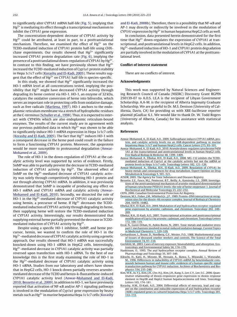
2 ogy Le
tHi
HmTcipIiig
Hsdcsspaaeot(atwM
aSiSindHMHumtosm
cHakHrktHtmC2rim
32 I.E.A. Amara et al. / Toxicol
o significantly alter CYP1A1 mRNA half-life (Fig. 5), implying thatg2+ is mediating its effect through a transcriptional mechanism to
nhibit the CYP1A1 gene expression.The concentration-dependent decrease of CYP1A1 activity by
g2+ could be attributed, at least in part, to a posttranslationalechanism. Therefore, we examined the effect of Hg2+ on the
CDD-mediated induction of CYP1A1 protein half-life using CHX-hase experiments. Our results showed that Hg2+ significantlyncreased CYP1A1 protein degradation rate (Fig. 6), implying theresence of a posttranslational down-regulation of CYP1A1 by Hg2+.
n contrast to this finding, we have previously shown that Hg2+
ncreased the TCDD-mediated induction of Cyp1a1 protein stabilityn Hepa 1c1c7 cells (Korashy and El-Kadi, 2005). These results sug-est that the effect of Hg2+ on CYP1A1 half-life is species-specific.
In this study, we showed that Hg2+ significantly increased theO-1 mRNA level at all concentrations tested, implying the pos-
ibility that Hg2+ might have decreased CYP1A1 activity throughegrading its heme content via HO-1. HO-1, an enzyme of 32 kDa,atalyzes the oxidative conversion of heme into biliverdin, whicherves an important role in protecting cells from oxidative damage,uch as free radicals (Marilena, 1997). HO-1 anchors to the endo-lasmic reticulum membrane via a stretch of hydrophobic residuest the C-terminus (Schuller et al., 1998). Thus, it is expected to inter-ct with CYP450s which are also endoplasmic reticulum-boundnzymes. The results of the current study are in agreement withur previous published data in which Hg2+ was shown to be ableo significantly induce HO-1 mRNA expression in Hepa 1c1c7 cellsKorashy and El-Kadi, 2005). The fact that Hg2+ induces HO-1 withconsequent decrease in the heme pool could result in the failure
o form a functioning CYP1A1 protein. Moreover, the apoproteinould be more susceptible to proteasomal degradation (Anwar-ohamed et al., 2009).The role of HO-1 in the down-regulation of CYP1A1 at the cat-
lytic activity level was supported by series of evidence. Firstly,nMP was able to partially prevent the decrease in TCDD-mediatednduction of CYP1A1 activity by Hg2+. The observed effect ofnMP on the Hg2+-mediated decrease of CYP1A1 catalytic activ-ty was solely through competitively inhibiting HO-1 protein andot through altering CYP1A1 mRNA. In addition, it was previouslyemonstrated that SnMP is incapable of producing any effect onO-1 mRNA and CYP1A1 mRNA and catalytic activity (Anwar-ohamed and El-Kadi, 2010). Secondly, we dissected the role ofO-1 in the Hg2+-mediated decrease of CYP1A1 catalytic activitysing hemin, a precursor of heme. If Hg2+ decreases the TCDD-ediated induction of CYP1A1 activity through degrading its heme,
hen supplying heme will restore the TCDD-mediated inductionf CYP1A1 activity. Interestingly, our results demonstrated thatupplying external heme partially prevented the decrease in TCDD-ediated induction of CYP1A1 activity by Hg2+.Despite using a specific HO-1 inhibitor, SnMP, and heme pre-
ursor, hemin, we wanted to confirm the role of HO-1 in theg2+-mediated decrease of CYP1A1 catalytic activity using a geneticpproach. Our results showed that HO-1 mRNA was successfullynocked-down using HO-1 siRNA in HepG2 cells. Interestingly,g2+-mediated decrease in CYP1A1 catalytic activity was partially
eversed upon transfection with HO-1 siRNA. To the best of ournowledge this is the first study examining the role of HO-1 inhe Hg2+-mediated decrease of CYP1A1 catalytic activity usingO-1 siRNA. Studies from our laboratory and others have shown
hat in HepG2 cells, HO-1 knock-down partially reverses arsenite-ediated decrease of the TCDD and benzo-k-fluoranthene-induced
YP1A1 catalytic activity level (Anwar-Mohamed and El-Kadi,010; Bessette et al., 2009). In addition to HO-1, we have previouslyeported that activation of NF-�B and/or AP-1 signaling pathwayss involved in the modulation of Cyp1a1 gene expression by heavy
etals such as Hg2+ in murine hepatoma Hepa 1c1c7 cells (Korashy
tters 199 (2010) 225–233
and El-Kadi, 2008b). Therefore, there is a possibility that NF-�B andAP-1 may directly or indirectly be involved in the modulation ofCYP1A1 expression by Hg2+ in human hepatoma HepG2 cells as well.
In conclusion, data presented herein demonstrated for the firsttime that Hg2+ down-regulates the expression of CYP1A1 at tran-scriptional, and posttranslational levels in HepG2 cells. In addition,Hg2+-mediated induction of HO-1 and CYP1A1 protein degradationare partially involved in the modulation of CYP1A1 at the posttrans-lational level.
Conflict of interest statement
There are no conflicts of interest.
Acknowledgments
This work was supported by Natural Sciences and Engineer-ing Research Council of Canada (NSERC) Discovery Grant RGPIN250139-07 to A.O.S. I.E.A is the recipient of Libyan GovernmentScholarship. A.A-M. is the recipient of Alberta Ingenuity GraduateScholarship. We are grateful to Dr. M.S. Denison (University of Cal-ifornia, Davis, CA) for providing us with XRE-luciferase reporterplasmid pGudLuc 6.1. We would like to thank Dr. W. Todd Rogers(University of Alberta, Canada) for his assistance with statisticalanalysis.
References
Anwar-Mohamed, A., El-Kadi, A.O., 2009. Sulforaphane induces CYP1A1 mRNA, pro-tein, and catalytic activity levels via an AhR-dependent pathway in murinehepatoma Hepa 1c1c7 and human HepG2 cells. Cancer Letters 275, 93–101.
Anwar-Mohamed, A., El-Kadi, A.O., 2010. Arsenite down-regulates cytochrome P4501A1 at the transcriptional and posttranslational levels in human HepG2 cells.Free Radical Biology & Medicine 48, 1399–1409.
Anwar-Mohamed, A., Elbekai, R.H., El-Kadi, A.O., 2008. MG-132 inhibits the TCDD-mediated induction of Cyp1a1 at the catalytic activity but not the mRNA orprotein levels in Hepa 1c1c7 cells. Toxicology Letters 182, 121–126.
Anwar-Mohamed, A., Elbekai, R.H., El-Kadi, A.O., 2009. Regulation of CYP1A1 byheavy metals and consequences for drug metabolism. Expert Opinion on DrugMetabolism & Toxicology 5, 501–521.
ATSDR, 2005. The Agency for Toxic Substances and Diseases Registry.Bessette, E.E., Fasco, M.J., Pentecost, B.T., Reilly, A., Kaminsky, L.S., 2009. Investiga-
tions of the posttranslational mechanism of arsenite-mediated downregulationof human cytochrome P4501A1 levels: the role of heme oxygenase-1. Journal ofBiochemical and Molecular Toxicology 23, 222–232.
CEPA, 2006. Canadian Environmental Protection Act Registry.Denison, M.S., Fisher, J.M., Whitlock Jr., J.P., 1989. Protein–DNA interactions at recog-
nition sites for the dioxin–Ah receptor complex. Journal of Biological Chemistry264, 16478–16482.
Elbekai, R.H., El-Kadi, A.O., 2004. Modulation of aryl hydrocarbon receptor-regulatedgene expression by arsenite, cadmium, and chromium. Toxicology 202, 249–269.
Elbekai, R.H., El-Kadi, A.O., 2007. Transcriptional activation and posttranscriptionalmodification of Cyp1a1 by arsenite, cadmium, and chromium. Toxicology Letters172, 106–119.
Ercal, N., Gurer-Orhan, H., Aykin-Burns, N., 2001. Toxic metals and oxidative stresspart I: mechanisms involved in metal-induced oxidative damage. Current Topicsin Medicinal Chemistry 1, 529–539.
Gerhardsson, L., Brune, D., Nordberg, G.F., Wester, P.O., 1988. Multielemental assayof tissues of deceased smelter workers and controls. The Science of the TotalEnvironment 74, 97–110.
Gochfeld, M., 2003. Cases of mercury exposure, bioavailability, and absorption. Eco-toxicology and Environmental Safety 56, 174–179.
Hankinson, O., 1995. The aryl hydrocarbon receptor complex. Annual Review ofPharmacology and Toxicology 35, 307–340.
Kikuchi, H., Kato, H., Mizuno, M., Hossain, A., Ikawa, S., Miyazaki, J., Watanabe,M., 1996. Differences in inducibility of CYP1A1-mRNA by benzimidazole com-pounds between human and mouse cells: evidences of a human-specific signaltransduction pathway for CYP1A1 induction. Archives of Biochemistry and Bio-physics 334, 235–240.
Kim, W.K., In, Y.J., Kim, J.H., Cho, H.J., Kim, J.H., Kang, S., Lee, C.Y., Lee, S.C., 2006. Quan-titative relationship of dioxin-responsive gene expression to dioxin response
element in Hep3B and HepG2 human hepatocarcinoma cell lines. ToxicologyLetters 165, 174–181.Korashy, H.M., El-Kadi, A.O., 2004. Differential effects of mercury, lead and cop-per on the constitutive and inducible expression of aryl hydrocarbon receptor(AHR)-regulated genes in cultured hepatoma Hepa 1c1c7 cells. Toxicology 201,153–172.

ogy Le
K
K
K
K
L
L
M
M
M
N
I.E.A. Amara et al. / Toxicol
orashy, H.M., El-Kadi, A.O., 2005. Regulatory mechanisms modulating the expres-sion of cytochrome P450 1A1 gene by heavy metals. Toxicological Sciences 88,39–51.
orashy, H.M., El-Kadi, A.O., 2008a. Modulation of TCDD-mediated induction ofcytochrome P450 1A1 by mercury, lead, and copper in human HepG2 cell line.Toxicology In Vitro 22, 154–158.
orashy, H.M., El-Kadi, A.O., 2008b. The role of redox-sensitive transcription factorsNF-kappaB and AP-1 in the modulation of the Cyp1a1 gene by mercury, lead,and copper. Free Radical Biology & Medicine 44, 795–806.
rusekopf, S., Kleeberg, U., Hildebrandt, A.G., Ruckpaul, K., 1997. Effects of benzim-idazole derivatives on cytochrome P450 1A1 expression in a human hepatomacell line. Xenobiotica 27, 1–9.
ipp, H.P., Schrenk, D., Wiesmuller, T., Hagenmaier, H., Bock, K.W., 1992. Assess-ment of biological activities of mixtures of polychlorinated dibenzo-p-dioxins(PCDDs) and their constituents in human HepG2 cells. Archives of Toxicology66, 220–223.
orenzen, A., Kennedy, S.W., 1993. A fluorescence-based protein assay for use witha microplate reader. Analytical Biochemistry 214, 346–348.
arilena, G., 1997. New physiological importance of two classic residual prod-ucts: carbon monoxide and bilirubin. Biochemical and Molecular Medicine 61,136–142.
cLemore, T.L., Adelberg, S., Liu, M.C., McMahon, N.A., Yu, S.J., Hubbard, W.C., Czer-winski, M., Wood, T.G., Storeng, R., Lubet, R.A., et al., 1990. Expression of CYP1A1gene in patients with lung cancer: evidence for cigarette smoke-induced geneexpression in normal lung tissue and for altered gene regulation in primarypulmonary carcinomas. Journal of the National Cancer Institute 82, 1333–1339.
eyer, B.K., Pray-Grant, M.G., Vanden Heuvel, J.P., Perdew, G.H., 1998. HepatitisB virus X-associated protein 2 is a subunit of the unliganded aryl hydrocarbonreceptor core complex and exhibits transcriptional enhancer activity. Molecularand Cellular Biology 18, 978–988.
ebert, D.W., Dalton, T.P., Okey, A.B., Gonzalez, F.J., 2004. Role of arylhydrocarbon receptor-mediated induction of the CYP1 enzymes in environ-
tters 199 (2010) 225–233 233
mental toxicity and cancer. Journal of Biological Chemistry 279, 23847–23850.
Ramadoss, P., Perdew, G.H., 2005. The transactivation domain of the Ah receptor isa key determinant of cellular localization and ligand-independent nucleocyto-plasmic shuttling properties. Biochemistry 44, 11148–11159.
Ramadoss, P., Petrulis, J.R., Hollingshead, B.D., Kusnadi, A., Perdew, G.H., 2004. Diver-gent roles of hepatitis B virus X-associated protein 2 (XAP2) in human versusmouse Ah receptor complexes. Biochemistry 43, 700–709.
Schuller, D.J., Wilks, A., Ortiz de Montellano, P., Poulos, T.L., 1998. Crystallization ofrecombinant human heme oxygenase-1. Protein Science 7, 1836–1838.
Shimada, T., Fujii-Kuriyama, Y., 2004. Metabolic activation of polycyclic aromatichydrocarbons to carcinogens by cytochromes P450 1A1 and 1B1. Cancer Science95, 1–6.
Snow, R.C., Barbieri, R.L., Frisch, R.E., 1989. Estrogen 2-hydroxylase oxidation andmenstrual function among elite oarswomen. The Journal of Clinical Endocrinol-ogy and Metabolism 69, 369–376.
Suzuki, T., Nohara, K., 2007. Regulatory factors involved in species-specific modula-tion of arylhydrocarbon receptor (AhR)-dependent gene expression in humansand mice. Journal of Biochemistry 142, 443–452.
Tezel, H., Ertas, O.S., Erakin, C., Kayali, A., 2001. Blood mercury levels of den-tal students and dentists at a dental school. British Dental Journal 191,449–452.
Vakharia, D.D., Liu, N., Pause, R., Fasco, M., Bessette, E., Zhang, Q.Y., Kaminsky, L.S.,2001. Polycyclic aromatic hydrocarbon/metal mixtures: effect on PAH induc-tion of CYP1A1 in human HEPG2 cells. Drug Metabolism and Disposition: TheBiological Fate of Chemicals 29, 999–1006.
Whitelaw, M.L., Gustafsson, J.A., Poellinger, L., 1994. Identification of transactivationand repression functions of the dioxin receptor and its basic helix-loop-helix/PAS partner factor Arnt: inducible versus constitutive modes of regulation.Molecular and Cellular Biology 14, 8343–8355.
Zalups, R.K., 2000. Molecular interactions with mercury in the kidney. Pharmacolog-cal Reviews 52, 113–143.
Copyright © 2022 FDOKUMEN