eNOS activation and NO function: structural motifs responsible for the posttranslational control of...
28
eNOS activation and NO function: Structural motifs responsible for the posttranslational control of endothelial nitric oxide synthase activity Ruslan Rafikov, Fabio V Fonseca 1 , Sanjiv Kumar, Daniel Pardo, Charles Darragh, Shawn Elms, David Fulton, and Stephen M Black Pulmonary Vascular Disease Program, Vascular Biology Center: CB-3211B, Georgia Health Sciences University, 1459 Laney Walker Boulevard, Augusta, Georgia 30912, USA 1 Department of Cell Biology, Cleveland Clinic, Cleveland, Ohio 44106, USA Abstract Rather than being a constitutive enzyme as was first suggested, endothelial nitric oxide synthase (eNOS) is dynamically regulated at the transcriptional, posttranscriptional, and posttranslational levels. This review will focus on how changes in eNOS function are conferred by various posttranslational modifications. The latest knowledge regarding eNOS targeting to the plasma membrane will be discussed as the role of protein phosphorylation as a modulator of catalytic activity. Furthermore, new data are presented that provide novel insights into how disruption of the eNOS dimer prevents eNOS uncoupling and the production of superoxide under conditions of elevated oxidative stress and identifies a novel regulatory region we have termed the ‘flexible arm’. Introduction Nitric oxide (NO) is synthesized by a family of enzymes called NO synthases (NOS). There are three mammalian NOS isoforms: neuronal (nNOS), endothelial (eNOS), and inducible (iNOS). They share 50–60% homology at the amino acid level and have an N-terminal oxygenase domain with heme-, L-arginine-, tetrahydrobiopterin (BH 4 )-binding domains, a central calmodulin (CaM)-binding region, and a C-terminal reductase domain with NADPH, FAD, and FMN binding sites (Stuehr 1997). Under physiological conditions, the dominant NOS isoform in the vasculature is eNOS, which rather than being a constitutive enzyme as was first suggested, is dynamically regulated at the transcriptional, posttranscriptional, and posttranslational levels. This review will focus on the posttranslational modifications that regulate eNOS activity and will conclude with the identification of a new structural component of eNOS that regulates enzyme function under conditions of oxidative stress. © 2011 Society for Endocrinology (Correspondence should be addressed to S M Black; [email protected]). This paper is one of three papers that form part of a Thematic review section on eNOS activation and NO function. The Guest Editor for this section was Ian Bird, University of Wisconsin-Madison, USA. Declaration of interest The authors declare that there is no conflict of interest that could be perceived as prejudicing the impartiality of the research reported. NIH Public Access Author Manuscript J Endocrinol. Author manuscript; available in PMC 2012 April 16. Published in final edited form as: J Endocrinol. 2011 September ; 210(3): 271–284. doi:10.1530/JOE-11-0083. NIH-PA Author Manuscript NIH-PA Author Manuscript NIH-PA Author Manuscript
-
Upload
universityofarizona -
Category
Documents
-
view
2 -
download
0
Transcript of eNOS activation and NO function: structural motifs responsible for the posttranslational control of...
eNOS activation and NO function: Structural motifs responsiblefor the posttranslational control of endothelial nitric oxidesynthase activity
Ruslan Rafikov, Fabio V Fonseca1, Sanjiv Kumar, Daniel Pardo, Charles Darragh, ShawnElms, David Fulton, and Stephen M BlackPulmonary Vascular Disease Program, Vascular Biology Center: CB-3211B, Georgia HealthSciences University, 1459 Laney Walker Boulevard, Augusta, Georgia 30912, USA1Department of Cell Biology, Cleveland Clinic, Cleveland, Ohio 44106, USA
AbstractRather than being a constitutive enzyme as was first suggested, endothelial nitric oxide synthase(eNOS) is dynamically regulated at the transcriptional, posttranscriptional, and posttranslationallevels. This review will focus on how changes in eNOS function are conferred by variousposttranslational modifications. The latest knowledge regarding eNOS targeting to the plasmamembrane will be discussed as the role of protein phosphorylation as a modulator of catalyticactivity. Furthermore, new data are presented that provide novel insights into how disruption ofthe eNOS dimer prevents eNOS uncoupling and the production of superoxide under conditions ofelevated oxidative stress and identifies a novel regulatory region we have termed the ‘flexiblearm’.
IntroductionNitric oxide (NO) is synthesized by a family of enzymes called NO synthases (NOS). Thereare three mammalian NOS isoforms: neuronal (nNOS), endothelial (eNOS), and inducible(iNOS). They share 50–60% homology at the amino acid level and have an N-terminaloxygenase domain with heme-, L-arginine-, tetrahydrobiopterin (BH4)-binding domains, acentral calmodulin (CaM)-binding region, and a C-terminal reductase domain with NADPH,FAD, and FMN binding sites (Stuehr 1997). Under physiological conditions, the dominantNOS isoform in the vasculature is eNOS, which rather than being a constitutive enzyme aswas first suggested, is dynamically regulated at the transcriptional, posttranscriptional, andposttranslational levels. This review will focus on the posttranslational modifications thatregulate eNOS activity and will conclude with the identification of a new structuralcomponent of eNOS that regulates enzyme function under conditions of oxidative stress.
© 2011 Society for Endocrinology(Correspondence should be addressed to S M Black; [email protected]).This paper is one of three papers that form part of a Thematic review section on eNOS activation and NO function. The Guest Editorfor this section was Ian Bird, University of Wisconsin-Madison, USA.Declaration of interestThe authors declare that there is no conflict of interest that could be perceived as prejudicing the impartiality of the research reported.
NIH Public AccessAuthor ManuscriptJ Endocrinol. Author manuscript; available in PMC 2012 April 16.
Published in final edited form as:J Endocrinol. 2011 September ; 210(3): 271–284. doi:10.1530/JOE-11-0083.
NIH
-PA Author Manuscript
NIH
-PA Author Manuscript
NIH
-PA Author Manuscript
Membrane targeting of eNOSIn contrast to the other NOS isoforms, eNOS contains a myristoyl group that is covalentlyattached to the glycine residue found at the second amino acid in the N-terminal region ofthe protein (Sessa et al. 1993). The co-translational addition of this myristoyl group enableseNOS to localize to cellular membranes and also serves as a mechanism to ensure that eNOSis in close proximity with other factors that regulate its function. Mutational studies haveshown that the myristoyl group is an absolute requirement for membrane localization andmaximum eNOS activity (Sakoda et al. 1995). In cells, the loss of N-myristoylation confersa cytosolic location and reduced eNOS activity, but in isolated activity assays with maximalcofactors, eNOS activity is equal to the wild-type enzyme (Sessa et al. 1993). It is thoughtthat myristoylation targets eNOS to the Golgi complex (Sessa et al. 1995), and although themyristoyl deficient eNOS can metabolize L-arginine and generate NO, it exhibits reducedcoupling to external stimuli such as calcium ionophores (Sessa et al. 1995). eNOS is alsosubject to posttranslational palmitoylation on two cysteine residues (Cys-15 and Cys-26)within the oxygenase domain of the enzyme. This modification is reversible and requires theprior N-myristoylation and membrane anchoring of eNOS. It is believed that palmitoylationstabilizes the association of eNOS with the membrane and may also be required for correctlocalization of the enzyme within specialized lipid domains (Liu et al. 1995). The role ofpalmitoylation in eNOS activation is controversial. Reports have indicated that agonist-induced activation of eNOS is associated with depalmitoylation and translocation frommembranes (Yeh et al. 1999). However, this study is contradicted by a similar report (Liu etal. 1995). Mutation of the palmitoylation sites has been shown to decrease agonist-stimulated eNOS activity, but to a lesser extent than for the myristoyl-deficient mutant(Shaul et al. 1996). The palmitoyl-deficient mutant also has an altered subcellulardistribution in that it is virtually absent from the plasma membrane (Robinson & Michel1995, Garcia-Cardena et al. 1996b). As with myristoyl-deficient eNOS, it should be notedthat the palmitoyl-deficient enzyme is not catalytically inferior to the wild-type enzyme asthe purified enzymes are kinetically identical (Liu et al. 1996). One particular site in theplasma membrane that is of great importance for eNOS function is the caveolae. Caveolaeare specialized invaginations of the plasma membrane that are present in most cell types butare particularly enriched within the membranes of endothelial cells (EC), adipocytes,fibroblasts, and smooth muscle cells. It is thought that caveolae are crucial sites for theorigination and integration of many signal transduction pathways (Anderson 1993). Studiesindicate that, in the plasma membrane, eNOS is located in caveolae (Shaul et al. 1996) andthat this location is important for eNOS activity. The localization of eNOS within thecaveolae renders the enzyme inactive due to the interaction of eNOS with caveolin-1 ( Ju etal. 1997). This interaction requires that eNOS be both myristoylated and palmitoylated, atleast in vivo (Sessa et al. 1993). The mechanism by which caveolin-1 reduces eNOS activityis by preventing CaM binding when calcium levels are low ( Ju et al. 1997, Michel et al.1997) and caveolin-1 over-expression reduces basal eNOS activity. The interaction sequencefor caveolin-1 in human eNOS has been located to amino acids 350–358, and when thisregion is mutated, eNOS activity is no longer affected by the level of caveolin-1 expression(Garcia-Cardena et al. 1997). However, it should be noted that there is also evidence foradditional caveolin-1 binding sites, particularly interactions within the electron transportdomain of eNOS, but the exact residues involved are yet to be described (Ghosh et al. 1998).Elevation of intracellular calcium levels or exposure of EC to shear stress causes caveolin-1to become displaced by the active calcium/CaM complex. This event may also coincide withdepalmitoylation (Robinson & Michel 1995) and translocation of eNOS to the cytosol(Michel et al. 1993). However, as mentioned previously, the depalmitoylation/translocationhypothesis has not been confirmed by all groups (Liu et al. 1995), and although thetranslocation of eNOS from membrane to cytosol has been demonstrated in a number ofstudies, its biological significance is not yet clear. It is also worth noting that eNOS has also
Rafikov et al. Page 2
J Endocrinol. Author manuscript; available in PMC 2012 April 16.
NIH
-PA Author Manuscript
NIH
-PA Author Manuscript
NIH
-PA Author Manuscript
been found within the Golgi complex both in vitro and in vivo (Sessa et al. 1995, Andries etal. 1998). Artificially restricting eNOS expression to the Golgi reveals that eNOS expressionon the cytoplasmic leaflet of the cis Golgi membrane results in an enzyme that is active butexhibits blunted capacity to generate NO versus the plasma membrane pool of eNOS (Fultonet al. 2004). Interestingly, targeting eNOS to regions of trans Golgi resulted in dramaticallyless activity suggesting that distinct regions of the Golgi are supportive or inhibitory toeNOS activity (Jagnandan et al. 2005). While the exact function of the Golgi eNOS isunclear at the present time, acute modulation of cholesterol levels dramatically impacts theactivity of eNOS at the plasma membrane but does not alter the output of the Golgi eNOS(Zhang et al. 2006). It may also be that the Golgi is the location for re-palmitoylation afteragonist-induced activation and depalmitoylation when the enzyme is recycled to the plasmamembrane via the Golgi. Indeed, the discovery of at least five enzymes including DHHC-21that are localized to the Golgi and promote eNOS palmitoylation provide strong evidence insupport of this hypothesis (Fernandez-Hernando et al. 2006). Interestingly, recent data haveshown that, at least under conditions of oxidative stress, eNOS trafficking to the plasmamembrane is positively regulated by caveolin-1 (Tian et al. 2010). This enrichment of eNOSin caveolae is called the compartmentation effect (Garcia-Cardena et al. 1996b, Shaul et al.1996) while the inhibitory effect of caveolin-1 is known as the clamp effect (Garcia-Cardenaet al. 1996a, Michel et al. 1997, Feron et al. 1998). Thus, changes in the expression ofcaveolin-1 can alter the balance of eNOS regulation and consequently alter NO generation.It has also been demonstrated that eNOS can interact with the 90 kDa heat-shock protein 90(Hsp90). Hsp90 is a molecular chaperone that can modulate protein folding and activity.Hsp90 appears to increase eNOS activity by facilitating the CaM-induced displacement ofcaveolin-1 from eNOS (Gratton et al. 2000), which can be inhibited with the Hsp90inhibitor, geldanamycin (Garcia-Cardena et al. 1998).
Phosphorylation of eNOSeNOS can be regulated by multiple phosphorylation sites at tyrosine (Y), serine (S), andthreonine (T) residues. The Y sites so far identified localize to Y81 and Y567, the S siteslocalize to S114, S615, S633, and S1177, and the T site localizes to T495 (using the humansequence nomenclature). The role of each phosphorylation site in regulating eNOS activitywill be discussed below.
Tyrosine phosphorylation and eNOSBoth eNOS and nNOS differ from iNOS in their primary dependence on the presence of thecalcium/CaM. Thus, eNOS activity can be acutely regulated by increases in theconcentration of intracellular calcium that occurs secondary to agonists such as bradykinin(Gosink & Forsberg 1993), estradiol (Goetz et al. 1999), and vascular endothelial growthfactor (VEGF) (Papapetropoulos et al. 1997). Fluid shear stress (FSS) can also activateeNOS in a different manner. The initial response of eNOS to FSS is calcium-dependent(Fleming & Busse 1995, Corson et al. 1996), but the long-term response appears to be lessdependent on changes in intracellular calcium levels. This long-term activation is reduced bytyrosine kinase inhibitors (Ayajiki et al. 1996, Corson et al. 1996) and recent data haveshown that the phosphorylation of tyrosine (Y) residues regulate the ability of eNOS toproduce NO (Fulton et al. 2005, 2008). The respective residues have been identified as Y81(Fulton et al. 2005) and Y657 (Fisslthaler et al. 2008). Interestingly these residues exertopposing effects on eNOS activity. Y81 is phosphorylated by pp60src and activates theenzyme (Fulton et al. 2005, 2008) while Y567 is phosphorylated by proline-rich tyrosinekinase 2 (PYK2) and appears to attenuate eNOS activity (Fisslthaler et al. 2008). Bothpp60src and PYK2 are activated by shear, so it is not readily apparent why it is necessary tohave opposing phosphorylation events that activate and inhibit eNOS activity. Onepossibility is that PYK2 activation may limit the generation of peroxynitrite from eNOS in
Rafikov et al. Page 3
J Endocrinol. Author manuscript; available in PMC 2012 April 16.
NIH
-PA Author Manuscript
NIH
-PA Author Manuscript
NIH
-PA Author Manuscript
response to sustained increases in flow (Fisslthaler et al. 2008). It has been noted thattyrosine phosphorylation appears to be most prominent in primary EC and may be lost whencells are cultured (Garcia-Cardena et al. 1996a, Fleming et al. 1998). This may account forthe studies that have found no eNOS tyrosine phosphorylation (Corson et al. 1996, Venemaet al. 1996, 1997, Dimmeler et al. 1999).
Serine and threonine phosphorylation and eNOSConsiderably more is known about the serine phosphorylation of eNOS. eNOS becomesrapidly serine phosphorylated when EC are exposed to FSS leading to phosphorylation ofS1177 (human enzyme) that is mediated by AKT1 (Fulton et al. 1999, Gallis et al. 1999)and other kinases such as protein kinase A (PKA) and AMPK (Chen et al. 1999, Michell etal. 2001). Increases in intracellular calcium levels activate CaM that in turn activates CaMkinase II, which also phosphorylates S1177 (Fleming et al. 2001). AKT1 signaling is thusimportant for both agonist and FSS activation of eNOS (Dimmeler et al. 1999). Althoughthe AKT1-mediated activation of eNOS by FSS has been reported to be independent ofchanges in intracellular calcium (Dimmeler et al. 1999), it is more likely that thephosphorylation of S1177 renders the enzyme more responsive to much lower levels ofcalcium (McCabe et al. 2000). FSS has also been shown to stimulate the phosphorylation ofeNOS at S633 within an internal autoinhibitory (AI) loop (see section The AL loop ofeNOS) and S1177 by a PKA-dependent mechanism (Boo et al. 2003). Mimicking this effectthrough increased expression of the active catalytic subunit of PKA also leads to thedephosphorylation of T495 (Boo et al. 2003). Using eNOS point mutants where S633 wasmutated to either A (to prevent phosphorylation) or D (to mimic the charge of the phosphategroup), it was then shown that NO production in S633D-expressing HEK-293 or bovineaortic EC was higher than that obtained from either wild-type eNOS or the S633A eNOSmutant (Boo et al. 2003). Importantly, S633D expressing cells were also able to generateNO in the presence of the calcium chelator, BAPTA-AM (Boo et al. 2003). In vitro analysisof mutants of eNOS containing phosphomimics (aspartic acid mutations) at S615 or S1177has also demonstrated that phosphorylation of these sites significantly lowers the EC50 forboth CaM binding and enzyme activation from the wild-type values of 180±2 and 397±23nM to 109±2 and 258±11 nM respectively (Tran et al. 2008) for S615 and to 109±2 and258±11 nM respectively for S1177 (Tran et al. 2009). Although introducing aspartic acid atboth S617 and S1177 did not enhance the Vmax of eNOS, the double phosphorylation mutantprotein does exhibit a further reduction in the EC50 for both CaM binding and enzymeactivation to 77±2 and 130±5 nM. Similarly, in cultured human aortic EC, the introductionof an alanine at aa615 (S615A, phospho-null) into the phosphomimic S1177D mutant eNOSsignificantly attenuates NO synthesis at resting calcium states (Ritchie et al. 2010).Conversely, mimicking the phosphorylation of S615 by introducing an aspartic acid(S615D) enhances NO generation (Ritchie et al. 2010). These data suggest that there iscooperation between AKT1-mediated phosphorylation of S615 and S1177 to enhance NOgeneration from eNOS at resting calcium concentrations.
Other factors such as bradykinin also stimulate NO synthesis by increasing thephosphorylation of S1177. However, many of these agents also induce phosphorylation ofeNOS at S615 and S633 while stimulating the dephosphorylation of eNOS at T495 (Harriset al. 2001). eNOS can be phosphorylated at T495 by PKC and AMPK and this is thought tobe associated with eNOS inhibition (Chen et al. 1999, Michell et al. 2001). In fact, theparadigm in the literature is that there is a yin–yang relationship between T495 and S1177with phosphorylation of one being associated with dephosphorylation of the other. However,this is not always the case as a recent study indicated that increases in S1177 could maintainNO signaling despite enhanced T495 phosphorylation (Li et al. 2007, Hsu et al. 2010).eNOS can also be phosphorylated at S114 and this is thought to produce a decrease in
Rafikov et al. Page 4
J Endocrinol. Author manuscript; available in PMC 2012 April 16.
NIH
-PA Author Manuscript
NIH
-PA Author Manuscript
NIH
-PA Author Manuscript
enzyme activity (Li et al. 2007). More recently, the MAPK Erk1/2 was shown to be thekinase responsible for S114 phosphorylation and this alters the ability of eNOS to bind theprolyl isomerase Pin1, which negatively impacts eNOS activity (Ruan et al. 2011). Theregulation of eNOS phosphorylation also involves the activity of the protein phosphatase 2A(PP2A; Greif et al. 2002, Church & Fulton 2006), PP1 (Schmitt et al. 2009), and calcineurin(PP2B; Kou et al. 2002), but the contribution of individual phosphatases to the site-specificdephosphorylation of eNOS remains largely unexplored.
The AI loop of eNOSWithin the three NOS isoforms, there are substantial differences in the rate of electron flowbetween reductase and oxygenase domains with eNOS having the lowest rate. Studies haveshown that this is due to sequences located within the reductase domain since chimeras inwhich the eNOS oxygenase domain was fused to the iNOS or nNOS reductase domainexhibited higher electron flow than wild-type eNOS while the opposite was true withchimeras in which the iNOS or nNOS oxygenase domains were fused to the eNOS reductasedomain (Nishida & Ortiz de Montellano 1999). By aligning the primary sequences of theNOS reductase domain, a 43–45 amino acid insert (at residue 595 of eNOS and 835 ofnNOS) was identified that was absent in iNOS (Nishida & Ortiz de Montellano 1999). Thisregion was termed the AI element (Nishida & Ortiz de Montellano 1999) as its deletion fromeither nNOS or eNOS leads to an increase in CaM-dependent NO synthesis in both enzymessuch that activity approached that found in iNOS (Nishida & Ortiz de Montellano 1999,Nishida & de Montellano 2001), although the Ca2+ independence found in iNOS was notobserved. Synthetic peptides based on the 45 amino acid insert ofeNOS 601SSPRPEQHKSYKIRFNSVSCSDPLVSSWRRKRK633 were found to prevent thebinding of CaM and the activation of both eNOS and nNOS (Salerno et al. 1997). Othershorter peptides also exhibited an inhibitory effect leading the authors to conclude that theinhibitory peptide requires the RRKRK motif (Salerno et al. 1997). They also hypothesizedthat there was another region in the peptide that may interact with EERKSYKVRF andEQHKSYKIRF sequences that are present as insertions within the reductase domains ofboth eNOS and nNOS (Salerno et al. 1997). An additional AI region (aa1165–1178) wasfound in the reductase of eNOS but absent in iNOS. Disruption of this region alone slightlyimproves eNOS calcium sensitivity, but deletion of both the 45aa and the 14aa domainsresults in an eNOS enzyme that is constitutively active and profoundly calcium insensitive(Chen & Wu 2003, Church & Fulton 2006). It is also noteworthy that the major eNOSphosphorylation sites (S615, S633, and S1177) lie within these two domains. Indeed thegeneration of an eNOS truncation mutant resulting from the deletion of the last 27 aminoacids of eNOS (1177–1205; the C-terminal 27 amino acids) produced an enzyme that had anEC50 for calcium that was five times less than the wild-type eNOS and a significant increasein catalytic activity (Lane & Gross 2002), which is very similar to the enzyme kinetics of theS1177D mutant eNOS which mimics the phosphorylation of S1177 (McCabe et al. 2000).Furthermore, it has been postulated that the AI element interacts with the FMN-bindingdomain to reduce electron flow (Lane & Gross 2002). However, as a full-length eNOS (ornNOS) protein has not as yet been crystallized, this possibility remains unproven. Inaddition, whether there are other ways to regulate the release of the AI element remainsunknown.
BH4 and eNOSBH4 is a cofactor essential for the catalytic activity of all three NOS isoforms (Kwon et al.1989, Tayeh & Marletta 1989, Mayer & Hemmens 1997, Gross et al. 2000). Studies indicatethat cellular BH4 levels have important consequences for the structure of NOS enzymes.BH4 binding causes NOS to shift its heme iron to a high-spin state, increases L-arginine
Rafikov et al. Page 5
J Endocrinol. Author manuscript; available in PMC 2012 April 16.
NIH
-PA Author Manuscript
NIH
-PA Author Manuscript
NIH
-PA Author Manuscript
binding, and, at least in some NOS isoforms, stabilizes the active dimeric form of theenzyme (Gross et al. 2000). Suboptimal concentrations of BH4 reduce the generation of NOand favor NOS ‘uncoupling’ leading to NOS-mediated reduction of oxygen and theformation of superoxide anions and hydrogen peroxide (H2O2) at the expense of NO.Enhanced BH4 degradation in arteries exposed to oxidative stress likely contribute to thepathogenesis of endothelial dysfunction in hypertension, hypercholesterolemia, diabetes,smoking, and ischemia–reperfusion (Milstien & Katusic 1999, Heitzer et al. 2000, Shinozakiet al. 2000, Tiefenbacher et al. 2000, Baker et al. 2001, Katusic 2001). Supporting thishypothesis, studies have shown that endothelial function can be normalized by BH4supplementation in experimental animal models of insulin resistance andhypercholesterolemia (Laursen et al. 1997, Shinozaki et al. 1999, 2000, Tiefenbacher et al.2000). BH4 is a potent reducing agent and exhibits antioxidant activity against bothsuperoxide anions and hydroxyl radicals (Kojima et al. 1995). Oxidative stress can lead toexcessive oxidation and depletion of BH4 and previous studies indicate that BH4 is amolecular target for oxidative stress and can cause ‘uncoupling’ of eNOS (Shinozaki et al.2000, Tiefenbacher et al. 2000, Katusic 2001). Consistent with these findings, vitamin C hasbeen shown to stimulate NOS activity via chemical stabilization of BH4 in cultured humanumbilical vein EC (Huang et al. 2000, Heller et al. 2001). This effect of vitamin C appearsto be independent of the chemical antagonism between vitamin C and superoxide anions(Baker et al. 2001).
The exact redox mechanism by which BH4 participates in the biosynthesis of NO is still notcompletely understood (Gross et al. 2000). However, accumulated evidence indicates thatoptimal concentrations of BH4 are fundamentally important for the normal function ofeNOS in EC. Information gained from the work of Klatt et al. (1995) indicates that BH4 isintimately involved in maintaining nNOS dimers. In contrast, the work of Rodríguez-Crespo& Ortiz de Montellano (1996) suggests that BH4 may not be required for eNOSdimerization. However, the study of N-terminal deletions in bovine eNOS revealed that thedeletion of 91 or 105 amino acids reduced the ability of the enzyme to dimerize (Rodríguez-Crespo et al. 1997). This suggests that some of the protein residues involved in dimerformation may be located in amino acids 52–105 of eNOS. The crystal structure for theheme domain of eNOS has been solved (Raman et al. 1998) and reveals that zinc ion istetrahedrally coordinated by two pairs of symmetry-related cysteine residues (correspondingto C94 and C99 from each monomer) predicted to be located at the eNOS dimer interface(Fig. 1).
Regulation of eNOS by dimerizationThe active form of eNOS enzyme exists as two identical subunits that form a head to tailhomodimer. However, the dimeric interface of the two eNOS monomers has not beenentirely elucidated. It has been previously shown that cysteines 94 and 99 of eNOS form azinc tetra-coordinated (ZnS4) cluster between each subunit (Fig. 1A). Zinc bound to thetetrathiolate cluster has also been shown to stabilize the dimer interface on the N-terminalregion of eNOS (Raman et al. 1998, Hemmens et al. 2000). However, the molecularmechanisms by which the ZnS4 cluster regulates the catalytic activity of eNOS are poorlyunderstood. Comparative analysis of bacterial NOS and cytochrome P450 structures, whichare the closest homologues to NOS, reveal the ZnS4 cluster to be a unique redox-sensitivestructural feature of eNOS (Fig. 1A). The bacterial NOS from Bacillus subtilis (bsNOS;Wang et al. 2007) also does not have a ZnS4 cluster (Fig. 2A). Interestingly, it has beenrecently shown that cytochrome P450 undergoes a structural rearrangement in the catalyticdomain (Scott et al. 2003, Savino et al. 2009). Previous X-ray structures of P450 have beensolved in a closed conformation, but recent studies have identified a structure with an openchannel allowing substrate access (Scott et al. 2003, Savino et al. 2009; Fig. 2B).
Rafikov et al. Page 6
J Endocrinol. Author manuscript; available in PMC 2012 April 16.
NIH
-PA Author Manuscript
NIH
-PA Author Manuscript
NIH
-PA Author Manuscript
Interestingly, within this structure, located between amino acids 100–109 within Helix B′(Honma et al. 2005), lies a flexible region of P450 (Fig. 2B). The N-terminal region ofeNOS is also poorly described in the available crystal structures for eNOS, and morespecifically, the N-terminal amino acids 105–125 is not present in the X-ray structure ofhuman eNOS. This region, which we have termed the flexible arm, links the ZnS4 clusterwith the rest of the protein structure. This flexible region appears to be conserved frommammalian P450s to NOS (Table 1) and it is possible that this sequence may be ofimportance in regulating the catalytic activity of eNOS.
Regulation of NO production during conditions of oxidative stress is very important for cellsurvival. Previous studies have shown that increased oxidative stress is often associated withdecreased NO levels (Wiseman et al. 2007, Yada et al. 2007, Gracia-Sancho et al. 2008).NO production, together with increased superoxide generation, leads to elevatedperoxynitrite (ONOO−) formation (Beckman et al. 1990). ONOO− is a powerful oxidant andnitrative agent that has been shown to corrupt protein function, mediate cellular dysfunction,and induce apoptosis (Poderoso 2009). It has been shown that the ZnS4 cluster is highlysensitive to oxidants such as ONOO−, NO, and H2O2 (Zou et al. 2002, Ravi et al. 2004,Tummala et al. 2008), and the oxidation of the ZnS4 cluster results in monomerization ofeNOS and inhibition of catalytic activity. Thus, the inhibition of NO synthesis throughdimer dissociation could play a protective role within EC experiencing increased superoxidegeneration. Another consideration is that, even after dimer dissociation, the monomericeNOS retains its heme and can continue to interact with reactive oxygen species (ROS) suchas H2O2 (Porasuphatana et al. 2001, Woodward et al. 2009). This would result in theformation of reactive iron–oxygen compounds on the heme. Formation of highly oxidativeheme states, in turn, could induce oxidation of surrounding proteins with further self-amplification of oxidative damage.
It is worthwhile noting that the effect of oxidants such as H2O2 on eNOS function iscomplex. It is now apparent that ROS such as the superoxide anion and H2O2 can act assecond messenger molecules (Griendling et al. 2000) altering the function of specificproteins; although in most cases, the mechanisms by which they interact with theirmolecular targets is still unclear. With respect to eNOS, the overall published data indicatethat H2O2 can both stimulate (Drummond et al. 2000) and inhibit (Wedgwood & Black2005, Kumar et al. 2008) eNOS expression and stimulate (Thomas et al. 2001, Aschner etal. 2007, Tian et al. 2010) and inhibit (Miyamoto et al. 1996) eNOS activity. Of particularimportance for eNOS is the kinase, pp60Src, that is the prototype non-receptor tyrosinekinase and is an important signaling molecule with many functions. pp60Src canphosphorylate phospholipase C (Marrero et al. 1995), complex with the epidermal growthfactor receptor (Eguchi et al. 1998), the focal adhesion protein, paxillin (Ishida et al. 1999),the janus kinase-2 (Sayeski et al. 1999), and mediate the activation of MAP kinases (Abe etal. 1997). Furthermore, pp60Src is activated by H2O2 leading to an increase inphosphorylation at Y418 (the autophosphorylation site) and Y215 (the SH2-domain) that areinhibited by antioxidants (Abe et al. 1997, Griendling et al. 2000, Ushio-Fukai et al. 2001).The activation of pp60Src by H2O2 stimulates eNOS activity via PI3 kinase and AKT1(Thomas et al. 2001). However, the effect of H2O2 on eNOS activity appears to be bothconcentration dependent and time dependent with lower levels stimulating activity (Aschneret al. 2007, Tian et al. 2010) and higher levels inhibiting the enzyme (Miyamoto et al.1996). The effect of H2O2 on eNOS activity may also be developmentally regulated(Wedgwood & Black 2005). When taken together, previously published studies from anumber of other groups suggest that there may also be differences in response to H2O2depending on the vascular bed. Related to these responses are the endogenous cellulardefense mechanisms including both small molecular weight antioxidants (vitamins C and E,reduced GSH, etc.) and antioxidant enzymes. These include CuZn- and Mn-SOD,
Rafikov et al. Page 7
J Endocrinol. Author manuscript; available in PMC 2012 April 16.
NIH
-PA Author Manuscript
NIH
-PA Author Manuscript
NIH
-PA Author Manuscript
glutathione peroxidase, and especially, catalase. Recent data has shown that theseantioxidant defense mechanisms, especially catalase, can be compromised under conditionsof endothelial dysfunction (Sharma et al. 2007). Thus, the effect of H2O2 on eNOS can beseparated into either a physiologic response (if catalase levels are maintained) or apathologic response (when catalase levels are compromised). Thus, under the physiologicconditions of acute increases in oxidative stress, eNOS will be activated through S1177phosphorylation (Fig. 3, right), while under sustained oxidative stress, eNOS dimerizationwill be compromised and NO signaling will be attenuated (Fig. 3, left).
The flexible arm of eNOS: a new regulatory regionThe molecular mechanism by which ZnS4 cluster oxidation results in eNOS inactivation andmonomerization still remains under investigation (Chen et al. 2010). The ZnS4 cluster isformed by four covalent bonds at the dimeric interface of eNOS. The ZnS4 cluster is ~20 Åaway from the catalytic center (Fig. 1A); this makes it unlikely that posttranslationalmodification of the cysteine residues in ZnS4 could directly affect eNOS activity. Despitethe distance between the ZnS4 cluster and the eNOS catalytic center, enzymatic activity isinhibited by ZnS4 disruption suggesting that this region provides significant conformationalchanges within eNOS (Zou et al. 2002, Ravi et al. 2004, Tummala et al. 2008). Thus, otherregions of the protein are implicated in regulating enzyme activity. The crystal structure ofeNOS (PDB ID 3NOS; Fischmann et al. 1999) lacks the region located between amino acids105–125 suggesting a general flexibility within this region. We have termed this sequencethe ‘flexible arm’. Utilizing available structures of bovine eNOS (pdb ID – 1FOO), wereconstructed the 105–125 amino acid region of human eNOS (Fig. 1A). Then, to evaluatepotential conformational changes in the flexible arm after ZnS4 disruption, we utilized amolecular dynamic (MD) approach. ZnS4 disruption was carried out by removing the Znatom from the control structure. MD simulations were performed for both structures (withand without the Zn atom) using the same initial parameters. We identified structural changesin the flexible arm region when the normal and ZnS4-disrupted eNOS structures were super-imposed (Fig. 1B). The MD simulations predicted that disruption of the ZnS4 cluster resultsin the movement of the flexible arm (blue coil Fig. 1B) to block the access of substrate to thecatalytic site (red arrow).
To test whether ZnS4 disruption affects access to the heme binding site, we purified threeZnS4 cluster mutants (C94A, C99A, and C94/99A) and identified a loss of a dimeric statusin each of the mutant proteins as determined by analytic gel filtration (Fig. 4A). We thensubjected the proteins to spectral analysis. We found that each protein contained a Soretband in the visible region characteristic of heme containing enzymes (Fig. 4B). The wild-type eNOS, in the presence of L-arginine and BH4 in the active site, has a Soret bandmaximum at 396 nm corresponding to high-spin iron (Berka et al. 2008). This absorptionmaximum is affected by a charge transfer from the sulfur of the cysteine at position 184 tothe iron of the heme (see insert Fig. 4B). It has been previously described that NOS enzymeslacking L-arginine and BH4 exhibit a shift in the Soret band to an absorption maximum at412 nm, indicative of a low-spin iron state (Berka et al. 2008). Interestingly, we observedthe same Soret shift to 412 nm for all the ZnS4 mutants (Fig. 4B). This may be explained byhindrance of heme access for L-arginine and BH4 in ZnS4 cluster mutants. To test theaccessibility of the heme in these mutants, we conducted cyanide and imidazole bindingexperiments. Cyanide is a very small molecule that can easily access the heme cavity, andby binding to the heme, it produces a strong absorption in the u.v.–VIS region of theenzyme’s spectrum (Yadav et al. 2003). We measured the iron–cyanide complex formationfor the wild-type eNOS, as well as the C94A, C99A, and C94/99A eNOS mutants usingincreasing concentrations of cyanide. Our data indicate that cyanide binding to wild-typeeNOS resulted in a binding-type equilibrium curve with saturation point around 10 mM
Rafikov et al. Page 8
J Endocrinol. Author manuscript; available in PMC 2012 April 16.
NIH
-PA Author Manuscript
NIH
-PA Author Manuscript
NIH
-PA Author Manuscript
KCN (Fig. 5A). Surprisingly, none of the ZnS4 cluster mutants exhibited detectable cyanidebinding (Fig. 5B–D). In addition, we tested imidazole binding to the heme in ZnS4 mutants.Imidazole competes with L-arginine for binding to the heme. Our data indicate that althoughimidazole was able to dose dependently bind to the heme site in wild-type eNOS (Fig. 6A),it was unable to bind to any of the ZnS4 cluster mutants (Fig. 6B–D). It is possible that thedisruption of the ZnS4 cluster alters either co-factor binding or the structure of the hemesuch that cyanide or imidazole cannot bind efficiently. To test this possibility, we utilizedtwo other mutant proteins. The W445A eNOS mutant has an altered BH4 binding ability(Joshi & Bauer 2008) while the C184A mutant removes the cysteine thiolate that acts as theproximal heme ligand (Chen et al. 1994). Our data indicate that both C184A (Fig. 7A and C)and W445A (Fig. 7B and D) have efficient cyanide and imidazole binding. These dataprovide clear evidence that the heme is accessible to both cyanide and imidazole in thesemutant proteins. Thus, alterations in either the heme environment or the alterations in BH4binding are unlikely to explain the lack of cyanide or imidazole binding in the ZnS4 clustermutant eNOS proteins. Although we did not investigate the access of other small moleculessuch as oxygen, NO, or H2O2, it is likely that the access to the heme would also beattenuated. This suggests that the disruption of ZnS4 cluster is more important in regulatingaccessibility to the heme versus altering BH4 binding affinity or an altered heme state.
We next determined the effect of the ZnS4 cluster mutants on NO and superoxide generationcompared to the wild-type protein. We found almost a total loss of NO production in C94A,C99A, and C94/99A mutants (Fig. 8A) and a significant reduction in superoxide production(Fig. 8B), suggesting that a functional ZnS4 cluster is required for both NO synthesis andeNOS uncoupling. These data are consistent with previous studies (Zou et al. 2002, Ravi etal. 2004). Further MD simulations predicted that the flexible arm movement is driven byformation of salt bridges between the flexible arm and the substrate channel wall. Thesesimulations further predict that interactions between three positively charged amino acidssituated in the flexible arm (Arg107, Lys108, and Arg112; Fig. 9) and negatively chargedamino acids from the adjacent surface (Asp264, Glu272, Asp478, and Glu75) are the drivingforce in the closure of the substrate channel (Fig. 9). To test this, we generated a flexiblearm mutant in which all the possible sites of salt bridges predicted from our MD simulationwere mutated in combination with a disrupted ZnS4 mutation. Thus, to the C94A/C99Amutant, we added R107A, K108A, and R112A mutations. We then determined how theflexible arm mutations affected dimerization using analytical gel filtration. Our data indicatethat this mutant exhibits partial eNOS dimer restoration (Fig. 10A), suggesting thatinteractions of the flexible arm with the wall of the substrate channel can overcome the needfor the ZnS4 cluster in forming a dimer. Our data also indicate that the Soret band of theflexible arm mutant has a maximum at 395 nm, indicative of BH4 and L-arginine binding tothe heme (Fig. 10B). In addition, the mutation of positive amino acids on the flexible armprevents the substrate channel from being blocked as both cyanide binding and imidazolewere able to dose dependently bind to the protein (Fig. 10C and D). Indeed the equilibriumcurve obtained for KCN binding (Fig. 10C) was similar to that of the wild-type protein (Fig.5A). Furthermore, although the C94A/C99A mutant lacks the ability to generate NO,mutation of the flexible arm restores NO production to levels found in the wild-type enzyme(Fig. 10E). However, superoxide generation by the flexible arm mutant was increased bytwofold compared with the WT eNOS (Fig. 10F), indicating that a significant amount ofeNOS uncoupling is occurring. This also resulted in the flexible arm mutant generatingsignificant peroxynitrite (Fig. 10G) that resulted in enhanced self-nitration of eNOS (Fig.10H). The increase in superoxide generation in the flexible arm mutant may be due either toincreased BH4 dissociation from the heme pocket or perhaps an increase in oxygen intakedue to the inability of the mutant proteins to close the substrate channel. However, thegeneration of NO and superoxide in close proximity results in the production of ONOO−,which results in increased self-nitration of the flexible arm mutant protein. Thus, our data
Rafikov et al. Page 9
J Endocrinol. Author manuscript; available in PMC 2012 April 16.
NIH
-PA Author Manuscript
NIH
-PA Author Manuscript
NIH
-PA Author Manuscript
suggest that the main role of the flexible arm is to regulate the heme access of molecularoxygen in order to prevent the enhanced superoxide generation and subsequent ONOO−
formation that ZnS4 cluster disruption would otherwise produce. Thus, under L-arginine andBH4-saturated conditions, the flexible arm is stabilized in the open position allowing accessto the heme. Decreases in L-arginine or BH4 levels are known to increase superoxidegeneration and contribute to oxidation of the ZnS4 cluster. This leads to the release of theflexible arm and closure of the heme cavity. This mechanism would limit superoxidegeneration by eNOS. Thus, the redox-sensitive nature of the ZnS4 cluster allowsconformational changes within eNOS that preserves enzyme integrity under conditions ofincreased oxidative stress. As the oxidation of cysteines in the ZnS4 is reversible, the ZnS4can be rapidly regenerated when the redox state of the cell returns to a reduced state. Whenthe ZnS4 cluster reforms, the flexible arm region of eNOS is restored to its normal positionbelow the substrate access cavity, allowing L-arginine and BH4 to bind heme and restoringthe capacity to generate NO. This process allows eNOS to rapidly cycle between activatedand inhibited conformations in response to the cellular redox environment and represents anew mechanism by which eNOS activity can be regulated.
ConclusionsThe mechanisms by which eNOS is posttranslationally regulated have been under activeinvestigation for nearly two decades. However, with our discovery of the flexible arm as aregulatory control element in eNOS, it is clear that new control mechanisms are still to beidentified and even previously identified regulatory mechanisms are still far from being fullyelucidated. Thus, there is still much to be learned regarding how eNOS is regulated underboth physiologic and pathologic conditions.
AcknowledgmentsFunding
This review was supported in part by grants HL60190 (to S M B), HL67841 (to S M B), HL72123 (to S M B),HL70061 (to S M B), HL084739 (to S M B), R21HD057406 (to S M B), HL085827 (to D F), and HL092446 (to DF) all from the National Institutes of Health, by a grant from the Foundation Leducq (to S M B), and a Seed Awardfrom the Cardiovascular Discovery Institute of the Medical College of Georgia (to S K). R R and F V F weresupported in part by NIH training Grant, 5T32HL06699.
ReferencesAbe J, Takahashi M, Ishida M, Lee JD, Berk BC. c-Src is required for oxidative stress-mediated
activation of big mitogen-activated protein kinase 1. Journal of Biological Chemistry. 1997;272:20389–20394.10.1074/jbc.272.33.20389 [PubMed: 9252345]
Anderson R. Caveolae: where incoming and outgoing messengers meet. PNAS. 1993; 90:10909–10913.10.1073/pnas.90.23.10909 [PubMed: 8248193]
Andries LJ, Brutsaert DL, Sys SU. Nonuniformity of endothelial constitutive nitric oxide synthasedistribution in cardiac endothelium. Circulation Research. 1998; 82:195–203.10.1161/01.RES.82.2.195 [PubMed: 9468190]
Aschner JL, Foster SL, Kaplowitz M, Zhang Y, Zeng H, Fike CD. Heat shock protein 90 modulatesendothelial nitric oxide synthase activity and vascular reactivity in the newborn piglet pulmonarycirculation. American Journal of Physiology. Lung Cellular and Molecular Physiology. 2007;292:L1515–L1525.10.1152/ajplung.00252.2006 [PubMed: 17337508]
Ayajiki K, Kindermann M, Hecker M, Fleming I, Busse R. Intracellular pH and tyrosinephosphorylation but not calcium determine shear stress-induced nitric oxide production in nativeendothelial cells. Circulation Research. 1996; 78:750–758.10.1161/01.RES.78.5.750 [PubMed:8620594]
Rafikov et al. Page 10
J Endocrinol. Author manuscript; available in PMC 2012 April 16.
NIH
-PA Author Manuscript
NIH
-PA Author Manuscript
NIH
-PA Author Manuscript
Baker TA, Milstien S, Katusic ZS. Effect of vitamin C on the availability of tetrahydrobiopterin inhuman endothelial cells. Journal of Cardiovascular Pharmacology. 2001; 37:333–338.10.1097/00005344-200103000-00012 [PubMed: 11243424]
Beckman JS, Beckman TW, Chen J, Marshall PA, Freeman BA. Apparent hydroxyl radical productionby peroxynitrite: implications for endothelial injury from nitric oxide and superoxide. PNAS. 1990;87:1620–1624.10.1073/pnas.87.4.1620 [PubMed: 2154753]
Berka V, Wang LH, Tsai AL. Oxygen-induced radical intermediates in the nNOS oxygenase domainregulated by L-arginine, tetrahydrobiopterin, and thiol. Biochemistry. 2008; 47:405–420.10.1021/bi701677r [PubMed: 18052254]
Boo YC, Sorescu GP, Bauer PM, Fulton D, Kemp BE, Harrison DG, Sessa WC, Jo H. Endothelial NOsynthase phosphorylated at SER635 produces NO without requiring intracellular calcium increase.Free Radical Biology and Medicine. 2003; 35:729–741.10.1016/S0891-5849(03)00397-6 [PubMed:14583337]
Chen PF, Wu KK. Structural elements contribute to the calcium/calmodulin dependence on enzymeactivation in human endothelial nitric-oxide synthase. Journal of Biological Chemistry. 2003;278:52392–52400.10.1074/jbc.M305469200 [PubMed: 14561757]
Chen PF, Tsai AL, Wu KK. Cysteine 184 of endothelial nitric oxide synthase is involved in hemecoordination and catalytic activity. Journal of Biological Chemistry. 1994; 269:25062–25066.[PubMed: 7523378]
Chen ZP, Mitchelhill KI, Michell BJ, Stapleton D, Rodriguez-Crespo I, Witters LA, Power DA, Ortizde Montellano PR, Kemp BE. AMP-activated protein kinase phosphorylation of endothelial NOsynthase. FEBS Letters. 1999; 443:285–289.10.1016/S0014-5793(98)01705-0 [PubMed:10025949]
Chen W, Druhan LJ, Chen CA, Hemann C, Chen YR, Berka V, Tsai AL, Zweier JL. Peroxynitriteinduces destruction of the tetrahydrobiopterin and heme in endothelial nitric oxide synthase:transition from reversible to irreversible enzyme inhibition. Biochemistry. 2010; 49:3129–3137.10.1021/bi9016632 [PubMed: 20184376]
Church JE, Fulton D. Differences in eNOS activity because of subcellular localization are dictated byphosphorylation state rather than the local calcium environment. Journal of Biological Chemistry.2006; 281:1477–1488.10.1074/jbc.M505968200 [PubMed: 16257964]
Corson MA, James NL, Latta SE, Nerem RM, Berk BC, Harrison DG. Phosphorylation of endothelialnitric oxide synthase in response to fluid shear stress. Circulation Research. 1996; 79:984–991.10.1161/01.RES.79.5.984 [PubMed: 8888690]
Dimmeler S, Fleming I, Fisslthaler B, Hermann C, Busse R, Zeiher AM. Activation of nitric oxidesynthase in endothelial cells by Akt-dependent phosphorylation. Nature. 1999; 399:601–605.10.1038/21224 [PubMed: 10376603]
Drummond G, Cai H, Davis M, Ramasamy S, Harrison D. Transcriptional and posttranscriptionalregulation of endothelial nitric oxide synthase expression by hydrogen peroxide. CirculationResearch. 2000; 86:347–354.10.1161/01.RES.86.3.347 [PubMed: 10679488]
Eguchi S, Numaguchi K, Iwasaki H, Matsumoto T, Yamakawa T, Utsunomiya H, Motley ED,Kawakatsu H, Owada KM, Hirata Y, et al. Calcium-dependent epidermal growth factor receptortransactivation mediates the angiotensin II-induced mitogen-activated protein kinase activation invascular smooth muscle cells. Journal of Biological Chemistry. 1998; 273:8890–8896.10.1074/jbc.273.15.8890 [PubMed: 9535870]
Fernandez-Hernando C, Fukata M, Bernatchez PN, Fukata Y, Lin MI, Bredt DS, Sessa WC.Identification of Golgi-localized acyl transferases that palmitoylate and regulate endothelial nitricoxide synthase. Journal of Cell Biology. 2006; 174:369–377.10.1083/jcb.200601051 [PubMed:16864653]
Feron O, Saldana F, Michel JB, Michel T. The endothelial nitric-oxide synthase-caveolin regulatorycycle. Journal of Biological Chemistry. 1998; 273:3125–3128.10.1074/jbc.273.6.3125 [PubMed:9452418]
Fischmann TO, Hruza A, Niu XD, Fossetta JD, Lunn CA, Dolphin E, Prongay AJ, Reichert P, LundellDJ, Narula SK, et al. Structural characterization of nitric oxide synthase isoforms reveals strikingactive-site conservation. Nature Structural Biology. 1999; 6:233–242.10.1038/6675
Rafikov et al. Page 11
J Endocrinol. Author manuscript; available in PMC 2012 April 16.
NIH
-PA Author Manuscript
NIH
-PA Author Manuscript
NIH
-PA Author Manuscript
Fisslthaler B, Loot AE, Mohamed A, Busse R, Fleming I. Inhibition of endothelial nitric oxidesynthase activity by proline-rich tyrosine kinase 2 in response to fluid shear stress and insulin.Circulation Research. 2008; 102:1520–1528.10.1161/CIRCRESAHA.108.172072 [PubMed:18483407]
Fleming I, Busse R. Control and consequences of endothelial nitric oxide formation. Advances inPharmacology. 1995; 34:187–206.10.1016/S1054-3589(08)61086-8 [PubMed: 8562434]
Fleming I, Bauersachs J, Fisslthaler B, Busse R. Ca2+-independent activation of the endothelial nitricoxide synthase in response to tyrosine phosphatase inhibitors and fluid shear stress. CirculationResearch. 1998; 82:686–695.10.1161/01.RES.82.6.686 [PubMed: 9546377]
Fleming I, Fisslthaler B, Dimmeler S, Kemp BE, Busse R. Phosphorylation of Thr(495) regulatesCa(2+)/calmodulin-dependent endothelial nitric oxide synthase activity. Circulation Research.2001; 88:E68–E75.10.1161/hh1101.092677 [PubMed: 11397791]
Fulton D, Gratton JP, McCabe TJ, Fontana J, Fujio Y, Walsh K, Franke TF, Papapetropoulos A, SessaWC. Regulation of endothelium-derived nitric oxide production by the protein kinase Akt. Nature.1999; 399:597–601.10.1038/21218 [PubMed: 10376602]
Fulton D, Babbitt R, Zoellner S, Fontana J, Acevedo L, McCabe TJ, Iwakiri Y, Sessa WC. Targetingof endothelial nitric-oxide synthase to the cytoplasmic face of the Golgi complex or plasmamembrane regulates Akt-versus calcium-dependent mechanisms for nitric oxide release. Journal ofBiological Chemistry. 2004; 279:30349–30357.10.1074/jbc. M402155200 [PubMed: 15136572]
Fulton D, Church JE, Ruan L, Li C, Sood SG, Kemp BE, Jennings IG, Venema RC. Src kinaseactivates endothelial nitric-oxide synthase by phosphorylating Tyr-83. Journal of BiologicalChemistry. 2005; 280:35943–35952.10.1074/jbc.M504606200 [PubMed: 16123043]
Fulton D, Ruan L, Sood SG, Li C, Zhang Q, Venema RC. Agonist-stimulated endothelial nitric oxidesynthase activation and vascular relaxation. Role of eNOS phosphorylation at Tyr83. CirculationResearch. 2008; 102:497–504.10.1161/CIRCRESAHA.107.162933 [PubMed: 18096817]
Gallis B, Corthals GL, Goodlett DR, Ueba H, Kim F, Presnell SR, Figeys D, Harrison DG, Berk BC,Aebersold R, et al. Identification of flow-dependent endothelial nitric-oxide synthasephosphorylation sites by mass spectrometry and regulation of phosphorylation and nitric oxideproduction by the phosphatidylinositol 3-kinase inhibitor LY294002. Journal of BiologicalChemistry. 1999; 274:30101–30108.10.1074/jbc.274.42. 30101 [PubMed: 10514497]
Garcia-Cardena G, Fan R, Stern DF, Liu J, Sessa WC. Endothelial nitric oxide synthase is regulated bytyrosine phosphorylation and interacts with caveolin-1. Journal of Biological Chemistry. 1996a;271:27237–27240.10.1074/jbc.271.44.27237 [PubMed: 8910295]
Garcia-Cardena G, Oh P, Liu J, Schnitzer JE, Sessa WC. Targeting of nitric oxide synthase toendothelial cell caveolae via palmitoylation: implications for nitric oxide signaling. PNAS. 1996b;93:6448–6453.10.1073/pnas.93.13.6448 [PubMed: 8692835]
Garcia-Cardena G, Martasek P, Masters BS, Skidd PM, Couet J, Li S, Lisanti MP, Sessa WC.Dissecting the interaction between nitric oxide synthase (NOS) and caveolin. Functionalsignificance of the nos caveolin binding domain in vivo. Journal of Biological Chemistry. 1997;272:25437–25440.10.1074/jbc.272.41.25437 [PubMed: 9325253]
Garcia-Cardena G, Fan R, Shah V, Sorrentino R, Cirino G, Papapetropoulos A, Sessa WC. Dynamicactivation of endothelial nitric oxide synthase by Hsp90. Nature. 1998; 392:821–824.10.1038/33934 [PubMed: 9580552]
Ghosh S, Gachhui R, Crooks C, Wu C, Lisanti M, Stuehr D. Interactions between caveolin 1 and thereductase domain of endothelial nitric-oxide synthase. Consequences for catalysis. Journal ofBiological Chemistry. 1998; 273:22267–22271.10.1074/jbc.273.35.22267 [PubMed: 9712842]
Goetz R, Thatte H, Prabhakar P, Cho M, Michel T, Golan D. Estradiol induces the calcium-dependenttranslocation of endothelial nitric oxide synthase. PNAS. 1999; 96:2788–2793.10.1073/pnas.96.6.2788 [PubMed: 10077589]
Gosink E, Forsberg E. Effects of ATP and bradykinin on endothelial cell Ca2+ homeostasis andformation of cGMP and prostacyclin. American Journal of Physiology. 1993; 265:C1620–C1629.[PubMed: 8279522]
Gracia-Sancho J, Lavina B, Rodriguez-Vilarrupla A, Garcia-Caldero H, Fernandez M, Bosch J,Garcia-Pagan JC. Increased oxidative stress in cirrhotic rat livers: a potential mechanism
Rafikov et al. Page 12
J Endocrinol. Author manuscript; available in PMC 2012 April 16.
NIH
-PA Author Manuscript
NIH
-PA Author Manuscript
NIH
-PA Author Manuscript
contributing to reduced nitric oxide bioavailability. Hepatology. 2008; 47:1248–1256.10.1002/hep.22166 [PubMed: 18273863]
Gratton JP, Fontana J, O’Connor DS, Garcia-Cardena G, McCabe TJ, Sessa WC. Reconstitution of anendothelial nitric-oxide synthase (eNOS), hsp90, and caveolin-1 complex in vitro. Evidence thathsp90 facilitates calmodulin stimulated displacement of eNOS from caveolin-1. Journal ofBiological Chemistry. 2000; 275:22268–22272.10.1074/jbc. M001644200 [PubMed: 10781589]
Greif DM, Kou R, Michel T. Site-specific dephosphorylation of endothelial nitric oxide synthase byprotein phosphatase 2A: evidence for crosstalk between phosphorylation sites. Biochemistry.2002; 41:15845–15853.10.1021/bi026732g [PubMed: 12501214]
Griendling KK, Sorescu D, Lassegue B, Ushio-Fukai M. Modulation of protein kinase activity andgene expression by reactive oxygen species and their role in vascular physiology andpathophysiology. Arteriosclerosis, Thrombosis, and Vascular Biology. 2000; 20:2175–2183.10.1161/01.ATV.20.10.2175
Grobe AC, Wells SM, Benavidez E, Oishi P, Azakie A, Fineman JR, Black SM. Increased oxidativestress in lambs with increased pulmonary blood flow and pulmonary hypertension: role ofNADPH oxidase and endothelial NO synthase. American Journal of Physiology. Lung Cellularand Molecular Physiology. 2006; 290:L1069–L1077.10.1152/ajplung.00408.2005 [PubMed:16684951]
Gross, S.; Jones, C.; Hattori, Y.; Raman, C. Tetrahydrobiopterin: an essential cofactor of nitric oxidesynthase with an elusive role. In: Jignarro, L., editor. Nitric Oxide Biology and Pathobiology. SanDiego, CA: Academic Press; 2000. p. 167-187.
Harris MB, Ju H, Venema VJ, Liang H, Zou R, Michell BJ, Chen ZP, Kemp BE, Venema RC.Reciprocal phosphorylation and regulation of endothelial nitric-oxide synthase in response tobradykinin stimulation. Journal of Biological Chemistry. 2001; 276:16587–16591.10.1074/jbc.M100229200 [PubMed: 11340086]
Heitzer T, Brockhoff C, Mayer B, Warnholtz A, Mollnau H, Henne S, Meinertz T, Munzel T.Tetrahydrobiopterin improves endothelium-dependent vasodilation in chronic smokers: evidencefor a dysfunctional nitric oxide synthase. Circulation Research. 2000; 86:E36–E41.10.1161/01.RES.86.2.e36 [PubMed: 10666424]
Heller R, Unbehaun A, Schellenberg B, Mayer B, Werner-Felmayer G, Werner ER. L-ascorbic acidpotentiates endothelial nitric oxide synthesis via a chemical stabilization of tetrahydrobiopterin.Journal of Biological Chemistry. 2001; 276:40–47.10.1074/jbc.M004392200 [PubMed: 11022034]
Hemmens B, Goessler W, Schmidt K, Mayer B. Role of bound zinc in dimer stabilization but notenzyme activity of neuronal nitric-oxide synthase. Journal of Biological Chemistry. 2000;275:35786–35791.10.1074/jbc.M005976200 [PubMed: 10954720]
Honma W, Li W, Liu H, Scott EE, Halpert JR. Functional role of residues in the helix B′ region ofcytochrome P450 2B1. Archives of Biochemistry and Biophysics. 2005; 435:157–165.10.1016/j.abb.2004.12.014 [PubMed: 15680917]
Hsu JH, Oishi P, Wiseman DA, Hou Y, Chikovani O, Datar S, Sajti E, Johengen MJ, Harmon C, BlackSM, et al. Nitric oxide alterations following acute ductal constriction in the fetal lamb: a role forsuperoxide. American Journal of Physiology. Lung Cellular and Molecular Physiology. 2010;298:L880–L887.10.1152/ajplung.00384.2009 [PubMed: 20363848]
Huang A, Vita JA, Venema RC, Keaney JF Jr. Ascorbic acid enhances endothelial nitric-oxidesynthase activity by increasing intracellular tetrahydrobiopterin. Journal of Biological Chemistry.2000; 275:17399–17406.10.1074/jbc.M002248200 [PubMed: 10749876]
Ishida T, Ishida M, Suero J, Takahashi M, Berk BC. Agonist-stimulated cytoskeletal reorganizationand signal transduction at focal adhesions in vascular smooth muscle cells require c-Src. Journal ofClinical Investigation. 1999; 103:789–797.10.1172/JCI4189 [PubMed: 10079099]
Jagnandan D, Sessa WC, Fulton D. Intracellular location regulates calcium-calmodulin-dependentactivation of organelle-restricted eNOS. American Journal of Physiology. Cell Physiology. 2005;289:C1024–C1033.10. 1152/ajpcell.00162.2005 [PubMed: 15917301]
Joshi MS, Bauer JA. Preliminary computational modeling of nitric oxide synthase 3 interactions withcaveolin-1: influence of exon 7 Glu298Asp polymorphism. Acta Biochimica et Biophysica Sinica.2008; 40:47–54.10.1111/j.1745-7270.2008.00369.x [PubMed: 18180853]
Rafikov et al. Page 13
J Endocrinol. Author manuscript; available in PMC 2012 April 16.
NIH
-PA Author Manuscript
NIH
-PA Author Manuscript
NIH
-PA Author Manuscript
Ju H, Zou R, Venema VJ, Venema RC. Direct interaction of endothelial nitric-oxide synthase andcaveolin-1 inhibits synthase activity. Journal of Biological Chemistry. 1997; 272:18522–18525.10.1074/jbc.272.30.18522 [PubMed: 9228013]
Katusic ZS. Vascular endothelial dysfunction: does tetrahydrobiopterin play a role? American Journalof Physiology. Heart and Circulatory Physiology. 2001; 281:H981–H986. [PubMed: 11514262]
Klatt P, Schmidt K, Lehner D, Glatter O, Bachinger HP, Mayer B. Structural analysis of porcine brainnitric oxide synthase reveals a role for tetrahydrobiopterin and L-arginine in the formation of anSDS-resistant dimer. EMBO Journal. 1995; 14:3687–3695. [PubMed: 7543842]
Kojima S, Ona S, Iizuka I, Arai T, Mori H, Kubota K. Antioxidative activity of 5, 6 ,7,8-tetrahydrobiopterin and its inhibitory effect on paraquat-induced cell toxicity in cultured rathepatocytes. Free Radical Research. 1995; 23:419–430.10.3109/10715769509065263 [PubMed:7581825]
Kou R, Greif D, Michel T. Dephosphorylation of endothelial nitric-oxide synthase by vascularendothelial growth factor. Implications for the vascular responses to cyclosporin A. Journal ofBiological Chemistry. 2002; 277:29669–29673.10.1074/jbc.M204519200 [PubMed: 12050171]
Kumar S, Sun X, Wedgwood S, Black SM. Hydrogen peroxide decreases endothelial nitric oxidesynthase promoter activity through the inhibition of AP-1 activity. American Journal ofPhysiology. Lung Cellular and Molecular Physiology. 2008; 295:L370–L377.10.1152/ajplung.90205.2008 [PubMed: 18556800]
Kwon NS, Nathan CF, Stuehr DJ. Reduced biopterin as a cofactor in the generation of nitrogen oxidesby murine macrophages. Journal of Biological Chemistry. 1989; 264:20496–20501. [PubMed:2584226]
Lane P, Gross SS. Disabling a C-terminal autoinhibitory control element in endothelial nitric-oxidesynthase by phosphorylation provides a molecular explanation for activation of vascular NOsynthesis by diverse physiological stimuli. Journal of Biological Chemistry. 2002; 277:19087–19094.10.1074/jbc.M200258200 [PubMed: 11839759]
Laursen JB, Rajagopalan S, Galis Z, Tarpey M, Freeman BA, Harrison DG. Role of superoxide inangiotensin II-induced but not catecholamine-induced hypertension. Circulation. 1997; 95:588–593.10.1161/01.CIR.95.3.588 [PubMed: 9024144]
Li C, Ruan L, Sood SG, Papapetropoulos A, Fulton D, Venema RC. Role of eNOS phosphorylation atSer-116 in regulation of eNOS activity in endothelial cells. Vascular Pharmacology. 2007;47:257–264.10.1016/j.vph.2007.07.001 [PubMed: 17822962]
Liu J, Garcia-Cardena G, Sessa WC. Biosynthesis and palmitoylation of endothelial nitric oxidesynthase: mutagenesis of palmitoylation sites, cysteines-15 and/or -26, argues againstdepalmitoylation-induced translocation of the enzyme. Biochemistry. 1995; 34:12333–12340.10.1021/bi00038a029 [PubMed: 7547976]
Liu J, Garcia-Cardena G, Sessa WC. Palmitoylation of endothelial nitric oxide synthase is necessaryfor optimal stimulated release of nitric oxide: implications for caveolae localization. Biochemistry.1996; 35:13277–13281.10.1021/bi961720e [PubMed: 8873592]
Marrero MB, Schieffer B, Paxton WG, Schieffer E, Bernstein KE. Electroporation of pp60c-srcantibodies inhibits the angiotensin II activation of phospholipase C-gamma 1 in rat aortic smoothmuscle cells. Journal of Biological Chemistry. 1995; 270:15734–15738.10.1074/jbc.270.26.15734[PubMed: 7541047]
Mayer B, Hemmens B. Biosynthesis and action of nitric oxide in mammalian cells. Trends inBiochemical Sciences. 1997; 22:477–481. (erratum appears in Trends Biochem Sci 1998 Feb;23(2):87). 10.1016/S0968-0004(97)01147-X [PubMed: 9433128]
McCabe TJ, Fulton D, Roman LJ, Sessa WC. Enhanced electron flux and reduced calmodulindissociation may explain “calcium-independent” eNOS activation by phosphorylation. Journal ofBiological Chemistry. 2000; 275:6123–6128.10.1074/jbc.275.9.6123 [PubMed: 10692402]
Michel T, Li G, Busconi L. Phosphorylation and subcellular translocation of endothelial nitric oxidesynthase. PNAS. 1993; 90:6252–6256.10.1073/pnas.90.13.6252 [PubMed: 7687064]
Michel JB, Feron O, Sacks D, Michel T. Reciprocal regulation of endothelial nitric-oxide synthase byCa2+-calmodulin and caveolin. Journal of Biological Chemistry. 1997; 272:15583–15586.10.1074/jbc.272.25. 15583 [PubMed: 9188442]
Rafikov et al. Page 14
J Endocrinol. Author manuscript; available in PMC 2012 April 16.
NIH
-PA Author Manuscript
NIH
-PA Author Manuscript
NIH
-PA Author Manuscript
Michell BJ, Chen Z, Tiganis T, Stapleton D, Katsis F, Power DA, Sim AT, Kemp BE. Coordinatedcontrol of endothelial nitric-oxide synthase phosphorylation by protein kinase C and the cAMP-dependent protein kinase. Journal of Biological Chemistry. 2001; 276:17625–17628.10.1074/jbc.C100122200 [PubMed: 11292821]
Milstien S, Katusic Z. Oxidation of tetrahydrobiopterin by peroxynitrite: implications for vascularendothelial function. Biochemical and Biophysical Research Communications. 1999; 263:681–684.10.1006/bbrc.1999.1422 [PubMed: 10512739]
Miyamoto Y, Akaike T, Yoshida M, Goto S, Horie H, Maeda H. Potentiation of nitric oxide-mediatedvasorelaxation by xanthine oxidase inhibitors. Proceedings of the Society for ExperimentalBiology and Medicine. 1996; 211:366–373. [PubMed: 8618943]
Nishida CR, de Montellano PR. Control of electron transfer in nitric-oxide synthases. Swapping ofautoinhibitory elements among nitric-oxide synthase isoforms. Journal of Biological Chemistry.2011; 276:20116–20124.10.1074/jbc.M101548200 [PubMed: 11264292]
Nishida CR, Ortiz de Montellano PR. Autoinhibition of endothelial nitric-oxide synthase.Identification of an electron transfer control element. Journal of Biological Chemistry. 1999;274:14692–14698.10.1074/jbc.274.21. 14692 [PubMed: 10329664]
Papapetropoulos A, Garcia Cardena G, Madri J, Sessa W. Nitric oxide production contributes to theangiogenic proterties of vascular endothelial growth factor in human endothelial cells. Journal ofClinical Investigation. 1997; 100:3131–3139.10.1172/JCI119868 [PubMed: 9399960]
Poderoso JJ. The formation of peroxynitrite in the applied physiology of mitochondrial nitric oxide.Archives of Biochemistry and Biophysics. 2009; 484:214–220.10.1016/j.abb.2008.12.020[PubMed: 19159609]
Porasuphatana S, Tsai P, Pou S, Rosen GM. Involvement of the perferryl complex of nitric oxidesynthase in the catalysis of secondary free radical formation. Biochimica et Biophysica Acta.2001; 1526:95–104. [PubMed: 11287127]
Raman CS, Li H, Martasek P, Kral V, Masters BS, Poulos TL. Crystal structure of constitutiveendothelial nitric oxide synthase: a paradigm for pterin function involving a novel metal center.Cell. 1998; 95:939–950.10.1016/S0092-8674(00)81718-3 [PubMed: 9875848]
Ravi K, Brennan LA, Levic S, Ross PA, Black SM. S-nitrosylation of endothelial nitric oxide synthaseis associated with monomerization and decreased enzyme activity. PNAS. 2004; 101:2619–2624.10.1073/pnas.0300464101 [PubMed: 14983058]
Ritchie SA, Kohlhaas CF, Boyd AR, Yalla KC, Walsh K, Connell JM, Salt IP. Insulin-stimulatedphosphorylation of endothelial nitric oxide synthase at serine-615 contributes to nitric oxidesynthesis. Biochemical Journal. 2010; 426:85–90.10.1042/BJ20091580 [PubMed: 19925457]
Robinson LJ, Michel T. Mutagenesis of palmitoylation sites in endothelial nitric oxide synthaseidentifies a novel motif for dual acylation and subcellular targeting. PNAS. 1995; 92:11776–11780.10.1073/pnas.92.25.11776 [PubMed: 8524847]
Rodríguez-Crespo I, Ortiz de Montellano PR. Human endothelial nitric oxide synthase: expression inEscherichia coli, coexpression with calmodulin, and characterization. Archives of Biochemistryand Biophysics. 1996; 336:151–156.10.1006/abbi.1996.0543 [PubMed: 8951046]
Rodríguez-Crespo I, Moënne-Loccoz P, Loehr TM, Ortiz de Montellano PR. Endothelial nitric oxidesynthase: modulations of the distal heme site produced by progressive N-terminal deletions.Biochemistry. 1997; 36:8530–8538.10.1021/bi970192j [PubMed: 9214298]
Ruan L, Torres CM, Qian J, Chen F, Mintz JD, Stepp DW, Fulton D, Venema RC. Pin1 prolylisomerase regulates endothelial nitric oxide synthase. Arteriosclerosis, Thrombosis, and VascularBiology. 2011; 31:392–398.10.1161/ATVBAHA.110.213181
Sakoda T, Hirata K, Kuroda R, Miki N, Suematsu M, Kawashima S, Yokoyama M. Myristoylation ofendothelial cell nitric oxide synthase is important for extracellular release of nitric oxide.Molecular and Cellular Biochemistry. 1995; 152:143–148.10.1007/BF01076076 [PubMed:8751160]
Salerno JC, Harris DE, Irizarry K, Patel B, Morales AJ, Smith SM, Martasek P, Roman LJ, MastersBS, Jones CL, et al. An autoinhibitory control element defines calcium-regulated isoforms of nitricoxide synthase. Journal of Biological Chemistry. 1997; 272:29769–29777.10.1074/jbc.272.47.29769 [PubMed: 9368047]
Rafikov et al. Page 15
J Endocrinol. Author manuscript; available in PMC 2012 April 16.
NIH
-PA Author Manuscript
NIH
-PA Author Manuscript
NIH
-PA Author Manuscript
Savino C, Montemiglio LC, Sciara G, Miele AE, Kendrew SG, Jemth P, Gianni S, Vallone B.Investigating the structural plasticity of a cytochrome P450: three-dimensional structures of P450EryK and binding to its physiological substrate. Journal of Biological Chemistry. 2009;284:29170–29179.10.1074/jbc.M109.003590 [PubMed: 19625248]
Sayeski PP, Ali MS, Hawks K, Frank SJ, Bernstein KE. The angiotensin II-dependent association ofJak2 and c-Src requires the N-terminus of Jak2 and the SH2 domain of c-Src. CirculationResearch. 1999; 84:1332–1338.10.1161/01.RES.84.11.1332 [PubMed: 10364571]
Schmitt CA, Heiss EH, Aristei Y, Severin T, Dirsch VM. Norfuraneol dephosphorylates eNOS atthreonine 495 and enhances eNOS activity in human endothelial cells. Cardiovascular Research.2009; 81:750–757.10.1093/cvr/cvn326 [PubMed: 19036824]
Scott EE, He YA, Wester MR, White MA, Chin CC, Halpert JR, Johnson EF, Stout CD. An openconformation of mammalian cytochrome P450 2B4 at 1.6-A resolution. PNAS. 2003; 100:13196–13201.10.1073/pnas. 2133986100 [PubMed: 14563924]
Sessa WC, Barber CM, Lynch KR. Mutation of N-myristoylation site converts endothelial cell nitricoxide synthase from a membrane to a cytosolic protein. Circulation Research. 1993; 72:921–924.10.1161/01.RES.72.4.921 [PubMed: 7680289]
Sessa WC, Garcia-Cardena G, Liu J, Keh A, Pollock JS, Bradley J, Thiru S, Braverman IM, DesaiKM. The Golgi association of endothelial nitric oxide synthase is necessary for the efficientsynthesis of nitric oxide. Journal of Biological Chemistry. 1995; 270:17641–17644.10.1074/jbc.270.30. 17641 [PubMed: 7543089]
Sharma S, Grobe AC, Wiseman DA, Kumar S, Englaish M, Najwer I, Benavidez E, Oishi P, Azakie A,Fineman JR, et al. Lung antioxidant enzymes are regulated by development and increasedpulmonary blood flow. American Journal of Physiology. Lung Cellular and Molecular Physiology.2007; 293:L960–L971.10.1152/ajplung.00449.2006 [PubMed: 17631609]
Shaul PW, Smart EJ, Robinson LJ, German Z, Yuhanna IS, Ying Y, Anderson RG, Michel T.Acylation targets emdothelial nitric-oxide synthase to plasmalemmal caveolae. Journal ofBiological Chemistry. 1996; 271:6518–6522.10.1074/jbc.271.11.6518 [PubMed: 8626455]
Shinozaki K, Kashiwagi A, Nishio Y, Okamura T, Yoshida Y, Masada M, Toda N, Kikkawa R.Abnormal biopterin metabolism is a major cause of impaired endothelium-dependent relaxationthrough nitric oxide/O2− imbalance in insulin-resistant rat aorta. Diabetes. 1999; 48:2437–2445.10.2337/diabetes.48.12.2437 [PubMed: 10580434]
Shinozaki K, Nishio Y, Okamura T, Yoshida Y, Maegawa H, Kojima H, Masada M, Toda N, KikkawaR, Kashiwagi A. Oral administration of tetrahydrobiopterin prevents endothelial dysfunction andvascular oxidative stress in the aortas of insulin-resistant rats. Circulation Research. 2000; 87:566–573.10.1161/01.RES.87.7.566 [PubMed: 11009561]
Stuehr DJ. Structure–function aspects in the nitric oxide synthases. Annual Review of Pharmacologyand Toxicology. 1997; 37:339–359.10.1146/annurev.pharmtox.37.1.339
Tayeh MA, Marletta MA. Macrophage oxidation of L-arginine to nitric oxide, nitrite, and nitrate.Tetrahydrobiopterin is required as a cofactor. Journal of Biological Chemistry. 1989; 264:19654–19658. [PubMed: 2584186]
Thomas S, Chen K, Keaney J Jr. Hydrogen peroxide activates endothelial nitric oxide synthasethrough coordinated phosphorylation and dephosphorylation via a phosphoinositide 3-kinase-dependent signaling pathway. Journal of Biological Chemistry. 2001; 277:6017–6024.10.1074/jbc.M109107200 [PubMed: 11744698]
Tian J, Hou Y, Lu Q, Wiseman DA, Vasconcelos Fonsesca F, Elms S, Fulton DJ, Black SM. A novelrole for caveolin-1 in regulating endothelial nitric oxide synthase activation in response to H2O2and shear stress. Free Radical Biology & Medicine. 2010; 49:159–170.10.1016/j.freeradbiomed.2010.03.023 [PubMed: 20353820]
Tiefenbacher CP, Bleeke T, Vahl C, Amann K, Vogt A, Kubler W. Endothelial dysfunction ofcoronary resistance arteries is improved by tetrahydrobiopterin in atherosclerosis. Circulation.2000; 102:2172–2179.10.1161/01.CIR.102.18.2172 [PubMed: 11056088]
Tran QK, Leonard J, Black DJ, Persechini A. Phosphorylation within an autoinhibitory domain inendothelial nitric oxide synthase reduces the Ca(2+) concentrations required for calmodulin tobind and activate the enzyme. Biochemistry. 2008; 47:7557–7566.10.1021/bi8003186 [PubMed:18558722]
Rafikov et al. Page 16
J Endocrinol. Author manuscript; available in PMC 2012 April 16.
NIH
-PA Author Manuscript
NIH
-PA Author Manuscript
NIH
-PA Author Manuscript

Tran QK, Leonard J, Black DJ, Nadeau OW, Boulatnikov IG, Persechini A. Effects of combinedphosphorylation at Ser-617 and Ser-1179 in endothelial nitric-oxide synthase on EC50(Ca2+)values for calmodulin binding and enzyme activation. Journal of Biological Chemistry. 2009;284:11892–11899.10.1074/jbc.M806205200 [PubMed: 19251696]
Tummala M, Ryzhov V, Ravi K, Black SM. Identification of the cysteine nitrosylation sites in humanendothelial nitric oxide synthase. DNA and Cell Biology. 2008; 27:25–33.10.1089/dna.2007.0655 [PubMed: 17941803]
Ushio-Fukai M, Griendling KK, Becker PL, Hilenski L, Halleran S, Alexander RW. Epidermal growthfactor receptor transactivation by angiotensin II requires reactive oxygen species in vascularsmooth muscle cells. Arteriosclerosis, Thrombosis, and Vascular Biology. 2001; 21:489–495.10.1161/01.ATV.21.4.489
Venema VJ, Marrero MB, Venema RC. Bradykinin-stimulated protein tyrosine phosphorylationpromotes endothelial nitric oxide synthase translocation to the cytoskeleton. Biochemical andBiophysical Research Communications. 1996; 226:703–710.10.1006/bbrc.1996.1417 [PubMed:8831678]
Venema VJ, Zou R, Ju H, Marrero MB, Venema RC. Caveolin-1 detergent solubility and associationwith endothelial nitric oxide synthase is modulated by tyrosine phosphorylation. Biochemical andBiophysical Research Communications. 1997; 236:155–161.10.1006/bbrc.1997.6921 [PubMed:9223444]
Wang ZQ, Lawson RJ, Buddha MR, Wei CC, Crane BR, Munro AW, Stuehr DJ. Bacterial flavodoxinssupport nitric oxide production by Bacillus subtilis nitric-oxide synthase. Journal of BiologicalChemistry. 2007; 282:2196–2202.10.1074/jbc.M608206200 [PubMed: 17127770]
Wedgwood S, Black SM. Endothelin-1 decreases endothelial NOS expression and activity throughETA receptor-mediated generation of hydrogen peroxide. American Journal of Physiology. LungCellular and Molecular Physiology. 2005; 288:L480–L487.10.1152/ajplung.00283.2004[PubMed: 15531748]
Wiseman DA, Wells SM, Hubbard M, Welker JE, Black SM. Alterations in zinc homeostasis underlieendothelial cell death induced by oxidative stress from acute exposure to hydrogen peroxide.American Journal of Physiology. Lung Cellular and Molecular Physiology. 2007; 292:L165–L177.10.1152/ajplung.00459.2005 [PubMed: 16936243]
Woodward JJ, Chang MM, Martin NI, Marletta MA. The second step of the nitric oxide synthasereaction: evidence for ferric-peroxo as the active oxidant. Journal of the American ChemicalSociety. 2009; 131:297–305.10.1021/ja807299t [PubMed: 19128180]
Yada T, Kaji S, Akasaka T, Mochizuki S, Ogasawara Y, Tanemoto K, Yoshida K, Kajiya F. Changesof asymmetric dimethylarginine, nitric oxide, tetrahydrobiopterin, and oxidative stress in patientswith acute myocardial infarction by medical treatments. Clinical Hemorheology andMicrocirculation. 2007; 37:269–276. [PubMed: 17726257]
Yadav J, Sagami I, Shimizu T. Cyanide binding study of neuronal nitric oxide synthase: effects ofinhibitors and mutations at the substrate binding site. Journal of Inorganic Biochemistry. 2003;95:25–30.10.1016/S0162-0134(03)00089-8 [PubMed: 12706538]
Yeh DC, Duncan JA, Yamashita S, Michel T. Depalmitoylation of endothelial nitric-oxide synthase byacyl-protein thioesterase 1 is potentiated by Ca(2+)-calmodulin. Journal of Biological Chemistry.1999; 274:33148–33154.10.1074/jbc.274.46.33148 [PubMed: 10551886]
Zhang Q, Church JE, Jagnandan D, Catravas JD, Sessa WC, Fulton D. Functional relevance of Golgi-and plasma membrane-localized endothelial NO synthase in reconstituted endothelial cells.Arteriosclerosis, Thrombosis, and Vascular Biology. 2006; 26:1015–1021.10.1161/01.ATV.0000216044. 49494.c4
Zou MH, Shi C, Cohen RA. Oxidation of the zinc–thiolate complex and uncoupling of endothelialnitric oxide synthase by peroxynitrite. Journal of Clinical Investigation. 2002; 109:817–826.10.1172/JCI200214442 [PubMed: 11901190]
Rafikov et al. Page 17
J Endocrinol. Author manuscript; available in PMC 2012 April 16.
NIH
-PA Author Manuscript
NIH
-PA Author Manuscript
NIH
-PA Author Manuscript
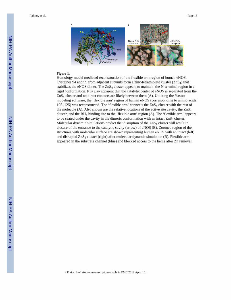
Figure 1.Homology model mediated reconstruction of the flexible arm region of human eNOS.Cysteines 94 and 99 from adjacent subunits form a zinc-tetrathiolate cluster (ZnS4) thatstabilizes the eNOS dimer. The ZnS4 cluster appears to maintain the N-terminal region in arigid conformation. It is also apparent that the catalytic center of eNOS is separated from theZnS4 cluster and no direct contacts are likely between them (A). Utilizing the Yasaramodeling software, the ‘flexible arm’ region of human eNOS (corresponding to amino acids105–125) was reconstructed. The ‘flexible arm’ connects the ZnS4 cluster with the rest ofthe molecule (A). Also shown are the relative locations of the active site cavity, the ZnS4cluster, and the BH4 binding site to the ‘flexible arm’ region (A). The ‘flexible arm’ appearsto be seated under the cavity in the dimeric conformation with an intact ZnS4 cluster.Molecular dynamic simulations predict that disruption of the ZnS4 cluster will result inclosure of the entrance to the catalytic cavity (arrow) of eNOS (B). Zoomed region of thestructures with molecular surface are shown representing human eNOS with an intact (left)and disrupted ZnS4 cluster (right) after molecular dynamic simulation (B). Flexible armappeared in the substrate channel (blue) and blocked access to the heme after Zn removal.
Rafikov et al. Page 18
J Endocrinol. Author manuscript; available in PMC 2012 April 16.
NIH
-PA Author Manuscript
NIH
-PA Author Manuscript
NIH
-PA Author Manuscript
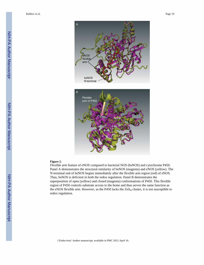
Figure 2.Flexible arm feature of eNOS compared to bacterial NOS (bsNOS) and cytochrome P450.Panel A demonstrates the structural similarity of bsNOS (magenta) and eNOS (yellow). TheN-terminal end of bsNOS begins immediately after the flexible arm region (red) of eNOS.Thus, bsNOS is deficient in both the redox regulation. Panel B demonstrates thesuperposition of open (yellow) and closed (magenta) conformations of P450. This flexibleregion of P450 controls substrate access to the heme and thus serves the same function asthe eNOS flexible arm. However, as the P450 lacks the ZnS4 cluster, it is not susceptible toredox regulation.
Rafikov et al. Page 19
J Endocrinol. Author manuscript; available in PMC 2012 April 16.
NIH
-PA Author Manuscript
NIH
-PA Author Manuscript
NIH
-PA Author Manuscript
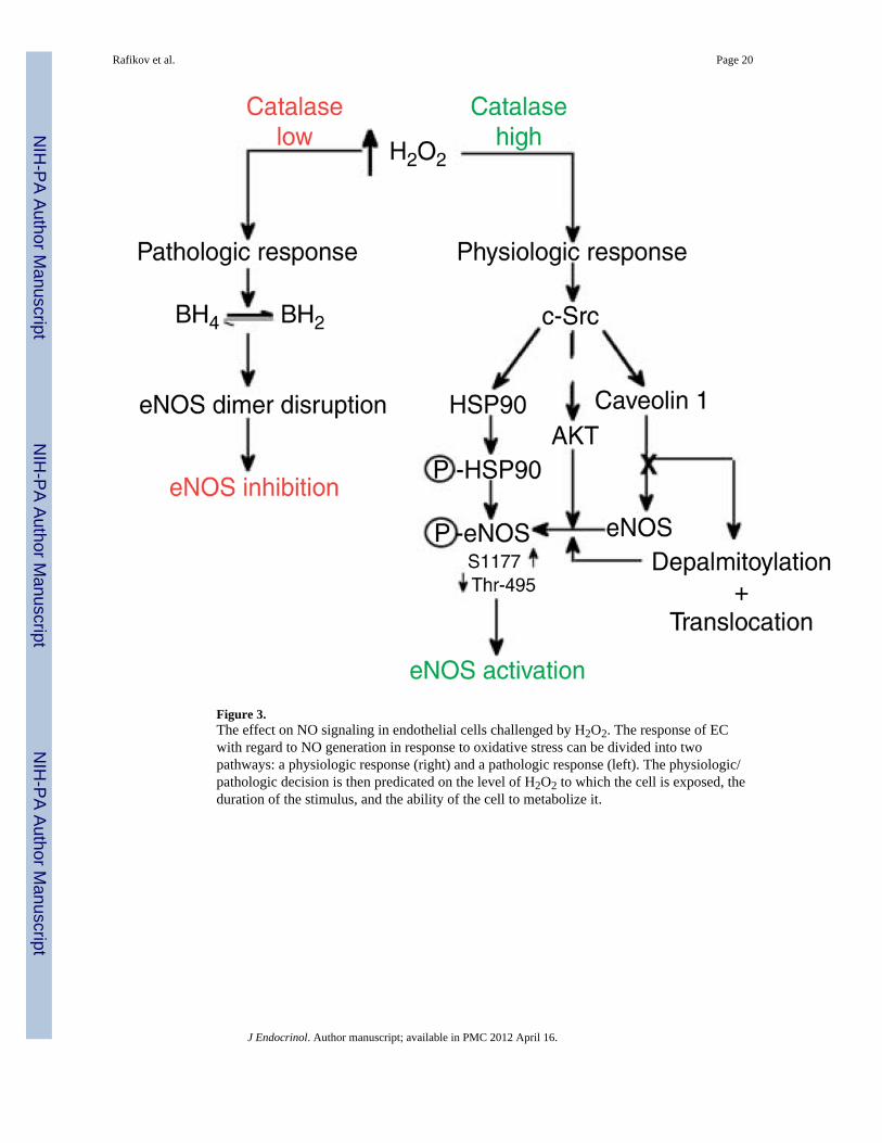
Figure 3.The effect on NO signaling in endothelial cells challenged by H2O2. The response of ECwith regard to NO generation in response to oxidative stress can be divided into twopathways: a physiologic response (right) and a pathologic response (left). The physiologic/pathologic decision is then predicated on the level of H2O2 to which the cell is exposed, theduration of the stimulus, and the ability of the cell to metabolize it.
Rafikov et al. Page 20
J Endocrinol. Author manuscript; available in PMC 2012 April 16.
NIH
-PA Author Manuscript
NIH
-PA Author Manuscript
NIH
-PA Author Manuscript
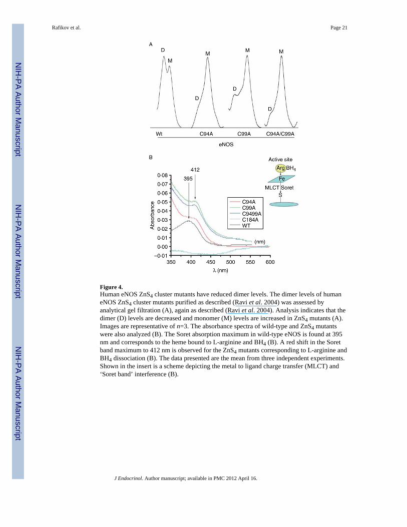
Figure 4.Human eNOS ZnS4 cluster mutants have reduced dimer levels. The dimer levels of humaneNOS ZnS4 cluster mutants purified as described (Ravi et al. 2004) was assessed byanalytical gel filtration (A), again as described (Ravi et al. 2004). Analysis indicates that thedimer (D) levels are decreased and monomer (M) levels are increased in ZnS4 mutants (A).Images are representative of n=3. The absorbance spectra of wild-type and ZnS4 mutantswere also analyzed (B). The Soret absorption maximum in wild-type eNOS is found at 395nm and corresponds to the heme bound to L-arginine and BH4 (B). A red shift in the Soretband maximum to 412 nm is observed for the ZnS4 mutants corresponding to L-arginine andBH4 dissociation (B). The data presented are the mean from three independent experiments.Shown in the insert is a scheme depicting the metal to ligand charge transfer (MLCT) and‘Soret band’ interference (B).
Rafikov et al. Page 21
J Endocrinol. Author manuscript; available in PMC 2012 April 16.
NIH
-PA Author Manuscript
NIH
-PA Author Manuscript
NIH
-PA Author Manuscript
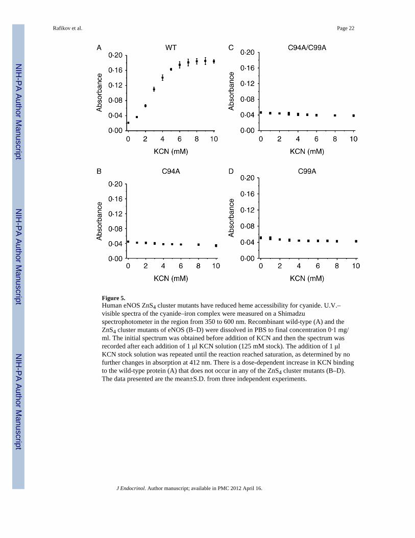
Figure 5.Human eNOS ZnS4 cluster mutants have reduced heme accessibility for cyanide. U.V.–visible spectra of the cyanide–iron complex were measured on a Shimadzuspectrophotometer in the region from 350 to 600 nm. Recombinant wild-type (A) and theZnS4 cluster mutants of eNOS (B–D) were dissolved in PBS to final concentration 0·1 mg/ml. The initial spectrum was obtained before addition of KCN and then the spectrum wasrecorded after each addition of 1 μl KCN solution (125 mM stock). The addition of 1 μlKCN stock solution was repeated until the reaction reached saturation, as determined by nofurther changes in absorption at 412 nm. There is a dose-dependent increase in KCN bindingto the wild-type protein (A) that does not occur in any of the ZnS4 cluster mutants (B–D).The data presented are the mean±S.D. from three independent experiments.
Rafikov et al. Page 22
J Endocrinol. Author manuscript; available in PMC 2012 April 16.
NIH
-PA Author Manuscript
NIH
-PA Author Manuscript
NIH
-PA Author Manuscript
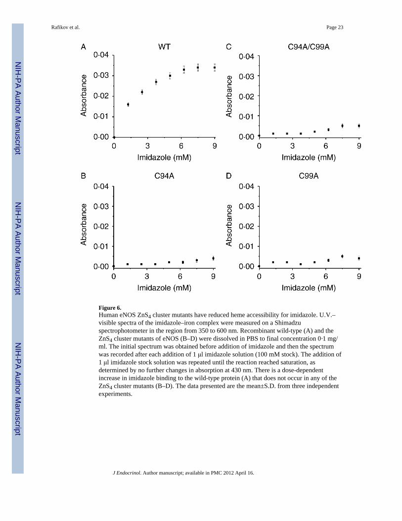
Figure 6.Human eNOS ZnS4 cluster mutants have reduced heme accessibility for imidazole. U.V.–visible spectra of the imidazole–iron complex were measured on a Shimadzuspectrophotometer in the region from 350 to 600 nm. Recombinant wild-type (A) and theZnS4 cluster mutants of eNOS (B–D) were dissolved in PBS to final concentration 0·1 mg/ml. The initial spectrum was obtained before addition of imidazole and then the spectrumwas recorded after each addition of 1 μl imidazole solution (100 mM stock). The addition of1 μl imidazole stock solution was repeated until the reaction reached saturation, asdetermined by no further changes in absorption at 430 nm. There is a dose-dependentincrease in imidazole binding to the wild-type protein (A) that does not occur in any of theZnS4 cluster mutants (B–D). The data presented are the mean±S.D. from three independentexperiments.
Rafikov et al. Page 23
J Endocrinol. Author manuscript; available in PMC 2012 April 16.
NIH
-PA Author Manuscript
NIH
-PA Author Manuscript
NIH
-PA Author Manuscript
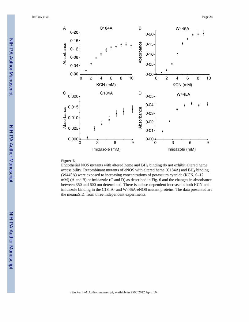
Figure 7.Endothelial NOS mutants with altered heme and BH4 binding do not exhibit altered hemeaccessibility. Recombinant mutants of eNOS with altered heme (C184A) and BH4 binding(W445A) were exposed to increasing concentrations of potassium cyanide (KCN, 0–12mM) (A and B) or imidazole (C and D) as described in Fig. 6 and the changes in absorbancebetween 350 and 600 nm determined. There is a dose-dependent increase in both KCN andimidazole binding in the C184A- and W445A-eNOS mutant proteins. The data presented arethe mean±S.D. from three independent experiments.
Rafikov et al. Page 24
J Endocrinol. Author manuscript; available in PMC 2012 April 16.
NIH
-PA Author Manuscript
NIH
-PA Author Manuscript
NIH
-PA Author Manuscript

Figure 8.NO and superoxide production by wild-type and the ZnS4 cluster mutants of eNOS.Recombinant wild-type eNOS and the ZnS4 cluster mutants (C94A, C99A, and C9499A)were incubated with L-arginine and the appropriate co-factors for 30 min at 37 °C and NO(A) and superoxide levels (B) determined as described (Grobe et al. 2006). There is asignificant reduction in both NO (A) and superoxide (B) generation in the ZnS4 clustermutants. Data are presented as mean±S.E.M.; n=3. *P<0·05 versus wild-typeeNOS. †P<0·05 versus C94A mutant.
Rafikov et al. Page 25
J Endocrinol. Author manuscript; available in PMC 2012 April 16.
NIH
-PA Author Manuscript
NIH
-PA Author Manuscript
NIH
-PA Author Manuscript

Figure 9.Molecular dynamic simulations identify the amino acids in the flexible arm of human eNOSresponsible for closure of the heme cavity. Superposition of intact eNOS (green) and withdisrupted ZnS4 (yellow) represents the flexible arm movement. Molecular dynamicsimulations predict conformational changes in the flexible arm region that are driven by saltbridge formation between positively charged amino acids (blue) of the flexible arm (Arg107,Lys108, and Arg112) and negatively charged amino acids on the wall of the substratechannel.
Rafikov et al. Page 26
J Endocrinol. Author manuscript; available in PMC 2012 April 16.
NIH
-PA Author Manuscript
NIH
-PA Author Manuscript
NIH
-PA Author Manuscript

Figure 10.The human eNOS flexible arm mutant undergoes self-nitration. Purified wild-typerecombinant eNOS was compared to the flexible arm mutant contained on a disrupted ZnS4cluster background (C94A/C99A/R107A/K108A/R112A). The flexible arm mutant hasdimer (D) and monomer (M) levels, as determined by analytic gel filtration, that are similarto those seen with wild-type eNOS (wt eNOS, A). The absorbance spectrum of the flexiblearm mutant contains a Soret band at 395 nm that is comparable with wild-type eNOS (B).The flexible arm mutation also allows both cyanide (C) and imidazole (D) to access theheme. The flexible arm mutant produces NO at levels comparable to the wild-type protein(E). However, the flexible arm mutant significantly generates greater levels of bothsuperoxide (F) and ONOO− (G) compared with the wild-type protein. Western blot analysisusing an antibody specific for 3-NT residues indicates that the flexible arm mutantundergoes increased self-nitration compared with either wild-type eNOS or the ZnS4 clustermutants (C94A, C99A, and C94A/99A, H). A representative image is shown. Data arepresented as mean±S.E.M.; n=3. *P<0·05 versus wild-type eNOS.
Rafikov et al. Page 27
J Endocrinol. Author manuscript; available in PMC 2012 April 16.
NIH
-PA Author Manuscript
NIH
-PA Author Manuscript
NIH
-PA Author Manuscript
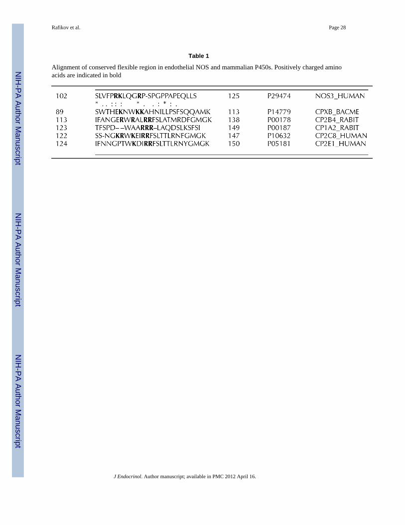
NIH
-PA Author Manuscript
NIH
-PA Author Manuscript
NIH
-PA Author Manuscript
Rafikov et al. Page 28
Table 1
Alignment of conserved flexible region in endothelial NOS and mammalian P450s. Positively charged aminoacids are indicated in bold
J Endocrinol. Author manuscript; available in PMC 2012 April 16.