Posttranslational regulation of CIRCADIAN CLOCK ASSOCIATED 1 (CCA1) in the circadian oscillator of...
46
Running title: Post-translational regulation of CCA1 Corresponding author: Rachel Green Department of Plant and Environmental Sciences, The Silberman Institute for Life Sciences, The Hebrew University, Givat Ram, Jerusalem 91904, Israel. Tel: +972 2 658 5391, Fax: +972 2 658 4425, [email protected] Journal research area: System Biology, Molecular Biology, and Gene Regulation Plant Physiology Preview. Published on April 1, 2009, as DOI:10.1104/pp.109.137414 Copyright 2009 by the American Society of Plant Biologists www.plant.org on March 14, 2014 - Published by www.plantphysiol.org Downloaded from Copyright © 2009 American Society of Plant Biologists. All rights reserved.
Transcript of Posttranslational regulation of CIRCADIAN CLOCK ASSOCIATED 1 (CCA1) in the circadian oscillator of...

Running title: Post-translational regulation of CCA1
Corresponding author:
Rachel Green
Department of Plant and Environmental Sciences, The Silberman Institute for Life Sciences,
The Hebrew University, Givat Ram, Jerusalem 91904, Israel.
Tel: +972 2 658 5391,
Fax: +972 2 658 4425,
Journal research area: System Biology, Molecular Biology, and Gene Regulation
Plant Physiology Preview. Published on April 1, 2009, as DOI:10.1104/pp.109.137414
Copyright 2009 by the American Society of Plant Biologists
www.plant.org on March 14, 2014 - Published by www.plantphysiol.orgDownloaded from Copyright © 2009 American Society of Plant Biologists. All rights reserved.

Post-translational regulation of CIRCADIAN CLOCK ASSOCIATED 1 (CCA1) in the
circadian oscillator of Arabidopsis thaliana
Esther Yakir1, Dror Hilman1, Ido Kron1, Miriam Hassidim1, Naomi Melamed-Book2 and Rachel
M. Green1.
1Department of Plant and Environmental Sciences, The Silberman Institute for Life Sciences,
The Hebrew University, Givat Ram, Jerusalem 91904, Israel.
2Bio-Imaging Unit, The Silberman Institute for Life Sciences,
The Hebrew University, Givat Ram, Jerusalem 91904, Israel.
www.plant.org on March 14, 2014 - Published by www.plantphysiol.orgDownloaded from Copyright © 2009 American Society of Plant Biologists. All rights reserved.

Financial source:
This work was supported by BSF grant 0378415 and DIP grant 0307712.
Corresponding author:
Rachel Green
www.plant.org on March 14, 2014 - Published by www.plantphysiol.orgDownloaded from Copyright © 2009 American Society of Plant Biologists. All rights reserved.

Abstract
As an adaptation to life in a world with predictable daily changes, most eukaryotes and
some prokaryotes have endogenous circadian (~24 hour) clocks. In plants, the circadian clock
regulates a diverse range of cellular and physiological events from gene expression and protein
phosphorylation to cellular calcium oscillations, hypocotyl growth, leaf movements and
photoperiod-dependant flowering. In Arabidopsis thaliana, as in other model organisms, such as
Drosophila and mice, circadian rhythms are generated by molecular oscillators that consist of
interlocking feedback loops involving a number of elements. CIRCADIAN CLOCK
ASSOCIATED 1 (CCA1) and LATE ELONGATED HYPOCOTYLS (LHY) are closely-related
single myb transcription factors that have been identified as key elements in the Arabidopsis
oscillator. Research in other model organisms has shown that post-translational regulation of
oscillator components plays a critical role in the generation of the ~24 hour cycles. To examine
the role of post-translational regulation of CCA1 and LHY in the Arabidopsis oscillator, we have
generated transgenic plants with tagged CCA1 and LHY under the control of their own
promoters. We have shown that these tagged proteins are functional and can restore normal
circadian rhythms to CCA1- and LHY-null plants. Using the tagged proteins, we demonstrate
that CCA1 can form both homodimers and heterodimers with LHY. Furthermore, we also show
that CCA1 is localized to the nucleus in vivo and that there is no significant delay between the
translation of CCA1 and its translocation to the nucleus. We discuss our findings in the context
of the functioning of the Arabidopsis oscillator.
www.plant.org on March 14, 2014 - Published by www.plantphysiol.orgDownloaded from Copyright © 2009 American Society of Plant Biologists. All rights reserved.

Introduction
The circadian, ~24 hour, clock has an enormous influence on the biology of plants and
controls a plethora of processes including hypocotyl growth, shade avoidance, leaf movements,
scent production and stomatal opening (Yakir et al., 2007). Consistent with the role of the
circadian clock in the regulation of a wide range of activities, transcription from approximately
one third of the genome (including non-coding genes) of the model plant, Arabidopsis thaliana,
is under circadian control (Michael and McClung, 2003; Covington et al., 2008; Michael et al.,
2008; Hazen et al., 2009). In addition, the circadian clock serves as a timekeeper to regulate
daylength-dependent processes such as flowering time and tuberization. The circadian systems
responsible for generating circadian rhythms are ubiquitous in eukaryotes and have also been
found in some prokaryotes (Dunlap, 1999). Conceptually, a circadian system can be divided into
three parts: the oscillator mechanism, input pathways and output pathways. Interestingly,
although their components differ, the basic oscillator mechanism appears to be well conserved in
all the eukaryotic model organisms that have been studied (Dunlap, 2006). Oscillators are
comprised of interlocking positive/negative feedback loops made up from clock proteins that
control their own rhythms. The oscillators can be entrained by signals from the environment such
as temperature and light changes. Output pathways from the oscillating clock proteins in turn
convey circadian rhythms to the various physiological and molecular processes.
The model of the Arabidopsis oscillator consists of interlocking feedback loops of several
components including CIRCADIAN CLOCK ASSOCIATED 1 (CCA1), LATE ELONGATED
HYPOCOTYL (LHY) and TIMING OF CAB EXPRESSION 1 (TOC1). CCA1 and LHY are
homologous myb-related transcription factors that show robust circadian oscillations in their RNA and
protein levels (Schaffer et al., 1998; Wang and Tobin, 1998). Over-expression of CCA1 or LHY in
transgenic Arabidopsis plants abrogates the circadian rhythmicity of clock-controlled processes,
including gene expression and leaf movements (Schaffer et al., 1998; Wang and Tobin, 1998; Thain et
al., 2004). Mutations in CCA1 and LHY result in a shorter period of circadian-controlled gene
expression and leaf movements than wild-type plants, while plants with mutations in both genes appear
to be unable to maintain sustained oscillations (Green and Tobin, 1999; Alabadi et al., 2002; Mizoguchi
et al., 2002). However, despite their homology, CCA1 and LHY may have somewhat different roles in
the oscillator mechanism (Gould et al., 2006; Zeilinger et al., 2006). TOC1 (also known as APRR1)
belongs to the APRR/TOC1 family of genes (Makino et al., 2000; Strayer et al., 2000) that encode
nuclear proteins with a region that is common to several transcription factors. Mutations in TOC1 also
www.plant.org on March 14, 2014 - Published by www.plantphysiol.orgDownloaded from Copyright © 2009 American Society of Plant Biologists. All rights reserved.

cause a short period phenotype (Somers et al., 1998). Other genes such as GIGANTEA (GI), EARLY
FLOWERING 4 (ELF4), LUX ARRHYTHMO (LUX), TIME FOR COFFEE (TIC), LIGHT
INSENSITIVE PERIOD1 (LIP1), ARABIDOPSIS PSEUDO-RESPONSE REGULATOR 3/5/7/9
(APPR3/5/7/9), LIGHT-REGULATED WD 1 and 2 (LWD1/2) and FIONA1 might operate in the
oscillator or close to it (Doyle et al., 2002; Hazen et al., 2005; Locke et al., 2005; Nakamichi et al.,
2005; Edwards et al., 2006; Gould et al., 2006; Ding et al., 2007; Kevei et al., 2007; McWatters et al.,
2007; Para et al., 2007; Kim et al., 2008; Wu et al., 2008).
Considerable progress has been made in determining the mechanisms by which the circadian
elements described above interact to generate circadian rhythms in Arabidopsis. In the least complex
model of the main oscillator, CCA1 and LHY expression rises before dawn and suppresses the
expression of TOC1 by binding to its promoter. In the evening, when CCA1 and LHY levels decrease,
TOC1 expression rises. TOC1 then activates CCA1 and LHY expression by an as yet unknown
mechanism (Alabadi et al., 2001). Recently, a combination of experimental and mathematical
modeling techniques (Locke et al., 2006; Zeilinger et al., 2006) have supported the idea that other
morning and evening genes such as PRR7, PRR9 and GI are involved in the oscillator, possibly by
forming two other additional feedback loops interacting with the CCA1/LHY/TOC1 loop.
However, although the CCA1/LHY/TOC1 transcription/translation feedback loop is clearly central to
the mechanism of the Arabidopsis oscillator, it is probably insufficient to create a cycle that takes ~24
hours. Thus, there are likely to be additional steps, including post-translational modifications, built in to
the cycling of oscillator components to ensure that the loop takes about a day to complete. Consistent
with the idea of post-translation modifications having a role in the oscillator, in the oscillators of two of
the best-studied organisms, mice and Drosophila, phosphorylation, protein interactions and cellular
localization play a crucial role in the regulation of the oscillator (Kwon et al., 2006; Meyer et al., 2006).
Post-translational modifications also play a role in the Arabidopsis oscillator. There is evidence
for the role of post-translational regulation of TOC1. ZTL (ZEITLUPE), an F-box protein, has been
shown to regulate TOC1 degradation, probably through a CUL1 (CULLIN1) containing SCF
complex, and ZTL itself is stabilized by interaction with GI (Mas et al., 2003; Kim et al., 2007;
Harmon et al., 2008). GI stability is regulated by CONSTITUTIVE PHOTOMORPHOGENIC 1
(COP1) and ELF3 (Yu et al., 2008). In addition, a phosphorylated form of TOC1 can interact with a
phosphorylated form of PRR3. The TOC1/PRR3 interaction competes with the TOC1/ZTL interaction
and possibly prevents TOC1 degradation in the vascular tissues (Para et al., 2007; Fujiwara et al.,
2008). Some work, mostly in vitro, has also started to show how CCA1 and LHY are modified. In
www.plant.org on March 14, 2014 - Published by www.plantphysiol.orgDownloaded from Copyright © 2009 American Society of Plant Biologists. All rights reserved.

westerns of total cellular protein extracts, CCA1 and LHY levels oscillate with a similar pattern,
peaking around dawn with no detectable protein late in the day (Wang and Tobin, 1998; Kim et al.,
2003). In addition, LHY may be degraded through the proteasomal pathway and DET1 (DE-
ETIOLATED 1) inhibits LHY turn-over (Wang and Tobin, 1998; Song and Carre, 2005). In vitro,
both CCA1 and LHY are phosphorylated by CK2 (Sugano et al., 1998) and when the CK2
phosphorylation sites in CCA1 are mutated, CCA1 activity is altered (Daniel et al., 2004). Moreover,
the DNA-binding activity of CCA1 from plant extracts requires phosphorylation by CK2 (Portolés and
Más, 2007). CCA1 and LHY have been shown to be localized to the nucleus (Wang et al., 1997; Carre
and Kim, 2002; Perales and Mas, 2007; Gutierrez et al., 2008) although there is no data about the
timing and control of this localization.
In general, however, little is known about the crucial post-translational localization and
interactions of CCA1 and LHY in the Arabidopsis oscillator in vivo. In order to study the post-
translational regulation of CCA1 and LHY, we have generated transgenic plants with tagged CCA1
and LHY proteins and have shown that these tagged proteins are fully functional. Using the tagged
proteins we demonstrate that in vivo CCA1 can interact with both itself and with LHY, moreover, after
translation CCA1 moves rapidly into the nucleus. Thus post-translational delays built into the
oscillators of other model organisms appear not to be present in the Arabidopsis circadian oscillator.
Results
CCA1 moves rapidly into the nucleus after translation
In order to be able to follow the sub-cellular localization and interactions of CCA1
protein, we generated transgenic plants with tagged CCA1. We made a construct of the CCA1
cDNA fused to three HA-tags and YFP (Yellow Fluorescent Protein) under the control of the
CCA1 promoter (1222bp up-stream of the start codon) shown in Figure 1A. The resulting
construct was used to transform CCA1-null plants (cca1-1; Green and Tobin, 1999). Two
transgenic lines, CCA1pro::CCA1-HA-YFP cca1-1#1 and CCA1pro::CCA1-HA-YFP cca1-1#2,
were chosen for further studies.
To determine whether CCA1 levels in the CCA1pro::CCA1-HA-YFP cca1-1 lines are
comparable to the levels in wild-type plants, CCA1pro::CCA1-HA-YFP cca1-1#1,
CCA1pro::CCA1-HA-YFP cca1-1#2 and wild-type plants were grown for two weeks in 14 hours
light:10 hours dark (LD). Tissue was harvested at zero and five hours after lights-on and mRNA
extracted. CCA1-HA-YFP mRNA levels in the transgenic plants and CCA1 mRNA levels in
www.plant.org on March 14, 2014 - Published by www.plantphysiol.orgDownloaded from Copyright © 2009 American Society of Plant Biologists. All rights reserved.

wild-type were determined by qRT-PCR. Figure 1B shows that the levels of CCA1-HA-YFP
mRNA in both of the transgenic lines are comparable to CCA1 levels in wild-type plants, with
higher levels at lights-on than five hours later. We then examined the accumulation of the CCA1-
HA-YFP protein. CCA1pro::CCA1-HA-YFP cca1-1#1 and CCA1pro::CCA1-HA-YFP cca1-1#2
plants were grown for two weeks in 14 hours light:10 hours dark (LD). Tissue was harvested at
intervals and protein extracted and determined by western analysis of total protein extracts using
an anti-HA antibody. Our results show that the levels of CCA1-HA-YFP protein in both
transgenic lines reach a maximum at 1.5 hours after lights-on (Figure 1C). Thus, the peak of
CCA1-HA-YFP is similar to that previously reported for CCA1 in wild-type plants (Wang and
Tobin, 1998). Taken together, our results suggest that CCA1-HA-YFP is expressed at both the
transcriptional and translational levels in the CCA1pro::CCA1-HA-YFP cca1-1#1 and
CCA1pro::CCA1-HA-YFP cca1-1#2 plants in a similar way to CCA1 in wild-type plants.
Our next goal was to check if, in addition to showing the correct pattern of accumulation
in LD, the CCA1-HA-YFP protein could also mimic the activity of wild-type CCA1. As a test
for CCA1-HA-YFP activity, we examined whether CCA1-HA-YFP could lengthen the period of
circadian-regulated leaf movement in the cca1-1 plants that have been shown previously to have
a shorter circadian period than wild-type plants (Mizoguchi et al., 2002). CCA1pro::CCA1-HA-
YFP cca1-1#1, CCA1pro::CCA1-HA-YFP cca1-1#2, cca1-1 and wild-type plants were grown for
seven days in LD before being transferred to continuous light (LL). Figure 1D shows that the
periods of leaf movement of the CCA1pro::CCA1-HA-YFP cca1-1#1 and CCA1pro::CCA1-HA-
YFP cca1-1#2 plants are longer (25.6±0.2 s.e. and 24.7±0.6 s.e. respectively) than those of
cca1-1 plants (22.9±0.2 s.e.) and resemble the wild-type (24.3±0.1 s.e.). The average traces for
leaf movements of the transgenic and control lines also showed that both transgenic lines were
rhythmic with, especially CCA1pro::CCA1-HA-YFP cca1-1#1, a pattern of rhythmicity close to
wild-type (Figure S1A). Thus, our results show that the CCA1-HA-YFP in the CCA1pro::CCA1-
HA-YFP cca1-1 plants has not only a similar pattern of expression to CCA1 in wild-type plants
but also a similar circadian activity.
CCA1 has been reported in the nuclei of wild-type plants at dawn (Gutierrez et al., 2008),
and we examined whether CCA1-HA-YFP could also be detected in the nuclei of
CCA1pro::CCA1-HA-YFP cca1-1 plants by examining the sub-cellular localization of
fluorescence from YFP. CCA1pro::CCA1-HA-YFP cca1-1#1, CCA1pro::CCA1-HA-YFP cca1-
1#2 and wild-type plants were grown for two weeks in LD and then examined by confocal
www.plant.org on March 14, 2014 - Published by www.plantphysiol.orgDownloaded from Copyright © 2009 American Society of Plant Biologists. All rights reserved.

microscopy. Figures 2A and S2A shows that three hours after lights-on there is a fluorescence
signal in both the epidermal and mesophyll cells of CCA1pro::CCA1-HA-YFP cca1-1#1 and
CCA1pro::CCA1-HA-YFP cca1-1#2 plants. Staining the cells with DAPI indicated that the
majority of the fluorescence in each cell was localized to the nucleus. We could not detect any
fluorescence signals in the nuclei of wild-type plants (Figure 2B). Thus CCA1-HA-YFP is found
in the nuclei of the CCA1pro::CCA1-HA-YFP cca1-1 plants.
Since in Drosophila the timing of PER and TIM movement into the nucleus is an
important part of the mechanism of the oscillator that allows it to cycle with ~24 hour period, we
examined the rate of CCA1-HA-YFP movement into the nucleus. CCA1pro::CCA1-HA-YFP
cca1-1#1 and CCA1pro::CCA1-HA-YFP cca1-1#2 plants were grown for two weeks in LD and
harvested at intervals. The nuclear and cytoplasmic fractions of the tissue were isolated (as
described in Materials and Methods) and examined by western analysis using an anti-HA
antibody. Figures 2C and S2B show that in both lines even at zero hours (lights-on) when total
protein levels are still very low, CCA1-HA-YFP is already in the nucleus in levels that are
similar to those in the cytoplasm. CCA-HA-YFP continues to accumulate in the nucleus before
declining. Figures 2C and S2B also show that in both lines, the levels of CCA1-HA-YFP protein
remain higher in the cytoplasm for longer. The fact that we observed high levels of CCA1-HA-
YFP protein in the cytoplasmic extracts at a time when the nuclear CCA1-HA-YFP levels have
declined, suggests that our nuclear extracts were free of significant amounts of cytoplasmic
contamination. To verify the purity of the nuclear extracts we used antibodies against histone 3
(Figure 2D), histone 4 (data not shown) and RNA polymerase II (Figure S2C) on westerns with
the cytoplasmic and nuclear fractions. Figures 2D and S2C show that the cytoplasmic fraction
was free from detectable contamination by nuclear material.
To confirm our findings that CCA1-HA-YFP moves into the nucleus soon after
translation and to examine the effects of light on its translocation, we also examined the sub-
cellular localization of fluorescence from YFP. CCA1pro::CCA1-HA-YFP cca1-1#1 and
CCA1pro::CCA1-HA-YFP cca1-1#2 plants were grown for two weeks in LD and then, at the end
of the last dark period, transferred to light or kept in the dark. YFP fluorescence was examined
by confocal microscopy. The levels of fluorescence in the nuclei of epidermis, stomata and
mesophyll cells were calculated and plotted as a function of time. As can be seen in Figures 2E
and S2D, at lights-on there were detectable levels of fluorescence in the nuclei. Fluorescence
levels reached a maximum around 1.5 hours after lights-on and then started to decrease. A
www.plant.org on March 14, 2014 - Published by www.plantphysiol.orgDownloaded from Copyright © 2009 American Society of Plant Biologists. All rights reserved.

comparison between the changes in CCA1-HA-YFP protein levels (Figure 1C) and the
fluorescence in the nucleus (Figures 2E and S2D) indicates that CCA1-HA-YFP protein can
move into the nucleus very soon after translation. The fact that we detected CCA1-HA-YFP in
the cytoplasm on westerns but little fluorescence from the YFP in the cytoplasm, is probably
because the CCA1-HA-YFP in the cytoplasm is more diffuse resulting in a weaker signal. Figure
2E also shows that the translocation of CCA1-HA-YFP protein into the nucleus is unaffected by
dark. This finding is consistent with the results shown in Figures 2C and S2B that there are
already detectable levels of CCA1-HA-YFP in the nucleus at lights-on. Taken together our
results show that CCA1-HA-YFP is able to move rapidly in to the nucleus after translation and
that this translocation is unaffected by light and dark.
CCA1 and LHY interact in vitro
Several structural studies have shown that in proteins containing two myb domains both
are necessary for DNA binding and it has been suggested that proteins containing only one myb
domain might bind DNA as dimers (Jin and Martin, 1999). Moreover, other proteins containing
single myb domains have been shown to dimerize (Bianchi et al., 1997). We therefore
hypothesized that CCA1 and LHY might function as heterodimers.
As a preliminary step to check whether CCA1 and LHY might function as heterodimers,
we determined whether CCA1 and LHY could form heterodimers in vitro. We expressed the
full-length CCA1 cDNA fused to three HA-tags (CCA1-HA[full]) and the full-length LHY cDNA
fused to a myc-tag (LHY-myc[full]) in E.coli. Extracts from the bacterial cultures were separated
by SDS-PAGE and CCA1-HA and LHY-myc identified by westerns with anti-HA and anti-myc
antibodies. Figure 3A and B shows that CCA1-HA and LHY-myc are expressed and recognized
correctly by the antibodies. The extracts were then used in reciprocal co-immunoprecipitation
(co-IP) experiments (as described in Materials and Methods). The co-IPs were done on a mixture
of CCA1-HA and LHY-myc protein extracts with either anti-HA antibody (Figure 3C and 3D
lane 7) or anti-myc antibody (Figure 3C and 3D lane 8). The immunoprecipitated proteins were
separated by SDS-PAGE and CCA1-HA and LHY-myc identified by westerns using the anti-
myc (Figure 3C) or anti-HA (Figure 3D) antibodies. Our results show that CCA1-HA was
immunoprecipitated by LHY-myc (Figure 3D lane 8) and that LHY-myc was
immunoprecipitated by CCA1-HA (Figure 3C lane 7). Thus, CCA1 and LHY form heteodimers
in vitro. As a control for the specificity of the antibodies, we showed that anti-HA precipitates
www.plant.org on March 14, 2014 - Published by www.plantphysiol.orgDownloaded from Copyright © 2009 American Society of Plant Biologists. All rights reserved.

CCA1-HA[full] and not LHY-myc[full] (3D lane 3 compared to Figure 3C lane 4) and that anti-
myc precipitates LHY-myc[full] and not CCA1-HA[full] (Figure 3C lane 6 compared to Figure
3D lane 5). As a further control, to check that there is a specific interaction between CCA1-
HA[full] and LHY rather than with the myc tag, we co-immunoprecipitated a mixture of CCA1-
HA[full] and GST-myc protein extract with anti-HA antibodies. No GST-myc band was detected
(Figure 3E lane 2).
To start to determine the domains in CCA1 that are necessary for its interactions with
LHY, we made two truncated CCA1-HA proteins and expressed them in E.coli. The constructs,
CCA1-HA[407] and CCA1-HA[223] contained the last 407 or 223 amino acids of CCA1
respectively with three HA tags (Figure 3F). Bacterial extracts of CCA1-HA[full], CCA1-
HA[407] and CCA1-HA[223] were used in co-IP experiments with LHY-myc[full] (as described
in Materials and Methods). The immunoprecipitated proteins were separated by SDS-PAGE and
LHY-myc[full] identified by westerns using the anti-myc antibodies. Figure 3G shows that only
CCA1-HA[full] immunoprecipitated LHY-myc[full] (lane 5 compared to lane 6 and 7). Since
LHY-myc[full] was not immunoprecipitated by either of the truncated CCA1-HA proteins, it is
likely that the region of CCA1 responsible for CCA1/LHY interactions is on the N’-terminal,
perhaps near the myb domain. However, we cannot rule out the possibility that the truncated
CCA1-HA proteins are mis-folded and that the correct folding of CCA1 is necessary for its
interaction with LHY. Taken together our results show that CCA1 and LHY interact in vitro and
that this interaction probably requires the N’-terminal of CCA1.
CCA1 and LHY interact in vivo
In order to be able to examine whether CCA1 and LHY also interact in vivo, we
generated transgenic plants with tagged LHY. We made a construct (LHY-myc) of the LHY
cDNA fused to a myc-tag under the control of the LHY promoter (1568bp up-stream of the start
codon) shown in Figure 4A. A line of plants with a T-DNA insertion in the LHY sequence was
obtained from the SALK institute. From this T-DNA line, we isolated a homozygous plant (LHY-
null) that did not express LHY (Figure S3). We then transformed LHY-null with the LHY-myc
construct to generate LHYpro::LHY-myc LHY-null.
To determine whether LHY-myc levels in the transgenic line are comparable to the levels
of LHY in wild-type plants, LHYpro::LHY-myc LHY-null and wild-type plants were grown for
two weeks in 14 hours light:10 hours dark (LD). Tissue was harvested at zero and five hours
www.plant.org on March 14, 2014 - Published by www.plantphysiol.orgDownloaded from Copyright © 2009 American Society of Plant Biologists. All rights reserved.

after lights-on and mRNA extracted. LHY-myc mRNA levels in the transgenic plants and LHY
mRNA levels in wild-type were determined by qRT-PCR. Figure 4B shows that the levels of
LHY-myc mRNA from the LHYpro::LHY-myc construct in the transgenic line are comparable to
LHY levels in wild-type plants. We then examined the accumulation of the LHY-myc protein.
LHYpro::LHY-myc LHY-null plants were grown for two weeks in 14 hours light:10 hours dark
(LD). Tissue was harvested in intervals and protein extracted and examined by western analysis
of total protein extracts using an anti-myc antibody. The LHY-myc protein levels peak around
dawn in the LHYpro::LHY-myc LHY-null plants (Figure 4C). Thus, the peak of LHY-myc is
close to that previously reported for LHY in wild-type plants (Kim et al., 2003)
In order to verify that the LHY-myc activity in plants is also similar to the endogenous
LHY activity, we checked whether LHYpro::LHY-myc could restore normal circadian rhythms of
leaf movements to LHY-null plants. Plants with mutated LHY have been shown to have a shorter
circadian period than wild-type plants (Mizoguchi et al., 2002). LHYpro::LHY-myc LHY-null,
LHY-null and wild-type plants were grown for seven days in LD before being transferred to LL.
Figure 4D demonstrates that the leaf movement period of LHYpro::LHY-myc LHY-null plants
(24±0.3 s.e.) is longer than that of the LHY-null plants (20.5±0.4 s.e.) and close to that of wild-
type plants (24±0.2 s.e.). We also observed that the average traces for leaf movements of the
LHYpro::LHY-myc LHY-null closely matched those of the wild-type control (Figure S1B). Thus,
our results show that the LHY-myc in the LHYpro::LHY-myc LHY-null plants has a similar
circadian activity to LHY in wild-type plants.
Our next goal was to examine whether CCA1 and LHY could interact in vivo. The
LHYpro::LHY-myc LHY-null plants were crossed into CCA1pro::CCA1-HA-YFP cca1-1#1
plants. The resulting LHYpro::LHY-myc LHY-null;CCA1pro::CCA1-HA-YFPcca1-1#1 plants
were grown in LD for four weeks. Tissue was harvested two hours after lights-on and used for
co-IP assays with anti-HA antibodies. The immunoprecipitated proteins were separated by SDS-
PAGE and LHY-myc identified by westerns using anti-myc. Our results show that the co-IP
resulted in a band the size of LHY-myc (Figure 5A lane 6) suggesting that CCA1-HA-YFP and
LHY-myc interact in vivo. In a control co-IP carried out without the anti-HA antibody, no
significant LHY-myc could be detected (Figure 5A lane 4). As a further control to show the that
the anti-HA antibody interacted specifically with CCA1-HA-YFP and not with LHY-myc, the
immunoprecipitation assay was also performed on LHYpro::LHY-myc LHY-null extracts. Figure
5A lane 2 shows that in the absence of CCA1-HA-YFP, no LHY-myc was precipitated. We also
www.plant.org on March 14, 2014 - Published by www.plantphysiol.orgDownloaded from Copyright © 2009 American Society of Plant Biologists. All rights reserved.

performed the reciprocal assay using the anti-myc antibody to co-IP CCA1-HA-YFP. Figure 5B
shows that CCA1-HA-YFP was precipitated only when the anti-myc antibody was used (Figure
5B lane 4 compared to lane 6). By contrast, the anti-myc antibody could not pull down CCA1-
HA-YFP from CCA1pro::CCA1-HA-YFP cca1-1#1 plants (Figure 5B lane 2). Our results clearly
show that CCA1 and LHY interact in vivo.
Since our results (Figure 1C) showed that the levels of CCA1-HA-YFP protein changes
during the day, we examined whether the in vivo interactions between CCA1-HA-YFP and
LHY-myc also show changes over time. We collected leaf samples from two-week old LD-
grown LHYpro::LHY-myc LHY-null;CCA1pro::CCA1-HA-YFPcca1-1#1 plants at intervals as
described (Figure 5C). The samples were subjected to co-IP with the anti-myc antibody, the
immunoprecipitated proteins were separated by SDS-PAGE and CCA1-HA-YFP identified using
anti-HA. Figure 5C shows that there is correlation between the levels of CCA1-HA-YFP and the
amount of CCA1-HA-YFP interacting with LHY-myc. Our results suggest that the interaction
between CCA1 and LHY depends mostly on their abundance and that they may form
heterodimers whenever they are present together in the cell.
CCA1 can form homodimers in vivo
Previous work in several labs has established that CCA1 and LHY are partially redundant
and that in the absent of LHY, CCA1 might function as a substitute for LHY in the circadian
system (Green and Tobin, 1999; Alabadi et al., 2002; Mizoguchi et al., 2002). In order to examine
whether CCA1 might also be able to form homodimers, we made a construct of the CCA1 cDNA
fused to a myc-tag under the control of the CCA1 promoter (1222bp up stream of the start codon;
Figure 6A). The resulting construct was used to transform cca1-1*LHY-null plants
(CCA1pro::CCA1-myc cca1-1*LHY-null). Figure 6B shows that the levels of CCA1-myc mRNA
in CCA1pro::CCA1-myc cca1-1*LHY-null#1 are comparable to the levels of CCA1 mRNA in
wild-type. In the CCA1pro::CCA1-myc cca1-1*LHY-null#2 plants CCA1-myc mRNA levels are
higher at zero hours than CCA1 mRNA in the wild-type but similar at five hours. Figure 6C
shows that, like CCA1-HA-YFP in the CCA1pro::CCA1-HA-YFP cca1-1#1 plants, the level of
CCA1-myc in the CCA1pro::CCA1-myc cca1-1*LHY-null#1 and CCA1pro::CCA1-myc cca1-
1*LHY-null#2 plants is highest 1.5 hours after lights-on, although the levels continue to remain
fairly high throughout the time course of the experiment.
www.plant.org on March 14, 2014 - Published by www.plantphysiol.orgDownloaded from Copyright © 2009 American Society of Plant Biologists. All rights reserved.

We also examined whether CCA1-myc could restore circadian rhythms of leaf
movements to cca1-1*LHY-null plants. Consistent with previous reports that plants with mutated
CCA1 and LHY have weak circadian rhythms (Mizoguchi et al., 2002), Figure 6D shows that
cca1-1*LHY-null plants do not have robust rhythms. The relative amplitude error (R.A.E.) is a
measurement of the robustness of a circadian rhythm (Somers et al., 2004) and fewer then 15%
of the cca1-1*LHY-null plants have an R.A.E. of less then 0.6, i.e. are significantly rhythmic. By
contrast, Figure 6D shows that both CCA1pro::CCA1-myc cca1-1*LHY-null#1 and
CCA1pro::CCA1-myc cca1-1*LHY-null#2 have robust circadian rhythms of leaf movements.
More then 85% of the CCA1pro::CCA1-myc cca1-1*LHY-null#1 plants and more than 80% of
the CCA1pro::CCA1-myc cca1-1*LHY-null#2 plants showed an R.A.E. of less then 0.6. In
addition, the average traces for leaf movements of the CCA1pro::CCA1-myc cca1-1*LHY-null
lines showed rhythmicity while the trace for cca1-1*LHY-null plants was arrhythmic (Figure
S1C). Thus, CCA1-myc in the CCA1pro::CCA1-myc cca1-1*LHY-null plants not only has a
similar pattern of expression to endogenous CCA1 in wild-type plants but also has similar
circadian activity.
To determine whether CCA1 can interact with itself, CCA1pro::CCA1-myc cca1-1*LHY-
null#1 and CCA1pro::CCA1-myc cca1-1*LHY-null#2 plants were crossed into CCA1pro::CCA1-
HA-YFP cca1-1#1 plants. The resulting CCA1pro::CCA1-myc cca1-1*LHY-
null;CCA1pro::CCA1-HA-YFPcca1-1#1 plants were grown for four weeks in LD. Tissue was
harvested two hours after lights-on and used for co-IP assays with anti-myc antibodies. The
immunoprecipitated proteins were separated by SDS-PAGE and CCA1-HA-YFP identified by
western analysis using anti-HA antibodies. Our previous results (Figure 5B lane 2) showed that
CCA1-HA-YFP is not precipitated by anti-myc antibodies in plants that do not express CCA1-
myc. Figure 6E shows that, in the CCA1pro::CCA1-myc cca1-1*LHY-null#1 and
CCA1pro::CCA1-myc cca1-1*LHY-null#2 plants that express both CCA1-HA-YFP and CCA1-
myc, the anti-myc antibodies precipitated a protein that could be detected on a western with anti-
HA. Thus it appears that CCA1 can interact with itself in vivo.
We also examined the in vivo interactions between CCA1-HA-YFP and CCA1-myc over
time. CCA1pro::CCA1-myc cca1-1 LHY-null;CCA1pro::CCA1-HA-YFPcca1-1#1 plants were
grown for two weeks in LD. Tissue was harvested at intervals and used for co-IP assays with
anti-myc antibodies. Figure 6F shows that there is correlation between the levels of CCA1-HA-
YFP and CCA1-myc and the amount of CCA1-HA-YFP interacting with CCA1-myc.
www.plant.org on March 14, 2014 - Published by www.plantphysiol.orgDownloaded from Copyright © 2009 American Society of Plant Biologists. All rights reserved.

Discussion
In other model organisms, such as mice and Drosophila, it has been established that post-
translation events such as dimerization, phosphorylation and cellular localization have important roles
in generating and maintaining the ~24 hour rhythms. PERIOD (PER) and TIMELESS (TIM), the two
main components of the negative feedback loop of the Drosophila oscillator, accumulate slowly
forming heterodimers in the cytoplasm before dissociating and moving separately to the nucleus.
(Meyer et al., 2006). In both the nucleus and the cytoplasm, phosphorylation by kinases such as
CASEIN KINASE1 (CK1) and CK2 controls the activity and stability of PER (Bae and Edery, 2006).
Similarly, in mouse cells BMAL1 shuttles between the cytoplasm and the nucleus where it regulates
the accumulation of another element of the oscillator loop, CLOCK (Kwon et al., 2006). In the nucleus,
CLOCK/BMAL1 heterodimers control expression of the clock genes, mPER1, mPER2, mPER3 and
CHRYPTOCHROME1 (CRY1) and CRY2. These mPER and CRY proteins form multimeric
complexes that re-enter the nucleus and repress the transcriptional activity of CLOCK and BMAL (Ko
and Takahashi, 2006; Kwon et al., 2006). Phosphorylation by kinases, especially CK1ε and CK1δ is
critical for regulating the stability and nuclear translocation of mPER and CRY.
While the basic model proposed for the Arabidopsis oscillator (Alabadi et al., 2001; Locke
et al., 2005) show clear parallels with those of other model organisms for example, mice and
Drosophila, it is clear that there are also differences in the mechanisms. The aim of the experiments
described in this paper was to examine post-translational regulation of CCA1 and LHY, the negative
elements in the Arabidopsis oscillator. In particular we wanted to examine if, like PER and TIM in
Drosophila and CLOCK and BMAL1, CCA1 and LHY form dimers and whether the timing of their
movement into the nucleus may add a significant delay to the oscillator mechanism to ensure that it
takes ~24 hours.
In order to examine the post-translational regulation of CCA1, we used the endogenous
sequence of CCA1 (including its promoter) attached to three HA tag and YFP in transgenic plants.
We first demonstrated that this construct could mimic the activity of endogenous CCA1. We
then followed the rate of CCA1-HA-YFP translocation to the nucleus by comparing the changes
in total levels of CCA1-HA-YFP protein during the course of the day (Figure 1C) to the levels of
CCA1-HA-YFP protein in the nucleus (Figure 2C and E and S2B and D). We examined the
levels of CCA1-HA-YFP protein in the nucleus both biochemically (Figure 2C and S2B) and
using microscopy (Figure 2E and S2D). Both approaches to examining the sub-cellular
localization of CCA1-HA-YFP gave essentially similar results and showed that the rate of
www.plant.org on March 14, 2014 - Published by www.plantphysiol.orgDownloaded from Copyright © 2009 American Society of Plant Biologists. All rights reserved.

increase in CCA1-HA-YFP levels in the nucleus is almost identical to the rate of increase in total
CCA1-HA-YFP levels in the cell. Furthermore, CCA1 protein can be detected in the nucleus
even before lights-on (Figure 2C). Thus it appears that, unlike the Drosophila oscillator, in the
Arabidopsis oscillator there is not necessarily a significant delay between the translation of a key
negative element and its initial translocation to the nucleus. We also observed that the levels of
CCA1-HA-YFP protein in the nucleus decrease more rapidly than the levels of protein in the
cytoplasm. This slightly different pattern of protein accumulation might indicate a time-
dependent mechanism for regulating CCA1 translocation or a difference in sub-cellular
degradation rates of CCA1.
In further experiments, we made transgenic plants harboring the endogenous sequence of
LHY (including its promoter) attached to a myc tag to examine whether CCA1 and LHY could
dimerize. Our results show that CCA1 and LHY do indeed interact and that this interaction appears to
occur whenever they are both present in the cell (Figure 5). Since CCA1 and LHY both have single
myb DNA binding domains and two myb domains may be required for their DNA binding, it is
possible that CCA1 and LHY bind DNA as dimers. However, further experiments will be required to
determine the significant of the CCA1/LHY interaction and its importance for sub-cellular localization
and transcriptional regulation. Moreover a recent report has shown that the oscillator mechanism in
roots, involves both CCA1 and LHY but is otherwise a simplified version of the oscillator found in the
aerial parts of the plant (James et al., 2008). It will be interesting to see how CCA1and LHY
interactions and localization are affected in the root.
We have also shown that CCA1 can dimerize with itself in vivo. There is clear evidence
for different biochemical activities of CCA1 and LHY in the circadian system (Gould et al.,
2006). Moreover, loss of either CCA1 or LHY shortens the period of circadian rhythms, so there
is only partial redundancy of CCA1 and LHY. It is possible that the different interactions of
CCA1, with itself or with LHY, affect its function as a transcriptional regulator. Thus, different
interactions of CCA1 may regulate the expression of different genes, different expression from
the same gene, or the same gene differently under different conditions. In future experiments it
will be interesting to determine when and under what conditions CCA1 and LHY form homo-
and heterodimers.
In conclusion, while there are clearly some similarities between the Arabidopsis circadian
oscillator and that of other model organisms in the regulation of the negative elements, for
www.plant.org on March 14, 2014 - Published by www.plantphysiol.orgDownloaded from Copyright © 2009 American Society of Plant Biologists. All rights reserved.

example their dimerisation, there are also differences, for example in the timing of their entry
into the nucleus.
Materials and Methods
Plant Materials and Growth
Arabidopsis (Arabidopsis thaliana) ecotype Columbia-0 (Col-0) was used for all
experiments unless stated otherwise. CCA1-null plants are cca1-1 plants (Green and Tobin,
1999) originally in WS but backcrossed six times into the Col-0 background. The LHY-null is
Salk_031092 obtain from the Salk Institute Genome Analysis Laboratory (SIGnAL) with a T-
DNA insertion in the third intron of LHY.
All seeds were imbibed and cold-treated at 4°C for 4 days to optimize germination. Plants
for transformation and for some of the co-immunoprecipitation (co-IP) assays were grown on
soil. For other purposes, plants were grown in petri dishes on Murashige and Skoog (Weigel and
Glazebrook, 2002) medium from Duchefa Biochemie supplemented with 0% sucrose (w/v) (leaf
movement assay), 1% sucrose (RNA and protein extractions) or 3% sucrose (confocal
microscopy). Unless otherwise stated, plants were grown for two weeks under 14:10 light:dark
(125 µE m–2 s–1) at a constant 23°C. Philips fluorescent lights, TLD 18W/29 and
TLD18W/33CW provided lighting for plant growth.
RNA Analysis
RNA extractions were carried out as previously described (Green and Tobin, 1999). 3.3µl
of 1.5µg/µl RNA samples were treated with DNase (DNA-freeTM from Ambion, Austin, Texas,
U.S.A.) according to the manufacturer's instructions. From each DNA-free RNA sample 5µl
aliquots were used as a template to produce cDNA, using Reverse-iT Max 1st Strand Synth Kit
from Abgene (Epsom, U.K.) with random-hexamer primers according to the manufacturer's
instructions. cDNA samples were diluted 5-fold and used as templates for the quantitative Real-
Time PCR reaction by using ABsolute SYBR Green ROX Mix from ABgene according to the
manufacturer's instructions. Reactions were performed in a Rotagene Real-Time PCR machine.
The primers for quantitative Real-Time PCR were: CCA1 forward,
TCCAGATAAGAAGTCACGCTCA, CCA1 reverse, TCTAGCGCTTGACCCATAGC; LHY
forward, GCTAAGGCAAGAAAGCCATA, LHY reverse, TGCCAAGCTCTTCCATAAAG;
www.plant.org on March 14, 2014 - Published by www.plantphysiol.orgDownloaded from Copyright © 2009 American Society of Plant Biologists. All rights reserved.

TUB2 forward, GGTTGAGCCTTACAACGCTACTCT, TUB2 reverse,
GTGGTTCAAATCACCAAAGCTGGG.
Protein Analysis
Protein extractions from plants were carried out as previously described (Somers et al.,
2004). Briefly, Arabidopsis plants were ground in liquid nitrogen before resuspending in
extraction buffer (50 mM Tris-Cl, pH 7.5, 150 mM NaCl, 0.5% Nonidet P-40, 1 mM EDTA, 3
mM DTT, 1:100 Protease Inhibitor Cocktail (P9599 from Sigma-Aldrich), 1 mM
phenylmethylsulfonyl fluoride) by gentle vortexing. The extracts were clarified by centrifugation
at 14,000g for 10 min at 4°C. Protein concentrations were determined using a commercial
protein assay reagent (Rc/Dc reagent; Bio-Rad). 100µg of protein were loaded onto each lane.
For the protein localization experiments, nuclei were separated from cytoplasm with
CELLYTPN1 CelLytic™ PN Isolation/Extraction Kit (Sigma-Aldrich), crude preparation. In
order to be able to determine the levels of protein in each subcellular compartment, nuclei and
cytoplasm fractions were loaded onto the western gel in proportion to their amounts in the total
protein extract calculated by their relative volumes in the original extractions.
For protein extractions from E.coli, cells were incubated with 1mM IPTG for three hours
for protein induction. The cells were collected by centrifugation and then lysed by incubation in
1mM EDTA, 1mM PMSF, 1:200 protease inhibitor cocktail (Sigma), 50 µg/ml DNaseI (Sigma),
10mM MgCl2, 10mg/ml Lysozyme, for 30 minutes on ice followed by disruption in a French-
press. Proteins were collected from the extract by centrifugation at 3000g for 10 minutes. Protein
concentrations were determined using a commercial protein assay reagent (Rc/Dc reagent; Bio-
Rad).
For the western blots, proteins were fractionated by SDS-PAGE (7% or 8%) and
transferred to nitrocellulose membrane (Amersham). Immunoblotting was performed using 1:500
monoclonal mouse anti-HA and anti-myc first antibodies from SANTA CRUZ (sc-7392 and sc-
40 respectively) and 1:5000 goat anti-mouse second antibody from KPL (474-1806) or 1:1000
polyclonal rabit anti-H3 first antibodies from Abcam (ab1791) or 1:1000 Phospho RNA
Polymerase II (S2) first antibodies from Bethyl (A300-654A) and 1:5000 goat anti-rabbit second
antibody from Sigma-Aldrich (A0545). Peroxidase chemo-luminance reactions were carried out
on the membranes using SuperSignalTM reagents from PIERCE and the membranes
photographed using a chemo-luminance sensitive camera Fujifilm LAS-3000. Levels of protein
www.plant.org on March 14, 2014 - Published by www.plantphysiol.orgDownloaded from Copyright © 2009 American Society of Plant Biologists. All rights reserved.

were calculated by ImageJ using the Mean Gray Value option. To verify the linearity of the
detection system, aliquots of CCA1-HA-YFP were run on a western and the antigen load plotted
against the detected signal (Figure S4).
Leaf Movement Analysis
One-week-old Arabidopsis plants grown on MS medium (Plant Materials and Growth) in
LD were transferred to 24-well cell culture plates (Greiner Labortechnik, Frickenhausen,
Germany), one plant per well. The plates were transferred to continuous white light (30-40 µmol
m-2 s-1) and leaf movement was recorded every 20 min over 7 days by Panasonic CCTV cameras,
model WV-BP120 (Matsushita Communications Industrial, Laguna, Philippines). Post-run
analysis was performed using the Kujata software program (Millar et al., 1995), and traces were
analyzed by FFT-NLLS (Plautz et al., 1997).
Preparation of the LHY and CCA1 Fusion Constructs
pBlueScriptTM plasmids containing the CCA1 cDNA and the LHY cDNA were donated
by Elaine Tobin. The endogenous stop codons of both CCA1 and LHY were mutated using a site
directed mutagenesis kit (Intron Biotechnology, MutaDirectTM kit). The 3HA tag sequence or the
myc tag sequence were introduced into CCA1 cDNA containing pBlueScriptTM down-stream of
the CCA1 cDNA between HindIII and BamHI, to create CCA1-HA[full] or CCA1-myc
respectively. The myc tag sequence was also introduced into LHY cDNA containing
pBlueScriptTM down-stream to the LHY cDNA between BamHI and NotI, to create LHY-
myc[full]. In order to express CCA1-HA[full] in E.coli cells, the CCA1-HA construct was
cloned into pHIS-parallel1 (pET22b, Dr. Peter Sheffield, University of Virginia) plasmid down-
steam of the ribosomal binding site. In order to express LHY-myc[full] in E.coli the LHY-myc
construct was cloned into pACYCDuet-1 (Novagen) plasmid down-steam of the ribosomal
binding site.
The CCA1-HA[407] construct was made by digesting CCA1-HA[full] with NdeI and
SacI and followed by ligation with an NdeI_SacI_linker_FP: TAT GGT GCT AGT GCC ATT
GGG GAG CT; NdeI_SacI_linker_RP: CCC CAA TGG CAC TAG CAC CA. The CCA1-
HA[223] constructs was made by digesting CCA1-HA[full] with NdeI and AgeI and ligation
with NdeI_AgeI_linker_FP: TAT GGG AGA CAG AAA ACA AGT TGA;
NdeI_AgeI_linker_RP: CCG GTC AAC TTG TTT TCT GTC TCC CA.
www.plant.org on March 14, 2014 - Published by www.plantphysiol.orgDownloaded from Copyright © 2009 American Society of Plant Biologists. All rights reserved.

The GST-myc control was created by cloning the myc tag into the EcoRI site in the
pGST-parallel1 (Dr. Peter Sheffield University of Virginia) plasmid down-stream of the GST
and in-frame.
CCA1pro::CCA1-HA-YFP was made by cloning the CCA1 promoter (1222bp up stream
of the start codon) into pBlueScriptTM with CCA1-HA with XhoI and BglII before the CCA1.
The stop codon was mutated by site-directed mutagenesis kit (Intron Biotechnology,
MutaDirectTM kit). The resulting CCA1-HA with the CCA1 promoter and without a stop codon
was cloned between the XhoI and BamHI sites in the vector 10 OP CE-YFP containing YFP
(CLONTECH) given by Yuval Eshed and the CCA1pro::CCA1-HA-YFP construct was cloned
into the NotI site in the binary vector pMLBART also obtained from Yuval Eshed (Goldshmidt
et al., 2008). The direction of the CCA1pro::CCA1-HA-YFP construct in the pMLBART vector
was checked by PCR. LHYpro::LHY-myc was made by cloning the LHY promoter (1568bp up
stream of the start codon) into pBlueScriptTM with LHY-myc with BstEII and NcoI before the
LHY. The LHY with LHY promoter was cloned into binary vector pMLBART.
CCA1pro::CCA1-myc was made by cloning the CCA1 promoter (1222bp up stream of the start
codon) into pBlueScriptTM with CCA1-myc with XhoI and BglII before the CCA1. The CCA1-
myc with CCA1 promoter was cloned into binary vector pMLBART.
Plant Transformation
GV3101::pMP90RK Agrobacteria containing the binary vectors described above was
cultured in Luria-Bertani medium at 28°C with agitation until OD600 nm = 1. Three-week-old
flowering Arabidopsis plants were dipped in floral dip medium for 5 minuets (Weigel and
Glazebrook, 2002). Plants were left horizontally in the dark for 24 hours, and then grown for
three more weeks until the seeds were ready for harvesting. Transformed plants were identified
by their resistant to 1% Basta (glufosinate ammonium).
Co-Immunoprecipitation
Co-immunoprecipitation experiments in plants were carried out as described in Weigel
and Glazebrook, 2002. Briefly, plant tissue samples were collected and ground in liquid nitrogen,
then resuspended in grinding buffer (50 mM Tris-Cl, pH 7.5, 150 mM NaCl, 10 mM MgCl2,
0.1% Nonidet P-40, 1 mM phenylmethylsulfonyl fluoride, Complete Protease Inhibitors (Roche))
and clarified by centrifugation twice at 16,000g for 10 minutes at 4°C. Protein concentrations
www.plant.org on March 14, 2014 - Published by www.plantphysiol.orgDownloaded from Copyright © 2009 American Society of Plant Biologists. All rights reserved.

were determined using a commercial protein assay reagent (Rc/Dc reagent; Bio-Rad). Aliquots
of 300µl of protein extracts were incubated with 10µl antibody for two hours at 4°C. 20µl of
protein A beads were added to each sample and then incubated for a further 24 hours at 4°C. The
samples were then washed with grinding buffer followed by centrifugation three times at 1,500g
for 5 minutes at 4°C. The precipitated proteins were fractionated by SDS-PAGE (Protein
Analysis) and analysed by western as described above.
For co-immunoprecipitations from E.coli extracts, cells were incubated with 1mM IPTG
for three hours for protein induction. Cells were collected by centrifugation and then lysed by
incubation in 1mM EDTA, 1mM PMSF, 1:200 protease inhibitor cocktail (Sigma), 50 µg/ml
DNaseI (Sigma), 10mM MgCl2, 10mg/ml Lysozyme, for 30 minutes on ice. The cells were
disrupted using a French-press and centrifuged at 3000g, for 10 minutes at 4°C. The supernatant
was than incubated for with 1/500 v/v antibody at 4oC over-night. 20µl of protein A/G agarose
beads were added to the supernatant and after 1 hour of incubation the samples were centrifuged
for 5 minutes at 1000g in 4oC and the resulting pellets washed with cold PBS (8 gr/l NaCl, 1.15
gr/l Na2HPO4, 0.2 gr/l KCl, 0.2 gr/l KH2PO4, pH 7.4) four times. Proteins were fractionated by
SDS-PAGE and analyzed by westerns, as described above.
Confocal Microscopy
Two-week-old Arabidopsis plants grown on MS medium (Plant Materials and Growth)
were examined using an FV-1000 Olympus Confocal Microscope with 40X/1.3 oil immersion
objective. Excitation and emission for DAPI stained cells: 405nm, 370nm to 430nm; excitation
and emission for YFP: 515nm, 535nm to 565nm; excitation and emission for chloroplast auto
fluorescence: 633nm, 655nm to 755nm. Levels of fluorescence were calculated by ImageJ using
the Mean Gray Value option.
Acknowledgments
We thank Elaine Tobin, Steve Knowles, Yuval Eshed and Alexander Goldshmidt for
their generous gifts of plasmids and seeds, Nir Ohad and Aviva Katz for technical advice, and
Shai Yerushalmi for his critical reading of the manuscript.
www.plant.org on March 14, 2014 - Published by www.plantphysiol.orgDownloaded from Copyright © 2009 American Society of Plant Biologists. All rights reserved.

Literature Cited
Alabadi D, Oyama T, Yanovsky MJ, Harmon FG, Mas P, Kay SA (2001) Reciprocal
Regulation Between TOC1 and LHY/CCA1 Within the Arabidopsis Circadian Clock.
Science 293: 880-883
Alabadi D, Yanovsky MJ, Mas P, Harmer SL, Kay SA (2002) Critical role for CCA1 and
LHY in maintaining circadian rhythmicity in Arabidopsis. Curr Biol 12: 757-761
Bae K, Edery I (2006) Regulating a circadian clock's period, phase and amplitude by
phosphorylation: insights from Drosophila. J Biochem (Tokyo) 140: 609-617
Bianchi A, Smith S, Chong L, Elias P, de Lange T (1997) TRF1 is a dimer and bends
telomeric DNA. EMBO J. 16: 1785–1794
Carre IA, Kim JY (2002) MYB transcription factors in the Arabidopsis circadian clock. J Exp
Bot 53: 1551-1557
Covington M, Maloof J, Straume M, Kay S, Harmer S (2008) Global transcriptome analysis
reveals circadian regulation of key pathways in plant growth and development. Genome
Biology 9: R130
Ding Z, Millar AJ, Davis AM, Davis SJ (2007) TIME FOR COFFEE Encodes a Nuclear
Regulator in the Arabidopsis thaliana Circadian Clock. Plant Cell 19: 1522-1536
Doyle MR, Davis SJ, Bastow RM, McWatters HG, Kozma-Bognar L, Nagy F, Millar AJ,
Amasino RM (2002) The ELF4 gene controls circadian rhythms and flowering time in
Arabidopsis thaliana. Nature 419: 74-77
Dunlap JC (2006) Physiology. Running a clock requires quality time together. Science 311:
184-186
Edwards KD, Anderson PE, Hall A, Salathia NS, Locke JCW, Lynn JR, Straume M, Smith
JQ, Millar AJ (2006) FLOWERING LOCUS C Mediates Natural Variation in the High-
Temperature Response of the Arabidopsis Circadian Clock. Plant Cell 18: 639-650
Fujiwara S, Wang L, Han L, Suh S-S, Salome PA, McClung CR, Somers DE (2008) Post-
translational Regulation of the Arabidopsis Circadian Clock through Selective Proteolysis
and Phosphorylation of Pseudo-response Regulator Proteins. J. Biol. Chem. 283: 23073-
23083
Goldshmidt A, Alvarez JP, Bowman JL and Eshed Y (2008) Signals Derived from YABBY
Gene Activities in Organ Primordia Regulate Growth and Partitioning of Arabidopsis
Shoot Apical Meristems. Plant Cell 20: 1217-1230
www.plant.org on March 14, 2014 - Published by www.plantphysiol.orgDownloaded from Copyright © 2009 American Society of Plant Biologists. All rights reserved.
Gould PD, Locke JCW, Larue C, Southern MM, Davis SJ, Hanano S, Moyle R, Milich R,
Putterill J, Millar AJ, Hall A (2006) The Molecular Basis of Temperature
Compensation in the Arabidopsis Circadian Clock. Plant Cell 18: 1177-1187
Green RM, Tobin EM (1999) Loss of the circadian clock-associated protein 1 in Arabidopsis
results in altered clock-regulated gene expression. PNAS 96: 4176-4179
Gutierrez RA, Stokes TL, Thum K, Xu X, Obertello M, Katari MS, Tanurdzic M, Dean A,
Nero DC, McClung CR, Coruzzi GM (2008) Systems approach identifies an organic
nitrogen-responsive gene network that is regulated by the master clock control gene
CCA1. PNAS 105: 4939-4944
Harmon F, Imaizumi T, Gray WM (2008) CUL1 regulates TOC1 protein stability in the
Arabidopsis circadian clock. Plant J. 55: 568-579
Hazen S, Naef F, Quisel T, Gendron J, Chen H, Ecker J, Borevitz J, Kay S (2009) Exploring
the transcriptional landscape of plant circadian rhythms using genome tiling arrays.
Genome Biology 10: R17
Hazen SP, Schultz TF, Pruneda-Paz JL, Borevitz JO, Ecker JR, Kay SA (2005) LUX
ARRHYTHMO encodes a Myb domain protein essential for circadian rhythms. PNAS
102: 10387-10392
James AB, Monreal JA, Nimmo GA, Kelly CL, Herzyk P, Jenkins GI, Nimmo HG (2008)
The Circadian Clock in Arabidopsis Roots Is a Simplified Slave Version of the Clock in
Shoots. Science 322: 1832-1835
Jin H, Martin C (1999) Multifunctionality and diversity within the plant MYB-gene family.
Plant Molecular Biology 41: 577-585
Kevei E, Gyula P, Feher B, Toth R, Viczian A, Kircher S, Rea D, Dorjgotov D, Schafer E,
Millar AJ, Kozma-Bognar L, Nagy F (2007) Arabidopsis thaliana circadian clock is
regulated by the small GTPase LIP1. Curr Biol 17: 1456-1464
Kim J, Kim Y, Yeom M, Kim J-H, Nam HG (2008) FIONA1 Is Essential for Regulating
Period Length in the Arabidopsis Circadian Clock. Plant Cell 20: 307-319
Kim JY, Song HR, Taylor BL, Carre IA (2003) Light-regulated translation mediates gated
induction of the Arabidopsis clock protein LHY. EMBO J 22: 935-944
Kim W-Y, Fujiwara S, Suh S-S, Kim J, Kim Y, Han L, David K, Putterill J, Nam HG,
Somers DE (2007) ZEITLUPE is a circadian photoreceptor stabilized by GIGANTEA in
blue light. Nature 449: 356-360
www.plant.org on March 14, 2014 - Published by www.plantphysiol.orgDownloaded from Copyright © 2009 American Society of Plant Biologists. All rights reserved.

Ko CH, Takahashi JS (2006) Molecular components of the mammalian circadian clock. Hum
Mol Genet 2: R271-277
Kwon I, Lee J, Chang SH, Jung NC, Lee BJ, Son GH, Kim K, Lee KH (2006) BMAL1
Shuttling Controls Transactivation and Degradation of the CLOCK/BMAL1
Heterodimer. Mol. Cell. Biol. 26: 7318-7330
Locke JC, Kozma-Bognar L, Gould PD, Feher B, Kevei E, Nagy F, Turner MS, Hall A,
Millar AJ (2006) Experimental validation of a predicted feedback loop in the multi-
oscillator clock of Arabidopsis thaliana. Mol Syst Biol 2: 59
Locke JCW, Southern MM, Kozma-Bognar L, Hibberd V, Brown PE, Turner MS, Millar
AJ (2005) Extension of a genetic network model by iterative experimentation and
mathematical analysis. Mol Syst Biol 1: E1-E9
Makino S, Kiba T, Imamura A, Hanaki N, Nakamura A, Suzuki T, Taniguchi M, Ueguchi
C, Sugiyama T, Mizuno T (2000) Genes encoding pseudo-response regulators: insight
into His-to-Asp phosphorelay and circadian rhythm in Arabidopsis thaliana. Plant Cell
Physiol. 41: 791-803
Mas P, Kim W-Y, Somers DE, Kay SA (2003) Targeted degradation of TOC1 by ZTL
modulates circadian function in Arabidopsis thaliana. Nature 426: 567-570
McWatters HG, Kolmos E, Hall A, Doyle MR, Amasino RM, Gyula P, Nagy F, Millar AJ,
Davis SJ (2007) ELF4 Is Required for Oscillatory Properties of the Circadian Clock.
Plant Physiol 144: 391-401
Meyer P, Saez L, Young MW (2006) PER-TIM Interactions in Living Drosophila Cells: An
Interval Timer for the Circadian Clock. Science 311: 226-229
Michael TP, McClung CR (2003) Enhancer Trapping Reveals Widespread Circadian Clock
Transcriptional Control in Arabidopsis. Plant Physiol. 132: 629-639
Michael TP, Mockler TC, Breton G, McEntee C, Byer A, Trout JD, Hazen SP, Shen R,
Priest HD, Sullivan CM, Givan SA, Yanovsky M, Hong F, Kay SA, Chory J (2008)
Network Discovery Pipeline Elucidates Conserved Time-of-Day-Specific cis-Regulatory
Modules. PLoS Genetics 4: e14
Millar AJ, Straume M, Chory J, Chua NH, Kay SA (1995) The regulation of circadian period
by phototransduction pathways in Arabidopsis. Science 267: 1163-1166
www.plant.org on March 14, 2014 - Published by www.plantphysiol.orgDownloaded from Copyright © 2009 American Society of Plant Biologists. All rights reserved.

Mizoguchi T, Wheatley K, Hanzawa Y, Wright L, Mizoguchi M, Song H-R, Carré IA,
Coupland G (2002) LHY and CCA1 Are Partially Redundant Genes Required to
Maintain Circadian Rhythms in Arabidopsis. Developmental Cell 2: 629-641
Nakamichi N, Kita M, Ito S, Yamashino T, Mizuno T (2005) PSEUDO-RESPONSE
REGULATORS, PRR9, PRR7 and PRR5, Together Play Essential Roles Close to the
Circadian Clock of Arabidopsis thaliana. Plant Cell Physiol. 46: 686-698
Para A, Farre EM, Imaizumi T, Pruneda-Paz JL, Harmon FG, Kay SA (2007) PRR3 Is a
Vascular Regulator of TOC1 Stability in the Arabidopsis Circadian Clock. Plant Cell 19:
3462-3473
Perales M, Mas P (2007) A Functional Link between Rhythmic Changes in Chromatin Structure
and the Arabidopsis Biological Clock. Plant Cell 19: 2111-2123
Plautz JD, Straume M, Stanewsky R, Jamison CF, Brandes C, Dowse HB, Hall JC, Kay SA
(1997) Quantitative analysis of Drosophila period gene transcription in living animals. J
Biol Rhythms 12: 204-217
Portolés S, Más P (2007) Altered oscillator function affects clock resonance and is responsible
for the reduced day-length sensitivity of CKB4 overexpressing plants. Plant J. 51: 966-
977
Schaffer R, Ramsay N, Samach A, Corden S, Putterill J, Carre IA, Coupland G (1998) The
late elongated hypocotyl mutation of Arabidopsis disrupts circadian rhythms and the
photoperiodic control of flowering. Cell 93: 1219-1229
Somers DE, Kim W-Y, Geng R (2004) The F-Box Protein ZEITLUPE Confers Dosage-
Dependent Control on the Circadian Clock, Photomorphogenesis, and Flowering Time.
Plant Cell 16: 769-782
Somers DE, Webb AAR, Pearson M, Kay SA (1998) The short-period mutant, toc1-1, alters
circadian clock regulation of multiple outputs throughout development in Arabidopsis
thaliana. Development. 125: 485-494
Song HR, Carre IA (2005) DET1 regulates the proteasomal degradation of LHY, a component
of the Arabidopsis circadian clock. Plant Mol Biol 57: 761-771
Strayer C, Oyama T, Schultz TF, Raman R, Somers DE, Mas P, Panda S, Kreps JA, Kay
SA (2000) Cloning of the Arabidopsis Clock Gene TOC1, an Autoregulatory Response
Regulator Homolog. Science 289: 768-771
www.plant.org on March 14, 2014 - Published by www.plantphysiol.orgDownloaded from Copyright © 2009 American Society of Plant Biologists. All rights reserved.

Sugano S, Andronis C, Green RM, Wang Z-Y, Tobin EM (1998) Protein kinase CK2
interacts with and phosphorylates the Arabidopsis circadian clock-associated 1 protein.
PNAS 95: 11020-11025
Thain SC, Vandenbussche F, Laarhoven LJ, Dowson-Day MJ, Wang ZY, Tobin EM,
Harren FJ, Millar AJ, Van Der Straeten D (2004) Circadian rhythms of ethylene
emission in Arabidopsis. Plant Physiol 136: 3751-3761
Wang ZY, Kenigsbuch D, Sun L, Harel E, Ong MS, Tobin EM (1997) A Myb-related
transcription factor is involved in the phytochrome regulation of an Arabidopsis Lhcb
gene. Plant Cell 9: 491-507
Wang ZY, Tobin EM (1998) Constitutive expression of the CIRCADIAN CLOCK
ASSOCIATED 1 (CCA1) gene disrupts circadian rhythms and suppresses its own
expression. Cell 93: 1207-1217
Wu J-F, Wang Y, Wu S-H (2008) Two new clock proteins, LWD1 and LWD2, regulate
Arabidopsis photoperiodic flowering. Plant Physiol.: pp.108.124917
Yakir E, Hilman D, Harir Y, Green RM (2007) Regulation of output from the plant circadian
clock. Febs J 274: 335-345
Yu J-W, Rubio V, Lee N-Y, Bai S, Lee S-Y, Kim S-S, Liu L, Zhang Y, Irigoyen ML,
Sullivan JA, Zhang Y, Lee I, Xie Q, Paek N-C, Deng XW (2008) COP1 and ELF3
Control Circadian Function and Photoperiodic Flowering by Regulating GI Stability.
Molecular Cell 32: 617-630
Zeilinger MN, Farre EM, Taylor SR, Kay SA, Doyle FJ, 3rd (2006) A novel computational
model of the circadian clock in Arabidopsis that incorporates PRR7 and PRR9. Mol Syst
Biol 2: 58
www.plant.org on March 14, 2014 - Published by www.plantphysiol.orgDownloaded from Copyright © 2009 American Society of Plant Biologists. All rights reserved.
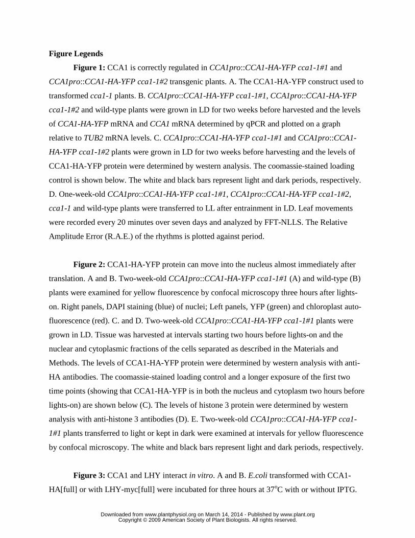
Figure Legends
Figure 1: CCA1 is correctly regulated in CCA1pro::CCA1-HA-YFP cca1-1#1 and
CCA1pro::CCA1-HA-YFP cca1-1#2 transgenic plants. A. The CCA1-HA-YFP construct used to
transformed cca1-1 plants. B. CCA1pro::CCA1-HA-YFP cca1-1#1, CCA1pro::CCA1-HA-YFP
cca1-1#2 and wild-type plants were grown in LD for two weeks before harvested and the levels
of CCA1-HA-YFP mRNA and CCA1 mRNA determined by qPCR and plotted on a graph
relative to TUB2 mRNA levels. C. CCA1pro::CCA1-HA-YFP cca1-1#1 and CCA1pro::CCA1-
HA-YFP cca1-1#2 plants were grown in LD for two weeks before harvesting and the levels of
CCA1-HA-YFP protein were determined by western analysis. The coomassie-stained loading
control is shown below. The white and black bars represent light and dark periods, respectively.
D. One-week-old CCA1pro::CCA1-HA-YFP cca1-1#1, CCA1pro::CCA1-HA-YFP cca1-1#2,
cca1-1 and wild-type plants were transferred to LL after entrainment in LD. Leaf movements
were recorded every 20 minutes over seven days and analyzed by FFT-NLLS. The Relative
Amplitude Error (R.A.E.) of the rhythms is plotted against period.
Figure 2: CCA1-HA-YFP protein can move into the nucleus almost immediately after
translation. A and B. Two-week-old CCA1pro::CCA1-HA-YFP cca1-1#1 (A) and wild-type (B)
plants were examined for yellow fluorescence by confocal microscopy three hours after lights-
on. Right panels, DAPI staining (blue) of nuclei; Left panels, YFP (green) and chloroplast auto-
fluorescence (red). C. and D. Two-week-old CCA1pro::CCA1-HA-YFP cca1-1#1 plants were
grown in LD. Tissue was harvested at intervals starting two hours before lights-on and the
nuclear and cytoplasmic fractions of the cells separated as described in the Materials and
Methods. The levels of CCA1-HA-YFP protein were determined by western analysis with anti-
HA antibodies. The coomassie-stained loading control and a longer exposure of the first two
time points (showing that CCA1-HA-YFP is in both the nucleus and cytoplasm two hours before
lights-on) are shown below (C). The levels of histone 3 protein were determined by western
analysis with anti-histone 3 antibodies (D). E. Two-week-old CCA1pro::CCA1-HA-YFP cca1-
1#1 plants transferred to light or kept in dark were examined at intervals for yellow fluorescence
by confocal microscopy. The white and black bars represent light and dark periods, respectively.
Figure 3: CCA1 and LHY interact in vitro. A and B. E.coli transformed with CCA1-
HA[full] or with LHY-myc[full] were incubated for three hours at 37oC with or without IPTG.
www.plant.org on March 14, 2014 - Published by www.plantphysiol.orgDownloaded from Copyright © 2009 American Society of Plant Biologists. All rights reserved.

Proteins were extracted and equal amounts of protein were loaded into SDS-PAGE gels and
analyzed by western anlysis with anti-HA (A) or anti-myc (B). C and D. E.coli transformed with
CCA1-HA[full] or with LHY-myc[full] were incubated for three hours at 37oC with IPTG.
Extracts were immunoprecipitated with anti-HA (lane 3 and 4) or anti-myc (lane 5 and 6) or
extracts were mixed and then co-immunoprecipitated with anti-HA (lane 7) or anti-myc (lane 8).
Proteins were analyzed by western anlysis with anti-myc (C) or anti-HA (D). E. Protein extracts
from cells of E.coli transformed with CCA1-HA[full] and cells of E.coli transformed with GST-
myc were mixed and used in co-IPs with anti-HA. The proteins pulled-down were analyzed on
westerns with anti-myc. F. CCA1-HA[full], CCA1-HA[407] and CCA1-HA[223] constructs. G.
Protein extracts from cells of E.coli transformed with CCA1-HA[full] (lane 5), CCA1-HA[407]
(lane 6) or with CCA1-HA[223] (lane 7) and cells of E.coli transformed with LHY-myc[full]
were mixed and co-immunoprecipitated with anti-HA. Input extract of CCA1-HA[full], CCA1-
HA[407] and CCA1-HA[223] constructs analyzed by westerns using anti-HA antibodies (lane 1,
2 and 3). Input extract of LHY-myc[full]construct analyzed by westerns using anti-myc
antibodies (lane 4). Co-IPs were analyzed by westerns using anti-myc antibodies (lane 5, 6 and
7).
Figure 4: LHY is correctly regulated in LHYpro::LHY-myc LHY-null transgenic plants.
A. The LHY-myc construct used to transforme to LHY-null plants. B. LHYpro::LHY-myc LHY-
null and wild-type plants were grown in LD for two weeks before harvested and the levels of
LHY-myc mRNA and LHY mRNA determined by qPCR and plotted on a graph relative to TUB2
mRNA levels. C. LHYpro::LHY-myc LHY-null plants were grown in LD for two weeks before
harvested and the levels of LHY-myc protein were determined by western analysis. The
coomassie-stained loading control is shown below. The white and black bars represent light and
dark periods, respectively. D. One-week-old LHYpro::LHY-myc LHY-null, LHY-null and wild-
type plants were transferred to LL. Leaf movements were recorded every 20 minutes over seven
days and analyzed by FFT-NLLS. The R.A.E. for the rhythms was plotted against the period.
Figure 5: CCA1 and LHY interact in vivo. A. LHYpro::LHY-myc LHY-
null;CCA1pro::CCA1-HA-YFPcca1-1#1 and LHYpro::LHY-myc LHY-null plants were grown in
LD for four weeks. Tissue was harvested and used for co-IPs with anti-HA (αHA). As a control,
the co-IP experiments were also carried out without anti-HA antibody (no αHA). Proteins were
www.plant.org on March 14, 2014 - Published by www.plantphysiol.orgDownloaded from Copyright © 2009 American Society of Plant Biologists. All rights reserved.

analyzed by western anlysis with anti-myc (upper row) or anti-HA (lower row). B.
LHYpro::LHY-myc LHY-null;CCA1pro::CCA1-HA-YFPcca1-1#1 and CCA1pro::CCA1-HA-
YFPcca1-1#1 plants were grown in LD for four weeks. Tissue was harvested and used for co-IPs
with anti-myc (αmyc). As a control the co-IP experiments were also carried out without anti-myc
antibody (no αmyc). Proteins were analyzed by western analysis with anti-HA (upper row) or
anti-myc (lower row). C. LHYpro::LHY-myc LHY-null;CCA1pro::CCA1-HA-YFPcca1-1#1
plants were grown in LD for two weeks. Tissue was harvested at intervals and used in a co-IP
experiment with anti-myc. The proteins were analyzed by western analysis with anti-HA (upper
row) or anti-myc (lower row). Lane 1-4, levels of CCA1-HA-YFP and LHY-myc in extracts,
lanes 5-8 CCA1-HA-YFP and LHY-myc pulled-down by anti-myc antibodies. The coomassie-
stained loading control is shown below.
Figure 6: CCA1 forms homodimers in vivo. A. The CCA1-myc construct used to
transform cca1-1*LHY-null plants. B. CCA1pro::CCA1-myc cca1-1*LHY-null#1,
CCA1pro::CCA1-myc cca1-1*LHY-null#2 and wild-type plants were grown in LD for two weeks
before harvested and the levels of CCA1-myc mRNA and CCA1 mRNA determined by qPCR
and plotted on a graph relative to TUB2 mRNA levels. C. Two-week-old CCA1pro::CCA1-myc
cca1-1*LHY-null#1 and CCA1pro::CCA1-myc cca1-1*LHY-null#2 plants were grown in LD.
The levels of CCA1-myc protein were determined by western analysis. The coomassie-stained
loading control is shown below. The white and black bars represent light and dark periods,
respectively. D. One-week-old CCA1pro::CCA1-myc cca1-1*LHY-null#1, CCA1pro::CCA1-myc
cca1-1*LHY-null#2 and cca1-1*LHY-null plants were transferred to LL after grown in LD. Leaf
movements were recorded every 20 min over seven days and analyzed by FFT-NLLS. The
R.A.E. was plotted against the period. E. Four-week-old CCA1pro::CCA1-myc cca1-1*LHY-
null#1;CCA1pro::CCA1-HA-YFPcca1-1#1 and CCA1pro::CCA1-myc cca1-1*LHY
null#2;CCA1pro::CCA1-HA-YFPcca1-1#1 plants were grown in LD. Tissue was harvested and
used for co-IPs carried out with anti-myc. Proteins were analyzed by western analysis with anti-
HA (upper row) or anti-myc (lower row). F. CCA1pro::CCA1-myc cca1-1*LHY
null#1;CCA1pro::CCA1-HA-YFPcca1-1#1 plants were grown in LD for two weeks. Tissue was
harvested at intervals and used in a co-IP experiment with anti-myc. The proteins were analyzed
by western analysis with anti-HA (upper row) or anti-myc (lower row). Lane 1-4, levels of
www.plant.org on March 14, 2014 - Published by www.plantphysiol.orgDownloaded from Copyright © 2009 American Society of Plant Biologists. All rights reserved.
CCA1-HA-YFP and CCA1-myc in extracts, lanes 5-8 CCA1-HA-YFP and CCA1-myc pulled-
down by anti-myc antibodies. The coomassie-stained loading control is shown below.
Figure S1: Leaf movement in the transgenic and control lines. CCA1pro::CCA1-HA-YFP
cca1-1#1, CCA1pro::CCA1-HA-YFP cca1-1#2, cca1-1 and wild-type plants (A), LHYpro::LHY-
myc LHY-null, LHY-null and wild-type plants (B) and CCA1pro::CCA1-myc cca1-1*LHY-null#1,
CCA1pro::CCA1-myc cca1-1*LHY-null#2 and cca1-1*LHY-null plants (C) were transferred to
LL after one week of growth in LD. Leaf movements were recorded every 20 minutes over seven
days and analyzed by FFT-NLLS. The average leaf position is plotted against time.
Figure S2: CCA1-HA-YFP is localized to the nucleus in CCA1pro::CCA1-HA-YFP
cca1-1#2 plants. A. Two-week-old CCA1pro::CCA1-HA-YFP cca1-1#2 plants were examined
for yellow fluorescence by confocal microscopy three hours after lights-on. Right panel, DAPI
staining (blue) of nuclei; Left panel, YFP (green) and chloroplast auto-fluorescence (red). B.
Two-week-old CCA1pro::CCA1-HA-YFP cca1-1#2 plants were grown in LD. Tissue was
harvested at intervals starting two hours before lights-on and the nuclear and cytoplasmic
fractions of the cells separated as described in the Materials and Methods. The levels of CCA1-
HA-YFP protein were determined by western analysis with anti-HA antibodies. The coomassie-
stained loading control is shown below. C. Two-week-old CCA1pro::CCA1-HA-YFP cca1-1#1
plants were grown in LD. Tissue was harvested at intervals starting two hours before lights-on
and the nuclear and cytoplasmic fractions of the cells separated as described in the Materials and
Methods. The levels of RNA Polymerase II protein were determined by western analysis with
anti-Phospho RNA polymerase II antibodies D. Two-week-old CCA1pro::CCA1-HA-YFP cca1-
1#2 plants were examined at intervals for yellow fluorescence by confocal microscopy. The
white and black bars represent light and dark periods, respectively.
Figure S3: LHY-null plants do not express LHY mRNA. Two-week-old LHY-null and
wild-type plants were grown in LD. The levels of LHY mRNA were determined by qPCR and
plotted on a graph relative to TUB2 mRNA expression.
Figure S4: The system used for protein detection is linear for the concentrations of
protein used in our experiments. Protein extracts of CCA1pro::CCA1-HA-YFP cca1-1#1 plants
www.plant.org on March 14, 2014 - Published by www.plantphysiol.orgDownloaded from Copyright © 2009 American Society of Plant Biologists. All rights reserved.

was diluted to three different concentrations and analyzed by western analysis with anti-HA
antibodies. The plot of HA signal against the relative protein amount is shown below.
www.plant.org on March 14, 2014 - Published by www.plantphysiol.orgDownloaded from Copyright © 2009 American Society of Plant Biologists. All rights reserved.
Wild-type
CCA1pro::CCA1-HA-YFP cca1-1#1
CCA1pro::CCA1-HA-YFP cca1-1#2
1222bp up stream of
CCA1 start codon
CCA1 coding
sequence
Three HA tags
YFP
A
B
CC
A1/
TU
B2
1
0.5
Time in light (hours)0 5
w
ww
.plant.org on M
arch 14, 2014 - Published by
ww
w.plantphysiol.org
Dow
nloaded from
Copyright ©
2009 Am
erican Society of P
lant Biologists. A
ll rights reserved.

cca1-1CCA1pro::CCA1-HA-YFP cca1-1#1
Wild-typeCCA1pro::CCA1-HA-YFP cca1-1#2
24 26 28Period (hours)
0.8
0.4
R.A
.E.
22
D
CCA1pro::CCA1-HA-YFP cca1-1#1-2 0 1.5 3 5 7 9
α-HA
Coomassie
CCA1pro::CCA1-HA-YFP cca1-1#2-2 0 1.5 3 5 7 9
α-HA
Coomassie
C
Time in light (hours)
Time in light (hours)
w
ww
.plant.org on M
arch 14, 2014 - Published by
ww
w.plantphysiol.org
Dow
nloaded from
Copyright ©
2009 Am
erican Society of P
lant Biologists. A
ll rights reserved.

YFP DAPIA
YFP DAPIB
w
ww
.plant.org on M
arch 14, 2014 - Published by
ww
w.plantphysiol.org
Dow
nloaded from
Copyright ©
2009 Am
erican Society of P
lant Biologists. A
ll rights reserved.

C
-2 0 1.5 3 5 7 9 -2 0 1.5 3 5 7 9
α-HA
Coomassie
Longer exposure
Time in light (hours)
-2 0 1.5 3 5 -2 0 1.5 3 5
D
α-H3
Coomassie
cytoplasmnucleus
cytoplasmnucleus
Time in light (hours)
w
ww
.plant.org on M
arch 14, 2014 - Published by
ww
w.plantphysiol.org
Dow
nloaded from
Copyright ©
2009 Am
erican Society of P
lant Biologists. A
ll rights reserved.

0
5
.9
-2 4 60 2Time in light (hours)
Rel
ativ
e Fl
uore
scen
ce 1
0.5
light
dark
E
w
ww
.plant.org on M
arch 14, 2014 - Published by
ww
w.plantphysiol.org
Dow
nloaded from
Copyright ©
2009 Am
erican Society of P
lant Biologists. A
ll rights reserved.

CCA1-HA[full]
+IPTG -IPTG
LHY-myc[full]
+IPTG -IPTG
1 2 3 4A
LHY-myc[full]
+IPTG -IPTG
CCA1-HA[full]
+IPTG -IPTG
1 2 3 4B
input
CCA1-HA [full]
LHY-myc [full]
CCA1-HA [full]
LHY-myc [full]
CCA1-HA [full]
LHY-myc [full]
CCA1-HA [full] +
IP (αHA)
LHY-myc [full]
CCA1-HA [full] +
LHY-myc [full]
IP (αmyc) co-IP (αHA)
co-IP (αmyc)
1 2 3 4 5 6 87C
w
ww
.plant.org on M
arch 14, 2014 - Published by
ww
w.plantphysiol.org
Dow
nloaded from
Copyright ©
2009 Am
erican Society of P
lant Biologists. A
ll rights reserved.

input
CCA1-HA [full]
LHY-myc [full]
CCA1-HA [full]
LHY-myc [full]
CCA1-HA [full]
LHY-myc [full]
CCA1-HA [full] +
IP (αHA)
LHY-myc [full]
CCA1-HA [full] +
LHY-myc [full]
IP (αmyc) co-IP (αHA)
co-IP (αmyc)
1 2 3 4 5 6 87D
27kD
input
GST-myc CCA1-HA [full] +
GST-myc
co-IP (αHA)
1 2E
w
ww
.plant.org on M
arch 14, 2014 - Published by
ww
w.plantphysiol.org
Dow
nloaded from
Copyright ©
2009 Am
erican Society of P
lant Biologists. A
ll rights reserved.

CCA1-HA[full]
CCA1-HA[407]
CCA1-HA[223]
CCA1 coding
sequence
Three HA tagsmyb
F
75
50
37
25
inputLHY-myc
[full]
co-IP (αHA)
CCA1-HA [full]
LHY-myc [full] +
CCA1-HA [407]
LHY-myc [full] +
CCA1-HA [223]
LHY-myc [full] +
1 2 3 4
inputCCA1-
HA [full]
CCA1-HA
[407]
CCA1-HA
[223]
5 6 7G
w
ww
.plant.org on M
arch 14, 2014 - Published by
ww
w.plantphysiol.org
Dow
nloaded from
Copyright ©
2009 Am
erican Society of P
lant Biologists. A
ll rights reserved.

09:00 14:00
Wild-type
LHYpro::LHY-myc LHY-null
Wild-type
LH
Y/T
UB
2
1
0.5
Time in light (hours)0 5
1568bp up stream of LHY
start codon
LHYcoding sequence
myc tag
A
B
w
ww
.plant.org on M
arch 14, 2014 - Published by
ww
w.plantphysiol.org
Dow
nloaded from
Copyright ©
2009 Am
erican Society of P
lant Biologists. A
ll rights reserved.

-2 0 1.5 3-3
α-myc
Coomassie
C
0
18 20 22 24 26
LHY-null
Wild-type
24 26 28
Period (hours)
0.8
0.4
R.A
.E.
20 22
LHYpro::LHY-myc LHY-null
D
Time in light (hours)
w
ww
.plant.org on M
arch 14, 2014 - Published by
ww
w.plantphysiol.org
Dow
nloaded from
Copyright ©
2009 Am
erican Society of P
lant Biologists. A
ll rights reserved.

co-IP
(no αmyc)
input co-IP
(αmyc)
CCA1-HA-YFP#1 + LHY-mycCCA1-HA-YFP#11 2 3 4 5 6
inputinput IP
(αmyc)
α-HA
α-myc
B
input co-IP
(no αHA)
input co-IP
(αHA)
CCA1-HA-YFP#1 + LHY-mycLHY-myc
input IP
(αHA)
1 2 3 4 5 6
α-myc
α-HA
A
w
ww
.plant.org on M
arch 14, 2014 - Published by
ww
w.plantphysiol.org
Dow
nloaded from
Copyright ©
2009 Am
erican Society of P
lant Biologists. A
ll rights reserved.

0 1.5 3 5
α-HA
Coomassie
Time in light (hours)
co-IP (αmyc)
α-myc
0 1.5 3 5
inputC
w
ww
.plant.org on M
arch 14, 2014 - Published by
ww
w.plantphysiol.org
Dow
nloaded from
Copyright ©
2009 Am
erican Society of P
lant Biologists. A
ll rights reserved.

1222bp up stream of
CCA1 start codon
CCA1 coding sequence
myc tag
A
Wild-type
CCA1pro::CCA1-myc
CCA1pro::CCA1-myc
CCA1pro::CCA1-myc cca1-1*LHY-null#1
Wild-type
CCA1pro::CCA1-myc cca1-1*LHY-null#2
CC
A1/
TU
B2
1
Time in light (hours)0 5
2
B
w
ww
.plant.org on M
arch 14, 2014 - Published by
ww
w.plantphysiol.org
Dow
nloaded from
Copyright ©
2009 Am
erican Society of P
lant Biologists. A
ll rights reserved.

-2 0 1.5 3 5
α-myc
Coomassie
Time in light (hours)C
-2 0 1.5 3 5
α-myc
Coomassie
Time in light (hours)
10.00 15.00 20.00 25.00 30.00 35.0025 30 35
1
0.6
R.A
.E.
201510
CCA1pro::CCA1-myc cca1-1*LHY-null#2
CCA1pro::CCA1-myc cca1-1*LHY-null#1
cca1-1*LHY-null
Period (hours)
D
w
ww
.plant.org on M
arch 14, 2014 - Published by
ww
w.plantphysiol.org
Dow
nloaded from
Copyright ©
2009 Am
erican Society of P
lant Biologists. A
ll rights reserved.

input co-IP(αmyc)
CCA1-HA-YFP#1 + LHY-myc#1
input co-IP(αmyc)
CCA1-HA-YFP#1 + LHY-myc#2
α-myc
α-HA
E
0 1.5 3 5
α-HA
Coomassie
Time in light (hours)
co-IP (αmyc)
α-myc
0 1.5 3 5
inputF
w
ww
.plant.org on M
arch 14, 2014 - Published by
ww
w.plantphysiol.org
Dow
nloaded from
Copyright ©
2009 Am
erican Society of P
lant Biologists. A
ll rights reserved.