
Disruption of Circadian Rhythm Genes in Obstructive Sleep ...
17
Citation: Gabryelska, A.; Turkiewicz, S.; Karuga, F.F.; Sochal, M.; Strzelecki, D.; Bialasiewicz, P. Disruption of Circadian Rhythm Genes in Obstructive Sleep Apnea Patients—Possible Mechanisms Involved and Clinical Implication. Int. J. Mol. Sci. 2022, 23, 709. https:// doi.org/10.3390/ijms23020709 Academic Editor: Abdelnaby Khalyfa Received: 17 November 2021 Accepted: 5 January 2022 Published: 10 January 2022 Publisher’s Note: MDPI stays neutral with regard to jurisdictional claims in published maps and institutional affil- iations. Copyright: © 2022 by the authors. Licensee MDPI, Basel, Switzerland. This article is an open access article distributed under the terms and conditions of the Creative Commons Attribution (CC BY) license (https:// creativecommons.org/licenses/by/ 4.0/). International Journal of Molecular Sciences Review Disruption of Circadian Rhythm Genes in Obstructive Sleep Apnea Patients—Possible Mechanisms Involved and Clinical Implication Agata Gabryelska 1, * , Szymon Turkiewicz 1 , Filip Franciszek Karuga 1 , Marcin Sochal 1 , Dominik Strzelecki 2 and Piotr Bialasiewicz 1 1 Department of Sleep Medicine and Metabolic Disorders, Medical University of Lodz, 92-215 Lodz, Poland; [email protected] (S.T.); fi[email protected] (F.F.K.); [email protected] (M.S.); [email protected] (P.B.) 2 Department of Affective and Psychotic Disorders, Medical University of Lodz, 92-215 Lodz, Poland; [email protected] * Correspondence: [email protected]; Tel.: +48-660796004 Abstract: Obstructive sleep apnea (OSA) is a chronic condition characterized by recurrent pauses in breathing caused by the collapse of the upper airways, which results in intermittent hypoxia and arousals during the night. The disorder is associated with a vast number of comorbidities affecting different systems, including cardiovascular, metabolic, psychiatric, and neurological complications. Due to abnormal sleep architecture, OSA patients are at high risk of circadian clock disruption, as has been reported in several recent studies. The circadian clock affects almost all daily behavioral patterns, as well as a plethora of physiological processes, and might be one of the key factors contributing to OSA complications. An intricate interaction between the circadian clock and hypoxia may further affect these processes, which has a strong foundation on the molecular level. Recent studies revealed an interaction between hypoxia-inducible factor 1 (HIF-1), a key regulator of oxygen metabolism, and elements of circadian clocks. This relationship has a strong base in the structure of involved elements, as HIF-1 as well as PER, CLOCK, and BMAL, belong to the same Per-Arnt-Sim domain family. Therefore, this review summarizes the available knowledge on the molecular mechanism of circadian clock disruption and its influence on the development and progression of OSA comorbidities. Keywords: obstructive sleep apnea (OSA); circadian clock; chronobiology; sleep disruption; hypoxia; polysomnography (PSG) 1. Master Circadian Clock and Influence of Light–Night Cycle The circadian clock is a complex, hierarchical timing system whose molecular elements are located in nearly every body cell. They are under the control of the master circadian pacemaker located in the suprachiasmatic nucleus (SCN) of the hypothalamus [1], which features a very similar molecular machinery to the peripheral circadian clock in the body cells. The most important function of SCN is collecting external cues from the retina, which enables the synchronization of the circadian clock with the light/dark cycle, and determines its duration over 24 h rhythm. The master clock generates a pronounced circadian rhythm of neuronal firing frequency, which, through a variety of direct and indirect output pathways, synchronizes other cells throughout the body [2]. Signals from the retina are transmitted by neurons from the retinohypothalamic tract, which axons project to the SCN, where they stimulate neurons by releasing glutamate and pituitary adenylate cyclase-activating polypeptide (PACAP) [3,4], which is a modulating protein [5]. Glutamate acts on N- methyl-D-aspartate receptors (NMDAR), which leads to signal transmission by increasing intracellular calcium and cyclic adenosine monophosphate (cAMP) synthesis in SCN cells [2], which in turn activates kinases, such as calcium/calmodulin-dependent protein Int. J. Mol. Sci. 2022, 23, 709. https://doi.org/10.3390/ijms23020709 https://www.mdpi.com/journal/ijms
-
Upload
khangminh22 -
Category
Documents
-
view
0 -
download
0
Transcript of Disruption of Circadian Rhythm Genes in Obstructive Sleep ...

�����������������
Citation: Gabryelska, A.; Turkiewicz,
S.; Karuga, F.F.; Sochal, M.; Strzelecki,
D.; Białasiewicz, P. Disruption of
Circadian Rhythm Genes in
Obstructive Sleep Apnea
Patients—Possible Mechanisms
Involved and Clinical Implication.
Int. J. Mol. Sci. 2022, 23, 709. https://
doi.org/10.3390/ijms23020709
Academic Editor: Abdelnaby Khalyfa
Received: 17 November 2021
Accepted: 5 January 2022
Published: 10 January 2022
Publisher’s Note: MDPI stays neutral
with regard to jurisdictional claims in
published maps and institutional affil-
iations.
Copyright: © 2022 by the authors.
Licensee MDPI, Basel, Switzerland.
This article is an open access article
distributed under the terms and
conditions of the Creative Commons
Attribution (CC BY) license (https://
creativecommons.org/licenses/by/
4.0/).
International Journal of
Molecular Sciences
Review
Disruption of Circadian Rhythm Genes in Obstructive SleepApnea Patients—Possible Mechanisms Involved andClinical ImplicationAgata Gabryelska 1,* , Szymon Turkiewicz 1 , Filip Franciszek Karuga 1, Marcin Sochal 1 ,Dominik Strzelecki 2 and Piotr Białasiewicz 1
1 Department of Sleep Medicine and Metabolic Disorders, Medical University of Lodz, 92-215 Lodz, Poland;[email protected] (S.T.); [email protected] (F.F.K.);[email protected] (M.S.); [email protected] (P.B.)
2 Department of Affective and Psychotic Disorders, Medical University of Lodz, 92-215 Lodz, Poland;[email protected]
* Correspondence: [email protected]; Tel.: +48-660796004
Abstract: Obstructive sleep apnea (OSA) is a chronic condition characterized by recurrent pauses inbreathing caused by the collapse of the upper airways, which results in intermittent hypoxia andarousals during the night. The disorder is associated with a vast number of comorbidities affectingdifferent systems, including cardiovascular, metabolic, psychiatric, and neurological complications.Due to abnormal sleep architecture, OSA patients are at high risk of circadian clock disruption, as hasbeen reported in several recent studies. The circadian clock affects almost all daily behavioral patterns,as well as a plethora of physiological processes, and might be one of the key factors contributing toOSA complications. An intricate interaction between the circadian clock and hypoxia may furtheraffect these processes, which has a strong foundation on the molecular level. Recent studies revealedan interaction between hypoxia-inducible factor 1 (HIF-1), a key regulator of oxygen metabolism, andelements of circadian clocks. This relationship has a strong base in the structure of involved elements,as HIF-1 as well as PER, CLOCK, and BMAL, belong to the same Per-Arnt-Sim domain family.Therefore, this review summarizes the available knowledge on the molecular mechanism of circadianclock disruption and its influence on the development and progression of OSA comorbidities.
Keywords: obstructive sleep apnea (OSA); circadian clock; chronobiology; sleep disruption; hypoxia;polysomnography (PSG)
1. Master Circadian Clock and Influence of Light–Night Cycle
The circadian clock is a complex, hierarchical timing system whose molecular elementsare located in nearly every body cell. They are under the control of the master circadianpacemaker located in the suprachiasmatic nucleus (SCN) of the hypothalamus [1], whichfeatures a very similar molecular machinery to the peripheral circadian clock in the bodycells. The most important function of SCN is collecting external cues from the retina, whichenables the synchronization of the circadian clock with the light/dark cycle, and determinesits duration over 24 h rhythm. The master clock generates a pronounced circadian rhythm ofneuronal firing frequency, which, through a variety of direct and indirect output pathways,synchronizes other cells throughout the body [2]. Signals from the retina are transmittedby neurons from the retinohypothalamic tract, which axons project to the SCN, wherethey stimulate neurons by releasing glutamate and pituitary adenylate cyclase-activatingpolypeptide (PACAP) [3,4], which is a modulating protein [5]. Glutamate acts on N-methyl-D-aspartate receptors (NMDAR), which leads to signal transmission by increasingintracellular calcium and cyclic adenosine monophosphate (cAMP) synthesis in SCNcells [2], which in turn activates kinases, such as calcium/calmodulin-dependent protein
Int. J. Mol. Sci. 2022, 23, 709. https://doi.org/10.3390/ijms23020709 https://www.mdpi.com/journal/ijms

Int. J. Mol. Sci. 2022, 23, 709 2 of 17
kinases (CamK), mitogen-activated protein kinases (MAPK) or protein kinase A (PKA) [6]and phosphorylates cAMP-responsive element-binding protein (CREB) [7]. PhosphorylatedCREB is an active transcription factor, which binds to calcium/cAMP regulatory elements(CREs) in promotors of repressors genes, including Per1 and Per2 [8], and stimulates theirtranscription.
Futhermore, the major circadian clock is autonomous to some extent, generatingcircadian rhythms by neuronal firing non-dependent from external stimuli [9]. SCN neuronsare heterogenic and they differ in their pacemaking ability, neuropeptide expression, andresponse to environmental timing cues, as well as the rhythms they control [6].
2. Molecular Mechanism of the Circadian Clock
The mammalian circadian clock is based on a transcriptional negative feedback loopbetween activators and repressors [10], whose function is regulated by kinases and phos-phatases [11]. The description of the respective genes is in Table 1. Furthermore, Aryl hydro-carbon receptor nuclear translocator-like (BMAL1) and clock circadian regulator (CLOCK)genes encode subunits of the heterodimeric basic helix-loop-helix PER-ARNT-SIM (bHLH-PAS) transcription factor [12]. BMAL1:CLOCK recognizes E-box motifs (5′CACGTC-3′) inpromotors of targeted genes including repressors, and lead to their transcription [13,14].Among repressors, there are families of Period (Per1, Per2, Per3) and Cryptochrome (Cry1,Cry2) genes [13]. Their protein products heterodimerize in the cytoplasm. PER:CRY under-goes phosphorylation by casein kinases (CKIδ and CKIε) and translocation to the nucleus,where the complex can act as an inhibitor of BMAL1:CLOCK-dependent transcription [1,2](see Figure 1). The cytoplasm and nucleus level of circadian clock repressors is regulatedby the E3 ubiquitin ligase complex (SCF-Fbxl3 complex) and proteasome-dependent path-ways of protein degradation [15]. The oscillation of repressors levels conditions the cyclictranscription of circadian clock-controlled output genes and the regulation of behavior [16],lipid, glucose, and redox metabolism [17], sleep [18], body temperature [19], and bloodpressure [20], endocrine [16], immune [21] or cardiovascular [22] function.
Table 1. Basic information about main circadian clock proteins [22].
Protein Name Gene Location onChromosome Size (Da) Size (Amino
Acids)Circadian Clock
Function
PER1 17p13.1 136,212 1290 repressor
PER2 2q37.3 136,579 1255 repressor
PER3 1p36.23 131,888 1201 repressor
Cry1 12q23.3 66,395 586 repressor
Cry2 11p11.2 66,947 593 repressor
BMAL1(ARNTL) 11p15.3 68,762 626 activator
CLOCK 4q12 95,304 846 activator
RORα 15q22.2 58,975 523 regulator
REV-ERBα 17q21.1 66,805 614 regulator
Nuclear retinoid-related orphan receptors ROR (α, β, γ) are transcription factors fromthe orphan nuclear receptor family, which bind to ROR response elements (RORE) in thepromotors of various genes [23]. The role of RORα in the circadian clock is to regulatethe transcription of mainly the BMAL1 gene [24], but also CLOCK, NPAS2 (neuronal PASdomain protein 2, a paralogue of CLOCK; creates a complex with BMAL1; BMAL1:NPAS2has the same function as BMAL1:CLOCK) [25], and CRY1 (see Figure 1). The BMAL:CLOCKcomplex, as a transcription factor, increases the expression of RORα, as well as its inhibitors,such as the Orphan nuclear receptor REV-ERBα. REV-ERBs are a group of two DNA bindingprotein isoforms, α and β. They bind to RORE in a promotor of BMAL1 and prevent RORα

Int. J. Mol. Sci. 2022, 23, 709 3 of 17
activity [24]. Both factors are part of the second feedback loop in the molecular mechanismof the circadian clock (see Figure 1). This feedback loop drives rhythmic changes in BMAL1transcription and introduces a delay in CRY1 mRNA expression that offsets it from genesregulated strictly by BMAL1:CLOCK. While rhythmic changes in BMAL1 abundance arenot required to drive the activators–repressors loop, the ROR/REV loop-induced delay inCRY1 expression is critical for proper circadian timing [1]. The connection of both feedbackloops ensures robustness against noise and environmental perturbations and keeps propercircadian timing [26]. Interestingly, PER2 can enhance BMAL1 expression by RORα eventhough it binds to REV-ERBα [27].
Int. J. Mol. Sci. 2022, 23, x FOR PEER REVIEW 3 of 17
Table 1. Basic information about main circadian clock proteins [22].
Protein Name Gene Location on
Chromosome Size (Da) Size (Amino
Acids) Circadian Clock
Function PER1 17p13.1 136,212 1290 repressor PER2 2q37.3 136,579 1255 repressor PER3 1p36.23 131,888 1201 repressor Cry1 12q23.3 66,395 586 repressor Cry2 11p11.2 66,947 593 repressor
BMAL1 (ARNTL) 11p15.3 68,762 626 activator
CLOCK 4q12 95,304 846 activator RORα 15q22.2 58,975 523 regulator
REV-ERBα 17q21.1 66,805 614 regulator
Figure 1. Circadian clock mechanism. BMAL1 and CLOCK are basic helix-loop-helix (bHLH)-PAS transcription factors, whose heterodimer transcripts a large number of genes, such as Per, Cry, Rora, Rev-Erb. PER and CRY are circadian clock repressors. They bind to each other and the PER:CRY complex undergoes phosphorylation, which enables translocation into the nucleus, where it can act as a repressor of BMAL1:CLOCK-dependent transcription. The second feedback loop of the circa-dian clock consists of two proteins, ROR and REV-ERB. ROR belongs to the same bHLH transcriptor factor family. ROR binds to RORE promotor sequence and transcripts circadian activators. The ex-pression of ROR inhibitor (REV-ERB) occurs at the same time as ROR.
3. Impact of Kinases and Phosphatases on the Circadian Clock The most important kinases regulating the circadian clock are the casein kinases, CK1
and CK2. Seven distinct genes are encoding CK1 isoforms, but only δ and ε influence cir-cadian clock proteins. CK1 acts on PERs, CRYs, and BMAL1. Various isoforms phosphor-ylate these proteins in different locations, which exerts an impact on the generated effect. The phosphorylation of circadian clock proteins by CK1δ leads to their stabilization. By contrast, CK1ε phosphorylation conduces to proteasome degradation. CK2 acts on PER2,
Figure 1. Circadian clock mechanism. BMAL1 and CLOCK are basic helix-loop-helix (bHLH)-PAStranscription factors, whose heterodimer transcripts a large number of genes, such as Per, Cry, Rora,Rev-Erb. PER and CRY are circadian clock repressors. They bind to each other and the PER:CRYcomplex undergoes phosphorylation, which enables translocation into the nucleus, where it can actas a repressor of BMAL1:CLOCK-dependent transcription. The second feedback loop of the circadianclock consists of two proteins, ROR and REV-ERB. ROR belongs to the same bHLH transcriptor factorfamily. ROR binds to RORE promotor sequence and transcripts circadian activators. The expressionof ROR inhibitor (REV-ERB) occurs at the same time as ROR.
3. Impact of Kinases and Phosphatases on the Circadian Clock
The most important kinases regulating the circadian clock are the casein kinases, CK1and CK2. Seven distinct genes are encoding CK1 isoforms, but only δ and ε influence circa-dian clock proteins. CK1 acts on PERs, CRYs, and BMAL1. Various isoforms phosphorylatethese proteins in different locations, which exerts an impact on the generated effect. Thephosphorylation of circadian clock proteins by CK1δ leads to their stabilization. By con-trast, CK1ε phosphorylation conduces to proteasome degradation. CK2 acts on PER2, butthere is inconsistent information about its effect. The phosphorylation of BMAL1 by CK2promotes transport to the cell nucleus [10]. Hirota et al. also found that CK1α-dependentphosphorylation can promote the proteasome degradation of PERs [28].
Glycogen synthase kinase 3β (GSK3β) is another kinase, which phosphorylates REV-ERBα in the mammalian circadian clock. It leads to the stabilization and acceleration

Int. J. Mol. Sci. 2022, 23, 709 4 of 17
of the inhibiting function of REV-ERBα on BMAL1 expression. Other targets of GSK3are BMAL1, CLOCK, and CRY2. Their phosphorylation by GSK3β destabilizes them,promoting proteasome degradation. Adenosine monophosphate-activated protein kinase(AMPK) also acts as a destabilizer of CRY1 [11].
Little is known about the role of phosphatases in the circadian clock. In mammalians,the effects of PP1 and PP5 seem to be significant. PP1 acts on PER2 and stabilizes it, whilePP5 activates CKIε by dephosphorylation [11].
4. Possible Molecular Mechanisms in OSA
Obstructive sleep apnea (OSA) is a common chronic sleep-related breathing disordercharacterized by recurrent pauses in breathing, which are caused by the collapse of theupper respiratory tract [29]. The prevalence of moderate-to-severe OSA in the generalpopulation reaches up to 50% in men and 23% in women, and the risk of OSA develop-ment increases with advancing age, male sex, and higher body mass index (BMI) [30]. Asa consequence, hypopneas and apneas lead to intermittent hypoxia (IH) [31], which ismediated, among others, by key factors in oxygen metabolism, such as hypoxia-induciblefactors (HIFs). HIFs are heterodimeric complexes, which consist of two subunits: α (HIFα) and β (HIF β) [32]. Both subunits belong to the basic helix-loop-helix PER-ARNT-SIM(bHLH-PAS) factor family (the same as BMAL1 and CLOCK), which are constitutivelyproduced in cells [32]. Subunit α is oxygen-sensitive [33]. During normoxia, HIF α under-goes hydroxylation and ubiquitin-dependent degradation, but in hypoxic conditions, itis stabilized and heterodimerizes with subunit β and p300 [34]. The formed complex istransported to the nucleus, where it functions as an active transcription factor [35].
Hypoxia is closely related to circadian clock disruption. Addamovich et al. studieddaily rhythms in oxygen and carbon dioxide in mice. They found changes in the levelsof both gases in Per1−/− mice during the dark phase compared with their wild-typecounterparts [36]. Moreover, clock gene expression was altered and the clock was phase-shifted. Another study connected HIF-1α with synchronizing cellular clocks and circadiangene expression. Short time-spans of decreased oxygenation caused the acceleration of theadaption to jet-lag [37]. Manella et al. noted that the response to hypoxia is time-dependent:different mechanisms may play the main role at different times during the day [38]. Theyalso found complete abrogation to hypoxia in Per1,2−/− mice and a lower expression ofcircadian clock components.
The relationship between hypoxia and the circadian clock is not clear, but it is mostlikely to be bidirectional. As outlined above, hypoxia disrupts the expression of circadianrhythm genes [39]. The main interaction mechanism is probably mutual transcriptionalregulation between HIF-1 and BMAL1:CLOCK. The Per1, Cry1, and CLOCK genes featureE-box-like hypoxia response elements (HRE) in their promotors, so they can be targetsfor HIF-1 [40] (see Figure 2a). Chilov et al. found increased levels of PER1 and CLOCKproteins in mouse brain cells with hypoxia [41]. Additionally, the HIF-1α gene promotorhas an E-box sequence, the target of circadian clock activators complex [42] (see Figure 2b).Kobayashi et al. found that PER2 mediates activation of HIF-1α by increasing its affinity toHRE in the study on cell cultures [43] (see Figure 2c). Moreover, HIF-1α accelerates Per2expression [42].

Int. J. Mol. Sci. 2022, 23, 709 5 of 17
Int. J. Mol. Sci. 2022, 23, x FOR PEER REVIEW 5 of 17
lations between evening PER1, CRY1, CLOCK, and evening HIF-1α protein levels in pa-tients with OSA have been reported [45]. All the protein levels were measured using ELISA assay. Similar outcomes were obtained among diabetes mellitus type 2 patients [46], patients with hepatocellular carcinoma [34], or varicose lesions [47]. However, early reports suggest that one-night effective continuous positive airway pressure (CPAP) treat-ment does not affect the level of HIF-1α in OSA patients [48,49].
It emerged that CLOCK and HIF-1α cooperate to induce vasopressin expression in the suprachiasmatic nucleus [50] or reprogram glucose metabolism in hepatocellular car-cinoma cells [51]. Both of them bind with MOP3 to enhance transcriptional activity, as does HIF-2α [52]. Peek et al. found that BMAL1 expression disruption leads to an in-creased level of HIF-1α and the overexpression of its metabolic targets: prolyl hydroxylase 3 (PHD3), vascular endothelial growth factor (VEGF), and lactate dehydrogenase A (LDHA). Moreover, the genetic stabilization of HIF-1α promotes changes in circadian transcription through the heterodimerization of both proteins [53]. The HIF-1α:BMAL1 complex can also increase the expression of PER2 the same way as HIF-1α:ARNT (aryl hydrocarbon receptor nuclear translocator) and CLOCK:BMAL1 complexes [54]. Another study has shown that BMAL1 gene silencing leads to the decreased expression of HIF-1α [55].
Figure 2. The possible molecular regulation mechanism between HIF-1 and circadian clock proteins. (a) HIF-1 can bind to HRE in promotors of circadian clock genes such as Per1, Cry1, and CLOCK. (b) Transcriptional activity of BMAL1:CLOCK leads to increased HIF-1 expression. (c) CRY1 and PER1 increase HIF-1 affinity to HRE, enhancing HIF-1 activity. (d) Influence of hypoxia-dependent acidification on circadian clock genes expression.
The circadian clock features many output genes, including albumin D-element bind-ing protein (DBP) and E4 binding protein 4 (E4BP4). DBP and E4BP4 are, respectively,
Figure 2. The possible molecular regulation mechanism between HIF-1 and circadian clock proteins.(a) HIF-1 can bind to HRE in promotors of circadian clock genes such as Per1, Cry1, and CLOCK.(b) Transcriptional activity of BMAL1:CLOCK leads to increased HIF-1 expression. (c) CRY1 andPER1 increase HIF-1 affinity to HRE, enhancing HIF-1 activity. (d) Influence of hypoxia-dependentacidification on circadian clock genes expression.
HIF-1 and circadian clock proteins present a relationship in Obstructive Sleep Apneapatients and their pathways are connected [42,44,45]. In a multivariate general linear modelfeaturing a concentration of all the circadian clock proteins as dependent variables, eveningHIF-1α protein level was the only significant covariant (p = 0.025). Positive correlationsbetween evening PER1, CRY1, CLOCK, and evening HIF-1α protein levels in patients withOSA have been reported [45]. All the protein levels were measured using ELISA assay.Similar outcomes were obtained among diabetes mellitus type 2 patients [46], patients withhepatocellular carcinoma [34], or varicose lesions [47]. However, early reports suggest thatone-night effective continuous positive airway pressure (CPAP) treatment does not affectthe level of HIF-1α in OSA patients [48,49].
It emerged that CLOCK and HIF-1α cooperate to induce vasopressin expressionin the suprachiasmatic nucleus [50] or reprogram glucose metabolism in hepatocellularcarcinoma cells [51]. Both of them bind with MOP3 to enhance transcriptional activity, asdoes HIF-2α [52]. Peek et al. found that BMAL1 expression disruption leads to an increasedlevel of HIF-1α and the overexpression of its metabolic targets: prolyl hydroxylase 3(PHD3), vascular endothelial growth factor (VEGF), and lactate dehydrogenase A (LDHA).Moreover, the genetic stabilization of HIF-1α promotes changes in circadian transcriptionthrough the heterodimerization of both proteins [53]. The HIF-1α:BMAL1 complex can alsoincrease the expression of PER2 the same way as HIF-1α:ARNT (aryl hydrocarbon receptornuclear translocator) and CLOCK:BMAL1 complexes [54]. Another study has shown thatBMAL1 gene silencing leads to the decreased expression of HIF-1α [55].

Int. J. Mol. Sci. 2022, 23, 709 6 of 17
The circadian clock features many output genes, including albumin D-element bindingprotein (DBP) and E4 binding protein 4 (E4BP4). DBP and E4BP4 are, respectively, positiveand negative regulators of the HIF-1β promotor. The disruption of this mechanism andARNT inhibition are conducive to the destruction of pancreatic islet β-cell, decreasedinsulin output, and diabetes mellitus development in mice [56].
Walton et al. proposed another mechanism of this complicated relationship based onthe hypoxic alteration of metabolic activity mediated by HIF-1α, which leads to acidificationand the spatial redistribution of lysosomes (see Figure 2d). Acid prevents mechanistictarget of rapamycin kinase (mTOR) localization to the lysosomal surface and its activation,which decreases circadian clock expression [57].
Based on the results of our study [45] and the above information, it seems that hypoxiamediated by HIF-1 is the most likely mechanism of circadian clock disruption in OSApatients. The first free molecular pathways can interfere with each other. HIF-1 increasedthe expression of circadian clock genes in the presence of HRE in their promotors and canalso intensify the transcription of HIF-1. Moreover, circadian clock repressors aggravateHIF-1 activity. All these create an ordered structure resembling a positive feedback loop(see Figure 3).
Int. J. Mol. Sci. 2022, 23, x FOR PEER REVIEW 6 of 17
positive and negative regulators of the HIF-1β promotor. The disruption of this mecha-nism and ARNT inhibition are conducive to the destruction of pancreatic islet β-cell, de-creased insulin output, and diabetes mellitus development in mice [56].
Walton et al. proposed another mechanism of this complicated relationship based on the hypoxic alteration of metabolic activity mediated by HIF-1α, which leads to acidifica-tion and the spatial redistribution of lysosomes (see Figure 2d). Acid prevents mechanistic target of rapamycin kinase (mTOR) localization to the lysosomal surface and its activation, which decreases circadian clock expression [57].
Based on the results of our study [45] and the above information, it seems that hy-poxia mediated by HIF-1 is the most likely mechanism of circadian clock disruption in OSA patients. The first free molecular pathways can interfere with each other. HIF-1 in-creased the expression of circadian clock genes in the presence of HRE in their promotors and can also intensify the transcription of HIF-1. Moreover, circadian clock repressors ag-gravate HIF-1 activity. All these create an ordered structure resembling a positive feed-back loop (see Figure 3).
Figure 3. Possible positive feedback loop, which can be responsible for circadian clock disruption in patients.
5. Clinical Implications of Circadian Rhythm Gene Disruption in OSA Patients Difficulty in waking up, problems with falling asleep, and daytime sleepiness are the
main symptoms of circadian disruption, which is defined as a misalignment between the central circadian clock, located in SCN, and the behavioral cycle. The diagnosis is based on a sleep diary, detailed patient history, and actigraphy [18]. The circadian misalign-ments are mainly caused by sleep disturbance, jet lag, night shifts, irregular shift work patterns, and dietary alterations [58,59]. Patients with OSA are in a high-risk group of developing disruption of the circadian rhythm. As described earlier, this phenomenon might be explained by the increased level of subunits α of HIF-1 in OSA patients, which is associated with the overexpression of circadian clock proteins, such as PER1 [45,60,61]. This is of great importance in the context of various complications, including the devel-opment of metabolic, cardiovascular, psychiatric, and neurodegenerative diseases [49,62,63]. The pathophysiological pathways involved in the development of the afore-mentioned complications in the group of patients suffering from OSA and/or circadian disruption are not well understood. Moreover, due to the interaction between HIF-1α and the circadian clock, it is difficult to determine whether the complications are caused by the influence of HIF-1 α or independently by the proteins determining circadian rhythms.
Figure 3. Possible positive feedback loop, which can be responsible for circadian clock disruptionin patients.
5. Clinical Implications of Circadian Rhythm Gene Disruption in OSA Patients
Difficulty in waking up, problems with falling asleep, and daytime sleepiness are themain symptoms of circadian disruption, which is defined as a misalignment between thecentral circadian clock, located in SCN, and the behavioral cycle. The diagnosis is based ona sleep diary, detailed patient history, and actigraphy [18]. The circadian misalignmentsare mainly caused by sleep disturbance, jet lag, night shifts, irregular shift work patterns,and dietary alterations [58,59]. Patients with OSA are in a high-risk group of developingdisruption of the circadian rhythm. As described earlier, this phenomenon might be ex-plained by the increased level of subunits α of HIF-1 in OSA patients, which is associatedwith the overexpression of circadian clock proteins, such as PER1 [45,60,61]. This is ofgreat importance in the context of various complications, including the development ofmetabolic, cardiovascular, psychiatric, and neurodegenerative diseases [49,62,63]. Thepathophysiological pathways involved in the development of the aforementioned compli-cations in the group of patients suffering from OSA and/or circadian disruption are notwell understood. Moreover, due to the interaction between HIF-1α and the circadian clock,it is difficult to determine whether the complications are caused by the influence of HIF-1
Int. J. Mol. Sci. 2022, 23, 709 7 of 17
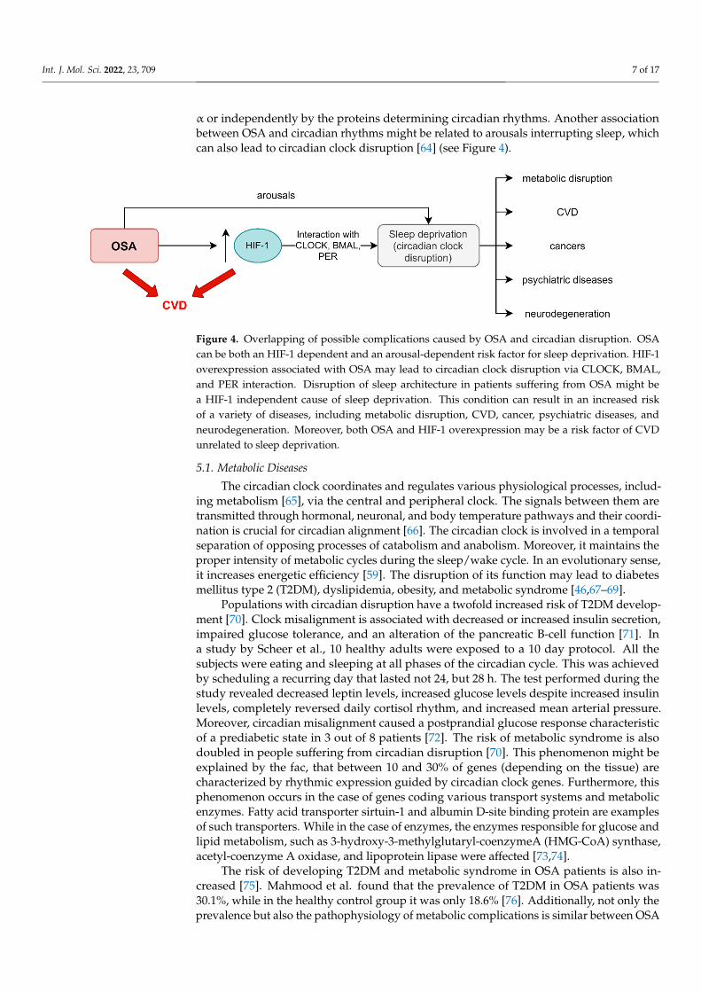
α or independently by the proteins determining circadian rhythms. Another associationbetween OSA and circadian rhythms might be related to arousals interrupting sleep, whichcan also lead to circadian clock disruption [64] (see Figure 4).
Int. J. Mol. Sci. 2022, 23, x FOR PEER REVIEW 7 of 17
Another association between OSA and circadian rhythms might be related to arousals in-terrupting sleep, which can also lead to circadian clock disruption [64] (see Figure 4).
Figure 4. Overlapping of possible complications caused by OSA and circadian disruption. OSA can be both an HIF-1 dependent and an arousal-dependent risk factor for sleep deprivation. HIF-1 over-expression associated with OSA may lead to circadian clock disruption via CLOCK, BMAL, and PER interaction. Disruption of sleep architecture in patients suffering from OSA might be a HIF-1 independent cause of sleep deprivation. This condition can result in an increased risk of a variety of diseases, including metabolic disruption, CVD, cancer, psychiatric diseases, and neurodegenera-tion. Moreover, both OSA and HIF-1 overexpression may be a risk factor of CVD unrelated to sleep deprivation.
5.1. Metabolic Diseases The circadian clock coordinates and regulates various physiological processes, in-
cluding metabolism [65], via the central and peripheral clock. The signals between them are transmitted through hormonal, neuronal, and body temperature pathways and their coordination is crucial for circadian alignment [66]. The circadian clock is involved in a temporal separation of opposing processes of catabolism and anabolism. Moreover, it maintains the proper intensity of metabolic cycles during the sleep/wake cycle. In an evo-lutionary sense, it increases energetic efficiency [59]. The disruption of its function may lead to diabetes mellitus type 2 (T2DM), dyslipidemia, obesity, and metabolic syndrome [46,67–69].
Populations with circadian disruption have a twofold increased risk of T2DM devel-opment [70]. Clock misalignment is associated with decreased or increased insulin secre-tion, impaired glucose tolerance, and an alteration of the pancreatic B-cell function [71]. In a study by Scheer et al., 10 healthy adults were exposed to a 10 day protocol. All the subjects were eating and sleeping at all phases of the circadian cycle. This was achieved by scheduling a recurring day that lasted not 24, but 28 h. The test performed during the study revealed decreased leptin levels, increased glucose levels despite increased insulin levels, completely reversed daily cortisol rhythm, and increased mean arterial pressure. Moreover, circadian misalignment caused a postprandial glucose response characteristic of a prediabetic state in 3 out of 8 patients [72]. The risk of metabolic syndrome is also doubled in people suffering from circadian disruption [70]. This phenomenon might be explained by the fac, that between 10 and 30% of genes (depending on the tissue) are char-acterized by rhythmic expression guided by circadian clock genes. Furthermore, this phe-nomenon occurs in the case of genes coding various transport systems and metabolic en-zymes. Fatty acid transporter sirtuin-1 and albumin D-site binding protein are examples of such transporters. While in the case of enzymes, the enzymes responsible for glucose and lipid metabolism, such as 3-hydroxy-3-methylglutaryl-coenzymeA (HMG-CoA) syn-thase, acetyl-coenzyme A oxidase, and lipoprotein lipase were affected [73,74].
The risk of developing T2DM and metabolic syndrome in OSA patients is also in-creased [75]. Mahmood et al. found that the prevalence of T2DM in OSA patients was 30.1%, while in the healthy control group it was only 18.6% [76]. Additionally, not only the prevalence but also the pathophysiology of metabolic complications is similar between
Figure 4. Overlapping of possible complications caused by OSA and circadian disruption. OSAcan be both an HIF-1 dependent and an arousal-dependent risk factor for sleep deprivation. HIF-1overexpression associated with OSA may lead to circadian clock disruption via CLOCK, BMAL,and PER interaction. Disruption of sleep architecture in patients suffering from OSA might bea HIF-1 independent cause of sleep deprivation. This condition can result in an increased riskof a variety of diseases, including metabolic disruption, CVD, cancer, psychiatric diseases, andneurodegeneration. Moreover, both OSA and HIF-1 overexpression may be a risk factor of CVDunrelated to sleep deprivation.
5.1. Metabolic Diseases
The circadian clock coordinates and regulates various physiological processes, includ-ing metabolism [65], via the central and peripheral clock. The signals between them aretransmitted through hormonal, neuronal, and body temperature pathways and their coordi-nation is crucial for circadian alignment [66]. The circadian clock is involved in a temporalseparation of opposing processes of catabolism and anabolism. Moreover, it maintains theproper intensity of metabolic cycles during the sleep/wake cycle. In an evolutionary sense,it increases energetic efficiency [59]. The disruption of its function may lead to diabetesmellitus type 2 (T2DM), dyslipidemia, obesity, and metabolic syndrome [46,67–69].
Populations with circadian disruption have a twofold increased risk of T2DM develop-ment [70]. Clock misalignment is associated with decreased or increased insulin secretion,impaired glucose tolerance, and an alteration of the pancreatic B-cell function [71]. Ina study by Scheer et al., 10 healthy adults were exposed to a 10 day protocol. All thesubjects were eating and sleeping at all phases of the circadian cycle. This was achievedby scheduling a recurring day that lasted not 24, but 28 h. The test performed during thestudy revealed decreased leptin levels, increased glucose levels despite increased insulinlevels, completely reversed daily cortisol rhythm, and increased mean arterial pressure.Moreover, circadian misalignment caused a postprandial glucose response characteristicof a prediabetic state in 3 out of 8 patients [72]. The risk of metabolic syndrome is alsodoubled in people suffering from circadian disruption [70]. This phenomenon might beexplained by the fac, that between 10 and 30% of genes (depending on the tissue) arecharacterized by rhythmic expression guided by circadian clock genes. Furthermore, thisphenomenon occurs in the case of genes coding various transport systems and metabolicenzymes. Fatty acid transporter sirtuin-1 and albumin D-site binding protein are examplesof such transporters. While in the case of enzymes, the enzymes responsible for glucose andlipid metabolism, such as 3-hydroxy-3-methylglutaryl-coenzymeA (HMG-CoA) synthase,acetyl-coenzyme A oxidase, and lipoprotein lipase were affected [73,74].
The risk of developing T2DM and metabolic syndrome in OSA patients is also in-creased [75]. Mahmood et al. found that the prevalence of T2DM in OSA patients was30.1%, while in the healthy control group it was only 18.6% [76]. Additionally, not only theprevalence but also the pathophysiology of metabolic complications is similar between OSA

Int. J. Mol. Sci. 2022, 23, 709 8 of 17
patients and patients with disrupted circadian rhythms. In both cases, it might be mediatedby HIF-1α and based on impaired glucose tolerance, increased insulin secretion, alter-ation in B-cell function, and the influence of metabolic enzymes such as acetyl-coenzymeA [49,77]. OSA subjects are also characterized by increased leptin levels [78] and impairedcortisol rhythmicity [79]. Furthermore, the effect of hypoxia may be aggravated by theinteraction of circadian components with HIF-1α [43].
Circadian clock disruption is also a potential mechanism of diabetes complications,such as diabetic retinopathy development. The excessive CLOCK-dependent expressionof DEC2 and VEGF [80] leads to incorrect neovascularization and in consequence diabeticretinopathy. Moreover, VEGF translation is powered by HIF-1α [81] (see Figure 5).
5.2. Cardiovascular Diseases
OSA is associated with increased cardiovascular disease (CVD) morbidity and mor-tality, commonly associated with obesity. The American Academy of Sleep Medicinerecommends dietary-induced weight loss and exercise as lifestyle treatment options forOSA. Low-fat diets are recommended for improving OSA severity and weight loss im-proves OSA severity and the CVD substrate [82]. The best example proving the importanceof circadian rhythms in cardiovascular diseases is the fact that myocardial infarction, my-ocardial ischemia, and sudden cardiac death occur more frequently in the morning than inthe evening [22]. This is in contrast to OSA patients, whose peak of cardiovascular risk is inthe middle of the night [83–85]. Moreover, patients suffering from circadian disruption areat a higher risk of cardiac ischemic events [86]. Other cardiac diseases, such as heart arrhyth-mias, are also affected by circadian rhythms. The electrical properties of the heart show24 h variation. Life-threatening arrhythmias, such as ventricular fibrillation, tend to occurin the morning after waking up [87]. The peak of premature ventricular beats detected bycontinuous Holter monitoring was determined between 6 a.m. and 12 noon [88]. Circadianrhythms also exert an influence on blood pressure. Physiologically, blood pressure dipsduring the night while resting by 10–20%; in the morning a significant increase in bloodpressure occurs, known as the “morning surge”. Blood pressure reaches a peak in theafternoon [20]. Furthermore, there are different circadian patterns among patients witharterial hypertension: dippers, whose blood pressure dips at night; non-dippers, for whomthere is no dip of blood pressure at night; and reverse-dippers, who present increasedblood pressure during the night [89]. A study by Kitamura et al. revealed that individuals’blood pressure pattern on the first days of night shift work changed from a dipper to anon-dipper pattern. Furthermore, this phenomenon was reversed after 4 days of nightshift work and the dipper pattern was restored [90]. Many mechanisms regulate bloodpressure during the 24 h cycle. It is well known that changes in sympathetic nervous systemtone are responsible for the “morning surge”. There have been several studies linking thecircadian rhythms of blood pressure with kidney and renal sodium homeostasis: higherdaytime sodium excretion with urine was associated with the presence of nocturnal dip inthe blood pressure [91], aldosteronism was shown to provoke the non-dipping type of hy-pertension [92], and unilateral nephrectomy was linked with the occurrence of non-dippingblood pressure patterns in patients [93]. In a study by Marques et al., the kidney tissuesfrom hypertensive and normotensive humans were compared. The results showed theupregulation of PER1 in hypertensive humans [94]. Dashti et al. showed the link betweensingle-nucleotide polymorphism in PER1, CRY1, CLOCK, and PER3 genes and systolicblood pressure. A study by Morris et al. revealed that circadian misalignment lastingonly 8 days leads to increased systolic and diastolic pressure by 3.0 mmHg and 1.5 mmHg,respectively [95]. Sudden morning increases in blood pressure, heart rate, sympatheticnervous system activity, prothrombic tendency, and vasoconstrictive hormones are thoughtto be an explanation of myocardial infarction and ischemia peak during the morning [22].Ischemia leads to hypoxia, which is responsible for HIF-1α protein stabilization in themyocardium and its expression. One of the genes activated by HIF-1α is VEGF, whichplays an important role in post-myocardial infarction cardiac angiogenesis [96,97]. A study

Int. J. Mol. Sci. 2022, 23, 709 9 of 17
by Koyonagi et al. found that protein Per2 expression reduced the hypoxic induction ofHIF-1α-dependent VEGF expression [98,99]. Since the expression of Per2 varies duringthe 24 h cycle, its fluctuation may alternate the cardiac response to ischemia dependingon the time of the ischemic episode. However, it is worth mentioning that angiogenesisis a long-term process; therefore, circadian clock disruption must be longer than a day toinfluence it significantly.
The treatment of cardiovascular diseases in patients with circadian rhythm disruptionsis complicated due to the influence of the circadian clock on the pharmacokinetics andpharmacodynamics of drugs [22]. Therefore, chronotherapy based on the understandingof circadian rhythms may help inappropriate drug selection and dosing, and improve thetreatment efficiency [100].
In OSA patients, HIF-1 α is also an important regulator of response to hypoxia. Thereis a vast number of genes regulating the cardiovascular system that are controlled bystabilized HIF1-α, e.g., genes regulating endothelin-1, erythropoietin, and leptin synthe-sis [101,102]. Therefore, moderate and severe OSA is associated with a significant increasein cardiovascular morbidity [103]. It is also worth mentioning that OSA individuals withcomorbid CVD present with higher HIF-1α compared to groups without cardiovascularcomplications [104]. Due to the cross-talk between clock genes and HIF-1α in OSA patients,the understanding of the pathophysiology of certain cardiovascular complications in OSAand circadian disruption patients is challenging (See Figure 5).
5.3. Psychiatric and Neurodegenerative Diseases
Circadian disruption has been found to be related to both psychiatric and neurode-generative diseases. Some examples include major depressive disorder, bipolar disease,schizophrenia, Alzheimer’s, and Parkinson’s disease [105,106]. Major depressive disorderis characterized by anhedonia, mood alterations, fatigue, changes in appetite and bodymass, irritability, and sleep disturbances, including both insomnia and excessive daytimesleepiness [107]. The relationship between major depressive disorder and circadian disrup-tion is bidirectional. On the one hand, depression leads to altered sleep architecture; on theother hand, people suffering from circadian misalignment are more prone to developing amajor depressive disorder. In a metanalysis, which included 11 studies, it was determinedthat night shift workers were at 40% higher risk of developing depression compared witha daytime worker control group [108]. The loss of circadian rhythm generated by thecircadian clock is one of the postulated factors leading to depression development. Thus,chronotherapy, which includes sleep deprivation, dark therapy, bright light therapy [109],and others, is a possible treatment option for depression [87]. Interestingly, the expres-sion of HIF-1 α increases three-fold increase in patients suffering from major depressivedisorder and 2.5 fold in patients suffering from bipolar disease. Moreover, patients in aremissive state are characterized by significantly lower HIF-1 α compared with patients ina depressive state [86]. The cause of HIF-1 α overexpression in patients suffering from theaforementioned diseases is not clear. One of the postulated factors is oxidative stress causedby an imbalance between the increased production of reactive oxygen species and a relativeshortage of antioxidant defense and increased HIF-1 α expression in the protective responseto oxidative stress [110–113], which is similar to the cellular senescence process in OSApatients [114]. Furthermore, increased HIF-1 levels in the brain may improve creatininemetabolism and correlate with a better treatment response to antidepressants [115]. On theother hand, HIF-1 α overexpression may interfere with the genes responsible for circadianclock regulation and modify circadian clockwork. Patients with OSA experience a higherprevalence of depression than healthy controls, even though HIF-1 α offers a protectivefunction against oxidative stress. Therefore, some patients suffering from depression mayalso improve after CPAP therapy. One of the explanations as to why depression is morefrequent in OSA patients might be the circadian disruption caused by arousals and HIF-1 αoverexpression [111,116,117].
Int. J. Mol. Sci. 2022, 23, 709 10 of 17
Bipolar affective disease is a chronic and complex disorder characterized by a combi-nation of different mood episodes including mania, hypomania, and depression. Circadiandisruption is a prevalent condition in patients with bipolar disease; however, a recentmeta-analysis, which included 42 clinical studies, did not establish an association betweencircadian disruption and bipolar disease incidence [118]. Despite this, it was reported thatbipolar disease can be induced by jet lag in the case of susceptible individuals [105]. Addi-tionally, OSA has been found to be a significant risk factor for bipolar disease. In a study ofKelly et al., 21% of patients with bipolar disease were also suffering from OSA [119]. Oneof the postulated mechanisms behind this phenomenon was the neurostructural changesseen in decreased gray matter concentration of the amygdala, dorsal lateral prefrontalcortex, hippocampus, cerebellum temporal lobe, caudate lobe, and other areas in the brainof OSA patients [119,120]. However, it is important to remember that many psychiatricdisorders, including bipolar disorder, cause an increase in body mass, which in itself maydirectly contribute to the development of OSA, since an increased body-mass index is amajor risk factor for OSA. Moreover, intermittent hypoxemia and circadian cycle disorderswith sleep fragmentation in pediatric subjects have shown an association with behavioraland neurocognitive disorders, with reduced school performance. The treatment of OSAproblems in children, mainly caused by tonsillar hypertrophy, led to the regression ofassociated symptoms [121]. This suggests the high probability of a mutual relationshipbetween OSA and psychiatric diseases. The focus in future studies should be on a newgeneration of drugs, such as aripiprazole, that are not likely to affect the body mass of anindividual, increases in which can aggravate OSA problems.
Schizophrenia is another severely disabling mental disorder, characterized by positiveand negative symptoms. One of the most prevalent manifestations of this disease iscircadian rhythm disruption. It occurs in around 80% of patients [122]. Similarly to the otherdiseases discussed above, circadian disruption may not only be a sign of disease, but it canalso be a cause. Skin fibroblasts, which were isolated from patients suffering from chronicschizophrenia, presented decreased expression of PER2 and CRY1 compared with healthycontrols [123]. Moreover, in a study by Sun et al., schizophrenia patients demonstratedaltered mRNA levels of PER1/2/3 and NPAS2 in white blood cells compared with a healthycontrol group [124]. Ying-Ying et al. reported that the prevalence of OSA was increased two-fold in schizophrenia patients compared with a healthy group [125]. Furthermore, in thesame study, the hazard ratio adjusted by gender, age, baseline comorbidities, and durationof antipsychotics use was lower for such comorbidities as hypertension, hyperlipidemia,or even diabetes compared with the presence of schizophrenia (HR = 1.61, HR = 1.55,HR = 1.53, and HR = 1.97, respectively) [125]. The interaction between HIF-1 α and CLOCKgenes and CLOCK gene alterations in schizophrenia patients seem to be among the possiblecauses of this prevalence.
Neurodegeneration is any pathological condition in which the nervous system losesits structure or function, or both. Due to increased global life expectancy, the prevalence ofneurodegenerative diseases is growing gradually. The disruption of sleep/wake cycles isamong the earliest manifestations of these diseases. Moreover, circadian rhythm disruptionmay be a cause of the neurodegeneration process. For example, the beta-amyloid peptide,which is linked with Alzheimer’s disease, is regulated by the rhythmically expressedpresenilin-2 gene in SCN [106]. Additionally, the presenilin-2 gene is regulated in peripheraltissues via CLOCK and BMAL1 [126]. No experimental studies have yet determined thatany alteration to clock genes affects presenilin-2 brain expression. Furthermore, a studyby Gu et al. found that certain single-nucleotide polymorphisms of PER1 and BMAL1 areassociated with an increased risk of Parkinson’s disease. Breen et al., similarly to Cai et al.,found that the expression of BMAL1 was decreased in patients suffering from Parkinson’sdisease [127,128]. In a study by Ping-Song et al. on 11,664 patients, it was discoveredthat patients with sleep apnea demonstrated a 1.85-fold higher risk of Parkinson’s diseasedevelopment compared with the control group [129]. In another study, patients sufferingfrom OSA demonstrated a 2.17-fold higher risk of developing Alzheimer’s disease than no-

Int. J. Mol. Sci. 2022, 23, 709 11 of 17
OSA patients [130]. Such a significant incidence of OSA in patients with neurodegenerativediseases suggests that the association of HIF-1 α and proteins regulating circadian genesmay play a substantial role [131]. Surprisingly, HIF-1 α is considered a neuroprotectivefactor and its activation might play a role in the future treatment of neurodegenerativedisorders. In addition, a trial on patients suffering from Alzheimer’s disease with OSArevealed improved cognition in the CPAP-treated group [132,133]. This suggests thepossible advantageous effects of the treatment not only on baseline OSA but also on itspsychiatric and neurogenerative comorbidities (see Figure 5).
Int. J. Mol. Sci. 2022, 23, x FOR PEER REVIEW 11 of 17
disease than no-OSA patients [130]. Such a significant incidence of OSA in patients with neurodegenerative diseases suggests that the association of HIF-1 α and proteins regulat-ing circadian genes may play a substantial role [131]. Surprisingly, HIF-1 α is considered a neuroprotective factor and its activation might play a role in the future treatment of neurodegenerative disorders. In addition, a trial on patients suffering from Alzheimer’s disease with OSA revealed improved cognition in the CPAP-treated group [132,133]. This suggests the possible advantageous effects of the treatment not only on baseline OSA but also on its psychiatric and neurogenerative comorbidities (see Figure 5).
Figure 5. Clinical implications of circadian clock disruption in OSA patients and their similarities in pathogenesis.
6. Conclusions Due to the pathophysiological association between OSA and circadian rhythm dis-
ruption, there seem to be overlapping risk factors for metabolic, cardiovascular, and neu-rological diseases. Available research proposes a molecular mechanism responsible for these processes. Screening for OSA and, eventually, CPAP therapy might improve the treatment outcome in the selected group of patients with concomitant circadian disrup-tion and certain metabolic, cardiovascular, and neurological diseases. Limited knowledge of the responsible mechanisms limits the possible implementation of more personalized and complex treatment for OSA patients focused on their multiple comorbidities.
Author Contributions: A.G. provided the overall concept and framework of the manuscript. A.G., S.T., and F.F.K. researched and identified appropriate articles and wrote the paper. S.T. was respon-sible for the visualization. A.G., M.S., D.S., and P.B. revised the manuscript. All authors approved the final version of the manuscript. All authors have read and agreed to the published version of the manuscript.
Funding: The study was supported by National Science Center, Poland, Preludium Grant no. 2018/31/N/NZ5/0393.
Figure 5. Clinical implications of circadian clock disruption in OSA patients and their similaritiesin pathogenesis.
6. Conclusions
Due to the pathophysiological association between OSA and circadian rhythm dis-ruption, there seem to be overlapping risk factors for metabolic, cardiovascular, and neu-rological diseases. Available research proposes a molecular mechanism responsible forthese processes. Screening for OSA and, eventually, CPAP therapy might improve thetreatment outcome in the selected group of patients with concomitant circadian disruptionand certain metabolic, cardiovascular, and neurological diseases. Limited knowledge ofthe responsible mechanisms limits the possible implementation of more personalized andcomplex treatment for OSA patients focused on their multiple comorbidities.
Author Contributions: A.G. provided the overall concept and framework of the manuscript. A.G.,S.T. and F.F.K. researched and identified appropriate articles and wrote the paper. S.T. was responsiblefor the visualization. A.G., M.S., D.S. and P.B. revised the manuscript. All authors have read andagreed to the published version of the manuscript.
Funding: The study was supported by National Science Center, Poland, Preludium Grant no.2018/31/N/NZ5/0393.
Conflicts of Interest: The authors declare that there is no conflict of interest regarding the publicationof this article.

Int. J. Mol. Sci. 2022, 23, 709 12 of 17
Abbreviations
AMPK adenosine monophosphate-activated protein kinasebHLH-PAS basic helix-loop-helix PER-ARNT-SIM
BMAL1/ARNTLbrain and muscle ARNT-like 1/aryl hydrocarbon receptor nucleartranslocator like
CamK calcium/calmodulin-dependent protein kinasescAMP cyclic adenosine monophosphate;CKI casein kinases
CLOCKclock circadian regulator/circadian locomotor output cycles proteinkaput
CPAP continuous positive airway pressure treatmentCREB phosphorylate cAMP-responsive element-binding proteinCry2 cryptochrome 2CVD cardio-vascular diseaseDBP albumin D-element binding proteinE4BP4 E4 binding protein 4E-box enhancer box; GSK3β—Glycogen synthase kinase 3βHIF hypoxia inducible factorHMG-CoA 3-hydroxy-3-methylglutaryl-coenzyme A synthaseHRE hypoxia response elementIH intermittent hypoxiaLDHA lactate dehydrogenase AMAPK mitogen-activated protein kinasesmTOR mechanistic target of rapamycin kinaseNMDAR N-methyl-D-aspartate receptorsNPAS2 neuronal PAS domain protein 2OSA obstructive sleep apneaPACAP pituitary adenylate cyclase—activating polypeptidePER1 period protein 1PER2 period protein 2PER3 period protein 3PHD3 prolyl hydroxylase 3PKA protein kinase APP1 protein phosphatase 1PP5 protein phosphatase 5REV-ERBα nuclear receptor subfamily 1 group D member 1ROR RAR-related orphan receptorRORE ROR response elementsRORα Nuclear retinoid-related orphan receptors αSCF-Fbxl3 E3 ubiquitin ligase complexE3 ubiquitin ligase complexSCN suprachiasmatic nucleusT2DM diabetes mellitus type 2VEGF vascular endothelial growth factor
References1. Partch, C.; Green, C.; Takahashi, J. Molecular architecture of the mammalian circadian clock. Trends Cell Biol. 2014, 24, 90–99.
[CrossRef] [PubMed]2. Welsh, D.; Takahashi, J.; Kay, S. Suprachiasmatic Nucleus: Cell Autonomy and Network Properties. Annu. Rev. Physiol. 2010, 72,
551–577. [CrossRef]3. Morin, L.; Allen, C. The circadian visual system. Brain Res. Rev. 2006, 51, 1–60. [CrossRef]4. Ozge, G.; Dogan, D.; Koylu, M.T.; Ayyildiz, O.; Akincioglu, D.; Mumcuoglu, T.; Mutlu, F.M. Retina nerve fiber layer and choroidal
thickness changes in obstructive sleep apnea syndrome. Postgrad. Med. 2016, 128, 317–322. [CrossRef]5. Kraves, S.; Weitz, C. A role for cardiotrophin-like cytokine in the circadian control of mammalian locomotor activity. Nat. Neurosci.
2006, 9, 212–219. [CrossRef] [PubMed]6. Meijer, J.H.; Schwartz, W.J. In search of the pathways for light-induced pacemaker resetting in the suprachiasmatic nucleus. J.
Biol. Rhythms 2003, 18, 235–249. [CrossRef]

Int. J. Mol. Sci. 2022, 23, 709 13 of 17
7. Ginty, D.; Kornhauser, J.; Thompson, M.; Bading, H.; Mayo, K.; Takahashi, J.; Greenberg, M. Regulation of CREB phosphorylationin the suprachiasmatic nucleus by light and a circadian clock. Science 1993, 260, 238–241. [CrossRef]
8. Travnickova-Bendova, Z.; Cermakian, N.; Reppert, S.; Sassone-Corsi, P. Bimodal regulation of mPeriod promoters by CREB-dependent signaling and CLOCK/BMAL1 activity. Proc. Natl. Acad. Sci. USA 2002, 99, 7728–7733. [CrossRef]
9. Aton, S.; Herzog, E. Come Together, Right Now: Synchronization of Rhythms in a Mammalian Circadian Clock. Neuron 2005, 48,531–534. [CrossRef] [PubMed]
10. Sato, T.; Yamada, R.; Ukai, H.; Baggs, J.; Miraglia, L.; Kobayashi, T.; Welsh, D.; Kay, S.; Ueda, H.; Hogenesch, J. Feedback repressionis required for mammalian circadian clock function. Nat. Genet. 2006, 38, 312–319. [CrossRef] [PubMed]
11. Reischl, S.; Kramer, A. Kinases, and phosphatases in the mammalian circadian clock. FEBS Lett. 2011, 585, 1393–1399. [CrossRef]12. Huang, N.; Chelliah, Y.; Shan, Y.; Taylor, C.; Yoo, S.; Partch, C.; Green, C.B.; Zhang, H.; Takahashi, J.S. Crystal Structure of the
Heterodimeric CLOCK: BMAL1 Transcriptional Activator Complex. Science 2012, 337, 189–194. [CrossRef] [PubMed]13. Gallego, M.; Virshup, D. Post-translational modifications regulate the ticking of the circadian clock. Nat. Rev. Mol. Cell Biol. 2007,
8, 139–148. [CrossRef] [PubMed]14. Freeman, S.; Kwon, H.; Portolano, N.; Parkin, G.; Venkatraman Girija, U.; Basran, J.; Fielding, A.J.; Fairall, L.; Svistunenko, D.A.;
Moody, P.C.E.; et al. Heme binding to human CLOCK affects interactions with the E-box. Proc. Natl. Acad. Sci. USA 2019, 116,19911–19916. [CrossRef] [PubMed]
15. Yoo, S.; Mohawk, J.; Siepka, S.; Shan, Y.; Huh, S.K.; Hong, H.K.; Kornblum, I.; Kumar, V.; Koike, N.; Xu, M.; et al. Competing E3Ubiquitin Ligases Govern Circadian Periodicity by Degradation of CRY in Nucleus and Cytoplasm. Cell 2013, 152, 1091–1105.[CrossRef] [PubMed]
16. Challet, E. The circadian regulation of food intake. Nat. Rev. Endocrinol. 2019, 15, 393–405. [CrossRef]17. Reinke, H.; Asher, G. Crosstalk between metabolism and circadian clocks. Nat. Rev. Mol. Cell Biol. 2019, 20, 227–241. [CrossRef]18. Zhu, L.; Zee, P. Circadian Rhythm Sleep Disorders. Neurol. Clin. 2012, 30, 1167–1191. [CrossRef]19. Nam, D.; Guo, B.; Chatterjee, S.; Chen, M.; Nelson, D.; Yechoor, V.K.; Ma, K. The adipocyte clock controls brown adipogenesis via
TGF-β/BMP signaling pathway. J. Cell Sci. 2015, 128, 1835–1847.20. Douma, L.; Gumz, M. Circadian clock-mediated regulation of blood pressure. Free Radic. Biol. Med. 2018, 119, 108–114. [CrossRef]21. Hergenhan, S.; Holtkamp, S.; Scheiermann, C. Molecular Interactions Between Components of the Circadian Clock and the
Immune System. J. Mol. Biol. 2020, 432, 3700–3713. [CrossRef]22. Portaluppi, F.; Tiseo, R.; Smolensky, M.; Hermida, R.; Ayala, D.; Fabbian, F. Circadian rhythms and cardiovascular health. Sleep
Med. Rev. 2012, 16, 151–166. [CrossRef] [PubMed]23. Genecards.org. 2021. Available online: https://www.genecards.org/ (accessed on 18 September 2021).24. Guillaumond, F.; Dardente, H.; Giguère, V.; Cermakian, N. Differential Control of Bmal1 Circadian Transcription by REV-ERB
and ROR Nuclear Receptors. J. Biol. Rhythm. 2005, 20, 391–403. [CrossRef]25. Debruyne, J. Oscillating perceptions: The ups and downs of the CLOCK protein in the mouse circadian system. J. Genet. 2008, 87,
437–446. [CrossRef]26. Brown, S.; Kowalska, E.; Dallmann, R. (Re)inventing the Circadian Feedback Loop. Dev. Cell 2012, 22, 477–487. [CrossRef]
[PubMed]27. Wang, M.; Zhong, Z.; Zhong, Y.; Zhang, W.; Wang, H. The Zebrafish Period2 Protein Positively Regulates the Circadian Clock
through Mediation of Retinoic Acid Receptor (RAR)-related Orphan Receptor α (Rorα). J. Biol. Chem. 2015, 290, 4367–4382.[CrossRef] [PubMed]
28. Hirota, T.; Lee, J.; Lewis, W.; Zhang, E.; Breton, G.; Liu, X.; Garcia, M.; Peters, E.C.; Etchegaray, J.P.; Traver, D.; et al. High-Throughput Chemical Screen Identifies a Novel Potent Modulator of Cellular Circadian Rhythms and Reveals CKIα as a ClockRegulatory Kinase. PLoS Biol. 2010, 8, e1000559. [CrossRef]
29. Lévy, P.; Kohler, M.; McNicholas, W.; Barbé, F.; McEvoy, R.D.; Somers, V.K.; Lavie, L.; Pépin, J.L. Obstructive sleep apnoeasyndrome. Nat. Rev. Dis. Primers 2015, 1, 1–21. [CrossRef]
30. Mokros, Ł.; Kuczynski, W.; Gabryelska, A.; Franczak, Ł.; Spałka, J.; Białasiewicz, P. High Negative Predictive Value of NormalBody Mass Index for Obstructive Sleep Apnea in the Lateral Sleeping Position. J. Clin. Sleep Med. 2018, 14, 985–990. [CrossRef]
31. Dewan, N.; Nieto, F.; Somers, V. Intermittent Hypoxemia and OSA. Chest 2015, 147, 266–274. [CrossRef]32. Semenza, G. Hypoxia-Inducible Factors in Physiology and Medicine. Cell 2012, 148, 399–408. [CrossRef]33. Kaelin, W.; Ratcliffe, P. Oxygen Sensing by Metazoans: The Central Role of the HIF Hydroxylase Pathway. Mol. Cell 2008, 30,
393–402. [CrossRef] [PubMed]34. Semenza, G. Hypoxia-Inducible Factor 1 (HIF-1) Pathway. Sci. STKE 2007, 2007, cm8. [CrossRef] [PubMed]35. Chachami, G.; Paraskeva, E.; Mingot, J.; Braliou, G.; Görlich, D.; Simos, G. Transport of hypoxia-inducible factor HIF-1α into the
nucleus involves importins 4 and 7. Biochem. Biophys. Res. Commun. 2009, 390, 235–240. [CrossRef] [PubMed]36. Adamovich, Y.; Ladeuix, B.; Sobel, J.; Manella, G.; Neufeld-Cohen, A.; Assadi, M.H.; Golik, M.; Kuperman, Y.; Tarasiuk, A.;
Koeners, M.P.; et al. Oxygen and Carbon Dioxide Rhythms Are Circadian Clock Controlled and Differentially Directed byBehavioral Signals. Cell Metab. 2019, 29, 1092–1103. [CrossRef]
37. Adamovich, Y.; Ladeuix, B.; Golik, M.; Koeners, M.P.; Asher, G. Rhythmic Oxygen Levels Reset Circadian Clocks through HIF1α.Cell Metab. 2017, 25, 93–101. [CrossRef]

Int. J. Mol. Sci. 2022, 23, 709 14 of 17
38. Manella, G.; Aviram, R.; Bolshette, N.; Muvkadi, S.; Golik, M.; Smith, D.F.; Asher, G. Hypoxia induces a time- and tissue-specificresponse that elicits intertissue circadian clock misalignment. Proc. Natl. Acad. Sci. USA 2020, 117, 779–786. [CrossRef]
39. Yu, C.; Yang, S.; Fang, X.; Jiang, J.; Sun, C.; Huang, T. Hypoxia disrupts the expression levels of circadian rhythm genes inhepatocellular carcinoma. Mol. Med. Rep. 2015, 11, 4002–4008. [CrossRef]
40. Bozek, K.; Kiełbasa, S.M.; Kramer, A.; Herzel, H. Promoter analysis of Mammalian clock-controlled genes. Genome Inf. 2007, 18,65–74.
41. Chilov, D.; Hofer, T.; Bauer, C.; Wenger, R.; Gassmann, M. Hypoxia affects expression of circadian genes PER1 and CLOCK inmouse brain. FASEB J. 2001, 15, 2613–2622. [CrossRef]
42. Okabe, T.; Kumagai, M.; Nakajima, Y.; Shirotake, S.; Kodaira, K.; Oyama, M.; Ueno, M.; Ikeda, M. The impact of HIF1α on thePer2 circadian rhythm in renal cancer cell lines. PLoS ONE 2014, 9, e109693. [CrossRef] [PubMed]
43. Kobayashi, M.; Morinibu, A.; Koyasu, S.; Goto, Y.; Hiraoka, M.; Harada, H. A circadian clock gene, PER 2, activates HIF-1 as aneffector molecule for recruitment of HIF-1α to promoter regions of its downstream genes. FEBS J. 2017, 284, 3804–3816. [CrossRef][PubMed]
44. Semenza, G. Oxygen Sensing, Homeostasis, and Disease. N. Engl. J. Med. 2011, 365, 537–547. [CrossRef] [PubMed]45. Gabryelska, A.; Sochal, M.; Turkiewicz, S.; Białasiewicz, P. Relationship between HIF-1 and Circadian Clock Proteins in Obstructive
Sleep Apnea Patients—Preliminary Study. J. Clin. Med. 2020, 9, 1599. [CrossRef] [PubMed]46. López-Cano, C.; Gutiérrez-Carrasquilla, L.; Barbé, F.; Sánchez, E.; Hernández, M.; Martí, R.; Ceperuelo-Mallafre, V.; Dalmases, M.;
Fernández-Veledo, S.; Vendrell, J.; et al. Effect of Type 2 Diabetes Mellitus on the Hypoxia-Inducible Factor 1-Alpha Expression. IsThere a Relationship with the Clock Genes? J. Clin. Med. 2020, 9, 2632. [CrossRef]
47. Gabryelska, A.; Stawski, R.; Sochal, M.; Szmyd, B.; Białasiewicz, P. Influence of one-night CPAP therapy on the changes of HIF-1αprotein in OSA patients: A pilot study. J. Sleep Res. 2020, 29, e12995. [CrossRef]
48. Tang, X.; Guo, D.; Lin, C.; Shi, Z.; Qian, R.; Fu, W.; Liu, J.; Li, X.; Fan, L. Upregulation of the gene expression of CLOCK iscorrelated with hypoxia-inducible factor 1α in advanced varicose lesions. Mol. Med. Rep. 2015, 12, 6164–6170. [CrossRef][PubMed]
49. Gabryelska, A.; Karuga, F.; Szmyd, B.; Białasiewicz, P. HIF-1α as a Mediator of Insulin Resistance, T2DM, and Its Complications:Potential Links with Obstructive Sleep Apnea. Front. Physiol. 2020, 11, 1035. [CrossRef]
50. Ghorbel, M.; Coulson, J.; Murphy, D. Crosstalk between hypoxic and circadian pathways: Cooperative roles for hypoxia-induciblefactor 1α and CLOCK in transcriptional activation of the vasopressin gene. Mol. Cell Neurosci. 2003, 22, 396–404. [CrossRef]
51. Yuan, P.; Yang, T.; Mu, J.; Zhao, J.; Yang, Y.; Yan, Z.; Hou, Y.; Chen, C.; Xing, J.; Zhang, H.; et al. Circadian clock gene NPAS2promotes reprogramming of glucose metabolism in hepatocellular carcinoma cells. Cancer Lett. 2020, 469, 498–509. [CrossRef]
52. Hogenesch, J.; Gu, Y.; Jain, S.; Bradfield, C. The basic-helix-loop-helix-PAS orphan MOP3 forms transcriptionally active complexeswith circadian and hypoxia factors. Proc. Natl. Acad. Sci. USA 1998, 95, 5474–5479. [CrossRef]
53. Peek, C.B.; Levine, D.C.; Cedernaes, J.; Taguchi, A.; Kobayashi, Y.; Tsai, S.J.; Bonar, N.A.; McNulty, M.R.; Ramsey, K.M.; Bass, J.Circadian Clock Interaction with HIF1αMediates Oxygenic Metabolism and Anaerobic Glycolysis in Skeletal Muscle. Cell Metab.2017, 25, 86–92. [CrossRef]
54. Peek, C.B. Metabolic Implications of Circadian-HIF Crosstalk. Trends Endocrinol. Metab. 2020, 31, 459–468. [CrossRef]55. Peek, C.; Affinati, A.; Ramsey, K.; Kuo, H.; Yu, W.; Sena, L.; Ilkayeva, O.; Marcheva, B.; Kobayashi, Y.; Omura, C.; et al. Circadian
Clock NAD + Cycle Drives Mitochondrial Oxidative Metabolism in Mice. Science 2013, 342, 1243417. [CrossRef] [PubMed]56. Nakabayashi, H.; Ohta, Y.; Yamamoto, M.; Susuki, Y.; Taguchi, A.; Tanabe, K.; Kondo, M.; Hatanaka, M.; Nagao, Y.; Tanizawa, Y.
Clock-controlled output gene Dbp is a regulator of Arnt/Hif-1β gene expression in pancreatic islet β-cells. Biochem. Biophys. Res.Commun. 2013, 434, 370–375. [CrossRef]
57. Walton, Z.; Patel, C.; Brooks, R.; Yu, Y.; Ibrahim-Hashim, A.; Riddle, M.; Porcu, A.; Jiang, T.; Ecker, B.L.; Tameire, F.; et al. AcidSuspends the Circadian Clock in Hypoxia through Inhibition of mTOR. Cell 2018, 174, 72–87. [CrossRef]
58. Baron, K.; Reid, K. Circadian misalignment, and health. Int. Rev. Psychiatry 2014, 26, 139–154. [CrossRef] [PubMed]59. Marcheva, B.; Ramsey, K.; Peek, C.; Affinati, A.; Maury, E.; Bass, J. Circadian Clocks and Metabolism. Handb. Exp. Pharmacol.
2013, 217, 127–155.60. Gabryelska, A.; Szmyd, B.; Panek, M.; Szemraj, J.; Kuna, P.; Białasiewicz, P. Serum Hypoxia-Inducible Factor-1α protein level as a
diagnostic marker of obstructive sleep apnea. Pol. Arch. Intern. Med. 2019, 130, 158–160. [CrossRef] [PubMed]61. Gabryelska, A.; Szmyd, B.; Szemraj, J.; Stawski, R.; Sochal, M.; Białasiewicz, P. Patients with obstructive sleep apnea present with
chronic upregulation of serum HIF-1α protein. J. Clin. Sleep Med. 2020, 16, 1761–1768. [CrossRef]62. Xie, Y.; Tang, Q.; Chen, G.; Xie, M.; Yu, S.; Zhao, J.; Chen, L. New Insights into the Circadian Rhythm, and Its Related Diseases.
Front. Physiol. 2019, 10, 682. [CrossRef] [PubMed]63. Pace, A.; Iannella, G.; Rossetti, V.; Visconti, I.C.; Gulotta, G.; Cavaliere, C.; De Vito, A.; Maniaci, A.; Cocuzza, S.; Magliulo, G.; et al.
Diagnosis of Obstructive Sleep Apnea in Patients with Allergic and Non-Allergic Rhinitis. Medicina 2020, 56, 454. [CrossRef]64. Noda, A.; Yasuma, F.; Okada, T.; Yokota, M. Influence of movement arousal on circardian rhythm of blood pressure in obstructive
sleep apnea syndrome. J. Hypertens. 2000, 18, 539–544. [CrossRef]65. Dallmann, R.; Viola, A.; Tarokh, L.; Cajochen, C.; Brown, S. The human circadian metabolome. Proc. Natl. Acad. Sci. USA 2012,
109, 2625–2629. [CrossRef] [PubMed]

Int. J. Mol. Sci. 2022, 23, 709 15 of 17
66. Mason, I.; Qian, J.; Adler, G.; Scheer, F. Impact of circadian disruption on glucose metabolism: Implications for type 2 diabetes.Diabetologia 2020, 63, 462–472. [CrossRef] [PubMed]
67. Shimba, S.; Ogawa, T.; Hitosugi, S.; Ichihashi, Y.; Nakadaira, Y.; Kobayashi, M.; Tezuka, M.; Kosuge, Y.; Ishige, K.; Ito, Y.; et al.Deficient of a Clock Gene, Brain and Muscle Arnt-Like Protein-1 (BMAL1), Induces Dyslipidemia and Ectopic Fat Formation.PLoS ONE 2011, 6, e25231. [CrossRef]
68. Serin, Y.; Acar Tek, N. Effect of Circadian Rhythm on Metabolic Processes and the Regulation of Energy Balance. Ann. Nutr.Metab. 2019, 74, 322–330. [CrossRef]
69. Gabryelska, A.; Chrzanowski, J.; Sochal, M.; Kaczmarski, P.; Turkiewicz, S.; Ditmer, M.; Karuga, F.F.; Czupryniak, L.; Białasiewicz,P. Nocturnal Oxygen Saturation Parameters as Independent Risk Factors for Type 2 Diabetes Mellitus among Obstructive SleepApnea Patients. J. Clin. Med. 2021, 10, 3770. [CrossRef]
70. Koopman, A.D.M.; Rauh, S.P.; van Riet, E.; Groeneveld, L.; van der Heijden, A.A.; Elders, P.J.; Dekker, J.M.; Nijpels, G.; Beulens,J.W.; Rutters, F. The Association between Social Jetlag, the Metabolic Syndrome, and Type 2 Diabetes Mellitus in the GeneralPopulation: The New Hoorn Study. J. Biol. Rhythm. 2017, 32, 359–368. [CrossRef]
71. Onaolapo, A.; Onaolapo, O. Circadian dysrhythmia-linked diabetes mellitus: Examining melatonin’s roles in prophylaxis andmanagement. World J. Diabetes 2018, 9, 99–114. [CrossRef]
72. Scheer, F.; Hilton, M.; Mantzoros, C.; Shea, S. Adverse metabolic and cardiovascular consequences of circadian misalignment.Proc. Natl. Acad. Sci. USA 2009, 106, 4453–4458. [CrossRef]
73. Gómez-Abellán, P.; Madrid, J.; Ordovás, J.; Garaulet, M. Aspectos cronobiológicos de la obesidad y el síndrome metabólico.Endocrinol. Nutr. 2012, 59, 50–61. [CrossRef]
74. Maury, E.; Ramsey, K.; Bass, J. Circadian Rhythms and Metabolic Syndrome. Circ. Res. 2010, 106, 447–462. [CrossRef] [PubMed]75. Gaines, J.; Vgontzas, A.; Fernandez-Mendoza, J.; Bixler, E. Obstructive sleep apnea and the metabolic syndrome: The road
to clinically meaningful phenotyping, improved prognosis, and personalized treatment. Sleep Med. Rev. 2018, 42, 211–219.[CrossRef]
76. Mahmood, K.; Akhter, N.; Eldeirawi, K.; Onal, E.; Christman, J.W.; Carley, D.W.; Herdegen, J.J. Prevalence of type 2 diabetes inpatients with obstructive sleep apnea in a multi-ethnic sample. J. Clin. Sleep Med. 2009, 5, 215–221. [CrossRef]
77. Nagao, A.; Kobayashi, M.; Koyasu, S.; Chow, C.; Harada, H. HIF-1-Dependent Reprogramming of Glucose Metabolic Pathway ofCancer Cells and Its Therapeutic Significance. Int. J. Mol. Sci. 2019, 20, 238. [CrossRef] [PubMed]
78. Tokuda, F.; Sando, Y.; Matsui, H.; Koike, H.; Yokoyama, T. Serum levels of adipocytokines, adiponectin and leptin, in patientswith obstructive sleep apnea syndrome. Intern. Med. 2008, 47, 1843–1849. [CrossRef] [PubMed]
79. Chopra, S.; Rathore, A.; Younas, H.; Pham, L.V.; Gu, C.; Beselman, A.; Kim, I.Y.; Wolfe, R.R.; Perin, J.; Polotsky, V.Y.; et al.Obstructive Sleep Apnea Dynamically Increases Nocturnal Plasma Free Fatty Acids, Glucose, and Cortisol During Sleep. J. Clin.Endocrinol. Metab. 2017, 102, 3172–3181. [CrossRef]
80. D’Souza, H.; Kapoor, K.G. Retinal vascular manifestations of obstructive sleep apnea. Curr. Opin. Ophthalmol. 2020, 31, 508–513.[CrossRef] [PubMed]
81. Zhang, D.; Lv, F.L.; Wang, G.H. Effects of HIF-1α on diabetic retinopathy angiogenesis and VEGF expression. Eur. Rev. Med.Pharmacol. Sci. 2018, 22, 5071–5076.
82. Dobrosielski, D.A.; Papandreou, C.; Patil, S.P.; Salas-Salvadó, J. Diet and exercise in the management of obstructive sleep apnoeaand cardiovascular disease risk. Eur. Respir. Rev. 2017, 26, 160110. [CrossRef]
83. Kuniyoshi, F.H.; Garcia-Touchard, A.; Gami, A.S.; Romero-Corral, A.; van der Walt, C.; Pusalavidyasagar, S.; Kara, T.; Caples,S.M.; Pressman, G.S.; Vasquez, E.C.; et al. Day-night variation of acute myocardial infarction in obstructive sleep apnea. J. Am.Coll. Cardiol. 2008, 52, 343–346. [CrossRef]
84. Gami, A.S.; Howard, D.E.; Olson, E.J.; Somers, V.K. Day-night pattern of sudden death in obstructive sleep apnea. N. Engl. J. Med.2005, 352, 1206–1214. [CrossRef]
85. Zeidan-Shwiri, T.; Aronson, D.; Atalla, K.; Blich, M.; Suleiman, M.; Marai, I.; Gepstein, L.; Lavie, L.; Lavie, P.; Boulos, M. Circadianpattern of life-threatening ventricular arrhythmia in patients with sleep-disordered breathing and implantable cardioverter-defibrillators. Heart Rhythm 2011, 8, 657–662. [CrossRef]
86. Gottlieb, E.; Landau, E.; Baxter, H.; Werden, E.; Howard, M.; Brodtmann, A. The bidirectional impact of sleep and circadianrhythm dysfunction in human ischaemic stroke: A systematic review. Sleep Med. Rev. 2019, 45, 54–69. [CrossRef] [PubMed]
87. Black, N.; D’Souza, A.; Wang, Y.; Piggins, H.; Dobrzynski, H.; Morris, G.; Boyett, M.R. Circadian rhythm of cardiac electrophysiol-ogy, arrhythmogenesis, and the underlying mechanisms. Heart Rhythm. 2019, 16, 298–307. [CrossRef] [PubMed]
88. Portaluppi, F.; Hermida, R. Circadian rhythms in cardiac arrhythmias and opportunities for their chronotherapy. Adv. Drug Deliv.Rev. 2007, 59, 940–951. [CrossRef]
89. Karaaggac, K.; Vatansever, F.; Tenekecioglu, E.; Arican Ozluk, O.; Kuzeytemiz, M.; Topal, D.; Yilmaz, M. The Relationship betweenNon-Dipper Blood Pressure and Thoracic Aortic Diameter in Metabolic Syndrome. Eurasian J. Med. 2014, 46, 120–125. [CrossRef][PubMed]
90. Kitamura, T.; Onishi, K.; Dohi, K.; Okinaka, T.; Ito, M.; Isaka, N.; Nakano, T. Circadian rhythm of blood pressure is transformedfrom a dipper to a non-dipper pattern in shift workers with hypertension. J. Hum. Hypertens. 2002, 16, 193–197. [CrossRef]
91. Bankir, L.; Bochud, M.; Maillard, M.; Bovet, P.; Gabriel, A.; Burnier, M. Nighttime Blood Pressure and Nocturnal Dipping AreAssociated with Daytime Urinary Sodium Excretion in African Subjects. Hypertension 2008, 51, 891–898. [CrossRef]

Int. J. Mol. Sci. 2022, 23, 709 16 of 17
92. Williams, D.; Croal, B.; Furnace, J.; Ross, S.; Witte, K.; Webster, M.; Critchen, W.; Webster, J. The prevalence of a raisedaldosterone—Renin ratio (ARR) among new referrals to a hypertension clinic. Blood Press. 2006, 15, 164–168. [CrossRef] [PubMed]
93. Goto, N.; Uchida, K.; Morozumi, K.; Ueki, T.; Matsuoka, S.; Katayama, A.; Haba, T.; Tominaga, Y.; Fukuda, M.; Nakao, A.; et al.Circadian Blood Pressure Rhythm Is Disturbed by Nephrectomy. Hypertens. Res. 2005, 28, 301–306. [CrossRef]
94. Marques, F.; Campain, A.; Tomaszewski, M.; Zukowska-Szczechowska, E.; Yang, Y.H.J.; Charchar, F.J.; Morris, B.J. Gene ExpressionProfiling Reveals Renin mRNA Overexpression in Human Hypertensive Kidneys and a Role for MicroRNAs. Hypertension 2011,58, 1093–1098. [CrossRef]
95. Morris, C.; Purvis, T.; Hu, K.; Scheer, F. Circadian misalignment increases cardiovascular disease risk factors in humans. Proc.Natl. Acad. Sci. USA 2016, 113, E1402–E1411. [CrossRef] [PubMed]
96. Zhao, T.; Zhao, W.; Chen, Y.; Ahokas, R.; Sun, Y. Vascular endothelial growth factor (VEGF)-A: Role on cardiac angiogenesisfollowing myocardial infarction. Microvasc. Res. 2010, 80, 188–194. [CrossRef]
97. Gabryelska, A.; Łukasik, Z.M.; Makowska, J.S.; Białasiewicz, P. Obstructive Sleep Apnea: From Intermittent Hypoxia toCardiovascular Complications via Blood Platelets. Front. Neurol. 2018, 9, 635. [CrossRef]
98. Koyanagi, S.; Kuramoto, Y.; Nakagawa, H.; Aramaki, H.; Ohdo, S.; Soeda, S.; Shimeno, H. A molecular mechanism regulatingcircadian expression of vascular endothelial growth factor in tumor cells. Cancer Res. 2003, 63, 7277–7283. [PubMed]
99. Virag, J.; Lust, R. Circadian influences on myocardial infarction. Front. Physiol. 2014, 5, 422. [CrossRef]100. Hermida, R.; Smolensky, M. Chronotherapy of hypertension. Curr. Opin. Nephrol. Hypertens. 2004, 13, 501–505. [CrossRef]101. Karkoulias, K.; Lykouras, D.; Sampsonas, F.; Drakatos, P.; Canova, S.; Tsoukalas, G.; Spiropoulos, K. The Role of Endothelin-1 in
Obstructive Sleep Apnea Syndrome and Pulmonary Arterial Hypertension: Pathogenesis and Endothelin-1 Antagonists. Curr.Med. Chem. 2010, 17, 1059–1066. [CrossRef]
102. Liu, M.; Galli, G.; Wang, Y.; Fan, Q.; Wang, Z.; Wang, X.; Xiao, W. Novel Therapeutic Targets for Hypoxia-Related CardiovascularDiseases: The Role of HIF-1. Front. Physiol. 2020, 11, 774. [CrossRef]
103. Yacoub, M.; Youssef, I.; Salifu, M.; McFarlane, S. Cardiovascular Disease Risk in Obstructive Sleep apnea: An Update. J. SleepDisord. Ther. 2018, 7, 155. [CrossRef]
104. Gabryelska, A.; Białasiewicz, P. Hitting two birds with one stone: The potential role of serum hypoxia-inducible factor-1αprotein levels in obstructive sleep apnea—Related cardiovascular disease. Authors’ reply. Pol. Arch. Intern. Med. 2020, 130, 162.[CrossRef]
105. Walker, W.; Walton, J.; DeVries, A.; Nelson, R. Circadian rhythm disruption and mental health. Transl. Psychiatry 2020, 10, 1–13.[CrossRef]
106. Hood, S.; Amir, S. Neurodegeneration, and the Circadian Clock. Front. Aging Neurosci. 2017, 9, 170. [CrossRef] [PubMed]107. Kennedy, S. Core symptoms of major depressive disorder: Relevance to diagnosis and treatment. Dialogues Clin. Neurosci. 2008,
10, 271–277.108. Lee, A.; Myung, S.; Cho, J.; Jung, Y.; Yoon, J.; Kim, M. Night Shift Work and Risk of Depression: Meta-analysis of Observational
Studies. J. Korean Med. Sci. 2017, 32, 1091. [CrossRef]109. Oldham, M.A.; Ciraulo, D.A. Bright light therapy for depression: A review of its effects on chronobiology and the autonomic
nervous system. Chronobiol. Int. 2014, 31, 305–319. [CrossRef] [PubMed]110. Maes, M.; Galecki, P.; Chang, Y.S.; Berk, M. A review on the oxidative and nitrosative stress (O&NS) pathways in major depression
and their possible contribution to the (neuro)degenerative processes in that illness. Prog. Neuropsychopharmacol. Biol. Psychiatry2011, 35, 676–692.
111. Shibata, T.; Yamagata, H.; Uchida, S.; Otsuki, K.; Hobara, T.; Higuchi, F.; Abe, N.; Watanabe, Y. The alteration of hypoxia induciblefactor-1 (HIF-1) and its target genes in mood disorder patients. Prog. Neuropsychopharmacol. Biol. Psychiatry 2013, 43, 222–229.[CrossRef] [PubMed]
112. Lacher, S.E.; Levings, D.C.; Freeman, S.; Slattery, M. Identification of a functional antioxidant response element at the HIF1Alocus. Redox Biol. 2018, 19, 401–411. [CrossRef]
113. Li, H.S.; Zhou, Y.N.; Li, L.; Li, S.F.; Long, D.; Chen, X.L.; Zhang, J.B.; Feng, L.; Li, Y.P. HIF-1α protects against oxidative stress bydirectly targeting mitochondria. Redox Biol. 2019, 25, 101109. [CrossRef]
114. Turkiewicz, S.; Ditmer, M.; Sochal, M.; Białasiewicz, P.; Strzelecki, D.; Gabryelska, A. Obstructive Sleep Apnea as an AccelerationTrigger of Cellular Senescence Processes through Telomere Shortening. Int. J. Mol. Sci. 2021, 22, 12536. [CrossRef]
115. Kang, I.; Kondo, D.; Kim, J.; Lyoo, I.K.; Yurgelun-Todd, D.; Hwang, J.; Renshaw, P.F. Elevating the level of hypoxia induciblefactor may be a new potential target for the treatment of depression. Med. Hypotheses 2021, 146, 110398. [CrossRef] [PubMed]
116. Liu, I.; Varinthra, P. Molecular basis for the association between depression and circadian rhythm. Tzu Chi Med. J. 2019, 31, 67.[CrossRef] [PubMed]
117. Jehan, S.; Auguste, E.; Pandi-Perumal, S.R.; Kalinowski, J.; Myers, A.K.; Zizi, F.; Rajanna, M.G.; Jean-Louis, G.; McFarlane, S.I.Depression, Obstructive Sleep Apnea and Psychosocial Health. Sleep Med. Disord. Int. J. 2017, 1, 00012.
118. Melo, M.; Abreu, R.; Linhares Neto, V.; de Bruin, P.; de Bruin, V. Chronotype and circadian rhythm in bipolar disorder: Asystematic review. Sleep Med. Rev. 2017, 34, 46–58. [CrossRef]
119. Kelly, T.; Douglas, L.; Denmark, L.; Brasuell, G.; Lieberman, D. The high prevalence of obstructive sleep apnea among patientswith bipolar disorders. J. Affect. Disord. 2013, 151, 54–58. [CrossRef]

Int. J. Mol. Sci. 2022, 23, 709 17 of 17
120. Joo, E.Y.; Tae, W.S.; Lee, M.J.; Kang, J.W.; Park, H.S.; Lee, J.Y.; Suh, M.; Hong, S.B. Reduced Brain Gray Matter Concentration inPatients with Obstructive Sleep Apnea Syndrome. Sleep 2010, 33, 235–241. [CrossRef]
121. Di Mauro, P.; Cocuzza, S.; Maniaci, A.; Ferlito, S.; Rasà, D.; Anzivino, R.; Vicini, C.; Iannella, G.; La Mantia, I. The Effect ofAdenotonsillectomy on Children’s Behavior and Cognitive Performance with Obstructive Sleep Apnea Syndrome: State of theArt. Children 2021, 8, 921. [CrossRef]
122. Ashton, A.; Jagannath, A. Disrupted Sleep and Circadian Rhythms in Schizophrenia and Their Interaction with DopamineSignaling. Front. Neurosci. 2020, 14, 636. [CrossRef] [PubMed]
123. Johansson, A.; Owe-Larsson, B.; Hetta, J.; Lundkvist, G. Altered circadian clock gene expression in patients with schizophrenia.Schizophr. Res. 2016, 174, 17–23. [CrossRef] [PubMed]
124. Sun, H.; Li, S.; Chen, F.; Zhang, Y.; Li, P.; Jin, M.; Sun, Y.; Wang, F.; Mi, W.F.; Shi, L. Diurnal neurobiological alterations afterexposure to clozapine in first-episode schizophrenia patients. Psychoneuroendocrinology 2016, 64, 108–116. [CrossRef]
125. Wu, Y.Y.; Chang, E.T.; Yang, Y.C.; Chen, S.F.; Hsu, C.Y.; Shen, Y.C. Risk of obstructive sleep apnea in patients with schizophrenia:A nationwide population-based cohort study. Soc. Psychiatry Psychiatr. Epidemiol. 2020, 55, 1671–1677. [CrossRef] [PubMed]
126. Bélanger, V.; Picard, N.; Cermakian, N. The Circadian Regulation of Presenilin-2Gene Expression. Chronobiol. Int. 2006, 23,747–766. [CrossRef] [PubMed]
127. Breen, D.P.; Vuono, R.; Nawarathna, U.; Fisher, K.; Shneerson, J.M.; Reddy, A.B.; Barker, R.A. Sleep, and circadian rhythmregulation in early Parkinson disease. JAMA Neurol. 2014, 71, 589–595. [CrossRef]
128. Cai, Y.; Liu, S.; Sothern, R.B.; Xu, S.; Chan, P. Expression of clock genes Per1 and Bmal1 in total leukocytes in health andParkinson’s disease. Eur. J. Neurol. 2010, 17, 550–554. [CrossRef]
129. Chou, P.S.; Lai, C.L.; Chou, Y.H.; Chang, W. Sleep apnea and the subsequent risk of Parkinson’s disease: A 3-year nationwidepopulation-based study. Neuropsychiatr. Dis. Treat. 2017, 13, 959–965. [CrossRef]
130. Tsai, M.S.; Li, H.Y.; Huang, C.G.; Wang, R.Y.L.; Chuang, L.P.; Chen, N.H.; Liu, C.H.; Yang, Y.H.; Liu, C.Y.; Hsu, C.M.; et al. Risk ofAlzheimer’s Disease in Obstructive Sleep Apnea Patients with or Without Treatment: Real-World Evidence. Laryngoscope 2020,130, 2292–2298. [CrossRef]
131. Kaminska, M.; Lafontaine, A.; Kimoff, R. The Interaction between Obstructive Sleep Apnea and Parkinsons Dis: PossibleMechanisms and Implications for Cognitive Function. Parkinson’s Dis. 2015, 2015, 1–11. [CrossRef]
132. Gaeta, A.; Benítez, I.; Jorge, C.; Torres, G.; Dakterzada, F.; Minguez, O.; Huerto, R.; Pujol, M.; Carnes, A.; Dalmases, M.; et al.Prevalence of obstructive sleep apnea in Alzheimer’s disease patients. J. Neurol. 2019, 267, 1012–1022. [CrossRef] [PubMed]
133. Ancoli-Israel, S.; Palmer, B.W.; Cooke, J.R.; Corey-Bloom, J.; Fiorentino, L.; Natarajan, L.; Liu, L.; Ayalon, L.; He, F.; Loredo, J.S.Cognitive effects of treating obstructive sleep apnea in Alzheimer’s disease: A randomized controlled study. J. Am. Geriatr. Soc.2008, 56, 2076–2081. [CrossRef] [PubMed]