Stopping time: The genetics of fly and mouse circadian clocks

Coupling governs entrainment range of circadianclocks
Ute Abraham1,3, Adrian E Granada2,3, Pal O Westermark2,3, Markus Heine1,3,4, Achim Kramer1,* and Hanspeter Herzel2
1 Laboratory of Chronobiology, Charite Universitatsmedizin Berlin, Berlin, Germany and 2 Institute for Theoretical Biology, Humboldt University Berlin, Berlin, Germany3 These authors contributed equally to this work4 Present address: Department of Anatomy II, University Medical Center Hamburg-Eppendorf, Hamburg 20246, Germany* Corresponding author. Laboratory of Chronobiology, Charite Universitatsmedizin Berlin, Hessische Str. 3-4, Berlin 10115, Germany. Tel.: þ 49 30 450 524263;Fax: þ 49 30 450 524942; E-mail: [email protected]
Received 31.3.10; accepted 7.10.10
Circadian clocks are endogenous oscillators driving daily rhythms in physiology and behavior.Synchronization of these timers to environmental light–dark cycles (‘entrainment’) is crucial for anorganism’s fitness. Little is known about which oscillator qualities determine entrainment, i.e.,entrainment range, phase and amplitude. In a systematic theoretical and experimental study, weuncovered these qualities for circadian oscillators in the suprachiasmatic nucleus (SCN—the masterclock in mammals) and the lung (a peripheral clock): (i) the ratio between stimulus (zeitgeber)strength and oscillator amplitude and (ii) the rigidity of the oscillatory system (relaxation rate uponperturbation) determine entrainment properties. Coupling among oscillators affects both qualitiesresulting in increased amplitude and rigidity. These principles explain our experimental findingsthat lung clocks entrain to extreme zeitgeber cycles, whereas SCN clocks do not. We confirmed ourtheoretical predictions by showing that pharmacological inhibition of coupling in the SCN leads tolarger ranges of entrainment. These differences between master and the peripheral clocks suggestthat coupling-induced rigidity in the SCN filters environmental noise to create a robust circadiansystem.Molecular Systems Biology 6: 438; published online 30 November 2010; doi:10.1038/msb.2010.92Subject Categories: computational methods; metabolic & regulatory networksKeywords: circadian clock; coupling; entrainment; mathematical modeling; oscillator
This is an open-access article distributed under the terms of the Creative Commons AttributionNoncommercial No Derivative Works 3.0 Unported License, which permits distribution and reproductioninanymedium,providedtheoriginalauthorandsourcearecredited.This licensedoesnotpermitcommercialexploitation or the creation of derivative works without specific permission.
Introduction
Daily rhythms in physiology, metabolism and behavior arecontrolled by an endogenous circadian timing system, whichhas evolved to synchronize an organism to periodicallyrecurring environmental conditions, such as light–dark ortemperature cycles. In mammals, the circadian system relieson cell-autonomous oscillators residing in almost every cell ofthe body. Essentially the same molecular components arearranged as interlocked transcriptional–translational feedbackloops generating B24-h rhythms at the molecular level (for areview see Reppert and Weaver, 2002). Precise synchroniza-tion of rhythms is an essential part of circadian organization,which usually follows hierarchically organized steps: (i)Periodic light detected by the eyes entrains (i.e., synchronizes)B20 000 neurons in the bilateral suprachiasmatic nucleus(SCN) of the hypothalamus via neuronal signals travelingalong the retinohypothalamic tract (for a review see Maywoodet al, 2007). (ii) Within SCN tissue, individual neuronssynchronize each other via neuropeptide coupling/synaptic
signaling and/or gap junctions (Shirakawa et al, 2001; Atonand Herzog, 2005). This generates precise and self-sustainedB24-h oscillations, e.g., of electrical activity in the SCN or oflocomotor behavior, even in the absence of externalsignals (Pittendrigh and Daan, 1976; Reppert and Weaver,2002; Herzog et al, 2004). Inhibition of this intra-SCNsynchronization shows that the periods of the individualSCN neurons are quite variable ranging from 20 to 28 h (Welshet al, 1995; Honma et al, 2004). (iii) Subsequently, the SCNsynchronizes other peripheral tissues orchestrating therhythmicity and phasing of their circadian clocks. Little isknown about the mechanisms of this communication;however, hormonal signals, sympathetic enervation and/orindirect cues, such as body temperature, feeding time andactivity rhythms, have been discussed (Levi and Schibler,2007). In contrast to the SCN, cellular clocks within aperipheral tissue seem to be much less coupled, leading torapid desynchronization of individual oscillator cells whena rhythmic environment is removed (Nagoshi et al, 2004;Liu et al, 2007).
Molecular Systems Biology 6; Article number 438; doi:10.1038/msb.2010.92Citation: Molecular Systems Biology 6:438& 2010 EMBO and Macmillan Publishers Limited All rights reserved 1744-4292/10www.molecularsystemsbiology.com
& 2010 EMBO and Macmillan Publishers Limited Molecular Systems Biology 2010 1

Many theoretical studies underline that coupled rhythms ofsimilar periods synchronize (Huygens, 1673; von Holst, 1939;Winfree, 1980; Kuramoto, 1984; Balanov et al, 2009). Theydiscuss detailed bifurcation diagrams, including toroidaloscillations and deterministic chaos (Glass et al, 1987;Pikovsky et al, 2001; Anishchenko et al, 2007). Even thoughin circadian rhythms complex nonlinear phenomena are foundunder certain experimental circumstances (Helfrich-Forster,2004; de la Iglesia et al, 2000, 2004), a stable entrainment isobserved in most physiological situations. Nevertheless, therobust and relatively fast synchronization of circadianrhythms in mammals is nontrivial due to the quite limitedsynchronization strengths (i.e., entrainment signals, intra-SCNsignals and signals from the SCN to the periphery). In general,immediate synchronization is not surprising when the stimuliare very strong (e.g., defibrillation of the heart). Yet a robustand fast synchronization in systems with weak synchroniza-tion signals probably requires a specific evolutionary design ofoscillator properties and coupling schemes. Little is knownabout which oscillator properties govern synchronizationcharacteristics of circadian oscillators, such as the range ofentrainment (i.e., the range of zeitgeber periods to which acell, a tissue or an organism is able to entrain (Pittendrigh andDaan, 1976; Chiesa et al, 2007)). In addition, the oscillatorproperties of SCN and peripheral tissues that govern entrain-ment behavior have not been systematically studied, althoughexamples indicate that entrainment properties are substan-tially different: The circadian oscillator in mouse liver entrainsto an inverted environmental temperature cycle, while the SCNdoes not (Brown et al, 2002). Moreover, SCN oscillationsusually exhibit moderate phase shifts in response to azeitgeber stimulus (Prosser and Gillette, 1989; Piggins et al,1995; Wisor and Takahashi, 1997; Gribkoff et al, 1998; Bestet al, 1999), while organotypic lung slices were recently shownto exhibit remarkably large phase responses (Gibbs et al,2009).
Here, we report a systematic theoretical and experimentalstudy of the entrainment range of the central circadian clock(SCN) on one hand and a peripheral clock (lung) on the otherhand. We subjected lung and SCN tissue slices to identicaltemperature cycles to compare their entrainment properties.We show that—in contrast to lung tissue—the SCN constitutes
a rigid oscillator that cannot easily entrain to temperaturecycles with experimental periods of 20 or 28 h. We further setout to understand theoretically which properties of a tissuedetermine the entrainment range. This approach allowed us toinfer features of the tissue, particularly about the involvedbiological oscillators and their coupling. Using genericoscillator models we show how the period, the relaxationrate, the coupling strength, as well as the ratio betweenzeitgeber strength and amplitude govern the entrainmentrange. In particular, simulations predict that reduced couplingstrength leads to a weak oscillatory state and consequently alarger entrainment range. We tested and confirmed thesepredictions experimentally by studying the entrainment ofSCN slices treated with drugs that attenuate coupling.
Results
Amplitude and zeitgeber strength determine theentrainment range
To set the stage for a systematic theoretical investigation ofentrainment, we first summarize established concepts ofentrainment and state our assumptions and definitions.Figure 1A schematically describes the concept of oscillatorentrainment to external signals (zeitgebers, e.g., light,temperature or food). Zeitgeber cycles interact with theendogenous oscillator in such a way that the frequencies arelocked in a 1:1 entrainment ratio, leading to a stable phaserelation between the external entraining signal and theentrained oscillator. Critically, upon release into constantconditions, entrained oscillations persist with a phasepredicted from the previous zeitgeber cycle. Note that we donot yet assume anything about the physical nature of theoscillator: it could be a single-cell biochemical oscillator(Leloup and Goldbeter, 2003; Forger and Peskin, 2003; Becker-Weimann et al, 2004) or a synchronized population of cells,such as the SCN. However, many details of the kineticmechanisms are not known, and in particular, the chosennonlinearities in detailed models, which shape the dynamicsof the self-sustained oscillations, are not based on experi-mental data. Consequently, we follow in this section thetradition of Winfree (1980), Kronauer et al (1982) and Glass
A B
Figure 1 Basic concepts of entrainment. (A) Schematic representation of a circadian rhythm entrained by 24-h temperature cycles. The quasi-square-wavetemperature cycle represents the zeitgeber cycle used in the experiments of this study (see Figures 4 and 6). Note that upon complete entrainment the phase anglebetween rhythmic variable and zeitgeber cycle is constant. (B) Schematic representation of the entrainment region. The entrainment region is dependent on zeitgeberperiod (T) and zeitgeber strength and is also known as 1:1 Arnold tongue. Small-amplitude oscillators exhibit a broader range of entrainment than large-amplitudeoscillators. For a constant zeitgeber strength, the entrainment region is confined between its lower (TLow) and upper (THigh) limit.
Coupling governs circadian entrainmentU Abraham et al
2 Molecular Systems Biology 2010 & 2010 EMBO and Macmillan Publishers Limited

and Mackey (1988) and study generic amplitude-phaseoscillators.
Figure 1B illustrates that the range of entrainment dependson zeitgeber strength and oscillator amplitude. Entrainment ismost easily achieved if the endogenous and zeitgeber periodsare similar. For example, the circadian behavior in laboratoryrodents can be entrained to zeitgeber cycles with deviations ofup to 2 h from the endogenous B24-h period. The entrainmentrange can be enlarged if the zeitgeber strength is increased.Thus, the entrainment region typically shows a tongue-shapedform (Berge et al, 1984). Alternatively, small-amplitudeoscillators can be entrained more easily as the effect of thezeitgeber is stronger (Pittendrigh et al, 1991; Vitaterna et al,2006; Brown et al, 2008).
Proceeding in general terms, we assume the oscillator can becharacterized by its amplitude, its intrinsic period and itsstability with respect to amplitude perturbations (amplituderelaxation rate or Floquet exponent (Guckenheimer andHolmes, 1983)). These general oscillator properties areconveniently parametrized by the Poincare oscillator (Glassand Mackey, 1988), which is given by the following equationsin polar coordinates with the radial coordinate r and phase f:
dr
dt¼ lrðA0 � rÞ
dfdt¼ 2p
t
ð1Þ
A0 and t denote amplitude and intrinsic period of the oscillator.The parameter l quantifies the relaxation of amplitudes to thestable oscillation (limit cycle) characterized by r¼A0. We termoscillators with small radial relaxation rates as l weakoscillators. Conversely, we term oscillators with large relaxa-tion rates as rigid oscillators. In terms of timescales, we definean oscillator as rigid if the radial relaxation time 1/l is fasterthan the intrinsic period, i.e., 1/lot. From dynamical systemstheory, it is known that a set of synchronized coupledoscillators can be dynamically described as a one-oscillatormodel, such as the Poincare oscillator, where the parametersand variables represent the whole synchronized population.Thus, we demonstrate (see below and SupplementaryInformation) that the results obtained by studying this generic
oscillator are general enough to apply to either single cells, or asynchronized rhythm generated by coupled oscillators.
Relaxation rate affects range of entrainment,entrained amplitude and phase shifting properties
Which properties of an oscillator are crucial for determining itsrange of entrainment? To test how the amplitude relaxationrate affects the response to zeitgeber cycles, i.e., theentrainment range, we calculated the entrainment range fordifferent relaxation rates l for the Poincare oscillator(Figure 2A). Such an illustration of the entrainment region isalso termed 1:1 ‘Arnold tongue’ (Berge et al, 1984). Even for arelative small zeitgeber strength, entrainment to cycles withdeviations of up to 2 h from the endogenous B24-h period canbe achieved (with A0¼1 and a zeitgeber strength of 0.05 h�1 inthese simulations). The Arnold tongue is somewhat skewed,i.e., an external signal with long periods can entrain theoscillator more easily. The comparison of the inner curves (fastamplitude relaxation l¼1 h�1) and the outer curves (slowerrelaxation l¼0.03 h�1) reveals that weak oscillators are moreeasily entrained. These particular values of the amplituderelaxation rates l are comparable with experimental measure-ments (Brown et al, 2008; Westermark et al, 2009). Our resultsfor the generic Poincare oscillator also hold for other oscillatormodels, such as the standard Hopf oscillator (Guckenheimerand Holmes, 1983) or the more realistic biophysical circadianclock Becker-Weimann–Bernard model (Becker-Weimannet al, 2004; Bernard et al, 2007) (see Supplementary Informa-tion and Supplementary Figures S1 and S2).
Interestingly, also the oscillator amplitude during entrain-ment depends on the amplitude relaxation rate l (Figure 2B).Weak oscillators exhibit a considerable amplitude expansion(Aent4A0) for zeitgeber periods close to the oscillator’sintrinsic period, and an amplitude reduction (AentoA0) forzeitgeber periods close to both upper and lower limit ofentrainment (LLE). Rigid oscillators exhibit almost noamplitude variations (AentEA0) within the range of entrain-ment. Some of these effects were described earlier inexperimental and theoretical studies (Wever, 1972; Aschoffand Pohl, 1978; Roenneberg et al, 2005; Kurosawa and
BA
Figure 2 Entrainment range and amplitude depend on the oscillator relaxation rate. (A) Numerically calculated entrainment region for a Poincare oscillator with radius1 plotted as a function of zeitgeber period and zeitgeber strength. The entrainment range is broader for weak oscillators with low relaxation rates l. (B) Entrainedamplitude of weak and rigid oscillators within the entrainment range. Weak oscillators exhibit a strong amplitude expansion for zeitgeber periods close to the oscillator’sintrinsic period (24 h), and an amplitude reduction for zeitgeber periods close to both upper and lower limit of entrainment. Rigid oscillators remain almost unperturbedalong the entrainment range. Numerical simulations results (dots) and analytically derived curves (lines) are in good agreement.
Coupling governs circadian entrainmentU Abraham et al
& 2010 EMBO and Macmillan Publishers Limited Molecular Systems Biology 2010 3
Goldbeter, 2006). Here, we also find amplitude expansionupon entrainment of lung tissue (Figure 4A and C, for detailssee below).
The effect of the amplitude relaxation rate l on entrainmentis also reflected in its effect on the shape of the phase–responsecurves (describing the response of the oscillator to singlezeitgeber pulses) of weak and rigid oscillators. AlreadyPittendrigh (1965) and later Honma et al (1985) argued thatentrainment range and phase–response curve amplitude areclosely related. We also find in our simulations that weakoscillators—while having a larger range of entrainment—alsoreact with overall larger phase shifts to a zeitgeber stimulusthan rigid oscillators (Supplementary Figure S3A). Experi-mentally, several studies have shown that the SCN reacts withsmall-to-moderate phase shifts upon zeitgeber stimuli (Wisorand Takahashi, 1997), whereas peripheral clocks such as thelung elicit large phase shifts upon zeitgeber stimuli (Gibbset al, 2009), suggesting that SCN might constitute a rigid andlung a weak oscillator. We confirmed these findings withforskolin as a zeitgeber—forskolin activates cAMP-mediatedsignaling to the clock (Yagita and Okamura, 2000; Obrietanet al, 1999; Brown et al, 2008)—in direct comparison of thesetwo tissues (Supplementary Figure S3B).
The ratio of zeitgeber strength to oscillatoramplitude determines the range of entrainment
It has been discussed earlier (Pittendrigh et al, 1991; Vitaternaet al, 2006; Brown et al, 2008; van der Leest et al, 2009) thatzeitgeber stimuli can have a strong effect on small-amplitudeoscillators. We hypothesized that a single quantity, the ratiobetween zeitgeber strength and oscillator amplitude, canpredict the entrainment range if the amplitude relaxation andthe oscillator period are known. To test this theoretically, wenumerically calculated the LLE Tlow as a function of thezeitgeber strength to oscillator–amplitude ratio (Figure 3 andSupplementary Figure S4). The dots represent values for the
Poincare oscillator for a large number of combinations ofzeitgeber strengths and amplitudes. In addition to thesenumerical results for the Poincare oscillator, we derivedanalytically the LLE under very general assumptions. Theanalytically derived relationship (solid curve) is in goodagreement with the numerical results. We conclude that theratio between the zeitgeber strength and oscillator amplitudeindeed determines the entrainment range for a given oscillatorperiod and relaxation rate l. In the Supplementary Informa-tion section, we apply an analytical theory of entrainment fromwhich the phase of entrainment Cent and entrained amplitudeAent are obtained. In Equation 2, these analytical results aregiven as functions of the oscillator period t, amplitude A0,zeitgeber strength B, zeitgeber period T, and amplituderelaxation rate l and detuning defined as D¼2p/t�2p/T.
Tlow ¼t
1þ ðBt=4pA0Þ
Cent ¼ arcsin2A0D
B
� �
Aent ¼l2A0
l2 þ D2þ
ffiffiffiffiffiffiffiffiffiffiffiffiffiffiffiffiffiffiffiffiffiffiffiffiffiffiffiffiffiffiffiffiffiffiffiffiffiffiffiffiffiffiffiffiffiffiffiffiffiffiffiffiffiffiffiffiffiffiffiffiffiffiffiffiffiffiffiffiffiffiffið2l2A0Þ2 � ðl2 þ D2Þð4l2A2
0 � B2Þ4ðl2 þ D2Þ2
s ð2Þ
Together, the results shown above (Figures 2 and 3) reflectvery general entrainment properties of simple, genericoscillator models: the entrainment behavior of an oscillatoris essentially determined by its intrinsic period, its amplituderelaxation rate and by the ratio between zeitgeber strength andamplitude. We justify these claims by analytical calculations,as well as by numerical simulations of a generic Hopf oscillatorand the more realistic biophysical circadian clock Becker-Weimann–Bernard model (see Supplementary Informationand Supplementary Figures S1, S2 and S3A).
Lung tissue behaves as a weak oscillator and SCNas a rigid oscillator
Based on our theoretical considerations elaborated above, wepredict that weak, small-amplitude oscillators exhibit a largerrange of entrainment compared with more rigid, high-amplitude oscillators. To test these predictions experimentally,we investigated the entrainment of circadian oscillators usingtemperature as a zeitgeber. Temperature is a key environ-mental factor for the regulation of circadian rhythms of manyspecies (Sweeney and Hastings, 1960; Rensing and Ruoff,2002). In addition to being an environmental input, tempera-ture cycles have also been shown to act as a synchronizingfactor in vivo (Brown et al, 2002). Mice usually experiencedaily temperature variations of up to 21C (Kapas and Krueger,1996), which are considered to contribute to tissue clocksynchronization in vivo. Thus, we used a quasi-square-wavetemperature profile ranging from 35.51C (cold) to 371C (warm)with gradual transitions between these two conditions (seeMaterials and methods). We tested two different temperaturecycles consisting of either 10 h of cold and 10 h of warm, or 14 hof cold and 14 h of warm. Hence, these cycles representzeitgeber periods T of 20 or 28 h, which both are presumablyclose to the borderline of the entrainment range for circadianoscillators. As readout for clock dynamics, we used abioluminescence-based mouse model, where the rhythmic
T Low
Figure 3 The ratio of zeitgeber strength to oscillator amplitude determinesentrainment range. The lower limit of entrainment is plotted as a function of theratio of zeitgeber strength to oscillator amplitude. The area under the analyticallyderived curve represents the entrainment region. The dots are numericallysimulated lower limits of entrainment for a Poincare oscillator with l¼1 h�1.Computational details are given in Material and methods, and theoretical detailsare provided in Supplementary Information.
Coupling governs circadian entrainmentU Abraham et al
4 Molecular Systems Biology 2010 & 2010 EMBO and Macmillan Publishers Limited

clock protein PER2 (a state variable for the circadian clock) isfused to luciferase as a reporter (Yoo et al, 2004).
Tissue explants from PER2::LUC mice were subjected totemperature cycles and simultaneously monitored for biolu-minescence rhythms. To test for predicted differences in theentrainment behavior of rigid and weak oscillators, we usedSCN tissue as an example of a putatively rigid oscillator (Wisorand Takahashi, 1997) and lung tissue as an example of aputatively weak, peripheral oscillator (Gibbs et al, 2009; seealso Supplementary Figure S3). Whereas explanted lung tissueentrained to both the 20-h and the 28-h temperature cycles,SCN tissue did not entrain to either (Figure 4). It took thelung tissue about three so-called ‘transient cycles’ (days 4–6,Figure 4B and D) to adopt a stable phase relationship to thezeitgeber cycles. This can be, for example, nicely seen in Figure4A, as initially two daily peaks of PER2 abundance: onedecreasing peak during the cold phase corresponding to thephase before entrainment, and a second, increasing peakduring the warm phase representing the new, entrained phase(Figure 4A). This increased amplitude of the second peakcorresponds nicely to our theoretical prediction, concerning
amplitude expansion upon entrainment (Figure 2B). Thephase of the entrained PER2 abundance persisted upon releaseinto constant temperature, strongly suggesting true entrain-ment of the lung clock to the temperature cycle rather thandirect temperature effects (so-called ‘masking’). In contrast tolung, SCN oscillation appeared unperturbed by temperature(e.g., we did not observe any transient effects on SCNoscillation, such as an increasing secondary peak of PER2abundance). This becomes even clearer in Figure 4B and D,where peak expression times in the lung moved along with thedaily progression of cold phases, while SCN tissue peakedunaltered at similar times each day, i.e., is presumably free-running. The successful entrainment of lung tissue, but notSCN tissue, to such extreme zeitgeber periods with relativelysmall temperature differences confirms the predictions of ourmodel. It further indicates that lung tissue is probably indeed aweak oscillator, whereas the SCN is a rigid oscillator.
Our theoretical considerations also predict that increasingthe zeitgeber strength enlarges the range of entrainment(Figure 2A), and at the same time affects the phase ofentrainment (Supplementary Figure S5A) as already noted by
8 16 00 8 16 0
2
4
6
8
10
12
Day
s
Time (h)B
LungSCN
A
0 2 4 6 8 10 120.75
1.00
1.25
20 h
Rel
. bio
lum
ines
cenc
eLungSCN
Time (days)
LungSCN
LungSCN
0 2 4 6 8 10 12 140
1.00
2.00
28 h
Rel
. bio
lum
ines
cenc
e
C8 16 00 8 16 0
2
4
6
8
10
12
Day
s
D Time (h)
LungSCN
Figure 4 Lung tissue behaves like a weak and the SCN like a rigid circadian oscillator. (A, C) Temperature entrainment experiment with SCN and lung slices fromPER2::LUC knockin mice. Tissues were cultured in luciferin-containing medium at 371C, and bioluminescence derived from rhythmic PER2-LUC abundance wascontinuously monitored. Between days 4 and 8 or 9, tissues were subjected to 20 or 28-h temperature cycles with 10 or 14 h 35.51C (blue boxes) and 10 h or 14 h 371Cbefore releasing them in constant 371C conditions. Periods before, during and after entrainment are given in Supplementary Table S1. (B, D) Double plots of the peaktimes (±s.d.; n¼4) of PER2-LUC bioluminescence. The circadian clock of lung tissue entrains to the DT¼1.51C temperature cycles (blue: low temperature), while theSCN clock does not: The bioluminescence peaks of the lung slices adopt a stable phase relation to the temperature cycle, while the SCN-derived peaks ‘run through’.After release in constant 371C (from day 9 on), lung-derived bioluminescence rhythms continue with a phase that is predicted from the previous temperature cycles, thusexcluding temperature-driven, so-called, masking effects. Source data is available for this figure at www.nature.com/msb.
Coupling governs circadian entrainmentU Abraham et al
& 2010 EMBO and Macmillan Publishers Limited Molecular Systems Biology 2010 5

others (Roenneberg et al, 2003). Thus, we speculated that—although having a smaller range of entrainment than lungclocks—SCN clocks might entrain to extreme zeitgeber cycles,if we would use a stronger zeitgeber. Therefore, we againapplied an extreme temperature cycle (T¼20 h) to lung andSCN explant cultures, but this time with increasing zeitgeberstrength (0.75, 1.5, 3 and 61C temperature variation). Wefound that (i) the SCN clock still did not entrain to thesestronger zeitgebers and (ii) the lung clocks entrained in everycase, but with a zeitgeber dose dependence of the phase ofentrainment, as predicted by theory (Supplementary Figure S5).Thus, the entrainment range of SCN clocks seems to be evensmaller than anticipated. To verify that SCN clocks are able toentrain to temperature cycles at all (as shown by others, e.g.,Herzog and Huckfeldt, 2003), we then applied a less extremezeitgeber cycle (T¼22 h) again with increasing zeitgeber strength(4, 6 and 81C temperature variation). Now, we observeentrainment for the two stronger zeitgebers (with, again,different phases of entrainment), but not for the weaker zeitgeber(Supplementary Figure S6). Together, these experiments nicelyrecapitulate the theoretical entrainment concepts illustrated bythe so-called Arnold tongue: whether an oscillator entrainsdepends on how close the zeitgeber cycle is to the intrinsic periodof the oscillator and how strong the zeitgeber is. In addition,zeitgeber strength also determines the phase of entrainment.
Coupling makes oscillators more rigid
What is the fundamental difference between lung and SCNtissue that so profoundly affects entrainment behavior? Assuggested earlier (Bernard et al, 2007; van der Leest et al, 2009),coupling of circadian rhythms can have a huge effect on theproperties of oscillators. So far, we have considered oscillators ina general sense: they could be individual cells or a synchronized,orchestrated population of a large number of coupled cells. Toinvestigate the influence of coupling strength on the entrainmentbehavior, we focus on coupled oscillators (e.g., SCN cellscoupled via neuropeptides, Aton and Herzog, 2005) and analyzeperiod, amplitudes and relaxation rate of the synchronizedsystem. As an elucidating example, we study two coupledPoincare oscillators described by the following equations:
dxi
dt¼ �gxiðri � A0Þ �
2pt
yi þM þ F
dyi
dt¼ �gyiðri � A0Þ þ
2pt
xi
ð3Þ
The single-uncoupled oscillator i has an intrinsic period oft¼24 h. Here, we consider two oscillators: i¼1 or 2 withri¼(xiþ yi)
1/2 being equivalent to the radial coordinate in thepolar representation of the oscillator (Equation 1). A0 is theamplitude of the stable limit cycle oscillation, and g is theamplitude relaxation rate of the single-non-coupled oscillator(note, in Equation 1 we use l for the relaxation rate to emphasizethat it characterizes either a single oscillator or an oscillatorysystem). Coupling is modeled as an average of the states of thetwo oscillators (a mean field M, Kuramoto, 1984):
M ¼ K
2ðx1 þ x2Þ
This type of coupling has been previously suggested for coupledSCN neurons assuming fast (relative to the 24-h oscillation
period) diffusion of coupling neuropeptides (Gonze et al, 2005;Locke et al, 2008). Finally, we allow the system to be entrainedby a square-wave forcing term F that represents, for example,light or temperature entrainment. Our conclusions derived fromthis simple system of two coupled oscillators (see below) arealso valid for more realistic biochemical circadian clock models(see Supplementary Information and Supplementary Figure S2).
To understand how coupling governs the dynamical proper-ties of a system of coupled oscillators, we first analyzed themodel (Equation 3) without entrainment (i.e., F¼0). In thiscase, the resulting limit cycle of the coupled system can beagain characterized by its period, amplitude and relaxationrate. We find that increasing coupling strength leads to adrastic increase of the amplitude most pronounced for smallvalues of g, the relaxation rate of the individual oscillators(Figure 5A)—a well-known resonance phenomenon (Guck-enheimer and Holmes, 1983). It implies that coupling ofresonating clock cells, i.e., weak oscillators, may lead toincreased amplitudes of the oscillating system and, as aconsequence (Figure 3), to a smaller range of entrainment.Interestingly, the amplitude relaxation rate of the coupledsystem l is primarily dominated by the coupling strength, and,surprisingly, this rate is independent of the radial relaxationrates g of the individual oscillators (Figure 5B). The couplingrather than the single-cell oscillators determines the amplituderelaxation rate of the synchronized system—with thedescribed implications for entrainment (Figure 2). Thisintriguing finding reflects the fact that deviations from thesynchronized state are damped via the coupling term(Pikovsky, 1984). In other words, in a network of oscillatorsalmost all perturbations affect synchrony. This impliesthat the relaxation to the equilibrium is strongly influencedby the coupling that brings the network back to synchrony.For sufficiently small coupling, the dominant amplituderelaxation rate of the coupled system is proportional to thecoupling strength as shown in Figure 5B, and determines therelaxation to synchrony rather than the individual oscillatorproperties. In summary, coupling makes the synchronizedoscillatory state more rigid in two aspects: (i) resonanceincreases the amplitude and (ii) coupling leads to fasterrelaxation.
These theoretical considerations now predict that decreas-ing the coupling should enlarge the range of entrainment, as(i) amplitude should decrease, and (ii) the coupled oscillatorystate should become weaker (i.e., the relaxation rate shoulddecrease). This is indeed the case for coupled Poincareoscillators (Equation 3): When we calculate the LLE as afunction of coupling strength (for different single-oscillatorrelaxation rates), we see that the LLE increases as the couplingstrength increases (Figure 5C), i.e., coupling makes the systemharder to entrain. Furthermore, the LLE also generallydecreases with increasing relaxation rates of the singleoscillators. In other words, the weaker the individualoscillators within a coupled system are, the harder is it toentrain this system. This at first glance seems counterintuitive(because for individual oscillators the opposite is true,Figure 2A), but is due to the amplitude expansion that occurswhen the single-oscillator relaxation rates are low (Figure 5A).This increase in amplitude of the coupled system leads tosmaller ranges of entrainment (Figure 3).
Coupling governs circadian entrainmentU Abraham et al
6 Molecular Systems Biology 2010 & 2010 EMBO and Macmillan Publishers Limited

Reducing coupling facilitates entrainment of SCNtissue
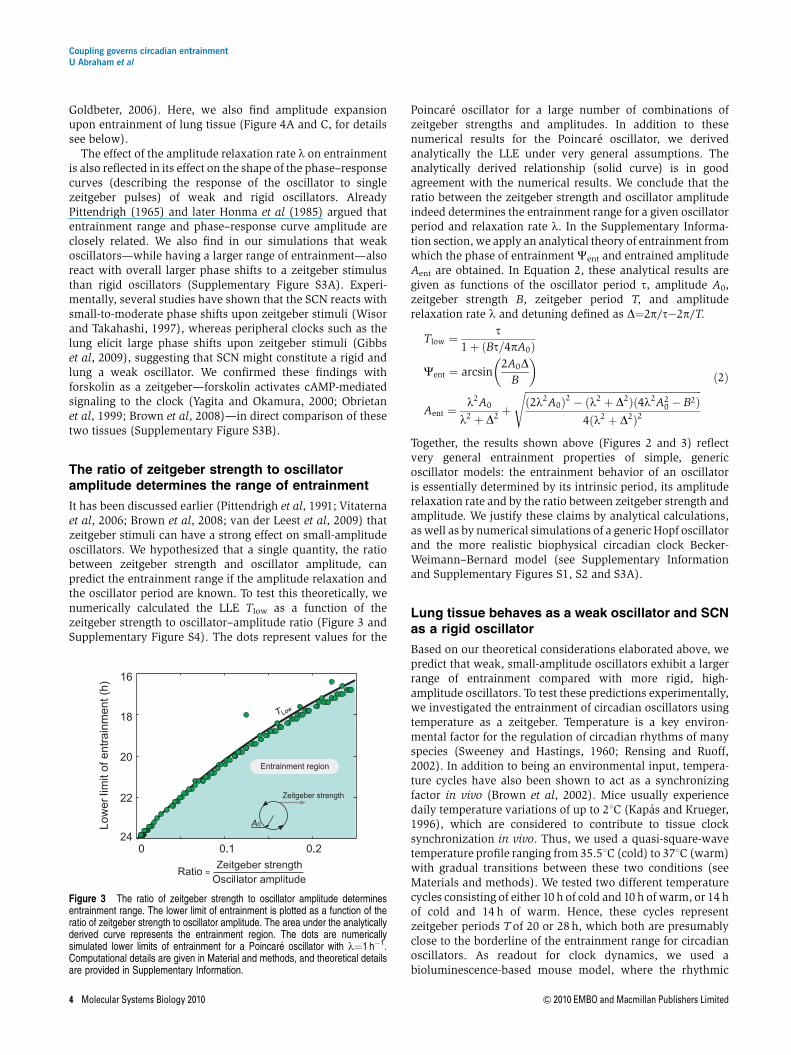
To test our theoretical prediction—attenuating coupling makesthe coupled oscillatory system weaker and thereby enlargesthe entrainment range—experimentally, we pharmacologi-cally reduced coupling in cultured SCN slices taken fromPER2::LUC mice. To this end, we subjected SCN slices toN-(Cis-2-phenyl-cyclopentyl)azacyclotridecan-2-imine-hydro-chloride (MDL)—a known inhibitor of adenylate cyclase(Hagen et al, 2006) that interferes with VIP-mediated coupling(O’Neill et al, 2008). PER2-LUC bioluminescence rhythms ofindividual SCN neurons were recorded using a high-resolutioncamera system (Figure 6A). SCN neurons of untreated slicesexhibit well-synchronized cellular rhythms, while treatmentwith MDL leads to their immediate desynchronization due tothe relatively wide distribution of individual cellular periods(Figure 6B and C; Supplementary Figure S7 and SupplementaryMovies). While the magnitude of PER2-LUC bioluminescenceis substantially reduced (and—as a consequence—also theabsolute amplitude values, as also described by O’Neill et al,2008), we did not find a significant effect of MDL on therelative amplitude of the circadian rhythms of single SCNneurons (Figure 6D). We determine relative amplitudesbecause the effect of a zeitgeber (e.g., the induction oftranscription) is more likely to be relative than absolute anddefine it as the ratio of the maximal/minimal values of anoscillating variable to its mean (see also Materials andmethods). Thus, we conclude that in this experimental setting,MDL reduces primarily cellular coupling with no detectableeffect on relative amplitude.
If reducing the coupling strength indeed makes an oscilla-tory system weaker, MDL-treated SCN slices should have awider range of entrainment than untreated SCN slices. To testthis, we entrained MDL-treated SCN slices to a 10-h 35.51C(cold)/10-h 371C (warm) temperature cycle. In contrast tountreated SCN slices that do not entrain to this 20-htemperature cycle (Figure 4), SCN slices treated with different
MDL concentrations entrained in a MDL dose-dependentmanner (Figure 6E and Supplementary Figure S8A). Thisclearly indicated an expanded range of entrainment, aspredicted by our theoretical arguments. In addition, whenwe used an alternative way to reduce the coupling betweenSCN neurons, i.e., application of tetrodotoxin (TTX), the SCNalso entrained to the 20-h temperature cycle. TTX is aninhibitor of voltage-gated sodium channels that prevents thegeneration of action potentials. TTX-treated SCN neurons havebeen described to lose the synchrony of their circadianoscillations, while also decreasing their amplitude signifi-cantly (Yamaguchi et al, 2003). Indeed, when analyzingoriginal single-cell time series data from the study reportedby Yamaguchi et al (2003), we also find a substantial (aboutthreefold) and highly significant reduction of relative ampli-tude upon TTX treatment (Po0.00001; Mann–Whitney test,one-tailed; n¼78 for untreated cells; n¼185 for TTX-treatedcells; not shown)—very different from the effects of MDL onrelative amplitude. Interestingly, the differential effect of thesedrugs on SCN single-cell amplitudes might also result indifferent phases of entrainment of MDL- and TTX-treated SCNslices (Brown et al, 2008), which we indeed see a trend for(Figure 6E and F). In addition, it is conceivable that MDL andTTX might also have a differential effect on coupling andthereby on the amplitude relaxation rate of the oscillatorysystem, which also can influence phase of entrainment(Supplementary Figure S8B).
In summary, subjecting cultured SCN slices to decouplingagents (MDL and TTX) leads to an expanded entrainmentrange. In the case of MDL, this is an amplitude-independenteffect and probably due to a weakening of the oscillatorsystem (i.e., reducing the amplitude relaxation rate l). Inthe case of TTX, also the amplitude of cellular rhythms isaffected, which leads to an expansion of the range ofentrainment. Thus, as predicted by our theoretical considera-tions, coupling governs the entrainment range of SCN tissuebecause it can affect both amplitude and rigidity of circadianoscillators.
A B C
Figure 5 Coupling constrains the entrainment range of oscillators. (A) Coupling increases the amplitude of the synchronized, coupled system. Given are numericallycalculated results of two coupled Poincare oscillators (see Equation 3). Coupling is quantified by the coupling strength parameter K. The amplitude increase uponcoupling is more pronounced, if the individual oscillators are weak, i.e., for small relaxation rates g. (B) Coupling makes oscillators more rigid by increasing the relaxationrate l of the coupled system. Shown are numerically calculated results of two coupled Poincare oscillators. Note that the effect of the coupling strength on the rigidity ofthe coupled system is not dependent on the amplitude relaxation rates g of the individual oscillators. As coupling slightly affects the period of the coupled system, therelaxation rate l was normalized by multiplying with the coupling-dependent period. (C) Coupling makes an oscillatory system harder to entrain. Two coupled Poincareoscillators were entrained by adding a periodic square-wave forcing of period T, alternating with equal duration and a forcing between 0 and 0.1 (term F in Equation 3).The lower limit of entrainment TLow of the coupled system was calculated for different coupling strengths K and different amplitude relaxation rates g of individualoscillators. As the coupling affects the intrinsic (non-forced) period itself, the lower limit of entrainment was normalized with respect to a constant intrinsic period of 24 h tovisualize the effects on the entrainment range that are entirely due to the effects on the rigidity and amplitude of the coupled system.
Coupling governs circadian entrainmentU Abraham et al
& 2010 EMBO and Macmillan Publishers Limited Molecular Systems Biology 2010 7
Discussion
Entrainment is one of the cornerstones of circadian biology. Inevolution, the phase of a rhythmic variable is selective rather
than its endogenous period. Thus, the synchronization ofendogenous rhythms to zeitgeber cycles of the environment(resulting in a specific phase of entrainment) is fundamentalfor the adaptive value of circadian clocks. In this study, we
8 16 00 8 16
Time (h)
2
4
6
8
10
12
Day
s
8 16 00 8 16
Time (h)
2
4
6
8
10
12
Day
s
00
E F
+MDL +TTX
C D
18 6
12
0
18 6
12
0
Time (h)
Untreated MDL
Untreated MDL
Rel
. am
plitu
de
0
2
A
Recording time (days)
Bio
lum
ines
cenc
e x1
04 Biolum
inescence x104
0 2 4 60
5
8 10 120
0.5
1.0MDL
10 µM MDL
B10 µM
Figure 6 Decoupling of SCN cells broadens the entrainment range of SCN tissue. (A) Circadian rhythms of individual SCN cells can be monitored. The digital imageshows a 10-min exposure of bioluminescence emitted from a medial section through the SCN of a PER2::LUC mouse. Bioluminescence reporting abundance of theclock protein PER2 was quantified from 130 single cells (marked by white circles) over the course of several days. Scale bar, 500 mm. (B) Representativebioluminescence traces from 10 different single cells. Typical for SCN slices, individual cells oscillate with a circadian period, and the majority of cells have theirbioluminescence peaks at similar times. After addition of the adenylyl cyclase inhibitor MDL on day 6, bioluminescence recording was resumed as shown for 10representative cells (note that representative cells before and after MDL treatment were not identical). While MDL addition reduced the magnitude of PER2 abundanceand lead to random phasing of peak bioluminescence, relative amplitudes of untreated and MDL-treated cells were similar. (C) Polar plots showing the peak times ofPER2 expression of individual SCN cells from the experiment shown in (B) during the third day of slice recording. Each dot represents the phase of a single cell. Single-cell phases in an untreated SCN slice (left) clustered significantly at 12 h (Rayleigh test, r¼0.8, Po0.0001, n¼49), but were randomly distributed upon MDL treatment(right, Rayleigh test, r¼0.26, P¼0.07, n¼40). Arrows in polar plots represent the mean vectors, the direction denotes the mean phase, and length measures thetendency of the data to cluster based upon a Rayleigh test, in which r-values range from 0 (randomly phased) to 1 (all cells peak at the same time). (D) The relativeamplitudes of single-cell oscillations do not vary with treatment (Mann–Whitney test, one-tailed, P¼0.47, n¼169 for untreated cells; n¼94 for MDL-treated cells).Displayed are normalized median amplitudes±quartiles±minima/maxima from three independent imaging experiments. For each experiment, individual amplitudevalues were normalized to the mean amplitudes of untreated cells. (E, F) SCN cultured in the presence of MDL (10 mM final concentration) or TTX (2-4 mM finalconcentration), respectively, entrain to a 20-h temperature cycle consisting of 10 h cold (35.51C, blue boxes) and 10 h warm (371C). Given are double plots of the peaktimes (±s.d.; n¼8 for MDL, and n¼6 for TTX) of PER2-LUC bioluminescence. Periods before, during and after entrainment are given in Supplementary Table S1. Thus,the range of entrainment for SCN slices is increased (compare with Figure 4) when SCN oscillators are decoupled (see B and C). Source data is available for this figure atwww.nature.com/msb.
Coupling governs circadian entrainmentU Abraham et al
8 Molecular Systems Biology 2010 & 2010 EMBO and Macmillan Publishers Limited
systematically investigated the properties of circadian oscilla-tors that are essential for entrainment behavior and describecoupling as a primary determinant.
As an experimental starting point of this study, wefound that the circadian oscillators of lung tissue have a largerrange of entrainment than SCN tissue—they readily entrainedto an experimental temperature cycle, whereas SCN tissuedid not (Figure 4). In theoretical analyses, we show that boththe ratio of amplitude and zeitgeber strength (Figure 3) and,importantly, inter-oscillator coupling are major determinantsfor entrainment (Figure 5). The reason for coupling beingcritical is twofold: (i) Coupling makes an oscillatory systemmore rigid, i.e., it relaxes faster in response to a perturbation,and (ii) coupling increases the amplitude of the oscillatorysystem. Both of these consequences of coupling lead to asmaller entrainment range, because zeitgeber stimuli affect theoscillatory system less if the relaxation is fast (Figure 2) andthe amplitude is high (Figure 1). From these theoreticalconsiderations, we conclude that the lung clock probablyconstitutes a weak oscillatory system, likely because a lack incoupling leads to a slow amplitude relaxation. (Circadianamplitude is not particularly low in lung (Figure 4)). Incontrast, the SCN constitutes a rigid oscillator, wherebycoupling and its described consequences probably are theprimary causes for this rigidity. We then tested thesetheoretical predictions by experimentally perturbing couplingin the SCN, and find that, indeed, reducing the couplingweakens the circadian oscillatory system in the SCN, whichresults in an enlargement of the entrainment range (Figure 6).
Why is the SCN designed to be a stronger circadian oscillatorthan peripheral organs? We speculate that the position of theSCN—as the tissue that conveys environmental timinginformation (i.e., light) to the rest of the body—makes itnecessary to create a circadian clock that is robust againstnoisy environmental stimuli. The SCN oscillator needsto be robust enough to be protected from environmentalnoise, but flexible enough to fulfill its function as anentrainable clock even in extreme photoperiods (i.e., seasons).By the same token, peripheral clocks are more protectedfrom the environmental zeitgebers due to intrinsic homeo-static mechanisms (e.g., temperature control in home-otherms). Thus, they do not necessarily need to develop astrong oscillatory system (e.g., by strengthening the coupling)rather they need to stay flexible enough to respond todirect or indirect signals from the SCN, such as hormonal,neural, temperature or metabolic signals. Such a designensures that only robust and persistent environmental signalstrigger an SCN resetting response, while SCN signals canrelatively easily be conveyed to the rest of the body. Thus, therobustness in the SCN clock likely serves as a filter forenvironmental noise.
These speculations are supported by observations suggest-ing that circadian clocks in primary sensory organs—such asthe eyes or the olfactory system—are more robust than otherperipheral clocks (Tosini and Menaker, 1996; Granados-Fuentes et al, 2004). These clocks are directly exposed toenvironmental zeitgebers, such as light and odor (Amir et al,1999). For example, the retinal clock seems to be specialamong extra-SCN oscillators in that one of its rhythmicfunctional outputs (melatonin secretion) persists for at least
5 days in culture without substantial dampening (Tosini andMenaker, 1996). In addition, while SCN lesion abolishes mostovert circadian rhythms, some rhythms in cornea and retinapersist (Scheving et al, 1983, Terman et al, 1993, Sakamotoet al, 2000), indicating a robust clock in the eye. In addition,the clock in the olfactory bulbs seems to be self-sustained andSCN independent, i.e., in vivo oscillation persists even in theabsence of the SCN (Abraham et al, 2005)—a property that hasnot been shown for other oscillators. In fact, clocks in otherbrain regions that are not directly exposed to environmentalsignals that may act as zeitgebers are usually much weaker,low-amplitude oscillators (for a review, see Guilding andPiggins, 2007). Although, to our knowledge, it has not yet beentested whether clocks in the eyes or olfactory bulbs are lessresponsive to zeitgeber stimuli than other brain clocks (as itwould be predicted for a robust clock), the fact that their cellsdo not quickly desynchronize suggests that some kind ofcoupling is in place in these tissues.
Coupling seems to have an important role for robustsynchronization, yet the effects of coupling and zeitgeber-to-amplitude ratio are probably well balanced. If the couplingbetween SCN neurons would be very strong (e.g., as strong asthe effect of constant light that substantially lengthensbehavioral periods in rodents, Daan and Pittendrigh,1976), the period of synchronized SCN neurons might bedifferent from the mean period of the dispersed neurons,which is clearly not the case (Honma et al, 1998). By thesame token, the zeitgeber-to-amplitude ratio together with theintra-SCN coupling allow environmental stimuli, such asexperimental 1-h light pulses (Comas et al, 2006), to phaseshift the SCN clock only moderately by maximally 3 h. Thisrestricts the entrainment range of circadian rhythms totypically 24±2 h (Pittendrigh and Daan, 1976). Thus, anecessary robustness against variable and noisy inputs isbalanced with a moderate responsiveness to zeitgeber stimulito allow daily entrainment, but probably also stable entrain-ment in different seasons.
As an additional means, daily or seasonal plasticity inthe coupling properties may contribute to the flexibility ofthe circadian system. In fact, in recent years it became obviousthat the phase distribution of the SCN neurons is stronglydependent on the photoperiod. In long photoperiods, thephases of SCN neurons are more broadened (Schaapet al, 2003) implying an altered, but not necessarily weakercoupling. Interestingly, this SCN-intrinsic encoding of thephotoperiod results in different responses toward zeitgeberstimuli (van der Leest et al, 2009), suggesting that the typeof coupling indeed may affect entrainment properties. Howthe coupling in long photoperiods is altered within the SCNis unclear at present. Further experiments are necessary, e.g.,by directly measuring amplitude relaxation rates, to determinethe rigidity of SCN (and peripheral oscillators) in differentzeitgeber periods.
In summary, using a combination of simulation studies,analytical calculations and experiments, we uncovered criticalfeatures for entrainment, such as zeitgeber-to-amplitude ratioand amplitude relaxation rate. Coupling is a primary factorthat governs these features explaining important differences inthe design of SCN and peripheral oscillators that ensure arobust, but also flexible circadian system.
Coupling governs circadian entrainmentU Abraham et al
& 2010 EMBO and Macmillan Publishers Limited Molecular Systems Biology 2010 9

Materials and methods
Numerical calculations
The simulations were designed to explore how the entrainment rangedepends on a few generic parameters that are representative for a largeclass of oscillators. Results presented in Figures 2 and 3 andSupplementary Figure S3 were numerically calculated using a forcedPoincare oscillator in Cartesian coordinates:
dx
dt¼ lxðA0 � rÞ � 2p
tyþ F
dy
dt¼ lyðA0 � rÞ þ 2p
tx
The parameters represent the oscillator period t, amplitude A0 andamplitude relaxation rate l. Introducing the radius r¼(xþ y)1/2 andf¼arctan(y/x) leads to our model in Equation 1. The oscillator isentrained by a forcing term F that represents light or temperatureentrainment. Without forcing, the oscillator has a 24-h period, i.e.,t¼24 h. In Figure 2A, a Poincare oscillator is entrained to square-wavezeitgeber cycles in analogy to the temperature cycles used in theexperiments. In Figure 2 and Supplementary Figure S3, the weakoscillator has an amplitude relaxation rate l¼0.03 h�1, the rigidoscillator has an amplitude relaxation rate l¼1 h�1 and bothoscillators have an amplitude A0¼1. In Figure 2B, the theoreticalcurves are plotted using our expression for the entrainment amplitudeAent (see Equation 2). In Figures 2B and 3, and Supplementary FigureS3, the Poincare oscillator is entrained to sinusoidal cycles, i.e.,F¼Bsin(2pt/T) with zeitgeber strength B, zeitgeber period Tand time t.Figure 3 shows 112 different ratios of B/A0 smaller than 0.25 withl¼1 h�1. Supplementary Figure S4 shows the LLE for a linear, aPoincare and a Hopf oscillator with an amplitude relaxation ratel¼0.01 h�1 for each model. Computational and mathematical detailsregarding square-wave entrainment and the linear oscillator are givenin the study by Granada and Herzel (2009).
The entrainment regions and lower limits of entrainment in Figures2 and 3, Supplementary Figures S1 and S3 were calculated followingthe same numerical protocol: (i) Choose an oscillator amplitude A0,relaxation rate l, zeitgeber strength B and zeitgeber period T.(ii) Define a set of 24 initial conditions equally distributed aroundthe limit cycle. (iii) Start the simulation and integrate 105 days. (iv) Foreach initial condition, the system is considered to reach entrainment ifthe mean phase difference of eight consecutive cycles is smaller than1 min. (v) If more than 70% of the 24 initial conditions reachentrainment, consider the oscillator to be entrained for the givenzeitgeber period Ti. (vi) For the LLE calculation take a shorter zeitgeberperiod Tj o Ti and repeat steps (iii–v) n times until a zeitgeber periodTn is reached, under which no entrainment is achieved. (vii) The limitof entrainment is the last zeitgeber period under which the oscillatorentrains, i.e., Tlow ¼ Tn�1. For the upper limit of entrainment take alonger zeitgeber period Tj4Ti for each loop. For Figure 2 andSupplementary Figure S1, the whole protocol is repeated for anincreasing zeitgeber strength B. In addition, for Figures 3 andSupplementary Figure S2, we repeated the protocol for 112 differentratios of B/A0 o0.25.
Numerical integrations were implemented in Matlab (The Math-Works, Inc.) and XPPAUT (Ermentrout, 2003) with the use of RobClewley’s XPP-Matlab interface. We used the CVODE integratorimplemented in XPPAUT with stepsize setting of 0.2 h�1, and errortolerance settings tol¼1e–5 and atol¼1e–5. To test our algorithm, werepeated the most sensitive numerically integrated results with thecontinuation software Cl_MatCont (Dhooge et al, 2008), which allowscomputation of bifurcations, limit cycles and Floquet exponents, andobserved a fairly good agreement. Results shown in Figures 5A–C wereentirely calculated with Cl_MatCont. The LLE is composed of a torusand a fold bifurcation (this is a general situation, see Balanov et al,2009, chapter 3), both were accurately computed with Cl_MatCont. Asthe intrinsic (unforced) period t of the coupled system (Equation 3)varies with the coupling strength K, this change (o1 h) slightly affectsthe LLE: the intrinsic period increases with increasing couplingstrength, making the effect shown in Figure 5C even more pronounced.To exclude this drift, and hence, to show that the coupling strength andthe amplitude relaxation rate of the single oscillators indeed exert a
strong bona fide effect on the LLE, the LLE was normalized such that
LLEnormalized ¼LLE
tðKÞ� 24h�1
where t(K) is period of the unforced but coupled system at couplingstrength K. Note that the unforced, uncoupled single-oscillator periodis 24 h. Continuation results were compared with standard numericalintegrations to assert accuracy and correctness.
Animals
Heterozygous male PER2::LUC knockin mice (Yoo et al, 2004;gift of DrJoseph Takahashi, Northwestern University, IL, USA) expressing aPERIOD2::LUCIFERASE fusion protein were bred and raised in ouranimal facility (FEM, Berlin, Germany), and maintained in a 12 h light/12 h dark cycle. All procedures were authorized by and performed inaccordance with guidelines and regulations of the German animalprotection law (Deutsches Tierschutzgesetz).
Bioluminescence recording and temperatureentrainment
Animals were killed by cervical dislocation and their brains and lungstransferred to chilled Hank’s buffered saline solution, pH 7.2. Fortissue culture, 300mm coronal sections of the brain and lung wereobtained with a tissue chopper. Brain slices containing the SCN wereidentified, and the bilateral, medial SCN dissected out using a pair ofscalpels. All tissues were cultured individually on a Millicellmembrane (Millipore) in a Petri dish with 1 ml of supplementedDulbecco’s Modified Eagle’s Medium (DMEM, for details seeSupplementary methods), containing 0.1 mM beetle luciferin (BioThe-ma, Sweden). Petri dishes were covered with glass slides, sealed withgrease and placed in temperature-adjustable light-tight boxes (Tech-nische Werkstatten Charite, Berlin, Germany), equipped with photo-multiplier tubes (HC135-11MOD, Hamamatsu, Japan) at 5% CO2 forbioluminescence recording. During bioluminescence recording, adaily temperature cycle with a period of 20 h (10 h of 35.51C and 10 hof 371C) was applied to each SCN and lung slice. In order to simulategradual temperature changes at dusk and dawn, each temperature stepcomprised a gradual temperature increase or decrease, respectively,over a course of 2 or 3 h. The first phase of the temperature entrainmentcycle was always the cold phase (10 h of 35.51C) and started at theminimum of PER2::LUC expression, as determined by online registra-tion. Temperature entrainment comprised five or six temperature cycles.To inhibit cAMP-dependent signaling, which has been described to becrucial for intra-SCN coupling (O’Neill et al, 2008), we supplementedthe culture medium with 10mM (final concentration) of the adenylylcyclase inhibitor MDL (Sigma). In an additional experiment, TTX(Sigma) were added to the culture medium (2–4mM final concentration)to block voltage-gated sodium channels, i.e., to inhibit neuronalcommunication via action potentials. As both drugs are solved inaqueous solution, solvent control experiments are those shown inFigure 4. Bioluminescence from all slices was recorded in 5-min bins forat least 13 days. For pharmacological treatments, cultures were brieflyremoved from the recording incubator, and a complete medium changewas performed. Recording was resumed immediately.
Single-cell recordings and image analysis
SCN tissues were cultured individually on pieces of Millicellmembrane, inverted and cultured in 100ml DMEM at 371C and 5%CO2 on the bottom of poly-D-lysine- and 5% laminine-coated 35-mmmicroscopic tissue culture dishes with grid (IBIDI, Germany). On day3–5, the culture medium was replaced by 500 ml DMEM supplementedwith 0.18 mg ml�1 NaHCO3 and 0.1 mM beetle luciferin. The Petri dishwas sealed with grease and placed in a light-tight imaging chamber.Bioluminescence imaging was carried out in complete darkness andconstant temperature with a 10� objective and an inverse setup,including an intensified digital camera (XR/Mega-10Z, StanfordPhotonics, USA). Images were stored in 10-min bins over the course
Coupling governs circadian entrainmentU Abraham et al
10 Molecular Systems Biology 2010 & 2010 EMBO and Macmillan Publishers Limited

of several days. For MDL treatment, culture medium was exchanged byDMEM supplemented with NaHCO3, 0.1 mM beetle luciferin, and10 mM MDL, and imaging was resumed immediately. Control treatment(DMEM supplemented with NaHCO3, luciferin, and the correspondingamount of MDL solvent (H2O)) did not have an effect on circadianoscillations (data not shown).
Ten-min exposures were stacked and resulting movies weresmoothed using a 15-image running average (Piper 1.3. software,Stanford Photonics, USA). Alternatively, 10-min exposures weresubjected twice to a Kalman filter (ImageJ software, NIH, USA). Singlebioluminescent cells in random parts of the SCN were manually tracedover several days and their gray values quantified using ImageJsoftware. Raw time series data were visually inspected and only cellsthat fulfilled certain criteria (Supplementary Information) wereincluded in further analysis. Note that due to technical limitationscells tracked before and after MDL treatment were not identical.
Analysis of time series data
Bioluminescence time series data from single cells and tissue sliceswere first trend eliminated, and then analyzed for their period, phaseand relative amplitude using ChronoStar 1.6 (Stephan Lorenzen,Institute for Theoretical Biology, Humboldt-University, Germany).Time series were trend eliminated by dividing values by a 24-h runningaverage, thereby normalizing the magnitude to 1 to be independent ofany measurement specifics, such as sensitivity/background of thephotomultiplier or efficacy of the luciferase. Resulting time seriesoscillate around 1 with amplitudes that are relative to its mean.Periods, phases and amplitudes were estimated by fitting the cosinewave function y¼a� exp(bt)� cos(2pt� 24/cþ d), which includes anexponential term for damping (a¼amplitude, b¼damping, c¼period,d¼phase). Statistical analyses were performed using GraphPad Prism4 software (GraphPad software, USA) and Rayleigh statistics afterBatschelet (1981).
Supplementary information
Supplementary information is available at the Molecular SystemsBiology website (www.nature.com/msb).
AcknowledgementsWe thank Stephan Lorenzen for developing the ChronoStar software,Bert Maier and Raik Paulat for help in designing and building thetemperature-adjustable light-tight boxes, Hitoshi Okamura for sendingus single SCN cell time series data from TTX-treatment experimentsand Joseph Takahashi for providing us with PER2::LUC mice. Thiswork was supported by the Deutsche Forschungsgemeinschaft (SFB618 and SPP 1395) and the Bernstein Center for ComputationalNeuroscience. Work in AK’s laboratory is supported by the 6th EUframework program EUCLOCK.
Author contributions: AK and HH designed research; UA and MHperformed experiments; AEG and POW performed theoretical studies;all authors analyzed the data; AK wrote the paper with contributionsfrom all other authors.
Conflict of interestThe authors declare that they have no conflict of interest.
References
Amir S, Cain S, Sullivan J, Robinson B, Stewart J (1999) Olfactorystimulation enhances light-induced phase shifts in free-runningactivity rhythms and Fos expression in the suprachiasmaticnucleus. Neuroscience 92: 1165–1170
Abraham U, Prior JL, Granados-Fuentes D, Piwnica-Worms DR,Herzog ED (2005) Independent circadian oscillations of Period1in specific brain areas in vivo and in vitro. J Neurosci 25: 8620–8626
Anishchenko VS, Astakhov V, Neiman A, Vadivasova T, Schimansky-Geier L (2007) Nonlinear Dynamics of Chaotic and StochasticSystems: Tutorial and Modern Developments. Springer-Verlag NewYork: LLC
Aton SJ, Herzog ED (2005) Come together, right.now: synchronizationof rhythms in a mammalian circadian clock. Neuron 48: 531–534
Aschoff J, Pohl H (1978) Phase relations between a circadianrhythm and its zeitgeber within the range of entrainment.Naturwissenschaften 65: 80–84
Balanov A, Janson N, Postnov D, Sosnovtseva O (2009) Synchronization:From Simple to Complex. Springer-Verlag New York: LLC
Batschelet E (1981) Circular statistics in biology. New York: AcademicPress
Becker-Weimann S, Wolf J, Herzel H, Kramer A (2004) Modelingfeedback loops of the mammalian circadian oscillator. Biophys J 87:3023–3034
Berge P, Pomeau Y, Vidal C (1984) Order within Chaos: Towards aDeterministic Approach to Turbulence. Paris, France: Hermann andWiley, John & Sons
Bernard S, Gonze D, Cajavec B, Herzel H, Kramer A (2007)Synchronization-induced rhythmicity of circadian oscillators inthe suprachiasmatic nucleus. PLoS Comput Biol 3: e68
Best JD, Maywood ES, Smith KL, Hastings MH (1999) Rapid resettingof the mammalian circadian clock. J Neurosci 19: 828–835
Brown SA, Kunz D, Dumas A, Westermark PO, Vanselow K, Tilmann-Wahnschaffe A, Herzel H, Kramer A (2008) Molecular insights intohuman daily behavior. Proc Natl Acad Sci USA 105: 1602–1607
Brown SA, Zumbrunn G, Fleury-Olela F, Preitner N, Schibler U (2002)Rhythms of mammalian body temperature can sustain peripheralcircadian clocks. Curr Biol 12: 1574–1583
Chiesa JJ, Dıez-Noguera A, Cambras T (2007) Effects of transient andcontinuous wheel running activity on the upper and lower limits ofentrainment to light-dark cycles in female hamsters. Chronobiol Int24: 215–234
Comas M, Beersma DGM, Spoelstra K, Daan S (2006) Phase andperiod responses of the circadian system of mice (Mus musculus)to light stimuli of different duration. J Biol Rhythms 21:362–372
Daan S, Pittendrigh CS (1976) A functional analysis of circadianpacemakers in nocturnalrodents III. Heavy water and constantlight: homeostasis of frequency? J comp Physiol A 106: 267–290
de la Iglesia HO, Cambras T, Schwartz WJ, Dıez-Noguera A (2004)Forced desynchronization of dual circadian oscillators within therat suprachiasmatic nucleus. Curr Biol 14: 796–800
de la Iglesia HO, Meyer J, Carpino A, Schwartz WJ (2000) Antiphaseoscillation of the left and right suprachiasmatic nuclei. Science 290:799–801
Dhooge A, Govaerts W, Kuznetsov YA, Meijer HGE, Sautois B (2008)New features of the software matcont for bifurcation analysis ofdynamical systems. Mathematical and Computer Modelling ofDynamical Systems: Methods, Tools and Applications inEngineering and Related Sciences 14: 147–175
Ermentrout B (2003) Simulating, analyzing, and animating dynamicalsystems: a guide to XPPAUT for researchers and students. ApplMech Rev 5: B53
Forger DB, Peskin CS (2003) A detailed predictive model ofthe mammalian circadian clock. Proc Natl Acad Sci USA 100:14806–14811
Gibbs JE, Beesley S, Plumb J, Singh D, Farrow S, Ray DW, Loudon ASI(2009) Circadian timing in the lung; a specific role for bronchiolarepithelial cells. Endocrinology 150: 268–276
Glass L, Guevara MR, Shrier A (1987) Universal bifurcations andthe classification of cardiac arrhythmias. Ann NY Acad Sci 504:168–178
Glass L, Mackey MC (1988) From Clocks to Chaos: The Rhythms of Life.Princeton, NJ: Princeton University Press
Coupling governs circadian entrainmentU Abraham et al
& 2010 EMBO and Macmillan Publishers Limited Molecular Systems Biology 2010 11

Gonze D, Bernard S, Waltermann C, Kramer A, Herzel H (2005)Spontaneous synchronization of coupled circadian oscillators.Biophys J 89: 120–129
Granada AE, Herzel H (2009) How to achieve fast entrainment? Thetimescale to synchronization. PLoS One 4: e7057
Granados-Fuentes D, Prolo LM, Abraham U, Herzog ED (2004) Thesuprachiasmatic nucleus entrains, but does not sustain, circadianrhythmicity in the olfactory bulb. J Neurosci 24: 615–619
Gribkoff VK, Pieschl RL, Wisialowski TA, van den Pol AN, Yocca FD(1998) Phase shifting of circadian rhythms and depression ofneuronal activity in the rat suprachiasmatic nucleus byneuropeptide Y: mediation by different receptor subtypes.J Neurosci 18: 3014–3022
Guckenheimer J, Holmes P (1983) Nonlinear Oscillations, DynamicalSystems, and Bifurcations of Vector Fields. New York: Springer-Verlag
Guilding C, Piggins HD (2007) Challenging the omnipotence of thesuprachiasmatic timekeeper: are circadian oscillators presentthroughout the mammalian brain? Eur J Neurosci 25: 3195–3216
Hagen BM, Bayguinov O, Sanders KM (2006) Vip and Pacap regulatelocalized Ca2+ transients via camp-dependent mechanism. Am JPhysiol Cell Physiol 291: C375–C385
Helfrich-Forster C (2004) The circadian clock in the brain: a structuraland functional comparison between mammals and insects. J CompPhysiol A Neuroethol Sens Neural Behav Physiol 190: 601–613
Herzog ED, Aton SJ, Numano R, Sakaki Y, Tei H (2004) Temporalprecision in the mammalian circadian system: a reliable clock fromless reliable neurons. J Biol Rhythms 19: 35–46
Herzog ED, Huckfeldt RM (2003) Circadian entrainment totemperature, but not light, in the isolated suprachiasmaticnucleus. J Neurophysiol 90: 763–770
Honma K, Honma S, Hiroshige T (1985) Response curve, free-runningperiod, and activity time in circadian locomotor rhythm of rats. JpnJ Physiol 35: 643–658
Honma S, Nakamura W, Shirakawa T, Honma K (2004) Diversity in thecircadian periods of single neurons of the rat suprachiasmaticnucleus depends on nuclear structure and intrinsic period. NeurosciLett 358: 173–176
Honma S, Shirakawa T, Katsuno Y, Namihira M, Honma K (1998)Circadian periods of single suprachiasmatic neurons in rats.Neurosci Lett 250: 157–160
Huygens C (1673) Horologium oscillatorium. Ames: Englishtranslation: The pendulum clock, Iowa State University Press1986 edition
Kapas L, Krueger JM (1996) Nitric oxide donors sin-1 and snappromote nonrapid-eye-movement sleep in rats. Brain Res Bull 41:293–298
Kronauer RE, Czeisler CA, Pilato SF, Moore-Ede MC, Weitzman ED(1982) Mathematical model of the human circadian system withtwo interacting oscillators. Am J Physiol 242: R3–R17
Kuramoto Y (1984) Chemical oscillations, waves, and turbulence.Berlin: Springer-Verlag
Kurosawa G, Goldbeter A (2006) Amplitude of circadian oscillationsentrained by 24-h light-dark cycles. J Theor Biol 242: 478–488
Leloup JC, Goldbeter A (2003) Toward a detailed computational modelfor the mammalian circadian clock. Proc Natl Acad Sci USA 100:7051–7056
Levi F, Schibler U (2007) Circadian rhythms: mechanisms andtherapeutic implications. Annu Rev Pharmacol Toxicol 47: 593–628
Liu AC, Welsh DK, Ko CH, Tran HG, Zhang EE, Priest AA, Buhr ED,Singer O, Meeker K, Verma IM, Doyle III FJ, Takahashi JS, Kay SA(2007) Intercellular coupling confers robustness against mutationsin the SCN circadian clock network. Cell 129: 605–616
Locke JCW, Westermark PO, Kramer A, Herzel H (2008) Globalparameter search reveals design principles of the mammaliancircadian clock. BMC Syst Biol 2: 22
Maywood ES, O’neill JS, Reddy AB, Chesham JE, Prosser HM,Kyriacou CP, Godinho SI, Nolan PM, Hastings MH (2007)Genetic and molecular analysis of the central and peripheral
circadian clockwork of mice. Cold Spring Harb Symp Quant Biol 72:85–94
Nagoshi E, Saini C, Bauer C, Laroche T, Naef F, Schibler U (2004)Circadian gene expression in individual fibroblasts: cell-auto-nomous and self-sustained oscillators pass time to daughter cells.Cell 119: 693–705
Obrietan K, Impey S, Smith D, Athos J, Storm DR (1999) Circadianregulation of cAMP response element-mediated gene expression inthe suprachiasmatic nuclei. J Biol Chem 274: 17748–17756
O’Neill JS, Maywood ES, Chesham JE, Takahashi JS, Hastings MH(2008) cAMP-dependent signaling as a core component of themammalian circadian pacemaker. Science 320: 949–953
Piggins HD, Antle MC, Rusak B (1995) Neuropeptides phase shift themammalian circadian pacemaker. J Neurosci 15: 5612–5622
Pikovsky A, Rosenblum M, Kurths J (2001) Synchronization: A universalconcept in nonlinear sciences. Cambridge, UK: Cambridge UniversityPress
Pikovsky AS (1984) On the interaction of strange attractors. Zeitschriftfur Physik B Condensed Matter 55: 149–154
Pittendrigh C (1965) In On the mechanism of the entrainment of acircadian rhythm by light cycles, Aschoff, J (ed), pp. 277–297.North-Holland, Amsterdam: Circadian Clocks
Pittendrigh C, Daan S (1976) The entrainment of circadian pacemakersin nocturnal rodents. IV. Entrainment: pacemaker as clock. J CompPhysiol A 106: 291–331
Pittendrigh CS, Kyner WT, Takamura T (1991) The amplitude ofcircadian oscillations: temperature dependence, latitudinalclines, and the photoperiodic time measurement. J Biol Rhythms6: 299–313
Prosser RA, Gillette MU (1989) The mammalian circadian clock inthe suprachiasmatic nuclei is reset in vitro by cAMP. J Neurosci 9:1073–1081
Rensing L, Ruoff P (2002) Temperature effect on entrainment, phaseshifting, and amplitude of circadian clocks and its molecular bases.Chronobiol Int 19: 807–864
Reppert SM, Weaver DR (2002) Coordination of circadian timing inmammals. Nature 418: 935–941
Roenneberg T, Dragovic Z, Merrow M (2005) Demasking biologicaloscillators: properties and principles of entrainment exemplifiedby the neurospora circadian clock. Proc Natl Acad Sci USA 102:7742–7747
Roenneberg T, Daan S, Merrow M (2003) The art of entrainment. J BiolRhythms 18: 183–194
Sakamoto K, Oishi K, Shiraishi M, Hamano S, Otsuka H, Miyake Y,Ishida N. (2000) Two circadian oscillatory mechanisms in themammalian retina. Neuroreport 11: 3995–3997
Schaap J, Albus H, VanderLeest HT, Eilers PH, Detari L, Meijer JH.(2003) Heterogeneity of rhythmic suprachiasmatic nucleusneurons: Implications for circadian waveform and photoperiodicencoding. Proc Natl Acad Sci USA 100: 15994–15999
Scheving LE, Tsai TH, Powell EW, Pasley JN, Halberg F, Dunn J. (1983)Bilateral lesions of suprachiasmatic nuclei affect circadian rhythmsin [3H]-thymidine incorporation into deoxyribonucleic acid inmouse intestinal tract, mitotic index of corneal epithelium, andserum corticosterone. Anat Rec 205: 239–249
Shirakawa T, Honma S, Honma K (2001) Multiple oscillators in thesuprachiasmatic nucleus. Chronobiol Int 18: 371–387
Sweeney BM, Hastings JW (1960) Effects of temperature upon diurnalrhythms. Cold Spring Harb Symp Quant Biol 25: 87–104
Terman JS, Reme CE, Terman M. (1993) Rod outer segment diskshedding in rats with lesions of the suprachiasmatic nucleus. BrainRes 605: 256–264
Tosini G, Menaker M (1996) Circadian rhythms in culturedmammalian retina. Science 272: 419–421
van der Leest HT, Rohling JHT, Michel S, Meijer JH (2009) Phaseshifting capacity of the circadian pacemaker determined by the SCNneuronal network organization. PLoS One 4: e4976
Vitaterna MH, Ko CH, Chang AM, Buhr ED, Fruechte EM, Schook A,Antoch MP, Turek FW, Takahashi JS (2006) The mouse clock
Coupling governs circadian entrainmentU Abraham et al
12 Molecular Systems Biology 2010 & 2010 EMBO and Macmillan Publishers Limited
mutation reduces circadian pacemaker amplitude and enhancesefficacy of resetting stimuli and phase-response curve amplitude.Proc Natl Acad Sci USA 103: 9327–9332
von Holst E (1939) Die relative Koordination als Phanomen und alsMethode zentralnervoser Funktionsanalyse, volume 42. SpringerBerlin/Heidelberg
Welsh DK, Logothetis DE, Meister M, Reppert SM (1995) Individualneurons dissociated from rat suprachiasmatic nucleus expressindependently phased circadian firing rhythms. Neuron 14:697–706
Westermark PO, Welsh DK, Okamura H, Herzel H (2009) Quantificationof circadian rhythms in single cells. PLoS Comput Biol 5: e1000580
Wever R (1972) Virtual synchronization towards the limits of the rangeof entrainment. J Theor Biol 36: 119–132
Winfree A (1980) The geometry of biological time. New York:Springer-Verlag
Wisor JP, Takahashi JS (1997) Regulation of the vgf gene in the goldenhamster suprachiasmatic nucleus by light and by the circadianclock. J Comp Neurol 378: 229–238
Yagita K, Okamura H (2000) Forskolin induces circadian geneexpression of rPer1, rPer2 and dbp in mammalian rat-1fibroblasts. FEBS Lett 465: 79–82
Yamaguchi S, Isejima H, Matsuo T, Okura R, Yagita K, Kobayashi M,Okamura H (2003) Synchronization of cellular clocks in thesuprachiasmatic nucleus. Science 302: 1408–1412
Yoo SH, Yamazaki S, Lowrey PL, Shimomura K, Ko CH,Buhr ED, Siepka SM, Hong HK, Oh WJ, Yoo OJ, Menaker M,Takahashi JS (2004) PERIOD2::LUCIFERASE real-time reportingof circadian dynamics reveals persistent circadian oscillationsin mouse peripheral tissues. Proc Natl Acad Sci USA 101:5339–5346
Molecular Systems Biology is an open-access journalpublished by European Molecular Biology Organiza-
tion and Nature Publishing Group. This work is licensed under aCreative Commons Attribution-Noncommercial-No DerivativeWorks 3.0 Unported License.
Coupling governs circadian entrainmentU Abraham et al
& 2010 EMBO and Macmillan Publishers Limited Molecular Systems Biology 2010 13
Copyright © 2022 FDOKUMEN