Stopping time: The genetics of fly and mouse circadian clocks
29
Annu. Rev. Neurosci. 2001. 24:1091–119 Copyright c 2001 by Annual Reviews. All rights reserved STOPPING TIME: The Genetics of Fly and Mouse Circadian Clocks Ravi Allada 2 , Patrick Emery 1,3 , Joseph S. Takahashi 1,2 , and Michael Rosbash 1,3 1 Howard Hughes Medical Institute 2 Northwestern University, Department of Neurobiology and Physiology, Evanston, Illinois 60208 3 Brandeis University, Department of Biology, Waltham, Massachusetts 02454; e-mail: [email protected], [email protected], [email protected], [email protected] Key Words circadian, bHLH-PAS, period, cryptochrome, casein kinase I epsilon ■ Abstract Forward genetic analyses in flies and mice have uncovered conserved transcriptional feedback loops at the heart of circadian pacemakers. Conserved mech- anisms of posttranslational regulation, most notably phosphorylation, appear to be im- portant for timing feedback. Transcript analyses have indicated that circadian clocks are not restricted to neurons but are found in several tissues. Comparisons between flies and mice highlight important differences in molecular circuitry and circadian or- ganization. Future studies of pacemaker mechanisms and their control of physiology and behavior will likely continue to rely on forward genetics. INTRODUCTION With the completion of the human genome sequence, the era of simple gene cloning and identification is nearing an end. With the advent of high throughput technolo- gies such as DNA microarrays, the temporal and spatial expression patterns of thousands of genes will be known. The challenge for neuroscience will be to func- tionally link genes, including their complex expression patterns, to the output of the nervous system: behavior. Genetics will play a crucial role in navigating through this genomic jungle. Forward genetics, the process of identifying mutant phenotypes to isolate genes, is best applied to a problem lacking molecular description. Forward genetics used to be “genetics,” but the advent of recombinant DNA and reverse genetics ne- cessitates a more useful term. Genetic screens require no prior hypothesis about the mechanism by which a system functions. The only requirements are an effi- cient means of mutagenesis and phenotypic screening. If there is an abundance of correlative molecular or cellular data, genetic analysis can also be helpful in 0147-006X/01/0621-1091$14.00 1091
-
Upload
utsouthwestern -
Category
Documents
-
view
0 -
download
0
Transcript of Stopping time: The genetics of fly and mouse circadian clocks

P1: FUM
April 6, 2001 11:34 Annual Reviews AR121-36
Annu. Rev. Neurosci. 2001. 24:1091–119Copyright c© 2001 by Annual Reviews. All rights reserved
STOPPING TIME: The Genetics of Flyand Mouse Circadian Clocks
Ravi Allada2, Patrick Emery1,3, Joseph S. Takahashi1,2,and Michael Rosbash1,31Howard Hughes Medical Institute2Northwestern University, Department of Neurobiology and Physiology, Evanston,Illinois 602083Brandeis University, Department of Biology, Waltham, Massachusetts 02454;e-mail: [email protected], [email protected],[email protected], [email protected]
Key Words circadian, bHLH-PAS, period, cryptochrome, casein kinase I epsilon
■ Abstract Forward genetic analyses in flies and mice have uncovered conservedtranscriptional feedback loops at the heart of circadian pacemakers. Conserved mech-anisms of posttranslational regulation, most notably phosphorylation, appear to be im-portant for timing feedback. Transcript analyses have indicated that circadian clocksare not restricted to neurons but are found in several tissues. Comparisons betweenflies and mice highlight important differences in molecular circuitry and circadian or-ganization. Future studies of pacemaker mechanisms and their control of physiologyand behavior will likely continue to rely on forward genetics.
INTRODUCTION
With the completion of the human genome sequence, the era of simple gene cloningand identification is nearing an end. With the advent of high throughput technolo-gies such as DNA microarrays, the temporal and spatial expression patterns ofthousands of genes will be known. The challenge for neuroscience will be to func-tionally link genes, including their complex expression patterns, to the output of thenervous system: behavior. Genetics will play a crucial role in navigating throughthis genomic jungle.
Forward genetics, the process of identifying mutant phenotypes to isolate genes,is best applied to a problem lacking molecular description. Forward genetics usedto be “genetics,” but the advent of recombinant DNA and reverse genetics ne-cessitates a more useful term. Genetic screens require no prior hypothesis aboutthe mechanism by which a system functions. The only requirements are an effi-cient means of mutagenesis and phenotypic screening. If there is an abundanceof correlative molecular or cellular data, genetic analysis can also be helpful in
0147-006X/01/0621-1091$14.00 1091
P1: FUM
April 6, 2001 11:34 Annual Reviews AR121-36
1092 ALLADA ET AL
determining which variables are critical. Distinguishing between observations thatare simply correlated with an output and those that are causal can be difficult. Byrandomly mutagenizing and assaying the consequence on a particular behavioralor physiologic parameter, one can identify key regulatory components.
Nowhere in neuroscience has the power of forward genetics been felt moreacutely than in circadian rhythms. Genetics has propelled rhythms research to ap-pear twice recently amongSciencemagazine’s breakthroughs of the year (1997,1998). In this review, we discuss the key discoveries and how they were made possi-ble by the confluence of genetics and genomics. We compare the molecular mech-anisms and functions of different animal circadian systems, focusing on the fruitfly, Drosophila melanogaster, and the mouse. For the sake of brevity and to avoidredundancy with other recent reviews (Dunlap 1999, Ishida et al 1999, Edery 2000,Hall 2000, King & Takahashi 2000, Scully & Kay 2000, Young 2000a,b), we focusour efforts on circadian pacemakers and their systemic organization and outputs.
BACKGROUND
Animals temporally organize their behavior and metabolism to adapt to andanticipate the progression through the 24-hour solar cycle. The earth’s 24-hourrotation has apparently dictated that all manner of life evolve to maximize fit-ness in this environment. Endogenous clocks serve to anticipate daily changes intheir environment. The overt manifestations of these clocks are circadian rhythms.They are defined by periodicity in the absence of exogenous cues. Circadianrhythms are therefore not simply driven by the environment but arise from in-ternal biological clocks. Circadian clocks only closely approximate but are notexactly 24 hours. Importantly, the timing of the clock is sensitive to light, amongother environmental cues. The daily cycle of light and darkness resets internalclocks to maintain synchrony between the external solar day and internal bio-logical clocks. Across a wide range of physiologic temperatures, the periodicityof rhythms is stable. This temperature compensation mechanism is particularlyrelevant in poikilotherms to prevent perturbations of the clock due to changesin the ambient temperature. However, even neural pacemaker tissues such as themammalian suprachiasmatic nucleus (SCN) when maintained in vitro also defendagainst changes in temperature (Ruby et al 1999). Temperature compensation isthought to reflect an underlying feature of the clock mechanism. Clocks have beentypically associated with regions of the nervous system, such as the SCN (Moore &Eichler 1972, Stephan & Zucker 1972). However, they are found in cells outsidethe nervous system and in organisms that do not have nervous systems. Evenunicellular prokaryotes (e.g. photosynthetic cyanobacteria) exhibit robust circa-dian rhythms (Johnson & Golden 1999). Determining the molecular nature of theseclocks awaited a forward genetic approach, first inDrosophilaand other organisms(Konopka & Benzer 1971, Bruce 1972, Feldman & Hoyle 1973) and later in themouse (Vitaterna et al 1994).
P1: FUM
April 6, 2001 11:34 Annual Reviews AR121-36
GENETICS OF CIRCADIAN CLOCKS 1093
BEHAVIOR, GENETICS, AND THE FRUIT FLY
In the modern era the study of the genetic basis of behavior was pioneered bySeymour Benzer and colleagues, using the fruit fly,Drosophila melanogaster.Benzer’s laboratory initiated genetic studies of many aspects of fly behavior, in-cluding circadian rhythms (Benzer 1971). The fruit fly had previously been aworkhorse of geneticists, aiding in the discovery of many of the core principles ofinheritance.Drosophilahas a generation time of only 10 days, and its long historyin the field of genetics has led to the development of many useful tools. Benzer’slaboratory hoped to apply the genetic techniques optimized in this model system toquestions related to behavior. Even then, much was known about circadian rhythmsin Drosophilaat the behavioral level. Fruit flies proceed through a series of char-acteristic developmental stages, beginning with the embryonic through a series ofthree larval stages to a pupal stage from which adults emerge or eclose. The fruit flyprefers to eclose at a particular time of day, reflecting gating by a circadian clock(Pittendrigh 1954). In fact, the wordDrosophilameans dew-loving, referring to itstendency to eclose in the morning. Ron Konopka, a graduate student in Benzer’slaboratory, designed a screen of mutagenized populations, looking for mutant fliesthat eclosed with a different circadian phase (Konopka & Benzer 1971). In nowclassic studies, Konopka identified three such strains. When studying the indivi-dual strains under constant conditions, he saw that one had a long period rhythmof 29 hours, the second had a short period rhythm of 19 hours, and the third hadno detectable rhythm. Remarkably, all three mutant phenotypes mapped to theX-chromosome to what appeared to be a single locus, which they calledperiod(per).
The identification of these behavioral mutants suggested that one could studygenes as a means to understanding behavior in general and circadian rhythms inparticular. In part, the success of this screen reflected the unusual precision of thisparticular behavioral assay. Few behavioral assays can detect small differenceson the order of 20% or less that distinguish their short and long period mutants.The ability to identify adult circadian rhythm mutants suggested that the mutatedgenes may not be vital to functions in development. If genes involved in circadianrhythms were also required for vital functions, then null mutations of such geneswould never live to eclosion. The ability to identify mutants suggested that thefunction of theperiodgene might be relatively circumscribed. As a result, we nowspeak of “circadian rhythm genes” or “circadian clock genes.”
TRANSCRIPTION AS NEURAL CODE: Drosophila Period
The work of Konopka and Benzer raised the possibility that forward geneticsmight more generally illuminate the circadian clockworks. However, the mutantsand their intriguing phenotypes shed little light on the actual clock mechanism.This level of insight awaited the cloning of theperiodgene and more precisely, a

P1: FUM
April 6, 2001 11:34 Annual Reviews AR121-36
1094 ALLADA ET AL
careful analysis of its temporal expression pattern. The cloning of PER was initiallymisleading, assigned to a group of cell surface proteins known as proteoglycans(Jackson et al 1986, Reddy et al 1986). This observation fit intercellular modelsof circadian clocks. However, more careful analysis ofper sequence, expression,and function changed this view dramatically. Both PER protein and RNA arerhythmically expressed (Siwicki et al 1988, Hardin et al 1990, Zerr et al 1990).Importantly, the timing of the RNA fluctuations was sensitive to point mutations intheperiodprotein (Hardin et al 1990). This set of observations placed these mole-cular fluctuations at the heart of the circadian pacemaker and led to the formulationof a model in which PER feeds back on its own transcription.
A clue to the mechanism of PER feedback came from sequence homologieswith the basic helix-loop-helix (bHLH) transcription factors,single-minded(sim),and the aryl hydrocarbon receptor nuclear translocator (ARNT) (Crews et al 1988).However,perdoes not contain a canonical bHLH or other DNA-binding domain.The conserved domain, termed PAS (forper-ARNT-sim), mediates dimeric PAS-PAS interactions (Huang et al 1993). In fact, it was shown that PER could inhibittranscriptional activation by ARNT and its bHLH-PAS partner, the aryl hydrocar-bon receptor (Ahr) (Lindebro et al 1995). It was proposed that PER might feedback by inhibiting the activity of such bHLH-PAS transcription factor(s). Gene-tics would be crucial in determining which of these factors is the relevant in vivotarget (see below).
For a decade,periodwas the only known circadian gene in animals. The inten-sive study ofperiod led to the elucidation of several principles concerning clockgenes. First, the gold standard for defining a clock gene is genetic: If one disruptsa gene, does it affect circadian rhythms? Second, genes that satisfy this first cri-terion are often (but not always) rhythmically transcribed. Third, clock proteinsfeed back and regulate their own transcription. Finally, there must be delays be-tween activation and inhibition in order to generate free-running oscillations. Inthe absence of such delays, the system will damp to steady state.
TRANSCRIPTIONAL FEEDBACK
Fly Transcriptional Activators: CLOCK and CYCLE
Genes on the positive arm of the cycle appear to be well conserved. InDroso-phila a pair of bHLH-PAS–containing transcription factors, CLOCK (CLK) andCYCLE (CYC), play roles as activators of clock genes. Arrhythmic mutant allelesof each gene abolish the rhythm ofperiodandtimeless(tim) transcription and RNAabundance and peg their levels near the trough of a dynamic cycle (Allada et al1998, Rutila et al 1998). Interestingly,Clk, but notcyc, RNA and protein levelscycle over a 24-hour period (Bae et al 1998, 2000; Darlington et al 1998; Leeet al 1998). A circadian enhancer from theperpromoter has been identified that isnecessary and sufficient to confer cycling to a reporter gene (Hao et al 1997).Of note, a known binding site for bHLH-PAS transcription factors, an E-box
P1: FUM
April 6, 2001 11:34 Annual Reviews AR121-36
GENETICS OF CIRCADIAN CLOCKS 1095
(CACGTG), is required for this activity (Hao et al 1997). Similar sequences havebeen identified in thetimelesspromoter (Darlington et al 1998). Transfection ofCLK into a CYC-expressingDrosophilacell line results in activation from cotrans-fectedperandtimelessenhancers in an E-box-dependent fashion (Darlington et al1998). CLK and CYC coimmunoprecipitate from fly head extracts and interact inyeast two-hybrid assays and as in vitro translated proteins (Darlington et al 1998,Lee et al 1999, Bae et al 2000). Thus, CLK and CYC appear to work together asa canonical heterodimeric transcription factor complex.
Mouse Transcriptional Activators: mCLOCK and BMAL1
For years following the cloning ofper, many interpreted the failure to identifymammalian homologs as a sign that the two systems are not well conserved, atleast at the molecular level. The recent expansion in both fly genes and theirmammalian orthologs proved these observers wrong. In the mouse, the activatorswork similarly but perhaps on targets somewhat distinct from those inDrosophila.Genetic evidence is currently available from the mouseClock mutant (mClock).mClockwas originally identified in a behavioral forward genetic screen for mu-tants with circadian rhythm phenotypes (Vitaterna et al 1994). Homozygous mu-tants exhibit long period (28 hr) rhythms, which damp to arrhythmicity (Vitaternaet al 1994, Antoch et al 1997, King et al 1997). In theseClockmutant mice, severalcycling genes appear to be downregulated, includingmouse Period1-3(mPer1-3),mouse Cryptochrome1-2(mCry1-2), andBmal1, implying a possible master reg-ulatory role as a transcriptional activator (Zylka et al 1998b, Kume et al 1999,Shearman et al 2000b). UnlikeDrosophila Clock, mClockdoes not cycle at theRNA level in the SCN (Shearman et al 2000b). However,Bmal1RNA levels ap-pear to cycle in the mouse and rat suprachiasmatic nucleus, though with a lowamplitude (Abe et al 1998, Oishi et al 1998a, Shearman et al 2000b). CLOCKand BMAL1 interact in two-hybrid assays and cooperatively activate from E-boxelements in themPer1promoter in transfection experiments (Gekakis et al 1998,Hogenesch et al 1998). It is tempting to equate mouse CLOCK and BMAL1 withfly CLOCK and CYCLE. Recent genetic evidence supports a role for BMAL1(Bunger et al 2000). Although the 5′ promoter region ofmPer1has been shownto confer cycling in vivo, it is not known if the E-boxes located there are neces-sary or sufficient for this cycling (Kuhlman et al 2000, Yamaguchi et al 2000,Yamazaki et al 2000). Complicating the picture is the presence of other ho-mologous bHLH-PAS transcription factors, such as MOP9. MOP9 appears toexceed BMAL1’s ability to activate with mCLOCK in transfection experiments(Hogenesch et al 2000). A MOP9 knockout will be required to distinguish the roleof this gene.
Fly Transcriptional Inhibitors: PERIOD and TIMELESS
The inhibitory complex inDrosophilaseems to consist of PER and TIM. Nullmutants of bothper and tim are completely arrhythmic in constant darkness
P1: FUM
April 6, 2001 11:34 Annual Reviews AR121-36
1096 ALLADA ET AL
(Konopka & Benzer 1971, Sehgal et al 1994). The levels ofperandtimRNAs bothoscillate daily (Hardin et al 1990, Sehgal et al 1995). In both arrhythmic mutantsthe levels of their own RNAs is middle to high in comparison with the dynamicrange of a daily cycle (Hardin et al 1990, Sehgal et al 1994). These measurementsof RNA levels have been largely confirmed by transcription run-on assays andin vivo measurements of promoter activity (Hardin et al 1992b, Brandes et al 1996,So & Rosbash 1997). Although TIM does not have a PAS domain, it coimmuno-precipitates with PER from fly head extracts (Zeng et al 1996). Furthermore,cotransfection of PER and TIM together represses CLK-mediated transcriptionfrom perandtim E-boxes (Darlington et al 1998, Rothenfluh et al 2000). PER andTIM coimmunoprecipitate with CLK and CYC from fly head extracts (Lee et al1998, Bae et al 2000). This association appears to be specific to times of fallingper andtim transcription. These four proteins have also been shown to associateas in vitro translated proteins (Lee et al 1999). Moreover, in vitro PER and TIMmodestly reduce CLK-CYC binding to its target E-box (Lee et al 1999). Thus,there is strong evidence that PER and TIM behave as direct biochemical inhibitorsof CLK/CYC-mediated transcriptional activation.
Is there functional specialization within this heterodimeric complex? TIMlevels, but not PER levels, are suppressed within minutes of exposure of the orga-nism to light (Hunter-Ensor et al 1996, Lee et al 1996, Myers et al 1996, Zeng et al1996). TIM may therefore function to link transcriptional repression to externaltemporal cues. Furthermore, PER protein levels, but notper RNA levels, are lowin tim0 mutants, suggesting a positive role for TIM in PER stabilization (Priceet al 1995). In addition to the presence of a PAS domain in PER, experimentalevidence more strongly links PER than TIM to repression. Disappearance of PERbut not TIM seems to correlate well with the turn-on ofper andtim transcription(Marrus et al 1996, So & Rosbash 1997). Light pulses that degrade TIM, in atimmutant (timUL), lead to rapid decreases inperRNA levels, perhaps by freeing PERmonomer (Rothenfluh et al 2000). Furthermore, transfection ofpermutants, whichare constitutively nuclear inDrosophilaS2 cells independently repress CLK/CYCtranscriptional activity without TIM (Rothenfluh et al 2000). Therefore, PER mayplay the role of primary repressor, and TIM may transport and/or stabilize PER(Figure 1).
Mouse Transcriptional Inhibitors: mCRY and mPER
In the mouse the molecular nature of the circadian inhibitor appears to be distinctfrom that ofDrosophila. The strongest candidates for components of an inhibitorycomplex are the cryptochromes. Cryptochromes are members of a blue-light sen-sitive family of proteins, which also includes UV-dependent DNA repair enzymes(photolyases) (Cashmore et al 1999). In flies strong evidence supports a role forcryptochrome in circadian photoreception (Emery et al 1998, 2000; Stanewskyet al 1998; Egan et al 1999; Ishikawa et al 1999). In mice genetic inactivation
P1: FUM
April 6, 2001 11:34 Annual Reviews AR121-36
GENETICS OF CIRCADIAN CLOCKS 1097
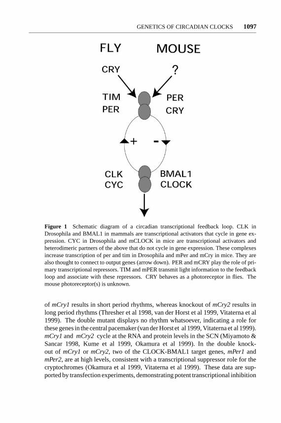
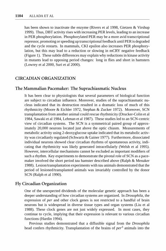
Figure 1 Schematic diagram of a circadian transcriptional feedback loop. CLK inDrosophila and BMAL1 in mammals are transcriptional activators that cycle in gene ex-pression. CYC in Drosophila and mCLOCK in mice are transcriptional activators andheterodimeric partners of the above that do not cycle in gene expression. These complexesincrease transcription of per and tim in Drosophila and mPer and mCry in mice. They arealso thought to connect to output genes (arrow down). PER and mCRY play the role of pri-mary transcriptional repressors. TIM and mPER transmit light information to the feedbackloop and associate with these repressors. CRY behaves as a photoreceptor in flies. Themouse photoreceptor(s) is unknown.
of mCry1results in short period rhythms, whereas knockout ofmCry2results inlong period rhythms (Thresher et al 1998, van der Horst et al 1999, Vitaterna et al1999). The double mutant displays no rhythm whatsoever, indicating a role forthese genes in the central pacemaker (van der Horst et al 1999, Vitaterna et al 1999).mCry1and mCry2 cycle at the RNA and protein levels in the SCN (Miyamoto &Sancar 1998, Kume et al 1999, Okamura et al 1999). In the double knock-out of mCry1or mCry2, two of the CLOCK-BMAL1 target genes,mPer1andmPer2, are at high levels, consistent with a transcriptional suppressor role for thecryptochromes (Okamura et al 1999, Vitaterna et al 1999). These data are sup-ported by transfection experiments, demonstrating potent transcriptional inhibition

P1: FUM
April 6, 2001 11:34 Annual Reviews AR121-36
1098 ALLADA ET AL
by bothmCry1or mCry2 expression (Kume et al 1999). Moreover, mCRY1/2 in-teract with mCLOCK and BMAL1 in yeast two-hybrid assays (Griffin et al 1999).These data support a biochemically direct inhibitory role for the mCRYs, parallel tothe role of PER-TIM or perhaps PER in the fly pacemaker mechanism (Figure 1).
One important question is whether mCRY1 and mCRY2 act alone or in concertwith mammalianPerand/orTim. Each CRY represses mCLOCK-BMAL1 activa-tion in heterologousDrosophilaS2 cells, which do not endogenously express dPERor dTIM (Saez & Young 1996, Shearman et al 2000b). The simplest model there-fore is one in which mCRY1 and/or mCRY2 repress mCLOCK-BMAL1-mediatedactivation. A mystery remains as to why themCry1and mCry2 single knock-outs have opposing period phenotypes, one shorter and one longer than 24 hours.Clearly, the transcriptional inhibitory assay of mCRY function in tissue culturecells, which does not significantly distinguish mCRY1 from mCRY2, does notexplain the whole story.
If mCLOCK and BMAL1 are the activators and mCRY1/2 are repressors, whatis the function of mPER? First, at least one of the mPERs satisfies the genetic goldstandard. A deletion of the PAS domain in mPER2 results in shortened periods,which grade into arrhythmicity (Zheng et al 1999). All three mouseperiodgenescycle at the RNA and protein levels (Shearman et al 1997, Sun et al 1997, Teiet al 1997, Takumi et al 1998, Zylka et al 1998b, Hastings et al 1999, Field et al2000). In these homozygous mutants, bothmPer2andmPer1transcript levels aresubstantially reduced (Zheng et al 1999). This is in contrast to flies in whichpertranscript levels are middle to high inper0 mutants (Hardin et al 1990). However,the nature of the mPER2 mutation complicates the interpretation of these results:A mutant protein might be produced and play a dominant-negative role. Indeed,transfection experiments show that this mutant allele is expressed and antago-nizes wild-type mPER2 cellular localization (Shearman et al 2000b). Therefore,a true null mutation would be useful to determine the precise role ofmPer2in theclock. Inactivation of themPer3gene also has a slightly short period phenotype(Shearman et al 2000a). Despite being the otherwise most well studied of thePeriodgenes,mPer1, when knocked out, has little effect on rhythms (Shearmanet al 2000b). Functional redundancy of themPergenes may be an obstacle togenetically deciphering their roles. Nonetheless, these data support a positive rolefor mPer2in promotingmPer1andmPer2RNA levels.
By analogy toDrosophila, one might expect mPER to be a transcriptionalrepressor of mCLOCK and BMAL1. The low RNA levels ofmPer1andmPer2in themPer2mutant animals are difficult to explain in this context. In fact, initialstudies of mPER1 showed that it modestly repressed mCLOCK-BMAL1 activation(Sangoram et al 1998). However, the modest inhibition by mPER1 is dwarfed bythat shown by either mCRY1 or mCRY2 alone (Kume et al 1999). In the absenceof contradictory evidence, the simplest model is that mammalianPeriodgenes donot behave like their repressing counterparts in flies.
How then doesmPer(s) participate in the feedback loop, specificallymPer2,for which there is the strongest genetic evidence? Recently, it was proposed that
P1: FUM
April 6, 2001 11:34 Annual Reviews AR121-36
GENETICS OF CIRCADIAN CLOCKS 1099
mPer2participates positively inBmal1RNA expression (Shearman et al 2000b).However,the peak values ofBmal1RNA are not dramatically affected inmPer2mutant animals (Shearman et al 2000b). Thus, there is no compelling evidencein favor of a specific function of mPER2. Interestingly, the mCRY and mPERproteins have been shown to engage in protein-protein contacts from SCN extracts(Field et al 2000). Furthermore, mPER2 protein levels are reduced inmCry1-mCry2 double knockout mice (Shearman et al 2000b). It is therefore possiblethat in vivo the mPERs modulate the repressing activity of the mCRYs by directphysical association.
The mammalianperiodgenes may also be involved in the response of the pace-maker to light. mPer1andmPer2RNAs as well as proteins are rapidly inducedby light in the SCN (Albrecht et al 1997, Shearman et al 1997, Shigeyoshi et al1997, Field et al 2000). Antisense oligonucleotides againstmPer1reduce phaseresetting, implicating mPER1 in this process (Akiyama et al 1999). However, theeffect of Period gene knockout on light-inducedmPerexpression has yet to bedetermined. The in vivo role of the mammalianPeriodgenes awaits true knockoutof each of these genes as well as double and triple mutant combinations. The inter-actions with mCRY as well as light responsiveness implicate mPER in transducinglight information, leading to a modulation of mCRY feedback, perhaps similar tothe role of TIM in flies (Figure 1).
A mammalian homolog ofDrosophila timelesshas also been identified. In-activation ofmTimeless(mTim) function in vivo results in early embryonic letha-lity (Gotter et al 2000). The role ofmTim has been confused by divergent re-sults. Some reports have shown no cycling ofmTimRNA; others show inductionby light (Koike et al 1998, Sangoram et al 1998, Zylka et al 1998a, Tischkauet al 1999). InDrosophila, timelessRNA and protein cycle in constant darknessconditions (Sehgal et al 1995, Hunter-Ensor et al 1996, Lee et al 1996, Myerset al 1996, Zeng et al 1996). Moreover,DrosophilaTIM is degraded by light(Hunter-Ensor et al 1996, Lee et al 1996, Myers et al 1996, Zeng et al 1996). Theonly reports of mTIM protein show no cycling and no light responsiveness(Hastings et al 1999, Field et al 2000). In addition, a newDrosophila gene,timeout, has been identified, which more closely resembles mTIM (Benna et al2000, Gotter et al 2000). It is unclear iftimeouthas any role in fly rhythms. Theseobservations have led to the proposal that the mammalianTimelessmay not beinvolved in circadian rhythms (Gotter et al 2000). However, mTIM coimmuno-precipitates from SCN with mCRY1 and mCRY2 (Field et al 2000). Because themTimknockout is lethal in mice, circadian rhythms cannot be assayed in theseanimals. Therefore, the jury will remain out on the in vivo role ofmTimuntil con-ditional knockouts are reported. Thedbt gene inDrosophilais just one exampleof a gene with developmental lethality but a clear function in the pacemaker (Priceet al 1998).
In summary (see Figure 1), theDrosophilabHLH-PAS DNA-binding proteins,CLK and CYC, activate transcription of theperiodandtimelessgenes. PER andTIM feed back and inhibit the activity of the CLK-CYC heterodimer. In the mouse,
P1: FUM
April 6, 2001 11:34 Annual Reviews AR121-36
1100 ALLADA ET AL
Clock andcycleorthologs (mCLOCK and BMAL1) probably activate themPerandmCrygenes. However, both CRYPTOCHROMES (mCRY1 and mCRY2) feedback and inhibit the mCLOCK-BMAL1 heterodimer. mPERs may transduce lightinformation to mCRYs. The role of mTIM remains unclear.
CODEPENDENT LOOPS?
One of the latest waves of circadian research deals with the role of positive andnegative interdependent feedback loops. The principle was first laid out in ex-aminations ofDrosophila ClockRNA but has infected other organisms, includingthe mouse. Likeper and tim, Clock is rhythmically expressed (Bae et al 1998,Darlington et al 1998). However,Clock RNA cycles antiphase toper and tim,peaking in the late night and early morning [Zeitgeber Time (ZT) 23, ZT 5; ZT 0=lights-on (“sunrise”); ZT 12= lights-off (“sunset”)]. Consistent with this alteredphase of cycling,Clocktranscript levels respond differently in arrhythmic mutantsand are low in theper0 andtim0 strains (Bae et al 1998);perandtim transcripts areat middle to peak levels in these mutants (Hardin et al 1990, Sehgal et al 1995). Incyc0andClkJrkmutants,Clk transcripts are high, whereasperandtim transcripts arelow (Glossop et al 1999). Finally, inper0; cyc0 andper0; ClkJrk, levels are similarto singlecyc0 andClkJrk mutants (Glossop et al 1999). These data led to a modelin which there are two interconnected feedback loops: the classic one regulatesper andtim transcription, with CLK/CYC acting positively and PER/TIM actingnegatively; the other regulatesClk transcription, with PER/TIM acting positivelyand CLK/CYC acting negatively. Epistasis analysis in double mutants indicatesthat the effects ofper andtim operate onClk RNA through CLK/CYC. Analysisof various mouse knockout strains of circadian rhythm genes came to similarconclusions, although the names have changed. mPER2 is proposed to act asa positive regulator ofBmal1transcripts (Shearman et al 2000b), although theseeffects are modest (see above).On the other hand, mCRY1 and mCRY2 are thoughtto play the major negative role onmPerandmCry transcription (see above).
Interdependent loops even appear to be involved in the circadian rhythms ofthe fungus,Neurospora crassa(Lee et al 2000). InNeurospora, frequency( frq)was the first clock component identified (reviewed in Dunlap 1999). Likeper, it isrhythmically expressed and feeds back negatively on its own transcription.Whitecollar-1 (WC-1) encodes a transcriptional activator that promotes expression of thefrq gene. Unlikefrq, WC-1RNA is not rhythmically expressed. However, its pro-tein product (WC-1) does oscillate, consistent with circadian posttranscriptionalregulation (Lee et al 2000). As expected,frq overexpression downregulates ex-pression of the endogenousfrq gene. On the other hand, this excessfrq positivelyregulates WC-1 expression.
One problem with the interdependent feedback models, particularly from fliesand mice, is the reliance on arrhythmic null mutants. Genetic studies of putative
P1: FUM
April 6, 2001 11:34 Annual Reviews AR121-36
GENETICS OF CIRCADIAN CLOCKS 1101
rhythm genes are the gold standard in determining whether or not they are clockgenes. However, analysis of mutant phenotypes can be difficult to interpret andeven misleading about the specific role of the gene. Like setting off a row ofdominoes, the absence of the gene(s) throughout development may result in con-sequences that may obscure true gene function in the adult. A second problem isthe absence of corroborating biochemical data to support interdependent loops.As all rhythms we measure are abolished byper0 andClkJrk, it is not surprisingthat per rhythms (or any rhythm) depend onClk or thatClk rhythms depend onper. Therefore, these analyses may lend little mechanistic insight into the exactnature of this regulation.
POSTRANSLATIONAL REGULATION
Gated Nuclear Entry?
Transcriptional feedback loops are ubiquitous in nature. A distinctive feature ofcircadian clocks is their timing. It has been proposed that it is the delay betweensynthesis and feedback that is necessary for circadian oscillations to occur. Further-more, the magnitude of this delay may dictate the daily oscillatory frequency. Evi-dence is accumulating that posttranslational regulation of certain circadian rhythmproteins is crucial to setting up these delays and therefore to timing feedback.
Several features of circadian rhythm gene expression appear to be subject toposttranslational regulation. In the case ofDrosophila per, the transport of PERfrom cytoplasm to nucleus seems to be circadianly gated, at least in the pacemakerlateral neurons (LNs) (Curtin et al 1995). As PER protein accumulates (ZT 8–17),immunohistochemical staining is detectable predominantly in the cytoplasm, ap-pearing in a so-called doughnut pattern. After ZT 17, PER staining rapidly (over thenext few hours) moves from predominantly cytoplasmic to predominantly nuclearin pacemaker neurons. These observations have been independently reproduced(Lee et al 1996, Matsumoto et al 1999). The importance of the gating of nuclearentry of PER is supported by genetic evidence. InperL mutants the timing ofnuclear entry is also delayed (Curtin et al 1995). These observations support amodel of regulated nuclear entry of PER.
PER association with its heterodimeric partner TIME has been implicatedin the temporal control of PER nuclear entry. TIME strongly associates withPER both in vitro and in vivo. Thetime gene was cloned in part by its abilityto interact with PER in a yeast two-hybrid assay (Gekakis et al 1995). Thesein vitro findings have been confirmed by in vivo observations as well. In coim-munoprecipitation experiments with fly head extracts, PER and TIM are stronglyassociated (Zeng et al 1996). Intim0 mutants, PER proteins levels are suppressed,even though RNA levels are relatively high, suggesting a role for TIM in PERstability (Price et al 1995). Furthermore, intim0 mutants, PER staining is constitu-tively cytoplasmic (Vosshall et al 1994). In culturedDrosophilaS2 cells, nuclear
P1: FUM
April 6, 2001 11:34 Annual Reviews AR121-36
1102 ALLADA ET AL
localization of transfected PER is completely dependent upon cotransfection ofTIM (Saez & Young 1996). Inper0 mutants, TIM protein is predominantly cyto-plasmic (Hunter-Ensor et al 1996, Myers et al 1996). These observations led to amodel in which association of PER with TIM is a prerequisite for nuclear co-entryof this heterodimer.
Though this model has been largely accepted, stray bits of evidence may ulti-mately undermine this nuclear entry model. Beyond the pacemaker lateral neurons,PER cycling is evident in several parts of the fly nervous system and in many dif-ferent tissues throughout the fly. If gating is a necessary step to generate circadianmacromolecular oscillations, then “doughnuts” should be observed in many otherPER-expressing cells. In stark contrast to the current model, no cytoplasmic accu-mulation is observed prior to nuclear entry, despite robust PER cycling, as noted byCurtin et al 1995. PER has been previously shown to be a transcriptional repressor,and its entry to the nucleus has been proposed to be required for its activity. Gatingof entry would be an elegant means of controlling the timing of repression andtherefore the clock. However, the timing of nuclear entry does not coincide withthe downturn ofper transcription and in fact occurs several hours too late at aboutthe time transcription has nearly reached trough levels (Curtin et al 1995, So &Rosbash 1997). Finally, gated nuclear entry has not yet been described for circa-dian systems of other organisms. If TIM is not gating PER nuclear entry, TIM’spredominant function inDrosophilamay therefore be to regulate PER stability.More careful studies in the ever-growing list of circadian model systems will berequired to determine if gated entry is a ubiquitous feature of pacemakers.
Phosphorylation
Phosphorylation may also impose a delay on PER feedback. The phosphorylationstate and hence the mobility of PER protein varies systematically over a 24-hourtime course (Edery et al 1994). PER accumulates during the late day/early night(ZT 8–16). As night progresses, PER migrates more slowly, i.e. at larger apparentmolecular weights. As PER disappears in the early morning, PER phosphorylationpeaks, as measured by mobility. Immunoprecipitation of PER and in vitro phos-phatase treatment returns PER to its baseline mobility, indicating a crucial roleof phosphorylation in these mobility changes. Studies of TIM protein show simi-lar progressive changes in mobility due to phosphorylation (Zeng et al 1996). Infact, TIM phosphorylation seems to occur in parallel with PER, implying similarregulation. The coincidence of the peak in phosphorylation with protein disap-pearance implicates phosphorylation as a key signal for protein degradation.
A major step forward in the understanding of the mechanistic basis of circadianphosphorylation awaited the identification ofdoubletime(dbt), a kinase involvedin circadian rhythms.dbt was originally identified in genetic screens for mutantswith altered circadian locomotor activity rhythms (Price et al 1998). Threedbtalleles were identified: one with short period rhythms (hence the mutant name;dbtS), one with long period rhythms (27 h;dbtL), and one homozygous lethalallele (dbtP). As predicted, the long and short period alleles alter in parallel the

P1: FUM
April 6, 2001 11:34 Annual Reviews AR121-36
GENETICS OF CIRCADIAN CLOCKS 1103
metabolism of PER and TIM expression, lending little mechanistic insight to thefunction of this gene (Price et al 1998). In this regard, the lethal allele turned outto be extremely important in establishing a model fordoubletimefunction.
It has been believed that a clock operates inDrosophila from early in larvaldevelopment (Brett 1955, Sehgal et al 1992). Subsequent studies have shown thatPER and TIM are rhythmically expressed in the larval central nervous system,including the precursors of the adult pacemaker neurons (Kaneko et al 1997).These 16–20 neurons, called the ventral lateral neurons (LNVS), are located deepinside each hemisphere of theDrosophilabrain and control circadian locomotoractivity (see below). As the lethaldbt allele,dbtP, did not induce lethality untilafter the larval stage, PER and TIM expression could be examined in homozygotes(Price et al 1998). In these homozygotes, PER and TIM cycling is abolished.PER specifically accumulates to very high levels in a hypophosphorylated form.The cloning ofdoubletimerevealed that it encodes a kinase that is homologous tohuman casein kinase I epsilon (Kloss et al 1998). DBT coimmunoprecipitates withPER in cotransfection experiments (Kloss et al 1998). This model posits that DBTphosphorylation of PER is a key signal for PER degradation. However, no studyhas yet shown that DBT directly phosphorylates PER inDrosophila, although thisis the likely working hypothesis.
Remarkably, genetic data also support a role for the mammalian homolog ofDBT in rhythms. These data involved a spontaneously mutant golden hamsternamedtau(Ralph & Menaker 1988).tau is a semidominant mutant with shortenedcircadian periods. Thetau locus was molecularly cloned and found to encode thehamster homolog ofDrosophila doubletimeor casein kinase I epsilon (Lowreyet al 2000). The shortened period of thetauhamster was attributed to the disruptedbiochemical activity of the mutant TAU kinase (Lowrey et al 2000). It was foundthat this enzyme has a markedly reduced maximal velocity (Vmax). The mutantprotein is still able to associate with PER, although its ability to phosphorylatePER in vitro is reduced. Purified human casein kinase I epsilon (hCKI epsilon)also phosphorylates human PER1 in vitro (Keesler et al 2000). Moreover, inco-transfection experiments, hCKI epsilon associates with and significantly shiftsthe mobility of hPER1 owing to phosphorylation (Keesler et al 2000). Consistentwith genetic evidence fromDrosophila, hCKI epsilon also destabilizes hPER1(Keesler et al 2000). Similar findings were made with mouse CKI (Vielhaberet al 2000). As in flies, casein kinase I epsilon appears to play a crucial role inthe circadian rhythms in mammals, and the enzymes appear to play similar roles inboth systems. Nonetheless, it remains unclear for both systems whether PER is thetrue target of DBT. In fact, preliminary mammalian studies do not find circadianregulation of electrophoretic mobility of mPERs in vivo as inDrosophila(Fieldet al 2000). Additional antibodies will help determine if mPERs undergo cyclicphosphorylation changes.
How mightdoubletimecontribute to circadian timekeeping? Current models ofDBT/CKI epsilon function focus on PER phosphorylation. One possibility is thatPER, through its association with DBT, imposes circadian regulation on thiskinase activity. Perhaps PER prevents DBT from autophosphorylation, which
P1: FUM
April 6, 2001 11:34 Annual Reviews AR121-36
1104 ALLADA ET AL
has been shown to inactivate the enzyme (Rivers et al 1998, Gietzen & Virshup1999). Thus, DBT activity rises with increasing PER levels, leading to an increasein PER phosphorylation. Phosphorylated PER may be a more avid transcriptionalrepressor, promoting or speeding up transcriptional feedback until PER is degradedand the cycle restarts. In mammals, CKI epsilon also increases PER phosphory-lation, but this may lead to a reduction or slowing in mCRY negative feedback(Figure 1). These subtle differences may explain why reductions in kinase activityin mutants lead to opposing period changes: long in flies and short in hamsters(Lowrey et al 2000, Suri et al 2000).
CIRCADIAN ORGANIZATION
The Mammalian Pacemaker: The Suprachiasmatic Nucleus
It has been clear to physiologists that several parameters of biological functionare subject to circadian influence. Moreover, studies of the suprachiasmatic nu-cleus indicated that its destruction resulted in a dramatic loss of much of thisrhythmicity (Moore & Eichler 1972, Stephan & Zucker 1972). Moreover, SCNtransplantation from another animal could rescue rhythmicity (Drucker-Colin et al1984, Sawaki et al 1984, Lehman et al 1987). These studies led to an SCN-centricview of circadian systems. The SCN is a symmetrical paired group of approx-imately 20,000 neurons located just above the optic chiasm. Measurements ofmetabolic activity using 2-deoxyglucose uptake indicated that its metabolic activ-ity was circadianly regulated (Schwartz & Gainer 1977). Furthermore, dissociatedindividual neurons showed clear circadian rhythms of spontaneous activity, indi-cating that rhythmicity was likely generated intracellularly (Welsh et al 1995).However, intercellular mechanisms cannot be excluded as important modifiers ofsuch a rhythm. Key experiments to demonstrate the pivotal role of SCN as a pace-maker involved the short periodtau hamster described above (Ralph & Menaker1988). Lesion/transplantation experiments withtauanimals demonstrated that theperiod of lesioned/transplanted animals was invariably controlled by the donorSCN (Ralph et al 1990).
Fly Circadian Organization
One of the unexpected dividends of the molecular genetic approach has been adeeper understanding of how circadian systems are organized. InDrosophila, theexpression ofper and other clock genes is not restricted to a handful of brainneurons but is widespread in diverse tissue types and organ systems (Liu et al1988). These clock genes are not just widely expressed. In most cases, theycontinue to cycle, implying that their expression is relevant to various circadianfunctions (Hardin 1994).
Previous studies demonstrated that a diffusible signal from theDrosophilahead confers rhythmicity. Transplantation of the brains ofperS animals into the
P1: FUM
April 6, 2001 11:34 Annual Reviews AR121-36
GENETICS OF CIRCADIAN CLOCKS 1105
abdomens of arrhythmicper0 animals rescued circadian activity rhythms (Handler& Konopka 1979). Based on these data, it might be expected that separat-ing peripheral oscillators from the central pacemaker neurons in theDrosophilahead would completely abolish cycling of gene expression. Consistent with thisview, developmental mutants that significantly disruptper-expressing LNVS resultin largely behaviorally arrhythmic flies. For example, thedisconnected(disco)mutation eliminates connections between photoreceptor cells in theDrosophilaeye and targets in the optic lobe (Steller et al 1987). Furthermore, PER-positivelateral neurons are not detectable (Zerr et al 1990). As a result of this disrup-tion in brain architecture, the flies are largely arrhythmic (Dushay et al 1989).However, peripheral cycling is robust in light-dark cycles and persists with somedamping over time in constant darkness (Hardin et al 1992a). Consistent withthese neuroanatomic mutants, decapitation does not abolish peripheral oscilla-tions (Hege et al 1997). Impressively, when the Malpighian tubules (aDrosophilakidney analog) are transplanted from one fly to another, these tubules retain thecircadian molecular cycling of the donor, even if out-of-phase with the recipient(Giebultowicz et al 2000). Therefore, although there are hormonal signals in flies,they do not strongly entrain peripheral molecular oscillators. Thus, the lateralneurons do not appear to be essential for peripheral cycling.
Surprisingly, these peripheral clocks respond and entrain to light. Transgenicflies containing theper promoter fused to firefly luciferase (per-luc) express thisreporter gene in the spatial and temporal distribution of thepergene (Brandes et al1996). Luciferase has been used previously as a method of continuous monitoringof gene expression in live organisms (Kay 1993). Because of the relatively rapidturnover of the luciferase enzyme, gene transcription changes are readily reflectedin enzyme activity changes. Thus, transgenicper-lucflies exhibit cycling biolumi-nescence when fed on the substrate luciferin. Although clocks are light-sensitive,the degree of bioluminescence is not sufficient to affect behavioral rhythms. Suchnoninvasive monitoring systems have made the repeated sampling of RNA or pro-tein values unnecessary and have allowed an “on-line” view of gene expression inindividual animals.
To test whether these peripheral oscillators respond to light and cycle indepen-dently, investigators systematically removed various parts of theDrosophilabodyfrom aper-luctransgenic animal (Plautz et al 1997). They then placed legs,wings,antennae, and other parts into culture media containing luciferin. Once the partswere separated from the central pacemaker, investigators observed that rhythmicgene expression in these persisted but appeared to damp over time in constant dark-ness. The luminescence technique could not determine if the loss of rhythmicitywas due to asynchronous oscillators between different cells or a loss of intrinsicoscillator amplitude within each cell. These separated tissues also demonstratedphotoreceptive properties (Plautz et al 1997). Robust rhythmicity, lost after sev-eral days in constant darkness, could be reinitiated by reexposure to light cycles.Taken together, these studies reveal how molecular genetic studies can illuminatethe organization of circadian systems.
P1: FUM
April 6, 2001 11:34 Annual Reviews AR121-36
1106 ALLADA ET AL
Mammalian Circadian Organization
Studies of the organization of theDrosophilacircadian system differ markedlywith that found in mammals. As mentioned above, lesioning of the SCN abolishesactivity rhythms, and transplants restore them to SCN-ablated animals (Ralph et al1990). Transplantation of SCNs in semipermeable capsules also restores rhythmi-city in hamsters (Silver et al 1996). Therefore, similar toDrosophila(Handler &Konopka 1979), diffusion of humoral molecules is sufficient to restore some rhyth-micity in mammals. As in the case of fly rhythm genes, mammalian rhythm genesare not restricted to the suprachiasmatic nucleus. For example,mClocktranscriptsare found in several neural and nonneural tissues, such as the heart and lungs (Kinget al 1997). Examination of the temporal profile ofPer expression in peripheraltissues (e.g. liver and skeletal muscle) finds robust molecular cycling as wellas an interesting temporal lag when compared with the oscillations in the SCN(Balsalobre et al 1998, Zylka et al 1998b). Even lymphocytes in peripheral bloodexhibit circadian gene cycling (Oishi et al 1998b).
Nevertheless, there are marked differences between flies and mammals in thecontrol of circadian rhythmicity in peripheral oscillators. Unlike the case of thefly, lesioning of the central pacemaker, the SCN, abolishes most peripheral rhyth-micity, at least as assayed at the tissue level (Sakamoto et al 1998). Again, lossof rhythmicity at the tissue level may be due either to asynchronous oscillationsbetween different cells or to a loss of intrinsic oscillation amplitude within eachcell. Although these observations, in general, differ substantially fromDrosophila,autonomous light-sensitive peripheral oscillators also exist in mammals: Hamsterretinal cells show robust and light-entrainable oscillation in culture (Tosini &Menaker 1996).
Studies usingper-driven luciferase reporters also emphasize the heavy re-liance of peripheral oscillators on central oscillators in mammals. Transgenic ratswere constructed using the mousemPer1promoter fused to a luciferase reporter(Yamazaki et al 2000). Various tissues and organs were removed from these ani-mals and assayed for rhythmic bioluminescence in vitro. In culture, the SCNs fromthese animals exhibited robust rhythms for up to 32 days. Peripheral oscillatorssuch as the liver, lung, and skeletal muscle were also rhythmic, but damped aftera few cycles. This damping of peripheral oscillators is reminiscent ofDrosophilaperipheral oscillators. The authors then examined the response to rapid changes,i.e. advances or delays, in the environmental light cycle as might occur to some-one experiencing jet lag. They observed that the SCN rhythm shifted rapidly tothe new environmental light regime. However, the bioluminescence rhythm in theperipheral tissues was either lost for several days or lagged for many days be-fore shifting to the new light cycle. The asynchrony between environmental lightcycles and the SCN on the one hand and peripheral oscillators on the other handmay explain the symptomatology of jet lag.
Immortalized cell lines derived from rat SCN neurons exhibit circadianrhythms of metabolism and gene expression (Earnest et al 1999a,b). Remarkably,
P1: FUM
April 6, 2001 11:34 Annual Reviews AR121-36
GENETICS OF CIRCADIAN CLOCKS 1107
transplantation of these SCN cell lines to the third ventricle restored circadianactivity rhythm to SCN-lesioned animals (Earnest et al 1999b). Thus, these SCNcell lines may release appropriate phase-setting signals. Transformed cell lines,such as Rat-1 fibroblasts, exhibit circadian patterns of gene expression after a briefexposure of high concentrations of serum (Balsalobre et al 1998). It has been pro-posed that serum contains important circadian phase-setting factors. Cycling ofgenes in rat lymphocytes suggests that similar humoral factors may drive circadianclocks in these cells (Oishi et al 1998b).
Prominent among circadianly regulated hormones in humans is melatonin.However, peripheral rhythms persist in melatonin-deficient mice (Zylka et al1998b). Another important class of circadian-controlled hormones is the glucocor-ticoids (Tronche et al 1998). Consistent with a possible role of glucocorticoids asoutput mediators, dexamethasone, a glucocorticoid analog, induces circadian rhy-thmicity in cultured Rat-1 fibroblasts (Balsalobre et al 2000). Moreover, dexam-ethasone transiently phase-shifts the rhythm of peripheral organs such as the liver.Finally, dexamethasone has no effect on the SCN rhythm, consistent with an ab-sence of glucocorticoid receptor expression there. However, liver-specific knock-out of the glucocorticoid receptor did not abolish circadian rhythmicity in the liver(Balsalobre et al 2000). Thus, glucocorticoids cannot be the sole entraining signals.
In zebrafish, rhythms of gene expression persist in peripheral organs such asthe kidney and heart in vitro, i.e. separated from the brain (Whitmore et al 1998).Furthermore, as in the case ofDrosophila, these peripheral oscillators appear tobe directly light sensitive (Whitmore et al 1998). In fact, a zebrafish cell linehas been shown to exhibit rhythmic gene expression in vitro (Whitmore et al2000). Moreover, these rhythms are also entrainable by light. These data are moreconsistent with the more autonomous fly circadian organization.
Why these differences in organization? In small and partially transparent ani-mals likeDrosophilaand zebrafish, light can easily penetrate into the body of theanimal. Therefore, the presence of a photoreceptor (CRY in flies) allows a verysimple way to synchronize the peripheral oscillators with the environment. On theother hand, larger and more opaque animals like mammals may need neurohor-monal control of circadian oscillations in internal organs. Strikingly, the mam-malian retina, which obviously receives light, has maintained light-sensitive cell-autonomous oscillation. The nature of the photoreceptor in this tissue or any mam-malian circadian photoreceptor is still mysterious. In zebrafish, though,zCRY4,which is in sequence much closer toDrosophila CRY than to the mammalianCRYs, looks like an interesting candidate for study (Kobayashi et al 2000).
CIRCADIAN OUTPUT
Direct Output Targets
Although much progress has been made in identifying clock genes, little is knownabout how these genes connect to outputs. In many organisms, various fractions
P1: FUM
April 6, 2001 11:34 Annual Reviews AR121-36
1108 ALLADA ET AL
of whole genomes appear to be under circadian control, from the vast majority inphotosynthetic cyanobacteria to 1–5% inDrosophila(Liu et al 1995, Van Gelderet al 1995). Given the size of theDrosophilagenome, hundreds of genes shouldcycle. These data implicate transcription in the control of various output genesand therefore in the control of behavioral and physiologic outputs. Most of theseclock-controlled genes are probably indirectly controlled; many fewer are likelyto be direct targets of clock genes. In our opinion, a direct interaction with a clockcomponent has not yet been conclusively demonstrated for any output gene.
What then should be the criteria for a direct output target? As identification ofcycling RNAs seems to be a straightforward strategy, it is likely that a large fractionof these will be controlled at the level of transcription. In this case, there shouldbe direct binding of a circadian transcription factor, such asClockto the promoterof the regulated gene. There are two main ways to show this convincingly. First,the construction of altered specificity mutants, i.e. mutants that alter DNA-bindingspecificity need to be coupled to mutants with correspondingly altered bindingsites to prove direct binding in vivo. Second, chromatin immunoprecipitationscould show that a factor is physically bound to its putative target in vivo. However,as these strategies are relatively difficult, studies of output gene regulation haveheavily relied on transient transfection into tissue culture cells. Typically, DNAsexpressing the relevant clock genes as well as the target promoters driving a reporterare transfected and allowed to express for 24–48 hours. The long temporal delaybetween expression and the downstream target gene allows for several intermediatesteps to occur. Thus, the observed effects could easily be indirect. The relevanceof the putative target site of the transcription factor needs to be assessed in vivo.Promoter elements need to confer cycling on reporter genes in transgenic animals.Mutation of the appropriate binding sites should abolish or reduce this cycling.
Most studies of output genes have tried to identify the targets of the CLK andCYC transcription complex, CACGTG. By simple randomness, this sequence willoccur approximately once every four kilobases. The random probability that onewill find such a sequence in the vicinity of a cycling gene is therefore relatively high.Moreover, expression is typically far higher in transfection experiments than whatwould be observed in vivo. The transfected target promoter DNA does not have thetypical repressing chromatin structure of genomic DNA. Thus, under these highlyartificial conditions, overexpressed transcription factors may find these vulnerableE-boxes, when they would not be accessible in vivo. Therefore, results based onlyon these types of experiments need to be interpreted very cautiously.
The neuropeptide gene,arginine vasopressin, is one putative output target inmammals. Besides many other functions, such as the physiologic control of waterbalance, this gene is also rhythmically expressed in the suprachiasmatic nucleus(Uhl & Reppert 1986). InClockmutants the levels ofvasopressinRNA are dra-matically reduced and rhythmicity is abolished in the SCN (Jin et al 1999). In cellculture experiments, mCLOCK and its partner BMAL1 activate from E-boxes inthevasopressinpromoter, providing a plausible explanation for the in vivo resultsand a model for how output might be generated (Jin et al 1999). However, itremains unclear whether the E-box sequence found in thevasopressinpromoter is
P1: FUM
April 6, 2001 11:34 Annual Reviews AR121-36
GENETICS OF CIRCADIAN CLOCKS 1109
required for its rhythmic expression in vivo. Moreover both the genetic evidenceand the cell culture data leave open the possibility that the effect of mCLOCK-BMAL1 on vasopressinis indirect.
A more likely direct target output gene is the D-box binding element protein(DBP). DBP is rhythmically expressed in the liver and SCN among other tissues(Fonjallaz et al 1996). DBP activates the promoters of many enzymes involvedin hepatic processes, including cholesterol metabolism (Lavery et al 1999). Micelacking DBP exhibit a subtle circadian behavioral phenotype, implying that DBPmay have a primary function in mediating output (Lopez-Molina et al 1997). Dele-tion of intragenic DBP regions substantially reduce promoter-driven expression(Ripperger et al 2000). Studies of in vivo promoter occupancy identified DNase Ihypersensitive sites, suggesting that an exclusive focus on E-boxes is overly sim-plistic. Protein binding to DNA often renders the local DNA sensitive to DNase I.In theDbpgenomic region, as many as five different loci, some of which are intra-genic, undergo circadian regulation of DNase I hypersensitivity. Control of outputgenes may therefore be much more complex than regulation by one or two fac-tors. E-box motifs were identified and shown to bind mCLOCK in vitro. However,this binding activity does not cycle, raising questions about whether mCLOCK isthe factor cyclically bound in vivo. Nonetheless, DBP regulates several enzymescircadianly expressed in the liver and is therefore an important link between thecircadian pacemaker and the final outputs.
In combination with genetic approaches, recent work inDrosophilahas focusedon molecular methods to find clock-controlled genes. In a search for genes thatare differentially expressed between wild-type andper0 animals, the zinc fingertranscription factorvrille (vri) was identified (Blau & Young 1999). Previous stud-ies of thevri gene demonstrated that it was required for embryonic development(George & Terracol 1997). Likeper, vri is rhythmically expressed in circadianpacemaker neurons (Blau & Young 1999). Furthermore, as in the case ofvaso-pressin, theDrosophilaorthologs of mCLOCK and BMAL1, CLOCK and CY-CLE, activate a reporter gene fused to thevri promoter, which contains E-boxtarget sites (Blau & Young 1999). Constitutive overexpression ofvri lengthenedbehavioral rhythms and reduced expression of theperiodandtimelessRNAs, indi-cating a potential role of this gene in the central pacemaker. This expression alsodownregulates the output molecule, pigment dispersing factor (PDF; see below).However, phenotypes due to misexpression or overexpression can be misleading,as they may induce functions that normal expression does not. Heterozygous dele-tions of thevri locus yield very subtle circadian phenotypes. Conditional rescueof the embryonic lethality and subsequent analysis of homozygous null mutantadults will be required to more fully specify the role ofvri in the pacemaker.
Using a similar subtractive hybridization approach, another clock-regulatedgene was identified calledtakeout(to) (So et al 2000).takeoutwas originallyidentified on the basis of its low expression in thecyc0 mutant. Studies of atakeoutmutant suggest that TAKEOUT protein is involved in the response to starvation(Sarov-Blat et al 2000).takeoutexpression is reduced in all circadian rhythmmutants tested, which distinguishes it from any other studied transcript. Although
P1: FUM
April 6, 2001 11:34 Annual Reviews AR121-36
1110 ALLADA ET AL
takeoutis also rhythmically expressed at the RNA level, the phase of this oscillationis distinct from other cycling RNAs fromClk, per, andtim. As in the case ofvri andvasopressin, a search was undertaken to identify E-box target sites. Not only wasan E-box identified, but there was also remarkable similarity in promoter regionsflanking the E-box, suggesting that this region was functional. Surprisingly, thisE-box was not sufficient to drive cycling when tested in vivo (which was not donefor vri, vasopressin, or Dbp promoters). Alternatively, the lack of cycling couldhave been due to an artifact of the transgene. Nonetheless, thetakeoutmRNAcycling is not likely due to a direct effect of CLK and CYC. These studies highlightthe importance of testing enhancer elements in vivo for circadian function.
Circadian Behavioral Output Molecules
Though the discovery of several new output genes and phenomena illustrates howpacemakers may generate output, they did not reveal any key mediator of beha-vioral rhythms. PDF is a neuropeptide that has been well studied in invertebratesand has been shown to phase-shift circadian clocks when injected into cockroaches(Petri & Stengl 1997). Initial studies withDrosophila pdf indicated that its ex-pression was largely restricted to the head and absent indiscomutants (Park 1998).These mutants are largely arrhythmic and lack pacemaker lateral neurons (Dushayet al 1989, Zerr et al 1990).pdf mRNA and protein are specifically expressedin a ventral subset of these lateral neurons (LNvs) (Helfrich-Forster 1995, Parket al 2000). Furthermore, PDF is rhythmically expressed in the termini of theseneurons (Park et al 2000). The role ofpdf in these neurons was more clearly elu-cidated by the discovery of apdf mutant,pdf01 (Renn et al 1999). Likediscomutants, these mutants display only weak or no rhythms. Becausediscomutantsexhibit far more neuroanatomical defects beyond just an absence of pacemakerlateral neurons, thepdf promoter was used to direct expression of the proapoptoticgenes,head-involution defective(hid) andreaper, to the ventral lateral neurons.This expression resulted in the complete ablation of these neurons. The behavioralconsequence was a circadian phenotype virtually identical to that ofpdf01, indicat-ing that the prinicipal mediator of the circadian signal from these key pacemakerneurons ispdf (Renn et al 1999).
Is PDF synaptically released? To address the role of chemical synaptic trans-mission, the tetanus-toxin light chain (TeTxLC) was expressed in all pacemaker-containing cells (Kaneko et al 2000). TeTxLC blocks synaptic transmission bycleaving the synaptic protein, synaptobrevin. Although clock gene cycling wasunaffected in these animals, rhythmic behavior was substantially reduced, indicat-ing a role for synaptic transmission. Surprisingly, targeted expression of TeTxLCexclusively to the LNvs did not affect circadian behavior,indicating thatpdf re-lease may operate through nonsynaptic mechanisms (Kaneko et al 2000). Further-more, other cells may also be relevant to behavioral rhythms. In contrast to theSCN, which relies on the eyes for synchronizing its activity with the environment,the LNVS also contain a circadian photoreceptor and are therefore directly light
P1: FUM
April 6, 2001 11:34 Annual Reviews AR121-36
GENETICS OF CIRCADIAN CLOCKS 1111
sensitive (Emery et al 2000). They are circadianly self-sufficient because theycontain an input pathway, a molecular pacemaker, and an output. There does notappear to be a mammalian ortholog of PDF. It will be interesting to see what themammalian version(s) of this output signal will be.
Sleep in Flies?
One of the most controversial notions regarding circadian output in flies is thatthe behavioral cycles of flies mimic the mammalian sleep-wake cycle. Close ob-servations of fly behavior during their 24 hour cycle demonstrate that flies canbe immobile for long periods of time (over 2 hours in some cases) (Hendrickset al 2000, Shaw et al 2000). During this immobile state, flies exhibit an increasedthreshold to arousing, sensory stimuli. Importantly, this state is homeostaticallyregulated, like sleep. Rest-deprived flies will increase their rest subsequently. Simi-lar stimulation during wake periods has no subsequent effect on rest behavior.Drugs that increase or decrease sleep in mammals, such as antihistamines andcaffeine, increase and decrease rest in flies. As in the case of circadian rhythms,molecular and genetic studies of sleep (or this sleep-like state if you prefer) in fliesmay prove to be very influential in our understanding of the neurobiology of sleep.
CONCLUSION
The problem of circadian rhythms has been reduced to the molecular realm: aproblem of understanding transcriptional feedback loops and the posttranslationalregulation of their loop components. The past few years have seen a remarkableincrease in the number of identified clock genes. There remains much to learn abouthow these genes regulate each other, especially in mammals, where we are truly atthe dawn of the molecular era. In the next few years an important focus will be onposttranslational regulation and feedback. The identification of additional kinasesand phosphatases as well as their regulatory features will be central to a betterunderstanding of timing. In addition to understanding how the clock couples tooutput, the identification of key output molecules in mammals may have medicalapplications. Many of these discoveries will undoubtedly continue to capitalizeon the power of forward genetics.
Visit the Annual Reviews home page at www.AnnualReviews.org
LITERATURE CITED
Abe H, Honma S, Namihira M, Tanahashi Y,Ikeda M, Honma K. 1998. Circadian rhythmand light responsiveness of BMAL1 expres-sion, a partner of mammalian clock gene
Clock, in the suprachiasmatic nucleus of rats.Neurosci. Lett.258:93–96
Akiyama M, Kouzu Y, Takahashi S, Waka-matsu H, Moriya T, et al. 1999. Inhibition of
P1: FUM
April 6, 2001 11:34 Annual Reviews AR121-36
1112 ALLADA ET AL
light- or glutamate-induced mPer1 expres-sion represses the phase shifts into the mousecircadian locomotor and suprachiasmatic fir-ing rhythms.J. Neurosci.19:1115–21
Albrecht U, Sun ZS, Eichele G, LeeCC. 1997. A differential response of twoputative mammalian circadian regulators,mper1 and mper2, to light.Cell 91:1055–64
Allada R, White NE, So WV, Hall JC,Rosbash M. 1998. A mutant Drosophila ho-molog of mammalian Clock disrupts circa-dian rhythms and transcription of period andtimeless.Cell 93:791–804
Antoch MP, Song EJ, Chang AM, VitaternaMH, Zhao Y, et al. 1997. Functional identifi-cation of the mouse circadian Clock gene bytransgenic BAC rescueCell 89:655–67
Bae K, Lee C, Hardin PE, Edery I. 2000.dCLOCK is present in limiting amountsand likely mediates daily interactions be-tween the dCLOCK–CYC transcription fac-tor and the PER-TIM complex.J. Neurosci.20:1746–53
Bae K, Lee C, Sidote D, Chuang KY, Edery I.1998. Circadian regulation of a Drosophilahomolog of the mammalian Clock gene:PER and TIM function as positive regulatorsMol. Cell. Biol.18:6142–51
Balsalobre A, Brown SA, Marcacci L, TroncheF, Kellendonk C, et al. 2000. Resetting ofcircadian time in peripheral tissues by gluco-corticoid signaling.Science289:2344–47
Balsalobre A, Damiola F, Schibler U. 1998. Aserum shock induces circadian gene expres-sion in mammalian tissue culture cells.Cell93:929–37
Benna C, Scannapieco P, Piccin A, SandrelliF, Zordan M, et al. 2000. A second timelessgene in Drosophila shares greater sequencesimilarity with mammalian tim.Curr. Biol.10:R512–13
Benzer S. 1971. From the gene to behavior.JAMA218:1015–22
Blau J, Young MW. 1999. Cycling vrille expres-sion is required for a functional Drosophilaclock.Cell 99:661–712
Brandes C, Plautz JD, Stanewsky R, Jamison
CF, Straume M, et al. 1996. Novel fea-tures of Drosophila period transcriptionrevealed by real-time luciferase reporting.Neuron16:687–92
Brett W. 1955. Persistent diurnal rhythmicity inDrosophila emergence.Ann. Entomol. Soc.Am.48:119–31
1997. Breakthrough of the year. The runners-up.Science278:2039–42
1998. Breakthrough of the year. The runners-up.Science282:2157–61
Bruce VG. 1972. Mutants of the biologicalclock in Chlamydomonas reinhardi.Genet-ics70:537–48
Bunger MK, Wilsbacher LD, Moran SM, Clen-denin C, Radcliffe LA, et al. 2000. Mop3 is anessential component of the master circadianpacemaker in mammals.Cell 103:1009–17
Cashmore AR, Jarillo JA, Wu YJ, Liu D.1999. Cryptochromes: blue light receptorsfor plants and animals.Science284:760–65
Crews ST, Thomas JB, Goodman CS. 1988.The Drosophila single-minded gene encodesa nuclear protein with sequence similarity tothe per gene product.Cell 52:143–51
Curtin KD, Huang ZJ, Rosbash M. 1995.Temporally regulated nuclear entry of theDrosophila period protein contributes to thecircadian clock.Neuron14:365–72
Darlington TK, Wager-Smith K, Ceriani MF,Staknis D, Gekakis N, et al. 1998. Closingthe circadian loop: CLOCK–induced tran-scription of its own inhibitors per and tim.Science280:1599–603
Drucker-Colin R, Aguilar-Roblero R, Garcia-Hernandez F, Fernandez-Cancino F, Bermu-dez Rattoni F. 1984. Fetal suprachiasmaticnucleus transplants: diurnal rhythm recov-ery of lesioned rats.Brain Res.311:353–57
Dunlap JC. 1999. Molecular bases for circadianclocks.Cell 96:271–90
Dushay MS, Rosbash M, Hall JC. 1989. Thedisconnected visual system mutations inDrosophila melanogasterdrastically disruptcircadian rhythms.J. Biol. Rhythms4:1–27
Earnest DJ, Liang FQ, DiGiorgio S, Gallagher
P1: FUM
April 6, 2001 11:34 Annual Reviews AR121-36
GENETICS OF CIRCADIAN CLOCKS 1113
M, Harvey B, et al. 1999a. Establishmentand characterization of adenoviral E1A im-mortalized cell lines derived from the ratsuprachiasmatic nucleus.J. Neurobiol.39:1–13
Earnest DJ, Liang FQ, Ratcliff M, Cassone VM.1999b. Immortal time: circadian clock prop-erties of rat suprachiasmatic cell lines.Sci-ence283:693–95
Edery I. 2000. Circadian rhythms in a nutshell.Phys. Genomics3:59–74
Edery I, Zwiebel LJ, Dembinska ME, RosbashM. 1994. Temporal phosphorylation of theDrosophila period protein.Proc. Natl. Acad.Sci. USA91:2260–64
Egan ES, Franklin TM, Hilderbrand-Chae MJ,McNeil GP, Roberts MA, et al. 1999. Anextraretinally expressed insect cryptochromewith similarity to the blue light photorecep-tors of mammals and plants.J. Neurosci.19:3665–73
Emery P, So WV, Kaneko M, Hall JC, RosbashM. 1998. CRY, a Drosophila clock and light-regulated cryptochrome, is a major contribu-tor to circadian rhythm resetting and photo-sensitivity.Cell 95:669–79
Emery P, Stanewsky R, Helfrich-Forster C,Emery-Le M, Hall JC, Rosbash M. 2000.Drosophila CRY is a deep brain circadianphotoreceptor.Neuron26:493–504
Feldman JF, Hoyle MN. 1973. Isolation of cir-cadian clock mutants of Neurospora crassa.Genetics75:605–13
Field MD, Maywood ES, O’Brien JA, WeaverDR, Reppert SM, Hastings MH. 2000. Anal-ysis of clock proteins in mouse SCN demon-strates phylogenetic divergence of the circa-dian clockwork and resetting mechanisms.Neuron25:437–47
Fonjallaz P, Ossipow V, Wanner G, SchiblerU. 1996. The two PAR leucine zipper pro-teins, TEF and DBP, display similar circa-dian and tissue-specific expression, but havedifferent target promoter preferences.EMBOJ. 15:351–62
Gekakis N, Saez L, Delahaye-Brown AM,Myers MP, Sehgal A, et al. 1995. Isolation of
timeless by PER protein interaction: defec-tive interaction between timeless protein andlong-period mutant PERL.Science270:811–15
Gekakis N, Staknis D, Nguyen HB, Davis FC,Wilsbacher LD, et al. 1998. Role of theCLOCK protein in the mammalian circadianmechanism.Science280:1564–69
George H, Terracol R. 1997. The vrille geneof Drosophila is a maternal enhancer of de-capentaplegic and encodes a new member ofthe bZIP family of transcription factors.Ge-netics146:1345–63
Giebultowicz JM, Stanewsky R, Hall JC, HegeDM. 2000. Transplanted Drosophila excre-tory tubules maintain circadian clock cy-cling out of phase with the host.Curr. Biol.10:107–10
Gietzen KF, Virshup DM. 1999. Identifica-tion of inhibitory autophosphorylation sitesin casein kinase I epsilonJ. Biol. Chem.274:32063–70
Glossop NR, Lyons LC, Hardin PE. 1999. Inter-locked feedback loops within the Drosophilacircadian oscillator.Science286:766–68
Gotter AL, Manganaro T, Weaver DR, Ko-lakowski LF Jr, Possidente B, et al. 2000. Atime-less function for mouse timeless.Nat.Neurosci.3:755–56
Griffin EA Jr, Staknis D, Weitz CJ. 1999.Light–independent role of CRY1 and CRY2in the mammalian circadian clock.Science286:768–71
Hall JC. 2000. Cryptochromes: sensory recep-tion, transduction, and clock functions sub-serving circadian systems.Curr. Opin. Neu-robiol. 10:456–66
Handler AM, Konopka RJ. 1979. Transplanta-tion of a circadian pacemaker in Drosophila.Nature279:236–38
Hao H, Allen DL, Hardin PE. 1997. A circadianenhancer mediates PER–dependent mRNAcycling in Drosophila melanogaster. Mol.Cell. Biol.17:3687–93
Hardin PE. 1994. Analysis of period mRNAcycling in Drosophila head and body tis-sues indicates that body oscillators behave

P1: FUM
April 6, 2001 11:34 Annual Reviews AR121-36
1114 ALLADA ET AL
differently from head oscillators.Mol. Cell.Biol. 7211–18
Hardin PE, Hall JC, Rosbash M. 1990. Feed-back of the Drosophila period gene producton circadian cycling of its messenger RNAlevels.Nature343:536–40
Hardin PE, Hall JC, Rosbash M. 1992a. Be-havioral and molecular analyses suggest thatcircadian output is disrupted by disconnectedmutants inD. melanogaster. EMBO J.11:1–6
Hardin PE, Hall JC, Rosbash M. 1992b. Circa-dian oscillations in period gene mRNA lev-els are transcriptionally regulated.Proc. Natl.Acad. Sci. USA89:11711–15
Hastings MH, Field MD, Maywood ES, WeaverDR, Reppert SM. 1999. Differential regula-tion of mPER1 and mTIM proteins in themouse suprachiasmatic nuclei: new insightsinto a core clock mechanism.J. Neurosci.19:RC11
Hege DM, Stanewsky R, Hall JC, Giebul-towicz JM. 1997. Rhythmic expression ofa PER-reporter in the Malpighian tubulesof decapitated Drosophila: evidence for abrain-independent circadian clock.J. Biol.Rhythms12:300–8
Helfrich-Forster C. 1995. The period clock geneis expressed in central nervous system neu-rons which also produce a neuropeptide thatreveals the projections of circadian pace-maker cells within the brain ofDrosophilamelanogaster. Proc. Natl. Acad. Sci. USA92:612–16
Hendricks JC, Finn SM, Panckeri KA, ChavkinJ, Williams JA, et al. 2000. Rest in Drosophilais a sleep-like state.Neuron25:129–38
Hogenesch JB, Gu YZ, Jain S, Bradfield CA.1998. The basic-helix-loop-helix-PAS or-phan MOP3 forms transcriptionally activecomplexes with circadian and hypoxia fac-tors.Proc. Natl. Acad. Sci. USA95:5474–79
Hogenesch JB, Gu YZ, Moran SM, ShimomuraK, Radcliffe LA, et al. 2000. The basic helix-loop-helix-PAS protein MOP9 is a brain-specific heterodimeric partner of circadianand hypoxia factors.J. Neurosci.20:RC83
Huang ZJ, Edery I, Rosbash M. 1993. PAS is adimerization domain common to Drosophilaperiod and several transcription factors.Na-ture364:259–62
Hunter-Ensor M, Ousley A, Sehgal A. 1996.Regulation of the Drosophila protein time-less suggests a mechanism for resetting thecircadian clock by lightCell 84:677–85
Ishida N, Kaneko M, Allada R. 1999. Biologicalclocks.Proc. Natl. Acad. Sci. USA96:8819–20. Erratum. 2000.Proc. Natl. Acad. Sci.USA97(16):9347
Ishikawa T, Matsumoto A, Kato T Jr, TogashiS, Ryo H, et al. 1999. DCRY is a Drosophilaphotoreceptor protein implicated in light en-trainment of circadian rhythm.Genes Cells4:57–65
Jackson FR, Bargiello TA, Yun SH, Young MW.1986. Product of per locus of Drosophilashares homology with proteoglycans.Nature320:185–88
Jin X, Shearman LP, Weaver DR, Zylka MJ,de Vries GJ, Reppert SM. 1999. A molec-ular mechanism regulating rhythmic outputfrom the suprachiasmatic circadian clock.Cell 96:57–68
Johnson CH, Golden SS. 1999. Circadian pro-grams in cyanobacteria adaptiveness andmechanismAnnu. Rev. Microbiol.53:389–409
Kaneko M, Helfrich-Forster C, Hall JC. 1997.Spatial and temporal expression of the periodand timeless genes in the developing nervoussystem of Drosophila: newly identified pace-maker candidates and novel features of clockgene product cycling.J. Neurosci.17:6745–60
Kaneko M, Park JH, Cheng Y, Hardin PE, HallJC. 2000. Disruption of synaptic transmis-sion or clock-gene-product oscillations in cir-cadian pacemaker cells of Drosophila causeabnormal behavioral rhythms.J. Neurobiol.43:207–33
Kay SA. 1993. Shedding light on clock con-trolled cab gene transcription in higherplants.Semin. Cell Biol.4:81–86
Keesler GA, Camacho F, Guo Y, Virshup D,
P1: FUM
April 6, 2001 11:34 Annual Reviews AR121-36
GENETICS OF CIRCADIAN CLOCKS 1115
Mondadori C, Yao Z. 2000. Phosphorylationand destabilization of human period I clockprotein by human casein kinase I epsilon.NeuroReport11:951–55
King DP, Takahashi JS. 2000. Moleculargenetics of circadian rhythms in mammals.Annu. Rev. Neurosci.23:713–42
King DP, Zhao Y, Sangoram AM, WilsbacherLD, Tanaka M, et al. 1997. Positional cloningof the mouse circadian clock gene.Cell89:641–53
Kloss B, Price JL, Saez L, Blau J, Rothen-fluh A, et al. 1998. The Drosophila clockgene double-time encodes a protein closely-related to human casein kinase-I-epsilon.Cell 94:97–107
Kobayashi Y, Ishikawa T, Hirayama J, DaiyasuH, Kanai S, et al. 2000. Molecular analysisof zebrafish photolyase/cryptochrome fam-ily: two types of cryptochromes present inzebrafish.Genes Cells5:725–38
Koike N, Hida A, Numano R, Hirose M, SakakiY, Tei H. 1998. Identification of the mam-malian homologues of the Drosophila time-less gene, Timeless1.FEBS Lett.441:427–31
Konopka RJ, Benzer S. 1971. Clock mutants ofDrosophila melanogaster. Proc. Natl. Acad.Sci. USA68:2112–16
Kuhlman SJ, Quintero JE, McMahon DG. 2000.GFP fluorescence reports Period 1 circadiangene regulation in the mammalian biologicalclock.NeuroReport11:1479–82
Kume K, Zylka MJ, Sriram S, Shearman LP,Weaver DR, et al. 1999. mCRY1 and mCRY2are essential components of the negative limbof the circadian clock feedback loop.Cell98:193–205
Lavery DJ, Lopez-Molina L, Margueron R,Fleury-Olela F, Conquet F, et al. 1999. Cir-cadian expression of the steroid 15 alpha-hydroxylase (Cyp2a4) and coumarin 7-hydroxylase (Cyp2a5) genes in mouse liveris regulated by the PAR leucine zipper tran-scription factor DBP.Mol. Cell. Biol. 19:6488–99
Lee C, Bae K, Edery I. 1998. The DrosophilaCLOCK protein undergoes daily rhythms
in abundance, phosphorylation, and interac-tions with the PER–TIM complex.Neuron21:857–67
Lee C, Bae K, Edery I. 1999. PER andTIM inhibit the DNA binding activity ofa Drosophila CLOCK-CYC/dBMAL1 het-erodimer without disrupting formation of theheterodimer: a basis for circadian transcrip-tion. Mol. Cell. Biol.19:5316–25
Lee C, Parikh V, Itsukaichi T, Bae K, EderyI. 1996. Resetting the Drosophila clock byphotic regulation of PER and a PER-TIMcomplex.Science271:1740–44
Lee K, Loros JJ, Dunlap JC. 2000. Intercon-nected feedback loops in the Neurospora cir-cadian system.Science289:107–10
Lehman MN, Silver R, Gladstone WR, KahnRM, Gibson M, Bittman EL. 1987. Circa-dian rhythmicity restored by neural trans-plant. Immunocytochemical characterizationof the graft and its integration with the hostbrain.J. Neurosci.7:1626–38
Lindebro MC, Poellinger L, Whitelaw ML.1995. Protein-protein interaction via PAS do-mains: role of the PAS domain in positive andnegative regulation of the bHLH/PAS dioxinreceptor-Arnt transcription factor complex.EMBO J.14:3528–39
Liu X, Lorenz L, Yu QN, Hall JC, RosbashM. 1988. Spatial and temporal expression ofthe period gene inDrosophila melanogaster.Genes Dev.2:228–38
Liu Y, Tsinoremas NF, Johnson CH, LebedevaNV, Golden SS, et al. 1995. Circadian orches-tration of gene expression in cyanobacteria.Genes Dev.9:1469–78
Lopez-Molina L, Conquet F, Dubois-DauphinM, Schibler U. 1997. The DBP gene is ex-pressed according to a circadian rhythm inthe suprachiasmatic nucleus and influencescircadian behavior.EMBO J.16:6762–71
Lowrey PL, Shimomura K, Antoch MP, Ya-mazaki S, Zemenides PD, et al. 2000.Positional syntenic cloning and functionalcharacterization of the mammalian circadianmutation tau.Science288:483–92
Marrus SB, Zeng H, Rosbash M. 1996. Effect

P1: FUM
April 6, 2001 11:34 Annual Reviews AR121-36
1116 ALLADA ET AL
of constant light and circadian entrainment ofperS flies: evidence for light-mediated delayof the negative feedback loop in Drosophila.EMBO J.15:6877–86
Matsumoto A, Tomioka K, Chiba Y, Tan-imura T. 1999. timrit Lengthens circadianperiod in a temperature-dependent mannerthrough suppression of PERIOD protein cy-cling and nuclear localization.Mol. Cell.Biol. 19:4343–54
Miyamoto Y, Sancar A. 1998. Vitamin B2-based blue-light photoreceptors in theretinohypothalamic tract as the photoactivepigments for setting the circadian clockin mammals.Proc. Natl. Acad. Sci. USA95:6097–102
Moore RY, Eichler VB. 1972. Loss of a circa-dian adrenal corticosterone rhythm followingsuprachiasmatic lesions in the rat.Brain Res.42:201–6
Myers MP, Wager-Smith K, Rothenfluh-Hilfiker A, Young MW. 1996. Light-induceddegradation of TIMELESS and entrainmentof the Drosophila circadian clock.Science271:1736–40
Oishi K, Sakamoto K, Okada T, Nagase T,Ishida N. 1998a. Antiphase circadian expres-sion between BMAL1 and period homologuemRNA in the suprachiasmatic nucleus andperipheral tissues of rats.Biochem. Biophys.Res. Commun.253:199–203
Oishi K, Sakamoto K, Okada T, Nagase T,Ishida N. 1998b. Humoral signals mediatethe circadian expression of rat period homo-logue (rPer2) mRNA in peripheral tissues.Neurosci. Lett.256:117–19
Okamura H, Miyake S, Sumi Y, YamaguchiS, Yasui A, et al. 1999. Photic induction ofmPer1 and mPer2 in cry-deficient mice lack-ing a biological clock.Science286:2531–34
Park JH, Hall JC. 1998. Isolation andchronobiological analysis of a neuropeptidepigment-dispersing factor gene inDrosop-hila melanogaster. J. Biol. Rhythms13:219–28
Park JH, Helfrich-Forster C, Lee G, Liu L, Ros-bash M, Hall JC. 2000. Differential regula-
tion of circadian pacemaker output by sepa-rate clock genes in Drosophila.Proc. Natl.Acad. Sci. USA97:3608–13
Petri B, Stengl M. 1997. Pigment-dispersinghormone shifts the phase of the circadianpacemaker of the cockroach Leucophaeamaderae.J. Neurosci.17:4087–93
Pittendrigh C. 1954. On temperature indepen-dence in the clock-system controlling emer-gence time in Drosophila.Proc. Natl. Acad.Sci. USA40:1018–29
Plautz JD, Kaneko M, Hall JC, Kay SA. 1997.Independent photoreceptive circadian clocksthroughout Drosophila.Science278:1632–35
Price JL, Blau J, Rothenfluh A, Abodeely M,Kloss B, Young MW. 1998. double-time isa novel Drosophila clock gene that regulatesPERIOD protein accumulation.Cell 94:83–95
Price JL, Dembinska ME, Young MW, Ros-bash M. 1995. Suppression of PERIOD pro-tein abundance and circadian cycling by theDrosophila clock mutation timeless.EMBOJ. 14:4044–49
Ralph MR, Foster RG, Davis FC, MenakerM. 1990. Transplanted suprachiasmatic nu-cleus determines circadian period.Science247:975–78
Ralph MR, Menaker M. 1988. A mutation of thecircadian system in golden hamsters.Science241:1225–27
Reddy P, Jacquier AC, Abovich N, Petersen G,Rosbash M. 1986. The period clock locus ofD. melanogastercodes for a proteoglycan.Cell 46:53–61
Renn SC, Park JH, Rosbash M, Hall JC, TaghertPH. 1999. A pdf neuropeptide gene muta-tion and ablation of PDF neurons each causesevere abnormalities of behavioral circadianrhythms in Drosophila.Cell 99:791–802. Er-ratum. 2000.Cell 101(1): following 113
Ripperger JA, Shearman LP, Reppert SM, Schi-bler U. 2000. CLOCK, an essential pace-maker component, controls expression of thecircadian transcription factor DBP.GenesDev.14:679–89.
P1: FUM
April 6, 2001 11:34 Annual Reviews AR121-36
GENETICS OF CIRCADIAN CLOCKS 1117
Rivers A, Gietzen KF, Vielhaber E, VirshupDM. 1998. Regulation of casein kinase I ep-silon and casein kinase I delta by an in vivofutile phosphorylation cycle.J. Biol. Chem.273:15980–84.
Rothenfluh A, Young MW, Saez L. 2000. ATIMELESS-independent function for PE-RIOD proteins in the Drosophila clock.Neu-ron 26:505–14
Ruby NF, Burns DE, Heller HC. 1999. Circa-dian rhythms in the suprachiasmatic nucleusare temperature-compensated and phase-shifted by heat pulses in vitro.J. Neurosci.19:8630–36
Rutila JE, Suri V, Le M, So WV, Rosbash M,Hall JC. 1998. CYCLE is a second bHLH-PAS clock protein essential for circadianrhythmicity and transcription of Drosophilaperiod and timeless.Cell 93:805–14
Saez L, Young MW. 1996. Regulation of nu-clear entry of the Drosophila clock proteinsperiod and timeless.Neuron17:911–20
Sakamoto K, Nagase T, Fukui H, HorikawaK, Okada T, et al. 1998. Multitissue circa-dian expression of rat period homolog (rPer2)mRNA is governed by the mammalian circa-dian clock, the suprachiasmatic nucleus inthe brain.J. Biol. Chem.273:27039–42
Sangoram AM, Saez L, Antoch MP, GekakisN, Staknis D, et al. 1998. Mammalian circa-dian autoregulatory loop: a timeless orthologand mPer1 interact and negatively regu-late CLOCK-BMAL1-induced transcription.Neuron21:1101–13
Sarov-Blat L, So WV, Liu L, Rosbash M.2000. The Drosophila takeout gene is a novelmolecular link between circadian rhythmsand feeding behavior.Cell 101:647–56
Sawaki Y, Nihonmatsu I, Kawamura H. 1984.Transplantation of the neonatal suprachias-matic nuclei into rats with complete bilat-eral suprachiasmatic lesions.Neurosci. Res.1:67–72
Schwartz WJ, Gainer H. 1977. Suprachias-matic nucleus: use of 14C-labeled deoxyglu-cose uptake as a functional marker.Science197:1089–91
Scully AL, Kay SA. 2000. Time flies forDrosophila.Cell 100:297–300
Sehgal A, Price J, Young MW. 1992. On-togeny of a biological clock inDrosophilamelanogaster. Proc. Natl. Acad. Sci. USA89:1423–27
Sehgal A, Price JL, Man B, Young MW. 1994.Loss of circadian behavioral rhythms and perRNA oscillations in the Drosophila mutanttimeless.Science263:1603–6
Sehgal A, Rothenfluh-Hilfiker A, Hunter-EnsorM, Chen Y, Myers MP, Young MW. 1995.Rhythmic expression of timeless: a basis forpromoting circadian cycles in period gene au-toregulation.Science270:808–10
Shaw PJ, Cirelli C, Greenspan RJ, TononiG. 2000. Correlates of sleep and wakingin Drosophila melanogaster. Science287:1834–37
Shearman LP, Jin X, Lee C, Reppert SM,Weaver DR. 2000a. Targeted disruption ofthe mPer3 gene: subtle effects on circadianclock function.Mol. Cell. Biol.20:6269–75
Shearman LP, Sriram S, Weaver DR, MaywoodES, Chaves I, et al. 2000b. Interacting molec-ular loops in the mammalian circadian clock.Science288:1013–19
Shearman LP, Zylka MJ, Weaver DR, Ko-lakowski LF Jr, Reppert SM. 1997. Twoperiod homologs: circadian expression andphotic regulation in the suprachiasmatic nu-clei. Neuron19:1261–69
Shigeyoshi Y, Taguchi K, Yamamoto S,Takekida S, Yan L, et al. 1997. Light-inducedresetting of a mammalian circadian clock isassociated with rapid induction of the mPer1transcript.Cell 91:1043–53
Silver R, LeSauter J, Tresco PA, Lehman MN.1996. A diffusible coupling signal fromthe transplanted suprachiasmatic nucleuscontrolling circadian locomotor rhythms.Nature382:810–13
Siwicki KK, Eastman C, Petersen G, RosbashM, Hall JC. 1988. Antibodies to the periodgene product of Drosophila reveal diverse tis-sue distribution and rhythmic changes in thevisual system.Neuron1:141–50
P1: FUM
April 6, 2001 11:34 Annual Reviews AR121-36
1118 ALLADA ET AL
So WV, Rosbash M. 1997. Post-transcriptionalregulation contributes to Drosophila clockgene mRNA cycling.EMBO J.16:7146–55
So WV, Sarov-Blat L, Kotarski CK, McDon-ald MJ, Allada R, Rosbash M. 2000. Take-out, a novel Drosophila gene under circadianclock transcriptional regulation.Mol. Cell.Biol. 20:6935–44
Stanewsky R, Kaneko M, Emery P, Beretta B,Wager-Smith K, et al. 1998. The cryb muta-tion identifies cryptochrome as a circadianphotoreceptor in Drosophila.Cell 95:681–92
Steller H, Fischbach KF, Rubin GM. 1987. Dis-connected: a locus required for neuronalpathway formation in the visual system ofDrosophila.Cell 50:1139–53
Stephan FK, Zucker I. 1972. Circadian rhythmsin drinking behavior and locomotor activityof rats are eliminated by hypothalamic le-sions.Proc. Natl. Acad. Sci. USA69:1583–86
Sun ZS, Albrecht U, Zhuchenko O, Bailey J,Eichele G, Lee CC. 1997. RIGUI, a putativemammalian ortholog of the Drosophila pe-riod gene.Cell 90:1003–11
Suri V, Hall JC, Rosbash M. 2000. Two noveldoubletime mutants alter circadian propertiesand eliminate the delay between RNA andprotein in Drosophila.J. Neurosci.20:7547–55
Takumi T, Taguchi K, Miyake S, SakakidaY, Takashima N, et al. 1998. A light-independent oscillatory gene mPer3 inmouse SCN and OVLT.EMBO J.17:4753–59
Tei H, Okamura H, Shigeyoshi Y, Fukuhara C,Ozawa R, et al. 1997. Circadian oscillation ofa mammalian homologue of the Drosophilaperiod gene.Nature389:512–16
Thresher RJ, Vitaterna MH, Miyamoto Y,Kazantsev A, Hsu DS, et al. 1998. Roleof mouse cryptochrome blue-light photore-ceptor in circadian photoresponses.Science282:1490–94
Tischkau Y, Barnes JA, Lin FJ, Myers EM,Barnes JW, et al. 1999. Oscillation and light
induction of timeless mRNA in the mam-malian circadian clock.J. Neurosci.19:RC15
Tosini G, Menaker M. 1996. Circadian rhythmsin cultured mammalian retina.Science272:419–21
Tronche F, Kellendonk C, Reichardt HM,Schutz G. 1998. Genetic dissection of glu-cocorticoid receptor function in mice.Curr.Opin. Genet. Dev.8:532–38
Uhl GR, Reppert SM. 1986. Suprachiasmaticnucleus vasopressin messenger RNA: circa-dian variation in normal and Brattleboro rats.Science232:390–93
van der Horst GT, Muijtjens M, Kobayashi K,Takano R, Kanno S, et al. 1999. MammalianCry1 and Cry2 are essential for maintenanceof circadian rhythms.Nature398:627–30
Van Gelder RN, Bae H, Palazzolo MJ, Kras-now MA. 1995. Extent and character ofcircadian gene expression inDrosophilamelanogaster: identification of twenty os-cillating mRNAs in the fly head.Curr. Biol.5:1424–36
Vielhaber E, Eide E, Rivers A, Gao ZH, Vir-shup DM. 2000. Nuclear entry of the circa-dian regulator mPER1 is controlled by mam-malian casein kinase I epsilon.Mol. Cell.Biol. 20:4888–99
Vitaterna MH, King DP, Chang AM, Korn-hauser JM, Lowrey PL, et al. 1994. Mu-tagenesis and mapping of a mouse gene,Clock, essential for circadian behavior.Sci-ence264:719–25
Vitaterna MH, Selby CP, Todo T, Niwa H,Thompson C, et al. 1999. Differential reg-ulation of mammalian period genes and cir-cadian rhythmicity by cryptochromes 1 and2. Proc. Natl. Acad. Sci. USA96:12114–19
Vosshall LB, Price JL, Sehgal A, Saez L, YoungMW. 1994. Block in nuclear localization ofperiod protein by a second clock mutation,timeless.Science263:1606–9
Welsh DK, Logothetis DE, Meister M, Rep-pert SM. 1995. Individual neurons dissoci-ated from rat suprachiasmatic nucleus ex-press independently phased circadian firingrhythms.Neuron14:697–706
P1: FUM
April 6, 2001 11:34 Annual Reviews AR121-36
GENETICS OF CIRCADIAN CLOCKS 1119
Whitmore D, Foulkes NS, Sassone-Corsi P.2000. Light acts directly on organs and cellsin culture to set the vertebrate circadianclock.Nature404:87–91
Whitmore D, Foulkes NS, Strahle U, Sassone-Corsi P. 1998. Zebrafish Clock rhythmic ex-pression reveals independent peripheral cir-cadian oscillators.Nat. Neurosci.1:701–7
Yamaguchi S, Mitsui S, Miyake S, Yan L, On-ishi H, et al. 2000. The 5′ upstream regionof mPer1 gene contains two promoters andis responsible for circadian oscillation.Curr.Biol. 10:873–76
Yamazaki S, Numano R, Abe M, Hida A, Taka-hashi R, et al. 2000. Resetting central andperipheral circadian oscillators in transgenicrats.Science288:682–85
Young MW. 2000a. Circadian rhythms. Mark-ing time for a kingdom.Science288:451–53
Young MW. 2000b. The tick-tock of the biolog-ical clock.Sci. Am.282:64–71
Zeng H, Qian Z, Myers MP, Rosbash M.
1996. A light-entrainment mechanism for theDrosophila circadian clock.Nature380:129–35
Zerr DM, Hall JC, Rosbash M, Siwicki KK.1990. Circadian fluctuations of period pro-tein immunoreactivity in the CNS and thevisual system of Drosophila.J. Neurosci.10:2749–62
Zheng B, Larkin DW, Albrecht U, Sun ZS, SageM, et al. 1999. The mPer2 gene encodes afunctional component of the mammalian cir-cadian clock.Nature400:169–73
Zylka MJ, Shearman LP, Levine JD, Jin X,Weaver DR, Reppert SM. 1998a. Molecu-lar analysis of mammalian timeless.Neuron21:1115–22
Zylka MJ, Shearman LP, Weaver DR, Rep-pert SM. 1998b. Three period homologs inmammals: differential light responses in thesuprachiasmatic circadian clock and oscil-lating transcripts outside of brain.Neuron20:1103–10