Expression pattern of circadian genes and steroidogenesis ...
7
RESEARCH Open Access Expression pattern of circadian genes and steroidogenesis-related genes after testosterone stimulation in the human ovary Minghui Chen 1,2 , Yanwen Xu 1,2 , Benyu Miao 1,2 , Hui Zhao 4 , Lu Luo 1,2 , Huijuan Shi 3 and Canquan Zhou 1,2* Abstract Background: Previous studies have shown that circadian genes might be involved in the development of polycystic ovarian syndrome (PCOS). Hyperandrogenism is a hallmark feature of PCOS. However, the effect of hyperandrogenism on circadian gene expression in human granulosa cells is unknown, and the general expression pattern of circadian genes in the human ovary is unclear. Methods: Expression of the circadian proteins CLOCK and PER2 in human ovaries was observed by immunohistochemistry. The mRNA expression patterns of the circadian genes CLOCK, PER2, and BMAL1, and the steroidogenesis-related genes STAR, CYP11A1, HSD3B2, and CYP19A1 in cultured human luteinized granulosa cells were analyzed over the course of 48 h after testosterone treatment by quantitative polymerase chain reaction. Results: Immunostaining of CLOCK and PER2 protein was detected in the granulosa cells of dominant antral follicles but was absent in the primordial, primary, or preantral follicles of human ovaries. After testosterone stimulation, expression of PER2 showed an oscillating pattern, with two peaks occurring at the 24th and 44th hours; expression of CLOCK increased significantly to the peak at the 24th hour, whereas expression of BMAL1 did not change significantly over time in human luteinized granulosa cells. Among the four steroidogenesis-related genes evaluated, only STAR displayed an oscillating expression pattern with two peaks occurring at the 24th and 40th hours after testosterone stimulation. Conclusions: Circadian genes are expressed in the dominant antral follicles of the human ovary. Oscillating expression of the circadian gene PER2 can be induced by testosterone in human granulosa cells in vitro. Expression of STAR also displayed an oscillating pattern after testosterone stimulation. Our results indicate a potential relationship between the circadian clock and steroidogenesis in the human ovary, and demonstrate the effect of testosterone on circadian gene expression in granulosa cells. Keywords: Circadian rhythm, Testosterone, Granulosa cells, Ovary, Human Abbreviations: BMAL1, Brain and muscle arnt-like protein 1; CLOCK, Circadian locomotor output cycles kaput; CYP11A1, Cholesterol side-chain cleavage cytochrome P450; CYP19A1, Cytochrome P450; HSD3B2, 3-β- hydroxysteroid dehydrogenase type II; PER, Period; qRT-PCR, Quantitative reverse transcription-polymerase chain reaction; STAR, Steroidal acute regulatory protein * Correspondence: [email protected] 1 Reproductive Medicine Center, The First Affiliated Hospital of Sun Yat-sen University, 58 2nd Zhongshan Road, Guangzhou GD510080, People’s Republic of China 2 Guangdong Provincial Key Laboratory of Reproductive Medicine, The First Affiliated Hospital of Sun Yat-sen University, 58 2nd Zhongshan Road, Guangzhou GD510080, People’s Republic of China Full list of author information is available at the end of the article © 2016 The Author(s). Open Access This article is distributed under the terms of the Creative Commons Attribution 4.0 International License (http://creativecommons.org/licenses/by/4.0/), which permits unrestricted use, distribution, and reproduction in any medium, provided you give appropriate credit to the original author(s) and the source, provide a link to the Creative Commons license, and indicate if changes were made. The Creative Commons Public Domain Dedication waiver (http://creativecommons.org/publicdomain/zero/1.0/) applies to the data made available in this article, unless otherwise stated. Chen et al. Journal of Ovarian Research (2016) 9:56 DOI 10.1186/s13048-016-0264-5
-
Upload
khangminh22 -
Category
Documents
-
view
0 -
download
0
Transcript of Expression pattern of circadian genes and steroidogenesis ...
RESEARCH Open Access
Expression pattern of circadian genes andsteroidogenesis-related genes aftertestosterone stimulation in the humanovaryMinghui Chen1,2, Yanwen Xu1,2, Benyu Miao1,2, Hui Zhao4, Lu Luo1,2, Huijuan Shi3 and Canquan Zhou1,2*
Abstract
Background: Previous studies have shown that circadian genes might be involved in the development ofpolycystic ovarian syndrome (PCOS). Hyperandrogenism is a hallmark feature of PCOS. However, the effect ofhyperandrogenism on circadian gene expression in human granulosa cells is unknown, and the general expressionpattern of circadian genes in the human ovary is unclear.
Methods: Expression of the circadian proteins CLOCK and PER2 in human ovaries was observed byimmunohistochemistry. The mRNA expression patterns of the circadian genes CLOCK, PER2, and BMAL1, and thesteroidogenesis-related genes STAR, CYP11A1, HSD3B2, and CYP19A1 in cultured human luteinized granulosa cellswere analyzed over the course of 48 h after testosterone treatment by quantitative polymerase chain reaction.
Results: Immunostaining of CLOCK and PER2 protein was detected in the granulosa cells of dominant antralfollicles but was absent in the primordial, primary, or preantral follicles of human ovaries. After testosteronestimulation, expression of PER2 showed an oscillating pattern, with two peaks occurring at the 24th and 44th hours;expression of CLOCK increased significantly to the peak at the 24th hour, whereas expression of BMAL1 did notchange significantly over time in human luteinized granulosa cells. Among the four steroidogenesis-related genesevaluated, only STAR displayed an oscillating expression pattern with two peaks occurring at the 24th and 40thhours after testosterone stimulation.
Conclusions: Circadian genes are expressed in the dominant antral follicles of the human ovary. Oscillatingexpression of the circadian gene PER2 can be induced by testosterone in human granulosa cells in vitro. Expressionof STAR also displayed an oscillating pattern after testosterone stimulation. Our results indicate a potentialrelationship between the circadian clock and steroidogenesis in the human ovary, and demonstrate the effect oftestosterone on circadian gene expression in granulosa cells.
Keywords: Circadian rhythm, Testosterone, Granulosa cells, Ovary, Human
Abbreviations: BMAL1, Brain and muscle arnt-like protein 1; CLOCK, Circadian locomotor output cycles kaput;CYP11A1, Cholesterol side-chain cleavage cytochrome P450; CYP19A1, Cytochrome P450; HSD3B2, 3-β-hydroxysteroid dehydrogenase type II; PER, Period; qRT-PCR, Quantitative reverse transcription-polymerase chainreaction; STAR, Steroidal acute regulatory protein
* Correspondence: [email protected] Medicine Center, The First Affiliated Hospital of Sun Yat-senUniversity, 58 2nd Zhongshan Road, Guangzhou GD510080, People’sRepublic of China2Guangdong Provincial Key Laboratory of Reproductive Medicine, The FirstAffiliated Hospital of Sun Yat-sen University, 58 2nd Zhongshan Road,Guangzhou GD510080, People’s Republic of ChinaFull list of author information is available at the end of the article
© 2016 The Author(s). Open Access This article is distributed under the terms of the Creative Commons Attribution 4.0International License (http://creativecommons.org/licenses/by/4.0/), which permits unrestricted use, distribution, andreproduction in any medium, provided you give appropriate credit to the original author(s) and the source, provide a link tothe Creative Commons license, and indicate if changes were made. The Creative Commons Public Domain Dedication waiver(http://creativecommons.org/publicdomain/zero/1.0/) applies to the data made available in this article, unless otherwise stated.
Chen et al. Journal of Ovarian Research (2016) 9:56 DOI 10.1186/s13048-016-0264-5

BackgroundA circadian clock is a biochemical mechanism that oscil-lates with a period of 24 h and is coordinated with theday–night cycle. In mammals, light signals perceived bythe retina are transmitted via the retino-hypothalamictract to the suprachiasmatic nuclei (SCN), which containthe master pacemaker for the generation of circadianrhythms [1]. The SCN synchronize countless subsidiaryoscillators existing in the peripheral tissues throughoutthe body [2]. The basis for maintaining the circadianrhythm is a molecular clock consisting of interlocked tran-scriptional/translational feedback loops. The proteinsencoded by the genes circadian locomotor output cycleskaput (Clock) and brain and muscle arnt-like protein 1(Bmal1) heterodimerize and promote the rhythmic tran-scription of the period (Per1, Per2) and cryptochrome(Cry1, Cry2) gene families, whereas modified PER–CRYcomplexes repress the activity of the CLOCK–BMAL1complex. Over several hours, PER–CRY complexes are de-graded, and the CLOCK–BMAL1 complex is eventuallyreleased from feedback inhibition [3–6].Quantitative reverse transcription-polymerase chain
reaction (qRT-PCR) analysis revealed that transcripts forthe core oscillator elements (Arntl, Clock, Per1, Per2,and Cry1) were present in the rat ovary [7]. Expressionof Arntl and Per2 was detected in the granulosa andtheca layers of growing and antral follicles, as well as inthe corpora lutea and stromal fibroblasts of the rat ovary[7]. Per1 and Per2 mRNAs in the rat ovary display rhyth-mic oscillation with a 24-h period [8]. Moreover, suchrhythmic expression of a circadian gene can be inducedin cultured ovarian cells. When cultured without anytreatment, no rhythmic pattern in the expression of ei-ther Per1 or Bmal1 transcripts was observed in chickengranulosa cells; however, both serum shock andluteinizing hormone (LH) treatment could induce arhythm of both Per1 and Bmal1 in these cells [9].Polycystic ovarian syndrome (PCOS) is the most com-
mon endocrine disorder of reproductive-age women. Arecent study highlighted the important role of circadiangenes in the development of PCOS. This study showedthat the level of BMAL1 expression in granulosa cells inthe PCOS group of women was lower than that of thegroup without PCOS. Estrogen synthesis and aromataseexpression were downregulated after BMAL1 knock-down and, conversely, were upregulated in KGN cells (agranulosa cell line) overexpressing BMAL1 [10]. Hyper-androgenism is a hallmark feature of PCOS and isstrongly implicated in the genesis of the disorder [11]. Ahigh testosterone level reflects a type of androgen ex-cess, which is one of the primary symptoms of PCOS. Inthe present study, we selected testosterone as a stimula-tor for cultured human granulosa cells and observed thetemporal expression patterns of circadian genes and
steroidogenesis-related genes in human luteinized gran-ulosa cells after testosterone stimulation. In addition, weevaluated the distribution of circadian protein expressionin human ovaries by immunohistochemistry.
MethodsImmunohistochemistryParaffin sections of normal ovarian tissue were obtainedfrom five women aged 29–35 years. All women hadundergone bilateral salpingo-oophorectomy with orwithout hysterectomy for a uterine or unilateral ovarianmalignant tumor before chemotherapy or radiotherapy.Informed consent was obtained from all patients. Afterdeparaffinization, antigen retrieval, and blocking in nor-mal goat serum (for CLOCK) or bovine serum (forPER2), the slides were incubated overnight at 4 °C inrabbit anti-human CLOCK polyclonal antibody dilutedto 20 μg/mL (catalogue no. ab 65033,Abcam) or goatanti-human PER2 polyclonal antibody diluted to 7.5 μg/mL (catalogue no. ab118489, Abcam), respectively. Slideswere washed and incubated with biotin-labeled goatanti-rabbit secondary antibody for CLOCK (CWBIO) orbovine anti-goat secondary antibody for PER2 (SantaCruz Biotechnology) for 30 min, then washed and incu-bated with horseradish peroxidase-labeled streptavidinfor 10 min. The peroxidase–antibody complex was visu-alized using 3, 3′-diaminobenzidine (CWBIO). Controlexperiments included samples treated in the same man-ner but with omission of the primary antibody. The sec-tions were counterstained with hematoxylin.
Cell cultureFor each experiment, human luteinized granulosa cellswere obtained from the follicle fluid collected during ovumaspiration of 10 patients undergoing in vitro fertilization.All patients underwent the long protocol of gonadotropin-releasing hormone agonist treatment (1.0 mg tiptorelinacetate; Ipsen Pharma Biotech, France) with human chori-onic gonadotropin as the trigger. The follicle fluid waspooled and centrifuged for 5 min at 2500 rpm. Granulosacells were purified using 50 % Percoll (Sigma) through gra-dient centrifugation for 10 min at 2000 rpm. Ovarian tissuefragments were then removed from the granulosa cell sus-pension with a 40-μm cell strainer (Becton-Dickinson).Purified granulosa cells were plated on a 12-well plate(3.0 × 10⁵/well) and cultured in Dulbecco’s modified Eaglemedium/Ham’s F12 supplemented with penicillin (100 U/mL), streptomycin (100 μg/mL), and 10 % fetal bovineserum (Gibco) in a 37 °C incubator with 5 % CO2. Cellswere cultured for 9 days to reach confluence prior to anytreatment. Cells were exposed to 100 ng/mL testosterone(Sigma) dissolved in serum-free medium for 2 h, washedwith serum-free medium once, and then cultured further inserum-free medium until harvested. Cells in the control
Chen et al. Journal of Ovarian Research (2016) 9:56 Page 2 of 7

group were cultured in serum-free medium withouttestosterone treatment. Samples were harvested every4 h starting at the beginning of treatment and con-tinuing until 48 h.
RNA extraction and quantificationRNA was extracted with High Pure RNA Isolation Kit(Roche) according to the manufacturer’s instructions, andquantitated by 260/280 UV spectrophotometry (NanoDropND-1000, Wilmington, DE, USA). Five hundred nanogramsof RNA were subjected to reverse transcription with oligo-dT primers using Roche Transcriptor First-strand cDNASynthesis Kit. The differential expression of target genemRNA in granulosa cells was quantified using the followingTaqMan® Gene Expression Assays (Applied Biosystems):CLOCK (Hs00231857_m1), PER2 (Hs00256143_m1),BMAL1 (Hs00154147_m1), steroidogenic acute regulatoryprotein (STAR; Hs00986559_g1), 3-β-hydroxysteroiddehydrogenase type II (HSD3B2; Hs00605123-m1), choles-terol side-chain cleavage cytochrome P450 (CYP11A1;Hs00167984_m1), and cytochrome P450 (CYP19A1;Hs00903413_m1). Quantification was accomplished withan Applied Biosystems 7500 real-time RT-PCR machineusing TaqMan® Fast Advanced Master Mix (Invitrogen).The relative mRNA levels were calculated using thecomparative cycle threshold method, using ACTB(Hs99999903_m1; Applied Biosystems) as a reference gene.
Statistical analysisData are presented as the least squares mean ± standarderror of the mean from three replicate experiments. Least-squares analysis of variance (ANOVA) implemented bySPSS 13.0 was used to analyze all data. Tukey’s multiple-comparison post-hoc test was used if equal variances werevalidated, or Dunnett’s T3 post-hoc test was used if equalvariances were not assumed, when ANOVA returned avalue of P ≤ 0.05. P ≤ 0.05 was considered a statistically sig-nificant difference.
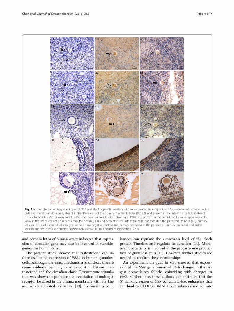
ResultsExpression of circadian proteins in the human ovaryImmunohistochemistry results showed positive expres-sion of CLOCK in the cumulus and mural granulosacells in dominant antral follicles and in interstitial cells,but no CLOCK expression was observed in the primor-dial follicles, primary follicles, and preantral follicles, andin the theca cells of antral follicles. Expression of PER2was observed in the cumulus cells and mural granulosacells in the dominant antral follicles and in interstitialcells, was weak in the theca cells of the dominant antralfollicle, but was absent in the primordial follicle, primaryfollicles, and preantral follicles (Fig. 1).
Expression patterns of circadian genes in humangranulosa cells stimulated by testosteroneTreatment of testosterone induced oscillations in PER2expression, with the first peak and bottom occurring atthe 24th and 32nd hours, respectively, and the secondpeak occurring at the 44th hour (P4 vs. 24 = 0.028, P24 vs. 32
= 0.041, P24 vs. 48 = 0.039, P 4 vs. 44 = 0.024). Expression ofCLOCK increased significantly at the 24th hour (P4 vs. 24
= 0.04), whereas expression of BMAL1 did not change sig-nificantly after testosterone stimulation. In the controlgroup, after changing to serum-free medium, the expres-sion levels of both PER2 (P4 vs. 16 = 0.016) and CLOCK (P4vs. 12 = 0.022, P4 vs. 16 = 0.031, P4 vs. 20 = 0.006, P4 vs. 36 =0.011) increased significantly; however, they did not dis-play an oscillating pattern. The expression of BMAL1 didnot change significantly over time (Fig. 2).
Expression patterns of steroidogenesis-related genes inhuman granulosa cells stimulated by testosteroneTreatment of testosterone induced oscillations in STARexpression, with the first and second peaks occurring atthe 24th and 40th hours, respectively (P4 vs. 24 < 0.001, P24vs. 32 < 0.001, P4 vs. 40 = 0.001). Expression of CYP19A1 ingranulosa cells was significantly repressed at the 12th hour(P4 vs. 12 = 0.003), whereas the expression levels ofHSD3B2 and CYP11A1 did not change significantly aftertestosterone stimulation. In the control group, changingto serum-free medium significantly stimulated STAR ex-pression (P4 vs. 24 = 0.028, P4 vs. 36 = 0.001, P4 vs. 40 = 0.021),but had no significant effects on the expression levels ofCYP11A1, HSD3B2, and CYP19A1 (Fig. 3).
DiscussionIn this study, we detected the presence of both PER2and CLOCK proteins in human dominant antral folli-cles, which were absent in the primordial, primary, andpreantral follicles. Moreover, CLOCK, PER2, and BMAL1mRNAs were present in human luteinized granulosacells. These distribution patterns of circadian genes inthe human ovary are similar to those reported for therat ovary. In the rat ovary, expression of Per2 and Bmal1was detected in growing and antral follicles, as well as inthe corpora lutea and stromal fibroblasts [7]. Circadiangenes have previously been found to be involved in ste-roidogenesis in granulosa cells. BMAL1 knock-down canlead to downregulation of estrogen synthesis and aroma-tase expression; conversely, overexpression of BMAL1resulted in upregulation of estrogen synthesis and aro-matase expression in KGN cells [10]. In addition, inhib-ition of the expression of Per2 with Per2-specific smallinterfering RNA stimulated the expression of Star ingranulosa cells from cows [12]. Both dominant antralfollicle and corpora lutea can produce steroids hormone.Expression of circadian genes in dominant antral follicle
Chen et al. Journal of Ovarian Research (2016) 9:56 Page 3 of 7
and corpora lutea of human ovary indicated that expres-sion of circadian gene may also be involved in steroido-genesis in human ovary.The present study showed that testosterone can in-
duce oscillating expression of PER2 in human granulosacells. Although the exact mechanism is unclear, there issome evidence pointing to an association between tes-tosterone and the circadian clock. Testosterone stimula-tion was shown to promote the association of androgenreceptor localized in the plasma membrane with Src kin-ase, which activated Src kinase [13]. Src-family tyrosine
kinases can regulate the expression level of the clockprotein Timeless and regulate its function [14]. More-over, Src activity is involved in the progesterone produc-tion of granulosa cells [15]. However, further studies areneeded to confirm these relationships.An experiment on quail in vivo showed that expres-
sion of the Star gene presented 24-h changes in the lar-gest preovulatory follicle, coinciding with changes inPer2. Furthermore, these authors demonstrated that the5′ flanking region of Star contains E-box enhancers thatcan bind to CLOCK–BMAL1 heterodimers and activate
Fig. 1 Immunohistochemistry staining of CLOCK and PER2 in paraffin sections of human ovaries. Staining of CLOCK was detected in the cumuluscells and mural granulosa cells, absent in the theca cells of the dominant antral follicles (D2, E2), and present in the interstitial cells, but absent inprimordial follicles (A2), primary follicles (B2), and preantral follicles (C2). Staining of PER2 was present in the cumulus cells, mural granulosa cells,weak in the theca cells of dominant antral follicles (D3, E3), and present in the interstitial cells, but absent in the primordial follicles (A3), primaryfollicles (B3), and preantral follicles (C3). A1 to E1 are negative controls (no primary antibody) of the primordial, primary, preantral, and antralfollicles and the cumulus complex, respectively. Bars = 50 μm. Original magnification, ×200
Chen et al. Journal of Ovarian Research (2016) 9:56 Page 4 of 7
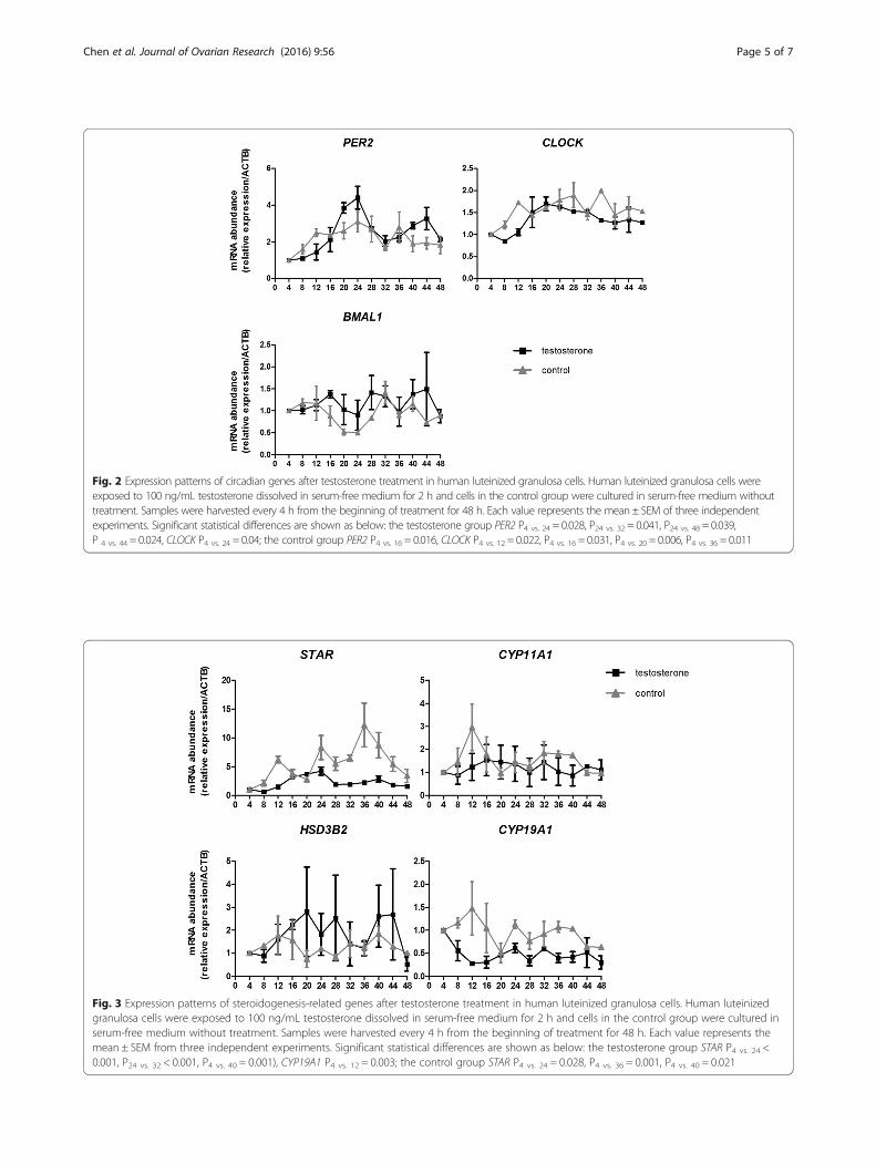
Fig. 2 Expression patterns of circadian genes after testosterone treatment in human luteinized granulosa cells. Human luteinized granulosa cells wereexposed to 100 ng/mL testosterone dissolved in serum-free medium for 2 h and cells in the control group were cultured in serum-free medium withouttreatment. Samples were harvested every 4 h from the beginning of treatment for 48 h. Each value represents the mean ± SEM of three independentexperiments. Significant statistical differences are shown as below: the testosterone group PER2 P4 vs. 24 = 0.028, P24 vs. 32 = 0.041, P24 vs. 48 = 0.039,P 4 vs. 44 = 0.024, CLOCK P4 vs. 24 = 0.04; the control group PER2 P4 vs. 16 = 0.016, CLOCK P4 vs. 12 = 0.022, P4 vs. 16 = 0.031, P4 vs. 20 = 0.006, P4 vs. 36 = 0.011
Fig. 3 Expression patterns of steroidogenesis-related genes after testosterone treatment in human luteinized granulosa cells. Human luteinizedgranulosa cells were exposed to 100 ng/mL testosterone dissolved in serum-free medium for 2 h and cells in the control group were cultured inserum-free medium without treatment. Samples were harvested every 4 h from the beginning of treatment for 48 h. Each value represents themean ± SEM from three independent experiments. Significant statistical differences are shown as below: the testosterone group STAR P4 vs. 24 <0.001, P24 vs. 32 < 0.001, P4 vs. 40 = 0.001), CYP19A1 P4 vs. 12 = 0.003; the control group STAR P4 vs. 24 = 0.028, P4 vs. 36 = 0.001, P4 vs. 40 = 0.021
Chen et al. Journal of Ovarian Research (2016) 9:56 Page 5 of 7
gene transcription [16]. Therefore, these findings indi-cated that Star is a clock-driven gene. Similarly, wefound that STAR expression showed an oscillating pat-tern in cultured human granulosa cells after testosteronestimulation, but the pattern did not completely coincidewith that of PER2. The difference between the results ofthe present study and those of the quail study may becaused by species-specific differences, the in vivo vs. invitro study design, or the stimulus used. In addition, wefound testosterone can decrease CYP19A1 expressionin human granulosa cells. Our results are in accord-ance with a recent study which showed that exposureto high level of testosterone could decrease bothmRNA and protein levels of aromatase in cultured lu-teinized granulosa cells isolated from non-PCOSwomen [17]. However, effect of testosterone on aro-matase expression in granulosa cells is different indifferent species. It has been reported that testoster-one stimulates Cyp19 promoter activity and theexpression of Cyp19 in granulosa cells from immaturefemale rats [18].A recent study pointed to the important role of
circadian genes in the development of PCOS and in-dicated that circadian clock is likely involved in ste-roidogenesis in granulosa cells [10]. Our resultsshowed that testosterone can affect circadian gene ex-pression in human granulosa cells. Therefore, hyper-androgenemia can affect circadian gene expression,resulting in the disorder of steroidogenesis in granu-losa cells leading to the development of PCOS. How-ever, a limitation of the present study is that we usedgranulosa cells from hormonally stimulated patients,because it is difficult to obtain granulosa cells fromhormonally unstimulated patients. Furthermore, thisstudy represents preliminary research on the expres-sion patterns of circadian genes and their relationshipwith steroidogenesis in the human ovary. Therefore,further studies are needed to elucidate the molecularmechanism.
ConclusionsWe found that the circadian genes CLOCK andPER2 are expressed in the dominant antral folliclesof the human ovary. Testosterone could induce os-cillating expression of the circadian gene PER2 incultured human granulosa cells. Moreover, expres-sion of STAR in human granulosa cells also dis-played an oscillating pattern after testosteronestimulation. Our results indicate a potential relation-ship between the circadian clock and steroidogenesisin the human ovary, and demonstrate the effect oftestosterone on circadian gene expression in granu-losa cells.
AcknowledgementsWe thank Jin-hu Guo for technical advice for sample harvesting, Chang Liufor technical advice for induction of circadian gene expression, Ping Xiao fortechnical advice on immunohistochemistry, and Ma Xiang for statisticaladvice.
FundingThis work was supported by grants from the National Basic ResearchProgram of China (973 program, 2012CB947600), Scientific Project of HealthIndustry (201002013), and Science and Technology Planning Project ofGuangdong Province, China (2008A030201028).
Availability of data and materialThe datasets generated during and/or analyzed during the current study arenot publicly available due to the on-going further analysis and study basedon the current study but are available from the corresponding author onreasonable request.
Authors’ contributionsMHC, YWX, and CQZ contributed to project design. MHC, BYM, and HZcarried out the cell culture and qRT-PCR. MHC, LL and HJS carried out theimmunohistochemistry. MHC, YWX, and HZ wrote the manuscript. All authorsdiscussed the results and commented on the manuscript. All authors readand approved the final manuscript.
Competing interestsThe authors declare that they have no competing interests.
Consent for publicationNot applicable.
Ethics approval and consent to participateThe use of normal human ovarian tissue and human granulosa cells fromthe follicle fluid obtained during ovum aspiration were approved by theinstitutional ethics committee of the First Affiliated Hospital of Sun Yat-senUniversity.
Author details1Reproductive Medicine Center, The First Affiliated Hospital of Sun Yat-senUniversity, 58 2nd Zhongshan Road, Guangzhou GD510080, People’sRepublic of China. 2Guangdong Provincial Key Laboratory of ReproductiveMedicine, The First Affiliated Hospital of Sun Yat-sen University, 58 2ndZhongshan Road, Guangzhou GD510080, People’s Republic of China.3Department of Pathology, The First Affiliated Hospital of Sun Yat-senUniversity, 58 2nd Zhongshan Road, Guangzhou GD510080, People’sRepublic of China. 4Department of Hepatic Surgery, The Third AffiliatedHospital of Sun Yat-sen University, 600 Tianhe Road, Guangzhou GD510630,People’s Republic of China.
Received: 20 May 2016 Accepted: 1 September 2016
References1. Hastings MH. Circadian clocks. Curr Biol. 1997;7:R670–2.2. Ko CH, Takahashi JS. Molecular components of the mammalian circadian
clock. Hum Mol Genet. 2006;15:R271–7.3. Takahashi JS, Hong HK, Ko CH, McDearmon EL. The genetics of mammalian
circadian order and disorder: implications for physiology and disease. NatRev Genet. 2008;9:764–75.
4. Gekakis N, Staknis D, Nguyen HB, Davis FC, Wilsbacher LD, King DP, et al.Role of the CLOCK protein in the mammalian circadian mechanism.Science. 1998;280:1564–9.
5. Shearman LP, Sriram S, Weaver DR, Maywood ES, Chaves I, Zheng B, et al.Interacting molecular loops in the mammalian circadian clock. Science.2000;288:1013–9.
6. Ueda HR, Hayashi S, Chen W, Sano M, Machida M, Shigeyoshi Y, et al.System-level identification of transcriptional circuits underlying mammaliancircadian clocks. Nat Genet. 2005;37:187–92.
7. Karman BN, Tischkau SA. Circadian clock gene expression in the ovary:effects of luteinizing hormone. Biol Reprod. 2006;75:624–32.
Chen et al. Journal of Ovarian Research (2016) 9:56 Page 6 of 7
8. Fahrenkrug J, Georg B, Hannibal J, Hindersson P, Gräs S. Diurnal rhythmicityof the clock genes Per1 and Per2 in the rat ovary. Endocrinology. 2006;147:3769–76.
9. Tischkau SA, Howell RE, Hickok JR, Krager SL, Bahr JM. The luteinizinghormone surge regulates circadian clock gene expression in the chickenovary. Chronobiol Int. 2011;28:10–20.
10. Zhang J, Liu J, Zhu K, Hong Y, Sun Y, Zhao X, Du Y, Chen ZJ. Effects ofBMAL1-SIRT1-positive cycle on estrogen synthesis in human ovariangranulosa cells: an implicative role of BMAL1 in PCOS. Endocrine. 2016;53:574-84.
11. Eisner JR, Barnett MA, Dumesic DA, Abbott DH. Ovarian hyperandrogenismin adult female rhesus monkeys exposed to prenatal androgen excess. FertilSteril. 2002;1:167–72.
12. Shimizu T, Hirai Y, Murayama C, Miyamoto A, Miyazaki H, Miyazaki K.Circadian Clock genes Per2 and clock regulate steroid production, cellproliferation, and luteinizing hormone receptor transcription in ovariangranulosa cells. Biochem Biophys Res Commun. 2011;412:132–5.
13. Cheng J, Watkins SC, Walker WH. Testosterone activates mitogen-activatedprotein kinase via Src kinase and the epidermal growth factor receptor insertoli cells. Endocrinology. 2007;148:2066–74.
14. O’Reilly LP, Zhang X, Smithgall TE. Individual Src-family tyrosine kinasesdirect the degradation or protection of the clock protein Timeless viadifferential ubiquitylation. Cell Signal. 2013;25:860–6.
15. Rice VM, Chaudhery AR, Oluola O, Limback SD, Roby KF, Terranova PF.Herbimycin, a tyrosine kinase inhibitor with Src selectivity, reducesprogesterone and estradiol secretion by human granulosa cells. Endocrine.2001;15:271–6.
16. Nakao N, Yasuo S, Nishimura A, Rupp GP, Echternkamp SE. Circadian clockgene regulation of steroidogenic acute regulatory protein gene expressionin preovulatory ovarian follicles. Endocrinology. 2007;148:3031–8.
17. Yang F, Ruan YC, Yang YJ, Wang K, Liang SS, Han YB, Teng XM. Yang JZFollicular hyperandrogenism downregulates aromatase in luteinizedgranulosa cells in polycystic ovary syndrome women. Reproduction. 2015;150:289–96.
18. Wu YG, Bennett J, Talla D, Stocco C. Testosterone, not 5α-dihydrotestosterone, stimulates LRH-1 leading to FSH-independentexpression of Cyp19 and P450scc in granulosa cells. Mol Endocrinol. 2011;25:656–68.
• We accept pre-submission inquiries
• Our selector tool helps you to find the most relevant journal
• We provide round the clock customer support
• Convenient online submission
• Thorough peer review
• Inclusion in PubMed and all major indexing services
• Maximum visibility for your research
Submit your manuscript atwww.biomedcentral.com/submit
Submit your next manuscript to BioMed Central and we will help you at every step:
Chen et al. Journal of Ovarian Research (2016) 9:56 Page 7 of 7