Circadian Regulation of Hippocampal Long-Term Potentiation
18
Circadian Regulation of Hippocampal Long-Term Potentiation Dipesh Chaudhury 1 , Louisa M. Wang, and Christopher S. Colwell 2 Department of Psychiatry and Biobehavioral Sciences, University of California, Los Angeles, California Abstract The goal of this study is to investigate the possible circadian regulation of hippocampal excitability and long-term potentiation (LTP) measured by stimulating the Schaffer collaterals (SC) and recording the field excitatory postsynaptic potential (fEPSP) from the CA1 dendritic layer or the population spike (PS) from the soma in brain slices of C3H and C57 mice. These 2 strains of mice were of interest because the C3H mice secrete melatonin rhythmically while the C57 mice do not. The authors found that the magnitude of the enhancement of the PS was significantly greater in LTP recorded from night slices compared to day slices of both C3H and C57 mice. They also found significant diurnal variation in the decay of LTP measured with fEPSPs, with the decay slower during the night in both strains of mice. There was evidence for a diurnal rhythm in the input/output function of pyramidal neurons measured at the soma in C57 but not C3H mice. Furthermore, LTP in the PS, measured in slices prepared during the day but recorded during the night, had a profile remarkably similar to the night group. Finally, PS recordings were carried out in slices from C3H mice maintained in constant darkness prior to experimentation. Again, the authors found that the magnitude of the enhancement of the PS was significantly greater in LTP recorded from subjective night slices compared to subjective day slices. These results provide the 1st evidence that an endogenous circadian oscillator modulates synaptic plasticity in the hippocampus. Keywords circadian rhythm; hippocampus; long-term potentiation; melatonin; synaptic plasticity; mice Daily rhythms in behavior and physiology have been shown to occur in most organisms, including humans. The physiological system responsible for these rhythms is known as the circadian system; in mammals, the core of this rhythm-generating system is localized to a site in the hypothalamus known as the SCN (Reppert and Weaver, 2002; Buijs et al., 2003). The endogenous rhythms generated by the SCN have a period close to, but not quite, 24 h. The function of the circadian system is thought to allow the temporal coordination of various physiological processes within an organism, as well as enabling an organism to temporally coordinate with the external world. To fulfill these functions, these rhythms must be synchronized to the exact 24-h cycle of the physical world. The dominant cue used by most organisms that enables their circadian system to synchronize to the environment is the daily cycle of light and dark. Within an organism, the biological clock regulates many physiological processes and behaviors. Recent data suggest that the SCN functions as a master clock that coordinates the activity of multiple, damped circadian oscillators outside the SCN (Yamazaki et al., 2000). 2To whom all correspondence should be addressed: Christopher S. Colwell, Department of Psychiatry and Biobehavioral Sciences, University of California, Los Angeles, 760 Westwood Plaza, Los Angeles, CA 90024-1759; e-mail: [email protected]. 1 Present address: Dipesh Chaudhury, Institut de Neurosciences Cognitives de la Méditerranée–INCM UMR 6193, CNRS–Université de la Méditerranée 31, Chemin Joseph Aiguier, 13402 Marseille Cedex 20, France. NIH Public Access Author Manuscript J Biol Rhythms. Author manuscript; available in PMC 2008 November 7. Published in final edited form as: J Biol Rhythms. 2005 June ; 20(3): 225–236. doi:10.1177/0748730405276352. NIH-PA Author Manuscript NIH-PA Author Manuscript NIH-PA Author Manuscript
Transcript of Circadian Regulation of Hippocampal Long-Term Potentiation

Circadian Regulation of Hippocampal Long-Term Potentiation
Dipesh Chaudhury1, Louisa M. Wang, and Christopher S. Colwell2Department of Psychiatry and Biobehavioral Sciences, University of California, Los Angeles,California
AbstractThe goal of this study is to investigate the possible circadian regulation of hippocampal excitabilityand long-term potentiation (LTP) measured by stimulating the Schaffer collaterals (SC) andrecording the field excitatory postsynaptic potential (fEPSP) from the CA1 dendritic layer or thepopulation spike (PS) from the soma in brain slices of C3H and C57 mice. These 2 strains of micewere of interest because the C3H mice secrete melatonin rhythmically while the C57 mice do not.The authors found that the magnitude of the enhancement of the PS was significantly greater in LTPrecorded from night slices compared to day slices of both C3H and C57 mice. They also foundsignificant diurnal variation in the decay of LTP measured with fEPSPs, with the decay slower duringthe night in both strains of mice. There was evidence for a diurnal rhythm in the input/output functionof pyramidal neurons measured at the soma in C57 but not C3H mice. Furthermore, LTP in the PS,measured in slices prepared during the day but recorded during the night, had a profile remarkablysimilar to the night group. Finally, PS recordings were carried out in slices from C3H mice maintainedin constant darkness prior to experimentation. Again, the authors found that the magnitude of theenhancement of the PS was significantly greater in LTP recorded from subjective night slicescompared to subjective day slices. These results provide the 1st evidence that an endogenouscircadian oscillator modulates synaptic plasticity in the hippocampus.
Keywordscircadian rhythm; hippocampus; long-term potentiation; melatonin; synaptic plasticity; mice
Daily rhythms in behavior and physiology have been shown to occur in most organisms,including humans. The physiological system responsible for these rhythms is known as thecircadian system; in mammals, the core of this rhythm-generating system is localized to a sitein the hypothalamus known as the SCN (Reppert and Weaver, 2002; Buijs et al., 2003). Theendogenous rhythms generated by the SCN have a period close to, but not quite, 24 h. Thefunction of the circadian system is thought to allow the temporal coordination of variousphysiological processes within an organism, as well as enabling an organism to temporallycoordinate with the external world. To fulfill these functions, these rhythms must besynchronized to the exact 24-h cycle of the physical world. The dominant cue used by mostorganisms that enables their circadian system to synchronize to the environment is the dailycycle of light and dark. Within an organism, the biological clock regulates many physiologicalprocesses and behaviors. Recent data suggest that the SCN functions as a master clock thatcoordinates the activity of multiple, damped circadian oscillators outside the SCN (Yamazakiet al., 2000).
2To whom all correspondence should be addressed: Christopher S. Colwell, Department of Psychiatry and Biobehavioral Sciences,University of California, Los Angeles, 760 Westwood Plaza, Los Angeles, CA 90024-1759; e-mail: [email protected] address: Dipesh Chaudhury, Institut de Neurosciences Cognitives de la Méditerranée–INCM UMR 6193, CNRS–Université dela Méditerranée 31, Chemin Joseph Aiguier, 13402 Marseille Cedex 20, France.
NIH Public AccessAuthor ManuscriptJ Biol Rhythms. Author manuscript; available in PMC 2008 November 7.
Published in final edited form as:J Biol Rhythms. 2005 June ; 20(3): 225–236. doi:10.1177/0748730405276352.
NIH
-PA Author Manuscript
NIH
-PA Author Manuscript
NIH
-PA Author Manuscript

It is well established that the hippocampus is critically involved in memory formation and thatsynaptic connections within the hippocampus are well known to undergo activity-dependentchanges in synaptic strength, referred to as long-term potentiation (LTP). LTP in the CA1pyramidal cell layer has been particularly well studied and can be measured by stimulating theSchaffer collaterals (SC) and recording the field excitatory postsynaptic potential (fEPSP) fromthe CA1 dendritic layer or the population spike (PS) from the soma. Changes in the strengthof the SC/CA1 synaptic connection are commonly viewed as a model for understandingactivity-dependent changes in synaptic strength that may ultimately be linked to learning andmemory (Martin et al., 2000; Sweatt, 2001). Previous work makes it clear that the strength ofLTP at the SC/CA1 synapse can be regulated by a number of signaling pathways, includingthose activated by modulatory neurotransmitters and hormones (Lisman, 2003; Silva, 2003).If LTP is indeed a neural correlate of learning and memory formation, then it seems logicalthat the many factors, including circadian rhythms, which modulate learning and memory, willalso influence LTP. Indeed, in vivo electro-physiological studies have described a diurnalrhythm of synaptic excitability in the dentate gyrus (DG) of monkeys and rats (Barnes et al.,1977; West and Deadwyler, 1980; Cauller et al., 1985). In addition, in vivo recordings fromanesthetized rats have also revealed a diurnal rhythm in LTP from hippocampal granule cells(Dana and Martinez, 1984). Finally, in vitro studies have reported diurnal variations in theincidence and magnitude of LTP in the CA1 region of the rat and hamster hippocampus, withthe largest responses occurring in the day (Harris and Teyler, 1983; Raghavan et al., 1999).
In the current study, we first examined the possibility of a diurnal variation in excitability andLTP in hippocampal brain slices of C3H and C57 mice. While this type of work has been donein rats and hamsters, extending this work to mice with their well-characterized genetics allowsa range of experiments not possible with other mammals. In addition, the daily rise and fall ofmelatonin (MEL) are under the control of the circadian system, and previous studies suggestthat this hormone can regulate synaptic plasticity (e.g., Collins and Davies, 1997; El-Sherif etal., 2003). Therefore, in the current study, we compared LTP in fEPSP and PS in a strain ofmice that secretes MEL rhythmically (C3H) to another strain (C57) that carries a mutation thatdoes not allow the production of MEL (Ebihara et al., 1986). Finally, we determined whetherthe day/night differences seen in a light-dark (LD) cycle continue when the animals weremaintained in constant darkness (DD). This experiment provides a critical test of the hypothesisthat an endogenous circadian oscillator drives the observed diurnal variations.
MATERIALS AND METHODSAnimals and Lighting Conditions
Two- to 4-month-old male mice (C-57 BL/6 or C3H) were purchased from Charles RiverLaboratories. The Animal Research Committee of the University of California, Los Angelesapproved the experimental protocols used in this study. Three types of experiments wereperformed. First, to investigate possible diurnal variations in any of the measured values,animals were maintained on a daily LD cycle consisting of 12 h of light followed by 12 h ofdark. It is already well established that cells in the SCN continue to show circadian oscillationswhen isolated from the animal in a hypothalamic brain slice preparation (e.g., Green andGillette, 1982), and we sought to determine if diurnal differences in LTP could also be foundin the hippocampus. For the day group, brain slices were prepared at ZT 3 and recordings madebetween ZT 4 and 12. For the night group, brain slices were prepared at ZT 15 and recordingsmade between ZT 16 and 24. By convention, ZT 12 is the time that the lights go off fororganisms held in an LD cycle. Second, some mice were kept in an LD cycle and killed in theday (ZT 11), and evoked responses were recorded at night (from ZT 13–18). With theseexperiments, we sought to determine if the hippocampal slices were able to transition from theday to the night state in vitro. Finally, experiments were conducted to determine if the rhythm
Chaudhury et al. Page 2
J Biol Rhythms. Author manuscript; available in PMC 2008 November 7.
NIH
-PA Author Manuscript
NIH
-PA Author Manuscript
NIH
-PA Author Manuscript

continues in DD and could be considered a circadian oscillation. For these experiments, micewere housed individually, and their wheel-running activity was recorded as revolutions per 3-min intervals and the time of activity onset determined. The running wheels and data acquisitionsystem were obtained from Mini-Mitter Company (Sunriver, OR). After entrainment to an LDcycle, the mice were placed into DD for a week to assess their free-running activity pattern.For the day group, brain slices were prepared at CT 3 and recordings made between CT 4 and12. For the night group, brain slices were prepared at CT 15 and recordings made between CT16 and 24. By convention, CT 12 is the time of activity onset for nocturnal organisms held inDD. All handling of animals was carried out either in the light portion of the LD cycle or inthe dark with the aid of an infrared viewer (FJW Industries, Palatine, IL).
Slice PreparationBrain slices of mice between 2 and 4 months of age were prepared using standard techniques.Mice were anesthetized with halothane and decapitated either in the day or the night asdescribed above. The brains were dissected and placed in cold oxygenated artificial cerebralspinal fluid (ACSF) containing (in mM) NaCl 130, NaHCO3 26, KCl 3, MgCl2 5, NaH2PO41.25, CaCl2 1, glucose 10 (pH 7.2–7.4; osmolality 290–300 mOsm). Coronal hippocampalslices (400 μM thick) were prepared using a microslicer (DSK Microslicer; Ted Pella, Redding,CA). Slices were then transferred into an ACSF, in which the CaCl2 was increased to 2 mMand MgCl2 was decreased to 2 mM. Slices were allowed to recover for at least 1 h prior tostarting electrophysiology recordings. Slices were constantly oxygenated with 95% O2–5%CO2. Besides the hippocampus, the slice contained some elements of the entorhinal cortex,parahippocampal gyrus, and subcortical white matter.
Electrophysiology RecordingBriefly, slices were placed in an interface chamber (Fine Science Tools, Foster City, CA) andcontinuously superfused with oxygenated ACSF (30 °C) at 2 to 3 mL/min. A bipolarstimulating electrode was constructed from nichrome wire (0.0015-inch diameter; A-MSystems, Carlborg, WA). The electrode was placed in the stratum radiatum in the CA1 regionof the hippocampus to stimulate presynaptic fibers arising from the CA3 pyramidal cells. Theslices were stimulated with negative current pulses with an A-M Systems stimulator at 0.02Hz (100- μs duration). The recording electrodes were pulled on a multistage puller (Sutter P-97,Novato, CA) and filled with ACSF (5–10 MΩ filled with ACSF). The field potentials weretypically 1 to 3 mV in amplitude and were amplified 100× with an Axon Instruments 2Aamplifier (Axon Instruments, Foster City, CA) and an external amplifier. Responses werefiltered at 5 kHz and digitized (10 kHz) using data acquisition and analysis programs (pClamp9;Axon Instruments).
Typically, stable baseline measurements were obtained within an hour after placinghippocampal slices in the interface chamber (0.02-Hz stimulation). At this point, postsynapticresponses were recorded for at least 10 min prior to the induction of LTP. During this time,input/output (I/O) relations were generated by varying the stimulation intensity from 0 to 100μA in steps of 10 μA. Information was also gathered about the peak amplitude, slope, andduration of evoked responses at half-maximal amplitude. These evoked responses were stableand changed less than 10% over the course of 60 min of recording (5% ± 8%, n = 5). For LTPexperiments, the baseline presynaptic stimulation was delivered at 0.02 Hz (100- μs duration)using a stimulation intensity that evoked approximately 50% of maximal postsynapticresponse. After tetanus, stimulation was again delivered at 0.02 Hz (100- μs duration). In mostcases, LTP was evoked by a single tetanizing stimulus (1 ×100 Hz, 1-sec duration). In somecases, a more robust form of LTP was evoked by repeated tetanizing stimuli (3 ×100 Hz, 1-sec duration, intertrial interval 15 sec). Different groups of animals were used for the PS andfEPSP recordings.
Chaudhury et al. Page 3
J Biol Rhythms. Author manuscript; available in PMC 2008 November 7.
NIH
-PA Author Manuscript
NIH
-PA Author Manuscript
NIH
-PA Author Manuscript

AnalysesFor LTP experiments, posttetanic responses were normalized to baseline, as is standard in thefield. We used 3 separate statistical analyses to assess possible between-group differences inLTP. First, possible differences in average LTP were assessed using the Friedman repeated-measures analysis of variance (RM ANOVA) on ranks followed by post hoc pairwisecomparison (Dunnett method). The α level for all analyses was set at 0.05. Next, the posttetanusdata were grouped into 10-min bins (10, 20, 30, 40, 50, 60 min) and pairwise comparisonsmade using the Tukey t test or the Mann-Whitney rank sum test. Finally, the posttetanus datafrom each slice were fitted with a nonlinear regression. The best fit (R > 0.9) was obtained withan equation that had an exponential decay and linear component (y = y0 +ae−bx + cx). Thisequation was used to determine the slope of the linear component of the curve (c in the equation)that we took for a measure of the rate of decay of LTP. The slope of the exponential decay wasdetermined in large part by the 1st couple of measurements after tetanus. This exponentialdecay did not significantly vary between groups. The best-fit values were then compared usingthe Tukey t test. In the text, the sample size (n) refers to the number of slices in each group. Inall cases, the slices in an experimental group came from at least 5 mice. Some control groupshad slices from just 3 mice. For example, the control slices prepared in the day and recordedin the late day were from just 3 mice. Values were considered significantly different if p <0.05. All tests were performed using SigmaStat (SPSS, Chicago, IL). In the text, values areshown as mean ± standard error of the mean (SEM).
RESULTSLTP in C3H Mice
A diagram of the hippocampus showing placement of the stimulating and recording electrodeis shown in Figure 1A. The 1st set of experiments was designed to determine if LTP recordedfrom the CA1 region varies as a function of time of day in C3H mice. For these experiments,we recorded fEPSP or PS from the CA1 region of hippocampal slices evoked by stimulationof the SC pathway before and after application of a high-frequency stimulus train (1 × 100 Hz,1-sec duration; Fig. 1B). To determine if the magnitude of LTP varies within the day or thenight, we compared data collected during ZT 4–6 (early day) with ZT 7–12 (late day) as wellas data collected between ZT 16–18 (early night) with ZT 19–24 (late night). There were nosignificant differences between the magnitude of LTP recorded during the early and latesegments of the day or night. Accordingly for these studies, the data collected between ZT 4–12 and ZT 16–24 were pooled to form the day and night groups, respectively (Fig. 1C). Overall,we found that the magnitude of the enhancement of the PS was greater in LTP recorded fromnight slices compared to day slices of C3H mice (Fig. 2A; RM ANOVA, χ2 = 146, p < 0.001).The average PS slope was 334% ± 12% (n = 7) of baseline during the night and 177% ± 15%(n = 9) of baseline during the day. The amplitude of the PS increased more (Mann-Whitneyrank sum test, T = 120.5, p = 0.014) during the night (pretetanus: −1.98 ± 0.2 mV; posttetanus:−5.0 ± 0.5 mV) than during the day (pretetanus: −1.28 ± 0.2 mV; posttetanus: −2.25 ± 0.4 mV).PS slopes were significantly larger in the night at all time points (Mann-Whitney rank sumtest, T = 82–87, p = 0.02–0.004). There were no significant differences in the latency-to-peakinduction (day: 1.6 ± 0.2 min; night: 2.4 ± 0.5 min). Finally, to measure the rate of decay ofthe potentiated PS, the posttetanus data collected from each slice were fitted to a nonlinearregression. The rate of decay was measured as the slope of the fitted equation. By this measure,the decay of LTP was faster in the day (slope = –0.48, R = 0.95) than in the night (slope = 1.6,R = 0.92), but this difference was not significant.
A day/night difference was also found in LTP recorded from the fEPSPs in C3H mice. Theaverage fEPSP slope was 165% ± 1.2% (n = 21) of baseline during the night and 152% ± 1.9%(n = 21) of baseline during the day (RM ANOVA, χ2 = 69, p < 0.001). Acomparison of the
Chaudhury et al. Page 4
J Biol Rhythms. Author manuscript; available in PMC 2008 November 7.
NIH
-PA Author Manuscript
NIH
-PA Author Manuscript
NIH
-PA Author Manuscript

fEPSP slopes indicated that slopes were significantly larger in the night only at the 50-min (ttest, t = −2.532, df = 40, p = 0.015) and 60-min (t test, t = −2.95, df = 40, p = 0.005) time points.While there were no significant day/night differences in the latency-to-peak induction (day:1.7 ± 0.2 min; night: 1.7 ± 0.1 min), the rate of decay of LTP was significantly faster in theday (slope = −0.66; R = 0.97) than the night (slope = −0.19; R = 0.93) groups (t test, t = −2.08,df = 38, p = 0.04). We did not see a diurnal difference in LTP in response to a stronger tetanizingprotocol (3 × 100 Hz, 1 sec, intertrial interval 15 sec; data not shown). The average fEPSPslope was 229% ± 2% of baseline during the night (n = 12) and 221% ± 5% of baseline duringthe day (n = 12). No day/night differences were found in the fEPSP slopes averaged into 10-min bins using a stronger tetanus (data not shown).
LTP in C57 MiceThe next set of experiments was designed to determine if LTP recorded from the CA1 regionalso varies as a function of time of day in C57 mice. Again, we compared data collected duringearly (ZT 4–6) and late (ZT 7–12) day as well as data collected between early (ZT 16–18) andlate (ZT 19–24) night. There were no significant differences between the magnitude of LTPrecorded during the early and late portions of the day or night. Accordingly for these studies,the data collected between ZT 4–12 and ZT 16–24 were pooled to form the day and nightgroups, respectively. We found that the magnitude of the enhancement of the PS was greaterin LTP recorded from night slices compared to day slices of C57 mice (Fig. 2B; RM ANOVA,χ2 = 74, p < 0.001). This diurnal difference was significant but more modest than that observedwith the C3H mice. The average PS slope was 168% ± 7% of baseline during the night (n =12) and 140% ± 5% of baseline during the day (n = 12). The amplitude of the PS increasedmore (t test, t = −2.508, p = 0.020) during the night (pretetanus: −1.5 ± 0.2 mV; posttetanus:−2.6 ± 0.4 mV) than during the day (pretetanus: −1.4 ± 0.1 mV; posttetanus: −1.8 ± 0.4 mV).Although the PS slopes were always larger during the night, these differences were onlysignificant at the 50-min (t test, t = −2.656, df = 22, p = 0.014) and 60-min (t test, t = −2.231,df = 22, p = 0.036) time points. There were no significant differences in the latency-to-peakinduction (day: 1.4 ± 0.1 sec; night: 1.7 ± 0.3 sec) or in the rate of the decay of LTP betweenthe day (slope = −0.28; R = 0.92) and night (slope = −0.16; R = 0.97) groups.
Some evidence for a diurnal difference was also found in LTP recorded from the fEPSPs inC57 mice. The average fEPSP slope was 151% ± 5% (n = 21) of baseline during the night and133% ± 41% (n = 28) of baseline during the day (RM ANOVA, χ2 = 27, p < 0.001). The fEPSPslopes were significantly larger in the night at the 50-min (t test, t = −2.446, df = 47, p = 0.018)and 60-min (t test, t = −2.656, df = 47, p = 0.011) time points. While there were no significantday/night differences in the latency-to-peak induction (day: 1.4 ± 0.1 sec; night: 1.4 ± 0.1 sec),the rate of decay of the LTP was faster in the day (slope = −0.21; R = 0.98) than the night (slope= 0.23; R = 0.94) groups. With the stronger protocol, no diurnal differences were observed(data not shown). The average fEPSP slope was 192% ± 1.7% of baseline during the night(n = 13) and 190% ± 1.6% of baseline during the day (n = 14). No day/night differences werefound in the fEPSP slopes averaged into 10-min bins using the stronger tetanus (data notshown).
Current-Response Relationships in C3H and C57 MiceWe also carried out experiments designed to determine whether there was a diurnal differencein evoked fEPSP and PS in hippocampal slices prepared from C3H or C57 mice measured inthe day (ZT 4–12) or night (ZT 16–24). For these experiments, we varied the stimulus intensityand recorded the resulting slope to characterize the I/O relationship for these evoked responses(Fig. 3). A change in the gain of the I/O curve could indicate a change in sensitivity of theneuron to excitatory input. The C3H mice did not exhibit a day/night difference in I/O curvesmeasured for either the fEPSP or the PS. In contrast, the slope I/O curves for the PS recorded
Chaudhury et al. Page 5
J Biol Rhythms. Author manuscript; available in PMC 2008 November 7.
NIH
-PA Author Manuscript
NIH
-PA Author Manuscript
NIH
-PA Author Manuscript

in the night from C57 mice were significantly larger then the slope I/O curves recorded in theday (t test, t = 3.315, df = 24, p = 0.003). The magnitude of the PS was larger in the night ateach of the stimulus intensities between 30 and 100 μA. No diurnal differences were found inthe I/O curves measured from the fEPSP of C57 mice. Other electrophysiological parametersof the evoked responses were not found to vary between day and night (Table 1).
Endogenous Rhythm in LTP in PS of C3H MiceFinally, 2 sets of experiments were performed on the C3H mice to explore the possibleendogenous nature of the day/night differences observed in the PS recordings. In the 1st set(Fig. 4A), C3H mice were kept in an LD cycle and killed in the day (1 h prior to lights-off atZT 11), and evoked responses were recorded at night (from ZT 13 onwards). With theseexperiments, we sought to determine if the hippocampal slices were able to transition from theday to the night state in vitro. The PS of slices prepared during the day, but recorded duringthe night, had an LTP profile remarkably similar to the night group. As described above, theaverage PS slope was 334% of baseline during the night and 177% of baseline during the day.The transition group exhibited an average PS slope of 288% ± 4% of baseline (n = 7), whichwas significantly greater than the day (RM ANOVA, χ2 = 146, p < 0.001) but not the night.For control slices prepared in the day and recorded in the late day (ZT 11–12), the PS slopewas 167% ± 6% of baseline (n = 3). The average PS amplitude was 180% ± 50% of baselineduring the night and 74% ± 9% of baseline during the day. The transition group exhibited anaverage PS amplitude of 129% ± 18% of baseline, which was significantly greater than the day(ANOVA, H = 9.2, p = 0.01) but not the night. Similar results were obtained with the fEPSPrecordings. As described above, the average fEPSP slope was 165% of baseline during thenight and 152% of baseline during the day. The transition group exhibited an average PS slopeof 172% ± 2% of baseline (n = 12). The transition group also exhibited a slower rate of decaythat is characteristic of the data collected during the night but not during the day. For controlslices prepared in the day and recorded in the late day (ZT 11–12), the fEPSP slope was 146%± 12% of baseline (n = 4).
To determine if the previously observed diurnal rhythm in PS LTP in C3H mice was anendogenous circadian oscillation that continues in constant conditions, recordings were carriedout in slices from mice that were initially entrained to an LD cycle and then maintained in DDprior to experimentation. Free-running activity rhythms of the mice were monitored, andanimals were killed at their individually calculated CT 3 or CT 15 (Fig. 4B). Overall, we foundthat the magnitude of the enhancement of the PS was significantly greater in LTP recordedfrom subjective night slices compared to subjective day slices (RM ANOVA, p = 0.008). Theaverage PS slope was 243% ± 4% of baseline during the night (n = 9) and 157% ± 2% ofbaseline during the day (n = 9). The amplitude of the PS increased more during the subjectivenight (pretetanus: −1.1 ± 0.1 mV; posttetanus: −2.9 ± 0.3 mV) than during the subjective day(pretetanus: −1.1 ± 0.1 mV; posttetanus: −1.6 ± 0.2 mV). A comparison of the PS slopesindicated that slopes were significantly larger in the night at the 10-, 30-, 40-, 50-, and 60-mintime points (Mann-Whitney rank sum test, T = 50–59, p = 0.02–0.004). There were nosignificant differences in the latency-to-peak induction or in the rate of the decay of LTPbetween the subjective day and night groups.
DISCUSSIONIn recent years, there has been a resurgence of interest in state-dependent changes in bothlearned behaviors and in the cellular/molecular events that may underlie learning and memory.In particular, there has been much discussion as to the possible relationship between sleep andlearning (e.g., Maquet, 2001; Siegel, 2001; Stickgold et al., 2001; Walker et al., 2002; Walkeret al., 2003). While the role of sleep in learning remains to be defined, sleep deprivation
Chaudhury et al. Page 6
J Biol Rhythms. Author manuscript; available in PMC 2008 November 7.
NIH
-PA Author Manuscript
NIH
-PA Author Manuscript
NIH
-PA Author Manuscript

protocols can be shown to have a negative impact on learning as well as affect hippocampalphysiology (Campbell et al., 2002; Davis et al., 2003; McDermott et al., 2003). Less studied,but perhaps equally important, is the temporal regulation imposed by the circadian timingsystem based in the SCN. The circadian clock is thought to broadly regulate the arousal of thenervous system. For example, the behavioral literature provides evidence for diurnal (e.g.,Holloway and Wansley, 1973a, 1973b; Wansley and Holloway, 1975; Stephan and Kovacevic,1978; Valentinuzzi et al., 2001) and circadian modulation in some learned behaviors(Chaudhury and Colwell, 2002; Fernandez et al., 2003; Valentinuzzi et al., 2004). Several invivo studies have found evidence for diurnal regulation of hippocampal excitability (Barneset al., 1977; West and Deadwyler, 1980; Cauller et al., 1985). These data also provided someindication that the rhythms may be under circadian variation. For example, Barnes andcolleagues (1977) showed that a rhythm in synaptic excitability continued in 1 blinded rat, andCauller and coworkers (1985) reported that the rhythm in responsiveness to perforant pathstimulation continued in 1 rat maintained in DD. While the role of the SCN in these rhythmshas not yet been established, a few studies have also provided evidence that synaptic plasticityin the hippocampus may also vary with the time of day (Harris and Teyler, 1983; Dana andMartinez, 1984; Raghavan et al., 1999).
In the current study, we found that the magnitude of the potentiation of the PS due to high-frequency stimulation (1 ×100 Hz) is larger in the night than in the day. This diurnal differencewas most strikingly illustrated in C3H mice, in which the night group exhibited an averagepotentiation that was about 70% larger than that measured during the day. Similarly, previousin vivo studies in the rat hippocampus found evidence for diurnal rhythms in LTP magnitude,which peaked during the night (Dana and Martinez, 1984; Leung et al., 2003). Other in vitroLTP studies found evidence for rhythms in the magnitude of PS LTP in CA1 neurons thatpeaked in the day in rats (Harris and Teyler, 1983) and hamsters (Raghavan et al., 1999). Sowhile day/night differences in LTP have now been found in several studies, the time of peakinduction appears to vary perhaps because of species differences or differences in the protocolsused to evoke LTP. In addition to diurnal differences in magnitude, we also found evidence tosuggest that LTP induced in the night was more stable than LTP measured during the day.Specifically, our data demonstrate that the decay in LTP in the fEPSP was slower at nightcompared to the day. This difference in the kinetics in the decay of LTP was observed in slicesfrom both C3H and C57 mice. Interestingly, REM sleep deprivation also causes a dramaticreduction in the stability of the maintenance phase of LTP in hippocampal slices (Davis et al.,2003). The day/night difference could be overcome with a stronger induction protocol (3 ×100Hz), at least within the 60-min posttetanus that we measured in the present study.
The circadian variation we observed in the magnitude of LTP recorded from the cell body layer(PS) was primarily an effect on amplitude, while the fEPSP exhibited more of an effect on thestability or rate of decay of LTP. The fEPSP slope is viewed as a measure of the excitatorydrive to the CA1 pyramidal neurons, whereas the population spike amplitude reflects thenumber of pyramidal neurons producing action potentials. The CA1 cell bodies are known toreceive strong γ-aminobutyric acid (GABA)–mediated innervations from interneurons withinthe hippocampal circuit to powerfully control the excitability of CA1 pyramidal neurons(Freund and Buzsáki, 1996) and are involved in the generation of network oscillations withinthe hippocampus (McBain et al., 1999). Thus, the striking diurnal variation in the magnitudeof the PS may be more the result of a change in the balance between inhibition and excitationrather then purely a regulation of glutamatergic synaptic transmission (see Marder andBuonomano, 2003).
Previous in vivo recordings from the hippocampal region have found some evidence for diurnalchanges in basic neural excitability. An earlier study investigating variation in the I/Orelationship in vivo found that evoked responses in the dentate gyrus and CA1 of rat hippocampi
Chaudhury et al. Page 7
J Biol Rhythms. Author manuscript; available in PMC 2008 November 7.
NIH
-PA Author Manuscript
NIH
-PA Author Manuscript
NIH
-PA Author Manuscript

varied as a function of the behavioral state of the animals (Winson and Abzug, 1977). Laterwork by West and Deadwyler (1980) showed that PS amplitude was greater in the light thanin the dark period of the rat and that spike amplitude was greater when the animals were intheir slow wave sleep state compared to other states. In addition, a report by Cauller andcolleagues (1985) using in vivo recordings found that fEPSP magnitude peaked in the nightwhile PS magnitude peaked in the day. These rhythms in hippocampal excitability maycontinue in the brain slice preparation. For example, in CA3 neurons, both depolarization-induced excitability and the magnitude of high-voltage-activated Ca2+ currents vary with thetime of day, with peaks during the night (Kole et al., 2001). Similarly, a study by Liu andcolleagues (2000) showed the existence of diurnal differences in synaptic excitability in thedentate gyrus and that blocking adenosine receptors eliminated the diurnal variation. In thepresent study, we investigated whether there was a diurnal variation in baseline excitability indendritic (fEPSP) and somatic (PS) regions of the postsynaptic CA1 pyramidal cells ofhippocampal slices from C57 and C3H mice. We found significant differences only with thePS recorded from C57 mice. At least 1 previous study found that the I/O curves are larger inC3H compared to C57 mice (Bampton et al., 1999), and our data confirm their observations.
Two experiments were performed to explore the possible endogenous nature of the day/nightdifferences observed in the PS recordings in the C3H mice. In 1 experiment, to determine ifthe previously observed diurnal rhythm in PS LTP in C3H mice is endogenous and presumablycircadian in nature, recordings were carried out in slices from mice maintained in DD prior toexperimentation. Free-running activity rhythms of the mice were monitored, and animals werekilled at their individually calculated subjective day or night. Again, we found that themagnitude of the enhancement of the PS was significantly greater in LTP recorded from brainslices in the subjective night compared to subjective day. Acute effects of light or theendogenous timing system can drive a rhythm seen in mice held in an LD cycle. The findingthat the rhythm continues in mice held in DD strongly implicates an endogenous timing systemas being responsible for the rhythm in LTP. Ideal circadian rhythm studies follow a measuredvariable in an individual through time and show that the magnitude varies with the circadiancycle. This is not possible for this type of LTP measured in acute hippocampal slices, butinstead we are able to demonstrate that the magnitude of the potentiation across a populationvaries as a function of time even when the organisms are held in constant conditions. In theother experiment, mice were killed in the light portion of an LD cycle while the evokedresponses were recorded at night. We reasoned that if the isolated hippocampus has someoscillatory properties, then the time of slice preparation should not be as important as the timethat LTP was recorded. We investigated whether slices prepared in the day but recorded in thenight would show potentiated evoked responses similar to those from slices prepared andrecorded in the day or in the night. The resulting LTP was strikingly similar in both the highermagnitude and slower decay kinetics to the previous night group. Control slices recorded inthe late day (ZT 11–12) still exhibited typical “day” magnitudes and kinetics of LTP. To us,this observation suggests that the hippocampal slices are not “frozen” at the time of slicepreparation but instead continue to change from the day to the night state in vitro.
These experiments do not allow us to know the anatomical location of the oscillator responsiblefor driving rhythms in hippocampal synaptic plasticity. One possibility, which we favor, is thatthis rhythm is due to some type of oscillator mechanism within the hippocampus itself. Ourfinding that the isolated hippocampal slice appears to transition from day to night supports thispossibility. Interestingly, at least 1 study has found that expression of circadian clock genesmPer1 and mPer2 mRNA in the hippocampus may be rhythmic, with peak expression in thenight (Wakamatsu et al., 2001). However, it is also possible that an oscillator in the SCN oranother brain region has long-lasting influences on the rhythmicity of hippocampal neurons.A slow-acting signal from another location could already have reached the hippocampus prior
Chaudhury et al. Page 8
J Biol Rhythms. Author manuscript; available in PMC 2008 November 7.
NIH
-PA Author Manuscript
NIH
-PA Author Manuscript
NIH
-PA Author Manuscript

to the preparation of the brain slice. While this signaling seems less likely than a directhippocampal circadian oscillator, it is also hard to exclude.
In the current study, we took advantage of strain differences between C57 and C3H mice toexamine the possible role of MELas a signal responsible for driving these rhythms in synapticplasticity. MELis a rhythmically synthesized hormone that has been suggested to be an outputmolecule by which the circadian system transmits temporal information. MEL mediates itseffects via high-affinity receptors found throughout the nervous system, including thehippocampus, the subiculum, and entorhinal cortex (Weaver et al., 1989; Siuciak et al.,1990; Musshoff et al., 2002). Electrophysiological studies of CA1 neurons have reported thatMEL increases the spontaneous firing rate (Musshoff et al., 2002) as well as inhibitsGABAA-mediated currents (Wan et al., 1999). Further-more, previous studies have found thatMELcan regulate synaptic plasticity measured in hippocampal neurons (Collins and Davies,1997; El-Sherif et al., 2003). Thus, it is possible that the rhythms in LTP are driven by a rhythmin the secretion of MEL. We addressed this hypothesis by comparing LTP in fEPSP and PS ina strain of mice that secretes MELrhythmically (C3H) to another strain (C57) that carries amutation that does not allow the production of MEL (Ebihara et al., 1986). The diurnal rhythmwas more robust in the MEL-secreting C3H mice and suggests that more work is needed toclarify the possible role of MEL as a regulator of LTP and learning. It is also clear that therhythms were still present in C57 mice that do not secrete MEL. Thus, while MEL may be animportant regulator of hippocampal physiology and synaptic plasticity, this hormone cannotbe solely responsible for the rhythms observed in the current study.
Circadian rhythms in hippocampal function could be directly, or indirectly, driven by a varietyof factors whose levels change with the daily circadian cycle. For example, the circadian systemhas a profound regulatory effect on activity levels in animals, and changes in activity levelsmay alter hippocampal physiology. Even access to a running wheel enhances performance onlearning tasks as well as the magnitude of LTP recorded from hippocampal brain slices duringthe day (e.g., van Praag et al., 1999; Farmer et al., 2004). A recent in vivo study demonstratedthat hippocampal LTP was larger when rats were active (night) than when sleeping or awakebut immobilized, and it provided evidence that cholinergic activity may be responsible (Leunget al., 2003). There have been reports of daily activity-driven fluctuations of adenosine in thehippocampus in vivo (Huston et al., 1996; Portas et al., 1997), and adenosine also modulateshippocampal synaptic transmission (Liu et al., 2000). Besides activity-driven changes, thelevels of many hormones are also known to vary rhythmically. For example, the levels ofcirculating cortisol vary with a strong diurnal rhythm, and a variety of evidence suggests thatthis hormone can affect hippocampal physiology and LTP (Lupien and McEwen, 1997;Pavlides et al., 2002). Dana and Martinez (1984) previously showed that in vivo PS LTP wasgreater at night in control animals, while in adrenalectomized rats, the effect was reversed.Taken as a whole, previous work has identified a large number of signaling molecules that alterthe magnitude of LTP (Lisman, 2003; Silva, 2003), and the levels of some of these moleculesare known to be rhythmic within the hippocampus. Thus, while it may be premature to speculateon the underlying mechanisms responsible for the circadian variation in hippocampal LTP, anumber of candidate processes can be investigated.
Finally, while it is always difficult to link cellular/molecular research to behavioral endpoints,we would like to consider the possible functional significance of these rhythms in synapticplasticity within the hippocampus. Among other functions, the circuitry in the hippocampushas been clearly linked to memory formation, especially declarative memories (i.e., theencoding, storage, and recall of memories for specific events). We do not yet know to whatextent our findings at the SC/CA1 synapse will apply to the other synapses in the hippocampus,nor is the relationship between LTP at a single synapse and learning always clear (e.g., Martinet al., 2000; Sweatt, 2001). Nevertheless, in the simplest case, we would speculate that the
Chaudhury et al. Page 9
J Biol Rhythms. Author manuscript; available in PMC 2008 November 7.
NIH
-PA Author Manuscript
NIH
-PA Author Manuscript
NIH
-PA Author Manuscript
rhythms that we see in LTP at the SC/CA1 synapse would lead to rhythms in hippocampal-dependent learned behaviors. Our finding that LTP peaked during the active phase of themouse’s activity cycle is consistent with previous data indicating that performance inhippocampal-dependent learned behaviors peaked during the night for rodents (Hoffmann andBalschun, 1992; Valentinuzzi et al., 2001; Valentinuzzi et al., 2004; but also see Chaudhuryand Colwell, 2002). Our finding that the strong tetanus protocol overcame the diurnalregulation is also consistent with behavioral data indicating that strongly trained rodents areless likely to express rhythms in learning (Chaudhury and Colwell, 2002; Valentinuzzi et al.,2004). More broadly, there is evidence that the time of day is an aspect of the physicalenvironment that can be learned much in the same way that an organism can learn spatialparameters. A number of studies have documented the ability of organisms to learn to go tospecific locations at specific times of the day (see Daan, 2000). This type of time-place learning(called Zeitgedächtnis or time memory), in which the time of day is used as a discriminatorycue for choosing among 2 or more food locations, has been described in several species (e.g.,Biebach et al., 1991; Mistlberger et al., 1996; Carr and Wilkie, 1997; Means et al., 2000). Theselearned associations of time and place may not involve the SCN-based circadian system butwould be expected to be dependent on an intact hippocampus. In general, time-of-day variationin learning and memory has been described in many species, including humans (e.g., Dijk etal., 1992). Unfortunately, the underlying mechanisms, including the possible involvement ofthe circadian system, have not been carefully investigated. To our mind, the work in the presentstudy certainly supports the hypothesis that the circadian system can regulate the ability ofanimals to learn and demonstrates that this regulation can occur at the level of cellularmechanisms that underlie learning.
ReferencesBampton ET, Gray RA, Large CH. Electrophysiological characterization of the dentate gyrus in five
inbred strains of mouse. Brain Res 1999;841:123–134. [PubMed: 10546995]Barnes CA, McNaughton BL, Goddard G, Douglas RM, Adamec K. Circadian rhythm of synaptic
excitability in rat and monkeys CNS. Science 1977;197:91–92. [PubMed: 194313]Biebach H, Falk H, Krebs JR. The effect of constant light and phase shifts on a learned time-place
association in garden warblers (Sylvia borin): Hourglass or circadian clock? J Biol Rhythms1991;6:353–365. [PubMed: 1773101]
Buijs RM, van Eden CG, Goncharuk VD, Kalsbeek A. The biological clock tunes the organs of the body:Timing by hormones and the autonomic nervous system. J Endocrinol 2003;177:17–26. [PubMed:12697033]
Campbell IG, Guinan MJ, Horowitz JM. Sleep deprivation impairs long-term potentiation in rathippocampal slices. J Neurophysiol 2002;88:1073–1076. [PubMed: 12163556]
Carr JA, Wilkie DM. Rats use an ordinal timer in a daily time-place learning task. J Exp Psychol1997;23:232–247.
Cauller LJ, Boulos Z, Goddard GV. Circadian rhythms in hippocampal responsiveness to perforant pathstimulation and their relation to behavioral state. Brain Res 1985;329:117–130. [PubMed: 3978437]
Chaudhury D, Colwell CS. Circadian modulation of learning and memory in fear-conditioned mice.Behav Brain Res 2002;133:95–108. [PubMed: 12048177]
Collins DR, Davies SN. Melatonin blocks the induction of long-term potentiation in an NMDAindependent manner. Brain Res 1997;767:162–165. [PubMed: 9365031]
Daan S. Learning and circadian behavior. J Biol Rhythms 2000;15:296–299. [PubMed: 10942260]Dana RC, Martinez JL. Effect of adrenalectomy on the circadian rhythm of LTP. Brain Res
1984;308:392–395. [PubMed: 6478217]Davis CJ, Harding JW, Wright JW. REM sleep deprivation-induced deficits in the latency-to-peak
induction and maintenance of long-term potentiation within the CA1 region of the hippocampus.Brain Res 2003;973:293–297. [PubMed: 12738073]
Chaudhury et al. Page 10
J Biol Rhythms. Author manuscript; available in PMC 2008 November 7.
NIH
-PA Author Manuscript
NIH
-PA Author Manuscript
NIH
-PA Author Manuscript
Dijk D, Duffy J, Czeisler C. Circadian and sleep/wake dependent aspects of subjective alertness andcognitive performance. J Sleep Res 1992;1:122–117. [PubMed: 10607038]
Ebihara S, Marks T, Hudson DJ, Menaker M. Genetic control of melatonin synthesis in the pineal glandof the mouse. Science 1986;231:491–493. [PubMed: 3941912]
El-Sherif Y, Tesoriero J, Hogan MV, Wieraszko A. Melatonin regulates neuronal plasticity in thehippocampus. J Neurosci Res 2003;72:454–460. [PubMed: 12704807]
Farmer J, Zhao X, Van Praag H, Wodtke K, Gage FH, Christie BR. Effects of voluntary exercise onsynaptic plasticity and gene expression in the dentate gyrus of adult male Sprague-Dawley rats invivo. Neurosci 2004;124:71–79.
Fernandez RI, Lyons LC, Levenson J, Khabour O, Eskin A. Circadian modulation of long-termsensitization in Aplysia. Proc Natl Acad Sci USA 2003;100:14415–14420. [PubMed: 14610272]
Freund TF, Buzsáki G. Interneurons of the hippocampus. Hippocampus 1996;6:347–470. [PubMed:8915675]
Green DJ, Gillette R. Circadian rhythm of firing rate recorded from single cells in the rat suprachiasmaticbrain slice. Brain Res 1982;245:198–200. [PubMed: 6889453]
Harris KM, Teyler TJ. Age differences in a circadian influence on hippocampal LTP. Brain Res1983;261:69–73. [PubMed: 6301629]
Hoffmann HJ, Balschun D. Circadian differences in maze performance of C57BL/6 OLA mice. BehavProc 1992;27:77–83.
Holloway FA, Wansley R. Multiphasic retention deficits at periodic intervals after passive-avoidancelearning. Science 1973a;180:208–210. [PubMed: 4694308]
Holloway FA, Wansley RA. Multiple retention deficits at periodic intervals after active and passiveavoidance learning. Behav Biol 1973b;9:1–14. [PubMed: 4738709]
Huston JP, Haas HL, Boix F, Pfister M, Decking U, Schrader J, Schwarting RK. Extracellular adenosinelevels in neostriatum and hippocampus during rest and activity periods of rats. Neurosci 1996;73:99–107.
Kole MH, Koolhaas JM, Luiten PG, Fuchs E. High-voltage-activated Ca2+ currents and the excitabilityof pyramidal neurons in the hippocampal CA3 subfield in rats depend on corticosterone and time ofday. Neurosci Lett 2001;307:53–56. [PubMed: 11516573]
Leung LS, Shen B, Rajakumar N, Ma J. Cholinergic activity enhances hippocampal long-termpotentiation in CA1 during walking in rats. J Neurosci 2003;23:9297–9304. [PubMed: 14561856]
Lisman J. Long-term potentiation: Outstanding questions and attempted synthesis. Philos Trans R SocLond B Biol Sci 2003;358:829–842. [PubMed: 12740130]
Liu DKC, Horner RL, Wojtowicz JM. Time of day determines modulation of synaptic transmission byadenosine in the rat hippocampal slices. Neurosci Lett 2000;282:200–202. [PubMed: 10717426]
Lupien SJ, McEwen BS. The acute effects of corticosteroids on cognition: Integration of animal andhuman model studies. Brain Res Brain Res Rev 1997;24:1–27. [PubMed: 9233540]
Maquet P. The role of sleep in learning and memory. Science 2001;294:1048–1052. [PubMed: 11691982]Marder CP, Buonomano DV. Differential effects of short- and long-term potentiation on cell firing in
the CA1 region of the hippocampus. J Neurosci 2003;23:112–121. [PubMed: 12514207]Martin SJ, Grimwood PD, Morris RG. Synaptic plasticity and memory: An evaluation of the hypothesis.
Annu Rev Neurosci 2000;23:649–711. [PubMed: 10845078]McBain CJ, Freund TF, Mody I. Glutamatergic synapses onto hippocampal interneurons: Precision
timing without lasting plasticity. Trends Neurosci 1999;22:228–235. [PubMed: 10322496]McDermott CM, LaHoste GJ, Chen C, Musto A, Bazan NG, Magee JC. Sleep deprivation causes
behavioral, synaptic, and membrane excitability alterations in hippocampal neurons. J Neurosci2003;23:9687–9695. [PubMed: 14573548]
Means LW, Ginn SR, Arolfo MP, Pence JD. Breakfast in the nook and dinner in the dining room: Time-of-day discrimination in rats. Behav Proc 2000;49:21–33.
Mistlberger RE, De Groot MH, Bossert JM, Marchant EG. Discrimination of circadian phase in intactand suprachiasmatic nuclei-ablated rats. Brain Res 1996;739:12–18. [PubMed: 8955919]
Chaudhury et al. Page 11
J Biol Rhythms. Author manuscript; available in PMC 2008 November 7.
NIH
-PA Author Manuscript
NIH
-PA Author Manuscript
NIH
-PA Author Manuscript

Musshoff U, Riewenherm D, Berger E, Fauteck JD, Speckmann EJ. Melatonin receptors in rathippocampus: Molecular and functional investigations. Hippocampus 2002;12:165–173. [PubMed:12000116]
Pavlides C, Nivon LG, McEwen BS. Effects of chronic stress on hippocampal long-term potentiation.Hippocampus 2002;12:245–257. [PubMed: 12000121]
Portas CM, Thakkar M, Rainnie DG, Greene RW, McCarley RW. Role of adenosine in behavioral statemodulation: A microdialysis study in the freely moving cat. Neuroscience 1997;79:225–235.[PubMed: 9178878]
Raghavan AV, Horowitz JM, Fuller CA. Diurnal modulation of long-term potentiation in the hamsterhippocampal slice. Brain Res 1999;833:311–314. [PubMed: 10375711]
Reppert SM, Weaver DR. Coordination of circadian timing in mammals. Nature 2002;418:935–941.[PubMed: 12198538]
Siegel JM. The REM sleep-memory consolidation hypothesis. Science 2001;294:1058–1063. [PubMed:11691984]
Silva AJ. Molecular and cellular cognitive studies of the role of synaptic plasticity in memory. J Neurobiol2003;54:224–237. [PubMed: 12486706]
Siuciak JA, Fang J-M, Dubocovich ML. Autoradiographic localization of 2-[125I]iodomelatonin bindingsites in the brains of C3H/HeN and C57BL/6J strains of mice. Eur J Pharmacol 1990;180:387–390.[PubMed: 2365011]
Stephan FK, Kovacevic NS. Multiple retention deficit in passive avoidance in rats is eliminated bysuprachiasmatic lesions. Behav Biol 1978;22:456–462. [PubMed: 567972]
Stickgold R, Hobson JA, Fosse R, Fosse M. Sleep, learning, and dreams: Off-line memory reprocessing.Science 2001;294:1052–1057. [PubMed: 11691983]
Sweatt JD. Memory mechanisms: The yin and yang of protein phosphorylation. Curr Biol 2001;11:R391–R394. [PubMed: 11378403]
Valentinuzzi V, Kolker D, Vitaterna M, Ferrari E, Takahashi J, Turek F. Effect of circadian phase oncontext and cued fear conditioning in C57BL/6J mice. Anim Learn Behav 2001;29:133–142.
Valentinuzzi VS, Menna-Barreto L, Xavier GF. Effect of circadian phase on performance of rats in theMorris water maze task. J Biol Rhythms 2004;19:312–324. [PubMed: 15245650]
van Praag H, Christie BR, Sejnowski TJ, Gage FH. Running enhances neurogenesis, learning, and long-term potentiation in mice. Proc Natl Acad Sci USA 1999;96:13427–13431. [PubMed: 10557337]
Wakamatsu H, Yoshinobu Y, Aida R, Moriya T, Akiyama M, Shibata S. Restricted-feeding-inducedanticipatory activity rhythm is associated with a phase-shift of the expression of mPer1 and mPer2mRNAin the cerebral cortex and hippocampus but not in the suprachiasmatic nucleus of mice. EurJ Neurosci 2001;13:1190–1196. [PubMed: 11285016]
Walker MP, Brakefield T, Hobson JA, Stickgold R. Dissociable stages of human memory consolidationand reconsolidation. Nature 2003;425:616–620. [PubMed: 14534587]
Walker MP, Brakefield T, Morgan A, Hobson JA, Stickgold R. Practice with sleep makes perfect: Sleep-dependent motor skill learning. Neuron 2002;35:205–211. [PubMed: 12123620]
Wan Q, Man HY, Liu F, Braunton J, Niznik HB, Pang SF, Brown GM, Wang YT. Differential modulationof GABAA receptor function by Mel1a and Mel1b receptors. Nat Neurosci 1999;2:401–403.[PubMed: 10321240]
Wansley RA, Holloway FA. Multiple retention deficits following one-trial appetitive training. BehavBiol 1975;14:135–149. [PubMed: 1137538]
Weaver DR, Rivkees SA, Reppert SM. Localization and characterization of melatonin receptors in rodentbrain by in vitro autoradiography. J Neurosci 1989;9:2581–2590. [PubMed: 2545841]
West MO, Deadwyler S. Circadian modulation of granule cell response to perforant path synaptic inputin the rat. Neurosci 1980;5:1597–1602.
Winson J, Abzug C. Gating of neuronal transmission varies with behavioral state. Science1977;196:1223–1225. [PubMed: 193192]
Yamazaki S, Numano R, Abe M, Hida A, Takahashi R, Ueda M, Block GD, Sakaki Y, Menaker M, TeiH. Resetting central and peripheral circadian oscillators in transgenic rats. Science 2000;288:682–685. [PubMed: 10784453]
Chaudhury et al. Page 12
J Biol Rhythms. Author manuscript; available in PMC 2008 November 7.
NIH
-PA Author Manuscript
NIH
-PA Author Manuscript
NIH
-PA Author Manuscript

Figure 1.Schematic of experimental design. (A) Schematic of the hippocampus, illustrating electrodeplacement for stimulating as well as recording electrodes. (B) Examples of a field excitatorypostsynaptic potential (fEPSP) and population spike (PS) before and after tetanus. (C)Experimental design in which day/night comparisons were made by preparing brain slices atZT 3 and recording between ZT 4–10 (day) or preparing tissue at ZT 15 and recording betweenZT 16–22 (night).
Chaudhury et al. Page 13
J Biol Rhythms. Author manuscript; available in PMC 2008 November 7.
NIH
-PA Author Manuscript
NIH
-PA Author Manuscript
NIH
-PA Author Manuscript

Figure 2.Diurnal variation in long-term potentiation (LTP) measured in the cell body region of CA1neurons in C3H and C57 mice. (A) Plots of population spike (PS) slope (normalized as apercentage of baseline) as a function of time measured in the day and night in C3H mice. (B)Plots of PS slope (normalized as a percentage of baseline) as a function of time measured inthe day and night in C57 mice. The tetanus of 1 × 100-Hz stimulation was given at time = 0.
Chaudhury et al. Page 14
J Biol Rhythms. Author manuscript; available in PMC 2008 November 7.
NIH
-PA Author Manuscript
NIH
-PA Author Manuscript
NIH
-PA Author Manuscript
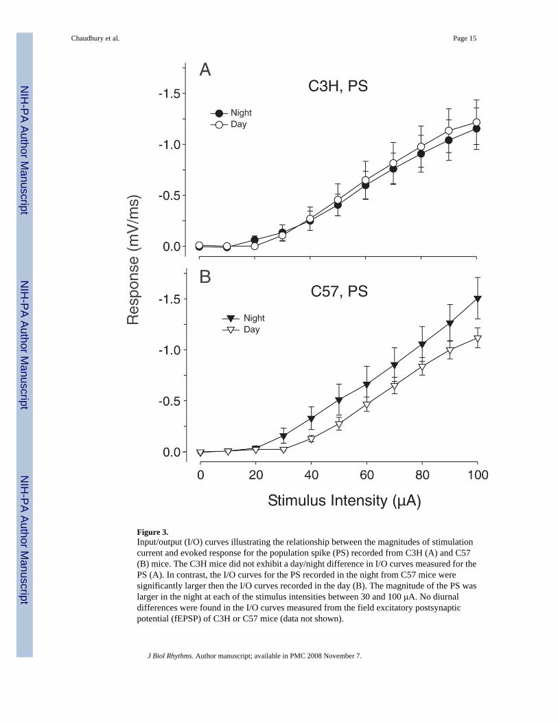
Figure 3.Input/output (I/O) curves illustrating the relationship between the magnitudes of stimulationcurrent and evoked response for the population spike (PS) recorded from C3H (A) and C57(B) mice. The C3H mice did not exhibit a day/night difference in I/O curves measured for thePS (A). In contrast, the I/O curves for the PS recorded in the night from C57 mice weresignificantly larger then the I/O curves recorded in the day (B). The magnitude of the PS waslarger in the night at each of the stimulus intensities between 30 and 100 μA. No diurnaldifferences were found in the I/O curves measured from the field excitatory postsynapticpotential (fEPSP) of C3H or C57 mice (data not shown).
Chaudhury et al. Page 15
J Biol Rhythms. Author manuscript; available in PMC 2008 November 7.
NIH
-PA Author Manuscript
NIH
-PA Author Manuscript
NIH
-PA Author Manuscript

Figure 4.Endogenous rhythms recorded from isolated hippocampal slices. (A) C3H mice were kept inan LD cycle and killed in the day (1 h prior to lights-off at ZT 11), and evoked responses wererecorded at night (from ZT 13–18). The population spike (PS) long-term potentiation (LTP)of slices prepared during the day but recorded during the night had the profile remarkablysimilar to the night group. Similar results were obtained with the field excitatory postsynapticpotential (fEPSP) recordings (data not shown). (B) Animals were maintained in DD, and theonset of activity was taken as CT 12. Plots of PS slope (normalized as a percentage of baseline)as a function of time were measured in the subjective day and subjective night. The PS LTP
Chaudhury et al. Page 16
J Biol Rhythms. Author manuscript; available in PMC 2008 November 7.
NIH
-PA Author Manuscript
NIH
-PA Author Manuscript
NIH
-PA Author Manuscript

was larger in the subjective night. For both sets of experiments, the tetanus of 1 ×100-Hzstimulation was given at time = 0.
Chaudhury et al. Page 17
J Biol Rhythms. Author manuscript; available in PMC 2008 November 7.
NIH
-PA Author Manuscript
NIH
-PA Author Manuscript
NIH
-PA Author Manuscript
NIH
-PA Author Manuscript
NIH
-PA Author Manuscript
NIH
-PA Author Manuscript
Chaudhury et al. Page 18
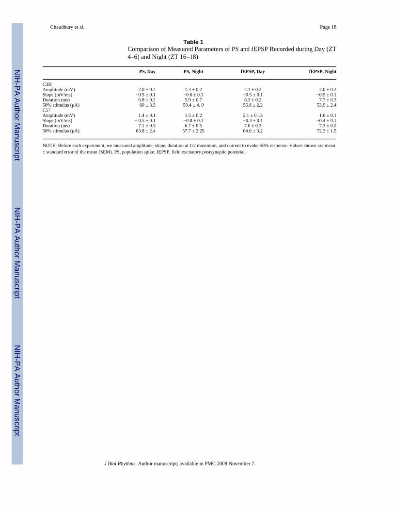
Table 1Comparison of Measured Parameters of PS and fEPSP Recorded during Day (ZT4–6) and Night (ZT 16–18)
PS, Day PS, Night fEPSP, Day fEPSP, Night
C3HAmplitude (mV) 2.0 ± 0.2 1.3 ± 0.2 2.1 ± 0.2 2.0 ± 0.2Slope (mV/ms) −0.5 ± 0.1 −0.6 ± 0.1 −0.5 ± 0.1 −0.5 ± 0.1Duration (ms) 6.8 ± 0.2 5.9 ± 0.7 8.3 ± 0.2 7.7 ± 0.350% stimulus (μA) 60 ± 3.5 59.4 ± 4. 0 56.8 ± 2.2 53.9 ± 2.4C57Amplitude (mV) 1.4 ± 0.1 1.5 ± 0.2 2.1 ± 0.13 1.6 ± 0.1Slope (mV/ms) − 0.5 ± 0.1 −0.8 ± 0.1 −0.3 ± 0.1 −0.4 ± 0.1Duration (ms) 7.1 ± 0.3 6.7 ± 0.5 7.0 ± 0.3 7.3 ± 0.250% stimulus (μA) 63.8 ± 2.4 57.7 ± 2.25 64.0 ± 3.2 72.3 ± 1.5
NOTE: Before each experiment, we measured amplitude, slope, duration at 1/2 maximum, and current to evoke 50% response. Values shown are mean± standard error of the mean (SEM). PS, population spike; fEPSP, field excitatory postsynaptic potential.
J Biol Rhythms. Author manuscript; available in PMC 2008 November 7.