Redox and Antioxidant Modulation of Circadian Rhythms - MDPI
14
molecules Article Redox and Antioxidant Modulation of Circadian Rhythms: Effects of Nitroxyl, N-Acetylcysteine and Glutathione Santiago Andrés Plano 1,† , Fernando Martín Baidanoff 2,† , Laura Lucía Trebucq 2 , Sebastián Ángel Suarez 3 , Fabio Doctorovich 3 , Diego Andrés Golombek 2, * and Juan José Chiesa 2 Citation: Plano, S.A.; Baidanoff, F.M.; Trebucq, L.L.; Suarez, S.Á.; Doctorovich, F.; Golombek, D.A.; Chiesa, J.J. Redox and Antioxidant Modulation of Circadian Rhythms: Effects of Nitroxyl, N-Acetylcysteine and Glutathione. Molecules 2021, 26, 2514. https://doi.org/10.3390/ molecules26092514 Academic Editors: Cristina Maccallini and Rosa Amoroso Received: 21 March 2021 Accepted: 23 April 2021 Published: 26 April 2021 Publisher’s Note: MDPI stays neutral with regard to jurisdictional claims in published maps and institutional affil- iations. Copyright: © 2021 by the authors. Licensee MDPI, Basel, Switzerland. This article is an open access article distributed under the terms and conditions of the Creative Commons Attribution (CC BY) license (https:// creativecommons.org/licenses/by/ 4.0/). 1 Institute for Biomedical Research (BIOMED), Catholic University of Argentina (UCA) and National Scientific and Technical Research Council (CONICET), C1107CABA Buenos Aires, Argentina; [email protected] 2 Laboratorio de Cronobiología, Departamento de Ciencia y Tecnología, Universidad Nacional de Quilmes (UNQ), Consejo Nacional de Investigaciones Científicas y Técnicas (CONICET), Roque Sáenz Peña 352, B1876BXD Bernal, Argentina; [email protected] (F.M.B.); [email protected] (L.L.T.); [email protected] (J.J.C.) 3 Departamento de Química Inorgánica, Analítica y Química Física/INQUIMAE-CONICET, Facultad de Ciencias Exactas y Naturales, Universidad de Buenos Aires, Ciudad Universitaria, Pab. II, C1428EHA Buenos Aires, Argentina; [email protected] (S.Á.S.); [email protected] (F.D.) * Correspondence: [email protected]; Tel.: +54-113657100 † These authors contributed equally to this work. Abstract: The circadian clock at the hypothalamic suprachiasmatic nucleus (SCN) entrains output rhythms to 24-h light cycles. To entrain by phase-advances, light signaling at the end of subjective night (circadian time 18, CT18) requires free radical nitric oxide (NO•) binding to soluble guanylate cyclase (sGC) heme group, activating the cyclic guanosine monophosphate (cGMP)-dependent pro- tein kinase (PKG). Phase-delays at CT14 seem to be independent of NO•, whose redox-related species were yet to be investigated. Here, the one-electron reduction of NO• nitroxyl was pharmacologi- cally delivered by Angeli’s salt (AS) donor to assess its modulation on phase-resetting of locomotor rhythms in hamsters. Intracerebroventricular AS generated nitroxyl at the SCN, promoting phase- delays at CT14, but potentiated light-induced phase-advances at CT18. Glutathione/glutathione disulfide (GSH/GSSG) couple measured in SCN homogenates showed higher values at CT14 (i.e., more reduced) than at CT18 (oxidized). In addition, administration of antioxidants N-acetylcysteine (NAC) and GSH induced delays per se at CT14 but did not affect light-induced advances at CT18. Thus, the relative of NO• nitroxyl generates phase-delays in a reductive SCN environment, while an oxidative favors photic-advances. These data suggest that circadian phase-locking mechanisms should include redox SCN environment, generating relatives of NO•, as well as coupling with the molecular oscillator. Keywords: suprachiasmatic nuclei; cGMP; Angeli’s salt; reactive nitrogen species 1. Introduction The mammalian circadian clock at the hypothalamic suprachiasmatic nuclei (SCN) orchestrates daily rhythms in a broad range of behavioral and physiological processes, driving autonomic and endocrine outputs which coordinate a network of oscillators in peripheral tissues [1]. This is achieved by the entrainment (i.e., phase-shifting) of the SCN oscillator activity to equal the 24-h light-dark cycle. Interacting transcriptional- translational feedback loops (TTFLs) of clock genes generate core oscillator activity at the SCN cells and most peripheral tissues. A transcriptional activator, formed by circadian locomotor output cycles kaput (CLOCK) and brain and muscle aryl hydrocarbon receptor nuclear translocator-like protein 1 (BMAL1) heterodimer, binds to enhancer elements at the promoter of repressor clock genes period (per) 1, 2, and 3, and cryptochrome (cry) 1 and 2, increasing their transcriptional activity. In turn, PERs and CRYs proteins accumulate in Molecules 2021, 26, 2514. https://doi.org/10.3390/molecules26092514 https://www.mdpi.com/journal/molecules
-
Upload
khangminh22 -
Category
Documents
-
view
1 -
download
0
Transcript of Redox and Antioxidant Modulation of Circadian Rhythms - MDPI

molecules
Article
Redox and Antioxidant Modulation of Circadian Rhythms:Effects of Nitroxyl, N-Acetylcysteine and Glutathione
Santiago Andrés Plano 1,† , Fernando Martín Baidanoff 2,†, Laura Lucía Trebucq 2, Sebastián Ángel Suarez 3 ,Fabio Doctorovich 3, Diego Andrés Golombek 2,* and Juan José Chiesa 2
�����������������
Citation: Plano, S.A.; Baidanoff, F.M.;
Trebucq, L.L.; Suarez, S.Á.;
Doctorovich, F.; Golombek, D.A.;
Chiesa, J.J. Redox and Antioxidant
Modulation of Circadian Rhythms:
Effects of Nitroxyl, N-Acetylcysteine
and Glutathione. Molecules 2021, 26,
2514. https://doi.org/10.3390/
molecules26092514
Academic Editors: Cristina Maccallini
and Rosa Amoroso
Received: 21 March 2021
Accepted: 23 April 2021
Published: 26 April 2021
Publisher’s Note: MDPI stays neutral
with regard to jurisdictional claims in
published maps and institutional affil-
iations.
Copyright: © 2021 by the authors.
Licensee MDPI, Basel, Switzerland.
This article is an open access article
distributed under the terms and
conditions of the Creative Commons
Attribution (CC BY) license (https://
creativecommons.org/licenses/by/
4.0/).
1 Institute for Biomedical Research (BIOMED), Catholic University of Argentina (UCA) and National Scientificand Technical Research Council (CONICET), C1107CABA Buenos Aires, Argentina;[email protected]
2 Laboratorio de Cronobiología, Departamento de Ciencia y Tecnología, Universidad Nacional de Quilmes (UNQ),Consejo Nacional de Investigaciones Científicas y Técnicas (CONICET), Roque Sáenz Peña 352,B1876BXD Bernal, Argentina; [email protected] (F.M.B.); [email protected] (L.L.T.);[email protected] (J.J.C.)
3 Departamento de Química Inorgánica, Analítica y Química Física/INQUIMAE-CONICET,Facultad de Ciencias Exactas y Naturales, Universidad de Buenos Aires, Ciudad Universitaria, Pab. II,C1428EHA Buenos Aires, Argentina; [email protected] (S.Á.S.); [email protected] (F.D.)
* Correspondence: [email protected]; Tel.: +54-113657100† These authors contributed equally to this work.
Abstract: The circadian clock at the hypothalamic suprachiasmatic nucleus (SCN) entrains outputrhythms to 24-h light cycles. To entrain by phase-advances, light signaling at the end of subjectivenight (circadian time 18, CT18) requires free radical nitric oxide (NO•) binding to soluble guanylatecyclase (sGC) heme group, activating the cyclic guanosine monophosphate (cGMP)-dependent pro-tein kinase (PKG). Phase-delays at CT14 seem to be independent of NO•, whose redox-related specieswere yet to be investigated. Here, the one-electron reduction of NO• nitroxyl was pharmacologi-cally delivered by Angeli’s salt (AS) donor to assess its modulation on phase-resetting of locomotorrhythms in hamsters. Intracerebroventricular AS generated nitroxyl at the SCN, promoting phase-delays at CT14, but potentiated light-induced phase-advances at CT18. Glutathione/glutathionedisulfide (GSH/GSSG) couple measured in SCN homogenates showed higher values at CT14 (i.e.,more reduced) than at CT18 (oxidized). In addition, administration of antioxidants N-acetylcysteine(NAC) and GSH induced delays per se at CT14 but did not affect light-induced advances at CT18.Thus, the relative of NO• nitroxyl generates phase-delays in a reductive SCN environment, whilean oxidative favors photic-advances. These data suggest that circadian phase-locking mechanismsshould include redox SCN environment, generating relatives of NO•, as well as coupling with themolecular oscillator.
Keywords: suprachiasmatic nuclei; cGMP; Angeli’s salt; reactive nitrogen species
1. Introduction
The mammalian circadian clock at the hypothalamic suprachiasmatic nuclei (SCN)orchestrates daily rhythms in a broad range of behavioral and physiological processes,driving autonomic and endocrine outputs which coordinate a network of oscillators inperipheral tissues [1]. This is achieved by the entrainment (i.e., phase-shifting) of theSCN oscillator activity to equal the 24-h light-dark cycle. Interacting transcriptional-translational feedback loops (TTFLs) of clock genes generate core oscillator activity at theSCN cells and most peripheral tissues. A transcriptional activator, formed by circadianlocomotor output cycles kaput (CLOCK) and brain and muscle aryl hydrocarbon receptornuclear translocator-like protein 1 (BMAL1) heterodimer, binds to enhancer elements atthe promoter of repressor clock genes period (per) 1, 2, and 3, and cryptochrome (cry) 1 and 2,increasing their transcriptional activity. In turn, PERs and CRYs proteins accumulate in
Molecules 2021, 26, 2514. https://doi.org/10.3390/molecules26092514 https://www.mdpi.com/journal/molecules

Molecules 2021, 26, 2514 2 of 14
the cytoplasm and heterodimerize, generating transcriptional repressors by interferingwith the CLOCK:BMAL heterodimers, closing the cycle each 24 h. Entrainment of the SCNclock to the light-dark cycles involves the communication of photic information throughseveral neurotransmitters and signal transduction pathways, whose endpoint effectorsconverge to modify the activity of clock genes at the transcriptional level [2]. Moreover, theTTFL circadian clockwork might be modulated by the redox state. Indeed, several redoxpairs have been shown to interact with either circadian clock proteins or transcriptionalregulators in vitro [3,4]. The SCN, NADP+/NADPH, FAD+/FADH, and dehydroascorbicacid/ascorbic acid redox couples, as well as the global protein glutathiolation, exhibitcircadian oscillations, while the resting electrical activity of SCN neurons is also sensitiveto oxidants and reductants [5].
Brief light stimulation during the circadian night (i.e., the time of behavioral ac-tivity in nocturnal rodents) generates a phase-shift of the SCN clock, and thereby itsbehavioral and physiological rhythmic outputs [6]. The specific circadian time (CT) atwhich the light pulse is applied is determinant of whether phase-delays or advancesare induced. At CT14 (i.e., 2 h after locomotor activity onset, CT12), a phase-delay oflocomotor rhythm is generated, while phase-advances occur after stimulation at the latecircadian night at CT18. Phase-shifts are transduced by the glutamatergic influx throughefferents from the retinal ganglion cells to ventral SCN neurons, which promotes cal-cium influx through glutamate-N-methyl-D-aspartate receptors (NMDAr) and triggerscalcium-dependent calmodulin-kinase phosphorylation of the nitric oxide synthase (nNOS),increasing its activity [7]. Downstream, the pathway for advances proceeds throughNO• coordination of regulatory ferrous heme on the soluble guanylyl cyclase (sGC), in-creasing cGMP synthesis and, subsequently, the cGMP-dependent protein kinase (PKGII)activity [8] to phosphorylate its dependent substrate [9]. At CT14, circadian phase-delaysproceed through other pathways (e.g., adenylate cyclase/cyclic adenosine monophosphate(cAMP)/cAMP-activated protein kinase (PKA)) [2] activated by calcium influx through theaperture of endoplasmic ryanodine type 2 receptor channels [10]. Downstream, both path-ways converge at cAMP-responding element-binding protein (CREB) phosphorylation [11]to activate CRE-mediated period 1–2 transcription [12,13], thereby setting the phase ofthe clock.
Various drugs can modulate circadian entrainment targeting NO• transduction, in-cluding organic nitroso derivatives such as S-Nitroso-N-acetylpenicillamine (SNAP, anS-nitrosothiol), or N-nitrosomelatonin (NOMel), both acting as NO• donors [14,15]. WhileNO• generation by NOMel at the SCN only potentiates photic phase-advances—anddoesn’t delay even at high doses [15]—administration of SNAP potentiates both photicdelays and advances [14]. At high doses SNAP can work as an NO• donor, increasingcGMP, but low doses generate S-nitrosation with opposite effects in cardiomiocytes [16]. Onthe other hand, the nNOS antagonist N-nitro-L-arginine methyl ester L-NAME attenuatesboth phase-advances and phase-delays of locomotor activity rhythms in hamsters [14,17].Extracellular scavenging of NO• with 2-Phenyl-4,4,5,5-tetramethylimidazoline-1-oxyl3-oxide (PTIO) blocks photic phase-advances, but not delays, which demonstrates its roleas a paracrine messenger, by decreasing ventral-dorsal communication of light message atthe SCN [18].
A hypothesis for circadian pathway bifurcation downstream of NO• [19] states thatthe photic activation of sGC hemoprotein occurs with diffusible, long-range (i.e., far fromits nNOS source) NO• concentrations [20,21], which, despite being a highly diffusible,membrane-permeable gaseous messenger, participates in an array of redox-dependent,secondary reactive nitrogen species, as S-nitrosothiols (i.e., S-nitrosated glutathione, GSNO),nitroxyl anion (NO−), and metalnitrosyl complexes [22]. In addition, redox forms ofNO• may act in so-called redox-dependent signaling to form S-nitrosation (i.e., the additionof a nitroso group by condensation of a thiol and the one-electron oxidation of NO•, nitrousacid) [20,23]. The one-electron reduced (and protonated at physiological pH) relative ofNO• nitroxyl (HNO, azanone, nitrosylhydride, among other names) can be generated

Molecules 2021, 26, 2514 3 of 14
by a number of biological processes: HNO is a by-product of S-nitrosothiols reactionwith cysteine thiols [24] and is even produced by nNOS [25]. Pharmacologically, it canbe generated by the decomposition of sodium trioxodinitrate (Na2[N2O3] Angeli’s salt,AS) together with sodium nitrite at physiological conditions [26]. HNO is widely studiedin the cardiovascular system [27], and can be easily oxidized to NO• at physiologicalconditions (but not the reciprocal reduction of NO•) [28]. However, NO• and nitroxylexhibit differential reactivity toward metalloproteins, as well as different thiophilicitydepending on redox intermediates, which might explain their distinctive physiologicalroles [28]. Moreover, while NO• increases cGMP through sGC, HNO activates a calcitoningene-related peptide [29], increasing cAMP, and also modulates calcium channels andryanodine receptors [30]. Thus, HNO is proposed as a signaling molecule with moredistinctive effects than NO• and is thus worthy of continued study regarding its role incircadian entrainment.
In this context, we investigated the role of NO• relative nitroxyl in the circadian clockentrainment under pharmacological administration of Angeli’s Salt, a diazeniumdiolate-based nitroxyl donor. In addition, the antioxidants N-acetylcysteine (NAC) and glutathione(GSH) were used to test whether the pharmacological modulation of the SCN-redox envi-ronment can affect circadian entrainment. In this way, we provide evidence of the role ofputative redox forms of NO•, as well as the redox modulation of SCN, driving this keycircadian function.
2. Results2.1. Effect of the Nitroxyl Donor Angeli’s Salt on Phase-Shifts of Locomotor Rhythms
Phase-shifting experiments under DD conditions were performed to assess the effectof Angeli’s Salt (AS) administration on photic entrainment. AS, a well-known nitroxyldonor, was delivered i.c.v. (1 µL, 100 µM) 5 min before a subsaturant LP at CT14 (to inducedelays) or CT18 (for advances). We found a circadian-dependent effect of AS over circadianphase-shifting. At CT14, this nitroxyl donor induced a phase-delay when administeredalone in the dark, as compared with the vehicle (AS dark: 66.8 min ± 26.3 min, n = 5; Vehicledark: 22.0 min ± 13.7 min, n = 9; p < 0.05 Tukey’s multiple comparisons test) (Figure 1A).AS did not affect light-induced phase-delays at CT14 (AS LP: 101.1 min ± 38.4 min, n = 7;Vehicle LP: 97.4 min ± 28.2 min, n = 5, respectively; p = ns Tukey’s multiple comparisonstest) (Two-way ANOVA; LP p < 0.0001, F = 24.82, DFn = 1, DFd = 22; Drug p > 0.05, F = 4.85;interaction p = ns, F = 3.47) (Figure 1A). On the other hand, at CT18 AS administrationpotentiated the effect of light, as it significantly increased phase-advances as comparedwith the vehicle (AS LP: 251.7 min ± 75.9 min, n = 4; Vehicle LP: 116.2 min ± 53.2 min, n = 4,respectively; p > 0.001 Tukey’s multiple comparisons test) (Figure 1B), but did not generateany effect on the circadian phase when delivered alone (AS Dark: 0.3 min ± 8.1 min, n = 4;Vehicle Dark: 4.7 min ± 34.9 min, n = 8, respectively; p = ns Tukey’s multiple comparisonstest) (Two-way ANOVA; LP p < 0.0001, F = 73.55, DFn = 1, DFd = 16; Drug p > 0.01, F = 9.03;interaction p < 0.01, F = 10.46). These results indicate that AS modulates light-inducedphase-shifts at CT18 but has no effect on light signaling at CT14 (i.e., AS modulates light-induced phase-advances but not light-induced phase-delays). On the other hand, ASadministration at CT14, without LP, has a photic-like effect—inducing phase-delays of thecircadian rhythm.
Molecules 2021, 26, 2514 4 of 14Molecules 2021, 26, x FOR PEER REVIEW 4 of 14
Figure 1. Effect of the nitroxyl donor AS on phase changes induced by light. Hamsters in constant darkness conditions, treated with an i.c.v injection of vehicle or 100 μM AS, 5 min before a sub-saturant light pulse (LP, 10 min, 50 lux) at two CTs: (A) CT14 and (B) CT18. Control animals (Dark) received only pharmacological treatment without the light pulse. Phase changes are shown as mean ± SEM. Two-way ANOVA, followed by Tukey’s multiple comparisons test, ns = not sig-nificant; * p < 0.05; *** p < 0.001 **** p < 0.0001. CT14 Dark: Veh n = 9, AS n = 5; CT14 LP: AS + LP n = 7, Veh + PL; CT18 Dark: Veh n = 4, AS n = 7; CT18 LP: Veh + LP and AS + LP n = 5. (C–E) are repre-sentative actograms showing: (C) the effect of a light pulse at CT14, inducing a phase-delay in the circadian locomotor rhythms; (D) the phase of an animal without light stimulation during the sub-jective night; and (E) the effect of light stimulation at CT18, which induces a phase-advance. The grey circle indicates the day and time of the LP.
Figure 1. Effect of the nitroxyl donor AS on phase changes induced by light. Hamsters in constantdarkness conditions, treated with an i.c.v injection of vehicle or 100 µM AS, 5 min before a subsaturantlight pulse (LP, 10 min, 50 lux) at two CTs: (A) CT14 and (B) CT18. Control animals (Dark) receivedonly pharmacological treatment without the light pulse. Phase changes are shown as mean ± SEM.Two-way ANOVA, followed by Tukey’s multiple comparisons test, ns = not significant; * p < 0.05;*** p < 0.001; **** p < 0.0001. CT14 Dark: Veh n = 9, AS n = 5; CT14 LP: AS + LP n = 7, Veh + PL; CT18Dark: Veh n = 4, AS n = 7; CT18 LP: Veh + LP and AS + LP n = 5. (C–E) are representative actogramsshowing: (C) the effect of a light pulse at CT14, inducing a phase-delay in the circadian locomotorrhythms; (D) the phase of an animal without light stimulation during the subjective night; and (E)the effect of light stimulation at CT18, which induces a phase-advance. The grey circle indicates theday and time of the LP.

Molecules 2021, 26, 2514 5 of 14
2.2. Electrochemical Detection of Nitroxyl (HNO) In Vivo at the SCN
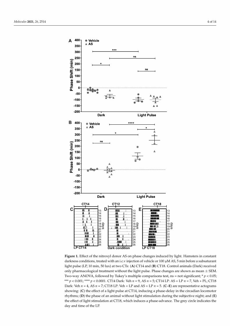
Decomposition of AS is known to produce both HNO and NO• [31,32]. To verify ifHNO was effectively generated at physiological conditions, time-resolved electrochemicalquantification was performed in vivo at the SCN in anesthetized hamsters. Raw data seriesshowed changes in current intensity measured in a representative individual for detectingHNO concentration at the SCN. Smoothed data allowed paired measures at baseline(vehicle-treated) and after AS microinjection in the same individual (Figure 2A). Differencesin current intensity were calculated as the difference between maximum current values afterAS treatment and baseline values. Current intensity (µAmperes, µA) showed increasedvalues after both 400 pmol and 800 pmol doses. A significant difference in this currentincrement was found regarding AS concentration (Figure 2B), showing higher incrementfor 800 pmol (0.111 µA ± 0.022 µA) as compared with 400 pmol (0.039 µA ± 0.033 µA)(p < 0.05 paired Student’s t-test; t = 5.14; df = 3), indicating HNO generation by AS in adose-dependent manner. Using a similar concentration order (400 and 800 pmol) as thatused for phase/shifting experiments (100 pmol) these results allowed us to verify thatHNO is pharmacologically generated by decomposition of AS after i.c.v. delivery of thedrug in the SCN.
2.3. Measurement of the Redox Pair GSH–GSSG at the SCN
Since HNO generated by AS may be favored under a reductive environment [33],the GSH/GSSG couple was measured at CT14 and CT18 in SCN tissue homogenatesas a reliable measure of the cellular redox state [34]. We found a significant differencein total GSH between CT14 and CT18 (10.29 µm/(mg/mL) ± 3.99 µm/(mg/mL) vs.16.42 µm/(mg/mL) ± 6.89 µm/(mg/mL), respectively) (p < 0.05 paired Student’s t-test;t = 2.43; df = 18; n = 6). When GSH was analyzed, we found no significant differences (CT14:8.73 µm/(mg/mL) ± 3.30 µm/(mg/mL) vs. CT18: 9.98 µm/(mg/mL) ± 3.91 µm/(mg/mL)).Nevertheless, GSSG was significantly higher at CT18 when compared with CT14 (CT14:0.13 µm/(mg/mL) ± 0.11 µm/(mg/mL) vs. CT18: 0.58 µm/(mg/mL) ± 0.31 µm/(mg/mL))(p < 0.01 paired t test; t = 3.99; df = 15; n = 6), indicating a more oxidized state at CT18 inthe SCN. This was confirmed when analyzing the GSH/GSSG ratio, resulting in signifi-cantly lower values for CT18 (CT14: 104.9 µm/(mg/mL) ± 42.38 µm/(mg/mL) vs. CT18:11.62 µm/(mg/mL) ± 3.37 µm/(mg/mL)) (p < 0.001 paired t-test; t = 5.38; df = 10; n = 6)(Figure 3). These results allowed us to hypothesize that pharmacological generation ofHNO could be favored under a reductive environment at CT14.
2.4. Effect of NAC and GSH Administration on Light-Induced Phase-Shifts
To test the effect of changing the redox environment on photic entrainment, we per-formed a set of experiments that used acute i.c.v. treatment with NAC or GSH, pairedwith a subsaturant LP. When compared with vehicles, the photically-induced shifts werenot affected by NAC, both at CT14 or CT18 (Figure 4A,B). Also, NAC delayed the phaseof locomotor rhythm at CT14 in a magnitude that was significantly different from thevehicle (Vehicle Dark: −23.71 min ± 19.54 min; NAC Dark: −104 min ± 49.7 min) butdid not affect light-induced phase-delays (Vehicle LP: −157.4 min ± 61.22 min; NAC LP:−85.63 min ± 37.29 min) (Two-way ANOVA; LP p < 0.001, F = 21.17, DFn = 1, DFd = 25;Drug p = ns, F = 0.06; interaction p < 0.001, F = 21.17). NAC had no effect over circa-dian synchronization at CT18 by itself (Vehicle Dark: −0.33 min ± 9.16 min; NAC Dark:−12 min ± 30.87 min) or with a concomitant light pulse (Vehicle LP: 102.6 min ± 33.83 min;NAC LP: 60 min ± 36.14 min) (Two-way ANOVA; LP p < 0.0001, F = 62.23, DFn = 1,DFd = 27; Drug p < 0.05, F = 5.99; interaction p = ns, F = 1.95). A similar result was foundfor GSH, which induced phase-delays at CT14 (Vehicle Dark: −12.17 min ± 6.67 min;GSH Dark: −70.8 min ± 23.38 min) and did not change the effect of light pulses (VehicleLP: −81.29 min ± 23.94 min; GSH LP: −55 min ± 15.186 min) (Two-way ANOVA; LPp < 0.01, F = 30.47, DFn = 1, DFd = 20; Drug p < 0.05, F = 12.01; interaction p < 0.0001,F = 30.47) (Figure 4C). Also, GSH had no effect at CT18 either by itself (Vehicle Dark:

Molecules 2021, 26, 2514 6 of 14
6.45 min ± 26.77 min; GSH Dark: −7 min ± 13.96 min) or interacting with light (Vehi-cle LP: 82.72 min ± 62.37 min; GSH LP: 63.43 min ± 34.64 min) (Two-way ANOVA; LPp < 0.0001, F = 25.42, DFn = 1, DFd = 40; Drug p = ns, F = 1.27; interaction p = ns, F = 1.27)(Figure 4D). These results show that antioxidants NAC and GSH are able to modulate thecircadian phase at CT14.
Molecules 2021, 26, x FOR PEER REVIEW 5 of 14
2.2. Electrochemical Detection of Nitroxyl (HNO) In Vivo at the SCN Decomposition of AS is known to produce both HNO and NO• [31,32]. To verify if
HNO was effectively generated at physiological conditions, time-resolved electrochemi-cal quantification was performed in vivo at the SCN in anesthetized hamsters. Raw data series showed changes in current intensity measured in a representative individual for detecting HNO concentration at the SCN. Smoothed data allowed paired measures at baseline (vehicle-treated) and after AS microinjection in the same individual (Figure 2A). Differences in current intensity were calculated as the difference between maximum cur-rent values after AS treatment and baseline values. Current intensity (μAmperes, μA) showed increased values after both 400 pmol and 800 pmol doses. A significant difference in this current increment was found regarding AS concentration (Figure 2B), showing higher increment for 800 pmol (0.111 μA ± 0.022 μA) as compared with 400 pmol (0.039 μA ± 0.033 μA) (p < 0.05 paired Student’s t-test; t = 5.14; df = 3), indicating HNO generation by AS in a dose-dependent manner. Using a similar concentration order (400 and 800 pmol) as that used for phase/shifting experiments (100 pmol) these results allowed us to verify that HNO is pharmacologically generated by decomposition of AS after i.c.v. deliv-ery of the drug in the SCN.
Figure 2. Electrochemical detection of nitroxyl (HNO) in vivo at the SCN. HNO generated in vivo by decomposition of Angeli’s Salt (AS) donor detected by time-resolved electrochemical quantifi-cation at the SCN in the pmolar range. (A) Representative graph of individual raw (Raw) and fil-tered (Smooth) data series using a moving average with a step of 300 samples to obtain 1 min-bin. After at least 2 min of baseline current register (white boxes), AS was delivered i.c.v from a 100
Figure 2. Electrochemical detection of nitroxyl (HNO) in vivo at the SCN. HNO generatedin vivo by decomposition of Angeli’s Salt (AS) donor detected by time-resolved electro-chemical quantification at the SCN in the pmolar range. (A) Representative graph ofindividual raw (Raw) and filtered (Smooth) data series using a moving average with astep of 300 samples to obtain 1 min-bin. After at least 2 min of baseline current register(white boxes), AS was delivered i.c.v from a 100 µM solution at 1.5 µL/min rate (greyboxes). Filled arrows indicate the time of injection with 400 pmol and 800 pmol of ASwhich corresponds with the registration of the baseline current, while dotted arrowsindicate maximum current, which correlates to maximum HNO production. (B) Deltacurrent values, calculated as the difference between maximum post-injection and basalprior-injection current values, for 400 pmol and 800 pmol AS. n = 4; * p < 0.05, paired t-test.

Molecules 2021, 26, 2514 7 of 14
Molecules 2021, 26, x FOR PEER REVIEW 6 of 14
μM solution at 1.5 μL/min rate (grey boxes). Filled arrows indicate the time of injection with 400 pmol and 800 pmol of AS which corresponds with the registration of the baseline current, while dotted arrows indicate maximum current, which correlates to maximum HNO production. (B) Delta current values, calculated as the difference between maximum post-injection and basal prior-injection current values, for 400 pmol and 800 pmol AS. n = 4; * p < 0.05, paired t-test.
2.3. Measurement of the Redox Pair GSH–GSSG at the SCN Since HNO generated by AS may be favored under a reductive environment [33], the
GSH/GSSG couple was measured at CT14 and CT18 in SCN tissue homogenates as a reli-able measure of the cellular redox state [34]. We found a significant difference in total GSH between CT14 and CT18 (10.29 μm/(mg/mL) ± 3.99 μm/(mg/mL) vs. 16.42 μm/(mg/mL) ± 6.89 μm/(mg/mL), respectively) (p < 0.05 paired Student’s t-test; t = 2.43; df = 18; n = 6). When GSH was analyzed, we found no significant differences (CT14: 8.73 μm/(mg/mL) ± 3.30 μm/(mg/mL) vs. CT18: 9.98 μm/(mg/mL) ± 3.91 μm/(mg/mL)). Nevertheless, GSSG was significantly higher at CT18 when compared with CT14 (CT14: 0.13 μm/(mg/mL) ± 0.11 μm/(mg/mL) vs. CT18: 0.58 μm/(mg/mL) ± 0.31 μm/(mg/mL)) (p < 0.01 paired t test; t = 3.99; df = 15; n = 6), indicating a more oxidized state at CT18 in the SCN. This was con-firmed when analyzing the GSH/GSSG ratio, resulting in significantly lower values for CT18 (CT14: 104.9 μm/(mg/mL) ± 42.38 μm/(mg/mL) vs. CT18: 11.62 μm/(mg/mL) ± 3.37 μm/(mg/mL)) (p < 0.001 paired t-test; t = 5.38; df = 10; n = 6) (Figure 3). These results al-lowed us to hypothesize that pharmacological generation of HNO could be favored under a reductive environment at CT14.
Figure 3. Glutathione couple measurement in the SCN at subjective night. The redox state was assessed as the GSH/GSSG ratio. Brains were collected at CT14 and CT18 and samples were frozen at −80 °C. SCNs from 2–3 individuals were pooled for measuring reduced (GSH) and total gluta-thione, inferring oxidized glutathione (GSSG) values using the equation GSSG = (total glutathi-one—GSH)/2. The rate relation was also calculated as GSH/GSSG, thus indicating a more oxidized environment when the relation was lower. Significantly lower values for CT18 were found, indi-cating a more oxidized environment at late subjective night (CT14: 104.9 μm/(mg/mL) ± 42.38 μm/(mg/mL) vs. CT18: 11.62 μm/(mg/mL) ± 3.37μm/(mg/mL)) (*** p < 0.001 paired t-test; t = 5.38; df = 10; n = 6).
2.4. Effect of NAC and GSH Administration on Light-Induced Phase-Shifts To test the effect of changing the redox environment on photic entrainment, we per-
formed a set of experiments that used acute i.c.v. treatment with NAC or GSH, paired with a subsaturant LP. When compared with vehicles, the photically-induced shifts were
Figure 3. Glutathione couple measurement in the SCN at subjective night. The redox state wasassessed as the GSH/GSSG ratio. Brains were collected at CT14 and CT18 and samples were frozen at−80 ◦C. SCNs from 2–3 individuals were pooled for measuring reduced (GSH) and total glutathione,inferring oxidized glutathione (GSSG) values using the equation GSSG = (total glutathione—GSH)/2.The rate relation was also calculated as GSH/GSSG, thus indicating a more oxidized environmentwhen the relation was lower. Significantly lower values for CT18 were found, indicating a moreoxidized environment at late subjective night (CT14: 104.9 µm/(mg/mL) ± 42.38 µm/(mg/mL) vs.CT18: 11.62 µm/(mg/mL) ± 3.37µm/(mg/mL)) (*** p < 0.001 paired t-test; t = 5.38; df = 10; n = 6).
Molecules 2021, 26, x FOR PEER REVIEW 7 of 14
not affected by NAC, both at CT14 or CT18 (Figure 4A,B). Also, NAC delayed the phase of locomotor rhythm at CT14 in a magnitude that was significantly different from the ve-hicle (Vehicle Dark: −23.71 min ± 19.54 min; NAC Dark: −104 min ± 49.7 min) but did not affect light-induced phase-delays (Vehicle LP: −157.4 min ± 61.22 min; NAC LP: −85.63 min ± 37.29 min) (Two-way ANOVA; LP p < 0.001, F = 21.17, DFn = 1, DFd = 25; Drug p = ns, F = 0.06; interaction p < 0.001, F = 21.17). NAC had no effect over circadian synchroni-zation at CT18 by itself (Vehicle Dark: −0.33 min ± 9.16 min; NAC Dark: −12 min ± 30.87 min) or with a concomitant light pulse (Vehicle LP: 102.6 min ± 33.83 min; NAC LP: 60 min ± 36.14 min) (Two-way ANOVA; LP p < 0.0001, F = 62.23, DFn = 1, DFd = 27; Drug p < 0.05, F = 5.99; interaction p = ns, F = 1.95). A similar result was found for GSH, which induced phase-delays at CT14 (Vehicle Dark: −12.17 min ± 6.67 min; GSH Dark: −70.8 min ± 23.38 min) and did not change the effect of light pulses (Vehicle LP: −81.29 min ± 23.94 min; GSH LP: −55 min ± 15.186 min) (Two-way ANOVA; LP p < 0.01, F = 30.47, DFn = 1, DFd = 20; Drug p < 0.05, F = 12.01; interaction p < 0.0001, F = 30.47) (Figure 4C). Also, GSH had no effect at CT18 either by itself (Vehicle Dark: 6.45 min ± 26.77 min; GSH Dark: −7 min ± 13.96 min) or interacting with light (Vehicle LP: 82.72 min ± 62.37 min; GSH LP: 63.43 min ± 34.64 min) (Two-way ANOVA; LP p < 0.0001, F = 25.42, DFn = 1, DFd = 40; Drug p = ns, F = 1.27; interaction p = ns, F = 1.27) (Figure 4D). These results show that antioxidants NAC and GSH are able to modulate the circadian phase at CT14.
Figure 4. Effect of antioxidants N-acetylcysteine (NAC) and glutathione (GSH) on circadian phase changes. Groups of hamsters in constant darkness conditions were treated with an i.c.v injection of either NAC (2 μL, 100 μM, or vehicle) (A,B) or GSH (1 μL, 100 μM) (C,D), 5 min before a subsatu-rant light pulse (10 min, 50 lux) delivered at CT14 or CT18. Controls for photic response (Dark) received only pharmacological treatment without light stimuli. Phase changes are shown as mean ± SEM. Two-way ANOVA, followed by Tukey’s multiple comparisons test, ns = not significant; * p
Figure 4. Cont.

Molecules 2021, 26, 2514 8 of 14
Molecules 2021, 26, x FOR PEER REVIEW 7 of 14
not affected by NAC, both at CT14 or CT18 (Figure 4A,B). Also, NAC delayed the phase of locomotor rhythm at CT14 in a magnitude that was significantly different from the ve-hicle (Vehicle Dark: −23.71 min ± 19.54 min; NAC Dark: −104 min ± 49.7 min) but did not affect light-induced phase-delays (Vehicle LP: −157.4 min ± 61.22 min; NAC LP: −85.63 min ± 37.29 min) (Two-way ANOVA; LP p < 0.001, F = 21.17, DFn = 1, DFd = 25; Drug p = ns, F = 0.06; interaction p < 0.001, F = 21.17). NAC had no effect over circadian synchroni-zation at CT18 by itself (Vehicle Dark: −0.33 min ± 9.16 min; NAC Dark: −12 min ± 30.87 min) or with a concomitant light pulse (Vehicle LP: 102.6 min ± 33.83 min; NAC LP: 60 min ± 36.14 min) (Two-way ANOVA; LP p < 0.0001, F = 62.23, DFn = 1, DFd = 27; Drug p < 0.05, F = 5.99; interaction p = ns, F = 1.95). A similar result was found for GSH, which induced phase-delays at CT14 (Vehicle Dark: −12.17 min ± 6.67 min; GSH Dark: −70.8 min ± 23.38 min) and did not change the effect of light pulses (Vehicle LP: −81.29 min ± 23.94 min; GSH LP: −55 min ± 15.186 min) (Two-way ANOVA; LP p < 0.01, F = 30.47, DFn = 1, DFd = 20; Drug p < 0.05, F = 12.01; interaction p < 0.0001, F = 30.47) (Figure 4C). Also, GSH had no effect at CT18 either by itself (Vehicle Dark: 6.45 min ± 26.77 min; GSH Dark: −7 min ± 13.96 min) or interacting with light (Vehicle LP: 82.72 min ± 62.37 min; GSH LP: 63.43 min ± 34.64 min) (Two-way ANOVA; LP p < 0.0001, F = 25.42, DFn = 1, DFd = 40; Drug p = ns, F = 1.27; interaction p = ns, F = 1.27) (Figure 4D). These results show that antioxidants NAC and GSH are able to modulate the circadian phase at CT14.
Figure 4. Effect of antioxidants N-acetylcysteine (NAC) and glutathione (GSH) on circadian phase changes. Groups of hamsters in constant darkness conditions were treated with an i.c.v injection of either NAC (2 μL, 100 μM, or vehicle) (A,B) or GSH (1 μL, 100 μM) (C,D), 5 min before a subsatu-rant light pulse (10 min, 50 lux) delivered at CT14 or CT18. Controls for photic response (Dark) received only pharmacological treatment without light stimuli. Phase changes are shown as mean ± SEM. Two-way ANOVA, followed by Tukey’s multiple comparisons test, ns = not significant; * p
Figure 4. Effect of antioxidants N-acetylcysteine (NAC) and glutathione (GSH) on circadian phase changes. Groups ofhamsters in constant darkness conditions were treated with an i.c.v injection of either NAC (2 µL, 100 µM, or vehicle)(A,B) or GSH (1 µL, 100 µM) (C,D), 5 min before a subsaturant light pulse (10 min, 50 lux) delivered at CT14 or CT18.Controls for photic response (Dark) received only pharmacological treatment without light stimuli. Phase changes areshown as mean ± SEM. Two-way ANOVA, followed by Tukey’s multiple comparisons test, ns = not significant; * p < 0.05;*** p < 0.001; **** p < 0.0001; for NAC experiment at CT14: vehicle Dark n = 7, vehicle LP n = 8, NAC Dark n = 7, NAC LPn = 8; at CT18: vehicle Dark n = 6, vehicle LP n = 8, NAC Dark n = 10, NAC LP n = 7. For GSH experiment at CT14: vehicleDark n = 6, vehicle LP n = 7, GSH Dark n = 5, GSH LP n = 6; at CT18: vehicle Dark n = 11, vehicle LP n = 17, GSH Dark n = 8,GSH LP n = 7).
3. Discussion
Evidence pointing to NO• as a diffusible, intra and extracellular messenger participat-ing in the photic transduction of circadian phase-advances is based on: (1) the increasedactivity in the nNOs/sGC/GMP/PKGII pathway after a light-pulse at CT18 [7,35–37]; (2)the indirect pharmacological reduction of phase-advances at CT18 by nNOS [14,15,17], sGC,and PKGII inhibition [35]; (3) scavenging of extracellular NO• [18]; and (4) enhancement ofphotic-advances by NO• donors as SNAP [14] and N-nitrosomelatonin [15]. Taken together,the current evidence indicates that a substantial increase in intra and extracellular NO• byits light-induced synthesis at the SCN should be expected at CT18 for phase-advances, ascompared with phase-delays.
Here we assessed the role of HNO, a redox form of NO•, in its ability to modulatephotic entrainment in the hamster SCN. This compound had two pharmacological effects:it generated phase-delays when delivered alone, and potentiated photic phase-advances.HNO was electrochemically measured in a dose-dependent relationship in the hamsterSCN after delivering the AS donor, verifying its ability to increase HNO in vivo. Themeasurement of the GSH/GSSG ratio estimates SCN, GSH, and GSSG concentrations ina 1–10 mM range [38]. We found a reductive environment at CT14, which may favor theaction of the HNO donor AS to generate phase-delays. Rate constants point that HNO oxi-dation is rather specific for thiols as reductants, while minor for nitrogenous species such asamines, indoles, and purines [29]. HNO oxidation should be determined by the availabilityof major cellular free thiols, such as glutathione, under reductive environment [20], whilean oxidative environment would induce oxidation to NO• and auto-dimerization and/orreaction with oxygen to form nitrous oxide, nitrate, nitrite [28,39], and peroxynitrite [40].Moreover, physiological detection of NO• by nNOS synthesis is dependent on oxidationof HNO (a primary product of nNOS) to NO•, catalyzed by the increase in superoxidedismutase activity under an oxidative environment [41], although this finding I challengedby [42]. Also, HNO can directly increase sGC enzymatic activity per se as found in purifiedlung extracts [43].

Molecules 2021, 26, 2514 9 of 14
Biological actions of HNO are largely explained due to its reactivity to free thiols(i.e., GSH) and cysteine residues. Most studies assessed its effects in the cardiovascularsystem, associated with the oxidation of free cysteine thiol residues [30], as well as withsarcoplasmic RyRs, increasing calcium release [44]. A few studies were done in the centralnervous system, comparing the effects of AS with the NO• donor diethylammonium(Z)-1-(N,N-diethylamino)diazen-1-ium-1,2-diolate (DEA/NO) in the hamster retina [45].Compared with DEA/NO, AS generates similar inhibition of nNOS activity and L-citrullinetransport and reduction of GSH and protein S-nitrosation, but less cGMP induction. High(400 nM), chronic (7 days) AS, administered intranigrostriatally as a model of neurode-generation, induces GSH depletion and dopaminergic neuronal loss [46]. The relativelyhigh GSH concentrations we measured at CT14 (1–10 mM) is not expected to change afterreacting with low HNO concentration generated by 100 pmol AS in the SCN. At low HNOconcentration, reaction with thiol proteins to form S-nitrosation is feasible without alteringfree thiol concentration [28], via thiol condensation with the one-electron oxidation of NO•,nitrous acid [20,23], or via dinitrosyl iron complexes [47]. In addition, to separate bothpathways based only on the global redox state (favoring the action of HNO at CT14) is asimplification, since in fact S-nitrosation should be favored under an oxidative environment;indeed, other factors such as target availability must be considered.
Although the SCN is very robust in keeping the circadian phase, here we report thatboth antioxidants GSH and NAC generate phase-delays of locomotor rhythms at CT14. Inpathologies involving oxidative stress, NAC has several possible mechanisms of actionas a therapeutic antioxidant: it can (1) act as a free-radical (RNS and ROS) and reactiveelectrophile scavenger; (2) increase the synthesis of GSH by its rapid deacetylation inthe cell to form cysteine-cystine; and (3) reduce disulfide bonds [48]. We found similarcircadian effects of GSH and NAC. In other models, NAC has been reported to delivercysteine-cystine increasing GSH in the brain [49] for long term treatment as needed inneurodegeneration [50]; however, our results suggest a faster activity that should happenwithin a 3-h time-window in order to induce circadian resetting through changes in per1–2activity. In addition, it was shown that the inhibition of the activity of nNOS/cGC/PKGIIinput pathway components [36,37] delayed the circadian phase, so we could hypothesizethat the phase-delays induced by GSH or NAC could be generated by a redox-dependentinhibition of the same pathway, as occurs with NOS activity and L-arginine uptake by AStreatment in the retina [45]. While Wang et al. [5] evidenced the effect of GSH on restingelectrical activity in SCN slices, few circadian studies were done with NAC, which hadinconsistent effects [51,52].
Measuring glutathione couple (GSH/GSSG ratio) in the SCN indicated a higherreductive environment at CT14, when compared with CT18. A recent study also showedhigh GSH concentration in rat brain tissue during the day [53]. In rat SCN organotypicslices, a relatively oxidized state at CT14 was reported by measuring glutathiolation [5](i.e., the addition of glutathione to protein thiols via oxidative intermediates GSSG and/orGSNO, [54]), and dehydroascorbic/ascorbic acid ratio. The methods used in this studydiffer from the ones reported here, since the authors sacrificed rats and recorded redoxstates in sliced brain. Our results do agree with others in which the redox state was derivedfrom glutathiolation state at CT6 vs. CT14 in SCN homogenates [55]. Experiments done byWang et al. [5] were the first to assess a circadian rhythm in SCN redox state, coupled withthe rhythm in membrane excitability by post-translational redox regulation of both leakand A-type K+ channels.
Several post-translational inputs from metabolic and redox pathways regulate theactivity and stability of core clock proteins, such as heme binding on REV-ERBa andREV-ERBb, regulating BMAL1 stability and repression [10], or the interplay between zincbinding and disulfide bond formation, stabilizing the PER2:CRY1 heterodimer [56]. Increas-ing the oxidative environment by acute treatment with hydrogen peroxide was shown todestabilize PER2, altering the phase and amplitude of PER2: LUC bioluminescence rhythmsin fibroblasts [57]. Indeed, a circadian oscillation of hydrogen peroxide determines a redox

Molecules 2021, 26, 2514 10 of 14
oscillation of CLOCK through Cys195 thiol oxidation, which promotes its interaction withBMAL1, enhances transcriptional activity of clock genes, and sets the molecular oscillatorperiod in vitro [58].
To conclude, our results support the idea that the redox state could modulate SCNclock messengers for entrainment pathways downstream of NO•, as well as work as aninput for circadian phase determination. Delivering nitroxyl by AS at the SCN exposes thisredox-dependent form of NO• as a noncanonical messenger in the control of the circadianphase. The putative targets for nitroxyl in the SCN—as well as the redox factors affectingthe circadian clock—remain to be established, whereas S-nitrosation of ryanodine receptorsand/or clock proteins emerge as strong candidates for involvement in this modulation.
4. Materials and Methods4.1. Pharmacological Modulation of Photic Entrainment4.1.1. Animals and Housing
Male, 3–4-month-old Syrian hamsters (Mesocricetus auratus) (Laboratorio Azul Di-agnóstico, Azul, Buenos Aires, Argentina) were used. Animals were put in group cages of5 individuals and acclimatized in stock rooms for at least two weeks under standardizedconditions (14 h light: 10 h dark (LD 14:10), 20 (±2) ◦C), before entering the experimentalprotocols. Light was set as 200 lux of white “cool” light at cage bedding level. Lights-OFFwas defined as zeitgeber time 12 (ZT12). Animals received rodent diet chow (ACA Coop-eración, San Nicolás, Buenos Aires, Argentina) and tap water ad-libitum, with wood chipbedding renewal every 4 days.
4.1.2. Drugs
Reduced glutathione (GSH) and N-acetyl-L-cysteine (NAC) (Sigma Aldrich, St. Louis,MO, USA) were diluted from stock solutions. Sodium trioxodinitrate (Na2[N2O3] Angeli’ssalt, AS) was synthesized at the laboratory of Fabio Doctorovich (INQUIMAE-CONICET,Universidad de Buenos Aires) as described in [59]. Fresh drug dilutions were used justbefore administration.
4.1.3. Surgeries and Intracerebroventricular Microinjections
For intracerebroventricular (i.c.v.) administrations, a 22-gauge steel guide cannula(Plastic Products, Roanoke, VA, USA) was implanted under isoflurane (5% in oxygen forinduction, 2% for maintenance, 200 mL/min) aimed at 1.0 mm above the target site betweenthe bilateral SCN (stereotaxic coordinates: anterior/posterior = +0.6, medial/lateral = 0.0,dorsal/ventral = −8.0 mm from bregma). After surgery, hamsters were kept under standard14:10 LD cycles to recover for 7 to 10 days before being transferred to constant darkness (DD)for the duration of the experiment. All microinjections were performed under darknessin agreement with DD conditions (i.e., CT14 and CT18) with the use of a dim red light toaim and gentle restraint of the animals for about 1 min to deliver 1 µL using a Hamiltonmicrosyringe (Reno, NV, USA). All animal procedures were performed in accordance withthe Institutional Animal Care and Use Committee of the Universidad Nacional de Quilmes.
4.1.4. Behavioral Experiments
The locomotor activity rhythm was obtained to assess the SCN clock output responseunder typical light-pulse experiments (i.e., nonparametric entrainment paradigm) (Figure 1,panels C–E) (see Statistical and Chronobiological Analyses, below). Wheel running activitywas recorded using a data acquisition system designed in our lab. Hamsters were trans-ferred into light-isolated closets with individual cages equipped with a running wheelto obtain time series of wheel revolutions accumulated, recorded each 5 min to a harddrive. For light-pulse (LP) experiments, the design includes two treatments as factors inan unpaired design: light induction (LP, or dark), and drug (drug, or vehicle VEH). Thevariable analyzed was the phase-shift, which was assessed in individual actograms (seeStatistical and Chronobiological Analyses). Activity onset was eye-fitted in actograms
Molecules 2021, 26, 2514 11 of 14
as a typical circadian phase marker, defined as circadian time 12 (CT12). The behavioralprotocols were as follows: hamsters were transferred from LD 14:10 to constant darkness(DD) for at least 15 days to obtain a stable free-running period under DD conditions. Then,a subsaturant LP (50 lux, 10 min) was delivered at two experimental CTs: CT14, to inducephase-delays, and at CT18, to induce advances. Drug delivery was set 5 min before theLP, in subgroups of hamsters that were separated to receive an i.c.v. injection of: AS (1 µL,100 µM), GSH (1 µL, 100 µM), or NAC (2 µL, 100 µM). For the duration of the protocols,animals received food and tap water ad libitum, and cages were cleaned and bedding wasreplaced every 7 days.
4.1.5. Electrochemical Detection of Nitroxyl In Vivo at the SCN
Nitroxyl generated in vivo by Angeli’s Salt donor was detected at the SCN in thepmolar range by time-resolved electrochemical quantification [60]. To this end, hamstersunder LD cycles were implanted under darkness at ZT14 with the sensing electrodecoupled to an internal cannula at SCN stereotaxic coordinates (see above in Surgeriesand Intracerebroventricular Microinjections) for acute infusions (1.5 µL/min). Currentintensity generated was monitored in real-time and registered each 0.2 s during the courseof the experiment. Once steady baseline recording was set under vehicle infusion, paireddrug-treatments were tested in each subject (using sequential microinjections of 2, 400, and800 pmol; see Figure 2) with vehicle wash-out prior to all drug administrations (200 pmoldosing did not generate evident changes in the signal, thus it was not included in theanalyses). Individual raw data series were filtered for high-frequency noise using a movingaverage with a step of 300 samples to obtain 1 min-bin series for the following calculations:two time-windows of 3 min data were averaged, one to set baseline values, taking 3 datapoints prior to drug injection, and the other post-drug infusion to set maximum, taking3 data points around the higher recorded value. Maximum and baseline were subtractedto calculate the change in the electrical current.
4.1.6. Measurement of the Redox Pair GSH/GSSG at the SCN
To study the redox state of the SCN at subjective night, we focused on the pairGSH–GSSG, the main redox couple in the cell [38]. Hamsters were kept under DD condi-tions and groups were set at CT14 or CT18. Individuals were euthanized by decapitationand their brains frozen in liquid nitrogen. For determination of GSH–GSSG levels, acommercial kit (Fluorometric—Green, Abcam, Cambridge, UK) with maximal detectionof 10 µM was used following the provider’s instructions. Briefly, a 1-mm thick coronalslice containing the SCN was sectioned from the brain. A tissue sample was extracted justabove the chiasm at the suprachiasmatic region. Samples corresponding to 2–3 brains werepooled (i.e., n = 1 corresponds to 2–3 pooled brains of the same experimental group) for eachdetermination of GSH and total glutathione (GSH + GSSG) values, inferring GSSG valuesusing the equation GSSG = (total glutathione − GSH)/2. Values obtained were relativizedin proportion to the total amount of protein, measured by Bradford Assay. Measurementswere performed using a Cytation 5 Imaging reader (Biotek Instruments, Winuschi, VT,USA), at Ex/Em = 490/520 nm. The GSH/GSSG ratio was calculated, indicating a moreoxidized environment when the value is lower.
4.1.7. Statistical and Chronobiological Analyses
Individuals were randomly assigned for all the experiments into their correspondinggroups. For the GSH/GSSG ratio experiment, each experimental sample is composed of apool of SCN tissue obtained from 2–3 hamster brains. Normal distributions were testedwith Shapiro–Wilk normality test and then parametric analyses were performed (t-test,one-way and two-way ANOVA, as indicated in the text) and post-hoc tests (Bonferroni andTukey’s multiple comparison tests) were applied when indicated. Results are presented asmean ± SEM of experimental datasets. Statistical analyses were performed with GraphPadPrism 8 (GraphPad Software, La Jolla, CA, USA) software. p-values lower than 0.05 were
Molecules 2021, 26, 2514 12 of 14
considered to reach statistical significance. Time-series of wheel running activity wererepresented in actograms plotted at modulo Tau (i.e., using the free running period of thecorresponding individual) to analyze the phase of locomotor rhythms (panels C–E in theFigure 1 show actograms of representative rhythms). An eye-fitted line set at the onsetsof locomotor activity were obtained in the actograms as a typical marker of the circadianphase, to obtain the times for CT12 and CT18. Phase-shifts were calculated by fitting aline through activity onsets 15 days prior to, and between 5 and 15 days after, the day oftreatment; the time difference between the extrapolation of these two lines on the day oftreatment (i.e., a phase-shift) was calculated. Chronobiological analyses were done usingan integrated package for chronobiology, El Temps (Antoni Díez Noguera, Universitat deBarcelona, Spain).
Author Contributions: S.A.P. and F.M.B. performed all the experiments, analyzed the results, andedited the manuscript; L.L.T. performed glutathione measurement and contributed to the manuscriptedition, S.Á.S. and F.D. designed the device, contributed to the electrochemical measurement ofnitroxyl, and synthesized Angeli´s salt; D.A.G. and J.J.C. designed the experiments, supervised theresults, and generated and revised the final version of the manuscript. All authors have read andagreed to the published version of the manuscript.
Funding: The author(s) disclosed receipt of the following financial support for the research, author-ship, and/or publication of this article: Materials and reagents used for this work were funded bygrants from the Ministerio de Ciencia, Tecnología e Innovación, Agencia Nacional de Promoción dela Investigación, el Desarrollo Tecnológico y la Innovación [PICT 2099-2014], Argentina; and the Uni-versidad Nacional de Quilmes [PUNQ 1310/19], Argentina. S.A.P., S.Á.S., F.D., D.A.G., and J.J.C. areresearchers from Consejo Nacional de Investigaciones Científicas y Técnicas (CONICET), Argentina.
Institutional Review Board Statement: The study was conducted according to the guidelinesof the Declaration of Helsinki and approved by the Institutional Review Board of UniversidadNacional de Quilmes.
Data Availability Statement: The datasets used and/or analyzed during the current study areavailable from the corresponding author on reasonable request.
Acknowledgments: The authors are thankful for institutional support from the Ministerio de Ciencia,Tecnología e Innovación, the Consejo Nacional de Investigaciones Científicas y Técnicas, and theUniversidad Nacional de Quilmes.
Conflicts of Interest: The authors declare no conflict of interest.
Sample Availability: Samples of the compounds are available from the corresponding author onreasonable request.
References1. Schibler, U.; Sassone-Corsi, P. A Web of Circadian Pacemakers. Cell 2002, 111, 919–922. [CrossRef]2. Hegazi, S.; Lowden, C.; Garcia, J.R.; Cheng, A.H.; Obrietan, K.; Levine, J.D.; Cheng, H.-Y.M. A Symphony of Signals: Inter-
cellular and Intracellular Signaling Mechanisms Underlying Circadian Timekeeping in Mice and Flies. Int. J. Mol. Sci. 2019,20, 2363. [CrossRef]
3. Asher, G.; Gatfield, D.; Stratmann, M.; Reinke, H.; Dibner, C.; Kreppel, F.; Mostoslavsky, R.; Alt, F.W.; Schibler, U. SIRT1 RegulatesCircadian Clock Gene Expression through PER2 Deacetylation. Cell 2008, 134, 317–328. [CrossRef] [PubMed]
4. Hirano, A.; Braas, D.; Fu, Y.-H.; Ptácek, L.J. FAD Regulates CRYPTOCHROME Protein Stability and Circadian Clock in Mice. CellRep. 2017, 19, 255–266. [CrossRef] [PubMed]
5. Wang, T.A.; Yu, Y.V.; Govindaiah, G.; Ye, X.; Artinian, L.; Coleman, T.P.; Sweedler, J.V.; Cox, C.L.; Gillette, M.U. Circadian Rhythmof Redox State Regulates Excitability in Suprachiasmatic Nucleus Neurons. Science 2012, 337, 839–842. [CrossRef] [PubMed]
6. Golombek, D.A.; Rosenstein, R.E. Physiology of Circadian Entrainment. Physiol. Rev. 2010, 90, 1063–1102. [CrossRef] [PubMed]7. Agostino, P.V.; Ferreyra, G.A.; Murad, A.D.; Watanabe, Y.; Golombek, D.A. Diurnal, circadian and photic regulation of
calcium/calmodulin-dependent kinase II and neuronal nitric oxide synthase in the hamster suprachiasmatic nuclei. Neurochem.Int. 2004, 44, 617–625. [CrossRef] [PubMed]
8. Golombek, D.A.; Agostino, P.V.; Plano, S.A.; Ferreyra, G.A. Signaling in the mammalian circadian clock: The NO/cGMP pathway.Neurochem. Int. 2004, 45, 929–936. [CrossRef]
9. Plano, S.A.; Alessandro, M.S.; Trebucq, L.L.; Endo, S.; Golombek, D.A.; Chiesa, J.J. Role of G-Substrate in the NO/cGMP/PKG SignalTransduction Pathway for Photic Entrainment of the Hamster Circadian Clock. ASN Neuro 2021, 13, 1759091420984920. [CrossRef]

Molecules 2021, 26, 2514 13 of 14
10. Ding, J.M.; Buchanan, G.F.; Tischkau, S.A.; Chen, D.; Kuriashkina, L.; Faiman, L.E.; Alster, J.M.; McPherson, P.S.; Campbell, K.P.;Gillette, M.U. A neuronal ryanodine receptor mediates light-induced phase delays of the circadian clock. Nature 1998,394, 381–384. [CrossRef]
11. Gau, D.; Lemberger, T.; von Gall, C.; Kretz, O.; Le Minh, N.; Gass, P.; Schmid, W.; Schibler, U.; Korf, H.W.; Schütz, G. Phosphoryla-tion of CREB Ser142 Regulates Light-Induced Phase Shifts of the Circadian Clock. Neuron 2002, 34, 245–253. [CrossRef]
12. Ding, J.M.; Faiman, L.E.; Hurst, W.J.; Kuriashkina, L.R.; Gillette, M.U. Resetting the Biological Clock: Mediation of NocturnalCREB Phosphorylation via Light, Glutamate, and Nitric Oxide. J. Neurosci. 1997, 17, 667–675. [CrossRef]
13. Tischkau, S.A.; Gallman, E.A.; Buchanan, G.F.; Gillette, M.U. Differential cAMP Gating of Glutamatergic Signaling RegulatesLong-Term State Changes in the Suprachiasmatic Circadian Clock. J. Neurosci. 2000, 20, 7830–7837. [CrossRef]
14. Melo, L.; Golombek, D.A.; Ralph, M.R. Regulation of circadian photic responses by nitric oxide. J. Biol. Rhythm. 1997, 12,319–326. [CrossRef] [PubMed]
15. Baidanoff, F.M.; Plano, S.A.; Doctorovich, F.; Suárez, S.A.; Golombek, D.A.; Chiesa, J.J. N-nitrosomelatonin enhances photicsynchronization of mammalian circadian rhythms. J. Neurochem. 2014, 129, 60–71. [CrossRef] [PubMed]
16. Gonzalez, D.R.; Fernández, I.C.; Ordenes, P.P.; Treuer, A.V.; Eller, G.; Boric, M.P. Differential role of S-nitrosylation and theNO–cGMP–PKG pathway in cardiac contractility. Nitric Oxide 2008, 18, 157–167. [CrossRef]
17. Watanabe, A.; Ono, M.; Shibata, S.; Watanabe, S. Effect of a nitric oxide synthase inhibitor, N-nitro-l-arginine methylester, on light-induced phase delay of circadian rhythm of wheel-running activity in golden hamsters. Neurosci. Lett. 1995, 192, 25–28. [CrossRef]
18. Plano, S.A.; Golombek, D.A.; Chiesa, J.J. Circadian entrainment to light-dark cycles involves extracellular nitric oxide communi-cation within the suprachiasmatic nuclei. Eur. J. Neurosci. 2010, 31, 876–882. [CrossRef] [PubMed]
19. Chiesa, J.J.; Baidanoff, F.M.; Golombek, D.A. Don’t just say no: Differential pathways and pharmacological responses to diversenitric oxide donors. Biochem. Pharmacol. 2018, 156, 1–9. [CrossRef] [PubMed]
20. Smith, B.C.; Marletta, M.A. Mechanisms of S-nitrosothiol formation and selectivity in nitric oxide signaling. Curr. Opin. Chem.Biol. 2012, 16, 498–506. [CrossRef] [PubMed]
21. Martínez-Ruiz, A.; Araújo, I.M.; Izquierdo-Álvarez, A.; Hernansanz-Agustín, P.; Lamas, S.; Serrador, J.M. Specificity in S-Nitrosylation: A Short-Range Mechanism for NO Signaling? Antioxid. Redox Signal. 2013, 19, 1220–1235. [CrossRef]
22. Hughes, M.N. Relationships between nitric oxide, nitroxyl ion, nitrosonium cation and peroxynitrite. Biochim. Biophys. ActaBioenerg. 1999, 1411, 263–272. [CrossRef]
23. Turell, L.; Zeida, A.; Trujillo, M. Mechanisms and consequences of protein cysteine oxidation: The role of the initial short-livedintermediates. Essays Biochem. 2020, 64, 55–66. [CrossRef] [PubMed]
24. Arnelle, D.R.; Stamler, J.S. NO+, NO, and NO− Donation by S-Nitrosothiols: Implications for Regulation of Physi-ological Functions by S-Nitrosylation and Acceleration of Disulfide Formation. Arch. Biochem. Biophys. 1995, 318,279–285. [CrossRef] [PubMed]
25. Stamler, J.S.; Meissner, G. Physiology of Nitric Oxide in Skeletal Muscle. Physiol. Rev. 2001, 81, 209–237. [CrossRef] [PubMed]26. Cammack, R.; Shergill, J.K.; Inalsingh, V.A.; Hughes, M.N. Applications of electron paramagnetic resonance spectroscopy
to study interactions of iron proteins in cells with nitric oxide. Spectrochim. Acta Part A Mol. Biomol. Spectrosc. 1998,54, 2393–2402. [CrossRef]
27. Parissis, J.; Bistola, V.; Ikonomidis, I.; Triposkiadis, F. Nitroxyl donors for acute heart failure: Promising newcomers. Eur. J. HeartFail. 2017, 19, 1333–1334. [CrossRef]
28. Fukuto, J.M. A recent history of nitroxyl chemistry, pharmacology and therapeutic potential. Br. J. Pharmacol. 2019,176, 135–146. [CrossRef]
29. Paolocci, N.; Saavedra, W.F.; Miranda, K.M.; Martignani, C.; Isoda, T.; Hare, J.M.; Espey, M.G.; Fukuto, J.M.; Feelisch, M.;Wink, D.A.; et al. Nitroxyl anion exerts redox-sensitive positive cardiac inotropy in vivo by calcitonin gene-related peptidesignaling. Proc. Natl. Acad. Sci. USA 2001, 98, 10463–10468. [CrossRef]
30. Choe, C.-U.; Lewerenz, J.; Gerloff, C.; Magnus, T.; Donzelli, S. Nitroxyl in the Central Nervous System. Antioxid. Redox Signal.2011, 14, 1699–1711. [CrossRef]
31. Dutton, A.S.; Fukuto, J.M.; Houk, K.N. Mechanisms of HNO and NO Production from Angeli’s Salt: Density Functional andCBS-QB3 Theory Predictions. J. Am. Chem. Soc. 2004, 126, 3795–3800. [CrossRef]
32. Amatore, C.; Arbault, S.; Ducrocq, C.; Hu, S.; Tapsoba, I. Angeli’s Salt (Na2N2O3) is a Precursor of HNO and NO: A VoltammetricStudy of the Reactive Intermediates Released by Angeli’s Salt Decomposition. ChemMedChem 2007, 2, 898–903. [CrossRef]
33. Suarez, S.A.; Vargas, P.; Doctorovich, F.A. Updating NO•/HNO interconversion under physiological conditions: A biologicalimplication overview. J. Inorg. Biochem. 2021, 216, 111333. [CrossRef]
34. Giustarini, D.; Colombo, G.; Garavaglia, M.L.; Astori, E.; Portinaro, N.M.; Reggiani, F.; Badalamenti, S.; Aloisi, A.M.; Santucci, A.;Rossi, R.; et al. Assessment of glutathione/glutathione disulphide ratio and S-glutathionylated proteins in human blood, solidtissues, and cultured cells. Free. Radic. Biol. Med. 2017, 112, 360–375. [CrossRef] [PubMed]
35. Ferreyra, G.A.; Golombek, D.A. Rhythmicity of the cGMP-related signal transduction pathway in the mammalian circadiansystem. Am. J. Physiol. Regul. Integr. Comp. Physiol. 2001, 280, R1348–R1355. [CrossRef]
36. Tischkau, S.A.; Mitchell, J.W.; Pace, L.A.; Barnes, J.W.; Barnes, J.A.; Gillette, M.U. Protein Kinase G Type II Is Required forNight-to-Day Progression of the Mammalian Circadian Clock. Neuron 2004, 43, 539–549. [CrossRef]
Molecules 2021, 26, 2514 14 of 14
37. Tischkau, S.A.; Weber, E.T.; Abbott, S.M.; Mitchell, J.W.; Gillette, M.U. Circadian Clock-Controlled Regulation of cGMP-ProteinKinase G in the Nocturnal Domain. J. Neurosci. 2003, 23, 7543–7550. [CrossRef] [PubMed]
38. Schafer, F.Q.; Buettner, G.R. Redox environment of the cell as viewed through the redox state of the glutathione disul-fide/glutathione couple. Free. Radic. Biol. Med. 2001, 30, 1191–1212. [CrossRef]
39. Miranda, K.M.; Paolocci, N.; Katori, T.; Thomas, D.D.; Ford, E.; Bartberger, M.D.; Espey, M.G.; Kass, D.A.; Feelisch, M.;Fukuto, J.M.; et al. A biochemical rationale for the discrete behavior of nitroxyl and nitric oxide in the cardiovascular system.Proc. Natl. Acad. Sci. USA 2003, 100, 9196–9201. [CrossRef] [PubMed]
40. Smulik, R.; Debski, D.; Zielonka, J.; Michałowski, B.; Adamus, J.; Marcinek, A.; Kalyanaraman, B.; Sikora, A. Nitroxyl (HNO)Reacts with Molecular Oxygen and Forms Peroxynitrite at Physiological pH: Biological implications. J. Biol. Chem. 2014,289, 35570–35581. [CrossRef]
41. Schmidt, H.H.H.W.; Hofmann, H.; Schindler, U.; Shutenko, Z.S.; Cunningham, D.D.; Feelisch, M. No ·NO from NO synthase.Proc. Natl. Acad. Sci. USA 1996, 93, 14492–14497. [CrossRef]
42. Xia, Y.; Zweier, J.L. Direct measurement of nitric oxide generation from nitric oxide synthase. Proc. Natl. Acad. Sci. USA 1997,94, 12705–12710. [CrossRef]
43. Miller, T.W.; Cherney, M.M.; Lee, A.J.; Francoleon, N.E.; Farmer, P.J.; King, S.B.; Hobbs, A.J.; Miranda, K.M.; Burstyn, J.N.;Fukuto, J.M. The Effects of Nitroxyl (HNO) on Soluble Guanylate Cyclase Activity: INTERACTIONS AT FERROUS HEME ANDCYSTEINE THIOLS. J. Biol. Chem. 2009, 284, 21788–21796. [CrossRef]
44. Cheong, E.; Tumbev, V.; Abramson, J.; Salama, G.; Stoyanovsky, D.A. Nitroxyl triggers Ca2+ release from skeletal and cardiacsarcoplasmic reticulum by oxidizing ryanodine receptors. Cell Calcium 2005, 37, 87–96. [CrossRef]
45. Sáenz, D.A.; Bari, S.E.; Salido, E.; Chianelli, M.; Rosenstein, R.E. Effect of nitroxyl on the hamster retinal nitridergic pathway.Neurochem. Int. 2007, 51, 424–432. [CrossRef] [PubMed]
46. Väänänen, A.J.; Moed, M.; Tuominen, R.K.; Helkamaa, T.H.; Wiksten, M.; Liesi, P.; Chiueh, C.C.; Rauhala, P. Angeli’s Salt InducesNeurotoxicity in Dopaminergic Neurons In Vivo and In Vitro. Free. Radic. Res. 2003, 37, 381–389. [CrossRef] [PubMed]
47. Vanin, A.F. What is the Mechanism of Nitric Oxide Conversion into Nitrosonium Ions Ensuring S-Nitrosating Processes in LivingOrganisms. Cell Biochem. Biophys. 2019, 77, 279–292. [CrossRef] [PubMed]
48. Zhitkovich, A. N-Acetylcysteine: Antioxidant, Aldehyde Scavenger, and More. Chem. Res. Toxicol. 2019, 32,1318–1319. [CrossRef] [PubMed]
49. Pocernich, C.B.; Cardin, A.L.; Racine, C.L.; Lauderback, C.M.; Butterfield, D.A. Glutathione elevation and its protective rolein acrolein-induced protein damage in synaptosomal membranes: Relevance to brain lipid peroxidation in neurodegenerativedisease. Neurochem. Int. 2001, 39, 141–149. [CrossRef]
50. Pocernich, C.B.; Butterfield, D.A. Elevation of glutathione as a therapeutic strategy in Alzheimer disease. Biochim. Biophys. ActaMol. Basis Dis. 2012, 1822, 625–630. [CrossRef]
51. Wrotek, S.; Jedrzejewski, T.; Piotrowski, J.; Kozak, W. N -Acetyl- l -cysteine exacerbates generation of IL-10 in cellsstimulated with endotoxin in vitro and produces antipyresis via IL-10 dependent pathway in vivo. Immunol. Lett. 2016,177, 1–5. [CrossRef] [PubMed]
52. Pilz, L.K.; Trojan, Y.; Quiles, C.L.; Benvenutti, R.; Melo, G.; Levandovski, R.; Hidalgo, M.P.L.; Elisabetsky, E. Effectsof N-acetylcysteine and imipramine in a model of acute rhythm disruption in BALB/c mice. Chronobiol. Int. 2015, 32,248–254. [CrossRef] [PubMed]
53. Ledezma, C.; Coria-Lucero, C.; Delsouc, M.B.; Casais, M.; Della Vedova, C.; Ramirez, D.; Devia, C.M.; Delgado, S.M.;Navigatore-Fonzo, L.; Anzulovich, A.C. Effect of an Intracerebroventricular Injection of Aggregated Beta-amyloid (1–42) on DailyRhythms of Oxidative Stress Parameters in the Prefrontal Cortex. Neuroscience 2021, 458, 99–107. [CrossRef] [PubMed]
54. Hill, B.G.; Bhatnagar, A. Protein S-glutathiolation: Redox-sensitive regulation of protein function. J. Mol. Cell. Cardiol. 2012,52, 559–567. [CrossRef]
55. Naseri Kouzehgarani, G.; Bothwell, M.Y.; Gillette, M.U. Circadian rhythm of redox state regulates membrane excitability inhippocampal CA1 neurons. Eur. J. Neurosci. 2020, 51, 34–46. [CrossRef]
56. Schmalen, I.; Reischl, S.; Wallach, T.; Klemz, R.; Grudziecki, A.; Prabu, J.R.; Benda, C.; Kramer, A.; Wolf, E. Interaction of CircadianClock Proteins CRY1 and PER2 Is Modulated by Zinc Binding and Disulfide Bond Formation. Cell 2014, 157, 1203–1215. [CrossRef]
57. Putker, M.; Crosby, P.; Feeney, K.A.; Hoyle, N.P.; Costa, A.S.; Gaude, E.; Frezza, C.; O’Neill, J.S. Mammalian CircadianPeriod, But Not Phase and Amplitude, Is Robust Against Redox and Metabolic Perturbations. Antioxid. Redox Signal. 2018,28, 507–520. [CrossRef]
58. Pei, J.-F.; Li, X.-K.; Li, W.-Q.; Gao, Q.; Zhang, Y.; Wang, X.-M.; Fu, J.-Q.; Cui, S.-S.; Qu, J.-H.; Zhao, X.; et al. Diurnal oscillations ofendogenous H2O2 sustained by p66Shc regulate circadian clocks. Nat. Cell Biol. 2019, 21, 1553–1564. [CrossRef]
59. Miranda, K.M.; Dutton, A.S.; Ridnour, L.A.; Foreman, C.A.; Ford, E.; Paolocci, N.; Katori, T.; Tocchetti, C.G.; Mancardi, D.;Thomas, D.D.; et al. Mechanism of Aerobic Decomposition of Angeli’s Salt (Sodium Trioxodinitrate) at Physiological pH. J. Am.Chem. Soc. 2005, 127, 722–731. [CrossRef]
60. Suarez, S.A.; Bikiel, D.E.; Wetzler, D.E.; Marti, M.A.; Doctorovich, F. Time-Resolved Electrochemical Quantification of Azanone(HNO) at Low Nanomolar Level. Anal. Chem. 2013, 85, 10262–10269. [CrossRef]