Computationally efficient sup-t transitive closure for sparse fuzzy binary relations
Upload
marianosamaniegoCategory
view
3download
0
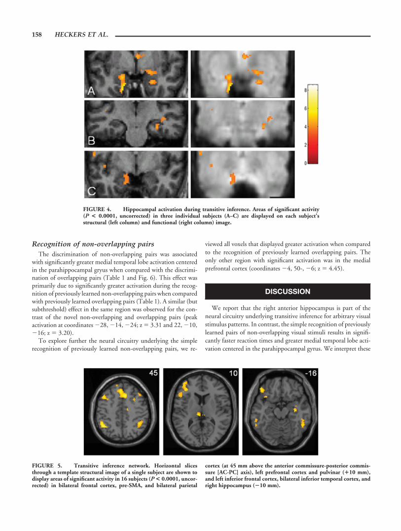
Hippocampal Activation During Transitive Inference in Humans
Stephan Heckers,1,2* Martin Zalesak,1,2 Anthony P. Weiss,1,2 Tali Ditman,1,2
and Debra Titone3
ABSTRACT: Studies in rodents have demonstrated that the integrationand flexible expression of memories, necessary for transitive inference,depend on an intact hippocampus. To test this hypothesis in humans, westudied brain activation during the discrimination of a series of overlap-ping and non-overlapping arbitrary visual stimulus pairs. We report thattransitive inference about overlapping pairs is associated with right ante-rior hippocampal activation, whereas recognition of non-overlappingstimulus pairs is associated with bilateral medial temporal lobe activationcentered in the anterior parahippocampal gyrus. We conclude that im-mediate access to simple stimulus-stimulus relationships is mediated viathe parahippocampal gyrus, whereas the flexible representation of mem-ory requires the recruitment of the hippocampus. © 2004 Wiley-Liss, Inc.
KEY WORDS: neuroimaging; fMRI; relational memory; parahippocam-pal gyrus
INTRODUCTION
The medial temporal lobe is essential for declarative memory, i.e., ourability to recollect facts and events (Squire and Zola-Morgan, 1991). Thisinference was drawn originally from studies of declarative memory functionin patients with medial temporal lobe damage (Scoville and Milner, 1957;Corkin, 2002) and was subsequently confirmed by animal studies (Eichen-baum and Cohen, 2001). The anatomical organization of hippocampus,surrounding cortices, and multimodal association cortex underlying declar-ative memory has now been well characterized in both the rodent and theprimate brain (Lavenex and Amaral, 2000; Witter et al., 2000). Despite thisremarkable progress, the precise contributions of the hippocampus and sur-rounding cortices (i.e., parahippocampal gyrus, entorhinal cortex, andperirhinal cortex) to the formation of declarative memory remain a matter ofdebate (Brown and Aggleton, 2001; Eichenbaum, 2000; Preston and Gab-rieli, 2002).
Experiments in rodents and nonhuman primates provide compelling ev-idence that the hippocampus and parahippocampal gyrus contribute differ-entially to recognition memory (Eichenbaum and Cohen, 2001). In short,
the parahippocampal gyrus is concerned with familiarityor recency of individual stimulus items, whereas the hip-pocampus is needed to recollect relations among items(Eichenbaum et al., 1994; Brown and Aggleton, 2001).Several recent functional neuroimaging studies havedemonstrated segregated roles of hippocampus and para-hippocampal gyrus for declarative memory in humans(Eldridge et al., 2000; Davachi and Wagner, 2002;Strange et al., 2002). However, these neuroimaging stud-ies of declarative memory in humans employ experimen-tal designs that cannot be evaluated further in animals. Inthe present report, we have followed the reverse strategy,i.e., translating an experimental design, which estab-lished a distinct memory function of the hippocampus inanimals, into a functional neuroimaging study in hu-mans.
We chose to study transitive inference, i.e., the abilityto infer the relationships between indirectly related itemsthat have not been presented together, based on previouslearning of a sequence of overlapping premise pairs (e.g.,A�C, if A�B and B�C). Using a hierarchically orderedset of five odors (A�B�C�D�E), Dusek and Eichen-baum (1997) demonstrated that disconnection of thehippocampus from either its cortical or subcortical path-way prevents rats from inferring the proper order forodors B and D. This and subsequent experiments in ro-dents (Fortin et al., 2002; Van Elzakker et al., 2003)support the theory that the hippocampus is necessary toestablish a flexible representation of memory.
We used a 2 � 2 factorial design to study the effectsof inference (novel vs previously learned pairings) andstimulus sequence (overlapping vs non-overlappingpairs) (Fig. 1). Initially, subjects were trained to dis-criminate arbitrary visual stimulus pairs. One set ofeight stimuli created four non-overlapping pairs (la-beled P in Fig. 1), whereas another set of five stimulicreated an overlapping sequence (labeled S). We thenstudied the ability to infer a relationship betweenitems that had not been presented together, based onprevious learning of a sequence of overlapping pairs(transitive inference) or non-overlapping pairs (non-transitive inference). We hypothesized that transitiveinferences about novel pairings derived from the over-lapping stimulus set (labeled IS) would be associatedwith hippocampal activation, whereas nontransitiveinferences about novel pairings derived from the non-overlapping stimulus set (labeled IP) would not.
1Department of Psychiatry, Massachusetts General Hospital, Boston,Massachusetts; 2Athinoula A. Martinos Center for Biomedical Imaging,Charlestown, Massachusetts; 3Department of Psychology, McGill Univer-sity, Montreal, CanadaGrant sponsor: National Institute of Mental Health (NIMH); Grant number:MH01763-01; Grant number: MH60272; Grant sponsor: MIND Institute;Grant sponsor: National Alliance for Research on Schizophrenia and De-pression (NARSAD); Grant sponsor: Essel Foundation.*Correspondence to: Stephan Heckers, Department of Psychiatry, Massa-chusetts General Hospital, East CNY-9112, Bldg. 149, Thirteenth Street,Charlestown, MA 02129. E-mail: [email protected] for publication 23 June 2003DOI 10.1002/hipo.10189
HIPPOCAMPUS 14:153–162 (2004)
© 2004 WILEY-LISS, INC.

MATERIALS AND METHODS
Subjects
We studied 16 healthy subjects (8 female and 8 male, ages 21-28, mean age 23.9, average estimated IQ (Blair and Spreen,1989) � 114), who gave informed consent in a manner approvedby the institutional review board of the Massachusetts GeneralHospital. No subject had a history of major medical, neurological,or psychiatric illness.
Stimuli and Paradigm
Stimuli
Thirteen visually distinctive pattern fills were selected fromthose provided by CorelDraw. Two sets of pattern fills (8 for thenon-overlapping pairs, and 5 for the overlapping pairs) were ran-domly assigned to pairs of pentagon and ellipsoid shapes for eachparticipant.
Training prior to scanning
Participants were informed that they would see pairs of visualpatterns on a computer screen, and that one pattern in each pairwould always hide a “smiling face” (e.g., �). They were thenshown each of the pairings, along with the correct answer in eachcase, and instructed to remember the correct location of the smil-ing face. Left/right position of individual patterns for each pair wascounterbalanced, and participants indicated their response bypressing “1” for the stimulus on the left and “2” for the stimulus onthe right. When participants made a correct guess during training,the selected visual pattern would move to reveal the smiling facereinforcement. When participants made an incorrect guess, theselected visual pattern would move, but the smiling face would notappear.
Participants were first trained and tested on the non-overlappingpairs, then on the overlapping pairs, and finally on a mixture ofnon-overlapping and overlapping pairs. This final testing sessionwas similar in format to that used in the noninference conditionsduring scanning, with one difference: stimuli pairs were presented
FIGURE 1. Stimulus set and experimental conditions. Prior toscanning, subjects were trained to discriminate non-overlapping pairs(P) and an overlapping sequence of pairs (S). The reinforced itemwithin each pair is shown on the left. During scanning, subjects wereasked to recollect the correct response for previously seen pairs (P and
S) and to infer the correct response for five novel pairings of non-overlapping pairs (IP) and overlapping pairs (IS). No letters wereshown in the experiment, and the presentation of the pairs and theposition of the two stimuli within each pair were randomized.
154 HECKERS ET AL.

randomly, rather than in blocked sessions, such that on each trialparticipants were equally likely to be presented with a pair of stim-uli from the overlapping set as they were to be presented with a pairof stimuli from the non-overlapping set. The training procedure,identical for non-overlapping and overlapping pairs, was brokeninto three blocks. The first training block consisted of 60 trials thatcontained twice as many of two of the four stimulus pairs. Forexample, during training of the overlapping stimulus set, partici-pants saw 20 instances of AB and BC, and 10 instances of CD andDE. Thus, the sequential stimuli during the first training blockwere “front loaded.” During training of the non-overlapping stim-ulus set, participants saw 20 instances of AB and CD, and 10instances of EF and GH. The second training block also consistedof 60 trials that contained twice as many of two of the four stimuluspairs from each set. In this second block, however, the pairs were“back loaded”. Thus, participants saw 20 instances of CD and DE,and 10 instances of AB and BC. During the second block of train-ing of the non-overlapping stimulus set, participants saw 20 in-stances of EF and GH, and 10 instances of AB and CD. The thirdtraining block consisted of 24 trials that contained equal numbersof the four stimulus pairs. Thus, for the training of both overlap-ping and non-overlapping pairs, participants saw six instances ofeach stimulus.
Overall, participants were presented with an equal number ofeach of the pairs in the overlapping and non-overlapping stimulussets. This method of training ensured that all participants wouldnot only be able to learn the correct response for each pairing butwould also be likely to hierarchically encode the overlapping stim-ulus set. Our previous work suggested that the initial front loadingof pairs was necessary for healthy participants to correctly judgeBD during the inference test trials (Titone et al., 2003).
Recognition task during fMRI scan
Subjects participated in two functional magnetic resonance im-aging (fMRI) scans, each lasting 5 min. Each scan started andended with 30 s of fixation trials. In between, blocks of 10 trials offour different types (P, S, IP, IS) were presented in the followingsequence: P, S, IP, IS, P, S, IP, IS. For each trial, subjects wereinstructed to indicate by pressing a button which pattern theyassociated with reinforcement, based on the previous training ses-sion. During scanning, however, the smiling face, used for rein-forcement during training, was not presented.
To avoid bias associated with particular object shapes and/orpatterns, we rotated the position of the fills within the two sets foreach participant (a total of 16 fills was used, each subject saw 13 ofthe 16 fills) and the two shapes across all subjects (8 subjects sawnon-overlapping pairs as pentagons and 8 subjects saw overlappingpairs as pentagons) (Fig. 1).
Functional Imaging
Subjects were scanned in a Siemens 1.5-tesla (T) Sonata high-speed echo-planar imaging device (Munich, Germany). Subjectslay on a padded scanner bed in a dimly illuminated room, wearingear plugs. Foam padding was used to stabilize the head. Stimuliwere generated using Presentation software (Neurobehavioral Sys-
tems) on a personal computer, projected onto a screen, and viewedby the subjects via a tilted mirror placed in front of their eyes.
Functional scanning began with an initial sagittal localizer scan.The two functional series lasted 5:10 min each. The first 10 s ofeach series were discarded, to equilibrate for scanner inhomogene-ity. During the remaining time of each series, 120 BOLD func-tional brain images were collected [TE/TR � 40/2,500 ms; 25coronal slices, perpendicular to the anterior commissure-posteriorcommissure (AC-PC) line and starting anterior at the frontal pole,5-mm thickness, 1-mm skip; voxel size 3.1 � 3.1 � 5 mm, FOV �200 mm; Flip angle � 90 degrees], to capture 80 trials lasting 3 seach, bracketed by two blocks of fixation trials, lasting 30 s each.
Data Analysis
Behavioral data
The accuracy data were analyzed using a repeated-measures 2(sequence type: overlapping, non-overlapping) � 2 (inferencetype: present, absent) analysis of variance (ANOVA). The latencyof correct responses was also analyzed using a repeated-measures2 � 2 ANOVA.
Functional neuroimaging data
All functional data were transformed into a common referencespace (MNI Talairach brain) and corrected for head motion usingSPM99 (Wellcome Department of Cognitive Neurology, Lon-don, UK). Functional images were smoothed using an 8-mm full-width/half-maximum (FWHM) Gaussian filter.
Functional images were analyzed in two stages in a mixed-effectsmodel. First, general linear models were created for each subject,which included the effects of session (1,2) and condition ([P], [S],[IP], [IS], and fixation baseline) to explain the variance of BOLDsignal change at each voxel. We tested for the main effects ofinference ([IP�IS] vs [P�S]) and sequence ([S�IS] vs [P�IP]), aswell as their interaction ([IS vs IP] vs ([S vs P]) across the twofunctional imaging sessions. Second, we pooled all individual con-trast images for the main effects and interactions into a one-samplet-test for within group effects. Activations were considered signif-icant at a voxel extent threshold of �50 voxels, with P � 0.0001,uncorrected for multiple comparisons. To disambiguate signifi-cant results of the main effects analysis, we followed up with anal-yses that included only two conditions (simple effect analysis).Activations were considered significant at P � 0.0001, uncor-rected for multiple comparisons.
RESULTS
Behavioral Data
Accuracy
The pattern of accuracy for each of the four conditions (P, S, IP,and IS; Fig. 1) was as follows: 98.0 � 4.0% (mean �SD) for P;95.9 � 6.9% for S; 94.5 � 15.0% for IP; and 91.7 � 10.4% for
_________________________________________________________ TRANSITIVE INFERENCE IN HUMANS 155

IS. Responses to non-overlapping pairs of items were not signifi-cantly different from responses to overlapping pairs (main effect ofsequence: F(1, 15) � 3.3, P � 0.09). Responses to previouslylearned pairs were not significantly different from responses tonovel pairings (main effect of inference: F(1, 15) � 1.8, P � 0.20).The interaction between the two main effects, i.e., sequence andinference, was not significant (F(1, 15) � 0.8, P � 0.78).
Response latency
Response latency of correct responses (in milliseconds) for each ofthe four conditions (P, S, IP, and IS; see Fig. 1) was as follows: 871.2 �194.7 (mean �SD) for P; 1138.3 � 234.6 for S; 868.4 � 289.0 forIP; and 1330.4 � 334.2 for IS (Fig. 2). Responses to overlapping pairswere significantly slower than responses to non-overlapping pairs
(main effect of sequence: F(1, 15) � 67.6, P � 0.0001). Responses tonovel pairings were significantly slower than responses to previouslylearned pairs (main effect of inference: F(1, 15) � 8.9, P � 0.009).Furthermore, the increase in reaction time associated with inferentialjudgments was significantly greater for the overlapping pairs than thenon-overlapping pairs (sequence-by-inference interaction: (F(1,15) � 19.9, P � 0.0001). Thus, whereas judgments not requiringtransitive processing resulted in no increase in reaction time, transitiveinferences required substantial additional processing (Fig. 2). Similarmain effects and interaction were found when latencies of all responseswere analyzed (main effect of sequence: F(1, 15) � 64.2, P � 0.0001;main effect of inference: F(1, 15) � 8.4, P � 0.01; sequence-by-inference interaction: F(1, 15) � 21.0, P � 0.0001). We will refer tothis significant sequence-by-inference interaction as the transitive in-ference effect. The transitive inference effect was more pronounced inthe BD trials of the IS condition, which resulted in significantly longerreaction time compared to the other four trial types of the same con-dition (i.e., AC, AD, CE, BE) (paired t-tests, all P � 0.01).
fMRI Data
Two behavioral effects, the transitive inference effect and therecognition of non-overlapping pairs, were associated with signif-icant medial temporal lobe activation.
Transitive inference
We investigated the transitive inference effect by testing whichvoxels showed a significant sequence-by-inference interaction(contrast: [IS � IP] � [S � P]). This analysis revealed significantright anterior hippocampal activation during transitive inference(Table 1 and Fig. 3). Activation of the same right anterior hip-pocampal region during transitive inference was confirmed in thesimple comparison of transitive versus nontransitive inference[IS�IP] and the comparison of transitive inference versus judg-ments on overlapping items [IS�S] (Table 1). Hippocampal acti-
FIGURE 2. Recall accuracy and reaction time. Bar graphs displaymean (�SD) reaction time for the four conditions: previously seen(P) and novel (IP) non-overlapping pairs and previously seen (S) andnovel (IS) overlapping pairs.
TABLE 1.
Medial Temporal Lobe Activation*
Effect Brain region z score MNI Talairach
Inference-by-Sequence InteractionContrast: [IS � IP] � [S � P] R anterior hippocampus 4.44 34, �14, �16Simple effects of transitive inferenceContrast: [IS] � [IP] R anterior Hippocampus 4.22 36, �6, �22
4.13 34, �10, �16Contrast: [IS] � [S] R anterior Hippocampus 3.89 34, �4, �14Recognition of non-overlapping pairsContrast: [P � IP] � [S � IS] R anterior Parahippocampal gyrus 4.97 24, �18, �20
L anterior Parahippocampal gyrus 4.90 �28, �16, �20Recognition of previously learned non-overlapping pairsContrast: [P] � [S] R anterior Parahippocampal gyrus 4.99 24, �16, �24
L anterior Parahippocampal gyrus 4.88 �28, �16, �22
*Letters in square brackets refer to the four conditions as indicated in Figure 1. P � pairs, S � sequence, IP � inferred pairs, IS � inferred sequence.Coordinates refer to the MNI305 stereotactic space, an approximation of Talairach space (Talairach and Tournoux, 1988).
156 HECKERS ET AL.

vation during transitive inference was also seen in individual sub-jects (Fig. 4).
We explored the neural circuitry underlying transitive infer-ence by reviewing all voxels with a significant sequence-by-inference interaction. A distributed network of brain regions,including the pre-supplementary motor area (pre-SMA), bilat-eral frontal cortex, bilateral parietal cortex, bilateral posteriortemporal cortex, and the pulvinar showed significant activationassociated with transitive inference (Table 2 and Fig. 5). Todisambiguate the two effects that contributed to the transitiveinference effect in these brain regions, we studied the maineffects of inference (novel pairs � previously learned pairs) and
sequence (overlapping pairs � non-overlapping pairs). Wefound significant main effects of inference and sequence inpre-SMA (peak activation at coordinates �4, �20, �52; z �5.64 and 2, 12, 56; z � 4.41 respectively), left prefrontal cortex(�52, 18, 34; z � 5.76 and �46, 28, 32; z � 4.58, respec-tively), and left parietal cortex (�46, �54, 50; z � 4.86 and�38, �52, 58; z � 5.47, respectively). In addition, a maineffect of sequence was observed in right prefrontal cortex (46, 4,54; z � 4.33), right parietal cortex (38, �48, 44; z � 5.16), andbilateral temporal cortex (�20, �58, �12; z � 5.22 and 26,�52, �20; z � 4.87). In contrast, neither of these two maineffects was present in the hippocampus.
FIGURE 3. Right anterior hippocampal activation during tran-sitive inference. A: Areas of significant activity in 16 subjects (P <0.0001, uncorrected) are mapped onto a template structural image ofa single subject. B: Contrast of parameter estimates (� 1 SE) for themagnitude of the hemodynamic response in right anterior hippocam-
pus for the four experimental conditions (i.e., previously seen (P) andnovel (IP) non-overlapping pairs and previously seen (S) and novel(IS) overlapping pairs) compared with the cross-hair fixation baselinecondition. The parameter estimates are collapsed across sessionswithin subjects and averaged across subjects.
_________________________________________________________ TRANSITIVE INFERENCE IN HUMANS 157
Recognition of non-overlapping pairsThe discrimination of non-overlapping pairs was associated
with significantly greater medial temporal lobe activation centeredin the parahippocampal gryus when compared with the discrimi-nation of overlapping pairs (Table 1 and Fig. 6). This effect wasprimarily due to significantly greater activation during the recog-nition of previously learned non-overlapping pairs when comparedwith previously learned overlapping pairs (Table 1). A similar (butsubthreshold) effect in the same region was observed for the con-trast of the novel non-overlapping and overlapping pairs (peakactivation at coordinates �28, �14, �24; z � 3.31 and 22, �10,�16; z � 3.20).
To explore further the neural circuitry underlying the simplerecognition of previously learned non-overlapping pairs, we re-
viewed all voxels that displayed greater activation when comparedto the recognition of previously learned overlapping pairs. Theonly other region with significant activation was in the medialprefrontal cortex (coordinates �4, 50-, �6; z � 4.45).
DISCUSSION
We report that the right anterior hippocampus is part of theneural circuitry underlying transitive inference for arbitrary visualstimulus patterns. In contrast, the simple recognition of previouslylearned pairs of non-overlapping visual stimuli results in signifi-cantly faster reaction times and greater medial temporal lobe acti-vation centered in the parahippocampal gyrus. We interpret these
FIGURE 4. Hippocampal activation during transitive inference. Areas of significant activity(P < 0.0001, uncorrected) in three individual subjects (A–C) are displayed on each subject’sstructural (left column) and functional (right column) image.
FIGURE 5. Transitive inference network. Horizontal slicesthrough a template structural image of a single subject are shown todisplay areas of significant activity in 16 subjects (P < 0.0001, uncor-rected) in bilateral frontal cortex, pre-SMA, and bilateral parietal
cortex (at 45 mm above the anterior commissure-posterior commis-sure [AC-PC] axis), left prefrontal cortex and pulvinar (�10 mm),and left inferior frontal cortex, bilateral inferior temporal cortex, andright hippocampus (�10 mm).
158 HECKERS ET AL.

data as evidence for an immediate access to previously learnedinformation via the parahippocampal gyrus, whereas the flexiblerepresentation of memory requires the recruitment of the hip-pocampus. Thus, this study confirms in humans the importance ofthe hippocampus for the orderly organization of stimulus relations,a finding previously demonstrated in rodents (Dusek and Eichen-baum, 1997; Van Elzakker et al., 2003), and extends recent fMRIstudies of relational memory in humans (Rombouts et al., 1997;Sperling et al., 2001; Davachi and Wagner, 2002).
Neural Circuitry of Transitive Inference
Inferential thinking is found in both humans and animals.Transitive inference, the paradigmatic example of inferentialthinking, has been described in humans as young as 4 years old(Bryant and Trabasso, 1971), in nonhuman primates (McGonigleand Chalmers, 1977), in rats (Davis, 1992; Roberts and Phelps,1994; Dusek and Eichenbaum, 1997), and in pigeons (von Fersenet al., 1991). It appears that transitive inference has evolvedthroughout phylogenesis as a beneficial strategy, for example, toinfer the rank order of animals in the habitat (Delius and Siemann,1998).
What are the cognitive mechanisms underlying transitive infer-ence (Harris and McGonigle, 1994; Wynne, 1998; Bryson, 2001),and what are their neural correlates? When subjects learn to dis-criminate a series of overlapping stimulus pairs, reaction timesincrease during the learning of pairs that do not include end items,but do not change for pairs with end items (Acuna et al., 2002b).
Furthermore, reaction times are shorter during subsequent recog-nition of novel pairs with more intervening items (symbolic dis-tance effect) (Moyer and Landauer, 1967; Acuna et al., 2002b).These two observations indicate that series of overlapping stimuluspairs are stored as a unified and flexible representation, whichprovides strong evidence for transitive inference as a general rela-tional memory capacity (Eichenbaum and Cohen, 2001). In con-trast, some of the results of instrumental transitive inference testingin animals can be explained by the value transfer theory, whichpredicts that the ability of a stimulus to attract a response is deter-mined by positive/negative values acquired during reinforcementtraining (Frank et al., 2003; Van Elzakker et al., 2003; von Fersenet al., 1991).
The present results suggest that a distributed but specific net-work of brain regions underlies transitive inference in humans. Wefound significant activation associated with transitive inference inthe pre-SMA (Brodmann area 6) and bilateral parietal, prefrontal,and inferior temporal cortices. This pattern is similar to that in therecent study by Acuna et al. (2002a), who trained healthy controlsubjects on a hierarchically ordered set of 11 different unicoloredshapes. The authors report significant brain activation in bilateralprefrontal cortex, pre-SMA, premotor area, insula, precuneus, andlateral posterior parietal cortex during the recognition of novelpairs of visual stimulus items. The slice selection used by Acuna etal. (2002a) did not cover the medial temporal lobe. Other recentstudies of transitive inference using verbal premise pairs (Goel andDolan, 2001) or iconic stimuli (Dickins et al., 2001) also reportedsignificant prefrontal and parietal cortex activation. Of interest,patients with prefrontal cortex damage exhibit deficits in the inte-gration of relations (Waltz et al., 1999), as do patients with schizo-phrenia (Titone et al., in press). Taken together, studies in humanshave established an important role of frontal and parietal multi-modal association cortices, both of which are closely connectedwith the hippocampus via the entorhinal cortex, for transitive in-ference.
Our main effects analysis demonstrated that pre-SMA, left pa-rietal cortex, and left prefrontal cortex contributed not only to thetransitive inference effect, but to all trials that involved inferences(transitive as well as nontransitive) and overlapping pairs (previ-ously seen as well as novel). Furthermore, right parietal, right pre-frontal, and bilateral temporal cortex activation was greater for alloverlapping pairs. In contrast, right hippocampal activation wasnot seen in the main effects analyses, but only in the interactionand the simple effects of transitive inference. This indicates that thehippocampus contributes uniquely to the ability to infer relation-ships between a sequence of items, while pre-SMA, parietal, andprefrontal cortex contribute more broadly to inferential judgmentsand the recognition of overlapping pairs of visual stimuli.
Distinct Patterns of MTL Activation forTransitive Inference and Recognition
Rodents trained to discriminate the overlapping stimulus setA�B�C�D�E cannot correctly discriminate the novel stimuluspair BD after hippocampal lesion, but they are still able to discrim-inate correctly the novel pairing of the two end items A and E
TABLE 2.
Transitive Inference Network*
Brain region H z scoreMNI Talairach
coordinates
Pre-SMA R 5.32 8 18 50Inferior temporal
gyrus (BA 37)L 5.14 �44 �52 �6
R 4.80 54 �50 �12Middle temporal
gyrus (BA 21)L 5.04 �58 �30 �8
Premotor cortex(BA 6)
L 4.99 �32 2 62
R 4.98 30 10 50Inferior frontal
gyrus (BA 47)L 4.95 �44 36 �8
L 4.42 �30 20 �12Inferior parietal
cortex (BA 40)L 4.87 �42 �54 46
R 4.72 46 �50 42Anterior cingulate
cortex (BA 24)L 4.79 �4 6 26
Pulvinar L 4.44 �8 �24 10Hippocampus R 4.44 34 �14 �16
*H indicates hemisphere. Coordinates refer to the MNI305 stereotacticspace, an approximation of Talairach space (Talairach and Tournoux,1988).
_________________________________________________________ TRANSITIVE INFERENCE IN HUMANS 159

(Dusek and Eichenbaum, 1997; Van Elzakker et al., 2003). Thepresent study provides novel evidence that, in humans, the flexiblerepresentation of a hierarchically ordered sequence of overlappingstimulus items recruits the hippocampus. Our study design differsfrom the animal studies in so far as we contrasted pairs that requiredifferent degrees of transitive inference (the four pairs AC, AD,BE, CE include one end item, whereas the BD trial does not) withnovel pairs of non-overlapping items (which are equivalent to theAE trials in the rat experiment). It is likely that individual trials orblocks of trials without end items would reveal even greater hip-pocampal activation.
A recent positron emission tomography (PET) study reportedhippocampal activation during learning of overlapping pairs andsubiculum activation during the recognition of novel non-overlap-ping pairs (Nagode and Pardo, 2002). Furthermore, previousfMRI studies of relational memory in humans have demonstratedhippocampal activation during the encoding (Rombouts et al.,1997; Sperling et al., 2001; Davachi and Wagner, 2002) and rec-ognition (Stark and Squire, 2001) of inter-item associations. Ourresults extend these reports by showing that associations that re-quire a flexible representation of a hierarchically ordered set ofstimuli are associated with significantly greater hippocampal acti-
FIGURE 6. Recognition of non-overlapping pairs. A: Areas ofsignificant activity in 16 subjects (P < 0.0001, uncorrected) in bilat-eral anterior parahippocampal gyrus are mapped onto a templatestructural image of a single subject. B: Contrast of parameter esti-mates (�1 SE) for the magnitude of the hemodynamic response inright anterior parahippocampal gyrus for the four experimental con-
ditions (i.e., previously seen [P] and novel [IP] non-overlapping pairsand previously seen [S] and novel [IS] overlapping pairs) comparedwith the cross-hair fixation baseline condition. The parameter esti-mates are collapsed across sessions within subjects and are averagedacross subjects.
160 HECKERS ET AL.

vation compared to a simple association between two items. Thisprovides support for the notion that the hippocampus supports theencoding and retrieving of sequences of events that compose epi-sodic memories (Eichenbaum and Cohen, 2001; Lisman, 1999).
The transitive inference trials resulted in significantly longerreaction time, but it is unlikely that simple task difficulty couldexplain the right anterior hippocampal activation. First, the reac-tion time for the previously seen overlapping pairs (condition S)was significantly longer than the one for the previously seen non-overlapping pairs (condition P), but the contrast (S-P) was notassociated with any hippocampal activation. Second, conditionswith shorter reaction time (P and IP) were associated with moresignificant medial temporal lobe activation than the conditionswith longer RT (S and IS). Both observations argue against thenotion that longer reaction time as an index of increased taskdifficulty translates into increased hippocampal activation.
While some have proposed that associative and single item rec-ognition memory segregate along the anatomical border of hip-pocampus and surrounding cortices (Brown and Aggleton, 2001),others have argued that such division of labor cannot be absolute(Suzuki and Eichenbaum, 2000; Stark and Squire, 2001; Stark etal., 2002). Our study provides evidence that the parahippocampalgyrus contributes to the recognition of previously learned non-overlapping pairs of visual stimulus patterns. Previous studies haveestablished that the parahippocampal gyrus is critical for paired-associate learning (Bunsey and Eichenbaum, 1993) and someforms of concurrent discrimination learning (Hood et al., 1999).Furthermore, it appears that the anterolateral aspects of the para-hippocampal gyrus (perirhinal cortex, Brodmann areas 35 and 36)contribute to recognition memory in an automatic fashion, toallow for the association of different sensory features of an object(Murray and Bussey, 1999; Murray and Richmond, 2001). It is inthis context that we interpret the greater medial temporal lobeactivation centered in the parahippocampal gyrus during the rec-ognition of previously learned non-overlapping pairs comparedwith the recognition of overlapping pairs. The number of pairs tobe remembered and the degree of item familiarity/novelty did notdiffer between the two conditions, but the number of items to beremembered did (eight items in the four non-overlapping and fiveitems in the four overlapping pairs). Despite the greater memoryload, the recognition of the non-overlapping pairs resulted in sig-nificantly faster reaction time. This suggests that simple stimulus-stimulus relationships can be represented by the parahippocampalgyrus (as evidenced by the greater activation), allowing for imme-diate access (as evidenced by the faster reaction time).
In conclusion, we have demonstrated hippocampal activationduring transitive inference in humans. Future experiments areneeded to dissect the different components of brain activationduring transitive inference in healthy and in patient populations(Titone et al., 2003). Such studies will provide valuable informa-tion about the functional connectivity underlying transitive infer-ence, whereas animal experiments allow us to study the cellular andmolecular mechanism in well-defined brain regions. Ultimately,experimental paradigms that can be explored in both animals andhumans will allow us to uncover the mechanisms of declarativememory.
Acknowledgments
This study was funded by National Institute of Mental Health(NIMH) grants MH01763-01 (to S.H.) and MH60272 (to D.T.),MIND Institute (S.H.), National Alliance for Research on Schizo-phrenia and Depression (NARSAD) (to D.T.), and the EsselFoundation (to D.T.).
REFERENCES
Acuna BD, Eliassen JC, Donoghue JP, Sanes JN. 2002a. Frontal andparietal lobe activation during transitive inference in humans. CerebCortex 12:1312–1321.
Acuna BD, Sanes JN, Donoghue JP. 2002b. Cognitive mechanisms oftransitive inference. Exp Brain Res 146:1–10.
Blair J, Spreen O. 1989. Predicting premorbid IQ: a revision of the na-tional adult reading test. Clin Neuropsychol 3:129–136.
Brown MW, Aggleton JP. 2001. Recognition memory: what are the rolesof the perirhinal cortex and hippocampus? Nat Rev Neurosci 2:51–61.
Bryant PE, Trabasso T. 1971. Transitive inferences and memory in youngchildren. Nature 232:456–458.
Bryson JJ. 2001. Intelligence by design: principles of modularity andcoordination for engineering. Cambridge, MA: MIT Press.
Bunsey M, Eichenbaum H. 1993. Critical role of the parahippocampalregion for paired-associate learning in rats. Behav Neurosci 107:740–747.
Corkin S. 2002. What’s new with the amnesic patient H.M.? Nat RevNeurosci 3:453–460.
Davachi L, Wagner AD. 2002. Hippocampal contributions to episodicencoding: insights from relational and item-based learning. J Neuro-physiol 88:982–990.
Davis H. 1992. Transitive inference in rats. J Comp Psychol 106:342–349.
Delius JD, Siemann M. 1998. Transitive responding in animals and hu-mans: exaptation rather than adaptation. Behav Processes 42:107–137.
Dickins DW, Singh KD, Roberts N, Burns P, Downes JJ, Jimmieson P,Bentall RP. 2001. An fMRI study of stimulus equivalence. NeuroRe-port 12:405–411.
Dusek JA, Eichenbaum H. 1997. The hippocampus and memory fororderly stimulus relations. Proc Natl Acad Sci USA 94:7109–7114.
Eichenbaum H. 2000. A cortical-hippocampal system for declarativememory. Nat Rev Neurosci 1:41–50.
Eichenbaum H, Cohen NJ. 2001. From conditioning to conscious recol-lection. Memory systems of the brain. Oxford: Oxford UniversityPress.
Eichenbaum H, Otto T, Cohen NJ. 1994. Two functional components ofthe hippocampal memory system. Brain Behav Sci 17:449–518.
Eldridge LL, Knowlton BJ, Furmanski CS, Bookheimer SY, Engel SA.2000. Remembering episodes: a selective role for the hippocampusduring retrieval. Nat Neurosci 3:1149–1152.
Fortin NJ, Agster KL, Eichenbaum HB. 2002. Critical role of the hip-pocampus in memory for sequences of events. Nat Neurosci 5:458–462.
Frank MJ, Rudy JW, O’Reilly RC. 2003. Transitivity, flexibility, con-junctive representations, and the hippocampus. II. A computationalanalysis. Hippocampus 13:299–312.
Goel V, Dolan RJ. 2001. Functional neuroanatomy of three-term rela-tional reasoning. Neuropsychologia 39:901–909.
Harris MR, McGonigle BO. 1994. A model of transitive choice. Q J ExpPsychol B 47:319–348.
_________________________________________________________ TRANSITIVE INFERENCE IN HUMANS 161

Hood KL, Postle BR, Corkin S. 1999. An evaluation of the concurrentdiscrimination task as a measure of habit learning: performance ofamnesic subjects. Neuropsychologia 37:1375–1386.
Lavenex P, Amaral DG. 2000. Hippocampal-neocortical interaction: ahierarchy of associativity. Hippocampus 10:420–430.
Lisman JE. 1999. Relating hippocampal circuitry to function: recall ofmemory sequences by reciprocal dentate-CA3 interactions. Neuron22:233–242.
McGonigle B, Chalmers M. 1977. Are monkeys logical? Nature 267:694–696.
Moyer RS, Landauer TK. 1967. Time required for judgments of numer-ical inequality. Nature 215:1519–1520.
Murray EA, Bussey TJ. 1999. Perceptual-mnemonic functions of theperirhinal cortex. Trends Cogn Sci 3:143–151.
Murray EA, Richmond BJ. 2001. Role of perirhinal cortex in objectperception, memory, and associations. Curr Opin Neurobiol 11:188–193.
Nagode JC, Pardo JV. 2002. Human hippocampal activation during tran-sitive inference. NeuroReport 13:939–944.
Preston AR, Gabrieli JDE. 2002. Different functions for different medialtemporal lobe structures? Learn Mem 9:215–217.
Roberts WA, Phelps MT. 1994. Transitive inference in rats: a test of thespatial coding hypothesis. Psychol Sci 5:368–374.
Rombouts SARB, Machielsen WCM, Witter MP, Barkhof F, LindeboomJ, Scheltens P. 1997. Visual association encoding activates the medialtemporal lobe: a functional magnetic resonance imaging study. Hip-pocampus 7:594–601.
Scoville WB, Milner B. 1957. Loss of recent memory after bilateral hip-pocampal lesions. J Neurol Neurosurg Psychiatry 20:11–21.
Sperling RA, Bates JF, Cocchiarella AJ, Schacter DL, Rosen BR, AlbertMS. 2001. Encoding novel face-name associations: a functional MRIstudy. Hum Brain Mapp 14:129–139.
Squire LR, Zola-Morgan S. 1991. The medial temporal lobe memorysystem. Science 253:1380–1386.
Stark CE, Squire LR. 2001. Simple and associative recognition memory inthe hippocampal region. Learn Mem 8:190–197.
Stark CE, Bayley PJ, Squire LR. 2002. Recognition memory for singleitems and for associations is similarly impaired following damage tothe hippocampal region. Learn Mem 9:238–242.
Strange BA, Otten LJ, Josephs O, Rugg MD, Dolan RJ. 2002. Dissociablehuman perirhinal, hippocampal, and parahippocampal roles duringverbal encoding. J Neurosci 22:523–528.
Suzuki WA, Eichenbaum H. 2000. The neurophysiology of memory.Ann NY Acad Sci 911:175–191.
Talairach J, Tournoux P. 1988. Co-planar stereotaxic atlas of the humanbrain. New York: Thieme.
Titone D, Ditman T, Holzman PS, Eichenbaum H, Levy DL. Transitiveinference in schizophrenia: impairments in relational memory organi-zation. Schizophr Res (in press).
Van Elzakker M, O’Reilly RC, Rudy JW. 2003. Transitivity, flexibility,conjunctive representations, and the hippocampus. I. An empiricalanalysis. Hippocampus 13:292–298.
von Fersen L, Wynne CDL, Delius JD, Staddon JER. 1991. Transitiveinference formation in pigeons. J Exp Psychol 17:334–341.
Waltz JA, Knowlton BJ, Holyoak KJ, Boone KB, Mishkin FS, de MenezesSantos M, Thomas CR, Miller BL. 1999. A system for relational rea-soning in human prefrontal cortex. Psychol Sci 10:119–125.
Witter MP, Wouterlood FG, Naber PA, Van Haeften T. 2000. Anatom-ical organization of the parahippocampal-hippocampal network. In:Scharfman HE, Witter MP, Schwarcz R, editors. The parahippocam-pal region. Implications for neurological and psychiatric diseases. NewYork: Annals of the New York Academy of Sciences. p 1–24.
Wynne CDL. 1998. A minimal model of transitive inference. In: WynneCDL, Staddon JER, editors. Models of action. Mahwah, NJ: LawrenceErlbaum. p 269–307.
162 HECKERS ET AL.
Copyright © 2022 FDOKUMEN