Comparative analysis of circadian clock genes in insects
17
© 2008 The Authors Journal compilation © 2008 The Royal Entomological Society 447 Insect Molecular Biology (2008) 17(5), 447– 463 Blackwell Publishing Ltd REVIEW ARTICLE Comparative analysis of circadian clock genes in insects F. Sandrelli*, R. Costa*, C. P. Kyriacou† and E. Rosato† *Department of Biology, University of Padova, Padova 35131, Italy; †Department of Genetics, University of Leicester, Leicester, LE1 7RH UK Summary After a slow start, the comparative analysis of clock genes in insects has developed into a mature area of study in recent years. Brain transplant or surgical interventions in larger insects defined much of the early work in this area, before the cloning of clock genes became possible. We discuss the evolution of clock genes, their key sequence differences, and their likely modes of regulation in several different insect orders. We also present their expression patterns in the brain, focusing particularly on Diptera, Lepidoptera, and Orthoptera, the most common non-genetic model insects studied. We also highlight the adaptive involve- ment of clock molecules in other complex phenotypes which require biological timing, such as social behaviour, diapause and migration. Keywords: circadian, genes, insects, expression, comparative. Introduction Circadian rhythms are important regulatory processes, which, almost universally, are used to harmonise physiology and behaviour with the 24 h geophysical cycles of light and temperature of our planet. The clock therefore is at the interface between the organism and the external environment and, as for other signalling pathways, it probes the outside world, transducing external changes to downstream effectors. In the last 25 years or so, the phenomenology which contributed to most of the circadian literature before about 1984, was replaced by the more pragmatic molecular genetic analysis of the clock, initially in Drosophila (Hall, 2003). At the cellular level, molecules which exemplify the progression of time are cyclically transformed. Phosphoryla- tion, degradation and altered subcellular localisation of clock proteins, result in modified expression of clock and clock controlled genes, ultimately coordinating the activities of the organism with its environment (aka ‘entrainment’) (Hall, 2003; Zheng & Sehgal, 2008). The same chain of events persists, with minor differences, also under constant ‘free-running’ conditions, suggesting that the internal milieu functions as a self-sustaining oscillation that continues in the absence of any periodic input. Such a design is a common feature of all circadian clocks (Dunlap, 1999) and it underlines the mechanistic constraints encountered (more than once) in the evolution of circadian systems. The identification of the mammalian genes Clock (Antoch et al., 1997; King et al., 1997b) and Period (Sun et al., 1997; Tei et al., 1997) revealed that vertebrate clocks have a common evolutionary origin with that of insects, although some interesting differences have also emerged (Reppert & Weaver, 2000; Yu & Hardin, 2006). This evolution- ary plasticity is observed between insect species and even within-species (Costa & Kyriacou, 1998; Helfrich-Forster, 2005c; Yuan et al., 2007; Kyriacou et al., 2008). Our review will focus on recent developments in the identity of functional molecular and neuronal components of the clock in insects. A molecular model for the clock The cloning of the Drosophila melanogaster period (per) gene paved the way for the modern molecular dissection of the circadian system (Hall, 2003). However, it also biased the way we think about the functioning and the evolution of the circadian clock. For many years Drosophila was the only tractable system among animals (in terms of genetics and molecular biology of the clock) until the identification of the mammalian clock genes in 1997 (Antoch et al., 1997; King et al., 1997a,b; Sun et al., 1997; Tei et al., 1997). At first glance the clocks of flies and mice are well conserved, the main molecular components are the same and there is only some variation on the general theme. However, these differences are intriguing, in particular the apparent swapping of the starring roles of key components (Clayton et al., 2001). At first this seemed to correlate with the phylogenetic distance between flies and mice, but in fact, some of the Received 1 May 2008; accepted after revision 23 June 2008. Correspondence: C. Kyriacou, Department of Genetics, University of Leicester, Leicester, LE1 7RH UK. Tel.: +44 116 2523430; fax: +44 116 2523378; e-mail: [email protected]
-
Upload
independent -
Category
Documents
-
view
0 -
download
0
Transcript of Comparative analysis of circadian clock genes in insects

© 2008 The AuthorsJournal compilation © 2008 The Royal Entomological Society
447
Insect Molecular Biology (2008)
17
(5), 447–463
Blackwell Publishing Ltd
REVIEW ARTICLE
Comparative analysis of circadian clock genes in insects
F. Sandrelli*, R. Costa*, C. P. Kyriacou† and E. Rosato†
*
Department of Biology, University of Padova, Padova 35131, Italy;
†
Department of Genetics, University of Leicester, Leicester, LE1 7RH UK
Summary
After a slow start, the comparative analysis of clockgenes in insects has developed into a mature area ofstudy in recent years. Brain transplant or surgicalinterventions in larger insects defined much of theearly work in this area, before the cloning of clockgenes became possible. We discuss the evolution ofclock genes, their key sequence differences, and theirlikely modes of regulation in several different insectorders. We also present their expression patterns inthe brain, focusing particularly on Diptera, Lepidoptera,and Orthoptera, the most common non-genetic modelinsects studied. We also highlight the adaptive involve-ment of clock molecules in other complex phenotypeswhich require biological timing, such as social behaviour,diapause and migration.
Keywords: circadian, genes, insects, expression,comparative.
Introduction
Circadian rhythms are important regulatory processes,which, almost universally, are used to harmonise physiologyand behaviour with the 24 h geophysical cycles of light andtemperature of our planet. The clock therefore is at theinterface between the organism and the external environmentand, as for other signalling pathways, it probes the outsideworld, transducing external changes to downstreameffectors. In the last 25 years or so, the phenomenologywhich contributed to most of the circadian literature beforeabout 1984, was replaced by the more pragmatic moleculargenetic analysis of the clock, initially in
Drosophila
(Hall,2003). At the cellular level, molecules which exemplify the
progression of time are cyclically transformed. Phosphoryla-tion, degradation and altered subcellular localisation ofclock proteins, result in modified expression of clock andclock controlled genes, ultimately coordinating the activitiesof the organism with its environment (aka ‘entrainment’)(Hall, 2003; Zheng & Sehgal, 2008). The same chain ofevents persists, with minor differences, also under constant‘free-running’ conditions, suggesting that the internal milieufunctions as a self-sustaining oscillation that continues inthe absence of any periodic input. Such a design is acommon feature of all circadian clocks (Dunlap, 1999) andit underlines the mechanistic constraints encountered(more than once) in the evolution of circadian systems.
The identification of the mammalian genes
Clock
(Antoch
et al
., 1997; King
et al
., 1997b) and
Period
(Sun
et al
., 1997; Tei
et al
., 1997) revealed that vertebrate clockshave a common evolutionary origin with that of insects,although some interesting differences have also emerged(Reppert & Weaver, 2000; Yu & Hardin, 2006). This evolution-ary plasticity is observed between insect species and evenwithin-species (Costa & Kyriacou, 1998; Helfrich-Forster,2005c; Yuan
et al
., 2007; Kyriacou
et al
., 2008). Our reviewwill focus on recent developments in the identity offunctional molecular and neuronal components of the clockin insects.
A molecular model for the clock
The cloning of the
Drosophila melanogaster period
(
per
)gene paved the way for the modern molecular dissection ofthe circadian system (Hall, 2003). However, it also biasedthe way we think about the functioning and the evolutionof the circadian clock. For many years
Drosophila
was theonly tractable system among animals (in terms of geneticsand molecular biology of the clock) until the identification ofthe mammalian clock genes in 1997 (Antoch
et al
., 1997;King
et al
., 1997a,b; Sun
et al
., 1997; Tei
et al
., 1997). Atfirst glance the clocks of flies and mice are well conserved,the main molecular components are the same and there isonly some variation on the general theme. However, thesedifferences are intriguing, in particular the apparent swappingof the starring roles of key components (Clayton
et al
.,2001). At first this seemed to correlate with the phylogeneticdistance between flies and mice, but in fact, some of the
Received 1 May 2008; accepted after revision 23 June 2008. Correspondence:C. Kyriacou, Department of Genetics, University of Leicester, Leicester,LE1 7RH UK. Tel.: +44 116 2523430; fax: +44 116 2523378; e-mail:[email protected]

448
F. Sandrelli
et al.
© 2008 The AuthorsJournal compilation © 2008 The Royal Entomological Society,
17
, 447–463
mouse-like features of these genes are already evidentwithin the insects (Zhu
et al
., 2005; Yuan
et al
., 2007).
The
Drosophila
model
Traditionally the fly clock is described as an ensembleof interlocked negative transcription/translation feedbackloops (Hall, 2003). In each loop positive elements drive thetranscription of negative elements that rhythmically feedback to inhibit the action of the former. However, recentwork suggests that post-translational modifications of clockproteins are more important than originally thought andthey might provide the fulcrum for the whole machinery(Zheng & Sehgal, 2008). This new wave of thinking has notproduced, as yet, a better ‘model’, thus we will describe theclock in the canonical fashion.
At the core of the clock are the transcriptional activatorsCLOCK (CLK) and CYCLE (CYC), that, as heterodimers, bindto E-box sequences on the promoters of
period
and
timeless
(
tim
) initiating their transcription (Hall, 2003). CLK, CYC andPER have a sequence similarity in that they all contain a
PAS domain, a dimerizing region that is found in an enormousfamily of proteins, many of which are involved in environmentalsensing (Gu
et al
., 2000). Seemingly, CLK/CYC are facilitatedin their function by NEJIRE (NEJ), the
Drosophila
ortho-logue of the CBP/p300 family of transcription co-activators,although an alternative scenario has also been suggested(Hung
et al
., 2007; Lim
et al
., 2007a) (see Fig. 1).After translation PER and TIM interact with each other
(via the PER-PAS domain, among others) and with anumber of kinases and phosphatases (see below) thatregulate stability, timing of nuclear entry and accumulation,and their ability to interact and repress the CLK/CYC dimer(Zheng & Sehgal, 2008). As a consequence, abundance,phosphorylation levels, nuclear/cytoplasmic ratio and therepressor function of these two proteins cycle in synchrony,reaching a peak at the end of the night (Zheng & Sehgal,2008). Although the details are still unclear, the formation ofa complex between PER and TIM is a dynamic process, asexemplified by the fact that the two proteins can accumulatein the nucleus with different kinetics, but still require eachother in order to do so (Shafer
et al
., 2002; Meyer
et al
., 2006).
Figure 1. The Drosophila molecular clock within a clock neuron. (1) The first negative feedback loop revolves around the cytoplasmic regulation of PER and TIM. Formation of a dynamic complex between the two molecules regulates phosphorylation/dephosphorylation by kinases [DBT, SGG, CK2] and phosphatases [PP1 and PP2A] and nuclear translocation and accumulation. In the nucleus PER inhibit CLK perhaps by facilitating its phosphorylation by DBT. CLK together with CYC binds E-box sequences on the promoter of clock and clock controlled genes. NJR helps transcriptional activation whereas CWO likely contributes to CLK/CYC repression. (2) The second loop is less well understood. VRI and PDP1ε are expressed under control of CLK. They accumulate with different kinetics and then feed back by repressing (VRI) and activating (PDP1ε) Clk transcription. Other factors likely contribute to this loop. (3) Post-synaptic pathways also mediate the regulation of the clock with modalities that are still unknown. (4) The clock generates rhythmic signals that are passed on by the neuron to downstream effectors. In this diagram some factors have not been included for the sake of simplicity.

Insect clock molecules
449
© 2008 The AuthorsJournal compilation © 2008 The Royal Entomological Society,
17
, 447–463
As mentioned above the complex also involves kinases,such as DOUBLETIME (DBT), SHAGGY (SGG) andCASEIN KINASE II (CKII), and phosphatases, such asprotein phosphatases 1 (PP1) and 2A (PP2A), thatphosphorylate/dephosphorylate PER and/or TIM and,once transported inside the nucleus, also CLK (Kloss
et al
., 1998; Price
et al
., 1998; Martinek
et al
., 2001; Lin
et al
., 2002a; 2005; Akten
et al
., 2003; Sathyanarayanan
et al
., 2004; Kim & Edery, 2006; Yu
et al
., 2006; Fang
et al
.,2007). Unlike PER and TIM, CLK levels are constantthroughout the 24 h but there are PER-dependent rhythmicchanges in its phosphorylation status that have implicationsfor its transcriptional activity (Houl
et al
., 2006; Kim &Edery, 2006; Yu
et al
., 2006). PER likely facilitates CLKphosphorylation by DBT, resulting in diminished CLK/CYCaffinity for DNA (Yu
et al
., 2006). This observation mightexplain why TIM is not an efficient repressor of CLK/CYCwhereas PER, once in the nucleus, is a more active repressorwhen alone (Rothenfluh
et al
., 2000).CYC is also a constitutively expressed protein (Rutila
et al
., 1998) hence it is particularly important that severalmechanisms intervene to impart rhythmicity to CLK/CYCactivity. Another transcriptional regulator CLOCKWORKORANGE (CWO) contributes to the inhibition of CLK/CYCperhaps by facilitating changes in chromatin structure(Kadener
et al
., 2007; Lim
et al
., 2007b; Matsumoto
et al
.,2007). However a more complicated mode of action hasalso been suggested, where CWO functions as a transcrip-tional activator at the beginning of the night, when PER islow and cytoplasmic, and as a repressor in the latenight-early morning, when PER levels are high and nuclear(Richier
et al
., 2008).Another feedback loop is centered around the rhythmic
expression of
Clk
, although its practical contribution to thefunctioning of the whole system is at present unclear.Initially CLK/CYC dimers bind to E-boxes on the promotersof
PAR domain protein 1
ε
(
Pdp1
ε
) and
vrille
(
vri
), drivingtheir robust rhythmic transcription. PDP1
ε
and VRI aretranscriptional regulators that cycle also at the protein level,but with different phases. VRI peaks before PDP1
ε
,outcompeting the latter for the same binding sites on the
Clk
promoter and causing repression of
Clk
transcription. Afew hours later increased PDP1
ε
displaces VRI andpromotes
Clk
mRNA production (Blau & Young, 1999;Cyran
et al
., 2003; Glossop
et al
., 2003). However, asmis-expression of
Pdp1
ε
in clock cells does not halt thecycling of
Clk
mRNA (Benito
et al
., 2007), this loop isprobably more complex than originally thought, perhapsrequiring additional, but as yet unknown elements.
Finally, light-resetting of the clock is mediated via thecanonical visual transduction system, as well as a blue-lightsensitive protein, CRYPTOCHROME (CRY) (Helfrich-Forster
et al
., 2001). CRY is particularly important in thatit also acts as a light gateway into the clock because, unlike
wild-type, flies carrying a nearly null mutation,
cry
b
, arerhythmic in constant light (LL), even though their visualsystem is intact (Emery
et al
., 2000).Various
cry
mutant genotypes also show reducedcircadian responses to brief light pulses (Emery
et al
.,1998; Stanewsky
et al
., 1998; Rosato
et al
., 2001; Dissel
et al
., 2004). One of the modes of action of CRY is to mediatethe light-degradation of TIM through the E3 ligase protein,JETLAG (JET) (Koh
et al
., 2006; Peschel
et al
., 2006). Thedegradation of TIM exposes PER to the kinase DBT, withrepercussions for PER stability and the feedback loop(Young, 1998). However, not all TIM containing neuronsexpress CRY at quantifiable levels, yet TIM is neverthelessrapidly degraded by light also in those cells (Picot
et al
.,2007; Yoshii
et al
., 2008). This suggests that communi-cation among neurons might be the most important factorfor the light-degradation of TIM and, in general, for entra-inment (Nitabach
et al
., 2002; Peng
et al
., 2003), so thatone of the functions of CRY may be to mediate cross-talkamong clock cells. In this regard, CRY can be found in theaxonal projections of the neurons in which it is expressed(Klarsfeld
et al
., 2004; Yoshii
et al
., 2008). While this hypo-thesis has yet to be verified, a recent knock-out of the genesuggests a dark function for CRY, highlighting its multipleroles in the
Drosophila
clock (Dolezelova
et al
., 2007).
Differences between
Drosophila
and mammalian clocks
Although in this review we focus on the clock of insects, itis convenient to refer briefly to the distinctive features of themammalian clock. This is because mammalian-like clockcharacteristics are found in insects other than
Drosophila
,suggesting that both flies and mammals specialised bydiverging from a common design. There are severaldifferences in the architecture of the mammalian and flyclocks, the most obvious being that in mammals, most clockcomponents are present in multiple copies, increasing thecomplexity and the redundancy of the system (Clayton
et al
., 2001). The mammalian CRY proteins (CRY1 andCRY2) substitute for
Drosophila
TIM as the partners ofPER proteins. CRY1 and CRY2 do not show circadian lightresponsiveness, but instead, they act as the main transcrip-tional repressors.
Bmal1
(the homologue of
Drosophilacyc
) substitutes for
Clk
as the rhythmic component of thesecond loop and contains the main transactivation domainof the CLK/BMAL1 complex (Reppert & Weaver, 2000; Yu& Hardin, 2006), which in
Drosophila
is carried by CLK(Allada
et al
., 1998; Rutila
et al
., 1998). Moreover, mammalian
Tim
(
mTim
) actually corresponds to the fly paralogue
timeout
/
tim2
(Benna
et al
., 2000; Gotter
et al
., 2000).Although both the fly (F. Sandrelli and R. Costa unpublishedobservations) and the mammalian (Barnes et al., 2003;Unsal-Kacmaz et al., 2005) timeout/tim2 genes seem to be

450 F. Sandrelli et al.
© 2008 The AuthorsJournal compilation © 2008 The Royal Entomological Society, 17, 447–463
involved with the circadian clock, at present their role is notcompletely clear. For the interested reader, several recentreviews offer a more detailed description of the mammalianclock (Ko & Takahashi, 2006; Levi & Schibler, 2007;Maywood et al., 2007).
The ancestral clock of insects
Clock genes sequences are available for several non-Drosophilid insect species. Figure 2 shows that in allLepidoptera analysed there are two cry genes, onesensitive to light, hence Drosophila-like (cry-d), the otherable to repress CLK/CYC mediated transcription in vitro asin mammals (cry-m) (Yuan et al., 2007). However, mammalian-like cry genes are also found in the honeybee Apismellifera and in the flour beetle Tribolium castaneum,both curiously lacking a copy of cry-d, whereas both typesof cry’s are found instead in the mosquitos Anophelesgambiae and Aedes aegypti (Zhu et al., 2005; Rubin et al.,2006; Yuan et al., 2007).
The mammalian-like TIM is an essential gene both inmouse (Gotter et al., 2000) and in Drosophila (F. Sandrelliand R. Costa, unpublished observations), and unlike itsparalogue, fly tim, it has been identified in every animalthat has been studied. Orthologues to fly tim are almostubiquitous among insects, with the exception of the honeybee(Rubin et al., 2006) and so tim would appear to be the resultof a more recent duplication of the ancestral essential genetimeout/tim2, even though its presence in sea urchins
dates the duplication event to pre-Cambrian times (Rubinet al., 2006).
Finally the main transactivation domain of the CLK/CYC(BMAL1) complex is generally found at the C-terminusof BMAL1 type proteins, whereas the poly-Q repeats foundin CLK only serve an ancillary function in transcriptionalactivation. However, Drosophila CYC is shorter and lacksthe transactivation domain and conversely, the poly-Qrepeats on CLK are expanded and provide the activationdomain necessary for the functioning of the complex(Chang et al., 2003; Rubin et al., 2006). In conclusion, theancestral clock of insects seemingly consisted of twoCRYs (-m and -d), two TIMs (-m and -d) and has CYC as themain transcriptional activator. In Drosophila, loss of cry-m(although a transcriptional repressor function might persistin CRY-d, – Collins et al., 2006), perhaps favoured by theloss of the CYC C-terminus and the rise of CLK as thedominant element of the complex, has brought TIM-d toacquire an important role in the main feedback loop andboosted the relevance of PER as a transcriptional repressor.
Anatomy of the clock in Drosophila
The anatomy of the clock is based predominantly onstaining with anti-PER and anti-TIM antibodies. There aresix main clusters of brain neurons, that are divided intothree groups of lateral and three groups of dorsal neurons(Hall, 2003) (see Fig. 3). The lateral neurons (LNs) arefurther subdivided into dorsal (1 cluster of ~6 cells, LNd),
Figure 2. Drosophila-like (-d) and mammalian-like (-m) clock components in representative insects. Insect orders: Hymenoptera, Coleoptera, Lepidoptera, Diptera. Mus musculus (mouse) is the outgroup. BTR = BMAL1 transactivation domain. CPQ = CLOCK poly-Q repeats. ? = unknown. = extensive poly-Q repeats from (Chang & Reppert, 2003; Zhu et al., 2005, 2008b; Rubin et al., 2006; Yuan et al., 2007).

Insect clock molecules 451
© 2008 The AuthorsJournal compilation © 2008 The Royal Entomological Society, 17, 447–463
Figure 3. Schematic representation of clock molecule expression in brains of Diptera and Lepidoptera. Top panel. Drosophila melanogaster ; middle panel, Musca domestica. Neurons expressing PER in Musca and Drosophila brains. Key: DN1, DN2 and DN3, dorsal neurons; PLNs, posterior lateral neurons; LNds, lateral neurons dorsal; s-LNvs, small lateral neurons ventral; l-LNvs, large lateral neurons ventral; Pn-LNv, PDF-null lateral neuron ventral; MNs, medial neurons; MLNs, medio-lateral neurons (Codd et al., 2007). Lower panel. Danaus plexippus. Regions expressing TIM, PER, CRY1 and/or CRY2 are highlighted in red. In these areas the four clock proteins are partially colocalized. Areas expressing TIM or CRY1 are indicated in magenta. In these regions the two clock proteins do not colocalise. CRY1 positive fibers are represented by continuous yellow lines. Projections of the dorsal rim area photoreceptors are indicated by dotted yellow lines. Neurons and fibres expressing CRY2 exclusively are represented in blue. Areas staining exclusively for TIM and PER are indicated in green and light blue, respectively. PL, Pars lateralis; PI, Pars intercerebralis; SOG, Subesophageal ganglion; CB, central body; LO, lobula; ME, medulla; LA, lamina; RE, retina (Modified from Sauman et al., 2005; Reppert, 2006; Zhu et al., 2008b).
452 F. Sandrelli et al.
© 2008 The AuthorsJournal compilation © 2008 The Royal Entomological Society, 17, 447–463
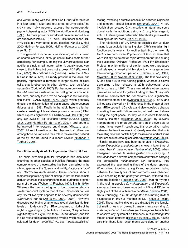
and ventral (LNv) with the latter also further differentiatedinto four large (l-LNv) and four small (s-LNv) cells. Thes-LNv and l-LNv neurons express the neuropeptide,pigment dispersing factor (PDF) (Helfrich-Forster & Homberg,1993). The more posterior and dorsal brain neurons (DNs),are subdivided into DN1 (~16 cells), DN2 (2 cells) and DN3(~40 cells in a very dorso-lateral position, Kaneko & Hall,2000; Helfrich-Forster, 2005a; Helfrich-Forster et al., 2007)(see Fig. 3).
This general clock neuron classification, which is basedon anatomical criteria, although useful, hides some furthercomplexity. For example, among the LNv group there is anadditional single small neuron, which is usually found veryclose to the l-LNvs but does not express PDF (Kaneko &Hall, 2000). This pdf-null LNv (pn-LNv), unlike the l-LNvs,but as in the s-LNvs, is already present in the larva, andpossibly represents a remnant of larger cluster of clockcells, that is observed in other diptera, such as Muscadomestica (Codd et al., 2007). Furthermore only two out ofthe ~16 neurons clustered in the DN1 group are found inthe larva, and only those two do not express the transcrip-tion factor GLASS (Shafer et al., 2006) which generallydirects the differentiation of opsin-based photoreceptors(Moses et al., 1989). Finally, in the adult there is a furthercluster consisting of three lateral posterior neurons (LPNs)which express high levels of TIM (Kaneko & Hall, 2000) andonly low levels of PER (Helfrich-Forster, 2005a,b; Shaferet al., 2006; Helfrich-Forster et al., 2007), and seem tobe involved in temperature entrainment (Miyasako et al.,2007). More information on the physiological differencesamong those neurons and their role in the circadian networkof the fly, can be found in a recent review (Nitabach &Taghert, 2008).
Functional analysis of clock genes in other fruit flies
The basic circadian plan for Drosophila has also beenexamined in other species of fruitflies. Probably the mostcomprehensive of these studies has been carried out in thesibling species of the Queensland fruitfly, Bactrocera tryoniand Bactrocera neohumeralis. These species show atemporal separation by time of mating, in that the former mateat dusk whereas the latter prefer to mate during the brighterparts of the day (Tychsen & Fletcher, 1971; Smith, 1979).Whereas the per orthologues of both species show asimilar transcript cycle to that of their Drosophila cousinsany Cry mRNA cycle appears to be severely dampened inBactrocera heads (An et al., 2002, 2004). Howeverdissected out brains or antennae reveal significantly highlevels of mid-daytime Cry mRNA compared to middle of thenight, suggesting a cycle. In both tissues, B. tryoni havesignificantly less Cry mRNA than B. neohumeralis, and thisis also reflected in corresponding hybrids which have beenselected for dusk (tryoni-like) vs. day (neohumeralis-like)
mating, revealing a putative association between Cry levelsand temporal sexual isolation (An et al., 2004). In situhybridisation revealed Cry transcripts in lateral and somedorsal cells. In addition, using a Drosophila reagent,anti-PER staining was detected in lateral cells, plus weakerstaining in dorsal areas (An et al., 2004).
The relationship of Cry levels to possible day vs. duskmating is particularly interesting given CRY’s circadian lightfunction and is relevant to another tephritid, the melon flyBactrocera curcurbitae. Populations of B. curcurbitae thatwere initially selected for rapid development time duringthe successful Okinawa Prefectural Fruit Fly EradicationProject, in which millions of sterile males were producedand released, showed a highly significant change in theirfree-running circadian periods (Shimizu et al., 1997;Miyatake, 2002; Koyama et al., 2004). The fast-developingS-Line had a 22 h free-running period, whereas a slowerdeveloping L-line, showed a 29 h behavioural cycle(Shimizu et al., 1997). These remarkable observationsparallel an old and forgotten finding in the Drosophilaliterature, namely that the per-mutants also significantlyaffect development time (Kyriacou et al., 1990). The S- andL-lines also showed a ~5 h difference in the phase of theirper mRNA cycles in LD cycles, and also revealed a changein mating time, with S-lines mating at dusk, and L-linesduring the night phase, so they were in effect temporallysexually isolated (Miyatake et al., 2002). By cleverlymanipulating the photoperiods so that the two sets ofmating times were in synchrony, this assortative matingbetween the two lines was lost, clearly revealing that onlythe mating time was contributing to the isolation, and not someother associated ethological factor (Miyatake et al., 2002).
Similar results have also been generated in Drosophila,where Drosophila pseudoobscura shows a later time ofmating than D. melanogaster (Tauber et al., 2003). Whentransgenic per-null D. melanogaster hosts carrying D.pseudoobscura per were compared to control flies carryingthe conspecific melanogaster per transgene, theyexpressed the later mating times of the donor species.When mixed together, a significant assortative matingbetween the two types of transformants was observedwhich according to the genotypes involved, reflected theirtemporal isolation (Tauber et al., 2003). Mating rhythmsin the sibling species D. melanogaster and Drosophilasimulans have also been reported in LD and DD to beslightly out of phase with each other (Sakai & Ishida, 2001).Not surprisingly, in D. melanogaster, this mating rhythmdisappears in per-null mutants in DD (Sakai & Ishida,2001). These mating rhythms are dictated by the female,but mating tests of per-null transformants carrying D.melanogaster or D. simulans per transgenes have failedto observe any systematic differences in D. melanogasterfemale choice patterns (Ritchie & Kyriacou, 1994). Havingsaid this, these latter experiments were done for another

Insect clock molecules 453
© 2008 The AuthorsJournal compilation © 2008 The Royal Entomological Society, 17, 447–463
purpose, and the time of the mating tests was not controlled,so no firm conclusions can be drawn.
Clock genes and photoperiodism
Another fruitfly, Chymomyza costata, has also receivedsome attention due mainly to its photoperiodic phenotypes,in which a mature larval diapause is programmed by shortdays (Kostal & Shimada, 2001). A naturally occurringnon-diapausing strain (npd, non-photoperiodic diapause)has been mapped to the timeless locus (Pavelka et al.,2003). Subsequent molecular analysis reveals a largenumber of amino acid differences between the wild-typeand the mutant strain, as well as a large deletion (~2 Kb) inthe 5′ UTR which removes the transcription start signal plusother regulatory sequences (Stehlik et al., 2008).
The mature npd larva shows very low levels of CctimmRNA, and no evidence for immunoreactivity to a con-specific CcTIM antiserum. Circadian cycles of Cctim arefound in the wild-type larval brain that are more robust inshort than long days, and two lateral dorsal neurons in eachhemisphere appear to express CcTIM, with more intensestaining at ZT2 and weakest at ZT20 in long days, but withmore intense (apparently cytoplasmic) staining during thenight in short days (Stehlik et al., 2008). Thus there seemsto be a correlation between the presence of photoperiodicdiapause and Cctim expression in this species, as well aschanges in CcTIM expression with different photoperiods.This is also seen in the diapausing pitcher plant mosquito,Wyomia smithii, in which Wstim expression changesconsistently with latitude in North America (Mathias et al.,2005). While linkage analysis may suggest that the Wstimlocus is associated with photoperiodism, it has beenargued that this is unlikely to involve the circadian clockdirectly (Bradshaw et al., 2006; Mathias et al., 2007).
The long standing debate on whether clock genes cancontribute to photoperiodic diapause in insects (Nunes &Saunders, 1999; Bradshaw et al., 2006) has been furtherstoked by the findings that in D. melanogaster, a new timnatural variant, ls-tim appears to be under natural selectionbecause it enhances the level of diapause in the Europeanseasonal environment (Tauber et al., 2007). The molecularbasis for this appears to be that the binding of the new TIMvariant protein to CRY is attenuated, leading to a morestable, light insensitive TIM, that leads to a reduced lightresponsiveness for both circadian and photoperiodicphenotypes (Sandrelli et al., 2007a). Such changes wouldbe favourable under the colder habitats, and seasonallyvarying exotic photoperiods of Europe, to which D. mela-nogaster migrated, out of eastern Africa ~15 000 years ago(Kyriacou et al., 2008). The new ls-tim variant, whichoriginated in southern Italy about 8000 years ago, thuscame under selection and spread throughout Europe(Tauber et al., 2007). However it is not yet completely clear
whether circadian behaviour and diapause induction aresimply sharing the same TIM-based light input mechanism,or whether the new tim variant affects diapause through thecircadian system (Bradshaw & Holzapfel, 2007; Kyriacouet al., 2008).
Clocks in other Diptera
The clock genes of a number of other flies have beencompared, the most detailed study so far is probablythat in Musca domestica, the housefly (Codd et al., 2007).In this species, the RNAs for Musca per, tim, and vri, arecycling and peak early in the subjective night, whereas theClk transcript cycles with an opposite phase. In LL, thesecycles were lost, and so these data are completely con-sistent with those from Drosophila, including the observationthat cyc does not cycle in DD in the housefly. However, as inBactrocera (An et al., 2004), and in contrast to Drosophila,Mdcry does not cycle in DD. It is not known whether Mdcrywould cycle in brains if, like Bactrocera, these were dis-sected out from the heads.
While Mdper transcript cycles in the head, the MdPERprotein is, surprisingly, produced constitutively as assessedby Western blots in both heads and thoraces, and analysedseparately in males and in females (Codd et al., 2007).A similar situation exists in the medfly, Ceratitis capitata(Mazzotta et al., 2005), whereas in the sheep blowfly,Lucilia cuprina, both gene products appear to cycle(Warman et al., 2000). MdTIM on the other hand showsthe expected cycles in LD and DD. In LL however, MdPERremains highly stable, whereas MdTIM degrades immedi-ately, so unlike Drosophila, MdPER does not apparentlyrequire MdTIM for stability.
Initial immunohistochemistry (IHC) using head sectionsrevealed various MdPER staining neurons, particularly inmedial and lateral areas in which the signal was alwayscytoplasmic at every time point (Codd et al., 2007). A moredetailed confocal analysis was then carried out using wholemounts with fluorescent secondary antibodies. Initially,groups of medial and medial lateral neurons were againdetected that appeared to be expressing high levels of con-stitutive cytoplasmic MdPER, but not MdTIM. An anti-PDHreagent also detected a group of neurons that seemedhomologous to the s-LNvs and l-LNvs of Drosophila (Pyzaet al., 2003; Codd et al., 2007), and, when re-examinedcarefully, these cells were also observed to co-expressMdTIM and MdPER (see Fig. 3). Furthermore the Muscas-LNvs showed nuclear MdPER/TIM staining at ZT24,whereas the l-LNvs showed a generally lower and morediffuse staining for both reagents in both cytoplasmic andnuclear compartments, rather different to the situation inDrosophila. A single sLNv neuron which in Drosophila doesnot express PDF (Pn-lNv) and is found closer to the l-LNvcluster, appears to have expanded to four neurons in

454 F. Sandrelli et al.
© 2008 The AuthorsJournal compilation © 2008 The Royal Entomological Society, 17, 447–463
Musca and shows nuclear localisation of MdPER/TIM at ZT24. Musca neurons equivalent to the Drosophila LNds werealso detected and these showed nuclear PER/TIM stainingat the end of the night. A small number of dorsal neuronswere also observed to colocalise MdPER and MdTIM. Itthus appears that the constitutive MdPER expressionobserved in Western blots comes from those very highly,constitutively cytoplasmically expressing medial/medial-lateral MdPER neurons that do not co-expressMdTIM (see Fig. 3).
This study reveals that the housefly has considerablesimilarities in clock gene expression to Drosophila, but alsosome notable differences. What, for example is the functionof the MdPER-reacting material in the medial-lateral cellswhich do not co-express MdTIM and therefore cannot berequired for MdTIM stability? Indeed, is it MdPER at all, orjust non-specific cross-reacting material? In the absence ofa negative genetic control, this is always a difficult question toanswer. The MdPER band in a Musca head Western blot,which is constitutively expressed, likely reflects the dynamicsof constitutive ‘MdPER’ in these medial neurons. This bandis the same size as the band seen in Drosophila per-nulltransformant carrying the Mdper transgene, so it seemsvery likely that this antigenic material in both blots and IHCis indeed MdPER (Piccin et al., 2000; Codd et al., 2007).
In addition, transformation of the Mdper gene into D. mela-nogaster per01 mutants, rescues rhythmicity very robustly,indeed far more strongly than the more closely phylogeneticallyrelated D. pseudoobscura per transgene (Piccin et al.,2000). This intriguing observation correlates with a higheramino acid identity in the PAS domain between Musca andD. melanogaster PER than between the two Drosophilaspecies (Piccin et al., 2000). As this region interactsphysically with TIM (Gekakis et al., 1995) this mightrepresent a case of intergenic coevolution, something thatcan be tested when the full MdTIM sequences becomeavailable. In fact one might expect under such circumstancesthat the PER interacting region of TIM to also be moresimilar between Musca and D. melanogaster, than betweenD. melanogaster and D. pseudoobscura.
The Musca study also provides something of a cautionarytale, in that, based on enzymatic-reactions, initial IHC revealedonly cytoplasmic, constitutive PER expression. When fluo-rescence based methods were used with confocal micro-scopy, the MdPER and MdTIM colocalising neurons withnuclear staining at appropriate phases of the circadian cycle,were revealed. Thus reports based on enzymatic IHC ona number of insect orders which generally report non-cycling ‘cytoplasmic PER’ (e.g. Zavodska et al., 2003b) mayhave to be revised when more sensitive methods are utilised.
Other Diptera that have been studied include the fleshfly,Sarcophaga crassipalpis, in which Scper and Sctim mRNAcycles have been detected in fly heads, but again, neitherSccry nor Sccyc transcripts show any significant temporal
pattern (Goto & Denlinger, 2002). In the yellow fever anddengue vector, Aedes aegypti, Aatim shows a weak cyclingwith a peak in mRNA abundance around the light-darktransition (Gentile et al., 2006). The hematophagous sandflyLutzomya longipalpis is unusual compared to Drosophila,in that both the LlClk and Llcyc transcripts cycle withhigher levels during the light phase (Meireles-Filho et al.,2006a,b). Llper and Lltim are also down regulated by blood-feeding, which also causes a reduction in locomotor activity(Meireles-Filho et al., 2006b). Lutzomya appears to have aC-terminal transactivation domain that is characteristic ofCYC in mosquitos (Gentile et al., 2006), bees (Rubin et al.,2006), and moths (Chang et al., 2003), but is absent inDrosophila (Rutila et al., 1998). Thus, and as mentionedearlier, the evolution of an extensive polyQ transactivationdomain in CLK might relieve similar pressure on the CYCpartner molecule to maintain its own.
Circadian clocks in Lepidoptera
In Lepidoptera, rhythmic behaviour in egg-hatching andadult eclosion (as well as a number of other rhythms suchas the behavioural activities in preparation for pupationobserved in Manduca sexta), appear to be driven by a pho-tosensitive circadian clock. In fact, circadian behaviouralrhythmicity in LD as well as in constant conditions hasbeen observed in a number of species (Sauman & Hashimi,1999; Froy et al., 2003). Most of the comparative circadianstudies conducted on Lepidoptera involve the giant silk-moth, Antheraea pernyi, the domestic silk moth Bombyxmori and the monarch butterfly, Danaus plexippus, (Sau-man & Hashimi, 1999; Chang et al., 2003; Sehadova et al.,2004; Takeda et al., 2004; Zhu et al., 2005, 2008a,b; Iwaiet al., 2006; Reppert, 2006; Trang le et al., 2006; Iwai et al.,2008). The latter species is particularly important given thepossible link between the circadian clock and annual longdistance migrations characteristic of this organism.
Molecular cloning and bioinformatic studies have identi-fied Lepidopteran orthologues and paralogues for most ofthe known Drosophila and/or mammalian clock genes (seeFig. 2). The A. pernyi genome harbors a family of three sex-linked per genes, of which only one (ApperZ) is essentialfor circadian clock functions (Gotter et al., 1999). Con-versely, B. mori has only a single copy of the per gene inboth males and females (Takeda et al., 2004; Iwai et al.,2006; Sandrelli et al., 2007b), that produces two BmPERisoforms, differing by 5 aa in length (Takeda et al., 2004)whereas in the monarch butterfly only a single Dpper cDNAhas been isolated (Froy et al., 2003). The PER proteins ofthese Lepidoptera show the canonical Drosophila PERdomains, including the nuclear localization signal (NLS),PAS A and B regions, cytoplasmic localization domain(CLD) and nuclear export signal (NES) (Reppert et al.,1994; Takeda et al., 2004).
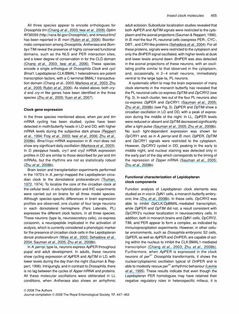
Insect clock molecules 455
© 2008 The AuthorsJournal compilation © 2008 The Royal Entomological Society, 17, 447–463
All three species appear to encode orthologues forDrosophila tim (Chang et al., 2003; Iwai et al., 2006) DptimAY36059 (http://rana.lbl.gov/Drosophila/), and timeout/tim2has been reported in B. mori (Rubin et al., 2006). Bioinfor-matic comparison among Drosophila, Antheraea and Bom-byx TIM reveal the presence of highly conserved functionaldomains, such as the NLS and PER interaction sites,and a lower degree of conservation in for the CLD domain(Chang et al., 2003; Iwai et al., 2006). These speciesencode a single orthologue of Drosophila Clock and cyc/Bmal1. Lepidopteran CLK/BMAL1 heterodimers are potenttranscription factors, with a C-terminal BMAL1 transactiva-tion domain (Chang et al., 2003; Markova et al., 2003; Zhuet al., 2005; Rubin et al., 2006). As stated above, both cry-d and cry-m like genes have been identified in the threespecies (Zhu et al., 2005; Yuan et al., 2007).
Clock gene expression
In the three species mentioned above, when per and timmRNA cycling has been studied, cycles have beendetected in moth/butterfly heads in LD and DD, with highermRNA levels during the subjective dark phase (Reppertet al., 1994; Froy et al., 2003; Iwai et al., 2006; Zhu et al.,2008b). Bma1l/cyc mRNA expression in B. mori does notshow any significant daily oscillation (Markova et al., 2003).In D. plexippus heads, cry1 and cry2 mRNA expressionprofiles in DD are similar to those described for per and timmRNAs, but the rhythms are not as statistically robust(Zhu et al., 2008b).
Brain lesion and transplantation experiments performedin the 1970’s in A. pernyi mapped the Lepidopteran circa-dian clock to the dorsolateral protocerebrum (Truman,1972, 1974). To localize the core of the circadian clock atthe cellular level, in situ hybridization and IHC experimentswere carried out on brains for all three model species.Although species-specific differences in brain expressionprofiles are observed, one cluster of four large neuronsin each dorsolateral hemisphere (Pars Lateralis, PL)expresses the different clock factors, in all three species.These neurons (type Ia1 neurosecretory cells), co-expresscorazonin, a neuropeptide implicated in the activation ofecdysis, which is currently considered a phenotypic markerfor the presence of circadian clock cells in the Lepidopterandorsal protocerebrum (Wise et al., 2002; Sehadova et al.,2004; Sauman et al., 2005; Zhu et al., 2008b).
In A. pernyi, type Ia1 neurons express ApPER throughoutpupal and adult development. In adults, these neuronsshow cycling expression of ApPER and ApTIM in LD, withlower levels during the day than the night (Sauman & Rep-pert, 1996). Intriguingly, and in contrast to Drosophila, thereis no lag between the cycles of Apper mRNA and proteins.All these molecular oscillations were obliterated in LLconditions, when Antheraea also shows an arrhythmic
adult eclosion. Subcellular localization studies revealed thatboth ApPER and ApTIM signals were restricted to the cyto-plasm and the axonal projections (Sauman & Reppert, 1996).In B. mori the four PL neuronal cells coexpress PER-, CYC-,DBT-, and CRY-like proteins (Sehadova et al., 2004). For allthese proteins, signals were restricted to the cytoplasm andonly the BmPER signal oscillated, with higher levels at duskand lower levels around dawn. BmPER was also detectedin the axonal projections of these neurons, with an oscil-lating profile similar to that observed in the cytoplasm,and, occasionally, in 2–4 small neurons, immediatelyventral to the large type-Ia1 PL neurons.
A systematic effort to map the brain expression of manyclock elements in the monarch butterfly has revealed thatthe PL neuronal cells co-express DpTIM and DpCRY2 (seeFig. 3). In each cluster, two out of the four PL neurons alsoco-express DpPER and DpCRY1 (Sauman et al., 2005;Zhu et al., 2008b) (see Fig. 3). DpPER and DpTIM show acircadian oscillation in LD and DD, with a peak of expres-sion during the middle of the night. In LL, DpPER levelswere reduced or absent and DpTIM decreased significantlyafter a light pulse (Sauman et al., 2005; Zhu et al., 2008b).No such light-dependent expression was shown forDpCRY1 and, as in A. pernyi and B. mori, DpPER, DpTIMand DpCRY1 signals were restricted to the cytoplasm.However, DpCRY2 cycled in DD, peaking in the early tomiddle night, and nuclear staining was detected only inthe early part of the day which corresponds to the timing ofthe repression of Dpper mRNA (Sauman et al., 2005;Zhu et al., 2008b).
Functional characterization of Lepidopteran clock components
Function analysis of Lepidopteran clock elements wasstudied ex in vivo in DpN1 cells, a monarch butterfly embry-onic line (Zhu et al., 2008b). In these cells, DpCRY2 wasable to inhibit DpCLK/DpBMAL-mediated transcription,while DpPER and DpTIM did not, a result consistent withDpCRY2’s nuclear localization in neurosecretory cells. Inaddition, both in monarch brains and DpN1 cells, DpCRY2,TIM, and PER appear to form a complex, as indicated byimmunoprecipitation experiments. However, in other cellu-lar environments, such as Drosophila embryonic S2 cells,DpPER, as well as ApPER and DmPER, are capable of act-ing within the nucleus to inhibit the CLK:BMAL1-mediatedtranscription (Chang et al., 2003; Zhu et al., 2008b).Furthermore, when ApPER is expressed in the clockneurons of per01 Drosophila transformants, it shows thenuclear/cytoplasmic oscillation typical of DmPER and isable to modestly rescue per01 arrhythmic behaviour (Levineet al., 1995). These results indicate that even though theLepidopteran PER homologues may have retained theirnegative regulatory roles in heterospecific milieus, it is

456 F. Sandrelli et al.
© 2008 The AuthorsJournal compilation © 2008 The Royal Entomological Society, 17, 447–463
DpCRY2 that acts as the major clock transcriptional repres-sor within the monarch. The DpCRY2 interaction withDpPER and DpTIM is probably cytoplasmic and may havea stabilizing function before nuclear translocation (Zhuet al., 2008b), as occurs in mammals (Lee et al., 2001).
In DpN1 cells, DpCRY1 is able to induce light-dependentDpTIM degradation (Zhu et al., 2008b). These results areconsistent with the ability of a Dpcry1 transgene (but notDpcry2) to partially rescue the circadian light responses ofthe cryb mutant in transgenic Drosophila (Sauman et al.,2005, Zhu et al., 2008b).
A feature that seems to be common at least to A. pernyiand B. mori is the expression of different clock elements inthe axonal projections of the PL neurons. This is parti-cularly evident for PER, which shows a daily oscillation inLD in both species (Sauman & Reppert, 1996; Sehadovaet al., 2004). In A. pernyi and B. mori, the dorsolateral PER-expressing neurons are very close to two neurons express-ing the prothoracicotropic hormone (PTTH), a criticalregulator of Lepidoptera postembryonic development(Kawakami et al., 1990; Sauman & Reppert, 1996). In addi-tion, BmPER staining has been identified in the B. morinerve fibre arborizations in the corpora cardiaca and cor-pora allata, organs involved in moulting and metamorpho-sis (Sehadova et al., 2004). These patterns suggest aregulatory activity for PER at the level of the neurohor-mones involved in postembryonic development. A role forBmPER in B. mori development has been suggested alsoby the observed reduction in egg-to-adult developmentaltime induced by Bmper dsRNAi (Sandrelli et al., 2007b)and might be due to a pleiotropic effect on development, asalready demonstrated for the Drosophila per clock gene(Kyriacou et al., 1990). This post-transcriptional manipula-tion of Bmper also generated a mild disruption of the egghatching rhythm (Sandrelli et al., 2007b), something whichwas also initially seen in A. pernyi with Apper antisenseinjections (Sauman et al., 1996), but this finding wassubsequently withdrawn (Sauman et al., 2000). Ironically,the corresponding results with Bombyx thus provide someadditional support for the initial finding in Antheraea.
In Bombyx, PER-, CYC-, DBT-, CRY-like signals havebeen observed also in some neurons of the pars intercere-bralis (PI), where these proteins apparently co-localize, andin two large neurons of the frontal ganglion, considered a‘peripheral’ component of the circadian oscillator that maybe implicated in some rhythmic activities controlled by thestomatogastric nervous system (Sehadova et al., 2004).In the monarch, DpPER, DpTIM, DpCRY1 and DpCRY2staining has also been detected in the suboesophagealganglion and PI, where a cluster of neurons co-expressDpTIM, DpCRY1 and DpPER (Sauman et al., 2005; Zhuet al., 2008b). DpPER expression was also identified in thecorpora cardiaca and for DpCRY2, in both corporae cardi-aca and allata. Moreover, clock proteins were also detected
in different regions of the optic lobe (Sauman et al., 2005;Reppert, 2007; Zhu et al., 2008b). Finally, a complex seriesof both DpCRY1- or CRY2-positive nerve fibers have beenrevealed in the brain (Sauman et al., 2005; Reppert, 2007;Zhu et al., 2008b) (see Fig. 3). These neuronal pathwaysseem to link the four PL clock neurons with different brainand eye structures implicated in the monarch’s annual3500–4000 km migration from North America to Mexico.The migratory state is characterized by reproductivediapause and increased longevity, induced by a reduction injuvenile hormone (JH) levels, and circumstantial evidenceimplicates a role for the circadian clock (Reppert, 2006).DpCRY1 positive pathways can be traced from the four PLneurons to PI, a neuronal region expressing clock proteinsand able to regulate the production of JH in the corporaallata (Reppert, 2006). It has been proposed that seasonalday-length variations could be registered by the four circa-dian PL neurons, and communicated via these DpCRY1positive fibers to the PI, which in turn activates themigratory state (Reppert, 2006).
Additional DpCRY1 positive pathways connect the PLclock neurons with the dorsal rim area of the eye, a struc-ture able to perceive UV polarized light, one of the input sig-nals for orientation during the long flight (Sauman et al.,2005; Reppert, 2006). Finally, DpCRY2 positive fibres canbe traced from the four PL neurons to the central brain com-plex (Zhu et al., 2008b), a region that has been recentlyidentified as the site of the sun compass in locusts (Heinze& Homberg, 2007). Zhu et al. (2008b) thus propose a dualrole for DpCRY2: that of a key factor in the feedback loopwhich generate the circadian oscillation in the four dorsola-teral clock neurons, and that of an output element that, withunknown mechanisms, regulates the circadian activity inthe central complex.
Cockroaches, crickets and other insects
It has been 40 years since the first surgical lesion studieswere published in which localisation of the circadian oscil-lator to the optic lobes was demonstrated in the cockroachLeucophaea maderae (Nishiitsutsuji-Uwo & Pittendrigh,1968). Subsequent and very elegant transplantation exper-iments revealed that optic lobes from a donor brain that hadbeen entrained to a different circadian period than the host,could, after several weeks, impart these altered periods tothe recipient (Page, 1982). A more recent modification ofthis theme has been the transplantation of the accessorymedulla (AMe), a small region of the optic lobe that showsimmunoreactivity to anti-PDH. When placed in hosts whoseown AMe had been removed, and who had been entrainedto a different LD cycle than the donor, behavioural rhythmic-ity could be restored, and this appeared to correlate withthe regeneration of PDH-ir fibres to the central brain (Reis-chig & Stengl, 2003). Similar correlations were obtained

Insect clock molecules 457
© 2008 The AuthorsJournal compilation © 2008 The Royal Entomological Society, 17, 447–463
when optic stalks were cut bilaterally, leading to arrhythmicbehaviour, which was restored in those animals that alsoshowed regenerated PDH-ir fibres to the central brain(Stengl & Homberg, 1994). In addition, GABA and Mas-allatotropin innervations in the accessory medulla havebeen strongly implicated in the photic entrainment pathway(Petri et al., 2002).
However, in the cricket Gryllus bimaculatus, it does notappear to be the AMe that is so important, but a more distalregion of the optic lobe, the lamina (Tomioka & Abdelsalam,2004). In other cricket species, Acheta domesticus, lesionand transplantation studies have implicated the neurose-cretory pars intercerebralis (Cymborowski, 1981). In thecricket Teleogryllus some neurons in the AMe are immuno-reactive for both anti-PER and anti-PDH, suggesting a Dro-sophila-centric view of clock anatomy (Lupien et al., 2003).In the ground cricket, Dianemobius nigrofasciatus immuno-reactivity to PER, DBT, CRY and CLK was observed pre-dominantly in the optic lobes, but intriguingly CYC was notobserved in the CLK expressing neurons in this region.CLK and CYC were also expressed in the pars intercere-bralis and suboesophageal ganglion (Shao et al., 2006,2008a). In contrast, in another ground cricket, Allenomo-bius allardi, these clock proteins appear to be particularlyenriched in the suboesophageal ganglion (Shao et al.,2006, 2008b). What does appear in all insect ordersstudied with antibodies to clock proteins, mostly fromDrosophila, or with anti-PDH, is that optic lobe stainingis a common feature (Helfrich-Forster, 2004, 2005c).However, so far, it is only in Drosophila and other dipterasuch as Musca, Teleogryllus, and the blood feedinghemipteran, Rhodnius prolixus, that PDF/PER co-localisationhas been observed. In the latter species, a concerted effortby Steel, Vafopoulou and co-workers have documentedthe circadian basis for the rhythm in PTTH, which controlssteroidogenesis from the prothoracic glands (PG) andinsect moulting (Steel & Vafopoulou, 2006). Eight lateralneurons in the optic lobes of Rhodnius adults co-expressrhythmic PER/TIM and PDF, and the axons of these‘clock’ neurons associate with PTTH cells, providing a linkbetween the clock and the neurohormonal system(Vafopoulou et al., 2007).
Finally, Hymenoptera reveal yet another dimension to theclock mechanism. In nurse bees which provide round theclock brood care, indirect measurements of their activity inthe hive suggest that circadian behaviour may be attenu-ated (Shemesh et al., 2007). However when placed inisolation, nurses revert back to a behavioural locomotorcycle within a couple of days. Temporal expression analysis ofthe ‘usual suspects’ putative clock molecules, reveals thateven under such optimal circadian conditions, molecularoscillations in nurse heads are severely compromised com-pared to foragers (Shemesh et al., 2007). An analysis ofclock protein cycling in the brain of such nurse bees would
be welcomed to investigate this apparent uncoupling ofmolecular and behavioural rhythms. Nevertheless, the takehome message of these studies is that there is a certaindevelopmental plasticity in the circadian system of bees.This is related to the social structuring demands of the col-ony, so that older bees that take on foraging roles haverobust behavioural and molecular cycles whereas youngernurses have their circadian cycles largely suppressedwithin the hive (Toma et al., 2000; Bloch et al., 2001).
Conclusions and future prospects
Hemi-metabolous insects such as cockroaches and crick-ets, have thus provided the best material for surgical manip-ulations, and together with expression studies of clockmolecules, they have significantly extended the ‘evo-devo’type of analysis of clock neuroanatomy. However, one com-mon ‘fly in the ointment’, so-to-speak, is that when clockprotein staining has been ‘demonstrated’ in insects, andthis usually means PER and typically with heterospecificanti-fly PER antibodies, the localisation is commonly exclu-sively cytoplasmic (Zavodska et al., 2003a,b). One notableexception is the hawkmoth, Manduca sexta, where PERnuclear antigenicity is observed in putative circadianneurons (Wise et al., 2002; Schuckel et al., 2007).
However, in general, one can surmise that either a novelform of clock gene regulation is present (as discussed ear-lier with some Musca and Antheraea neurons), or that theanti-PER reagents are identifying cross-reacting material.The absence of a negative control (ie a per-null mutant) innon-model insects provides a caveat that has already beendiscussed earlier in relation to the Musca PER story (Coddet al., 2007). This cautionary element can most practicallybe alleviated by colocalising both the mRNA and protein tothe neuronal regions in question (Zhu et al., 2008b). Onefurther important consideration that comes from these var-ious studies that we have discussed, will probably turn outto be a general rule of thumb. In insects that have both fly-like (CRY1) and vertebrate-like (CRY2) CRYs, they arelikely to use CRY2 rather than PER or TIM as the mainnegative regulator, as elegantly demonstrated recently inLepidoptera (Zhu et al., 2008b).
One of the most exciting parts of this comparative workhowever, apart from examining how evolution has tweakedthe roles of the various clock genes, is the way the clockmay impact on other complex phenotypes such as devel-opment (Kyriacou et al., 1990; Shimizu et al., 1997; Miyat-ake, 2002; Koyama et al., 2004; Sandrelli et al., 2007b),mating (Tychsen & Fletcher, 1971; Smith, 1979; Sakai &Ishida, 2001; An et al., 2002, 2004; Miyatake et al., 2002;Tauber et al., 2003), navigation (Sauman et al., 2005; Zhuet al., 2008b) and social (Toma et al., 2000; Bloch et al.,2001) behaviour in different species, revealing that clockproteins will have extensive pleiotropic effects. This has

458 F. Sandrelli et al.
© 2008 The AuthorsJournal compilation © 2008 The Royal Entomological Society, 17, 447–463
been brought to focus most dramatically by microarraystudies in Drosophila when a clock mutant background isused. The vast majority of genes that normally show circadianmRNA cycles in wild type, become non-rhythmic, asexpected. However many non-rhythmic genes, that likelyhave non-circadian functions, also have their basic transcriptlevels altered in clock mutant backgrounds (Claridge-Changet al., 2001; McDonald & Rosbash, 2001; Ceriani et al.,2002; Lin et al., 2002b; Ueda et al., 2002). Conseqeuntlywe should perhaps not be surprised if more than just timingphenotypes are eventually linked to clock genes.
In our review we have focused on behavioural rhythmsand ‘brain’ clocks which, likely, are unique in their positionof being entrained directly by retinal input. However periph-eral clocks are common in insects and, at least in manycases, can be entrained by light. It would be interesting forfuture studies to address how light entrainment of periph-eral clocks is achieved in species that do not have a lightsensitive CRY and to compare the molecular organisationof peripheral and central clocks. Conversely, in insects suchas cockroaches and crickets where available data suggestthat retinal photoreceptors are both necessary and suffi-cient for entrainment and that there are no extra-retinalphotoreceptors involved, it would be interesting to analyse
the implications on the molecular organization of the clock,particularly in relation to CRY’s photoreceptive function.
In conclusion, after a slow start, the comparative analysisof clock molecules in insects is rapidly gaining pace. Unfor-tunately due to space limitations we could not cover all ofthis work. However we hope to have given a flavour of whatis out there, and of the evolutionary plasticity of the circa-dian system that exists within the insects. Table 1 providesa summary of the different species we have discussed.
Acknowledgements
CPK acknowledges a Royal Society Wolfson ResearchMerit Award and grants from the Wellcome Trust and theHuman Frontiers Science Programme. CPK and ER alsoacknowledge support from the BBSRC and NERC. CPKand RC thank the European Community for grants from theBiotechnology Programme, ERB-B104-CT960096 andEUCLOCK, 018 741. RC thanks the Ministero dell’Univer-sità e della Ricerca (MIUR) and Agenzia Spaziale Italiana(ASI, DCMC grant). FS acknowledges Università di Padovagrant CPDA074398. We thank Alexandre Peixoto for com-ments on the manuscript and Matteo Simonetti for graphi-cal support.
Table 1. Summary of species and references
SPECIES ORDER REFERENCES
Acheta domesticus Orthoptera Cymborowski, 1981Aedes aegypti Diptera (Nematocera) Zhu et al., 2005; Gentile et al., 2006; Rubin et al., 2006; Yuan et al., 2007Allenomobius allardi Orthoptera Shao et al., 2006, 2008bAnopheles gambiae Diptera (Nematocera) Zhu et al., 2005; Rubin et al., 2006; Yuan et al., 2007Antheraea pernyi Lepidoptera Truman, 1972, 1974; Gotter et al., 1999; Levine et al., 1995; Reppert et al., 1994; Sauman et al.,
1996, 2000; Sauman & Hashimi, 1999; Chang et al., 2003Apis mellifera Hymenoptera Toma et al., 2000; Bloch et al., 2001; Shemesh et al., 2007Bactrocera curcurbitae Diptera (Brachycera) Shimizu et al., 1997; Miyatake, 2002; Miyatake et al., 2002; Koyama et al., 2004Bactrocera neohumeralis Diptera (Brachycera) Tychsen & Fletcher, 1971; Smith, 1979; An et al., 2004Bactrocera tryoni Diptera (Brachycera) Tychsen & Fletcher, 1971; Smith, 1979; An et al., 2004Bombyx mori Lepidoptera Kawakami et al., 1990; Markova et al., 2003; Sehadova et al., 2004; Takeda et al., 2004; Iwai et al.,
2006; Trang le et al., 2006; Sandrelli et al., 2007b; Iwai et al., 2008Ceratitis capitata Diptera (Brachycera) Mazzotta et al., 2005Chymomyza costata Diptera (Brachycera) Kostal & Shimada, 2001; Pavelka et al., 2003; Stehlik et al., 2008Danaus plexippus Lepidoptera Froy et al., 2003; Sauman et al., 2005; Reppert, 2006, 2007; Zhu et al., 2008a,bDianemobius nigrofasciatus Orthoptera Shao et al., 2006, 2008aDrosophila pseudoobscura Diptera (Brachycera) Tauber et al., 2003; Piccin et al., 2000Drosophila simulans Diptera (Brachycera) Sakai & Ishida, 2001Gryllus bimaculatus Orthoptera Tomioka & Abdelsalam, 2004 Leucophaea maderae Blattaria Nishiitsutsuji-Uwo & Pittendrigh, 1968; Page, 1982; Stengl & Homberg, 1994; Petri et al., 2002;
Reischig & Stengl, 2003Lucilia cuprina Diptera (Brachycera) Warman et al., 2000Lutzomya longipalpis Diptera (Brachycera) Meireles-Filho et al., 2006a,bManduca sexta Lepidoptera Wise et al., 2002; Schuckel et al., 2007Musca domestica Diptera (Brachycera) Piccin et al., 2000; Codd et al., 2007Rhodnius prolixus Hemyptera Steel & Vafopoulou, 2006; Vafopoulou et al., 2007Sarcophaga crassipalpis Diptera (Brachycera) Goto & Denlinger, 2002Teleogryllus commodus Orthoptera Lupien et al., 2003Teleogryllus oceanicus Orthoptera Lupien et al., 2003Wyomia smithii Diptera (Nematocera) Mathias et al., 2005, 2007; Bradshaw et al., 2006

Insect clock molecules 459
© 2008 The AuthorsJournal compilation © 2008 The Royal Entomological Society, 17, 447–463
Note
An interesting analysis of PER and PDF colocalisation inthree neurons within the optic lobes of two sibling speciesof cockroaches (Blattella germanica and B. bisignata) hasjust appeared (Wen & Lee, 2008).
References
Akten, B., Jauch, E., Genova, G.K., Kim, E.Y., Edery, I., Raabe, T.et al. (2003) A role for CK2 in the Drosophila circadian oscilla-tor. Nat Neurosci 6: 251–257.
Allada, R., White, N.E., So, W.V., Hall, J.C. and Rosbash, M.(1998) A mutant Drosophila homolog of mammalian Clockdisrupts circadian rhythms and transcription of period and time-less. Cell 93: 791–804.
An, X., Wilkes, K., Bastian, Y., Morrow, J.L., Frommer, M. and Rap-hael, K.A. (2002) The period gene in two species of tephritidfruit fly differentiated by mating behaviour. Insect Mol Biol 11:419–430.
An, X., Tebo, M., Song, S., Frommer, M. and Raphael, K.A. (2004)The cryptochrome (cry) gene and a mating isolation mecha-nism in tephritid fruit flies. Genetics 168: 2025–2036.
Antoch, M.P., Song, E.J., Chang, A.M., Vitaterna, M.H., Zhao, Y.,Wilsbacher, L.D. et al. (1997) Functional identification of themouse circadian Clock gene by transgenic BAC rescue. Cell89: 655–667.
Barnes, J.W., Tischkau, S.A., Barnes, J.A., Mitchell, J.W., Bur-goon, P.W., Hickok, J.R. et al. (2003) Requirement of mamma-lian timeless for circadian rhythmicity. Science 302: 439–442.
Benito, J., Zheng, H. and Hardin, P.E. (2007) PDP1epsilon func-tions downstream of the circadian oscillator to mediate behav-ioral rhythms. J Neurosci 27: 2539–2547.
Benna, C., Scannapieco, P., Piccin, A., Sandrelli, F., Zordan, M.,Rosato, E. et al. (2000) A second timeless gene in Drosophilashares greater sequence similarity with mammalian tim. CurrBiol 10: R512–R513.
Blau, J. and Young, M.W. (1999) Cycling vrille expression isrequired for a functional Drosophila clock. Cell 99: 661–671.
Bloch, G., Toma, D.P. and Robinson, G.E. (2001) Behavioral rhyth-micity, age, division of labor and period expression in the honeybee brain. J Biol Rhythms 16: 444–456.
Bradshaw, W.E. and Holzapfel, C.M. (2007) Tantalizing timeless.Science 316: 1851–1852.
Bradshaw, W.E., Holzapfel, C.M. and Mathias, D. (2006) Circadianrhythmicity and photoperiodism in the pitcher-plant mosquito:can the seasonal timer evolve independently of the circadianclock? Am Nat 167: 601–605.
Ceriani, M.F., Hogenesch, J.B., Yanovsky, M., Panda, S., Straume,M. and Kay, S.A. (2002) Genome-wide expression analysisin Drosophila reveals genes controlling circadian behavior. JNeurosci 22: 9305–9319.
Chang, D.C. and Reppert, S.M. (2003) A novel C-terminal domainof Drosophila PERIOD inhibits dCLOCK:CYCLE-mediatedtranscription. Curr Biol 13: 758–762.
Chang, D.C., McWatters, H.G., Williams, J.A., Gotter, A.L., Levine,J.D. and Reppert, S.M. (2003) Constructing a feedback loopwith circadian clock molecules from the silkmoth, Antheraeapernyi. J Biol Chem 278: 38149–38158.
Claridge-Chang, A., Wijnen, H., Naef, F., Boothroyd, C., Rajewsky,
N. and Young, M.W. (2001) Circadian regulation of geneexpression systems in the Drosophila head. Neuron 32: 657–671.
Clayton, J.D., Kyriacou, C.P. and Reppert, S.M. (2001) Keepingtime with the human genome. Nature 409: 829–831.
Codd, V., Dolezel, D., Stehlik, J., Piccin, A., Garner, K.J., Racey,S.N. et al. (2007) Circadian rhythm gene regulation in thehousefly Musca domestica. Genetics 177: 1539–1551.
Collins, B., Mazzoni, E.O., Stanewsky, R. and Blau, J. (2006)Drosophila CRYPTOCHROME is a circadian transcriptionalrepressor.Curr Biol. 16: 441–449.
Costa, R. and Kyriacou, C.P. (1998) Functional and evolutionaryimplications of natural variation in clock genes. Curr Opin Neu-robiol 8: 659–664.
Cymborowski, B. (1981) Transplantation of circadian pacemaker inthe house cricket, Acheta domesticus L. J Interdiscipl CycleRes 12: 133–140.
Cyran, S.A., Buchsbaum, A.M., Reddy, K.L., Lin, M.C., Glossop,N.R., Hardin, P.E. et al. (2003) Vrille, Pdp1, and dClock form asecond feedback loop in the Drosophila circadian clock. Cell112: 329–341.
Dissel, S., Codd, V., Fedic, R., Garner, K.J., Costa, R., Kyriacou,C.P. et al. (2004) A constitutively active cryptochrome in Dro-sophila melanogaster. Nat Neurosci 7: 834–840.
Dolezelova, E., Dolezel, D. and Hall, J.C. (2007) Rhythm defectscaused by newly engineered null mutations in Drosophila’scryptochrome gene. Genetics 177: 329–345.
Dunlap, J.C. (1999) Molecular bases for circadian clocks. Cell 96:271–290.
Emery, P., So, W.V., Kaneko, M., Hall, J.C. and Rosbash, M. (1998)CRY, a Drosophila clock and light-regulated cryptochrome, is amajor contributor to circadian rhythm resetting and photosen-sitivity. Cell 95: 669–679.
Emery, P., Stanewsky, R., Hall, J.C. and Rosbash, M. (2000) Aunique circadian-rhythm photoreceptor. Nature 404: 456–457.
Fang, Y., Sathyanarayanan, S. and Sehgal, A. (2007) Post-translational regulation of the Drosophila circadian clock requiresprotein phosphatase 1 (PP1). Genes Dev 21: 1506–1518.
Froy, O., Gotter, A.L., Casselman, A.L. and Reppert, S.M. (2003)Illuminating the circadian clock in monarch butterfly migration.Science 300: 1303–1305.
Gekakis, N., Saez, L., Delahaye-Brown, A.M., Myers, M.P., Seh-gal, A., Young, M.W. et al. (1995) Isolation of timeless by PERprotein interaction: defective interaction between timeless pro-tein and long-period mutant PERL. Science 270: 811–815.
Gentile, C., Meireles-Filho, A.C., Britto, C., Lima, J.B., Valle, D. andPeixoto, A.A. (2006) Cloning and daily expression of the time-less gene in Aedes aegypti (diptera:Culicidae). Insect BiochemMol Biol 36: 878–884.
Glossop, N.R., Houl, J.H., Zheng, H., Ng, F.S., Dudek, S.M. andHardin, P.E. (2003) VRILLE feeds back to control circadiantranscription of clock in the Drosophila circadian oscillator.Neuron 37: 249–261.
Goto, S.G. and Denlinger, D.L. (2002) Short-day and long-dayexpression patterns of genes involved in the flesh fly clockmechanism: Period, timeless, cycle and cryptochrome. J InsectPhysiol 48: 803–816.
Gotter, A.L., Levine, J.D. and Reppert, S.M. (1999) Sex-linkedperiod genes in the silkmoth, Antheraea pernyi: implications forcircadian clock regulation and the evolution of sex chromo-somes. Neuron 24: 953–965.

460 F. Sandrelli et al.
© 2008 The AuthorsJournal compilation © 2008 The Royal Entomological Society, 17, 447–463
Gotter, A.L., Manganaro, T., Weaver, D.R., Kolakowski, L.F., Jr,Possidente, B., Sriram, S. et al. (2000) A time-less function formouse timeless. Nat Neurosci 3: 755–756.
Gu, Y.Z., Hogenesch, J.B. and Bradfield, C.A. (2000) The PASsuperfamily: sensors of environmental and developmental sig-nals. Annu Rev Pharmacol Toxicol 40: 519–561.
Hall, J.C. (2003) Genetics and molecular biology of rhythms in Dro-sophila and other insects. Adv Genet 48: 1–280.
Heinze, S. and Homberg, U. (2007) Maplike representation ofcelestial E-vector orientations in the brain of an insect. Science(New York, NY) 315: 995–997.
Helfrich-Forster, C. (2004) The circadian clock in the brain: a struc-tural and functional comparison between mammals andinsects. J Comp Physiol A Neuroethol Sens Neural BehavPhysiol 190: 601–613.
Helfrich-Forster, C. (2005a) Techniques that revealed the networkof the circadian clock of Drosophila. Meth Enzymol 393:439–451.
Helfrich-Forster, C. (2005b) Neurobiology of the fruit fly’s circadianclock. Genes Brain Behav 4: 65–76.
Helfrich-Forster, C. (2005c) Organization of endogenous clocks ininsects. Biochem Soc Trans 33: 957–961.
Helfrich-Forster, C. and Homberg, U. (1993) Pigment-dispersinghormone-immunoreactive neurons in the nervous system ofwild-type Drosophila melanogaster and of several mutants withaltered circadian rhythmicity. J Comp Neurol 337: 177–190.
Helfrich-Forster, C., Winter, C., Hofbauer, A., Hall, J.C. andStanewsky, R. (2001) The circadian clock of fruit flies is blindafter elimination of all known photoreceptors. Neuron 30: 249–261.
Helfrich-Forster, C., Yoshii, T., Wulbeck, C., Grieshaber, E.,Rieger, D., Bachleitner, W. et al. (2007) The lateral and dorsalneurons of Drosophila melanogaster: new insights about theirmorphology and function. Cold Spring Harbor Symp Quant Biol72: 517–525.
Houl, J.H., Yu, W., Dudek, S.M. and Hardin, P.E. (2006) DrosophilaCLOCK is constitutively expressed in circadian oscillator andnon-oscillator cells. J Biol Rhythms 21: 93–103.
Hung, H.C., Maurer, C., Kay, S.A. and Weber, F. (2007) Circadiantranscription depends on limiting amounts of the transcriptionco-activator nejire/CBP. J Biol Chem 282: 31349–31357.
Iwai, S., Fukui, Y., Fujiwara, Y. and Takeda, M. (2006) Structureand expressions of two circadian clock genes, period and time-less in the commercial silkmoth, Bombyx mori. J Insect Physiol52: 625–637.
Iwai, S., Thi Dieu Trang, L., Sehadova, H. and Takeda, M. (2008)Expression analyses of casein kinase 2alpha and caseinkinase 2beta in the silkmoth, Bombyx mori. Comp BiochemPhysiol B Biochem Mol Biol 149: 38–46.
Kadener, S., Stoleru, D., McDonald, M., Nawathean, P. and Ros-bash, M. (2007) Clockwork orange is a transcriptional repres-sor and a new Drosophila circadian pacemaker component.Genes Dev 21: 1675–1686.
Kaneko, M. and Hall, J.C. (2000) Neuroanatomy of cells expressingclock genes in Drosophila: transgenic manipulation of the periodand timeless genes to mark the perikarya of circadian pacemakerneurons and their projections. J Comp Neurol 422: 66–94.
Kawakami, A., Kataoka, H., Oka, T., Mizoguchi, A., Kimura-Kawakami, M., Adachi, T. et al. (1990) Molecular cloning of theBombyx mori prothoracicotropic hormone. Science (New York,NY) 247: 1333–1335.
Kim, E.Y. and Edery, I. (2006) Balance between DBT/CKIepsilonkinase and protein phosphatase activities regulate phosphor-ylation and stability of Drosophila CLOCK protein. Proc NatlAcad Sci USA 103: 6178–6183.
King, D.P., Vitaterna, M.H., Chang, A.M., Dove, W.F., Pinto, L.H.,Turek, F.W. et al. (1997a) The mouse Clock mutation behavesas an antimorph and maps within the W19H deletion, distal ofkit. Genetics 146: 1049–1060.
King, D.P., Zhao, Y., Sangoram, A.M., Wilsbacher, L.D., Tanaka,M., Antoch, M.P. et al. (1997b) Positional cloning of the mousecircadian Clock gene. Cell 89: 641–653.
Klarsfeld, A., Malpel, S., Michard-Vanhee, C., Picot, M., Chelot, E.and Rouyer, F. (2004) Novel features of cryptochrome-mediated photoreception in the brain circadian clock ofDrosophila. J Neurosci 24: 1468–1477.
Kloss, B., Price, J.L., Saez, L., Blau, J., Rothenfluh, A., Wesley,C.S. et al. (1998) The Drosophila clock gene double-timeencodes a protein closely related to human casein kinase iep-silon. Cell 94: 97–107.
Ko, C.H. and Takahashi, J.S. (2006) Molecular components of themammalian circadian clock. Hum Mol Genet 15 Spec No 2:R271–7.
Koh, K., Zheng, X. and Sehgal, A. (2006) JETLAG resets the Dro-sophila circadian clock by promoting light-induced degradationof TIMELESS. Science 312: 1809–1812.
Kostal, V. and Shimada, K. (2001) Malfunction of circadian clock inthe non-photoperiodic-diapause mutants of the drosophilid fly,Chymomyza costata. J Insect Physiol 47: 1269–1274.
Koyama, J., Kakinohana, H. and Miyatake, T. (2004) Eradication ofthe melon fly, Bactrocera cucurbitae, in Japan: importance ofbehavior, ecology, genetics, and evolution. Annu Rev Entomol49: 331–349.
Kyriacou, C.P., Oldroyd, M., Wood, J., Sharp, M. and Hill, M. (1990)Clock mutations alter developmental timing in Drosophila.Heredity 64 (Pt 3): 395–401.
Kyriacou, C.P., Peixoto, A.A., Sandrelli, F., Costa, R. and Tauber,E. (2008) Clines in clock genes: fine-tuning circadian rhythmsto the environment. Trends Genet 24: 124–132.
Lee, C., Etchegaray, J.P., Cagampang, F.R., Loudon, A.S. andReppert, S.M. (2001) Posttranslational mechanisms regulatethe mammalian circadian clock. Cell 107: 855–867.
Levi, F. and Schibler, U. (2007) Circadian rhythms: mechanismsand therapeutic implications. Annu Rev Pharmacol Toxicol 47:593–628.
Levine, J.D., Sauman, I., Imbalzano, M., Reppert, S.M. and Jack-son, F.R. (1995) Period protein from the giant silkmoth Anther-aea pernyi functions as a circadian clock element in Drosophilamelanogaster. Neuron 15: 147–157.
Lim, C., Lee, J., Choi, C., Kim, J., Doh, E. and Choe, J. (2007a)Functional role of CREB-binding protein in the circadian clocksystem of Drosophila melanogaster. Mol Cell Biol 27: 4876–4890.
Lim, C., Chung, B.Y., Pitman, J.L., McGill, J.J., Pradhan, S., Lee,J. et al. (2007b) Clockwork orange encodes a transcriptionalrepressor important for circadian-clock amplitude in Dro-sophila. Curr Biol 17: 1082–1089.
Lin, J.M., Kilman, V.L., Keegan, K., Paddock, B., Emery-Le, M.,Rosbash, M. et al. (2002a) A role for casein kinase 2alpha inthe Drosophila circadian clock. Nature 420: 816–820.
Lin, J.M., Schroeder, A. and Allada, R. (2005) In vivo circadianfunction of casein kinase 2 phosphorylation sites in DrosophilaPERIOD. J Neurosci 25: 11175–11183.

Insect clock molecules 461
© 2008 The AuthorsJournal compilation © 2008 The Royal Entomological Society, 17, 447–463
Lin, Y., Han, M., Shimada, B., Wang, L., Gibler, T.M., Amarakone,A. et al. (2002b) Influence of the period-dependent circadianclock on diurnal, circadian, and aperiodic gene expression inDrosophila melanogaster. Proc Natl Acad Sci USA 99: 9562–9567.
Lupien, M., Marshall, S., Leser, W., Pollack, G.S. and Honegger,H.W. (2003) Antibodies against the PER protein of Drosophilalabel neurons in the optic lobe, central brain, and thoracic gan-glia of the crickets Teleogryllus commodus and Teleogryllusoceanicus. Cell Tissue Res 312: 377–391.
Markova, E.P., Ueda, H., Sakamoto, K., Oishi, K., Shimada, T. andTakeda, M. (2003) Cloning of cyc (Bmal1) homolog in Bombyxmori: structural analysis and tissue specific distributions. CompBiochem Physiol B Biochem Mol Biol 134: 535–542.
Martinek, S., Inonog, S., Manoukian, A.S. and Young, M.W. (2001)A role for the segment polarity gene shaggy/GSK-3 in the Dro-sophila circadian clock. Cell 105: 769–779.
Mathias, D., Jacky, L., Bradshaw, W.E. and Holzapfel, C.M. (2005)Geographic and developmental variation in expression of thecircadian rhythm gene, timeless, in the pitcher-plant mosquito,Wyeomyia smithii. J Insect Physiol 51: 661–667.
Mathias, D., Jacky, L., Bradshaw, W.E. and Holzapfel, C.M. (2007)Quantitative trait loci associated with photoperiodic responseand stage of diapause in the pitcher-plant mosquito, Wyeomyiasmithii. Genetics 176: 391–402.
Matsumoto, A., Ukai-Tadenuma, M., Yamada, R.G., Houl, J., Uno,K.D., Kasukawa, T. et al. (2007) A functional genomics strategyreveals clockwork orange as a transcriptional regulator in theDrosophila circadian clock. Genes Dev 21: 1687–1700.
Maywood, E.S., O’Neill, J.S., Reddy, A.B., Chesham, J.E.,Prosser, H.M., Kyriacou, C.P. et al. (2007) Genetic and molec-ular analysis of the central and peripheral circadian clockworkof mice. Cold Spring Harbor Symp Quant Biol 72: 85–94.
Mazzotta, G.M., Sandrelli, F., Zordan, M.A., Mason, M., Benna, C.,Cisotto, P. et al. (2005) The clock gene period in the medflyCeratitis capitata. Genet Res 86: 13–30.
McDonald, M.J. and Rosbash, M. (2001) Microarray analysis andorganization of circadian gene expression in Drosophila. Cell107: 567–578.
Meireles-Filho, A.C., Amoretty, P.R., Souza, N.A., Kyriacou, C.P.and Peixoto, A.A. (2006a) Rhythmic expression of the cyclegene in a hematophagous insect vector. BMC Mol Biol 7:38.
Meireles-Filho, A.C., da S Rivas, G.B., Gesto, J.S., Machado,R.C., Britto, C., de Souza, N.A. et al. (2006b) The biologicalclock of an hematophagous insect: locomotor activity rhythms,circadian expression and downregulation after a blood meal.FEBS Lett 580: 2–8.
Meyer, P., Saez, L. and Young, M.W. (2006) PER-TIM interactionsin living Drosophila cells: an interval timer for the circadianclock. Science (New York NY) 311: 226–229.
Miyasako, Y., Umezaki, Y. and Tomioka, K. (2007) Separate setsof cerebral clock neurons are responsible for light and temper-ature entrainment of Drosophila circadian locomotor rhythms.J Biol Rhythms 22: 115–126.
Miyatake, T. (2002) Pleiotropic effect, clock genes and reproduc-tive isolation. Popul Ecol 44: 201–207.
Miyatake, T., Matsumoto, A., Matsuyama, T., Ueda, H.R.,Toyosato, T. and Tanimura, T. (2002) The period gene andallochronic reproductive isolation in Bactrocera cucurbitae.Proc R Soc Lond B Biol Sci 269: 2467–2472.
Moses, K., Ellis, M.C. and Rubin, G.M. (1989) The glass geneencodes a zinc-finger protein required by Drosophila photore-ceptor cells. Nature 340: 531–536.
Nishiitsutsuji-Uwo, J. and Pittendrigh, C.S. (1968) Centralnervous system control of circadian rhythmicity in the cock-roach. III. the optic lobes, locus of driving oscillation? Zeitshriftfor Vergleichende Physiologie 58: 14–46.
Nitabach, M.N. and Taghert, P.H. (2008) Organization of theDrosophila circadian control circuit. Curr Biol 18: R84–R93.
Nitabach, M.N., Blau, J. and Holmes, T.C. (2002) Electrical silenc-ing of Drosophila pacemaker neurons stops the free-runningcircadian clock. Cell 109: 485–495.
Nunes, M.V. and Saunders, D. (1999) Photoperiodic time meas-urement in insects: a review of clock models. J Biol Rhythm JBiol Rhythm 14: 84–104.
Page, T.L. (1982) Transplantation of the cockroach circadian pace-maker. Science (New York, NY) 216: 73–75.
Pavelka, J., Shimada, K. and Kostal, V. (2003) TIMELESS:a link between fly’s circadian and photoperiodic clocks? Eur JEntomol 100: 255–265.
Peng, Y., Stoleru, D., Levine, J.D., Hall, J.C. and Rosbash, M.(2003) Drosophila free-running rhythms require intercellularcommunication. PLoS Biol 1: E13.
Peschel, N., Veleri, S. and Stanewsky, R. (2006) Veela defines amolecular link between cryptochrome and timeless in the light-input pathway to Drosophila’s circadian clock. Proc Natl AcadSci USA 103: 17313–17318.
Petri, B., Homberg, U., Loesel, R. and Stengl, M. (2002) Evidencefor a role of GABA and mas-allatotropin in photic entrainmentof the circadian clock of the cockroach Leucophaea maderae.J Exp Biol 205: 1459–1469.
Piccin, A., Couchman, M., Clayton, J.D., Chalmers, D., Costa, R.and Kyriacou, C.P. (2000) The clock gene period of the house-fly, Musca domestica, rescues behavioral rhythmicity inDrosophila melanogaster: evidence for intermolecular coevolu-tion? Genetics 154: 747–758.
Picot, M., Cusumano, P., Klarsfeld, A., Ueda, R. and Rouyer, F.(2007) Light activates output from evening neurons and inhibitsoutput from morning neurons in the Drosophila circadian clock.PLoS Biol 5: e315.
Price, J.L., Blau, J., Rothenfluh, A., Abodeely, M., Kloss, B. andYoung, M.W. (1998) Double-time is a novel Drosophila clock genethat regulates PERIOD protein accumulation. Cell 94: 83–95.
Pyza, E., Siuta, T. and Tanimura, T. (2003) Development of PDF-immunoreactive cells, possible clock neurons, in the houseflyMusca domestica. Microsc Res Tech 62: 103–113.
Reischig, T. and Stengl, M. (2003) Ectopic transplantation of theaccessory medulla restores circadian locomotor rhythms inarrhythmic cockroaches (Leucophaea maderae). J Exp Biol206: 1877–1886.
Reppert, S.M. (2006) A colorful model of the circadian clock. Cell124: 233–236.
Reppert, S.M. (2007) The ancestral circadian clock of monarchbutterflies: role in time-compensated sun compass orientation.Cold Spring Harbor Symp Quant Biol 72: 113–118.
Reppert, S.M. and Weaver, D.R. (2000) Comparing clockworks:mouse vs. fly. J Biol Rhythms 15: 357–364.
Reppert, S.M., Tsai, T., Roca, A.L. and Sauman, I. (1994) Cloningof a structural and functional homolog of the circadian clockgene period from the giant silkmoth Antheraea pernyi. Neuron13: 1167–1176.

462 F. Sandrelli et al.
© 2008 The AuthorsJournal compilation © 2008 The Royal Entomological Society, 17, 447–463
Richier, B., Michard-Vanhee, C., Lamouroux, A., Papin, C. andRouyer, F. (2008) The clockwork orange Drosophila proteinfunctions as both an activator and a repressor of clock geneexpression. J Biol Rhythms 23: 103–116.
Ritchie, M.G. and Kyriacou, C.P. (1994) Reproductive isolation andthe period gene of Drosophila. Mol Ecol 3: 595–599.
Rosato, E., Codd, V., Mazzotta, G., Piccin, A., Zordan, M., Costa,R. et al. (2001) Light-dependent interaction between Dro-sophila CRY and the clock protein PER mediated by the car-boxy terminus of CRY. Curr Biol 11: 909–917.
Rothenfluh, A., Young, M.W. and Saez, L. (2000) A TIMELESS-independent function for PERIOD proteins in the Drosophilaclock. Neuron 26: 505–514.
Rubin, R., Shemesh, Y., Cohen, M., Elgavish, S., Robertson, H.M.and Bloch, G. (2006) Molecular and phylogenetic analysesreveal mammalian-like clockwork in the honey bee (Apis mel-lifera) and shed new light on the molecular evolution of the cir-cadian clock. Genome Res 16: 1352–1365.
Rutila, J.E., Suri, V., Le, M., So, W.V., Rosbash, M. and Hall, J.C.(1998) CYCLE is a second bHLH-PAS clock protein essentialfor circadian rhythmicity and transcription of Drosophila periodand timeless. Cell 93: 805–814.
Sakai, T. and Ishida, N. (2001) Circadian rhythms of female matingactivity governed by clock genes in Drosophila. Proc Natl AcadSci USA 98: 9221–9225.
Sandrelli, F., Tauber, E., Pegoraro, M., Mazzotta, G., Cisotto, P.,Landskron, J. et al. (2007a) A molecular basis for natural selec-tion at the timeless locus in Drosophila melanogaster. Science(New York, NY) 316: 1898–1900.
Sandrelli, F., Cappellozza, S., Benna, C., Saviane, A., Mastella, A.,Mazzotta, G.M. et al. (2007b) Phenotypic effects induced byknock-down of the period clock gene in Bombyx mori. GenetRes 89: 73–84.
Sathyanarayanan, S., Zheng, X., Xiao, R. and Sehgal, A. (2004)Posttranslational regulation of Drosophila PERIOD protein byprotein phosphatase 2A. Cell 116: 603–615.
Sauman, I. and Hashimi, H. (1999) Insect clocks: what are theytelling us besides time? Entomol Sci 2: 589–596.
Sauman, I. and Reppert, S.M. (1996) Circadian clock neurons inthe silkmoth Antheraea pernyi: novel mechanisms of periodprotein regulation. Neuron 17: 889–900.
Sauman, I., Tsai, T., Roca, A.L. and Reppert, S.M. (1996) Periodprotein is necessary for circadian control of egg hatchingbehavior in the silkmoth Antheraea pernyi. Neuron 17: 901–909.
Sauman, I., Tsai, T., Roca, A.L. and Reppert, S.M. (2000) Erratumof period protein is necessary for circadian control of egghatching behaviour in the silkmoth Antheraea pernyi. Neuron27: 189.
Sauman, I., Briscoe, A.D., Zhu, H., Shi, D., Froy, O., Stalleicken, J.et al. (2005) Connecting the navigational clock to sun compassinput in monarch butterfly brain. Neuron 46: 457–467.
Schuckel, J., Siwicki, K.K. and Stengl, M. (2007) Putative circadianpacemaker cells in the antenna of the hawkmoth Manducasexta. Cell Tissue Res 330: 271–278.
Sehadova, H., Markova, E.P., Sehnal, F. and Takeda, M. (2004)Distribution of circadian clock-related proteins in the cephalicnervous system of the silkworm, Bombyx mori. J Biol Rhythms19: 466–482.
Shafer, O.T., Rosbash, M. and Truman, J.W. (2002) Sequentialnuclear accumulation of the clock proteins period and timeless
in the pacemaker neurons of Drosophila melanogaster. J Neu-rosci 22: 5946–5954.
Shafer, O.T., Helfrich-Forster, C., Renn, S.C. and Taghert, P.H.(2006) Reevaluation of Drosophila melanogaster’s neuronalcircadian pacemakers reveals new neuronal classes. J CompNeurol 498: 180–193.
Shao, Q.M., Sehadova, H., Ichihara, N., Sehnal, F. and Takeda, M.(2006) Immunoreactivities to three circadian clock proteins intwo ground crickets suggest interspecific diversity of the circa-dian clock structure. J Biol Rhythms 21: 118–131.
Shao, Q.M., Bembenek, J., Trang le, T.D., Hiragaki, S. and Takeda,M. (2008a) Molecular structure, expression patterns, and local-ization of the circadian transcription modulator CYCLE inthe cricket, Dianemobius nigrofasciatus. J Insect Physiol 54:403–413.
Shao, Q.M., Hiragaki, S. and Takeda, M. (2008b) Co-localizationand unique distributions of two clock proteins CYCLE andCLOCK in the cephalic ganglia of the ground cricket, Allonemo-bius allardi. Cell Tissue Res 331: 435–446.
Shemesh, Y., Cohen, M. and Bloch, G. (2007) Natural plasticity incircadian rhythms is mediated by reorganization in the molec-ular clockwork in honeybees. FASEB J 21: 2304–2311.
Shimizu, T., Miyatake, T., Watari, Y. and Arai, T. (1997) A gene plei-otropically controlling developmental and circadian periods inthe melon fly Bactrocera cucurbitae (diptera: Tephritidae).Heredity 79: 600–605.
Smith, P.H. (1979) Genetic manipulation of the circadian clock’stiming of sexual behavior in the queensland fruit flies, Dacustryoni and Dacus neohumeralis. Physiol Entomol 4: 71–78.
Stanewsky, R., Kaneko, M., Emery, P., Beretta, B., Wager-Smith,K., Kay, S.A. et al. (1998) The cryb mutation identifies crypto-chrome as a circadian photoreceptor in Drosophila. Cell 95:681–692.
Steel, C.G. and Vafopoulou, X. (2006) Circadian orchestration ofdevelopmental hormones in the insect, Rhodnius prolixus.Comp Biochem Physiol A Mol Integr Physiol 144: 351–364.
Stehlik, J., Zavodska, R., Shimada, K., Sauman, I. and Kostal, V.(2008) Photoperiodic induction of diapause requires regulatedtranscription of timeless in the larval brain of Chymomyza cos-tata. J Biol Rhythms 23: 129–139.
Stengl, M. and Homberg, U. (1994) Pigment-dispersing hormone-immunoreactive neurons in the cockroach Leucophaea mad-erae share properties with circadian pacemaker neurons. JComp Physiol [A] 175: 203–213.
Sun, Z.S., Albrecht, U., Zhuchenko, O., Bailey, J., Eichele, G. andLee, C.C. (1997) RIGUI, a putative mammalian ortholog of theDrosophila period gene. Cell 90: 1003–1011.
Takeda, Y., Chuman, Y., Shirasu, N., Sato, S., Matsushima, A.,Kaneki, A. et al. (2004) Structural analysis and identification ofnovel isoforms of the circadian clock gene period in the silkmoth Bombyx mori. Zoolog Sci 21: 903–915.
Tauber, E., Roe, H., Costa, R., Hennessy, J.M. and Kyriacou, C.P.(2003) Temporal mating isolation driven by a behavioral genein Drosophila. Curr Biol 13: 140–145.
Tauber, E., Zordan, M., Sandrelli, F., Pegoraro, M., Osterwalder,N., Breda, C. et al. (2007) Natural selection favors a newlyderived timeless allele in Drosophila melanogaster. Science(New York, NY) 316: 1895–1898.
Tei, H., Okamura, H., Shigeyoshi, Y., Fukuhara, C., Ozawa, R.,Hirose, M. et al. (1997) Circadian oscillation of a mammalianhomologue of the Drosophila period gene. Nature 389: 512–516.
Insect clock molecules 463
© 2008 The AuthorsJournal compilation © 2008 The Royal Entomological Society, 17, 447–463
Toma, D.P., Bloch, G., Moore, D. and Robinson, G.E. (2000) Changesin period mRNA levels in the brain and division of labor in honeybee colonies. Proc Natl Acad Sci USA 97: 6914–6919.
Tomioka, K. and Abdelsalam, S. (2004) Circadian organization inhemimetabolous insects. Zoolog Sci 21: 1153–1162.
Trang le, T.D., Sehadova, H., Ichihara, N., Iwai, S., Mita, K. andTakeda, M. (2006) Casein kinases I of the silkworm, Bombyxmori: their possible roles in circadian timing and developmentaldetermination. J Biol Rhythms 21: 335–349.
Truman, J.W. (1972) Physiology of insect rhythms 2 Silkmoth brainas location of biological clock controlling eclosion. J CompPhysiol 81: 99–114.
Truman, J.W. (1974) Physiology of insect rhythms, IV: role of thebrain in regulation of the flight rhythm of the giant silkmoths.J Comp Physiol 95: 281–296.
Tychsen, P.H. and Fletcher B.S., (1971) Studies on the rhythm ofmating in the queensland fruit fly, Dacus tryoni. J Insect Physiol17: 2139–2156.
Ueda, H.R., Matsumoto, A., Kawamura, M., Iino, M., Tanimura, T.and Hashimoto, S. (2002) Genome-wide transcriptional orche-stration of circadian rhythms in Drosophila. J Biol Chem 277:14048–14052.
Unsal-Kacmaz, K., Mullen, T.E., Kaufmann, W.K. and Sancar, A.(2005) Coupling of human circadian and cell cycles by the time-less protein. Mol Cell Biol 25: 3109–3116.
Vafopoulou, X., Steel, C.G. and Terry, K.L. (2007) Neuroanatomi-cal relations of prothoracicotropic hormone neurons with thecircadian timekeeping system in the brain of larval and adultRhodnius prolixus (hemiptera). J Comp Neurol 503: 511–524.
Warman, G.R., Newcomb, R.D., Lewis, R.D. and Evans, C.W.(2000) Analysis of the circadian clock gene period in the sheepblow fly Lucilia cuprina. Genet Res 75: 257–267.
Wen, C.-H. and Lee, H.-J. (2008) Mapping the cellular network ofthe circadian clock in two cockroach species. Arch InsectBiochem & Physiol 68: 215–231.
Wise, S., Davis, N.T., Tyndale, E., Noveral, J., Folwell, M.G.,Bedian, V. et al. (2002) Neuroanatomical studies of periodgene expression in the hawkmoth, Manduca sexta. J CompNeurol 447: 366–380.
Yoshii, T., Todo, T., Wulbeck, C., Stanewsky, R. and Helfrich-Forster, C. (2008) Cryptochrome is present in the compoundeyes and a subset of Drosophila’s clock neurons. J CompNeurol 508: 952–966.
Young, M.W. (1998) The molecular control of circadian behavioralrhythms and their entrainment in Drosophila. Annu Rev Bio-chem 67: 135–152.
Yu, W. and Hardin, P.E. (2006) Circadian oscillators of Drosophilaand mammals. J Cell Sci 119: 4793–4795.
Yu, W., Zheng, H., Houl, J.H., Dauwalder, B. and Hardin, P.E.(2006) PER-dependent rhythms in CLK phosphorylation andE-box binding regulate circadian transcription. Genes Dev 20:723–733.
Yuan, Q., Metterville, D., Briscoe, A.D. and Reppert, S.M. (2007)Insect cryptochromes: gene duplication and loss define diverseways to construct insect circadian clocks. Mol Biol Evolution24: 948–955.
Zavodska, R., Sauman, I. and Sehnal, F. (2003a) The cycling anddistribution of PER-like antigen in relation to neurons recog-nized by the antisera to PTTH and EH in Thermobia domestica.Insect Biochem Mol Biol 33: 1227–1238.
Zavodska, R., Sauman, I. and Sehnal, F. (2003b) Distribution ofPER protein, pigment-dispersing hormone, prothoracicotropichormone, and eclosion hormone in the cephalic nervous sys-tem of insects. J Biol Rhythms 18: 106–122.
Zheng, X. and Sehgal, A. (2008) Probing the relative importanceof molecular oscillations in the circadian clock. Genetics 178:1147–1155.
Zhu, H., Yuan, Q., Briscoe, A.D., Froy, O., Casselman, A. andReppert, S.M. (2005) The two CRYs of the butterfly. Curr Biol15: R953–R954.
Zhu, H., Casselman, A. and Reppert, S.M. (2008a) Chasingmigration genes: a brain expressed sequence tag resource forsummer and migratory monarch butterflies (Danaus plexippus).PLoS ONE 3: e1345.
Zhu, H., Sauman, I., Yuan, Q., Casselman, A., Emery-Le, M.,Emery, P. et al. (2008b) Cryptochromes define a novel circa-dian clock mechanism in monarch butterflies that may underliesun compass navigation. PLoS Biol 6: e4.