Analysis of the circadian clock gene period in the sheep blow ...
11
Genet. Res., Camb. (2000), 75, pp. 257–267. With 5 figures. Printed in the United Kingdom # 2000 Cambridge University Press 257 Analysis of the circadian clock gene period in the sheep blow fly Lucilia cuprina G. R. WARMAN*, R.D.NEWCOMB#, R. D. LEWIS" C. W. EVANS" " School of Biological Sciences, Uniersity of Auckland, Priate Bag 92019, Auckland, New Zealand # HortResearch, Mt Albert Research Centre, Priate Bag 92169, Auckland, New Zealand (Receied 9 June 1999 and in reised form 17 September 1999) Summary We have isolated a homologue of the period (per) gene from the Australian sheep blow fly, Lucilia cuprina, as part of a comparative approach to the analysis of dipteran circadian systems. Sequence analysis of the 4 kb per cDNA revealed the conservation of three functional domains, namely the PAS dimerization motif, and the nuclear and cytoplasmic localization domains. A fourth domain, the threonine–glycine (TG) repeat region, is also conserved in L. cuprina per but has been severely truncated. No length variation was found in the TG repeat of L. cuprina or L. sericata collected from several different latitudinal zones. Expression analysis indicated a diel oscillation in per mRNA in LD 12: 12 with a period of 24 h and a peak at Zt 12. PER-immunoreactive protein oscillations were also demonstrated, with peak immunoreactivity lagging approximately 3 h behind peak mRNA levels. These results show the existence of a Drosophila-like circadian system in a calliphorid fly. They also provide evidence for the conservation of per function across the Diptera, and confirm the relevance of the Drosophila system as a model for fly circadian rhythms. 1. Introduction Circadian rhythms derive from a temperature- compensated endogenous oscillator which in nature is entrained on a daily basis by light and temperature cycles (Saunders, 1977). This timing system, which mediates rhythmic output to various elements of behaviour and physiology, arguably confers a selective advantage as it allows the prediction of rhythmic environmental phenomena, thus enabling organisms to prepare themselves physiologically and behaviour- ally for the onset of biologically important events. The genetic control of endogenous rhythmicity has been studied extensively in Drosophila melanogaster and is known to rely on the action of at least five central clock genes : period, timeless (Sehgal et al., 1994 ; Myers et al., 1995 ; Gekakais et al., 1995), dClock, dBmal1 (Cyc) (Allada et al., 1998 ; Darlington et al., 1998 ; Rutila et al., 1998) and doubletime (Price et al., 1998). The products of these genes interact to * Corresponding author. Present address: School of Biological Sciences, University of Surrey, Guildford GU2 5XH, U.K. generate a time-delayed negative feedback loop which results in the circadian expression of per and tim mRNAs and proteins, and subsequently rhythmic behaviour and physiology. period (per) was the first circadian clock gene to be identified (Konopka & Benzer, 1971) and cloned (Bargiello et al., 1984 ; Reddy et al., 1984). The per mRNA along with the PER protein oscillate with a period equal to that of the behavioural rhythm (Siwicki et al., 1988 ; Zerr et al., 1990 ; Hardin et al., 1990), and per’s function is necessary for the rhythmic expression of many processes in the insect life cycle from egg hatching (Sauman et al., 1996), eclosion and adult activity patterns (Konopka & Benzer, 1971) through to ultradian courtship song rhythms (Kyriacou et al., 1993 ; Konopka et al., 1996). There are at least three motifs in D. melanogaster PER known to be important in the correct functioning of the clock. These comprise a PAS dimerization domain, which encodes two highly degenerate 51 amino acid repeats and which mediates the dimer- ization of PER protein to its heterodimeric partner TIM (Huang et al., 1993 ; Gekakais et al., 1995), a nuclear localization signal (NLS), which mediates https://doi.org/10.1017/S0016672399004425 Downloaded from https://www.cambridge.org/core. IP address: 65.21.229.84, on 10 Jan 2022 at 19:33:30, subject to the Cambridge Core terms of use, available at https://www.cambridge.org/core/terms.
-
Upload
khangminh22 -
Category
Documents
-
view
0 -
download
0
Transcript of Analysis of the circadian clock gene period in the sheep blow ...

Genet. Res., Camb. (2000), 75, pp. 257–267. With 5 figures. Printed in the United Kingdom # 2000 Cambridge University Press 257
Analysis of the circadian clock gene period in the sheep
blow fly Lucilia cuprina
G. R. WARMAN*, R. D. NEWCOMB#, R. D. LEWIS" C. W. EVANS"
"School of Biological Sciences, Uni�ersity of Auckland, Pri�ate Bag 92019, Auckland, New Zealand#HortResearch, Mt Albert Research Centre, Pri�ate Bag 92169, Auckland, New Zealand
(Recei�ed 9 June 1999 and in re�ised form 17 September 1999)
Summary
We have isolated a homologue of the period (per) gene from the Australian sheep blow fly, Lucilia
cuprina, as part of a comparative approach to the analysis of dipteran circadian systems. Sequence
analysis of the 4 kb per cDNA revealed the conservation of three functional domains, namely the
PAS dimerization motif, and the nuclear and cytoplasmic localization domains. A fourth domain,
the threonine–glycine (TG) repeat region, is also conserved in L. cuprina per but has been severely
truncated. No length variation was found in the TG repeat of L. cuprina or L. sericata collected
from several different latitudinal zones. Expression analysis indicated a diel oscillation in per
mRNA in LD 12:12 with a period of 24 h and a peak at Zt 12. PER-immunoreactive protein
oscillations were also demonstrated, with peak immunoreactivity lagging approximately 3 h behind
peak mRNA levels. These results show the existence of a Drosophila-like circadian system in a
calliphorid fly. They also provide evidence for the conservation of per function across the Diptera,
and confirm the relevance of the Drosophila system as a model for fly circadian rhythms.
1. Introduction
Circadian rhythms derive from a temperature-
compensated endogenous oscillator which in nature is
entrained on a daily basis by light and temperature
cycles (Saunders, 1977). This timing system, which
mediates rhythmic output to various elements of
behaviour and physiology, arguably confers a selective
advantage as it allows the prediction of rhythmic
environmental phenomena, thus enabling organisms
to prepare themselves physiologically and behaviour-
ally for the onset of biologically important events.
The genetic control of endogenous rhythmicity has
been studied extensively in Drosophila melanogaster
and is known to rely on the action of at least five
central clock genes : period, timeless (Sehgal et al.,
1994; Myers et al., 1995; Gekakais et al., 1995),
dClock, dBmal1 (Cyc) (Allada et al., 1998; Darlington
et al., 1998; Rutila et al., 1998) and doubletime (Price
et al., 1998). The products of these genes interact to
* Corresponding author. Present address: School of BiologicalSciences, University of Surrey, Guildford GU2 5XH, U.K.
generate a time-delayed negative feedback loop which
results in the circadian expression of per and tim
mRNAs and proteins, and subsequently rhythmic
behaviour and physiology.
period (per) was the first circadian clock gene to be
identified (Konopka & Benzer, 1971) and cloned
(Bargiello et al., 1984; Reddy et al., 1984). The per
mRNA along with the PER protein oscillate with a
period equal to that of the behavioural rhythm (Siwicki
et al., 1988; Zerr et al., 1990; Hardin et al., 1990), and
per’s function is necessary for the rhythmic expression
of many processes in the insect life cycle from egg
hatching (Sauman et al., 1996), eclosion and adult
activity patterns (Konopka & Benzer, 1971) through
to ultradian courtship song rhythms (Kyriacou et al.,
1993; Konopka et al., 1996).
There are at least three motifs in D. melanogaster
PER known to be important in the correct functioning
of the clock. These comprise a PAS dimerization
domain, which encodes two highly degenerate 51
amino acid repeats and which mediates the dimer-
ization of PER protein to its heterodimeric partner
TIM (Huang et al., 1993; Gekakais et al., 1995), a
nuclear localization signal (NLS), which mediates
https://doi.org/10.1017/S0016672399004425Downloaded from https://www.cambridge.org/core. IP address: 65.21.229.84, on 10 Jan 2022 at 19:33:30, subject to the Cambridge Core terms of use, available at https://www.cambridge.org/core/terms.

G. R. Warman et al. 258
nuclear entry of the PER–TIM complex (Baylies et
al., 1993; Vosshall et al., 1994), and a cytoplasmic
localization signal (CLS) involved in the retention of
PER in the cytoplasm until nuclear entry is ap-
propriate (Saez & Young, 1996). The presence of the
PER–TIM complex in the nucleus results in altered
transcription of their genes thus completing the
feedback loop.
A fourth region, the threonine–glycine (TG) repeat
region, has been proposed to provide thermal stability
of the circadian phenotype since the rescue of
arrhythmic per! flies with a per gene lacking the repeat
produces a rhythmic fly lacking temperature com-
penzation (Ewer et al., 1990). A North–South lati-
tudinal cline in TG repeat length in European D.
melanogaster supports the hypothesis that repeat
length is involved in adaptation to different thermal
environments (Costa et al., 1992; Sawyer et al., 1997).
It has also been shown that coevolving regions flanking
the TG repeat are involved in the temperature
compenzation of the circadian clock (Peixoto et al.,
1993, 1998; Nielsen et al., 1994) especially in non-
drosophilid species which show a stable TG repeat
length (Nielsen et al., 1994).
Isolation of full-length per homologues from a
number of species including Drosophila (Colot et al.,
1988), the giant silkmoth Antheraea pernyi (Reppert et
al., 1994) and mammals (Tei et al., 1997; Sun et al.,
1997; Zylka et al., 1998a), together with the isolation
of partial per fragments from numerous other species
(Reppert et al., 1994; Nielsen et al.,1994; Regier et al.,
1998), has illustrated the wide degree of conservation
of this central clock component within the Metazoa.
Despite its evolutionary maintenance, period shows
surprisingly high levels of sequence divergence be-
tween species (Regier et al., 1998). The subtle
functioning of these homologues is also amazingly
diverse. In Drosophila, cycling of the per and tim
mRNAs and proteins and the nuclear entry of the
PER–TIM dimer are essential for the generation of a
circadian rhythm. However, in the eight lateral neuron
(LN) cells of the lepidopteran brain, oscillations of a
sense and an antisense per mRNA circumvent the
need for nuclear entry of the PER protein (Sauman et
al., 1996). The functional diversity of per is further
illustrated by the murine system. Mice possess three
per homologues which respond differentially to light
at particular times of the circadian cycle and which
dimerize to each other in addition to mouse Tim
(Sangoram et al., 1998; Zylka et al., 1998a, b).
Given the diverse structure and function of per in
different lineages, we have examined whether the
Drosophila model for per function is relevant to other
dipterans by analysing per in the Australian sheep
blow fly Lucilia cuprina.
L. cuprina is a calliphorid dipteran whose circadian
rhythms have been analysed extensively. Many be-
havioural patterns in its life cycle have been shown to
be controlled by the circadian clock, from the timing
of larval exodus (Smith et al., 1981) through to
eclosion and adult activity (Smith, 1983, 1987). A
naturally occurring clock mutant (ary) which results
in the arrhythmicity of eclosion and adult activity
patterns has been mapped to chromosome V in L.
cuprina (Smith, 1987). The well-described circadian
system of L. cuprina, in addition to the availability of
extensive genetic resources (Weller & Foster, 1993),
makes it a useful comparative model to analyse period
gene structure and expression in the Diptera. To these
ends we have isolated the full-length per cDNA from
L. cuprina, and have conducted expression analyses
both at the mRNA and protein levels.
2. Materials and methods
(i) Fly rearing
A mixed laboratory strain of L. cuprina originally
obtained from AgResearch, Wallaceville, New
Zealand was cultured in LD 12:12 (25 °C). Adult flies
were supplied with sugar and water, and protein
requirements were satisfied with slices of sheep liver.
Liver was also used to egg flies. Eggs laid on liver were
transferred to a culturing medium (consisting of 50%
minced liver and 50% cat food). Post-feeding larvae
were transferred to containers filled with Vermiculite
and were left to pupate and eclose.
(ii) cDNA isolation
Total RNA was extracted from 10 fly heads in 1 ml of
Trizol Reagent (Life Technologies) according to the
manufacturer’s instructions, and was subsequently
precipitated using 0±5 ml isopropanol. After washing
with 75% ethanol, RNA was resuspended in 20 µl of
DEPC-treated H#O and DNase treated for 15 min
using amplification grade DNAse I (Life Tech-
nologies). Following denaturation at 70 °C for 10
min, 5 µl of RNA was reverse transcribed for 1 h at
42 °C using M-MLV reverse transcriptase (Life
Technologies) according to the manufacturer’s in-
structions and primed with 10 µ oligodT"&
.
Degenerate oligonucleotide primers (PerF1 and
PerR2) used for the amplification of a per cDNA
fragment from L. cuprina were designed using con-
sensus data from 10 different published insect per
sequences, and the unpublished sequence of house fly
(courtesy of Dr A. Piccin and Prof. C. P. Kyriacou,
University of Leicester). The sequences of PerF1
(GGN MGN WSN TTY ATH GAY TTY GTN CA)
and PerR2 (TTN TCR TTR TAR TTN ARY TGR
TTR TA) correspond to amino acid positions 271–279
and 613–701 of the D. melanogaster PER protein
sequence.
https://doi.org/10.1017/S0016672399004425Downloaded from https://www.cambridge.org/core. IP address: 65.21.229.84, on 10 Jan 2022 at 19:33:30, subject to the Cambridge Core terms of use, available at https://www.cambridge.org/core/terms.

The period gene in the sheep blow fly 259
Table 1. Geographic location and latitude of samples of L. cuprina and
L. sericata used for the sequence analysis of the TG repeat region of per
Site (L. cuprina) Latitude Site (L. sericata) Latitude
Baton Rouge (USA) 30° 30« N Vancouver (Canada) 49° 15« NKluang (Malaysia) 3° 0« N Bloemfontein (SA) 29° 6« SBloemfontein (SA) 29° 6« S Canberra (Australia) 35° 15« SCanberra (Australia) 35° 15« S Wallaceville (NZ) 41° 7« SWanganui (NZ) 39° 56« S Rakaia (NZ) 43° 45« S
SA, South Africa; NZ, New Zealand.
Hotstart PCR was used to amplify fragments from
1 µl of head cDNA in a 50 µl reaction mix containing
50 m-KCl, 10 m-Tris-Cl (pH 8±3), 1±5 m-MgCl#,
50 pmol of each primer, 100 µ dNTP and 1 U
AmpliTaq DNA polymerase (Perkin Elmer). Ampli-
fication was conducted in a Hybaid Omn-E thermal
cycler under the following conditions : 94 °C 3 min; 30
cycles of 94 °C 30 s, 45 °C 30 s, 72 °C 1 min.
Amplicons were tested for per-homology by Southern
transfer onto positively charged nylon membrane
(Hybond N). The membrane was probed with a
fragment of house fly per (a gift from Dr A. Piccin and
Prof. C. P. Kyriacou, University of Leicester) which
had been random primed with [α$#P] dCTP (using the
Life Technologies random prime kit). Prehybridiz-
ation and hybridization were conducted at 45 °C in
0±25 Na#HPO
%(pH 7±2) containing 7% SDS and
1m EDTA. Membranes were washed twice in
2¬SSC, 0±1% SDS (45 °C, 30 min) and twice in
1¬SSC, 0±1% SDS (45 °C, 30 min) before exposure
to X-ray film at ®80 °C for 48 h. Autoradiographs
were subsequently developed to visualize per-reac-
tivity.
Positively hybridizing fragments were purified from
a 1% agarose TAE gel using ‘GlassMax’ (Life
Technologies) and were cloned into pGEM-T
(Promega). Plasmid DNA was purified using a
modified alkaline lysis method (Feliciello & Chinali,
1993) and sequenced using PRISM Dye Deoxy
Terminator Cycle Sequencing (Applied Biosystems)
on a 373A automated sequencer (Applied Biosystems).
The per fragment isolated above was radiolabelled
and used to probe a random-primed L. cuprina adult
head λ GT10 cDNA library (a gift from Dr P. East,
CSIRO). Prehybridization and hybridization were
conducted at 55 °C in 0±25 -Na#HPO
%(pH 7±2)
containing 5% SDS. Inserts from positive recom-
binant phage clones were subcloned into pUC 18 and
sequenced as above by primer walking.
3« RACE was employed to isolate the remaining 3«region of the L. cuprina per cDNA according to Ma et
al. (1994). RNA isolation and reverse transcription
were conducted as above with the exception that a
modified oligo-dT primer RoR
idT (ATC GAT GGT
CGA CGC ATG CGG ATC CAA AGC TTG AAT
TCG AGC TCT TTT TTT TTT TTT TTT) (including
two specific priming sites) was used for the reverse
transcription. PCR was conducted as above using a
primer designed to the 3« end of the per cDNA clone
and a reverse primer specific for one of the synthetic
priming sites generated on the end of the cDNA by the
modified oligo-dT primer. Thermal cycling was carried
out using the following program: 94 °C 3 min; 35
cycles of 94 °C 15 s, 60 °C 30 s, 72 °C 1 min.
Amplicons were purified using a ‘Qiaquick’ PCR
purification kit (Qiagen) and were cloned as above.
Positive transformants were initially screened by their
restriction profile, and Southern hybridization with an
overlapping per cDNA fragment. Positively hybrid-
izing clones were then sequenced as above.
(iii) Geographic analysis of TG repeat length
PCR of an 733 bp fragment of per was conducted
using genomic DNA samples of L. cuprina and the
sibling species L. sericata collected from eight different
geographic locations (Table 1).
PCR was conducted using the following cycling
conditions : 94 °C 3 min; 35 cycles of 94 °C 15 s, 55 °C30 s, 72 °C 1 min. Amplicons were sequenced directly
as above and the sequences aligned using Sequencher
(Gene Codes Corp.). Phylogenetic analysis was con-
ducted using PAUP 3±1 (Swofford, 1993).
(iv) Quantitati�e competiti�e RT-PCR
Primers QPerF1 (TGG AAT ACC AAT AGC CGA
ATC ACG C), QPerR2 (CAA TAC CGA CAC
TGC TGC ACT ACT C) and QComp (TGG AAT
ACC AAT AGC CGA ATC ACG CGA TTG ACA
TTC CGC GAA GCA CC) were designed to enable
the amplification of a per fragment and a per
competitor. Amplification of a per cDNA fragment
with QPerF1 and R2 produced an amplicon of 359 bp.
Amplification with the QPerF1}QComp combination
https://doi.org/10.1017/S0016672399004425Downloaded from https://www.cambridge.org/core. IP address: 65.21.229.84, on 10 Jan 2022 at 19:33:30, subject to the Cambridge Core terms of use, available at https://www.cambridge.org/core/terms.

G. R. Warman et al. 260
produced a shorter amplicon (the competitor) of
238 bp which contained identical priming sites to the
QPerF1}R2 product. Both these fragments were
cloned into pGEM-T as described above and were
sequenced to check their integrity and orientation.
The plasmids were then linearized and a sense RNA
strand of each fragment was transcribed using a
Promega in �itro transcription kit. RNA was DNase
treated using Promega RQ1 RNase-free DNaseI,
purified byphenol}chloroform extraction, and ethanol
precipitated. Purified RNA was resuspended in
DEPC-treated H#O and quantified using a Genequant
RNA}DNA Calculator (Pharmacia).
Quantitative competitive RT-PCR was undertaken
using the standard curve methodology of Tsai &
Wiltbank (1996). To generate a standard curve, RT-
PCR was conducted using constant amounts of
competitor RNA (10 pg) and serial dilutions of in
�itro transcribed native template between 100 pg and
0±5 pg. The specific primer QPerR2 was used to prime
the reverse transcription as opposed to oligo-dT. The
PCR conditions used were as follows: 94 °C 3 min; 35
cycles of 94 °C 15 s, 65 °C 30 s, 72 °C 1 min. Gel
images were quantified using Scion image (NIH) and
data plotted using Microsoft Excel 7.
Experimental samples of 10 fly heads were taken
every 2 h for 48 h from flies held in LD 12:12. Total
RNA was extracted and quantified and 5 µg was used
in each RT-PCR with 10 pg of competitor. Samples
were visualized and quantified as above. Relative
band intensities were converted into pg of per mRNA
per µg of total RNA using the standard curve
measurements.
(v) Northern dot blots
A 291 bp fragment of L. cuprina RP49 cDNA was
isolated using redundant PCR and primers RP49F1
(CAC CAG TCG GAT CGN TAT GCC) and
RP49R2 (GAC AGC TGC TTG GCN CGN TC)
designed to a consensus RP49 sequence from D.
acanthoptera, D. subobscura and D. pseudoobscura.
The fragment was cloned, sequenced, linearized and
transcribed as an antisense DIG-labelled probe using
a Promega in �itro transcription kit and DIG RNA
labelling mix (Boehringer Mannheim). This probe,
along with sense and antisense DIG-labelled per
probes transcribed from the QPerF1}R2 fragment,
were used to probe dot blots of 20 µg total RNA.
Prehybridization and hybridization were conducted
usingDIG-EasyHybe (BoehringerManheim) at 60 °C.
Subsequent washes were conducted according to the
instructions in the DIG protocols handbook
(Boehringer Manheim) and the reaction was visualized
using a chemiluminescant system (Boehringer
Manheim).
(vi) Western blotting
Samples of 10 fly heads were taken at 3 h intervals
from flies held in LD 12:12 and frozen in liquid N#.
Heads were subsequently homogenized in 50 m-Tris-
Cl (pH 7±5), 10% glycerol, 5 m magnesium acetate,
0±2 mM-EDTA containing 1 m-DTT, 1m-PMSF,
0±1% leupeptin and 0±1% aprotinin (Persichetti et al.,
1993). Protein quantification was conducted using the
Bradford assay (Bradford, 1976), and 40 µg of each
sample was resolved on an 8% SDS-polyacrylamide
gel. Following separation and semi-dry Western
blotting, the membrane was blocked for 1±5 h in
1¬PBS 0±1% Tween 20 (PBS-T) and incubated for
1±5 h with the primary PER antibody PER no. 107 (a
gift from Dr L. Saez, Rockefeller University) diluted
1 :500 in PBS-T. Incubation with a secondary HRP-
linked anti rabbit IgG antibody (Amershsam), diluted
1 :1000 in PBS-T was conducted for 1 h and positive
immunoreactivity was visualized using an enhanced
chemiluminescent system (Amersham).
3. Results
(i) per encodes an inferred protein of 1037 amino
acids
Isolation of a 3996 bp L. cuprina per cDNA (GenBank
accession number Y19108) encoding an inferred
protein of 1037 amino acids (MW 110 kDa) was
achieved in three steps. Isolation of an initial 1±2 kb
fragment (corresponding to nucleotide positions
813–2103 of D. melanogaster per) using degenerate
PCR facilitated the screening of a L. cuprina adult
head cDNA library. Screening of this library detected
a 2±8 kb per cDNA which extended from the initiation
methionine through to nucleotide 2316 (corresponding
to nucleotide position 2811 in D. melanogaster). The
remaining 800 bp of coding sequence and 346 bp of 3«untranslated region were obtained using further
redundant PCR and 3« RACE. The entire L. cuprina
per cDNA (Fig. 1) was constructed by joining these
overlapping fragments.
The sequence shown in Fig. 1 includes 425 bp of 5«untranscribed sequence; however, the CAP sequence
is not evident at the extreme 5« end. The initiation
ATG codon is embedded in a good Kozak consensus
sequence (CAAAATGGAA). There is a predicted
polyadenylation signal from nucleotides 3397–3403
(Fig. 1). Codon and nucleotide composition in both
coding (59%) and non-coding (72%) regions are
biased towards A and Ts.
The overall identity of the inferred PER protein to
Musca domestica, D. melanogaster and Antheraea
pernyi PER is 84%, 73% and 37% respectively
(unpublished Musca domestica sequence donated by
Dr A. Piccin and Prof. C. P. Kyriacou, University of
Leicester). However, within the PAS domain and the
https://doi.org/10.1017/S0016672399004425Downloaded from https://www.cambridge.org/core. IP address: 65.21.229.84, on 10 Jan 2022 at 19:33:30, subject to the Cambridge Core terms of use, available at https://www.cambridge.org/core/terms.
The period gene in the sheep blow fly 261
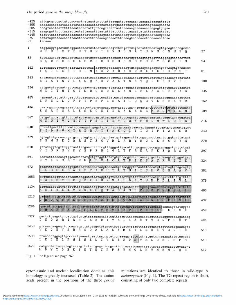
Fig. 1. For legend see page 262.
cytoplasmic and nuclear localization domains, this
homology is greatly increased (Table 2). The amino
acids present in the positions of the three period
mutations are identical to those in wild-type D.
melanogaster (Fig. 1). The TG repeat region is short,
consisting of only two complete repeats.
https://doi.org/10.1017/S0016672399004425Downloaded from https://www.cambridge.org/core. IP address: 65.21.229.84, on 10 Jan 2022 at 19:33:30, subject to the Cambridge Core terms of use, available at https://www.cambridge.org/core/terms.

G. R. Warman et al. 262
Fig. 1. Nucleotide and inferred amino acid sequence of L. cuprina per cDNA (Genbank accession number Y19108).Motifs corresponding to D. melanogaster functional domains are highlighted: , nuclear localization signal (aa 65–80) ;
, PAS A domain (aa 183–232) ; , PAS B domain (aa 307–396) ; , cytoplasmic localization signal (aa 394–454) ; ,sites of period mutations in D. melanogaster : (I) PerL mutation (aa 186), (II) per! mutation and (III) pers mutation.Nucleotide numbering is on the left and starts at the adenine of the predicted ATG start of translation. Amino acidnumbering is on the right. The predicted stop of translation is marked with an asterisk and the predictedpolyadenylation signal is underlined.
(ii) TG repeat length does not �ary with latitude in
L. cuprina or L. sericata
No polymorphism in TG repeat length was found
across any of the samples analysed. All the flies
analysed maintained the two complete repeat form
(TGTGT) as shown in Fig. 1. However, two amino
acid encoding polymorphisms were detected in the
733 bp fragment spanning the repeat. All five L.
sericata strains contained an alanine at amino acid
595 while all L. cuprina strains contained a glutamic
acid at this position. The L. cuprina strain LS2
https://doi.org/10.1017/S0016672399004425Downloaded from https://www.cambridge.org/core. IP address: 65.21.229.84, on 10 Jan 2022 at 19:33:30, subject to the Cambridge Core terms of use, available at https://www.cambridge.org/core/terms.
The period gene in the sheep blow fly 263
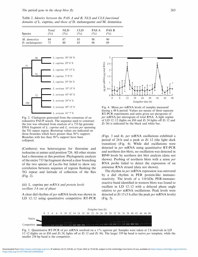
Table 2. Identity between the PAS A and B, NLS and CLS functional
domains of L. cuprina, and those of D. melanogaster and M. domestica
SpeciesTotal(%)
NLD(%)
CLD(%)
PAS A(%)
PAS B(%)
M. domestica 84 87 85 98 90D. melanogaster 73 80 85 96 89
L. cuprina 30° 30′ Ν
L. cuprina 29° 6′ S
L. cuprina 35° 15′ S
L. cuprina 3° 0′ Ν
L. cuprina 39° 56′ S
L. sericata 35° 15′ S
L. sericata 43° 45′ S
L. sericata 29° 6′ S
L. sericata 49° 15′ S
L. sericata 41° 7′ S
90
51
91
72
100
Fig. 2. Cladogram generated from the consensus of anexhaustive PAUP search. The sequence used to constructthe tree was obtained from analysis of a 733 bp genomicDNA fragment of L. cuprina and L. sericata per spanningthe TG repeat region. Bootstrap values are indicated onthose branches which have greater than 50% support.Branches with less than 50% support have beencollapsed.
(Canberra) was heterozygous for threonine and
isoleucine at amino acid position 726. All other strains
had a threonine at this position. Phylogenetic analysis
of the entire 733 bp fragment showed a clear branching
of the two species of Lucilia but failed to show any
correlation between sequence of regions flanking the
TG repeat and latitude of collection of the flies
(Fig. 2).
(iii) L. cuprina per mRNA and protein le�els
oscillate 3 h out of phase
A clear diel rhythm of per mRNA levels was shown in
LD 12:12 using quantitative competitive RT-PCR
0 2 6 8 104 12 14 16 18 20 22 24 26 28 30 32 34 36 38 40 42 44 46 48
Native
Competitior
Zeitgeber time (h)
Fig. 3. Quantitative RT-PCR of per mRNA resolved on a 1% agarose gel. Samples were taken at 2 h intervals in LD12:12 (lights on at Zt0 and Zt 24, lights off at Zt 12 and Zt 36). The larger 359 bp band is native per template, while thesmaller 238 bp band is the competitor.
3
2·5
2
1
0
1·5
0·5
pg p
er m
RN
A/í
g to
tal R
NA
0 6 12 18 24 30 36 42 48
Zeitgeber time (h)
Fig. 4. Mean per mRNA levels of samples measuredduring a 48 h period. Values are means of three separateRT-PCR experiments and units given are picograms ofper mRNA per microgram of total RNA. A light regimeof LD 12:12 (lights on Zt0 and Zt 24 lights off Zt 12 andZt 36) is indicated by the black and white bar.
(Figs. 3 and 4). per mRNA oscillations exhibited a
period of 24 h and a peak at Zt 12 (the light–dark
transition) (Fig. 4). While diel oscillations were
detected in per mRNA using quantitative RT-PCR
and northern dot-blots, no oscillation was detected in
RP49 levels by northern dot blot analysis (data not
shown). Probing of northern blots with a sense per
RNA probe failed to detect the expression of an
antisense RNA strand (data not shown).
The rhythm in per mRNA expression was mirrored
by a diel rhythm in PER protein-like immuno-
reactivity. The levels of a 110 kDa PER-immuno-
reactive band identified in western blots was found to
oscillate in LD 12:12 with a delayed phase angle
relative to per mRNA oscillations. Peak levels were
detected at Zt 15 (3 h after the peak per mRNA levels)
(Fig. 5).
https://doi.org/10.1017/S0016672399004425Downloaded from https://www.cambridge.org/core. IP address: 65.21.229.84, on 10 Jan 2022 at 19:33:30, subject to the Cambridge Core terms of use, available at https://www.cambridge.org/core/terms.

G. R. Warman et al. 264
MW C 0 3 2118151296200 kD
113 kD
82 kD
Zeitgeber time (h)
Fig. 5. Autoradiograph of western blot (ECL) of Per-lir levels in L. cuprina maintained in LD 12:12. Samples takenevery 3 h were resolved on an 8% SDS acrylamide gel and Westerns probed with Per no. 107 polyclonal antibody (a giftfrom L. Saez). The reactive band detected has an estimated molecular weight of approximately 110 kDa. Lanes 1 and 2show a Coomassie-stained molecular weight marker (BioRad) and a total protein sample. Lanes 3–10 indicate theamount of Per-lir in samples taken from the time points noted.
4. Discussion
per is a central clock gene involved in the generation
of the circadian feedback loop and has been extensively
investigated in D. melanogaster. We have isolated an
entire per cDNA homologue from a representative of
a second dipteran family : the sheep blow fly Lucilia
cuprina (Calliphoridae). The predicted protein of
1037 amino acids is 73% identical to Drosophila PER.
This is within the range of comparisons of other
nuclear genes : 73% for the LcEcR ecdysone receptor
gene (Hannan & Hill, 1997), 74% for the notch
homologue (Chen et al., 1998), 64% for the E3 gene
(Newcomb et al., 1997) and 78% for the white gene
(Garcia et al., 1996). As with other L. cuprina genes,
the levels of AT bias are high (Garcia et al., 1996;
Newcomb et al., 1997), thus preventing informative
nucleotide comparisons from being made with other
flies such as Drosophila.
In contrast to the Diptera, lepidopteran per has
been noted for its extremely rapid rate of evolution
(Schmid and Tautz, 1997; Regier et al., 1998). Within
the PAS domain lepidopteran per has evolved 5 to 40
times more rapidly than other nuclear genes (Regier et
al., 1998). The difference in rates of per evolution
between the Diptera and the Lepidoptera cannot be
attributed to an evolutionary time scale, as molecular
clock evidence shows that the radiation of the higher
flies and the moths both occurred during the Cre-
taceous period (Beverley & Wilson, 1984).
The presence of particular regions of conservation
and variability in the per coding sequence first
mentioned by Colot et al. (1988) was evident in L.
cuprina, and many of the conserved regions were
associated with functional domains (Table 1).
Possession of a PAS domain and in many cases a
TG repeat are hallmarks of a clock gene. PAS
domains and TG repeats have been found in clock
genes from a wide range of species, from Drosophila
through to the bread mould Neurospora crassa (Citri
et al., 1987; Kay, 1997; Linden & Macino, 1997), and
the analysis of these regions may provide evidence for
the evolutionary origins of circadian clocks (Kay,
1997). Particularly high conservation of the PAS,
CLD and NLD domains between D. melanogaster
and L. cuprina suggests that the L. cuprina feedback
loop may function in a similar manner to that of
Drosophila, and we may predict the existence of a
timeless homologue in the sheep blow fly. The high
degree of sequence divergence of per within the
Lepidoptera may reflect the different functional nature
of the circadian feedback loop in moths.
The two-repeat form of the TG motif evident in L.
cuprina is characteristic of all non-drosophilid flies,
which have been shown to maintain this stable repeat
length (Nielsen et al., 1994). Despite the different
habitat ranges and upper lethal limits of L. cuprina
and L. sericata, the lack of variation in TG repeat
length, and the lack of correlation of flanking se-
quences with latitude of strain collection (Fig. 2),
provide no evidence for the involvement of this region
in temperature compensation of the circadian clock in
these species (as it has done in Drosophila). The lack
of variation in this region does not, however, pre-
clude its involvement in temperature compensation.
Similarly it does not rule out the involvement of
other regions such as the PAS domain in conferring
temperature compenzation (Huang et al., 1995; Curtin
et al., 1995).
L. cuprina maintains a 22 h free-running period in
constant conditions (Smith, 1983) compared with the
24 h period of Drosophila. The amino acids which give
rise to changes in period length when altered in
Drosophila have been conserved in the wild-type form
of L. cuprina per (Fig. 1). This suggests that the
shorter free-running period in Lucilia is attributable
either to the action of a different amino acid mutation
in per, or to a difference in another functional element
of the L. cuprina feedback loop.
In Drosophila, per was localized by mapping three
free-running period mutations to the left arm of the X
chromosome (Konopka & Benzer, 1971). The map-
ping of L. cuprina per using clones isolated in the
present work has shown per to occupy a position
homologous to that of per in the Drosophila genome
(P. Batterham, personal communication 1998). This
https://doi.org/10.1017/S0016672399004425Downloaded from https://www.cambridge.org/core. IP address: 65.21.229.84, on 10 Jan 2022 at 19:33:30, subject to the Cambridge Core terms of use, available at https://www.cambridge.org/core/terms.
The period gene in the sheep blow fly 265
mapping also indicates that the ary mutation, which
maps to chromosome V (3L equivalent in Drosophila)
and has severe effects on the circadian phenotype
(Smith, 1987), is either a Lucilia homologue of another
clock element or a novel clock gene. There also
remains the possibility that ary corresponds to the
arrhythmic XR allele of D. pseudoobscura (Smith,
1987).
The expression studies conducted here demonstrate
that both per mRNA and protein levels oscillate in a
diel manner. This provides further evidence that the
per homologue in Lucilia is a functional element of the
circadian feedback loop. The expression pattern of per
mRNA in L. cuprina is remarkably similar to that of
D. melanogaster, with peak levels maintaining com-
parable phase angles in both species (mRNA levels
peak at Zt 12 in L. cuprina and Zt 13 in D.
melanogaster). As in D. melanogaster, peak protein
levels lag significantly behind mRNA levels ; however,
the phase angle of PER protein oscillations in Lucilia
is advanced with respect to the Drosophila system
(peak PER-lir levels occur at Zt 15 in L. cuprina
compared with Zt19–20 in Drosophila). It may be
possible that the shortened phase angle is an artefact
of the entrainment of the free-running period to 24 h
by the light cycle, as has been noted in per! mutants
(Marrus et al., 1996) ; however, this shortened phase
angle may also reflect the protracted 22 h behavioural
circadian cycle in Lucilia. The observation of a shorter
phase angle in Lucilia may have significant impli-
cations for the L. cuprina feedback loop, as the
kinetics of transcription and translation are such that
the approximately 3 h delay observed in Lucilia does
not require an independent delay mechanism, whereas
the 4–6 h delay observed in Drosophila does (U.
Schibler, personal communication 1998).
Despite the diversity of per expression in the insect
species previously investigated, the pattern in Lucilia
corresponds closely with that of Drosophila. In
addition to these oscillations, no antisense RNA has
been detected, thus eliminating the possibility of
cycling sense and antisense mRNA strands as is found
in Lepidoptera. The implications of these data for the
use of Drosophila as a fly model are important. As this
is the first system which appears to mirror Drosophila
closely, these findings enhance the relevance of the
Drosophila model. Therefore, the Drosophila system
may be the rule rather than the exception, at least in
the Diptera.
The authors wish to thank Dr L. Huynen (MasseyUniversity, N.Z.) and Dr C. Millar (University of Auck-land, N.Z.) for their technical advice. The kind donation ofthe Per no.107 polyclonal antibody by Dr L. Saez and DrM. Young (Rockefeller University, New York) made de-tection of the L. cuprina Per-lir possible. Many thanks alsogo to Dr P. East (CSIRO, Australia) for the donation of aL. cuprina cDNA library, and to Dr D. Gleeson for
supplying DNA strains of L. cuprina and L. sericata. Thiswork was supported by a University of Auckland Staffresearch grant, and by an AGMARDT doctoral scholarshipto G.R.W.
References
Allada, R., While, N., So, W. V, Hall, J. C. & Rosbash, M.(1998). A mutant Drosophila homolog of mammalianClock disrupts circadian rhythms and transcription ofperiod and timeless. Cell 93, 791–804.
Bargiello, T. A., Jackson, F. R. & Young, M. W. (1984).Restoration of circadian behavioral rhythms by genetransfer in Drosophila. Nature 312, 752–754.
Baylies, M. K, Weiner, L., Vosshall, L. B., Saez, L. &Young, M. W. (1993). Genetic, molecular, and cellularstudies of the per locus and its products in Drosophilamelanogaster. In Molecular Genetics of Biological Rhythms(ed. M. W. Young), pp. 123–154. New York: MarcelDekker.
Beverley, S. M. & Wilson, A. C. (1984). Molecular evolutionin Drosophila and the higher Diptera. II. A time scale forfly evolution. Journal of Molecular E�olution 21, 1–13.
Bradford, M. N. (1976). A rapid and sensitive method forthe quantification of microgram quantities of proteinutilizing the principle of protein-dye binding. AnalyticalBiochemistry 72, 248–254.
Chen, Z., Newsome, T., McKenzie, J. A. & Batterham, P.(1998). Molecular characterization of the notch hom-ologue from the Australian sheep blowfly, Lucilia cuprina.Insect Biochemistry and Molecular Biology 28, 601–612.
Citri, Y., Colot, H. V., Jacquier, A. C., Yu, Q., Hall, J. C.,Baltimore, D. & Rosbash, M. (1987). A family ofunusually spliced biologically active transcripts encodedby a Drosophila clock gene. Nature 326, 42–47.
Colot, H. V., Hall, J. C. & Rosbash, M. (1988). Interspecificcomparison of the period gene of Drosophila reveals largeblocks of non-conserved coding DNA. EMBO Journal 7,3929–3937.
Costa, R., Peixoto, A. A., Barbujani, G. & Kyriacou, C. P.(1992). A latitudinal cline in a Drosophila clock gene.Proceedings of the Royal Society of London, Series B 250,43–49.
Curtin, K. D., Huang, Z. J. & Rosbash, M. (1995).Temporally regulated nuclear entry of the Drosophilaperiod protein contributes to the circadian clock. Neuron14, 365–372.
Darlington, T. K., Wager-Smith, K., Ceriani, M. F.,Staknis, D., Gekakis, N., Steeves, T. D. L., Weitz, C. J.,Takahashi, J. S. & Kay, S. A. (1998). Closing the circadianloop: CLOCK-induced transcription of its own inhibitorsper and tim. Science 280, 1599–1602.
Ewer, J., Hamblen-Coyle, M., Rosbash, M. & Hall, J. C.(1990). Requirement for period gene expression in theadult and not during development for locomotor activityrhythms of imaginal Drosophila melanogaster. Journal ofNeurogenetics 7, 31–73.
Feliciello, I. & Chinalli, G. (1993). A modified alkaline lysismethod for the preparation of highly purified plasmidDNA from Escherichia coli. Analytical Biochemistry 212,394–401.
Garcia, R. L., Perkins, H. D. & Howells, A. J. (1996). Thestructure, sequence and developmental pattern of ex-pression of the white gene in the blowfly Lucilia cuprina.Insect Molecular Biology 5, 251–260.
Gekakis, N., Saez, L., Delahaye-Brown, A., Myers, M. P.,Sehgal, A., Young, M. W. & Weitz, C. J. (1995). Isolationof timeless by PER protein interaction: Defective in-
https://doi.org/10.1017/S0016672399004425Downloaded from https://www.cambridge.org/core. IP address: 65.21.229.84, on 10 Jan 2022 at 19:33:30, subject to the Cambridge Core terms of use, available at https://www.cambridge.org/core/terms.

G. R. Warman et al. 266
teraction between timeless protein and long-period mutantPerL. Science 270, 811–815.
Hannan, G. N. & Hill, R. J. (1997). Cloning and charac-terization of LcEcR: a functional ecdysone receptor fromthe seep blowfly Lucilia cuprina. Insect Biochemistry andMolecular Biology 27, 479–488.
Hardin, P. E., Hall, J. C. & Rosbash, M. (1990). Feedbackof the Drosophila period gene product on circadian cyclingof its messenger RNA levels. Nature 343, 536–540.
Huang, Z. J., Edery, I. & Rosbash, M. (1993). PAS is adimerization domain common to Drosohila period andseveral transcription factors. Nature 364, 259–262.
Huang, Z. J., Curtin, K. D. & Rosbash, M. (1995). PERprotein interactions and temperature compenzation of acircadian clock in Drosophila. Science 267, 1169–1172.
Kay, S. A. (1997). PAS, present and future : clues to theorigins of circadian clocks. Science 276, 753–754.
Konopka, R. J. & Benzer, S. (1971). Clock mutants ofDrosophila melanogaster. Proceedings of the NationalAcademy of Sciences of the USA 68, 2112–2116.
Konopka, R. J., Kyriacou, C. P. & Hall, J. C. (1996).Mosaic analysis in the CNS of circadian and courtshiprhythms affected by a period clock mutation. Journal ofNeurogenetics 11, 117–139.
Kyriacou, C. P., Geenacre, J. R., Thackeray, J. R. & Hall,J. C. (1993). Genetic and molecular analysis of songrhythms in Drosophila. In Molecular Genetics of BiologicalRhythms (ed. M. W. Young), pp. 171–194. New York:Marcel Dekker.
Linden, H. & Macino, G. (1997). White collar 2, a partnerin blue-light signal transduction, controlling expression oflight-regulated gens in Neurospora crassa. The EMBOJournal 16 (1), 98–109.
Ma, P. W. K., Knipple, D. C. & Roelofs, W. L. (1994).Structural organization of the Helioco�erpa zea geneencoding the precursor protein for pheremone bio-synthesis-activating neuropeptide and other neuro-peptides. Proceedings of the National Academy of Sciencesof the USA 91, 6506–6510.
Marrus, S. B., Zeng, H. & Rosbash, M. (1996). Effect ofconstant light and circadian entrainment of pers flies :evidence for light mediated delay of the negative feedbackloop. EMBO Journal 15, 6877–6886.
Myers, M. P., Wager-Smith, K., Wesley, C. S., Young,M. W. & Sehgal, A. (1995). Positional cloning andsequence analysis of the Drosophila clock gene timeless.Science 270, 805–808.
Newcomb, R. D., Campbell, P. M., Russell, R. J. &Oakshott, J. G. (1997). cDNA cloning, bacilovirus-expression and kinetic properties of esterase, E3, involvedin organophosphorus resistance in Lucilia cuprina. InsectBiochemistry and Molecular Biology 27,15–25.
Nielsen, J., Peixoto, A. A., Piccin, A., Costa, R., Kyriacou,C. P. & Chalmers, D. (1994). Big flies, small repeats : theThr-Gly repeat region on the period gene in Diptera.Molecular Biology and E�olution 11, 839–853.
Peixoto, A. A., Campesan, S., Costa, R. & Kyriacou, C. P.(1993). Molecular evolution of a repetitive region withinthe per gene of Drosophila. Molecular Biology andE�olution 10, 127–139.
Peixoto, A. A., Hennessy, J. M., Townson, I., Hasan, G.,Rosbash, M., Costa, R. & Kyriacou, C. P. (1998).Molecular coevolution within a Drosophila clock gene.Proceedings of the National Academy of Sciences of theUSA 95, 4475–4480.
Persichetti, F., Ambrose, C. M., Ge, P., McNeil, S. M.,Srinidhi, J., Anderson, M. A., Jenkins, B., Barnes, G. T.,
Duyao, M. P., Kanaley, L., Wexler, N. S., Myers, R. H.,Bird, E. D., Vonsattel, J.-P., MacDonald, M. E. &Gusella, J. F. (1993). Normal and expanded Huntington’sdisease gene alleles produce distinguishable proteins dueto translation across the CAG repeat. Molecular Medicine1, 374–383.
Price, J. L., Blau, J., Rothenfluh, A., Abodeely, M., Kloss,B. & Young, M. W. (1998). double-time is a novelDrosophila clock gene that regulates PERIOD proteinaccumulation. Cell 94, 83–95.
Reddy, P., Zehring, W. A., Wheeler, D. A., Pirrotta, V.,Hadfield, C., Hall, J. C. & Rosbash, M. (1984). Molecularanalysis of the period locus in Drosophila melanogasterand identification of a transcript involved in biologicalrhythms. Cell 38, 701–710.
Regier, J. C., Fang, Q. Q., Mitter, C., Peigler, R. S.,Friedlander, T. P. & Solis, M. A. (1998). Evolution andphylogenetic utility of the period gene in Lepidoptera.Molecular Biology and E�olution 15, 1172–1182.
Reppert, S. M., Tsai, T., Roca, A. L. & Sauman, I. (1994).Cloning of a structural and functional homolog of thecircadian clock gene period from the giant silkmothAntheraea pernyi. Neuron 13, 1167–1176.
Rutila, J. E., Suri, V., Le, M., So, V., Rosbash, M. & Hall,J. C. (1998). CYCLE is a second bHLH-PAS clockprotein essential for circadian rhythmicity and tran-scription of Drosophila period and timeless. Cell 93,805–814.
Saez, L. & Young, M. W. (1996). Regulation of nuclearentry of the Drosophila clock proteins Period andTimeless. Neuron 17, 911–920.
Sangoram, A. M., Saez, L., Antoch, M. P., Gekakis, N.,Staknis, D., Whiteley, A., Fruechte, E. M., Vitaterna,M. H., Shimomura, K., King, D. P., Young, M. W.,Weitz, C. J. & Takahashi, J. S. (1998). Mammaliancircadian autoregulatory loop: a timeless ortholog andmPer1 interact and negatively regulate CLOCK-BMAL1-induced transcription. Neuron 21, 1101–1113.
Sauman, I., Tsai, T., Roca, A. L. & Reppert, S. M. (1996).Period protein is necessary for circadian control of egghatching behavior in the silkmoth Antheraea pernyi.Neuron 17, 901–909.
Saunders, D. S. (1977). An Introduction to BiologicalRhythms. London: Blackie.
Sawyer, L. A., Hennessy, J. M., Peixoto, A. A., Rosato, E.,Parkinson, H., Costa, R. & Kyriacou, C. P. (1997).Natural variation in a Drosophila clock gene andtemperature compenzation. Science 278, 2117–2120.
Schmid, K. J. & Tautz, D. (1997). A screen for fast evolvinggenes from Drosophila. Proceedings of the NationalAcademy of Sciences of the USA 94, 9746–9750.
Sehgal, A., Price, J. C., Man, B. & Young, M. W. (1994).Loss of circadian behavioral rhythms and per mRNAoscillations in the Drosophila mutant timeless. Science263, 1603–1606.
Siwicki, K. K., Eastman, C., Petersen, G., Rosbash, M. &Hall, J. C. (1988). Antibodies to the period gene productof Drosophila reveal diverse tissue distribution andrhythmic changes in the visual system. Neuron 1, 141–150.
Smith, P. H. (1983). Circadian control of spontaneous flightactivity in the blowfly Lucilia cuprina. PhysiologicalEntomology 8, 73–82.
Smith P. H. (1987). Naturally occurring arrhythmicity ineclosion and activity in Lucilia cuprina : its genetic basis.Physiological Entomology 12, 99–107.
Smith, P. H., Dallwitz, R., Wardaugh, K. G., Vogt, W. G.
https://doi.org/10.1017/S0016672399004425Downloaded from https://www.cambridge.org/core. IP address: 65.21.229.84, on 10 Jan 2022 at 19:33:30, subject to the Cambridge Core terms of use, available at https://www.cambridge.org/core/terms.
The period gene in the sheep blow fly 267
& Woodburn, T. L. (1981). Timing of larval exodus fromsheep and carrion in the sheep blowfly Lucilia cuprina.Entomologia Experimentalis et Applicata 30, 157–162.
Sun, Z. H., Albrecht, U., Zhuchenko, O., Bailey, J.,Eichele, G. & Lee, C. C. (1997). RIGUI, a putativemammalian ortholog of the Drosophila period gene. Cell90, 1003–1011.
Swofford, D. L. (1993). PAUP: phylogenetic analysis usingparsimony, version 3.1. Distributed by the Illinois NaturalHistory Survey.
Tei, H., Okamura, H., Shigeyoshi, Y., Fukuhara, C.,Ozawa, R., Hirose, M. & Sakaki, Y. (1997). Circadianoscillation of a mammalian homolog of the Drosophilaperiod gene. Nature 389, 512–516.
Tsai, S.-J. & Wiltbank, M. C. (1996). Quantification ofmRNA using competitive RT-PCR with standard curvemethodology. BioTechniques 21, 862–866.
Vosshall, L. B., Price, J. C., Sehgal, A., Saez, L. & Young,
M. W. (1994). Block in nuclear localization of periodprotein by a second clock mutation, timeless. Science 263,1606–1609.
Weller, G. L. & Foster, G. G. (1993). Genetic maps of thesheep blow fly Lucilia cuprina : linkage-group correlationswith other dipteran genera. Genome 36, 495–506.
Zerr, D. M., Hall, J. C., Rosbash, M. & Siwicki, K. K.(1990). Circadian fluctuations of period immunoreactivityin the CNS and visual system of Drosophila. Journal ofNeuroscience 10, 2749–2762.
Zylka, M. J., Shearman, L. P., Weaver, D. R., & Reppert,S. M. (1998a). Three period homologs in mammals :differential light responses in the suprachiasmatic cir-cadian clock and oscillating transcripts outside of brain.Neuron 20, 1103–1110.
Zylka, M. J., Shearman, L. P., Levine, J. D, Jin, X., Wever,D. R. & Reppert, S. M. (1998b). Molecular analysis ofmammalian timeless. Neuron 21, 1115–1122.
https://doi.org/10.1017/S0016672399004425Downloaded from https://www.cambridge.org/core. IP address: 65.21.229.84, on 10 Jan 2022 at 19:33:30, subject to the Cambridge Core terms of use, available at https://www.cambridge.org/core/terms.