SIRT1 Regulates Circadian Clock Gene Expression through PER2 Deacetylation
12
SIRT1 Regulates Circadian Clock Gene Expression through PER2 Deacetylation Gad Asher, 1 David Gatfield, 1 Markus Stratmann, 1 Hans Reinke, 1 Charna Dibner, 1 Florian Kreppel, 2 Raul Mostoslavsky, 3 Frederick W. Alt, 4 and Ueli Schibler 1, * 1 Department of Molecular Biology, Sciences III, University of Geneva, 30, Quai Ernest Ansermet, CH-1211 Geneva-4, Switzerland 2 Division of Gene Therapy, University of Ulm, Helmholtzstrasse 8/1, D-89081 Ulm, Germany 3 Massachusetts General Hospital Cancer Center, Harvard Medical School, Boston, MA 02114, USA 4 Howard Hughes Medical Institute, Children’s Hospital, Center for Blood Research, and Department of Genetics, Harvard University Medical School, Boston, MA 02115, USA *Correspondence: [email protected] DOI 10.1016/j.cell.2008.06.050 SUMMARY The mammalian circadian timing system is com- posed of a central pacemaker in the suprachiasmatic nucleus of the brain that synchronizes countless subsidiary oscillators in peripheral tissues. The rhythm-generating mechanism is thought to rely on a feedback loop involving positively and negatively acting transcription factors. BMAL1 and CLOCK activate the expression of Period (Per) and Crypto- chrome (Cry) genes, and once PER and CRY proteins accumulate to a critical level they form complexes with BMAL1-CLOCK heterodimers and thereby repress the transcription of their own genes. Here, we show that SIRT1, an NAD + -dependent protein deacetylase, is required for high-magnitude circa- dian transcription of several core clock genes, in- cluding Bmal1, Rorg, Per2, and Cry1. SIRT1 binds CLOCK-BMAL1 in a circadian manner and promotes the deacetylation and degradation of PER2. Given the NAD + dependence of SIRT1 deacetylase activity, it is likely that SIRT1 connects cellular metabolism to the circadian core clockwork circuitry. INTRODUCTION The physiology and behavior of mammals are subject to daily oscillations driven by an endogenous circadian clock (Albrecht and Eichele, 2003; Reppert and Weaver, 2002). In mammals, the circadian timing system is composed of a central pacemaker in the brain’s suprachiasmatic nucleus (SCN) and subsidiary oscillators in most peripheral tissues. While light-dark cycles are the predominant Zeitgebers (timing cues) for the SCN pace- maker, cyclic feeding behavior is a strong Zeitgeber for clocks operating in many peripheral tissues (Albrecht and Eichele, 2003; Damiola et al., 2000; Reppert and Weaver, 2002; Stokkan et al., 2001). It is therefore likely that the SCN synchronizes peripheral oscillators by imposing rest-activity rhythms and thus feeding-fasting cycles. The molecular oscillator in both master and subsidiary clocks (Balsalobre et al., 1998; Yagita et al., 2001) is thought to rely on a negative transcriptional feedback loop (Hardin et al., 1990; Lowrey and Takahashi, 2000; Reppert and Weaver, 2002). The PAS domain helix-loop-helix proteins BMAL1 and CLOCK (or its paralog NPAS2 [DeBruyne et al., 2007; Reick et al., 2001]) bind as heterodimers to regulatory elements of Cry and Per genes and stimulate the transcription of these genes. Once the repres- sor proteins CRY and PER have reached a critical concentration, they attenuate the activity of BMAL1-CLOCK heterodimers and thereby repress the transcription of their own genes. In addition, an interconnecting feedback loop involving orphan nuclear re- ceptors of the REV-ERB and ROR families regulates the expres- sion of Bmal1 (Preitner et al., 2002; Sato et al., 2004). Several lines of evidence suggest a strong interplay between metabolism and the circadian clock (Kaasik and Lee, 2004; Rutter et al., 2002; Tu and McKnight, 2006). The dominance of feeding cycles as a Zeitgeber for peripheral clocks implies that the circadian clock plays an important role in nutrient process- ing and energy homeostasis. Indeed, transcriptome profiling studies revealed that many genes involved in metabolism are rhythmically expressed (Akhtar et al., 2002; Duffield et al., 2002; Kornmann et al., 2007; Panda et al., 2002; Storch et al., 2002; Walker and Hogenesch, 2005). Furthermore, at least in vitro, the DNA-binding activity of BMAL1-CLOCK is strongly influenced by the ratio of reduced to oxidized NAD cofactors, which are often considered as a readout of the cellular metabolic state (Rutter et al., 2001). SIRT1 is the mammalian homolog of yeast Sir2, an NAD + - dependent deacetylase involved in transcriptional silencing, genome stability, and longevity (Blander and Guarente, 2004; Dali-Youcef et al., 2007). The SIRT1 catalytic reaction involves the breakdown of one NAD + molecule for each deacetylated acetyl lysine and the generation of nicotinamide and O-acetyl- ADP-ribose. SIRT1 was found to deacetylate not only histones but also several transcriptional regulatory proteins involved in the control of metabolism, including members of the FOXO protein family (Bordone et al., 2006; Brunet et al., 2004; Motta et al., 2004), peroxisome proliferator-activated receptor gamma (PPARg) coactivator 1a (PGC1a), and the nuclear receptor LXR (Li et al., 2007; Rodgers et al., 2005). Cell 134, 317–328, July 25, 2008 ª2008 Elsevier Inc. 317
-
Upload
independent -
Category
Documents
-
view
0 -
download
0
Transcript of SIRT1 Regulates Circadian Clock Gene Expression through PER2 Deacetylation

SIRT1 Regulates Circadian Clock GeneExpression through PER2 DeacetylationGad Asher,1 David Gatfield,1 Markus Stratmann,1 Hans Reinke,1 Charna Dibner,1 Florian Kreppel,2 Raul Mostoslavsky,3
Frederick W. Alt,4 and Ueli Schibler1,*1Department of Molecular Biology, Sciences III, University of Geneva, 30, Quai Ernest Ansermet, CH-1211 Geneva-4, Switzerland2Division of Gene Therapy, University of Ulm, Helmholtzstrasse 8/1, D-89081 Ulm, Germany3Massachusetts General Hospital Cancer Center, Harvard Medical School, Boston, MA 02114, USA4Howard Hughes Medical Institute, Children’s Hospital, Center for Blood Research, and Department of Genetics,
Harvard University Medical School, Boston, MA 02115, USA
*Correspondence: [email protected]
DOI 10.1016/j.cell.2008.06.050
SUMMARY
The mammalian circadian timing system is com-posed of a central pacemaker in the suprachiasmaticnucleus of the brain that synchronizes countlesssubsidiary oscillators in peripheral tissues. Therhythm-generating mechanism is thought to rely ona feedback loop involving positively and negativelyacting transcription factors. BMAL1 and CLOCKactivate the expression of Period (Per) and Crypto-chrome (Cry) genes, and once PER and CRY proteinsaccumulate to a critical level they form complexeswith BMAL1-CLOCK heterodimers and therebyrepress the transcription of their own genes. Here,we show that SIRT1, an NAD+-dependent proteindeacetylase, is required for high-magnitude circa-dian transcription of several core clock genes, in-cluding Bmal1, Rorg, Per2, and Cry1. SIRT1 bindsCLOCK-BMAL1 in a circadian manner and promotesthe deacetylation and degradation of PER2. Giventhe NAD+ dependence of SIRT1 deacetylase activity,it is likely that SIRT1 connects cellular metabolism tothe circadian core clockwork circuitry.
INTRODUCTION
The physiology and behavior of mammals are subject to daily
oscillations driven by an endogenous circadian clock (Albrecht
and Eichele, 2003; Reppert and Weaver, 2002). In mammals,
the circadian timing system is composed of a central pacemaker
in the brain’s suprachiasmatic nucleus (SCN) and subsidiary
oscillators in most peripheral tissues. While light-dark cycles
are the predominant Zeitgebers (timing cues) for the SCN pace-
maker, cyclic feeding behavior is a strong Zeitgeber for clocks
operating in many peripheral tissues (Albrecht and Eichele,
2003; Damiola et al., 2000; Reppert and Weaver, 2002; Stokkan
et al., 2001). It is therefore likely that the SCN synchronizes
peripheral oscillators by imposing rest-activity rhythms and
thus feeding-fasting cycles.
The molecular oscillator in both master and subsidiary clocks
(Balsalobre et al., 1998; Yagita et al., 2001) is thought to rely on
a negative transcriptional feedback loop (Hardin et al., 1990;
Lowrey and Takahashi, 2000; Reppert and Weaver, 2002). The
PAS domain helix-loop-helix proteins BMAL1 and CLOCK (or
its paralog NPAS2 [DeBruyne et al., 2007; Reick et al., 2001])
bind as heterodimers to regulatory elements of Cry and Per genes
and stimulate the transcription of these genes. Once the repres-
sor proteins CRY and PER have reached a critical concentration,
they attenuate the activity of BMAL1-CLOCK heterodimers and
thereby repress the transcription of their own genes. In addition,
an interconnecting feedback loop involving orphan nuclear re-
ceptors of the REV-ERB and ROR families regulates the expres-
sion of Bmal1 (Preitner et al., 2002; Sato et al., 2004).
Several lines of evidence suggest a strong interplay between
metabolism and the circadian clock (Kaasik and Lee, 2004;
Rutter et al., 2002; Tu and McKnight, 2006). The dominance of
feeding cycles as a Zeitgeber for peripheral clocks implies that
the circadian clock plays an important role in nutrient process-
ing and energy homeostasis. Indeed, transcriptome profiling
studies revealed that many genes involved in metabolism
are rhythmically expressed (Akhtar et al., 2002; Duffield et al.,
2002; Kornmann et al., 2007; Panda et al., 2002; Storch et al.,
2002; Walker and Hogenesch, 2005). Furthermore, at least
in vitro, the DNA-binding activity of BMAL1-CLOCK is strongly
influenced by the ratio of reduced to oxidized NAD cofactors,
which are often considered as a readout of the cellular metabolic
state (Rutter et al., 2001).
SIRT1 is the mammalian homolog of yeast Sir2, an NAD+-
dependent deacetylase involved in transcriptional silencing,
genome stability, and longevity (Blander and Guarente, 2004;
Dali-Youcef et al., 2007). The SIRT1 catalytic reaction involves
the breakdown of one NAD+ molecule for each deacetylated
acetyl lysine and the generation of nicotinamide and O-acetyl-
ADP-ribose. SIRT1 was found to deacetylate not only histones
but also several transcriptional regulatory proteins involved in
the control of metabolism, including members of the FOXO
protein family (Bordone et al., 2006; Brunet et al., 2004; Motta
et al., 2004), peroxisome proliferator-activated receptor gamma
(PPARg) coactivator 1a (PGC1a), and the nuclear receptor LXR
(Li et al., 2007; Rodgers et al., 2005).
Cell 134, 317–328, July 25, 2008 ª2008 Elsevier Inc. 317

Figure 1. Circadian Expression of SIRT1 Protein
Mice were sacrificed at 4 hr intervals; liver nuclear protein extracts and total RNA were prepared.
(A) Protein extracts were analyzed by immunoblotting.
(B) RNA was analyzed by quantitative TaqMan real-time PCR using specific TaqMan probes.
MEFs were synchronized by a dexamethasone shock and samples were collected at 4 hr intervals, starting 24 hr after the shock.
(C) Protein extracts were analyzed by immunoblotting.
(D) RNA was analyzed by quantitative TaqMan real-time PCR using specific TaqMan probes.
Plotted values are the mean values ± standard deviation (SD) from three independent experiments.
Here we show that SIRT1 is expressed in a circadian manner
and that it is required for high-magnitude circadian expression
of several core clock genes. Moreover, we present evidence
that SIRT1 binds to CLOCK-BMAL1 heterodimers and promotes
the deacetylation and degradation of PER2.
RESULTS
Circadian Expression of SIRT1 Protein in Mouse Liverand in Cultured FibroblastsIn order to investigate whether SIRT1 might be involved in circa-
dian rhythm, we first examined the temporal expression of SIRT1
in mouse liver. Mice were sacrificed at 4 hr intervals around the
clock, and liver nuclear proteins were prepared and analyzed.
The results showed that SIRT1 accumulated in a circadian man-
ner with maximal and minimal levels reached at around Zeitgeber
time (ZT) 16 and ZT4, respectively (Figure 1A). In parallel, we also
followed the temporal expression of known core clock proteins.
As reported previously, circadian changes in PER2 protein
318 Cell 134, 317–328, July 25, 2008 ª2008 Elsevier Inc.
expression as well as changes in BMAL1 phosphorylation were
observed (Figure 1A) (Kornmann et al., 2007; Lee et al., 2001;
Preitner et al., 2002; Ripperger and Schibler, 2006). To test
whether the daily changes in SIRT1 protein expression were
due to corresponding changes in Sirt1 mRNA accumulation,
we analyzed mouse liver RNA harvested around the clock
by quantitative TaqMan real-time PCR. In contrast to several
well-established circadian transcripts such as Per2, Rorg, and
Rev-Erba mRNAs, the levels of Sirt1 mRNA were nearly constant
throughout the day (Figure 1B). Hence, posttranscriptional regu-
latory mechanisms must have accounted for the observed circa-
dian changes in SIRT1 protein levels.
SIRT1 protein was also expressed in a circadian manner in
dexamethasone-synchronized cultured mouse embryonic fibro-
blasts (MEFs) (Figure 1C) and NIH 3T3 cells (Figure S1A available
online). Again, no significant changes in Sirt1 mRNA accumula-
tion were observed (Figure 1D). As expected Per2 and
Cry1 mRNA levels were clearly rhythmic in these cells
(Figure 1D). Finally, we examined temporal SIRT1 accumulation

Figure 2. SIRT1 Deacetylase Activity Is Required for the High-Magnitude Oscillation of the Bmal1-Luciferase Reporter
(A) NIH 3T3 cells were transfected with a 200 ng/plate Bmal1-luciferase reporter gene either alone (blue) or together with an HA-Flag-SIRT1 expression vector
(green). The small insert shows the cells transfected with the Bmal1-luciferase reporter gene alone at a higher magnification.
(B) NIH 3T3 cells were transfected with a 200 ng/plate Bmal1-luciferase reporter gene either alone (blue) or together with an HA-Flag-SIRT1 H363Y expression
vector (green).
(C) NIH 3T3 cells were transfected with a 1 mg/plate Bmal1-luciferase reporter gene either with pU6 empty vector (blue) or together with pU6-Sirt1 siRNA expres-
sion vector (red).
(D) Immunoblot analysis of protein extracts obtained from NIH 3T3 cells transfected with pU6 empty vector or pU6-Sirt1 siRNA expression vector.
(E) Two WT MEF cell lines (dark and light blue) and two Sirt1 KO MEF cell lines (dark and light orange), all obtained from different embryos harvested from the same
pregnant female, were transduced with Bmal1-luciferase adenovirus.
Cells were synchronized by a dexamethasone shock and bioluminescence was recorded using photomultiplier tubes.
in synchronized NIH 3T3 fibroblasts by immunohistochemistry
experiments. SIRT1 staining was mostly nuclear and more in-
tense at 36 hr than at 24 hr following the dexamethasone shock
(Figures S1C and S1D). Interestingly, even in knockout (KO)
MEFs stably expressing human SIRT1 from an expression vector
containing the SV40 promoter and unrelated 50- and 30-untrans-
lated regions, human SIRT1 protein was expressed differentially
at two examined time points (Figure S1B). This experiment fa-
vors a mechanism involving changes in protein stability rather
than translation rates in circadian SIRT1 protein accumulation.
SIRT1 Deacetylase Activity Is Requiredfor High-Magnitude Bmal1 ExpressionIn order to examine whether SIRT1 might influence circadian
gene expression, we followed real-time bioluminescence
recordings of a Bmal1-luciferase reporter whose expression is
driven by the Bmal1 promoter (Nagoshi et al., 2004). Cotransfec-
tions of a SIRT1 expression vector together with the Bmal1-
luciferase reporter plasmid resulted in a dramatic increase in
the magnitude of the bioluminescence oscillations (Figure 2A).
The deacetylase activity of SIRT1 was required for this effect
since cotransfection of SIRT1 H363Y, a catalytically inactive,
dominant-negative SIRT1 mutant version (Vaziri et al., 2001),
virtually abolished the circadian bioluminescence oscillations
generated by the Bmal1-luciferase reporter gene (Figure 2B).
We also tested the effect of SIRT1 expression on reporter
genes driven by other promoters, in particular Per2-luciferase,
Dbp-luciferase, Rev-Erba-luciferase, and CMV-luciferase.
Whereas SIRT1 did not significantly affect CMV-luciferase or
Rev-Erba-luciferase expression, it increased the magnitude of
the bioluminescence oscillations of Per2-luciferase and Dbp-
luciferase (Figure S2).
Nicotinamide (NAM), a product of the SIRT1 deacetylation
reaction, was reported to inhibit SIRT1 activity (Bitterman
et al., 2002). Treatment of NIH 3T3 cells stably expressing the
Bmal1-luciferase reporter gene with a moderate concentration
Cell 134, 317–328, July 25, 2008 ª2008 Elsevier Inc. 319

of NAM (10 mM) had no significant effect on the daily biolumines-
cence oscillations (Figure S3A). A higher concentration of NAM
(50 mM) resulted in the dampening of circadian Bmal1-luciferase
expression (Figure S3A). However, the most conspicuous effect
of NAM treatment was a dramatic period lengthening (Fig-
ure S3A), a phenotype that was not observed in genetic loss-
of-function experiments (see below) and, therefore, was unlikely
to involve SIRT1. In contrast, Sirtinol, a more specific and potent
inhibitor of SIRT1 deacetylation activity (Grozinger et al., 2001),
strongly dampened the circadian Bmal1-luciferase reporter
gene expression (Figure S3B) and thereby closely phenocopied
the phenotypes observed in genetic Sirt1 loss-of-function
experiments.
To further scrutinize possible roles of SIRT1 on the clock func-
tion we performed loss-of-function experiments. Cotransfection
of a Sirt1 siRNA expression vector with the Bmal1-luciferase
reporter gene attenuated the circadian oscillations (Figure 2C)
in a dose-dependent manner (Figure S4A). The effect of Sirt1
siRNA expression on endogenous SIRT1 protein accumulation
was verified by analysis of protein extracts from the cells that
were used for the Bmal1-luciferase recordings (Figure 2D). Since
not all cells contributing to the protein extract were transfected,
the downregulation of SIRT1 accumulation in transfected cells
must have been very efficient. The expression of Sirt1 siRNA
also resulted in a decrease in the bioluminescence oscillations
of Per2-luciferase and to a lesser extent of Dbp-luciferase oscil-
lations (Figures S4B and S4C) but did not affect the biolumines-
cence of CMV-luciferase or Rev-Erba-luciferase (Figures S4D
and S4E). We also examined the effect of SIRT1 knockdown
on Bmal1-luciferase reporter at a single-cell resolution. As can
be concluded from a comparison of Movies S1 (control) and
S2 (Sirt1 siRNA) only a small proportion of cells displayed strong
bioluminescence cycles when Sirt1 expression was diminished.
Experiments with MEFs from wild-type (WT) and Sirt1 KO em-
bryos, transduced with an adenoviral vector harboring the
Bmal1-luciferase reporter gene, substantiated the observations
made with Sirt1 siRNA-expressing cells. Thus, the magnitude
of bioluminescence cycles was considerably higher in WT
MEFs than in Sirt1 KO MEFs (Figure 2E). A closer inspection of
our loss-of-function data also revealed a modest phase advance
for the temporal expression of Bmal1-luciferase, Per2-luciferase,
and Dbp-luciferase in the absence of SIRT1 (Figures 2E and S4).
However, in none of these experiments were significant differ-
ences in period lengths noticed.
SIRT1 Influences the Expression of EndogenousCore Clock GenesTo examine whether SIRT1 also affected the expression of en-
dogenous circadian genes, we analyzed the levels of various
transcripts in WT and Sirt1 KO MEFs by quantitative TaqMan
real-time PCR. The levels of endogenous Bmal1 mRNA were
reduced to around 40% in nonsynchronized Sirt1 KO MEFs
compared to WT MEFs (Figure 3A). Clock, Per1, and Cry1
mRNA accumulation was attenuated to a similar extent in Sirt1
KO MEFs (Figure 3A), while Per2 and Rorg transcript levels in
Sirt1 KO cells only amounted to 20% and 10%, respectively, of
those observed in WT cells (Figure 3A). In contrast, the mRNA
levels of Rev-Erba, Dbp, bTrcp (an F box protein targeting PER
320 Cell 134, 317–328, July 25, 2008 ª2008 Elsevier Inc.
proteins for ubiquitination and degradation), and Cki3 (casein
kinase I3) were only slightly affected by the absence of SIRT1
(Figure 3A). The strong repression of Rorg mRNA expression in
the Sirt1 KO MEFs incited us to examine the levels of the other
two Ror paralogs, Rora and Rorb . However, no significant
differences were observed for Rora mRNA levels, and Rorb, a
neuron-specific Ror isoform (Dzhagalov et al., 2004), was unde-
tectable in both WT and Sirt1 KO MEFs (Figure 3B).
We also examined the accumulation of various clock proteins
from nonsynchronized WT and Sirt1 KO MEFs. In keeping with
the changes observed for their mRNA levels, both BMAL1 and
CLOCK protein levels were significantly downregulated in the
Sirt1 KO MEFs (Figure 3C). However, although the Per2 mRNA
level was strongly decreased in Sirt1 KO MEFs (Figure 3A),
PER2 protein accumulation was actually higher in these cells
(Figure 3C). CRY1 protein accumulation was also slightly ele-
vated in the absence of SIRT1 (Figure 3C), in contrast to the
downregulation of its mRNA level (Figure 3A).
To verify whether the described changes in clock gene expres-
sion were indeed due to the absence of SIRT1 in the KO MEFs,
we generated rescue cell lines from Sirt1 KO MEFs that stably
expressed a human SIRT1 cDNA. The comparison of WT, Sirt1
KO, and two human SIRT1-rescued KO MEFs cell lines showed
that the mRNA levels encoding the different clock transcription
factors and human SIRT1 were restored to nearly normal levels
in both rescued cell lines (Figure 3D). Similarly, the magnitude
of the bioluminescence oscillations of Bmal1-luciferase re-
porter were reestablished in the human SIRT-rescued KO
MEFs (Figure S5).
The observed changes in clock gene mRNA and protein levels
were obtained in nonsynchronized cells and thus reflected
average expression levels. Thus, we also wished to monitor
the circadian expression of these genes in synchronized MEFs.
In accordance with the observations made in the experiments
with the Bmal1-luciferase reporter (Figure 2E), endogenous
Bmal1 mRNA was expressed at low and nearly invariable levels
throughout the day (Figure 4A). Similarly, Rorg mRNA accumula-
tion was strongly repressed in the absence of SIRT1 (Figure 4A).
Per2 and Cry1 mRNA were still expressed in a circadian manner
in Sirt1 KO MEFs, but with a significantly reduced magnitude
(Figure 4A). Again, no significant changes in Rev-Erba and Dbp
mRNA accumulation were noticed (Figure 4A). These results
were thus in keeping with the changes observed in our analysis
of nonsynchronized MEFs (Figure 3A).
Similarly to the changes observed for Bmal1 mRNA expres-
sion (Figure 4A), BMAL1 protein levels were significantly downre-
gulated in the Sirt1 KO MEFs (Figure 4B). In contrast, both PER2
and CRY1 protein levels were elevated and relatively constant in
the absence of SIRT1 (Figure 4B), in spite of their diminished
mRNA levels (Figure 4A). Again, SIRT1 accumulated in a circa-
dian manner with maximal expression between 32 to 36 hr
following the dexamethasone shock (Figure 4B).
SIRT1 Binds to CLOCK-BMAL1 and PER2in a Circadian FashionSIRT1 was previously reported to deacetylate several tran-
scriptional regulatory proteins (Blander and Guarente, 2004;
Dali-Youcef et al., 2007), including the basic helix-loop-helix

Figure 3. Analysis of mRNA and Protein Levels of Core Clock Proteins in Nonsynchronized WT, Sirt1 KO, and Sirt1 Rescue MEFs
(A) RNA extracts from nonsynchronized WT and Sirt1 KO MEFs were analyzed by quantitative TaqMan real-time PCR using specific TaqMan probes.
(B) RNA extracts from nonsynchronized WT and Sirt1 KO MEFs were analyzed for the mRNA expression of different ROR isoforms by quantitative TaqMan real-
time PCR.
(C) Protein extracts from nonsynchronized WT and Sirt1 KO MEFs were examined by immunoblotting.
(D) RNA extracts from nonsynchronized WT, Sirt1 KO, and two different monoclonal Sirt1 rescue MEF lines expressing human SIRT1 were analyzed by quan-
titative TaqMan real-time PCR using specific TaqMan probes.
Plotted values are the mean values ± SD from three independent experiments.
repressors HES1 and HEY2 (Takata and Ishikawa, 2003). Since,
similarly to HES1 and HEY2, both CLOCK and BMAL1 contain
helix-loop-helix domains, we first examined whether SIRT1
might interact with these transcription factors. Both endogenous
CLOCK and BMAL1 coimmunoprecipitated with SIRT1 in ex-
tracts obtained from mouse liver nuclei (Figure 5A) and from cul-
tured NIH 3T3 fibroblasts (Figure 5B). In addition, coimmunos-
taining experiments for SIRT1 and CLOCK in NIH 3T3 cells
showed that at least a fraction of these proteins colocalized in
the nucleus (Figure 5C).
The binding of SIRT1 to CLOCK and BMAL1 prompted us to
examine whether SIRT1 interacted with additional core clock
proteins in a circadian manner. We thus analyzed the different
binding partners of CLOCK around the clock in mouse liver nu-
clear extracts (Figure 5D). As expected, immunoprecipitaiton of
CLOCK resulted in the coimmunoprecipitation of BMAL1, with
maximal binding around ZT8 (Figure 5E). In contrast, maximal in-
teractions of the repressors PER2 and CRY1 with CLOCK were
observed around ZT0 (Figure 5E). After a longer exposure
weak binding of PER2 to CLOCK was also detected around
ZT4 (data not shown). Importantly, an immunoprecipitation ex-
periment with SIRT1 antibody with the same protein extracts
showed that SIRT1 bound to CLOCK in a circadian manner
with maximal binding around ZT4 (Figure 5F). A similar experi-
ment was conducted with whole-cell extracts from NIH 3T3
cells synchronized by a dexamethasone shock. The binding of
CLOCK-BMAL1 to SIRT1 was circadian with maximal binding
between 36 and 42 hr after the dexamethasone treatment
(Figure 5G). Interestingly, PER2 could also be detected in
SIRT1-associated complexes around 42 hr after the dexameth-
asone shock.
SIRT1 Deacetylates PER2To examine whether CLOCK, BMAL1, or PER2 are acetylated
and thereby potential substrates for SIRT1 deacetylation
activity, we performed immunoprecpitation experiments with
Cell 134, 317–328, July 25, 2008 ª2008 Elsevier Inc. 321
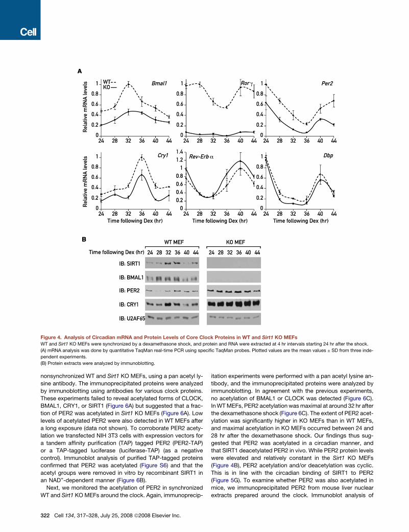
Figure 4. Analysis of Circadian mRNA and Protein Levels of Core Clock Proteins in WT and Sirt1 KO MEFs
WT and Sirt1 KO MEFs were synchronized by a dexamethasone shock, and protein and RNA were extracted at 4 hr intervals starting 24 hr after the shock.
(A) mRNA analysis was done by quantitative TaqMan real-time PCR using specific TaqMan probes. Plotted values are the mean values ± SD from three inde-
pendent experiments.
(B) Protein extracts were analyzed by immunoblotting.
nonsynchronized WT and Sirt1 KO MEFs, using a pan acetyl ly-
sine antibody. The immunoprecipitated proteins were analyzed
by immunoblotting using antibodies for various clock proteins.
These experiments failed to reveal acetylated forms of CLOCK,
BMAL1, CRY1, or SIRT1 (Figure 6A) but suggested that a frac-
tion of PER2 was acetylated in Sirt1 KO MEFs (Figure 6A). Low
levels of acetylated PER2 were also detected in WT MEFs after
a long exposure (data not shown). To corroborate PER2 acety-
lation we transfected NIH 3T3 cells with expression vectors for
a tandem affinity purification (TAP) tagged PER2 (PER2-TAP)
or a TAP-tagged luciferase (luciferase-TAP) (as a negative
control). Immunoblot analysis of purified TAP-tagged proteins
confirmed that PER2 was acetylated (Figure S6) and that the
acetyl groups were removed in vitro by recombinant SIRT1 in
an NAD+-dependent manner (Figure 6B).
Next, we monitored the acetylation of PER2 in synchronized
WT and Sirt1 KO MEFs around the clock. Again, immunoprecip-
322 Cell 134, 317–328, July 25, 2008 ª2008 Elsevier Inc.
itation experiments were performed with a pan acetyl lysine an-
tibody, and the immunoprecipitated proteins were analyzed by
immunoblotting. In agreement with the previous experiments,
no acetylation of BMAL1 or CLOCK was detected (Figure 6C).
In WT MEFs, PER2 acetylation was maximal at around 32 hr after
the dexamethasone shock (Figure 6C). The extent of PER2 acet-
ylation was significantly higher in KO MEFs than in WT MEFs,
and maximal acetylation in KO MEFs occurred between 24 and
28 hr after the dexamethasone shock. Our findings thus sug-
gested that PER2 was acetylated in a circadian manner, and
that SIRT1 deacetylated PER2 in vivo. While PER2 protein levels
were elevated and relatively constant in the Sirt1 KO MEFs
(Figure 4B), PER2 acetylation and/or deacetylation was cyclic.
This is in line with the circadian binding of SIRT1 to PER2
(Figure 5G). To examine whether PER2 was also acetylated in
mice, we immunoprecipitated PER2 from mouse liver nuclear
extracts prepared around the clock. Immunoblot analysis of

Figure 5. SIRT1 Binds to CLOCK, BMAL1, and PER2 in a Circadian Manner
SIRT1 was immunoprecipitated from mouse liver nuclear extracts (A) and from NIH 3T3 cells (B). The immunoprecipitated proteins were analyzed by immuno-
blotting. Rabbit yeast RAP1 antibody was used as a negative control.
(C) Immunostaining of SIRT1 (red) and CLOCK (green) in NIH 3T3 cells was performed with rabbit SIRT1 and CLOCK antibodies. In blue: DAPI staining. In yellow:
merge of SIRT1 and CLOCK staining.
(D) Mice were sacrificed at 4 hr intervals, and liver nuclear extracts were analyzed by immunoblotting.
(E) CLOCK was immunoprecipitated from mouse liver nuclear extracts, and the immunoprecipitated proteins were analyzed by immunoblotting. Rabbit yeast
RAP1 antibody was used as a negative control.
(F) SIRT1 was immunoprecipitated from mouse liver nuclear extracts, and the immunoprecipitated proteins were analyzed by immunoblotting.
(G) NIH 3T3 cells were synchronized by a dexamethasone shock, and protein extracts were prepared at 6 hr intervals, starting 24 hr after the shock. SIRT1 was
immunoprecipitated from NIH 3T3 cells, and the immunoprecipitated proteins were analyzed by immunoblotting.
precipitated proteins with the pan acetyl lysine antibody showed
that PER2 was acetylated also in mouse liver (Figure 6D).
SIRT1-Dependent Deacetylation of PER2 DeterminesPER2 Protein StabilityThe elevated PER2 acetylation and accumulation in Sirt1 KO
MEFs on one hand and the reduced Per2 mRNA levels on the
other hand raised the possibility that acetylation of PER2 stabi-
lized the protein. We thus compared the decay of PER2 protein
in the presence and absence of SIRT1. To this end, WT and Sirt1
KO MEFs were incubated with or without cycloheximide 24 hr
after synchronization and PER2 protein levels were recorded
during 4 hr. The results indicated that the protein half-life of
PER2 was significantly prolonged in the absence of SIRT1
(Figures 7A and 7B). In keeping with these observations, the
coexpression of SIRT1 together with PER2-TAP significantly
reduced the accumulation (Figure 7C) and acetylation of PER2-
TAP (Figure 7D), whereas knockdown of SIRT1 expression with
Sirt1 siRNA resulted in a significant increase in PER2 accumula-
tion and acetylation (Figure 7E).
To further address the dependency of PER2 degradation upon
deacetylation by SIRT1 we performed an in vitro assay with
purified PER2-TAP and extracts obtained from WT and Sirt1 KO
MEFs. PER2 was deacetylated only in the presence of extracts
Cell 134, 317–328, July 25, 2008 ª2008 Elsevier Inc. 323

Figure 6. SIRT1 Deacetylates PER2
(A) Protein extracts from nonsynchronized WT and Sirt1 KO MEFs were subjected to immunoprecipitation with rabbit pan acetyl lysine antibody, and the immu-
noprecipitated proteins were analyzed by immunoblotting.
(B) Purified PER2-TAP was incubated in the absence or presence of recombinant SIRT1 and NAD+ for 3 hr at 30�, and samples were analyzed by immunoblotting.
(C) WT and Sirt1 KO MEFs were synchronized by a dexamethasone shock, and protein extracts were prepared at 4 hr intervals, starting 24 hr after the shock.
Immunoprecipitation experiments were performed with rabbit pan acetyl lysine antibody, and the immunoprecipitated proteins were analyzed by immunoblotting.
(D) Mice were sacrificed at 4 hr intervals and liver nuclear extracts were prepared. PER2 was immunoprecipitated, and the immunoprecipitated proteins were
analyzed by immunoblotting. Rabbit yeast RAP1 antibody was used as a negative control.
from WT MEFs together with NAD+. Deacetylation of PER2 re-
sulted in PER2 degradation, which was blocked in the presence
of the proteasome inhibitor MG132 (Figure 7F).
DISCUSSION
Modulation of Circadian Oscillator Function by ProteinAcetylation and DeacetylationWe identified SIRT1 as a regulator of circadian gene expression.
SIRT1 accumulates in a circadian manner in mouse hepatocytes
and cultured fibroblasts and is required for high-magnitude cir-
cadian transcription of several core clock genes, including
Bmal1, Rorg, Per2, and Cry1. SIRT1 binds to CLOCK-BMAL1
and PER2 in a circadian manner and supports the deacetylation
and degradation of PER2. In the absence of SIRT1, constitutively
high protein levels of PER2 may lead to the repression of Per1,
Per2, Cry1, and Rorg mRNA expression. Repression of RORg,
an activator of Bmal1 transcription, is likely to account for the
dampening of Bmal1 mRNA and protein expression in Sirt1 KO
cells (Figure 7G).
The enzyme(s) responsible for PER2 acetylation remain(s) to
be identified, but the acetyltransferase activity of CLOCK (Doi
et al., 2006) or p300, a coactivator associated with CLOCK-
BMAL1 heterodimer (Etchegaray et al., 2003), are attractive can-
didates. PGC1a, a recently identified key player in circadian
oscillator function (Liu et al., 2007), may also affect PER2 acety-
lation via stimulating the acetyl transferase activity of p300
(Wallberg et al., 2003).
Although our results cannot rigorously exclude a more compli-
cated scenario, they suggest that SIRT1 deacetylates PER2
directly. Thus, SIRT1 is associated with CLOCK-BMAL1-PER2
complexes, purified PER2 is deacetylated in vitro by extracts ob-
324 Cell 134, 317–328, July 25, 2008 ª2008 Elsevier Inc.
tained from wild-type but not from Sirt1 KO MEFs, and recombi-
nant SIRT1 deacetylates purified PER2 in vitro in a NAD+-depen-
dent manner. The latter result should, however, be interpreted
with caution since the in vitro substrate specificity of recombi-
nant SIRT1 is rather promiscuous (Blander et al., 2005).
Other posttranslational modifications such as phosphoryla-
tion, sumoylation, histone acetylation, and methylation have
already been shown to play a key role in circadian gene expres-
sion (Gallego and Virshup, 2007). For example, sumoylation of
BMAL1 has been shown to play an important role in BMAL1 ac-
cumulation and clock rhythmicity (Cardone et al., 2005). Like-
wise, phosphorylation of BMAL1 either by Casein Kinase I (CKI)
(Eide et al., 2002) or by mitogen-activated protein kinases
(MAPK) (Sanada et al., 2002) modulates BMAL1-CLOCK-depen-
dent transcription. Recently, CLOCK was reported to acetylate
BMAL1, thereby facilitating repression of BMAL1-CLOCK-de-
pendent transcription (Hirayama et al., 2007). We suppose that
due to the sensitivity of our immunoblot experiments, the acety-
lated fraction of endogenous BMAL1 in liver and fibroblast ex-
tracts was not revealed. CKI has been reported to phosphorylate
PER2 protein, thereby regulating PER2 protein stability (Eide
et al., 2005). Likewise, CRY degradation mediated by the F box
protein SCFFbxl3 has been demonstrated to be required for
normal oscillator function (Busino et al., 2007; Godinho et al.,
2007; Siepka et al., 2007).
SIRT1 Affects Circadian Transcriptionin a Gene-Specific MannerThe extent to which SIRT1 affects circadian transcription ap-
pears to be target gene specific. For example, in the absence
of SIRT1, Rorg mRNA levels are strongly repressed and Per2
mRNA levels are significantly downregulated, whereas Rev-Erba

Figure 7. SIRT1-Dependent PER2 Deacetylation Determines PER2 Protein Stability
(A) WT and Sirt1 KO MEFs were synchronized by a dexamethasone shock, and 24 hr after the shock cells were untreated or treated with cycloheximide. Cells
were harvested 1, 2, 3, and 4 hr following the treatment, and protein extracts were analyzed by immunoblotting.
(B) The graph illustrates the quantification of PER2 by densitometry of triplicate experiments (mean ± standard error).
(C) NIH 3T3 cells were transfected with PER2-TAP expression vector either alone or together with HA-FLAG-human SIRT1 expression vector. Protein extracts
were analyzed by immunoblotting.
(D) PER2-TAP was purified from NIH 3T3 cells transfected with PER2-TAP expression vector either alone or together with HA-FLAG-human SIRT1 expression
vector and analyzed by immunoblotting.
(E) NIH 3T3 cells were transfected with the V5-PER2 expression vector either alone or together with the Sirt1 siRNA expression vector. Protein extracts were
prepared, and immunoprecipitation experiments were performed with mouse V5 antibody. The immunoprecipitated proteins were analyzed by immunoblotting.
(F) Purified PER2-TAP was incubated for 3 hr at 30� with protein extract obtained from WT or Sirt1 KO MEFs in the absence or presence of 100 mM NAD+ or 25 mM
MG132, and samples were analyzed by immunoblotting.
(G) Hypothetical model showing the possible role of SIRT1 in circadian oscillator function. BMAL1-CLOCK heterodimers bind and activate transcription of the Per,
Cry, Rre-Erba, and Rorg genes. Once the PER and CRY proteins accumulate to a critical level, they form complexes with BMAL1-CLOCK and thereby repress
their own transcription. In addition, there is an interconnecting feedback loop in which REV-ERBa represses and RORg activates Bmal1 transcription. SIRT1
binds CLOCK-BMAL1 complexes and promotes PER2 deacetylation and degradation.
and Dbp mRNA levels are only slightly affected (Figure 3). Con-
ceivably, BMAL1 and CLOCK bind their DNA cognate sites in
Rev-Erba and Dbp with a higher affinity than those present in
Per2 and Rorg. The reduced BMAL1-CLOCK levels in Sirt1 KO
cells might then still support high-amplitude/magnitude Rev-
Erba and Dbp transcription.
PGC1a was found to be expressed in a circadian manner
and to stimulate Bmal1 transcription as a coactivator of the
ROR family of nuclear orphan receptors (Liu et al., 2007).
SIRT1 deacetylates PGC1a and thereby modulates its coacti-
vator activity (Rodgers et al., 2005). Thus, it is possible that
the downregulation of Bmal1 expression in the absence of
SIRT1 is caused by a combination of diminished RORg
expression and impaired PGC1a coactivation. Indeed, Pgc1a
KO mice exhibit abnormal diurnal rhythms of activity, body
temperature, and metabolic rate (Liu et al., 2007). Unfortu-
nately, such studies cannot be performed with Sirt1-deficient
mice because their postnatal survival rates are very poor and
the few surviving mice exhibit many developmental defects
(Cheng et al., 2003; McBurney et al., 2003).
Regulation of PER2 Protein DegradationIn mammals, the stability of PER proteins is regulated by
the F-box-containing E3 ubiquitin ligase bTrCP (Gallego and
Cell 134, 317–328, July 25, 2008 ª2008 Elsevier Inc. 325

Virshup, 2007). PER phosphorylation by CKI3 promotes the re-
cruitment of bTrCP complexes, which in turn mediates the ubiq-
uitination and proteasomal degradation of PER (Eide et al., 2005;
Shirogane et al., 2005). Our results that both PER2 and acety-
lated PER2 levels are elevated in the absence of SIRT1 suggest
that SIRT1-mediated deacetylation enhances the rate of PER2
degradation. Since ubiquitination and acetylation occur on lysine
residues, it is conceivable that the same lysine residues can be
either acetylated or ubiquitinated. If true, acetylated PER2 could
no longer be ubiquitinated and degraded by the proteasome,
which would explain the augmented PER2 levels in Sirt1 KO
cells. Interestingly, CRY1 protein levels are also elevated in the
absence of SIRT1, in spite of reduced Cry1 mRNA levels (Fig-
ure 4). It has been reported that PER2 inhibits the ubiquitination
and degradation of CRY proteins (Yagita et al., 2002). Thus, the
higher accumulation of PER2 in Sirt1 KO cells may account for
the elevated CRY1 levels in these cells.
The Circadian Accumulation and Activityof SIRT1 ProteinCircadian SIRT1 protein accumulation appears to be con-
trolled by posttranscriptional mechanisms as no significant
changes in Sirt1 mRNA were observed (Figure 1). Surprisingly,
temporal SIRT1 accumulation does not correlate with the cir-
cadian interaction of SIRT1 with its identified core clock bind-
ing partners. For example, in mouse liver nuclei, maximal
SIRT1 protein expression is observed at around ZT16,
whereas its maximal binding to CLOCK occurs at around
ZT4, when SIRT1 levels are minimal. Similarly, in NIH 3T3
cells, maximal SIRT1 protein expression occurs between 32
and 36 hr after the dexamethasone shock, while its maximal
binding to the core clock components is observed around
42 hr. Therefore, SIRT1 might also regulate the expression
of circadian output genes expressed with a different phase,
possibly through activation of coactivators and transcription
factors such as PGC1a, FOXO, or LXR (Dali-Youcef et al.,
2007; Li et al., 2007) or through the circadian deacetylation
of histones in nucleosomes associated with clock-controlled
genes (Belden et al., 2007; Curtis et al., 2004; Etchegaray
et al., 2003; Naruse et al., 2004; Ripperger and Schibler,
2006). Future experiments with Sirt1-deficient and -proficient
cells should shed light on the role of SIRT1 in these additional
mechanisms involved in the regulation of circadian gene
expression.
EXPERIMENTAL PROCEDURES
Cells and Cell Culture
NIH 3T3 cells and NIH 3T3-Bmal1-luciferase cells stably expressing Bmal1-lu-
ciferase reporter were grown as previously described (Nagoshi et al., 2004).
WT and Sirt1 KO MEFs were grown in Dulbecco’s modified Eagle’s medium
(DMEM) supplemented with 15% FBS, 100 units/ml penicillin, 100 mg/ml
streptomycin, 2 mM glutamine, 8 mM nonessential amino acids (Sigma),
1 mM Na-Pyruvate, 0.006 mM b-mercaptoethanol, and 18 mM HEPES
(pH 7.0) and cultured at 37�C in a humidified incubator with 5.6% CO2. Cells
were synchronized with 100 nM dexamethasone and real-time biolumines-
cence was recorded (Nagoshi et al., 2004). Nicotinamide and dexamethasone
were prepared in H2O and ethanol, respectively. Trichostatin a, cycloheximide,
and sirtinol (Sigma) were dissolved in DMSO.
326 Cell 134, 317–328, July 25, 2008 ª2008 Elsevier Inc.
Plasmids and Transfections
The following plasmids were used: Bmal1-luciferase (Nagoshi et al., 2004),
Per2(E-BOX2)-luciferase (Yoo et al., 2005), Dbp-luciferase (Brown et al.,
2005), Rev-Erba-luciferase, and CMV-luciferase; pCDNA HA-Flag-SIRT1 en-
coding WT human SIRT1, pCDNA HA-Flag-SIRT1 H363Y encoding inactive
deacetylase mutant of SIRT1, pBabe human SIRT1, pU6-siRNA-Sirt1, and
pU6-empty vector (Cohen et al., 2004); and pEF5/FRT7V5-PER2, pCMV
PER2-TAP, and pCMV luciferase-TAP. Transient transfections of NIH 3T3 cells
were carried out with FuGENE Transfection Reagent (Roche) according to the
manufacturer’s instructions.
Generation and Transduction of Bmal1-Luciferase
Expressing Adenovirus
Bmal1-luciferase (Nagoshi et al., 2004) cassettes was cloned into pCV100
plasmid, first-generation adenoviral vector was amplified in N52.E6-producer
cells, and viruses were generated and purified as previously described (Krep-
pel et al., 2002).
MEFs were incubated with Bmal1-luciferase expressing adenovirus at an
multiplicity of infection (moi) of 5000 for 6 hr, cells were thoroughly washed
with PBS, and the medium was replaced. Forty-eight hours after transduction
cells were shocked with dexamethasone and real-time bioluminescence was
recorded (Nagoshi et al., 2004).
Immunostaining
Cells were fixed with 4% paraformaldehyde for 10 min at room temperature.
Fixed cells were permeabilized with 0.5% Triton X-100 in TBS and washed
with TBS containing 0.1% Triton X-100 (TBS-T). Samples were blocked with
2% BSA in TBS-T followed by incubation with rabbit anti-SIRT1 and rat anti-
CLOCK antibodies. Samples were washed with TBS-T and incubated with
alexa 594-conjugated anti-rabbit and with FITC-conjugated anti-rat secondary
antibodies. Nuclei were stained with DAPI. Microscopic images were obtained
using a Leica SP2 confocal microscope.
RNA Analysis by Real-Time Quantitative PCR
RNA extraction and transcript quantification by TaqMan real-time PCR tech-
nology was performed as previously described (Preitner et al., 2003), using
an ABI PRISM 7700 Sequence Detection System from PE-Applied Biosys-
tems. The real-time PCR data were normalized to 45S pre-mRNA. Primers
and probes are listed in Table S1.
Protein Extraction and Immunoblot Analysis
Proteins from mouse liver nuclei and cultured fibroblasts were prepared
according to the NUN procedure (Lavery and Schibler, 1993). Trichostatin A
was added during the extraction. SDS-PAGE and immunoblot analysis were
performed according to standard protocols. Antibodies used were rabbit
CRY1, PER2, BMAL1, and CLOCK (kindly provided by S. Brown and J. Rip-
perger) and rabbit SIRT1 (Upstate), human-SIRT1 (Santa Cruz), pan acetylated
lysine (Cell Signaling), TAP (OPEN BIOSYSTEMS), mouse V5 (Invitrogen), and
U2AF65 (Sigma).
Coimmunoprecipitation Experiments
Coimmunoprecipitation experiments were carried out with mouse liver nuclear
extracts or with whole-cell NUN extracts. Extracts were incubated for 12 hr
with the indicated antibodies at 4�C and further incubated with protein A beads
(Roche) for an additional 2 hr at 4�C. The beads were collected by centrifuga-
tion and washed with NP40 buffer (100 mM Tris-HCL pH 7.5, 150 mM NaCl,
2 mM EDTA, and 1% NP40). Laemmli sample buffer was added and samples
were heated at 95�C for 5 min and loaded on a polyacrylamide-SDS PAGE.
Purification of the C-terminal TAP-tagged PER2 and TAP-tagged luciferase
proteins were performed according to standard protocol as previously
described (Puig et al., 2001).
In Vitro Deacetylation Assay
Purified PER2-TAP protein was incubated in deacetylation buffer (50 mM Tris-
HCL pH 8, 50 mM NaCl, 4 mM MgCl2) in the presence of purified recombinant
human SIRT1 (BioMol, 5U) or in the presence of protein extracts from WT or
Sirt1 KO MEFs lysed in RIPA lysis buffer (150 mM NaCl, 1% NP-40 [vol/vol],
0.5% Na-deoxycholate [DOC vol/vol], 0.1% SDS [vol/vol], 50 mM Tris-Hcl
pH 8, 1 mM dithiothreitol [DTT]). Reactions were carried out in the presence
or absence of 100 mM NAD+ for 3 hr at 30�.
SUPPLEMENTAL DATA
Supplemental Data include six figures, one table, and two movies and can be
found with this article online at http://www.cell.com/cgi/content/full/134/2/
317/DC1/.
ACKNOWLEDGMENTS
We thank S. Brown and J. Ripperger for the CRY1, PER2, BMAL1, and CLOCK
antibodies; A. Kramer, J. Takahashi, and J. Ripperger for the pEF5/FRT7V5-
PER2, Per2(E-BOX2)-luciferase, and Rev-Erba-luciferase plasmids, respec-
tively; D. Welsh for advice on single-cell luminescence imaging; and N. Roggi
for the artwork. This research was supported by the Swiss National Foundation
(through an individual research grant to U.S. and the National Center of Com-
petence in Research Program Frontiers in Genetics), the State of Geneva, the
Louis Jeantet Foundation of Medicine, the Bonizzi-Theler Stiftuung, and the 6th
European Framework Project EUCLOCK. G.A. and H.R. received long-term
fellowships from EMBO and Human Frontier Science Program. D.G. received
long-term fellowships from FEBS and Human Frontier Science Program.
Received: October 24, 2007
Revised: March 17, 2008
Accepted: June 23, 2008
Published: July 24, 2008
REFERENCES
Akhtar, R.A., Reddy, A.B., Maywood, E.S., Clayton, J.D., King, V.M., Smith,
A.G., Gant, T.W., Hastings, M.H., and Kyriacou, C.P. (2002). Circadian cycling
of the mouse liver transcriptome, as revealed by cDNA microarray, is driven by
the suprachiasmatic nucleus. Curr. Biol. 12, 540–550.
Albrecht, U., and Eichele, G. (2003). The mammalian circadian clock. Curr.
Opin. Genet. Dev. 13, 271–277.
Balsalobre, A., Damiola, F., and Schibler, U. (1998). A serum shock induces cir-
cadian gene expression in mammalian tissue culture cells. Cell 93, 929–937.
Belden, W.J., Loros, J.J., and Dunlap, J.C. (2007). Execution of the circadian
negative feedback loop in Neurospora requires the ATP-dependent chroma-
tin-remodeling enzyme CLOCKSWITCH. Mol. Cell 25, 587–600.
Bitterman, K.J., Anderson, R.M., Cohen, H.Y., Latorre-Esteves, M., and
Sinclair, D.A. (2002). Inhibition of silencing and accelerated aging by nicotin-
amide, a putative negative regulator of yeast sir2 and human SIRT1. J. Biol.
Chem. 277, 45099–45107.
Blander, G., and Guarente, L. (2004). The Sir2 family of protein deacetylases.
Annu. Rev. Biochem. 73, 417–435.
Blander, G., Olejnik, J., Krzymanska-Olejnik, E., McDonagh, T., Haigis, M.,
Yaffe, M.B., and Guarente, L. (2005). SIRT1 shows no substrate specificity
in vitro. J. Biol. Chem. 280, 9780–9785.
Bordone, L., Motta, M.C., Picard, F., Robinson, A., Jhala, U.S., Apfeld, J.,
McDonagh, T., Lemieux, M., McBurney, M., Szilvasi, A., et al. (2006). Sirt1 reg-
ulates insulin secretion by repressing UCP2 in pancreatic beta cells. PLoS Biol.
4, e31. 10.1371/journal.pbio.0040031.
Brown, S.A., Ripperger, J., Kadener, S., Fleury-Olela, F., Vilbois, F., Rosbash,
M., and Schibler, U. (2005). PERIOD1-associated proteins modulate the neg-
ative limb of the mammalian circadian oscillator. Science 308, 693–696.
Brunet, A., Sweeney, L.B., Sturgill, J.F., Chua, K.F., Greer, P.L., Lin, Y., Tran, H.,
Ross, S.E., Mostoslavsky, R., Cohen, H.Y., et al. (2004). Stress-dependent reg-
ulation of FOXO transcription factors by the SIRT1 deacetylase. Science 303,
2011–2015.
Busino, L., Bassermann, F., Maiolica, A., Lee, C., Nolan, P.M., Godinho, S.I.,
Draetta, G.F., and Pagano, M. (2007). SCFFbxl3 controls the oscillation of
the circadian clock by directing the degradation of cryptochrome proteins.
Science 316, 900–904.
Cardone, L., Hirayama, J., Giordano, F., Tamaru, T., Palvimo, J.J., and
Sassone-Corsi, P. (2005). Circadian clock control by SUMOylation of
BMAL1. Science 309, 1390–1394.
Cheng, H.L., Mostoslavsky, R., Saito, S., Manis, J.P., Gu, Y., Patel, P., Bron-
son, R., Appella, E., Alt, F.W., and Chua, K.F. (2003). Developmental defects
and p53 hyperacetylation in Sir2 homolog (SIRT1)-deficient mice. Proc. Natl.
Acad. Sci. USA 100, 10794–10799.
Cohen, H.Y., Miller, C., Bitterman, K.J., Wall, N.R., Hekking, B., Kessler, B.,
Howitz, K.T., Gorospe, M., de Cabo, R., and Sinclair, D.A. (2004). Calorie re-
striction promotes mammalian cell survival by inducing the SIRT1 deacetylase.
Science 305, 390–392.
Curtis, A.M., Seo, S.B., Westgate, E.J., Rudic, R.D., Smyth, E.M., Chakravarti,
D., FitzGerald, G.A., and McNamara, P. (2004). Histone acetyltransferase-de-
pendent chromatin remodeling and the vascular clock. J. Biol. Chem. 279,
7091–7097.
Dali-Youcef, N., Lagouge, M., Froelich, S., Koehl, C., Schoonjans, K., and
Auwerx, J. (2007). Sirtuins: The ‘magnificent seven’, function, metabolism
and longevity. Ann. Med. 39, 335–345.
Damiola, F., Le Minh, N., Preitner, N., Kornmann, B., Fleury-Olela, F., and Schi-
bler, U. (2000). Restricted feeding uncouples circadian oscillators in peripheral
tissues from the central pacemaker in the suprachiasmatic nucleus. Genes
Dev. 14, 2950–2961.
DeBruyne, J.P., Weaver, D.R., and Reppert, S.M. (2007). CLOCK and NPAS2
have overlapping roles in the suprachiasmatic circadian clock. Nat. Neurosci.
10, 543–545.
Doi, M., Hirayama, J., and Sassone-Corsi, P. (2006). Circadian regulator
CLOCK is a histone acetyltransferase. Cell 125, 497–508.
Duffield, G.E., Best, J.D., Meurers, B.H., Bittner, A., Loros, J.J., and Dunlap,
J.C. (2002). Circadian programs of transcriptional activation, signaling, and
protein turnover revealed by microarray analysis of mammalian cells. Curr.
Biol. 12, 551–557.
Dzhagalov, I., Zhang, N., and He, Y.W. (2004). The roles of orphan nuclear
receptors in the development and function of the immune system. Cell. Mol.
Immunol. 1, 401–407.
Eide, E.J., Vielhaber, E.L., Hinz, W.A., and Virshup, D.M. (2002). The circadian
regulatory proteins BMAL1 and cryptochromes are substrates of casein kinase
Iepsilon. J. Biol. Chem. 277, 17248–17254.
Eide, E.J., Woolf, M.F., Kang, H., Woolf, P., Hurst, W., Camacho, F., Vielhaber,
E.L., Giovanni, A., and Virshup, D.M. (2005). Control of mammalian circadian
rhythm by CKIepsilon-regulated proteasome-mediated PER2 degradation.
Mol. Cell. Biol. 25, 2795–2807.
Etchegaray, J.P., Lee, C., Wade, P.A., and Reppert, S.M. (2003). Rhythmic
histone acetylation underlies transcription in the mammalian circadian clock.
Nature 421, 177–182.
Gallego, M., and Virshup, D.M. (2007). Post-translational modifications regu-
late the ticking of the circadian clock. Nat. Rev. Mol. Cell Biol. 8, 139–148.
Godinho, S.I., Maywood, E.S., Shaw, L., Tucci, V., Barnard, A.R., Busino, L.,
Pagano, M., Kendall, R., Quwailid, M.M., Romero, M.R., et al. (2007). The af-
ter-hours mutant reveals a role for Fbxl3 in determining mammalian circadian
period. Science 316, 897–900.
Grozinger, C.M., Chao, E.D., Blackwell, H.E., Moazed, D., and Schreiber, S.L.
(2001). Identification of a class of small molecule inhibitors of the sirtuin family
of NAD-dependent deacetylases by phenotypic screening. J. Biol. Chem. 276,
38837–38843.
Hardin, P.E., Hall, J.C., and Rosbash, M. (1990). Feedback of the Drosophila
period gene product on circadian cycling of its messenger RNA levels. Nature
343, 536–540.
Hirayama, J., Sahar, S., Grimaldi, B., Tamaru, T., Takamatsu, K., Nakahata, Y.,
and Sassone-Corsi, P. (2007). CLOCK-mediated acetylation of BMAL1 con-
trols circadian function. Nature 450, 1086–1090.
Cell 134, 317–328, July 25, 2008 ª2008 Elsevier Inc. 327

Kaasik, K., and Lee, C.C. (2004). Reciprocal regulation of haem biosynthesis
and the circadian clock in mammals. Nature 430, 467–471.
Kornmann, B., Schaad, O., Bujard, H., Takahashi, J.S., and Schibler, U. (2007).
System-driven and oscillator-dependent circadian transcription in mice with
a conditionally active liver clock. PLoS Biol. 5, e34. 10.1371/journal.pbio.
0050034.
Kreppel, F., Biermann, V., Kochanek, S., and Schiedner, G. (2002). A DNA-
based method to assay total and infectious particle contents and helper virus
contamination in high-capacity adenoviral vector preparations. Hum. Gene
Ther. 13, 1151–1156.
Lavery, D.J., and Schibler, U. (1993). Circadian transcription of the cholesterol
7 alpha hydroxylase gene may involve the liver-enriched bZIP protein DBP.
Genes Dev. 7, 1871–1884.
Lee, C., Etchegaray, J.P., Cagampang, F.R., Loudon, A.S., and Reppert, S.M.
(2001). Posttranslational mechanisms regulate the mammalian circadian
clock. Cell 107, 855–867.
Li, X., Zhang, S., Blander, G., Tse, J.G., Krieger, M., and Guarente, L. (2007).
SIRT1 deacetylates and positively regulates the nuclear receptor LXR. Mol.
Cell 28, 91–106.
Liu, C., Li, S., Liu, T., Borjigin, J., and Lin, J.D. (2007). Transcriptional coactiva-
tor PGC-1alpha integrates the mammalian clock and energy metabolism.
Nature 447, 477–481.
Lowrey, P.L., and Takahashi, J.S. (2000). Genetics of the mammalian circadian
system: Photic entrainment, circadian pacemaker mechanisms, and post-
translational regulation. Annu. Rev. Genet. 34, 533–562.
McBurney, M.W., Yang, X., Jardine, K., Hixon, M., Boekelheide, K., Webb,
J.R., Lansdorp, P.M., and Lemieux, M. (2003). The mammalian SIR2alpha
protein has a role in embryogenesis and gametogenesis. Mol. Cell. Biol. 23,
38–54.
Motta, M.C., Divecha, N., Lemieux, M., Kamel, C., Chen, D., Gu, W., Bultsma,
Y., McBurney, M., and Guarente, L. (2004). Mammalian SIRT1 represses fork-
head transcription factors. Cell 116, 551–563.
Nagoshi, E., Saini, C., Bauer, C., Laroche, T., Naef, F., and Schibler, U. (2004).
Circadian gene expression in individual fibroblasts: cell-autonomous and self-
sustained oscillators pass time to daughter cells. Cell 119, 693–705.
Naruse, Y., Oh-hashi, K., Iijima, N., Naruse, M., Yoshioka, H., and Tanaka, M.
(2004). Circadian and light-induced transcription of clock gene Per1 depends
on histone acetylation and deacetylation. Mol. Cell. Biol. 24, 6278–6287.
Panda, S., Antoch, M.P., Miller, B.H., Su, A.I., Schook, A.B., Straume, M.,
Schultz, P.G., Kay, S.A., Takahashi, J.S., and Hogenesch, J.B. (2002). Coordi-
nated transcription of key pathways in the mouse by the circadian clock. Cell
109, 307–320.
Preitner, N., Damiola, F., Lopez-Molina, L., Zakany, J., Duboule, D., Albrecht,
U., and Schibler, U. (2002). The orphan nuclear receptor REV-ERBalpha con-
trols circadian transcription within the positive limb of the mammalian circa-
dian oscillator. Cell 110, 251–260.
Preitner, N., Brown, S., Ripperger, J., Le-Minh, N., Damiola, F., and Schibler,
U. (2003). Orphan nuclear receptors, molecular clockwork, and the entrain-
ment of peripheral oscillators. Novartis Found. Symp. 253, 89–99.
Puig, O., Caspary, F., Rigaut, G., Rutz, B., Bouveret, E., Bragado-Nilsson, E.,
Wilm, M., and Seraphin, B. (2001). The tandem affinity purification (TAP)
method: a general procedure of protein complex purification. Methods 24,
218–229.
Reick, M., Garcia, J.A., Dudley, C., and McKnight, S.L. (2001). NPAS2: an an-
alog of clock operative in the mammalian forebrain. Science 293, 506–509.
Reppert, S.M., and Weaver, D.R. (2002). Coordination of circadian timing in
mammals. Nature 418, 935–941.
328 Cell 134, 317–328, July 25, 2008 ª2008 Elsevier Inc.
Ripperger, J.A., and Schibler, U. (2006). Rhythmic CLOCK-BMAL1 binding to
multiple E-box motifs drives circadian Dbp transcription and chromatin transi-
tions. Nat. Genet. 38, 369–374.
Rodgers, J.T., Lerin, C., Haas, W., Gygi, S.P., Spiegelman, B.M., and Puig-
server, P. (2005). Nutrient control of glucose homeostasis through a complex
of PGC-1alpha and SIRT1. Nature 434, 113–118.
Rutter, J., Reick, M., Wu, L.C., and McKnight, S.L. (2001). Regulation of clock
and NPAS2 DNA binding by the redox state of NAD cofactors. Science 293,
510–514.
Rutter, J., Reick, M., and McKnight, S.L. (2002). Metabolism and the control of
circadian rhythms. Annu. Rev. Biochem. 71, 307–331.
Sanada, K., Okano, T., and Fukada, Y. (2002). Mitogen-activated protein ki-
nase phosphorylates and negatively regulates basic helix-loop-helix-PAS
transcription factor BMAL1. J. Biol. Chem. 277, 267–271.
Sato, T.K., Panda, S., Miraglia, L.J., Reyes, T.M., Rudic, R.D., McNamara, P.,
Naik, K.A., FitzGerald, G.A., Kay, S.A., and Hogenesch, J.B. (2004). A func-
tional genomics strategy reveals Rora as a component of the mammalian
circadian clock. Neuron 43, 527–537.
Shirogane, T., Jin, J., Ang, X.L., and Harper, J.W. (2005). SCFbeta-TRCP con-
trols clock-dependent transcription via casein kinase 1-dependent degrada-
tion of the mammalian period-1 (Per1) protein. J. Biol. Chem. 280, 26863–
26872.
Siepka, S.M., Yoo, S.H., Park, J., Song, W., Kumar, V., Hu, Y., Lee, C., and
Takahashi, J.S. (2007). Circadian mutant Overtime reveals F-box protein
FBXL3 regulation of cryptochrome and period gene expression. Cell 129,
1011–1023.
Stokkan, K.A., Yamazaki, S., Tei, H., Sakaki, Y., and Menaker, M. (2001).
Entrainment of the circadian clock in the liver by feeding. Science 291, 490–
493.
Storch, K.F., Lipan, O., Leykin, I., Viswanathan, N., Davis, F.C., Wong, W.H.,
and Weitz, C.J. (2002). Extensive and divergent circadian gene expression in
liver and heart. Nature 417, 78–83.
Takata, T., and Ishikawa, F. (2003). Human Sir2-related protein SIRT1 associ-
ates with the bHLH repressors HES1 and HEY2 and is involved in HES1- and
HEY2-mediated transcriptional repression. Biochem. Biophys. Res. Commun.
301, 250–257.
Tu, B.P., and McKnight, S.L. (2006). Metabolic cycles as an underlying basis of
biological oscillations. Nat. Rev. Mol. Cell Biol. 7, 696–701.
Vaziri, H., Dessain, S.K., Ng Eaton, E., Imai, S.I., Frye, R.A., Pandita, T.K.,
Guarente, L., and Weinberg, R.A. (2001). hSIR2(SIRT1) functions as an NAD-
dependent p53 deacetylase. Cell 107, 149–159.
Walker, J.R., and Hogenesch, J.B. (2005). RNA profiling in circadian biology.
Methods Enzymol. 393, 366–376.
Wallberg, A.E., Yamamura, S., Malik, S., Spiegelman, B.M., and Roeder, R.G.
(2003). Coordination of p300-mediated chromatin remodeling and TRAP/
mediator function through coactivator PGC-1alpha. Mol. Cell 12, 1137–1149.
Yagita, K., Tamanini, F., van Der Horst, G.T., and Okamura, H. (2001). Molec-
ular mechanisms of the biological clock in cultured fibroblasts. Science 292,
278–281.
Yagita, K., Tamanini, F., Yasuda, M., Hoeijmakers, J.H., van der Horst, G.T.,
and Okamura, H. (2002). Nucleocytoplasmic shuttling and mCRY-dependent
inhibition of ubiquitylation of the mPER2 clock protein. EMBO J. 21, 1301–
1314.
Yoo, S.H., Ko, C.H., Lowrey, P.L., Buhr, E.D., Song, E.J., Chang, S., Yoo, O.J.,
Yamazaki, S., Lee, C., and Takahashi, J.S. (2005). A noncanonical E-box en-
hancer drives mouse Period2 circadian oscillations in vivo. Proc. Natl. Acad.
Sci. USA 102, 2608–2613.