SIRT1 Regulates the Function of the Nijmegen Breakage Syndrome Protein
24
SIRT1 regulates the function of the Nijmegen breakage syndrome protein Zhigang Yuan 1 , Xiaohong Zhang 1 , Nilanjan Sengupta 1 , William S. Lane 2 , and Edward Seto 1,* 1 H. Lee Moffitt Cancer Center & Research Institute, Tampa, Florida 33612, USA 2 Microchemistry and Proteomics Analysis Facility, Harvard University, Cambridge, Massachusetts 02138, USA Summary MRE11-RAD50-NBS1 (MRN) is a conserved nuclease complex that exhibits properties of a DNA damage sensor and is critical in regulating cellular responses to DNA double-strand breaks. NBS1, which is mutated in the human genetic disease Nijmegen breakage syndrome, serves as the regulatory subunit of MRN. of NBS1 by the ATM kinase is necessary for both activation of the S-phase checkpoint and for efficient DNA damage repair response. Here, we report that NBS1 is an acetylated protein and that the acetylation level is tightly regulated by the SIRT1 deacetylase. SIRT1 associates with the MRN complex and, importantly, maintains NBS1 in a hypoacetylated state, which is required for ionizing radiation-induced NBS1 Ser343 phosphorylation. Our results demonstrate the presence of crosstalk between two different posttranslational modifications in NBS1 and strongly suggest that deacetylation of NBS1 by SIRT1 plays a key role in the dynamic regulation of the DNA damage response and in the maintenance of genomic stability. Introduction Nijmegen breakage syndrome (NBS) is a rare autosomal recessive condition of chromosomal instability that is clinically manifested by symptoms including microcephaly, a distinct facial appearance, growth retardation, immunodeficiency, radiation sensitivity, and a strong predisposition to lymphoid malignancy (van der Burgt et al., 1996; Shiloh, 1997; Digweed and Sperling, 2004). Cells from NBS patients exhibit radiation hypersensitivity, radioresistant DNA synthesis (RDS), chromosomal instability, and cell cycle checkpoint defect (Tauchi et al., 2002). Mutations in NBS1 (also known as nibrin or p95), the product of the Nijmegen breakage syndrome gene, are responsible for NBS (Varon et al., 1998). The N-terminus of NBS1 protein contains a forkhead-associated (FHA) domain adjacent to a breast cancer carboxy-terminal (BRCT) domain, both of which are commonly found in cell cycle checkpoint proteins. The C-terminus of NBS1 is required for induction of MRN complex-mediated apoptosis in response to irradiation (Stracker et al., 2007). The ATM protein kinase, a multi-tasking DNA damage sensor, is mutated in individuals with the radiosensitivity disorder ataxia-telangiectasia. Following cellular exposure to ionizing radiation (IR), ATM undergoes rapid autophosphorylation at Ser1981, resulting in the Correspondence: E-mail: [email protected]. Publisher's Disclaimer: This is a PDF file of an unedited manuscript that has been accepted for publication. As a service to our customers we are providing this early version of the manuscript. The manuscript will undergo copyediting, typesetting, and review of the resulting proof before it is published in its final citable form. Please note that during the production process errors may be discovered which could affect the content, and all legal disclaimers that apply to the journal pertain. NIH Public Access Author Manuscript Mol Cell. Author manuscript; available in PMC 2009 May 8. Published in final edited form as: Mol Cell. 2007 July 6; 27(1): 149–162. doi:10.1016/j.molcel.2007.05.029. NIH-PA Author Manuscript NIH-PA Author Manuscript NIH-PA Author Manuscript
-
Upload
independent -
Category
Documents
-
view
0 -
download
0
Transcript of SIRT1 Regulates the Function of the Nijmegen Breakage Syndrome Protein

SIRT1 regulates the function of the Nijmegen breakage syndromeprotein
Zhigang Yuan1, Xiaohong Zhang1, Nilanjan Sengupta1, William S. Lane2, and EdwardSeto1,*
1 H. Lee Moffitt Cancer Center & Research Institute, Tampa, Florida 33612, USA
2 Microchemistry and Proteomics Analysis Facility, Harvard University, Cambridge, Massachusetts 02138,USA
SummaryMRE11-RAD50-NBS1 (MRN) is a conserved nuclease complex that exhibits properties of a DNAdamage sensor and is critical in regulating cellular responses to DNA double-strand breaks. NBS1,which is mutated in the human genetic disease Nijmegen breakage syndrome, serves as the regulatorysubunit of MRN. of NBS1 by the ATM kinase is necessary for both activation of the S-phasecheckpoint and for efficient DNA damage repair response. Here, we report that NBS1 is an acetylatedprotein and that the acetylation level is tightly regulated by the SIRT1 deacetylase. SIRT1 associateswith the MRN complex and, importantly, maintains NBS1 in a hypoacetylated state, which is requiredfor ionizing radiation-induced NBS1 Ser343 phosphorylation. Our results demonstrate the presenceof crosstalk between two different posttranslational modifications in NBS1 and strongly suggest thatdeacetylation of NBS1 by SIRT1 plays a key role in the dynamic regulation of the DNA damageresponse and in the maintenance of genomic stability.
IntroductionNijmegen breakage syndrome (NBS) is a rare autosomal recessive condition of chromosomalinstability that is clinically manifested by symptoms including microcephaly, a distinct facialappearance, growth retardation, immunodeficiency, radiation sensitivity, and a strongpredisposition to lymphoid malignancy (van der Burgt et al., 1996; Shiloh, 1997; Digweed andSperling, 2004). Cells from NBS patients exhibit radiation hypersensitivity, radioresistantDNA synthesis (RDS), chromosomal instability, and cell cycle checkpoint defect (Tauchi etal., 2002). Mutations in NBS1 (also known as nibrin or p95), the product of the Nijmegenbreakage syndrome gene, are responsible for NBS (Varon et al., 1998). The N-terminus ofNBS1 protein contains a forkhead-associated (FHA) domain adjacent to a breast cancercarboxy-terminal (BRCT) domain, both of which are commonly found in cell cycle checkpointproteins. The C-terminus of NBS1 is required for induction of MRN complex-mediatedapoptosis in response to irradiation (Stracker et al., 2007).
The ATM protein kinase, a multi-tasking DNA damage sensor, is mutated in individuals withthe radiosensitivity disorder ataxia-telangiectasia. Following cellular exposure to ionizingradiation (IR), ATM undergoes rapid autophosphorylation at Ser1981, resulting in the
Correspondence: E-mail: [email protected]'s Disclaimer: This is a PDF file of an unedited manuscript that has been accepted for publication. As a service to our customerswe are providing this early version of the manuscript. The manuscript will undergo copyediting, typesetting, and review of the resultingproof before it is published in its final citable form. Please note that during the production process errors may be discovered which couldaffect the content, and all legal disclaimers that apply to the journal pertain.
NIH Public AccessAuthor ManuscriptMol Cell. Author manuscript; available in PMC 2009 May 8.
Published in final edited form as:Mol Cell. 2007 July 6; 27(1): 149–162. doi:10.1016/j.molcel.2007.05.029.
NIH
-PA Author Manuscript
NIH
-PA Author Manuscript
NIH
-PA Author Manuscript

conversion of the inactive dimer form to active monomers (Bakkenist and Kastan, 2003).Activated ATM phosphorylates a number of cellular substrates including NBS1 (Lim et al.,2000; Wu et al., 2000; Zhao et al., 2000). NBS1 associates with MRE11 and RAD50 to forma protein complex (MRN complex) involved in detection, signaling, and repair of DNAdamage. Using an Nbs1 knockout cell line, NBS1 was shown to be essential for homologousrecombination DNA repair in vertebrate cells (Tauchi et al., 2002). Although phosphorylationof NBS1 does not affect MRN association, this modification is functionally important sincemutant NBS1 (S343A) cannot completely complement radiosensitivity in cell lines lackingfunctional NBS1 (NBS cells) (Gatei et al., 2000; Lim et al., 2000; Zhao et al., 2000). In additionto serving as a downstream effector of ATM, NBS1 may function in activating ATM(Cerosaletti et al., 2006; Lee and Paull, 2005; You et al., 2005). In fact, NBS1 phosphorylationmay be required for activation of the S-phase checkpoint by stimulating ATM-mediatedphosphorylation of Chk2 (Lee and Paull, 2004).
Besides phosphorylation, the functions and activities of an increasing number of proteins havebeen found to be regulated by posttranslational acetylation on the ε-amino group of lysines(Glozak et al., 2005; Kouzarides, 2000; Yang, 2004). This modification prevents positivecharges from forming on the amino group of lysines and, as a result, has a significant impacton the electrostatic properties of the protein. Over thirty proteins have been reported to possesslysine acetyltransferase activity, and many of these enzymes were first thought to specificallyacetylate histones, but later were found to have a wide range of protein substrates in additionto histones (Sterner and Berger, 2000; Roth et al., 2001; Yang, 2004). Also, manyacetyltransferases, including p300, CBP (CREB-binding protein), and PCAF (p300/CBP-associated factor) are transcriptional co-activators.
Like many covalent protein modifications, posttranslational lysine acetylation is highlyreversible, and increasing evidences suggest that acetylation/deacetylation, likephosphorylation, is important in the regulation of a number of biological processes(Kouzarides, 2000). Thus, in order to fully understand the pathways that modulate the functionsof NBS1, it is important to determine whether NBS1 undergoes acetylation and if so, whetherthis modification is reversibly regulated by deacetylation.
Histone deacetylases (HDACs) are enzymes that catalyze the removal of acetyl moieties fromthe ε-amino groups of conserved lysine residues in the amino terminal tail of histones. Theremoval of this modification strengthens histone-DNA interactions and may generate specificdocking surfaces for proteins that regulate chromatin folding and/or transcription. Results fromnumerous studies overwhelmingly support the prediction that HDACs play crucial roles ingene transcription and most likely affect all eukaryotic biological processes that involvechromatin. Recent studies have shown that many non-histone proteins can serve as HDACsubstrates and many HDACs, like histone acetyltransferases (HATs), regulate importantbiological processes that extend beyond histones and gene transcription (Glozak and Seto,2005).
In humans, HDACs are divided into three categories: the class I RPD3-like proteins (HDAC1,HDAC2, HDAC3, HDAC8); the class II HDA1-like proteins (HDAC4, HDAC5, HDAC6,HDAC7, HDAC9, and HDAC10); and the class III Sir2-like proteins (SIRT1, SIRT2, SIRT3,SIRT4, SIRT5, SIRT6, and SIRT7). The class III proteins do not exhibit any sequencesimilarity to the other HDAC family members and differ from the other HDACs in that theyrequire the cofactor NAD+ for activity. Whereas the NAD+-dependent class III deacetylasesare specifically inhibited by nicotinamide, class I and II HDACs are specifically sensitive tothe inhibitor tricohostatin A (TSA).
Yuan et al. Page 2
Mol Cell. Author manuscript; available in PMC 2009 May 8.
NIH
-PA Author Manuscript
NIH
-PA Author Manuscript
NIH
-PA Author Manuscript

Of the seven human Sir2-like proteins (sirtuins), SIRT1 is most similar to the yeast Sir2 protein,which is the prototypic class III HDAC (Guarente, 2006; Blander and Guarente, 2004). Invitro, SIRT1 preferentially deacetylates histones H4K16 and H3K9, interacts with anddeacetylates histone H1 at K26, and may mediate heterochromatin formation (Vaquero et al.,2004). Besides histones, more than a dozen nonhistone proteins have been found to serve assubstrates for SIRT1 (Blander and Guarente, 2004; Haigis and Guarente, 2006). SIRT1regulates the tumor suppressor protein p53 and FOXO3 to suppress apoptosis and promote cellsurvival (Brunet et al., 2004; Giannakou and Partridge, 2004; Luo et al., 2001; Vaziri et al.,2001). SIRT1 plays a role in several biological processes including stress resistance,metabolism, differentiation, and aging (Haigis and Guarente, 2006). In addition, thymocytesderived from mice lacking SIRT1 exhibit increased sensitivity to γ-irradiation (Cheng et al.,2003).
In the present study, we show that the NBS1 protein is acetylated. Furthermore, we found thatSIRT1 binds to and deacetylates NBS1 in vitro and in vivo and, importantly, maintains NBS1in a hypoacetylated state that is required for IR-induced NBS1 phosphorylation. Induction ofNBS1 hyperacetylation greatly reduces NBS1 phosphorylation. Consistent with theimportance of hypoacetylated NBS1 in DNA damage repair, cell survival decreases and RDSincreases in NBS1-deficient cells complemented with hyperacetylated NBS1. Together, ourfindings strongly implicate crosstalk between two different posttranslational modifications inthe function of NBS1. Additionally, our results uncovered NBS1 as a substrate for SIRT1 andprovide convincing evidence that SIRT1 acts upstream of NBS1.
ResultsNBS1 is an Acetylated Protein
The NBS1 protein (accession BAA28616) contains three lysine acetylation consensus motifs,Kx1–2x/KK (Yang, 2004), at residues 233–237 (KGHKK), 683–687 (KNFKK), and 686–690(KKFKK). Thus, in addition to phosphorylation, NBS1 is potentially modified byposttranslational lysine acetylation. To determine if NBS1 is indeed a substrate foracetyltransferases, 293T cells were co-transfected with plasmids expressing a Myc-taggedNBS1 protein and three different acetyltransferases (PCAF, p300, or CBP). Acetylated NBS1was readily detected in cells that overexpressed PCAF or p300 but not in cells thatoverexpressed CBP (Figure 1A). The level of NBS1 acetylation by PCAF was dose-dependent(Figure 1B). of purified Myc-NBS1 by LC tandem mass spectrometry (LC-MS/MS) revealedthat 10 of the 70 lysine residues were acetylated. These included K208, K233, K334, K441,K504, K544, K665, K690, K698, and K715 (Figure 1C). of all ten sites from lysine to arginineresulted in a dramatic decrease in the overall NBS1 acetylation levels as determined by Westernblot analysis with anti-acetyl-lysine antibodies (Figure 1D). small amount of residualacetylation can still be detected by Western blotting with anti-acetyl-lysine antibodies,suggesting that there could be minor acetylation sites that escaped mass spectrometry analysis.
Class III HDACs Regulate NBS1 Acetylation LevelTo examine whether NBS1 might be regulated by deacetylation, we immunoprecipitated Myc-NBS1 from 293T cells treated with HDAC inhibitors. shown in Figure 1E, treatment of cellswith TSA, a class I and II HDAC inhibitor, did not affect the acetylation status of NBS1.contrast, cells treated with nicotinamide, a class III (Sir2 family) HDAC inhibitor, resulted ina notable increase in acetylation of NBS1. Treatment with a combination of TSA andnicotinamide was not associated with any additional increase in NBS1 acetylation whencompared to treatment with nicotinamide alone. Thus, NBS1 might be deacetylated by classIII HDACs but not by class I or II HDACs. Hyperacetylation of NBS1 is specific tonicotinamide treatment since the NBS1-acetylation mutant did not respond to this HDAC
Yuan et al. Page 3
Mol Cell. Author manuscript; available in PMC 2009 May 8.
NIH
-PA Author Manuscript
NIH
-PA Author Manuscript
NIH
-PA Author Manuscript

inhibitor (Figure 1F). Anti-acetyl-lysine Western blot analysis of anti-NBS1immunoprecipitates obtained under high stringency conditions in which neither MRE11 norRAD50 co-precipitated with NBS1, revealed that nicotinamide enhanced the acetylation stateof endogenous NBS1 (Figure 1G), providing proof that endogenous NBS1 is regulated by classIII HDACs.
NBS1 Interacts with SIRT1An interesting feature of the class III HDACs is their subcellular location (Michishita et al.,2005). SIRT1, SIRT6, and SIRT7 are present in the nucleus while SIRT2 is located in thecytosol despite the possibility of deacetylating H4K16 during mitosis (Vaquero et al., 2006).SIRT3, SIRT4, and SIRT5 are localized in the mitochondria. Of the three nuclear SIRTs, SIRT1has been shown to possess robust deacetylase enzymatic activity, while SIRT6 and SIRT7 havebeen reported to have very weak or no deacetylase activity (Liszt et al., 2005; North et al.,2003). Therefore, SIRT1 is likely responsible for the deacetylation of NBS1.
Since most, if not all, deacetylase substrates interact with their respective enzymes, we firsttested the ability of NBS1 to interact with SIRT1. As shown in Figure 2A, Flag-SIRT1 co-precipitated with Myc-NBS1. Similarly, Myc-NBS1 co-precipitated with Flag-SIRT1 (Figure2B). The SIRT1-NBS1 interaction is highly specific since none of the other Sir2 familymembers (SIRT2, SIRT3, SIRT4, SIRT5, SIRT6, or SIRT7) co-precipitated with NBS1 underidentical conditions (Figure 2C). This association of SIRT1 and NBS1 also occurs in theabsence of overexpression, epitope tagging of either protein, or IR treatment (Figure 2D).Because the subunits of the MRN complex most likely do not act individually, MRE11 andRAD50 also co-precipitated with SIRT1 as expected, suggesting that SIRT1 associates withthe whole MRN complex. Analyses of three different GST-NBS1 deletions indicated thatSIRT1 interacts with the C-terminus of NBS1 (residues 572–754), a segment that contains theMRE11 and ATM interaction region (Figure 2E).
SIRT1 Deacetylates NBS1To test whether SIRT1 deacetylates NBS1, we examined the effect of RNAi-mediated SIRT1knockdown on NBS1 acetylation in 293T cells. BS/U6-templated siRNA efficiently silencedexpression of SIRT1 but not the control protein β-actin, as monitored by Western blot analysis(Figure 3A). In agreement with the finding that SIRT1 binds NBS1, acetylation of both Myc-NBS1 (Figure 3A) and endogenous NBS1 (Figure 3B) was significantly increased as a resultof SIRT1 knockdown. Unlike knock-down of SIRT1, depletion of SIRT6 or SIRT7 did notaffect NBS1 acetylation levels (Figure 3C). Furthermore, PCAF-acetylated NBS1 wasdeacetylated by wild-type SIRT1 (Figure 3D), but not by a catalytic-defective SIRT1 mutant(H363Y) (Figure 3D) or by SIRT2, SIRT3, SIRT4, SIRT6, SIRT7 (Figure 3E). In vitrodeacetylation assays using purified SIRT1 in the presence of NAD+ further confirmed thatNBS1 is a substrate of SIRT1 (Figure 3F). Collectively, our data strongly suggest that SIRT1modulates the acetylation status of NBS1 via its deacetylase activity.
Acetylation Inhibits NBS1 PhosphorylationThe balance between acetylation and deacetylation may have functional consequences forNBS1. Previous work has shown that ATM-mediated phosphorylation of NBS1 at Ser343 inresponse to DNA damage is critical for the DNA damage responses and for activation of thecell cycle S-phase checkpoint (Lim et al., 2000; Wu et al., 2000; Zhao et al., 2000). Wehypothesized that different posttranslational modifications of NBS1 affect the phosphorylationof NBS1 at Ser343. NBS cells (GM07166) expressing Myc-NBS1 alone or in combinationwith Flag-PCAF were irradiated, and the time course of NBS1 Ser343 phosphorylation wasexamined via Western blot analysis. As shown in Figure 4A, overexpression of PCAF, whichacetylates NBS1, reduced the levels of NBS1 phosphorylation at all times examined. Similar
Yuan et al. Page 4
Mol Cell. Author manuscript; available in PMC 2009 May 8.
NIH
-PA Author Manuscript
NIH
-PA Author Manuscript
NIH
-PA Author Manuscript

results were obtained when NBS cells, complemented with Myc-NBS1, were treated withnicotinamide to induce NBS1 hyperacetylation (Figure 4B). The decrease in NBS1 Ser343phosphorylation was not due to a decrease in ATM protein levels (Figures 4A and 4B), adecrease in activated ATM (as measured by phospho-S1981 of ATM; Figures 4A and 4B), ora decrease in ATM-NBS1 interaction (Figure 4C). Treatment with nicotinamide had no effecton NBS1 Ser343 phosphorylation in an NBS1 mutant that was refractory to acetylation/deacetylation (Figure 4D).
To further examine the effect of acetylation/deacetylation on NBS1 Ser343 phosphorylation,we constructed four plasmids that express NBS1 acetylation-mimic mutants, with lysine toglutamine changes, and introduced them into NBS cells. As shown in Figure 4E, consistentwith our observation that acetylation of NBS1 inhibits phosphorylation of NBS1, mutation oflysine to glutamine in NBS1 resulted in a significant decrease in NBS1 Ser343 phosphorylation.Importantly, the decrease in NBS1 phosphorylation is strictly proportional to the number oflysine mutations (i.e., the more lysine to glutamine change, the more decrease in NBS1phosphorylation).
Presence of SIRT1 Affects NBS1 phosphorylationBecause SIRT1 is the chief regulator of the acetylation status of NBS1 (Figure 3), wedetermined whether NBS1 Ser343 phosphorylation was directly affected by the presence ofSIRT1. As shown in Figure 5A, Nbs1 Ser343 was hypophosphorylated in Sirt1−/− MEFscompared to wild-type MEFs. Likewise, depletion of SIRT1 by siRNA resulted in a significantdecrease in NBS1 Ser343 phosphorylation (Figure 5B).
Acetylation of NBS1 Affects RDSAlthough results from one study suggest that ATM phosphorylation of NBS1 contributes tothe formation of IR-induced foci in cells (Zhao et al., 2000), other studies convincinglydemonstrated that NBS1 phosphorylation does not affect formation of IR-induced foci and thatthe MRN complex is recruited to foci independently of ATM (Lim et al., 2000). We examinedthe ability of the NBS1 acetylation-defective mutant to associate with MRE11 and RAD50 andfound that no significant differences exist between mutant and wild-type NBS1 (Figure S1).Furthermore, no significant differences in NBS1 foci formation could be detected betweenSirt1+/+ and Sirt1−/− MEFs following IR treatment (data not shown). Therefore, our datasuggest that, similar to phosphorylation, acetylation of NBS1 does not play a role in therecruitment of MRN to γH2AX domains surrounding DNA DSBs.
Unlike IR-induced foci formation, phosphorylation of NBS1 is clearly involved in regulatingthe intra-S-phase checkpoint (Lim et al., 2000; Zhao et al., 2000). A common phenotype ofNBS cells is the loss of the IR-induced S-phase checkpoint, leading to RDS. Using an RDSassay, we examined the effects of NBS1 acetylation on cell cycle checkpoints activated byDNA damage. NBS cells expressing Myc-NBS1 and enhanced green fluorescent protein(EGFP) were irradiated or mock-treated immediately after 25-bromodeoxyuridine (BrdU)treatment, and the ratios of BrdU-positive/EGFP-positive cells to EGFP-positive cells weredetermined. Compared to wild-type NBS1, PCAF-hyperacetylated NBS1 was ineffective atinhibiting RDS, suggesting that hypoacetylation of NBS1 is crucial for intra-S-phasecheckpoint activation (Figure 6A). In contrast, PCAF did not alter the ability of the acetylation-refractory mutant NBS1 to inhibit RDS. Similarly, in control experiments, PCAF had no effecton the NBS1 phosphorylation mutant (S343A) in inhibiting RDS. Western blot analysisconfirmed that Ser343 phosphorylation of wildtype (Myc-NBS1), but not the acetylationmutant (Myc-NBS1mt), decreased with increased acetylation in NBS cells.
Yuan et al. Page 5
Mol Cell. Author manuscript; available in PMC 2009 May 8.
NIH
-PA Author Manuscript
NIH
-PA Author Manuscript
NIH
-PA Author Manuscript
To be certain that NBS1 acetylation/deacetylation affects RDS, we transfected NBS cells(GM07166) with plasmids expressing wild-type or mutated NBS1, in the presence or absenceof plasmids that express PCAF. Stable clones were selected by growth in G418 plus or minuspuromycin, and cells stably expressing NBS1 and/or PCAF were treated with IR and examinedfor DNA synthesis using radiolabeled thymidine. Consistent with transient transfection results,acetylation of wild-type, but not acetylation-defective, NBS1 by PCAF significantly reducedits ability to inhibit RDS (Figure 6B).
Finally, consistent with observations that NBS1 hyperacetylation prevents its ability to inhibitRDS in transfected NBS cells, we found that Sirt1−/− MEFs, which containhypophosphorylated and hyperacetylated Nbs1, display a higher level of RDS compared toSirt1+/+ cells (Figure 6C).
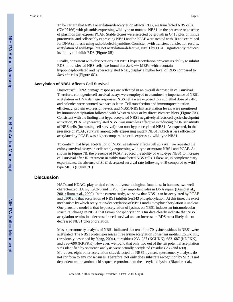
Acetylation of NBS1 Affects Cell SurvivalUnsuccessful DNA damage responses are reflected in an overall decrease in cell survival.Therefore, clonogenic cell survival assays were employed to examine the importance of NBS1acetylation in DNA damage responses. NBS cells were exposed to a sublethal dose of γ-IR,and colonies were counted two weeks later. Cell transfection and immunoprecipitationefficiency, protein expression levels, and NBS1/NBS1mt acetylation levels were monitoredby immunoprecipitation followed with Western blots or by direct Western blots (Figure 7A).Consistent with the finding that hyperacetylated NBS1 negatively affects cell cycle checkpointactivation, PCAF-hyperacetylated NBS1 was much less effective in reducing the IR sensitivityof NBS cells (increasing cell survival) than non-hyperacetylated NBS1. As expected, in thepresence of PCAF, survival among cells expressing mutant NBS1, which is less efficientlyacetylated by PCAF, was higher compared to cells expressing wild-type NBS1.
To confirm that hyperacetylation of NBS1 negatively affects cell survival, we repeated thecolony survival assays in cells stably expressing wild-type or mutant NBS1 and PCAF. Asshown in Figure 7B, the presence of PCAF reduced the ability of wild-type NBS1 to increasecell survival after IR treatment in stably transfected NBS cells. Likewise, in complementaryexperiments, the absence of Sirt1 decreased survival rate following γ-IR compared to wild-type MEFs (Figure 7C).
DiscussionHATs and HDACs play critical roles in diverse biological functions. In humans, two well-characterized HATs, hGCN5 and TIP60, play important roles in DNA repair (Brand et al.,2001; Ikura et al., 2000). In the current study, we show that NBS1 can be acetylated by PCAFand p300 and that acetylation of NBS1 inhibits Ser343 phosphorylation. At this time, the exactmechanism by which acetylation/deacetylation of NBS1 modulates phosphorylation is unclear.One plausible model is that hypoacetylation of lysines on NBS1 induces an intramolecularstructural change in NBS1 that favors phosphorylation. Our data clearly indicate that NBS1acetylation results in a decrease in cell survival and an increase in RDS most likely due todecreased NBS1 phosphorylation.
Mass spectrometry analysis of NBS1 indicated that ten of the 70 lysine residues in NBS1 wereacetylated. The NBS1 protein possesses three lysine acetylation consensus motifs, Kx1–2x/KK,(previously described by Yang, 2004), at residues 233–237 (KGHKK), 683–687 (KNFKK),and 686–690 (KKFKK). However, we found that only two out of the ten potential acetylationsites identified by sequence analysis were actually acetylated (residues 233 and 690).Moreover, eight other acetylation sites detected on NBS1 by mass spectrometry analysis donot conform to any consensuses. Therefore, not only does substrate recognition by SIRT1 notdependent on the amino acid sequence proximate to the acetylated lysine (Blander et al.,
Yuan et al. Page 6
Mol Cell. Author manuscript; available in PMC 2009 May 8.
NIH
-PA Author Manuscript
NIH
-PA Author Manuscript
NIH
-PA Author Manuscript

2005), it appears that the NBS1 acetyltransferase(s) similarly does not have strict sequencerequirements.
Although we cannot rule out the possibility that in addition to p300 and PCAF moreacetyltransferases can modify NBS1, our data clearly demonstrate that NBS1 is deacetylatedpredominantly, if not exclusively, by SIRT1. Our results are consistent with a model in whichSIRT1 is required to maintain hypoacetylated NBS1, which promotes efficient IR-inducedNBS1 phosphorylation and the ensuing cellular responses to DNA damage. Thus, an additionalfunction of SIRT1 may be to maintain NBS1 in a constant hypoacetylated state. Alternatively,IR may induce SIRT1 activity in order to regulate the acetylation status of NBS1; however,this possibility cannot be tested directly due to the low basal levels of acetylated NBS1. Despitethis dilemma, these results are in accordance with the notion that SIRT1, in addition to silencingtranscription, plays an important role in suppressing genomic instability and, consequently, inregulating aging in mammals (Lombard et al., 2005).
Many evidences suggest a close link between histone deacetylation and DNA repair. Forexample, the yeast Sin3/Rpd3 complex has been shown to modulate DNA DSB repair throughthe deacetylation of histone H4K16 (Jazayeri et al., 2004). The Sir2 and Rpd3 proteins arerecruited to HO lesions during homology-directed repair (Tamburini and Tyler, 2005).RPD3 and HOS2 are required for the activation of DNA damage-inducible genes RNR3 andHUG1 (Sharma et al., 2007). Furthermore, HDAC4 is recruited to DSBs in vivo and, therefore,is implicated in DNA repair in mammalian cells (Kao et al., 2003). Knockdown of HDAC4expression by RNAi increases radiosensitivity and blocks G2/M checkpoint maintenance.Also, it was reported that the SMRT-HDAC3 complex is involved in cellular recovery fromDNA DSBs in mammalian cells (Yu et al., 2006). SMRT and HDAC3 are required fortranscriptional repression by Ku70, a subunit of the DNA-PK DSB repair complex.
Although p53 is known to be a substrate for SIRT1 (Luo et al., 2001; Vaziri et al., 2001), thecontribution of SIRT1 to p53-mediated DNA damage repair is controversial. While one studysuggests that SIRT1 modulates p53-dependent DNA-damage responses (Chen et al., 2005),other reports argue that SIRT1 does not affect p53-mediated biological activities (Kamel et al.,2006; Solomon et al., 2006). More recently, SIRT6 was found to be a chromatin-associatedprotein that promotes normal base excision repair but does not play a role in regulating cellcycle checkpoints (Mostoslavsky et al., 2006). Our finding that SIRT1 deacetylates NBS1 andis intimately involved in the regulation of the intra-S-phase checkpoint, coupled with the recentreport that DNA damage induces SIRT1 expression (Wang et al., 2006), raises the intriguingpossibility that HDACs ensure genomic stability through a variety of mechanisms involvingmultiple pathways.
In the MRN complex, besides acetylation/deacetylation and phosphorylation of NBS1, theactivities and functions of MRE11 are regulated by posttranslational modifications. MRE11undergoes cell cycle-dependent phosphorylation in response to several genotoxic agents, andthis modification requires NBS1 but not ATM (Dong et al., 1999; Yuan et al., 2002). Inaddition, MRE11 is methylated at arginine residues by PRMT1, and mutation of the argininesseverely impairs the exonuclease activity of MRE11 although the mutated protein still formsthe MRN complex (Boisvert et al., 2005). In sharp contrast to the hypoacetylation of NBS1,cells containing hypomethylated MRE11 display intra-S-phase DNA damage checkpointdefects. In preliminary studies, we found that MRE11, but not RAD50, is acetylated in the cell(data not shown). Because SIRT1 associates with the entire MRN complex, future studies arewarranted to determine whether MRE11, like NBS1, is similarly regulated by SIRT1 and ifso, whether acetylation/deacetylation of MRE11 interacts with other MRE11 modifications toultimately affect the DNA DSB repair pathway.
Yuan et al. Page 7
Mol Cell. Author manuscript; available in PMC 2009 May 8.
NIH
-PA Author Manuscript
NIH
-PA Author Manuscript
NIH
-PA Author Manuscript
Experimental ProceduresPlasmids, Antibodies, and Viruses
Details of all plasmid constructions and sources of recombinant viruses and antibodies areprovided in the Supplementary Experimental Procedures.
Cell Culture, Transfection, and Adenovirus InfectionSirt1+/− mice were time-mated to screen for homozygous Sirt1−/− mutant embryos by PCR.MEFs were generated from 13.5 dpc embryos using standard methods. HeLa, 293T, and murineSirt1+/+ and Sirt1−/− fibroblasts were grown in Dulbecco’s modified Eagle’s medium (DMEM)supplemented with 10% fetal calf serum (FCS) and penicillin/streptomycin (pen/strep). TheNBS cell line (GM07166) was obtained from Coriell Cell Repository and grown in minimumessential medium (MEM) with 10% FCS and pen/strep. All transfections were normalized withequal amounts of parental vector DNA. Transfections were performed with Lipofectamine2000 (Invitrogen) according to the manufacturer’s instructions. To generate stable cell lines,NBS cells (GM 07166) transfected with vector alone or various expression plasmids weregrown in the presence of G418 (200 μg/ml, for cells that received NBS1 expression plasmids)and puromycin (0.5 μg/ml, for cells that received PCAF expression plasmids) for 10 days.Resistant colonies were isolated, pooled together, and grown for further analysis. viralinfection, HeLa cells were infected with adenovirus for 24 h in DMEM with 0.5% BSA aspreviously described (Rodgers et al., 2005).
Immunoprecipitation and Western Blot AnalysisFor immunoprecipitations, cells were lysed in buffer (50 mM Tris-HCl [pH 7.5], 1 mM EDTA,1% NP-40, and protease inhibitor cocktail) containing either 500 mM NaCl (high stringency)or 150 mM NaCl (low stringency). The lysates were incubated with the primary antibodyovernight at 4°C. The resultant immunocomplexes were collected, washed four times in lysisbuffer, and resolved by SDS-PAGE. For immunoblotting, samples were transferred ontonitrocellulose membranes. Membranes were probed with the appropriate antibodies. Proteinsof interest were visualized using the Chemiluminescent Detection Kit (Pierce).
Ion Trap Mass Spectrometry293T cells were transfected with the Myc-NBS1 expression plasmid and treated with TSA (1.3μM) and nicotinamide (20 mM) overnight. The cells were then lysed in high stringency buffercontaining 10 mM NaB and 10 mM nicotinamide. Cell extracts were subjected toimmunoprecipitation with anti-Myc antibodies. The immune complexes were resolved bySDS-PAGE and stained with colloidal Blue (Invitrogen). The Myc-NBS1 sequence wasanalyzed with an in-house algorithm (EnzOpt) for a dual enzyme strategy maximizingproteotypic peptide coverage of all lysines. The appropriate Myc-NBS1 gel band was excisedand divided into two parts. Each part was subjected to in-gel reduction andcarboxyamidomethylation followed by separate tryptic or chymotryptic digestion. Acetylatedpeptides from each digest were detected and sequenced using microcapillary reverse-phaseHPLC nano-electrospray tandem mass spectrometry (μLC-MS/MS) on a Thermo LTQ linearquadrupole ion trap mass spectrometer. Data analysis was facilitated with SEQUEST and theProteomics Browser Suite (Thermo).
GST Pull-Down AssayGST and GST-NBS1 deletion mutants were expressed and purified from bacteria usingstandard methods. Equimolar quantities of the various purified proteins were conjugated toglutathione-Sepharose beads and incubated with HeLa whole cell lysates for 1 h at 4°C. After
Yuan et al. Page 8
Mol Cell. Author manuscript; available in PMC 2009 May 8.
NIH
-PA Author Manuscript
NIH
-PA Author Manuscript
NIH
-PA Author Manuscript
extensive washing, bound proteins were eluted and analyzed by Western blotting with anti-SIRT1 antibodies.
In Vitro Deacetylation AssayIn vitro deacetylation of NBS1 by SIRT1 was performed using an approach similar to that usedpreviously for detection of PGC-1α acetylation (Nemoto et al., 2005).
RDS AssayThe RDS assay was performed as described previously (Zhao et al., 2000) with minormodifications. Briefly, NBS cells were transfected with either pEGFP-C3 (Clontech) orpEGFP-C3 and the Myc-NBS1 expression plasmid at a ratio of 1:10. In some experiments,cells were also transfected with an additional plasmid encoding Flag-PCAF. Cells were -irradiated with 15 Gy, incubated for 1 h at 37°C, and then incubated for an additional 2 h in amedium containing 100 μM of BrdU. BrdU incorporation was detected using an anti-BrdUantibody following immunostaining procedures described previously (Zhang et al., 2004).
Alternative RDS assays were performed as described by Lim et al. (2000) and by Zhao et al.(2002). Briefly, cells were labeled for 24 h in medium containing 10 nCi/ml of 14C-thymidine.The cells were washed once with PBS and then incubated for 24 h in a nonradioactive medium.After treatment with IR or left untreated, cells were incubated at 37°C for 30 min and pulse-labeled for 30 min in a medium containing 2.5 μCi/ml of 3H-thymidine. Cells were then washedwith PBS and lysed with 0.5 ml of 0.2 M NaOH. Radioactivity was quantified in a liquidscintillation counter, and the resulting ratios of 3H/14C were calculated and compared withratios of non-irradiated cells.
Colony Survival AssayColony survival assays were performed as previously described (Zhao et al., 2002) with minormodifications. Briefly, NBS cells were plated in quadruplicate (1,000 cells per 60-mm tissueculture dish). The cells were γ-irradiated with 0, 2, or 5 Gy. After two weeks, dishes werewashed with PBS, fixed in ice-cold methanol for 15 min, and then stained with Giemsa stainfor 30 min. Colonies on each plate were quantified and expressed as the percentage of theunirradiated control.
Supplementary MaterialRefer to Web version on PubMed Central for supplementary material.
AcknowledgementsWe would like to thank A. Ghosh, R. Goodman, I. Horikawa, M. Kastan, M. Leid, J. Petrini, P. Puigserver, and E.Verdin for providing plasmids. We would also like to thank R. Mostoslavsky and K. Chua for Sirt1+/− mice and J.Neveu and R. Robinson for LC-MS/MS. We also thank J. Koomen, N. Luetteke, A. Monteiro, and H.G. Wang forhelpful discussions and for critical reading of the manuscript. We appreciate the assistance from the Moffitt CancerCenter Core Facility. This work was supported by grants from the National Institutes of Health and the Kaul Foundation(E.S.) and by fellowships from the American Heart Association (Z.Y. and N.S.).
ReferencesBakkenist CJ, Kastan MB. DNA damage activates ATM through intermolecular autophosphorylation
and dimer dissociation. Nature 2003;421:499–506. [PubMed: 12556884]Blander G, Guarente L. The Sir2 family of protein deacetylases. Annu Rev Biochem 2004;73:417–435.
[PubMed: 15189148]
Yuan et al. Page 9
Mol Cell. Author manuscript; available in PMC 2009 May 8.
NIH
-PA Author Manuscript
NIH
-PA Author Manuscript
NIH
-PA Author Manuscript
Blander G, Olejnik J, Krzymanska-Olejnik E, McDonagh T, Haigis M, Yaffe MB, Guarente L. SIRT1shows no substrate specificity in vitro. J Biol Chem 2005;280:9780–9785. [PubMed: 15640142]
Boisvert FM, Dery U, Masson JY, Richard S. Arginine methylation of MRE11 by PRMT1 is requiredfor DNA damage checkpoint control. Genes Dev 2005;19:671–676. [PubMed: 15741314]
Brand M, Moggs JG, Oulad-Abdelghani M, Lejeune F, Dilworth FJ, Stevenin J, Almouzni G, Tora L.UV-damaged DNA-binding protein in the TFTC complex links DNA damage recognition tonucleosome acetylation. EMBO J 2001;20:3187–3196. [PubMed: 11406595]
Brunet A, Sweeney LB, Sturgill JF, Chua KF, Greer PL, Lin Y, Tran H, Ross SE, Mostoslavsky R, CohenHY, et al. Stress-dependent regulation of FOXO transcription factors by the SIRT1 deacetylase.Science 2004;303:2011–2015. [PubMed: 14976264]
Cerosaletti K, Wright J, Concannon P. Active role for nibrin in the kinetics of atm activation. Mol CellBiol 2006;26:1691–1699. [PubMed: 16478990]
Chen WY, Wang DH, Yen RC, Luo J, Gu W, Baylin SB. Tumor suppressor HIC1 directly regulatesSIRT1 to modulate p53-dependent DNA-damage responses. Cell 2005;123:437–448. [PubMed:16269335]
Cheng HL, Mostoslavsky R, Saito S, Manis JP, Gu Y, Patel P, Bronson R, Appella E, Alt FW, Chua KF.Developmental defects and p53 hyperacetylation in Sir2 homolog (SIRT1)-deficient mice. Proc NatlAcad Sci USA 2003;100:10794–10799. [PubMed: 12960381]
Digweed M, Sperling K. Nijmegen breakage syndrome: clinical manifestation of defective response toDNA double-strand breaks. DNA Repair 2004;3:1207–1217. [PubMed: 15279809]
Dong Z, Zhong Q, Chen PL. The Nijmegen breakage syndrome protein is essential for Mre11phosphorylation upon DNA damage. J Biol Chem 1999;274:19513–19516. [PubMed: 10391882]
Gatei M, Young D, Cerosaletti KM, Desai-Mehta A, Spring K, Kozlov S, Lavin MF, Gatti RA,Concannon P, Khanna K. ATM-dependent phosphorylation of nibrin in response to radiationexposure. Nat Genet 2000;25:115–119. [PubMed: 10802669]
Giannakou ME, Partridge L. The interaction between FOXO and SIRT1: tipping the balance towardssurvival. Trends Cell Biol 2004;14:408–412. [PubMed: 15308206]
Glozak MA, Sengupta N, Zhang X, Seto E. Acetylation and deacetylation of non-histone proteins. Gene2005;363:15–23. [PubMed: 16289629]
Guarente L. Sirtuins as potential targets for metabolic syndrome. Nature 2006;444:868–874. [PubMed:17167475]
Haigis MC, Guarente LP. Mammalian sirtuins--emerging roles in physiology, aging, and calorierestriction. Genes Dev 2006;20:2913–2921. [PubMed: 17079682]
Ikura T, Ogryzko VV, Grigoriev M, Groisman R, Wang J, Horikoshi M, Scully R, Qin J, Nakatani Y.Involvement of the TIP60 histone acetylase complex in DNA repair and apoptosis. Cell2000;102:463–473. [PubMed: 10966108]
Jazayeri A, McAinsh AD, Jackson SP. Saccharomyces cerevisiae Sin3p facilitates DNA double-strandbreak repair. Proc Natl Acad Sci USA 2004;101:1644–1649. [PubMed: 14711989]
Kamel C, Abrol M, Jardine K, He X, McBurney MW. SirT1 fails to affect p53-mediated biologicalfunctions. Aging Cell 2006;5:81–88. [PubMed: 16441846]
Kao GD, McKenna WG, Guenther MG, Muschel RJ, Lazar MA, Yen TJ. Histone deacetylase 4 interactswith 53BP1 to mediate the DNA damage response. J Cell Biol 2003;160:1017–1027. [PubMed:12668657]
Kobayshi J, Tauchi H, Sakamoto S, Nakamura A, Morishima K, Matsuura S, Kobayashi T, Tamai K,Tanimoto K, Komatsu K. NBS1 localizes to gamma-H2AX foci through interaction with the FHA/BRCT domain. Curr Biol 2002;12:1846–1851. [PubMed: 12419185]
Kouzarides T. Acetylation: a regulatory modification to rival phosphorylation? EMBO J 2000;19:1176–1179. [PubMed: 10716917]
Lee JH, Paull TT. Direct activation of the ATM protein kinase by the Mre11/Rad50/Nbs1 complex.Science 2004;304:93–96. [PubMed: 15064416]
Lee JH, Paull TT. ATM activation by DNA double-strand breaks through the Mre11-Rad50-Nbs1complex. Science 2005;308:551–554. [PubMed: 15790808]
Yuan et al. Page 10
Mol Cell. Author manuscript; available in PMC 2009 May 8.
NIH
-PA Author Manuscript
NIH
-PA Author Manuscript
NIH
-PA Author Manuscript

Lim DS, Kim ST, Xu B, Maser RS, Lin J, Petrini JH, Kastan MB. ATM phosphorylates p95/nbs1 in anS-phase checkpoint pathway. Nature 2000;404:613–617. [PubMed: 10766245]
Liszt G, Ford E, Kurtev M, Guarente L. Mouse Sir2 homolog SIRT6 is a nuclear ADP-ribosyltransferase.J Biol Chem 2005;280:21313–21320. [PubMed: 15795229]
Lombard DB, Chua KF, Mostoslavsky R, Franco S, Gostissa M, Alt FW. DNA repair, genome stability,and aging. Cell 2005;120:497–512. [PubMed: 15734682]
Luo J, Nikolaev AY, Imai S, Chen D, Su F, Shiloh A, Guarente L, Gu W. Negative control of p53 bySir2alpha promotes cell survival under stress. Cell 2001;107:137–148. [PubMed: 11672522]
Michishita E, Park JY, Burneskis JM, Barrett JC, Horikawa I. Evolutionarily conserved and nonconservedcellular localizations and functions of human SIRT proteins. Mol Biol Cell 2005;16:4623–4635.[PubMed: 16079181]
Mirzoeva OK, Petrini JH. DNA damage-dependent nuclear dynamics of the Mre11 complex. Mol CellBiol 2001;21:281–288. [PubMed: 11113202]
Mostoslavsky R, Chua KF, Lombard DB, Pang WW, Fischer MR, Gellon L, Liu P, Mostoslavsky G,Franco S, Murphy MM, et al. Genomic instability and aging-like phenotype in the absence ofmammalian SIRT6. Cell 2006;124:315–329. [PubMed: 16439206]
Nemoto S, Fergusson MM, Finkel T. SIRT1 functionally interacts with the metabolic regulator andtranscriptional coactivator PGC-1α. J Biol Chem 2005;280:16456–16460. [PubMed: 15716268]
North BJ, Marshall BL, Borra MT, Denu JM, Verdin E. The human Sir2 ortholog, SIRT2, is an NAD+-dependent tubulin deacetylase. Mol Cell 2003;11:437–444. [PubMed: 12620231]
Rodgers JT, Lerin C, Haas W, Gygi SP, Spiegelman BM, Puigserver P. Nutrient control of glucosehomeostasis through a complex of PGC-1alpha and SIRT1. Nature 2005;434:113–118. [PubMed:15744310]
Roth SY, Denu JM, Allis CD. Histone acetyltransferases. Annu Rev Biochem 2001;70:81–120. [PubMed:11395403]
Sharma VM, Tomar RS, Dempsey AE, Reese JC. Histone deacetylase RPD3 and HOS2 regulate thetranscriptional activation of DNA damage-inducible genes. Mol Cell Biol 2007;27:3199–3210.[PubMed: 17296735]
Shiloh Y. Ataxia-telangiectasia and the Nijmegen breakage syndrome: related disorders but genes apart.Annu Rev Genet 1997;31:635–662. [PubMed: 9442910]
Solomon JM, Pasupuleti R, Xu L, McDonagh T, Curtis R, DiStefano PS, Huber LJ. Inhibition of SIRT1catalytic activity increases p53 acetylation but does not alter cell survival following DNA damage.Mol Cell Biol 2006;26:28–38. [PubMed: 16354677]
Sterner DE, Berger SL. Acetylation of histones and transcription-related factors. Microbiol Mol Biol R2000;64:435–459.
Stracker TH, Morales M, Couto SS, Hussein H, Petrini JHJ. The carboxy terminus of NBS1 is requiredfor induction of apoptosis by the MRE11 complex. Nature 2007;447:218–221. [PubMed: 17429352]
Tamburini BA, Tyler JK. Localized histone acetylation and deacetylation triggered by the homologousrecombination pathway of double-strand DNA repair. Mol Cell Biol 2005;25:4903–4913. [PubMed:15923609]
Tauchi H, Kobayashi J, Morishima K, van Gent DC, Shiraishi T, Verkaik NS, vanHeems D, Ito E,Nakamura A, Sonoda E, et al. Nbs1 is essential for DNA repair by homologous recombination inhigher vertebrate cells. Nature 2002;420:93–98. [PubMed: 12422221]
Tauchi H, Matsuura S, Kobayashi J, Sakamoto S, Komatsu K. Nijmegen Breakage Syndrome gene,NBS1, and molecular links to factors for genome stability. Oncogene 2002;21:8967–8980. [PubMed:12483513]
van der Burgt I, Chrzanowska KH, Smeets D, Weemaes C. Nijmegen breakage syndrome. J Med Genet1996;33:153–156. [PubMed: 8929954]
Vaquero A, Scher M, Lee D, Erdjument-Bromage H, Tempst P, Reinberg D. Human SirT1 interacts withhistone H1 and promotes formation of facultative heterochromatin. Mol Cell 2004;16:93–105.[PubMed: 15469825]
Vaquero A, Scher MB, Lee DH, Sutton A, Cheng HL, Alt FW, Serrano L, Sternglanz R, Reinberg D.SirT2 is a histone deacetylase with preference for histone H4 Lys 16 during mitosis. Genes Dev2006;20:1256–1261. [PubMed: 16648462]
Yuan et al. Page 11
Mol Cell. Author manuscript; available in PMC 2009 May 8.
NIH
-PA Author Manuscript
NIH
-PA Author Manuscript
NIH
-PA Author Manuscript

Varon R, Vissinga C, Platzer M, Cerosaletti KM, Saar K, Beckmann G, Seemanova E, Cooper PR, NowakNJ, Stumm M, et al. Nibrin, a novel DNA double-strand break repair protein, is mutated in Nijmegenbreakage syndrome. Cell 1998;93:467–476. [PubMed: 9590180]
Vaziri H, Dessain SK, Ng Eaton E, Imai SI, Frye RA, Pandita TK, Guarente L, Weinberg RA. hSIR2(SIRT1) functions as an NAD-dependent p53 deacetylase. Cell 2001;107:149–159. [PubMed:11672523]
Wang C, Chen L, Hou X, Li Z, Kabra N, Ma Y, Nemoto S, Finkel T, Gu W, Cress WD, Chen J. Interactionsbetween E2F1 and SirT1 regulate apoptotic response to DNA damage. Nat Cell Biol 2006;8:1025–1031. [PubMed: 16892051]
Wu X, Ranganathan V, Weisman DS, Heine WF, Ciccone DN, O’Neill TB, Crick KE, Pierce KA, LaneWS, Rathbun G, et al. ATM phosphorylation of Nijmegen breakage syndrome protein is required ina DNA damage response. Nature 2000;405:477–482. [PubMed: 10839545]
Yang XJ. Lysine acetylation and the bromodomain: a new partnership for signaling. Bioessays2004;26:1076–1087. [PubMed: 15382140]
You Z, Chahwan C, Bailis J, Hunter T, Russell P. ATM activation and its recruitment to damaged DNArequire binding to the C terminus of Nbs1. Mol Cell Biol 2005;25:5363–5379. [PubMed: 15964794]
Yu J, Palmer C, Alenghat T, Li Y, Kao G, Lazar MA. The corepressor silencing mediator for retinoidand thyroid hormone receptor facilitates cellular recovery from DNA double-strand breaks. CancerRes 2006;66:9316–9322. [PubMed: 16982777]
Yuan SS, Su JH, Hou MF, Yang FW, Zhao S, Lee EY. Arsenic-induced Mre11 phosphorylation is cellcycle-dependent and defective in NBS cells. DNA Repair 2002;1:137–142. [PubMed: 12509260]
Zhang X, Wharton W, Yuan Z, Tsai SC, Olashaw N, Seto E. Activation of the growth-differentiationfactor 11 gene by the histone deacetylase (HDAC) inhibitor trichostatin A and repression by HDAC3.Mol Cell Biol 2004;24:5106–5118. [PubMed: 15169878]
Zhao S, Renthal W, Lee EY. Functional analysis of FHA and BRCT domains of NBS1 in chromatinassociation and DNA damage responses. Nucleic Acids Res 2002;30:4815–4822. [PubMed:12433983]
Zhao S, Weng YC, Yuan SS, Lin YT, Hsu HC, Lin SC, Gerbino E, Song MH, Zdzienicka MZ, Gatti RA,et al. Functional link between ataxia-telangiectasia and Nijmegen breakage syndrome gene products.Nature 2000;405:473–477. [PubMed: 10839544]
Yuan et al. Page 12
Mol Cell. Author manuscript; available in PMC 2009 May 8.
NIH
-PA Author Manuscript
NIH
-PA Author Manuscript
NIH
-PA Author Manuscript

Figure 1. NBS1 is Acetylated(A) 293T cells were co-transfected with equal amounts (4 μg) of Myc-NBS1 ± one of thefollowing expression plasmids: Flag-PCAF, HA-p300, or HA-CBP. Cell lysates wereimmunoprecipitated (IP) under high stringency conditions using anti-Myc antibodies.Immunoprecipitates were subjected to Western blot (IB) analysis using anti-acetyl-lysine(AcK) antibodies. The blot was stripped and re-probed with anti-Myc antibodies to confirmequal immunoprecipitation efficiency and loading. As CBP (but not PCAF) is known toacetylate p53-K382, a Western blot was performed with anti-AcK382-p53 to confirm that theexpressed CBP was functional. (B) 293T cells were co-transfected with Myc-NBS1 (4 μg) andFlag-PCAF (0, 0.5, 1, or 4 μg). Cell lysates were subjected to immunoprecipitation/Western
Yuan et al. Page 13
Mol Cell. Author manuscript; available in PMC 2009 May 8.
NIH
-PA Author Manuscript
NIH
-PA Author Manuscript
NIH
-PA Author Manuscript

blot analysis using the indicated antibodies. Western blot analysis of whole extracts wasperformed to assess PCAF expression (bottom panel). (C) Myc-NBS1, which was expressedand purified from 293T cells, was digested with trypsin and subjected to ITMS. Positions ofthe unambiguously-identified acetylated lysine residues are shown. FHA, forkhead-associateddomain; BRCT, BRCA1 C-terminal domain; MIR, MRE11-interacting domain; AIR, ATM-interacting domain. (D) 293T cells were co-transfected with Myc-NBS1 (wild-type or lysineto arginine mutant [mt]) and Flag-PCAF (4 μg each), as indicated. Total and acetylated NBS1levels were analyzed by immunoprecipitation/Western blotting. (E) 293T cells weretransfected with plasmids that expressed Myc-NBS1 (4 μg). Twenty-four hours post-transfection, cells were left untreated or treated overnight with TSA (1.3 μM), nicotinamide(20 mM), or TSA plus nicotinamide. Cell lysates were subjected to immunoprecipitation/Western blotting. Whole cell extracts were probed with anti-Ac-tubulin and anti-Ac-H4K16to confirm that TSA is functional under the treatment condition (lower panels). (F) 293T cellswere transfected with plasmids (4 μg each) expressing wild-type or mutant Myc-NBS1 asindicated and then treated with 20 mM nicotinamide overnight. Myc-NBS1 acetylation andprotein expression were examined by immunoprecipitation/Western blotting. (G) HeLa cellswere treated with nicotinamide (20 mM) or left untreated overnight. Cell lysates were thenimmunoprecipitated under high stringency conditions with monoclonal anti-NBS1 antibodies.Endogenous acetylated NBS1 was analyzed by Western blotting with a mixture of polyclonalanti-acetyl-lysine antibodies.
Yuan et al. Page 14
Mol Cell. Author manuscript; available in PMC 2009 May 8.
NIH
-PA Author Manuscript
NIH
-PA Author Manuscript
NIH
-PA Author Manuscript

Figure 2. SIRT1 Interacts With NBS1(A, B, C) 293T cells were co-transfected with plasmids (4 μg each) encoding the indicatedMyc and Flag fusion proteins. Anti-Myc and anti-Flag immunoprecipitates obtained under lowstringency conditions were analyzed by Western blotting with the indicated antibodies. (D)HeLa cells were irradiated (10 Gy) or left untreated. One hour after irradiation, endogenousSIRT1 was immunoprecipitated under low stringency conditions with anti-SIRT1 polyclonalantibodies. Immune complexes were analyzed by Western blotting with anti-NBS1, anti-MRE11, anti-RAD50, or anti-SIRT1 antibodies. For maximum clarity, the image gathered forthe left upper middle panel (IB anti-MRE11) was under-exposed compared to the right uppermiddle panel. (E) GST-NBS1 deletion mutants coupled to Sepharose beads were incubated
Yuan et al. Page 15
Mol Cell. Author manuscript; available in PMC 2009 May 8.
NIH
-PA Author Manuscript
NIH
-PA Author Manuscript
NIH
-PA Author Manuscript
with whole HeLa cell extracts. After the beads were washed, bound proteins were eluted andanalyzed by Western blotting with an anti-SIRT1 antibody. The blot was stripped and re-probedwith anti-GST antibodies to confirm equal quantities of GST proteins in each reaction.
Yuan et al. Page 16
Mol Cell. Author manuscript; available in PMC 2009 May 8.
NIH
-PA Author Manuscript
NIH
-PA Author Manuscript
NIH
-PA Author Manuscript

Figure 3. SIRT1 Deacetylates NBS1(A) 293T cells were co-transfected with equal amounts (4 μg) of Myc-NBS1 plasmid and eithera plasmid encoding pBS/U6-SIRT1 or control siRNA. Western blots were performed with theindicated antibodies to assess the levels of SIRT1, β-actin, total Myc-NBS1, and acetylatedMyc-NBS1. (B) 293T cells were infected with either adenovirus that expresses control siRNAor adenovirus that expresses SIRT1 siRNA. Western blots were performed with the indicatedantibodies to assess the acetylation of endogenous NBS1 and NBS1 immunoprecipitationefficiency. (C) Upper panels, 293T cells were co-transfected with equal amounts (4 μg) ofMyc-NBS1 plasmid and either a plasmid encoding SIRT1 siRNA, SIRT6 siRNA, SIRT7siRNA, or control siRNA. Western blots were performed with the indicated antibodies to assess
Yuan et al. Page 17
Mol Cell. Author manuscript; available in PMC 2009 May 8.
NIH
-PA Author Manuscript
NIH
-PA Author Manuscript
NIH
-PA Author Manuscript

the levels of total Myc-NBS1 and acetylated Myc-NBS1. Bottom panels, similar experimentswere performed with over-expression of Flag-SIRT6 and Flag-SIRT7 to show that SIRT6 andSIRT7 siRNAs are functional. D) 293T cells were co-transfected with plasmids that expressHA-NBS1, Flag-PCAF, and either a wild-type or a catalytically-defective Myc-SIRT1.Acetylation of HA-NBS1 and all protein levels were determined with direct Western blottingor immunoprecipitations followed by Western blotting using the indicated antibodies. (E) 293Tcells were co-transfected with plasmids that express Myc-NBS1, Flag-PCAF, and differentFlag-tagged SIRTs. Acetylation of Myc-NBS1 and all protein levels were determined withdirect Western blotting or immunoprecipitations followed by Western blotting using theindicated antibodies. (F) 293T cells were transfected with Myc-NBS1 and Flag-PCAF. Anti-Myc immunoprecipitates were incubated with recombinant GST-SIRT1 in the presence orabsence of NAD+ (+, 1mM; +++, 10 mM) and nicotinamide (10 mM) at 30°C for 1 h. Westernblot analysis of acetylated Myc-NBS1 was then performed using anti-acetyl-lysine (AcK)antibodies.
Yuan et al. Page 18
Mol Cell. Author manuscript; available in PMC 2009 May 8.
NIH
-PA Author Manuscript
NIH
-PA Author Manuscript
NIH
-PA Author Manuscript

Figure 4. NBS1 Acetylation Status Affects IR-induced NBS1 Phosphorylation(A) The NBS cell line GM07166 was transfected with equal amounts (4 μg) of Myc-NBS1expression plasmid and Flag-PCAF expression plasmid or empty vector. Twenty-four hourspost-transfection, cells were irradiated (6 Gy) or left untreated. Cell lysates were subjected toWestern blot analysis or to high stringency immunoprecipitation/Western blot analysis, asindicated. (B) NBS cells transfected with Myc-NBS1 expression plasmid (4 μg) were treatedovernight with 20 mM of nicotinamide or left untreated. The cells were then irradiated (6 Gy)or left untreated. Cell lysates were prepared after the indicated time and subjected to directWestern blot analysis or high stringency immunoprecipitation/Western blot analysis using theindicated antibodies. (C) To examine the effect of nicotinamide on ATM-NBS1 interactions,
Yuan et al. Page 19
Mol Cell. Author manuscript; available in PMC 2009 May 8.
NIH
-PA Author Manuscript
NIH
-PA Author Manuscript
NIH
-PA Author Manuscript

ATM and NBS1 were immunoprecipitated with anti-ATM antibodies and anti-NBS1antibodies, respectively, from extracts prepared from HeLa cells treated ± nicotinamide. NBS1and ATM were identified in the immunoprecipitates by Western blotting with anti-NBS1 andanti-ATM, respectively. (D) NBS cells were transfected with plasmids encoding either wild-type or acetylation-defective Myc-NBS1 (4 μg). Parallel cultures were then irradiated or leftunirradiated in the presence or absence of nicotinamide. Lysates were analyzed by Westernblots with the indicated antibodies. (E) NBS cells were transfected with plasmids encodingeither wild-type Myc-NBS1 or acetylation-mimic Myc-NBS1 mutants (4 μg). Parallel cultureswere then irradiated or left unirradiated and lysates were analyzed by Western blots with theindicated antibodies.
Yuan et al. Page 20
Mol Cell. Author manuscript; available in PMC 2009 May 8.
NIH
-PA Author Manuscript
NIH
-PA Author Manuscript
NIH
-PA Author Manuscript
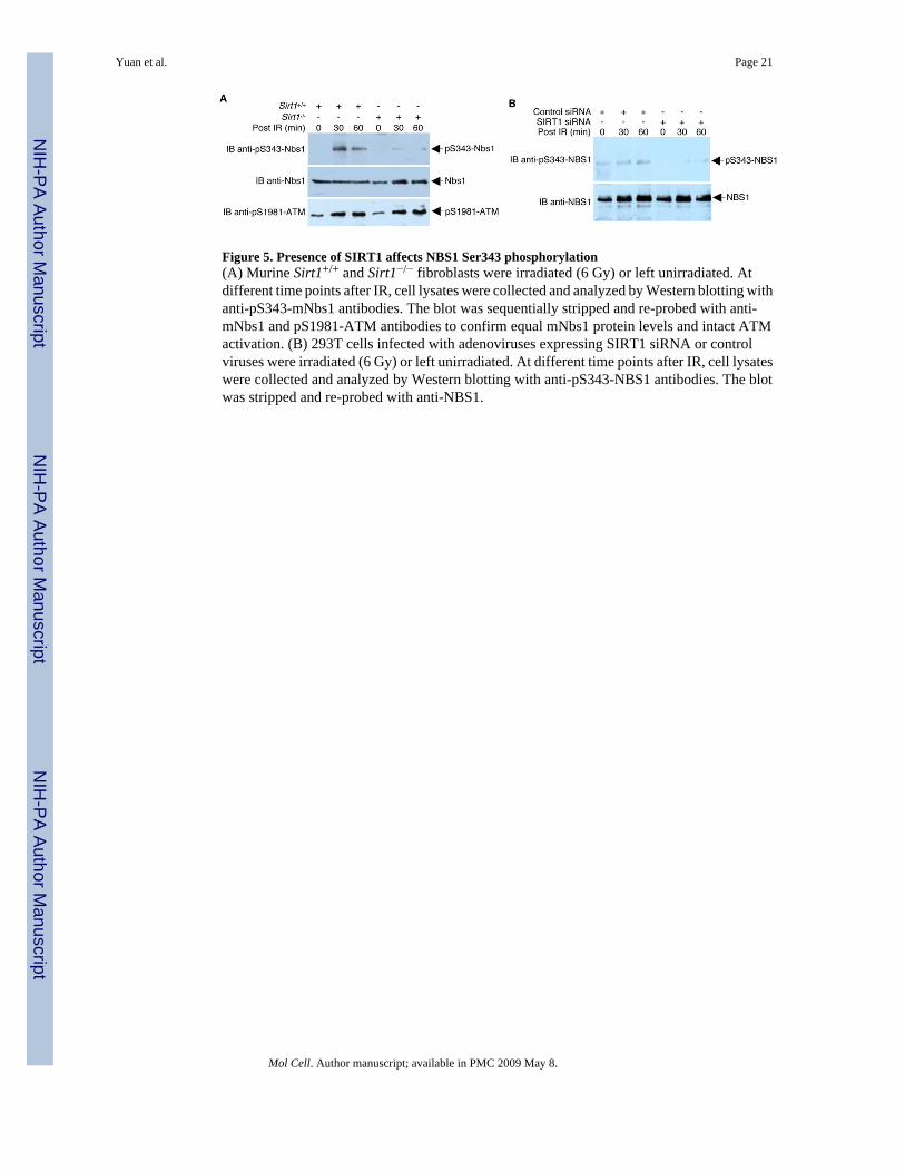
Figure 5. Presence of SIRT1 affects NBS1 Ser343 phosphorylation(A) Murine Sirt1+/+ and Sirt1−/− fibroblasts were irradiated (6 Gy) or left unirradiated. Atdifferent time points after IR, cell lysates were collected and analyzed by Western blotting withanti-pS343-mNbs1 antibodies. The blot was sequentially stripped and re-probed with anti-mNbs1 and pS1981-ATM antibodies to confirm equal mNbs1 protein levels and intact ATMactivation. (B) 293T cells infected with adenoviruses expressing SIRT1 siRNA or controlviruses were irradiated (6 Gy) or left unirradiated. At different time points after IR, cell lysateswere collected and analyzed by Western blotting with anti-pS343-NBS1 antibodies. The blotwas stripped and re-probed with anti-NBS1.
Yuan et al. Page 21
Mol Cell. Author manuscript; available in PMC 2009 May 8.
NIH
-PA Author Manuscript
NIH
-PA Author Manuscript
NIH
-PA Author Manuscript

Figure 6. NBS1 Acetylation Affects Radioresistant DNA synthesis (RDS)(A) NBS cells were transfected with plasmids encoding the indicated proteins. Twenty-fourhours after transfection, cells were irradiated (15 Gy) or left unirradiated. Left panel, RDS wasdetermined by comparing the ratio of BrdU+/EGFP+ cells to EGFP+ cells. The bar representsthe ratio in the IR-treated cells divided by the ratio in the untreated cells. Right panels, to assessimmunoprecipitation efficiency, protein expression, and NBS1 acetylation/phosphorylation,cell lysates were subjected to Western blot analysis or to high stringency immunoprecipitation/Western blot analysis, as indicated. Representative blots are shown. (B) Left panel, stablytransfected NBS cells (GM 07166) were treated with 15 Gy of IR or left untreated, cells wereradiolabeled with thymidine for RDS analysis. Right panels, representative blots to monitorimmunoprecipitation efficiency, protein expression, and NBS1 acetylation/phosphorylation.(C) 2 × 10⁴ Sirt1+/+ or Sirt1−/− MEFs were plated on 35 mm dishes for 48 h. RDS assayswere performed using the radio-labeling method to determine inhibition of DNA synthesisafter 15 Gy of IR. Error bars denote the standard deviation (SD). The data are expressed asmean ± SD from three separate experiments.
Yuan et al. Page 22
Mol Cell. Author manuscript; available in PMC 2009 May 8.
NIH
-PA Author Manuscript
NIH
-PA Author Manuscript
NIH
-PA Author Manuscript

Figure 7. NBS1 Acetylation Affects Cell Survival(A) Left panel, NBS cells transfected with plasmids (4 μg each) encoding the indicated proteinswere irradiated (2 Gy) or left unirradiated. The surviving colonies were counted two weekslater. Right panels, direct Western blots and high stringency immunoprecipitation/Western blotanalyses were performed to determine NBS1 acetylation, immunoprecipitation efficiency, andprotein expression levels. Representative blots are shown. (B) Left panel, stably transfectedNBS cells (GM 07166) were treated with 2 or 5 Gy of IR or left untreated and surviving colonieswere counted two weeks later. Right panels, direct Western blots and high stringencyimmunoprecipitation/Western blot analyses were performed to determine NBS1 acetylation/phosphorylation, immunoprecipitation efficiency, and protein expression levels. Althoughsimilar results were obtained from 2 Gy and 5 Gy IR treatments, representative blots from 5Gy treatments only are shown for simplicity. (C) Sirt1+/+ and Sirt1−/− MEFs were irradiatedwith 2 or 5 Gy of IR or left unirradiated. The surviving colonies were counted two weeks later.Fractional cell survival is the fraction of colonies surviving irradiation divided by the total
Yuan et al. Page 23
Mol Cell. Author manuscript; available in PMC 2009 May 8.
NIH
-PA Author Manuscript
NIH
-PA Author Manuscript
NIH
-PA Author Manuscript
number of colonies in the unirradiated parallel culture. Error bars denote the standard deviation(SD). The data are expressed as mean ± SD from three separate experiments.
Yuan et al. Page 24
Mol Cell. Author manuscript; available in PMC 2009 May 8.
NIH
-PA Author Manuscript
NIH
-PA Author Manuscript
NIH
-PA Author Manuscript