A novel defect in mitochondrial p53 accumulation following DNA damage confers apoptosis resistance...
9
Please cite this article in press as: V. Turinetto, et al., A novel defect in mitochondrial p53 accumulation following DNA damage confers apoptosis resistance in Ataxia Telangiectasia and Nijmegen Breakage Syndrome T-cells, DNA Repair (2010), doi:10.1016/j.dnarep.2010.09.003 ARTICLE IN PRESS G Model DNAREP-1431; No. of Pages 9 DNA Repair xxx (2010) xxx–xxx Contents lists available at ScienceDirect DNA Repair journal homepage: www.elsevier.com/locate/dnarepair A novel defect in mitochondrial p53 accumulation following DNA damage confers apoptosis resistance in Ataxia Telangiectasia and Nijmegen Breakage Syndrome T-cells Valentina Turinetto a , Paola Porcedda a , Valentina Minieri a , Luca Orlando a , Erica Lantelme a , Lisa Accomasso a , Antonio Amoroso b , Mario De Marchi a , Laura Zannini c , Domenico Delia c , Claudia Giachino a,∗ a Department of Clinical and Biological Sciences, University of Turin, Regione Gonzole 10, 10043 Orbassano (Turin), Italy b Department of Genetics, Biology and Biochemistry, University of Turin, Italy c Department of Experimental Oncology, Fondazione IRCCS Istituto Nazionale Tumori, Milan, Italy article info Article history: Received 25 January 2010 Received in revised form 9 September 2010 Accepted 13 September 2010 Available online xxx Keywords: Ataxia Telangiectasia Nijmegen Breakage Syndrome p53 Apoptosis DNA damage abstract We have previously shown that whereas T-cells from normal individuals undergo accumulation of p53 and apoptosis when treated with the genotoxic agent Actinomycin D (ActD), those from Ataxia Telang- iectasia (AT) and Nijmegen Breakage Syndrome (NBS) patients resist ActD-induced apoptosis [1]. We have now found similar resistance by the p53-null Jurkat T-cell line and by siRNA p53-knockdown nor- mal T-cells. This evidence that ActD initiates a p53-dependent apoptotic responce prompted us to look for defective p53 accumulation by AT and NBS T-cells. Surprisingly the total p53 level was only slightly reduced compared to normal T cells but its intracellular localization was highly defective: p53 was poorly accumulated in the cytosol and nearly undetectable in mitochondria. In accordance with the depen- dence of ActD-induced apoptosis on a mitochondrial p53 function, in control T-cells specific inhibition of mitochondrial p53 translocation with pifithrin reduced apoptosis by 86%, whereas treatment with pifithrin, which blocks p53-mediated transcription, had no effect. We also showed that nuclear export is not required for mitochondrial p53 translocation. Observation of an altered p53 ubiquitination pat- tern and Mdm2 accumulation in ActD-treated AT and NBS T-cells provided a mechanistic link to their defective extranuclear p53 localization. Our results disclose an undescribed defect in mitochondrial p53 accumulation in AT and NBS T-cells that makes them resistant to apoptosis following unrepairable DNA damage. © 2010 Elsevier B.V. All rights reserved. 1. Introduction DNA damage-induced apoptosis and precise DNA repair are critical cellular function, and both are needed to prevent cancer [2–6]. Defective apoptosis contributes to mutagenesis, carcinogen- esis, and the resistance of cancer cells to a variety of therapeutic agents [6]. DNA damage-induced apoptosis involves the partic- ipation of several factors with both overlapping and distinct functions, that are largely mediated by p53 [7,8]. In conditions of genotoxic stress, the increased abundance of p53 orchestrates Abbreviations: ActD, Actinomycin D; AT, Ataxia Telangiectasia; DSB, DNA double-strand break; IR, ionizing radiation; LMB, leptomycin B; NBS, Nijmegen Breakage Syndrome; -PFT, pifithrin; -PFT, pifithrin; PI, propidium iodide. ∗ Corresponding author. Tel.: +39 0116705425; fax: +39 0119038639. E-mail address: [email protected] (C. Giachino). multiple responses that involve both transcription-dependent and transcription-independent mechanisms [9]. In the nucleus, p53 acts as a transcription factor to induce pro-apoptotic and/or cell cycle arresting programs by transactivating genes encoding pro- apoptotic and/or negative regulators of cell cycle progression [10]. Additionally, a direct apoptotic role of the p53 protein at the mito- chondrial compartment in response to genotoxic stress has been amply demonstrated [11,12]. Mitochondrial p53 participates in the intrinsic apoptosis pathway by interacting with members of the Bcl-2 family to induce mitochondrial outer membrane permeabi- lization, thereby triggering the release of pro-apoptotic factors from the mitochondrial intermembrane space [13,14]. p53 turnover is regulated by Mdm2, which binds p53 and functions as an ubiqui- tin E3 ligase to promote its degradation by the proteasome [15,16]. It mediates both poly- and monoubiquitylation of p53, and while abundant levels of Mdm2 promote p53 poly-ubiquitylation and degradation, limiting levels of Mdm2 promote p53 monoubiqui- tylation on one or few lysines [17]. 1568-7864/$ – see front matter © 2010 Elsevier B.V. All rights reserved. doi:10.1016/j.dnarep.2010.09.003
-
Upload
independent -
Category
Documents
-
view
1 -
download
0
Transcript of A novel defect in mitochondrial p53 accumulation following DNA damage confers apoptosis resistance...
G
D
AcS
VLDa
b
c
a
ARRAA
KANpAD
1
c[eaifo
dB
1d
ARTICLE IN PRESSModel
NAREP-1431; No. of Pages 9
DNA Repair xxx (2010) xxx–xxx
Contents lists available at ScienceDirect
DNA Repair
journa l homepage: www.e lsev ier .com/ locate /dnarepai r
novel defect in mitochondrial p53 accumulation following DNA damageonfers apoptosis resistance in Ataxia Telangiectasia and Nijmegen Breakageyndrome T-cells
alentina Turinettoa, Paola Porceddaa, Valentina Minieri a, Luca Orlandoa, Erica Lantelmea,isa Accomassoa, Antonio Amorosob, Mario De Marchia, Laura Zannini c,omenico Deliac, Claudia Giachinoa,∗
Department of Clinical and Biological Sciences, University of Turin, Regione Gonzole 10, 10043 Orbassano (Turin), ItalyDepartment of Genetics, Biology and Biochemistry, University of Turin, ItalyDepartment of Experimental Oncology, Fondazione IRCCS Istituto Nazionale Tumori, Milan, Italy
r t i c l e i n f o
rticle history:eceived 25 January 2010eceived in revised form 9 September 2010ccepted 13 September 2010vailable online xxx
eywords:taxia Telangiectasiaijmegen Breakage Syndrome53
a b s t r a c t
We have previously shown that whereas T-cells from normal individuals undergo accumulation of p53and apoptosis when treated with the genotoxic agent Actinomycin D (ActD), those from Ataxia Telang-iectasia (AT) and Nijmegen Breakage Syndrome (NBS) patients resist ActD-induced apoptosis [1]. Wehave now found similar resistance by the p53-null Jurkat T-cell line and by siRNA p53-knockdown nor-mal T-cells. This evidence that ActD initiates a p53-dependent apoptotic responce prompted us to lookfor defective p53 accumulation by AT and NBS T-cells. Surprisingly the total p53 level was only slightlyreduced compared to normal T cells but its intracellular localization was highly defective: p53 was poorlyaccumulated in the cytosol and nearly undetectable in mitochondria. In accordance with the depen-dence of ActD-induced apoptosis on a mitochondrial p53 function, in control T-cells specific inhibition
poptosisNA damage
of mitochondrial p53 translocation with � pifithrin reduced apoptosis by 86%, whereas treatment with� pifithrin, which blocks p53-mediated transcription, had no effect. We also showed that nuclear exportis not required for mitochondrial p53 translocation. Observation of an altered p53 ubiquitination pat-tern and Mdm2 accumulation in ActD-treated AT and NBS T-cells provided a mechanistic link to theirdefective extranuclear p53 localization.
Our results disclose an undescribed defect in mitochondrial p53 accumulation in AT and NBS T-cellst to a
that makes them resistan. Introduction
DNA damage-induced apoptosis and precise DNA repair areritical cellular function, and both are needed to prevent cancer2–6]. Defective apoptosis contributes to mutagenesis, carcinogen-
Please cite this article in press as: V. Turinetto, et al., A novel defect in mitocresistance in Ataxia Telangiectasia and Nijmegen Breakage Syndrome T-ce
sis, and the resistance of cancer cells to a variety of therapeuticgents [6]. DNA damage-induced apoptosis involves the partic-pation of several factors with both overlapping and distinctunctions, that are largely mediated by p53 [7,8]. In conditionsf genotoxic stress, the increased abundance of p53 orchestrates
Abbreviations: ActD, Actinomycin D; AT, Ataxia Telangiectasia; DSB, DNAouble-strand break; IR, ionizing radiation; LMB, leptomycin B; NBS, Nijmegenreakage Syndrome; �-PFT, � pifithrin; �-PFT, � pifithrin; PI, propidium iodide.∗ Corresponding author. Tel.: +39 0116705425; fax: +39 0119038639.
E-mail address: [email protected] (C. Giachino).
568-7864/$ – see front matter © 2010 Elsevier B.V. All rights reserved.oi:10.1016/j.dnarep.2010.09.003
poptosis following unrepairable DNA damage.© 2010 Elsevier B.V. All rights reserved.
multiple responses that involve both transcription-dependent andtranscription-independent mechanisms [9]. In the nucleus, p53acts as a transcription factor to induce pro-apoptotic and/or cellcycle arresting programs by transactivating genes encoding pro-apoptotic and/or negative regulators of cell cycle progression [10].Additionally, a direct apoptotic role of the p53 protein at the mito-chondrial compartment in response to genotoxic stress has beenamply demonstrated [11,12]. Mitochondrial p53 participates in theintrinsic apoptosis pathway by interacting with members of theBcl-2 family to induce mitochondrial outer membrane permeabi-lization, thereby triggering the release of pro-apoptotic factors fromthe mitochondrial intermembrane space [13,14]. p53 turnover isregulated by Mdm2, which binds p53 and functions as an ubiqui-
hondrial p53 accumulation following DNA damage confers apoptosislls, DNA Repair (2010), doi:10.1016/j.dnarep.2010.09.003
tin E3 ligase to promote its degradation by the proteasome [15,16].It mediates both poly- and monoubiquitylation of p53, and whileabundant levels of Mdm2 promote p53 poly-ubiquitylation anddegradation, limiting levels of Mdm2 promote p53 monoubiqui-tylation on one or few lysines [17].
ING
D
2 A Repa
rtsaosldD[aNclNfgo
pptDtc
lDtcidtrAhbmr
mdtamtp
2
2
pwi6aPi10mM
ARTICLEModel
NAREP-1431; No. of Pages 9
V. Turinetto et al. / DN
ATM and NBS1 proteins are key regulators of the DNA damageesponse. ATM is a central component of the signal transduc-ion pathway activated by DNA double-strand breaks (DSBs), andynchronizes DNA repair with the induction of p53-dependentpoptosis [4,18]. Individuals who inherit germline ATM mutationsn both alleles develop AT, a disorder characterised by progres-ive neurodegeneration and predisposition to the development ofymphoid malignancies [19,20]. The cellular consequences of ATMysfunction include chromosomal radiosensitivity, radioresistantNA synthesis, loss of cell cycle checkpoints, and p53 dysfunction
18]. NBS1 acts by forming a complex with RAD50/MRE11 and byctivating ATM. It is mutated in the NBS and cell lines derived fromBS patients display hypersensitivity to ionizing radiation (IR),hromosome instability, and defects in cell cycle checkpoint fol-owing DNA damage [20,21]. The direct interconnection of ATM andBS1, e.g. ATM phosphorylates NBS1 and NBS1 in turn is required
or full ATM activation [22–24] accounts for the common patho-enetic mechanisms and the similar cellular and clinical phenotypef AT and NBS [20,25,26].
ActD is an anti-tumor antibiotic used in many chemotherapeuticrotocols [27–29]. It intercalates into DNA and interferes with RNAolymerases and DNA topoisomerases, thus inhibiting transcrip-ion and inducing various types of DNA damage [30–32], includingSBs [1]. This multiplicity of action is shared with many other geno-
oxic agents such as cisplatin and the topoisomerase I inhibitoramptothecin [33–35].
We have previously reported a defective p53 accumulation fol-owing IR in AT and NBS cells [36–39] and, like others, found thatSBs in these cells are not completely repaired [1,40]. Nevertheless,
hese residual DSBs did not induce apoptosis in patients’s quiescentells, as also confirmed by our ongoing studies on genomic stabilityn T cells. Thus, the mechanism of radiation induced response is wellefined in patients’ cells [41] at least to the point of accounting forhe radiosensitivity of proliferating cells versus the characteristicadioresistance of quiescent cells. In contrast, we have shown thatctD treatment induces an accumulation of unrepairable DSBs andigh amounts of total p53, followed by apoptosis in normal T-cellsut not T cells from AT and NBS patients [1]. In this case, both theechanism of p53 activity after ActD treatment and the underlying
esistance of AT and NBS cells are still unknown.Here we show that ActD-induced apoptosis depends on a
itochondrial p53-dependent pathway and Bax activation, andemonstrate for the first time that AT and NBS T-cells are charac-erised by almost undetectable mitochondrial p53 accumulation,n altered p53 ubiquitination pattern and increased Mdm2 accu-ulation. These findings open new perspectives in understanding
he multifaceted function of ATM and NBS1 in regulating apoptoticathways.
. Material and methods
.1. Cell isolation, cell culture and drug treatment
Peripheral blood from four healthy donors, three AT and two NBSatients was collected after signed informed consent. AT patientsere either homozygous or compound heterozygous for truncat-
ng mutations; NBS patients were homozygous for the common57del5 mutation. PBMC were isolated and T-cell lines, generateds described [42], were maintained by periodic stimulation withHA (Gibco-Invitrogen, Paisley, UK) and irradiated allogeneic PBMC
Please cite this article in press as: V. Turinetto, et al., A novel defect in mitocresistance in Ataxia Telangiectasia and Nijmegen Breakage Syndrome T-ce
n complete RPMI medium (RPMI 1640 added with 1% kanamycin,% sodium piruvate, 1% L-glutamine, 1% nonessential amino acids,.1% �-mercapto-ethanol) (all from Gibco-Invitrogen), supple-ented with 5% human serum (BioWitthaker, Cambrex, Baltimore,D, USA) and 200 U/ml recombinant IL2 (from the myeloma pro-
PRESSir xxx (2010) xxx–xxx
ducing cell line IL2-t6, kindly provided by Dr A. Lanzavecchia,IRB, Bellinzona, Switzerland). All experiments were conducted onresting T cells (>95% in G0/G1). The Jurkat human leukemia cellline was purchased from the American Type Culture Collection(Rockville, MD, USA), and grown in complete RPMI medium sup-plemented with 10% heat-inactivated fetal bovine serum (Gibco,Invitrogen). ActD was supplied by Sigma–Aldrich Co. (St. Luis, MO,USA) and used at doses ranging from 0.05 to 0.5 �g/ml. CulturedT cells were �-irradiated (2 Gy) using a 6 MV accelerator (Elekta)at a dose of 2 Gy/min. �-pifithrin (�-PFT) and �-pifithrin (�-PFT)(Sigma–Aldrich Co.) were used at 30 �M and 10 �M respectively,and added 20 min before ActD. Leptomycin B (LMB) (Applichem,Germany) was used at 10 nM and added 30 min before ActD.
2.2. Flow cytometry
Cell viability was determined by propidium iodide (PI)(Sigma–Aldrich Co.) staining; PI was used at the final concentra-tion of 1 �g/ml and maintained at room temperature for 15 minin the dark before the analysis. Cell survival was expressed afternormalization to medium alone or supplemented with the drug’ssolvent (DMSO). Apoptosis was defined through double stainingwith anti-annexinV-FITC (BD PharMingen, San Diego, CA, USA) andPI, in accordance with the manufacturer’s instructions. Caspaseactivation was analyzed with anti-Active Caspase-3 (BD PharMin-gen) as primary antibody and a PE-conjugated goat anti-rabbit (BDPharMingen) as secondary antibody, using the Cytofix/CytopermKit (BD PharMingen). Stained cells were analyzed on a FACScan(Becton Dikinson & Co., San Jose, CA, USA). Statistical analysis of cellsurvival and caspase-3 activation was performed with either thetest of independence or the Student’s t-test. The significance wereexpressed in the figures with the asterisks: *p < 0.05; **p < 0.01;***p < 0.005.
2.3. Gene silencing by siRNA
The Amaxa Nucleofector system (Lonza, Cologne, Germany) andAmaxa Human T-cell Nucleofector Kit were used for electropora-tion. Briefly, T-cells were resuspended in Nucleofector Solution,Scramble (ON-TARGET Nontargeting pool, Thermo Scientific, Rock-ford, IL, USA) or p53 specific (ON-TARGETplus SMART pool, HumanTP53, Thermo Scientific) siRNA oligos (30 pmol) were added to6 × 106 T-cells/100 �L transfection buffer and nucleofected usingthe Amaxa Nucleofector II Device and program V-024, according tothe manufacturer’s instructions. Nucleofected T-cells were main-tained in fresh medium for 48 h, then either treated with ActD for24 h or left untreated. Treated and untreated cells were harvestedat the same time point.
2.4. Immunofluorescence
Approximately 400,000 T-cells for each condition were col-lected, fixed with 4% paraformaldehyde, permeabilized with 0.5%Triton X-100, and blocked with 6% bovine serum albumin and 2.5%normal goat serum. They were stained with anti-p53 (clone DO1),anti-BAX (clone 2D2), anti-Mdm2 (clone H221) specific Abs (allfrom Santa Cruz Biotechnology, Santa Cruz, CA, USA), and withAlexa 546-conjugated goat anti-mouse as secondary Ab (Molec-ular Probes, Invitrogen). Green Fluorescent Mitotraker (MolecularProbes, Invitrogen) was used at the final concentration of 100 nM.Stained cells were transferred to poly-l-lysine-coated coverslips
hondrial p53 accumulation following DNA damage confers apoptosislls, DNA Repair (2010), doi:10.1016/j.dnarep.2010.09.003
and slides were mounted with Mowiol (Calbiochem, San Diego, CA,USA). Fluorescence images were obtained with a 510 Carl Zeissconfocal laser microscope using a 63× objective. Colocalizationanalyses were performed with LSM510 Image examiner softwarefrom Zeiss, selecting the cytosolic area of 20 different cells for each
ING
D
A Repa
csZawe
2
lcsaTbnPMt2eaStmotiwBbi
2
cg(wC((-s(afiKatml
3
3nA
tidw
ARTICLEModel
NAREP-1431; No. of Pages 9
V. Turinetto et al. / DN
ondition, in two independent experiments. Fluorescence inten-ity analysis was performed with Image examiner software fromeiss, selecting 20 different cells for each condition elaborated asrbitrary units/pixel. Statistical analysis of fluorescence intensityas performed with the Student’s t-test. The significance were
xpressed in the figure with the asterisks: *p < 0.05; **p < 0.01.
.5. Cell extracts and subcellular fractionation
Cells were harvested and washed with PBS, pelleted, andysed in Laemmli buffer (0.125 M Tris–HCl [pH 6.8], 5% SDS)ontaining as inhibitors 1 mM phenylmethylsulfonyl fluoride, pep-tatin (10 �g/ml), aprotinin (100 KIU/ml), leupeptin (10 �g/ml)nd 1 mM sodium orthovanadate (Na3VO4) (all from Calbiochem).otal lysates were boiled for 2 min, sonicated, and quantitatedy the micro-bicinchoninic acid method (Thermo Scientific). Foruclear protein isolation, cells were harvested and washed withBS, pelleted and lysed in 10 mM Hepes, 10 mM KCl, 1.5 mMgCl2, 0.7% NP-40 (all from Sigma–Aldrich) buffer containing pro-
ease inhibitors; subsequently, nuclear proteins were isolated with0 mM Hepes, 0.42 M NaCl, 1.5 mM MgCl2, 0.2 mM EDTA, 25% glyc-rol (all from Sigma–Aldrich) buffer containing protease inhibitorsnd quantitated by the micro-bicinchoninic acid method (Thermocientific) before analysis by immunoblotting. For subcellular frac-ionation, 2 × 10⁷ T-cells were harvested and washed with PBS;
itochondrial and cytosolic fractions were isolated with the usef the Mitochondria Isolation Kit for cultured cells (Thermo Scien-ific, reagent-based method). The mitochondrial pellet was lysedn Laemmli buffer, and the cytosolic supernatant was concentrated
ith a Microcon device (size cut-off 10 kDa) (Millipore Corporation,edford, MA, USA). Both fractions were quantitated by the micro-icinchoninic acid method (Thermo Scientific) before analysis by
mmunoblotting.
.6. Immunoblot analysis
40 �g of total lysates and 20 �g of nuclear, mitochondrial andytosolic fractions were size fractionated by SDS-PAGE 7–10%els and electroblotted onto polyvinylidene difluoride membranesAmersham, GE Healthcare, Buckinghamshhire, UK). After blockingith 5% nonfat dried milk in PBS plus 0.1% Tween (Sigma–Aldricho.), the membranes were incubated with anti-p53 (clone DO7)YLEM, Avezzano, Italy), -Tom40 (H-300), -lamin A (H-102), -Mdm2H-221) (Santa Cruz Biotechnology), -vinculin (clone hVIN-1),�-actin (clone AC-72) (Sigma–Aldrich Co.) specific Abs, andubsequently with peroxidase-conjugated secondary antibodiesAmersham, GE Healthcare). The immunoreactive bands were visu-lized by ECL Super Signal (Thermo Scientific) on autoradiographiclms. Autoradiographic bands were scanned and quantified byodak 1D Image Analysis Software. p53 monoubiquitination wasssessed through long exposures of p53 immunoblots to evaluatehe appearance of a signal in the range between 60 kDa (one p53
onoubiquitylated site) and 120 kDa (all eight p53 monoubiquity-ated sites), as described [17,43].
. Results
.1. T-cells from AT and NBS patients are resistant to aon-redundant p53-dependent apoptotic pathway induced byctD
Please cite this article in press as: V. Turinetto, et al., A novel defect in mitocresistance in Ataxia Telangiectasia and Nijmegen Breakage Syndrome T-ce
To clarify the mechanism that makes AT and NBS T-cells resis-ant to ActD-induced apoptosis, we explored the death eventsnitiated by ActD. Control cells harvested 15 h after ActD treatmentid not display any appreciable signs of apoptosis (data not shown),hereas after 24 h there was an evident increase in the fraction
PRESSir xxx (2010) xxx–xxx 3
of active caspase-3-positive and annexinV-positive/PI-negative, i.e.early apoptotic, cells (Fig. 1A and B). By contrast, similarly treatedAT and NBS T-cells completely lacked caspase-3 activation, andwere defective in phosphatidylserine translocation from the innerto the outer leaflet of the membrane (Fig. 1A and B).
As in our previous study [1], control T-cells treated with ActDaccumulated high amounts of total p53 (Fig. 1C). To determinewhether p53 contributed to the stress response elicited by ActD,we examined the response of the p53-null Jurkat T-cell line [44].Our western blot analysis confirmed that p53 was not expressedin Jurkat cells, even after ActD treatment (Fig. 1C). As shown inFig. 1D, they were resistant to ActD-induced apoptosis. Their viabil-ity at 72 h was more than 60% (Jurkat versus control cells: p < 0.0001with the test of independence), similar to AT and NBS T-cells [1], andwere neither active caspase-3-positive nor AnnexinV-positive/PI-negative at 24 h (Fig. 1A and B). As a second way to assess thep53 dependence of ActD-activated apoptotic pathway, we depletedp53 by siRNA in control T-cell lines. Immunoblot analysis con-firmed that p53 levels decreased after p53 siRNA nucleofection(Fig. 1E). We found that p53 knockdown significantly increased sur-vival (Fig. 1F) and decreased caspase-3 activation (Fig. 1G) followingActD treatment, further suggesting that p53 is required to activateActD apoptotic pathway.
These findings support the conclusion that ActD induces ap53-dependent apoptotic pathway in normal T-cells, and that theresistance of AT and NBS T-cells to ActD-induced apoptosis is dueto its defective activation.
3.2. Defective ActD-induced p53 cytoplasmic accumulation andBax activation in AT and NBS T-cells
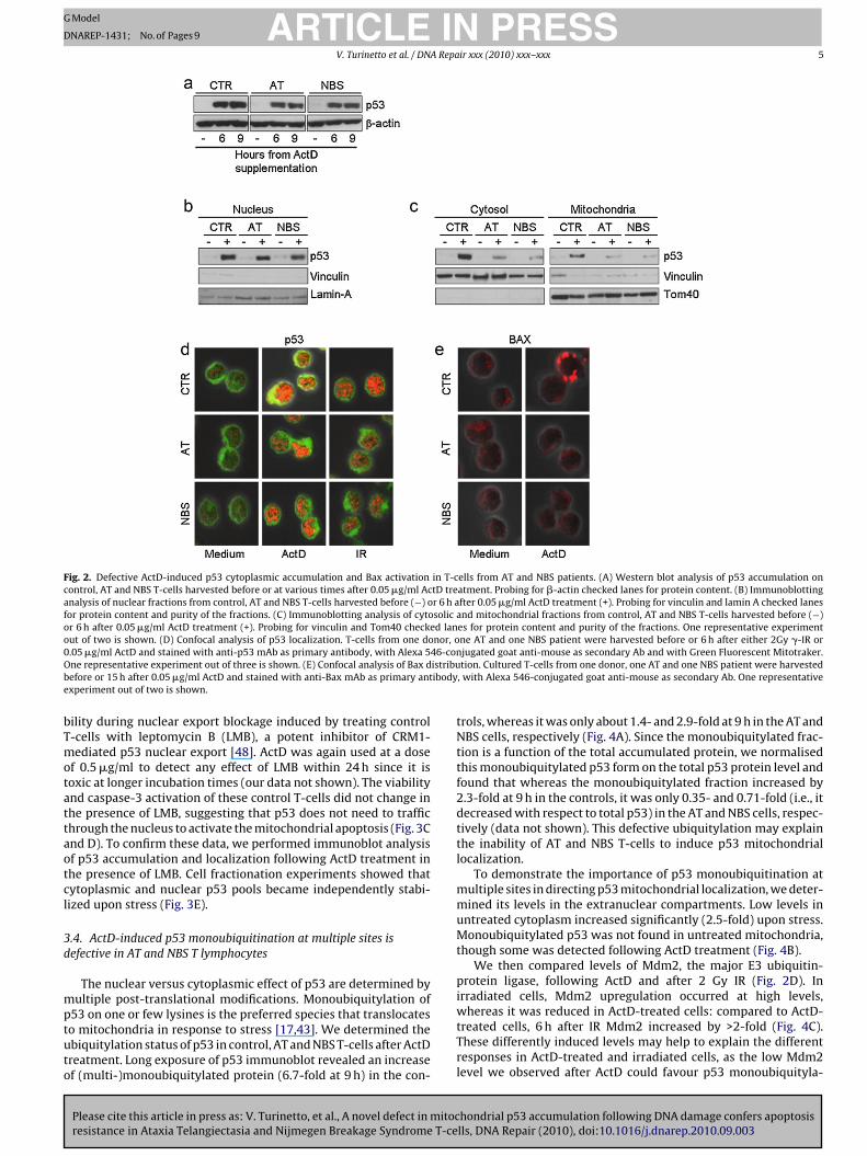
The similar apoptotic defects observed in AT and NBS T-cells andin p53-deficient Jurkat T-cells prompted us to look for defectivep53 accumulation by AT and NBS T-cells following ActD treatment.Surprisingly, their total p53 protein level after 6 h was only slightlyreduced to 78% and 98% of that in control T-cells in AT and NBS T-cells respectively (Fig. 2A), whereas p53 accumulation induced byIR was clearly defective (about 10% and 28% that of control T-cells,respectively), as assessed by both western blot analysis [36] andconfocal microscopy (Fig. 2D), as already reported [37–39].
We thus employed cell fractionation and immunoblot analysisin a more detailed search for major defects in p53 subcellular local-ization in control, AT and NBS T-cells 6 h after ActD-treatment. Thep53 detected in nuclear extracts from the patients was only slightlydefective, being about 81% in AT and 77% in NBS cells compared tothe controls (Fig. 2B). In addition, p53 was clearly visible in both thecytosol and the mitochondria of the controls, whereas its accumu-lation was highly defective in the cytosolic fraction of AT and NBST-cells (about 20% in AT and 22% in NBS compared to the controls),and nearly undetectable in their mitochondrial fraction (about 6%in AT and 16% in NBS compared to the controls) (Fig. 2C).
The distribution of p53 was then determined by immunfluo-rescence staining and confocal microscopy with a cytosolic probe(Mitotraker green) in conjunction with the anti-p53 antibody (red).In control T-cells p53 was not only present in the nucleus, but also inthe cytosol 6 h after ActD, as highlighted by the yellow signal in thedouble-staining with the cytosolic probe (Fig. 2D). In AT and NBS T-cells, on the contrary, p53 nuclear accumulation alone was evidentat the same time point (Fig. 2D). Colocalization analysis confirmedthese data: p53 was present in about 40% of the cytosolic area ofActD-treated normal T-cells, but in less than 5% of ActD-treated
hondrial p53 accumulation following DNA damage confers apoptosislls, DNA Repair (2010), doi:10.1016/j.dnarep.2010.09.003
patients’ T-cells (data not shown). After 2 Gy IR, a genotoxic stim-ulus that does not induce apoptosis in resting T-cells [1], p53 wasexclusively nuclear in both control and patients’ T-cells (Fig. 2D).
Cytosolic accumulation of p53 directly activates Bax to insertand oligomerize into the mitochondrial membrane, thus leading

ARTICLE IN PRESSG Model
DNAREP-1431; No. of Pages 9
4 V. Turinetto et al. / DNA Repair xxx (2010) xxx–xxx
Fig. 1. Selective resistance to ActD-induced apoptosis of T-cells from AT and NBS patients. (A) Cultured T-cells from one control, one AT and one NBS patient, and JurkatT-cells were kept for 24 h in medium alone or with 0.05 �g/ml ActD, then harvested and stained with anti-active caspase-3 antibody. Percentages of active caspase-3+ cellsare indicated. (B) Cultured T-cells from one control, one AT and one NBS patient and Jurkat T-cells were kept for 24 h in medium alone or with 0.05 �g/ml ActD, then harvestedand stained with anti-annexinV antibody and PI. Percentages of PI− annexinV+ cells are indicated. (C) Western blot analysis of p53 accumulation on control and Jurkat T-cellsharvested before or at various times after 0.05 �g/ml ActD treatment. Probing for �-actin checked lanes for protein content. (D) Cultured T-cells from three controls (�) andJurkat T-cells (*) were kept in medium alone or in medium containing 0.05 �g/ml of ActD, harvested at 24, 48 and 72 h and analyzed by flow cytometry after staining withPI. Viability is shown as mean ± SD of three independent experiments. (E) Western blot analysis of p53 in control T-cells transfected with either scrambled or p53 siRNAa roteint zed toC mediua
t3caabavaeIt
cad
3t
p
nd treated for 24 h with 0.05 �g/ml ActD. Probing for vinculin checked lanes for preated with 0.05 �g/ml ActD and analyzed at 24 h by PI staining. Data are normaliontrol T-cells transfected with either scrambled or p53 siRNA were kept for 24 h inntibody. Percentages of active caspase-3+ cells are indicated.
o the cascade of mitochondria-mediated apoptosis and caspase-activation [13]. We therefore sought to find out whether high
ytosolic p53 levels in ActD-treated cells were coupled with Baxctivation. We monitored Bax distribution by immunofluorescencend confocal microscopy at 9–15–24 h. In the control T-cells, Bax’saseline cytosolic distribution remained until 15 h after ActD, whendot-like pattern was displayed by about 30% of these cells; this
alue increased to 35% at 24 h, concomitant with the beginning ofpoptosis (Fig. 2E and data not shown). When treated AT cells werexamined, no changes in this baseline distribution were detected.n NBS cells, Bax was activated at significantly lower levels than inhe controls (6% of positive cells at 24 h) (Fig. 2E).
These results suggested that cytoplasmic p53 accumulation andhanges in Bax distribution are involved in ActD-induced apoptosis,nd that this extra-nuclear accumulation and Bax activation areefective in AT cells and, to a lesser extent, in NBS cells.
Please cite this article in press as: V. Turinetto, et al., A novel defect in mitocresistance in Ataxia Telangiectasia and Nijmegen Breakage Syndrome T-ce
.3. ActD-induced apoptosis is dependent on the mitochondrialargeting of p53 from an extranuclear pool
To clarify the role of p53 in ActD-induced apoptosis, we usedharmacological manipulations to distinguish its nuclear and mito-
content. (F) Control T-cells transfected with either scrambled or p53 siRNA weresurvival in medium alone (bars, mean ± SD of two independent experiments). (G)m alone or with 0.05 �g/ml ActD, harvested and stained with anti-active caspase-3
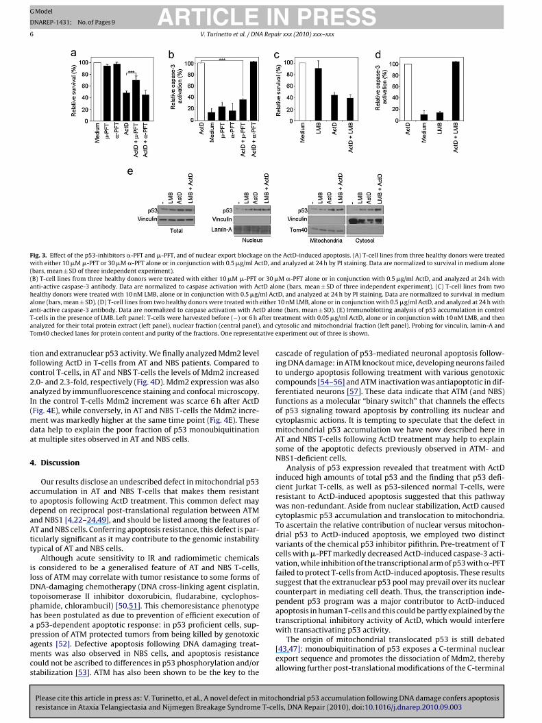
chondrial functions. Control T-cells were preincubated with twop53 inhibitors that act through different mechanisms: �-pifithrin(�-PFT) reversibly blocks the p53-mediated transactivation of p53-dependent responsive genes [45]; �-PFT blocks the interaction ofp53 with Bcl-xL and Bcl-2, and selectively inhibits p53 translocationto the mitochondria, without affecting its genomic transactivationfunction [46].
We used 0.5 �g/ml ActD in order to detect any effect of PFTwithin 24 h. At longer incubation times, the activity of �-PFT and�-PFT is partially lost (our data not shown). While the viability levelof ActD-treated control T-cells did not change in the presence of �-PFT, preincubation with �-PFT conferred significant resistance toActD-induced apoptosis (70% viability at 24 h) (Fig. 3A). To demon-strate that blockage of mitochondrial p53 increases viability byinhibiting apoptosis, we assessed caspase-3 activation in control T-cells treated with ActD, either alone or in the presence of �-PFT or�-PFT. �-PFT preincubation reduced caspase-3 activation by about
hondrial p53 accumulation following DNA damage confers apoptosislls, DNA Repair (2010), doi:10.1016/j.dnarep.2010.09.003
86% compared to ActD alone (Fig. 3B), whereas �-PFT treatmenthad no effect, as expected. These data suggested that ActD-inducedapoptosis depends on mitochondrial p53 functions.
To further investigate the origin of mitochondrial translocatedp53, a still debated issue [43,47], we assessed the effects on cell via-
ARTICLE IN PRESSG Model
DNAREP-1431; No. of Pages 9
V. Turinetto et al. / DNA Repair xxx (2010) xxx–xxx 5
Fig. 2. Defective ActD-induced p53 cytoplasmic accumulation and Bax activation in T-cells from AT and NBS patients. (A) Western blot analysis of p53 accumulation oncontrol, AT and NBS T-cells harvested before or at various times after 0.05 �g/ml ActD treatment. Probing for �-actin checked lanes for protein content. (B) Immunoblottinganalysis of nuclear fractions from control, AT and NBS T-cells harvested before (−) or 6 h after 0.05 �g/ml ActD treatment (+). Probing for vinculin and lamin A checked lanesfor protein content and purity of the fractions. (C) Immunoblotting analysis of cytosolic and mitochondrial fractions from control, AT and NBS T-cells harvested before (−)or 6 h after 0.05 �g/ml ActD treatment (+). Probing for vinculin and Tom40 checked lanes for protein content and purity of the fractions. One representative experimentout of two is shown. (D) Confocal analysis of p53 localization. T-cells from one donor, one AT and one NBS patient were harvested before or 6 h after either 2Gy �-IR or0 46-conO istribub ibodye
bTmotattaotcl
3d
mptuto
.05 �g/ml ActD and stained with anti-p53 mAb as primary antibody, with Alexa 5ne representative experiment out of three is shown. (E) Confocal analysis of Bax defore or 15 h after 0.05 �g/ml ActD and stained with anti-Bax mAb as primary antxperiment out of two is shown.
ility during nuclear export blockage induced by treating control-cells with leptomycin B (LMB), a potent inhibitor of CRM1-ediated p53 nuclear export [48]. ActD was again used at a dose
f 0.5 �g/ml to detect any effect of LMB within 24 h since it isoxic at longer incubation times (our data not shown). The viabilitynd caspase-3 activation of these control T-cells did not change inhe presence of LMB, suggesting that p53 does not need to traffichrough the nucleus to activate the mitochondrial apoptosis (Fig. 3Cnd D). To confirm these data, we performed immunoblot analysisf p53 accumulation and localization following ActD treatment inhe presence of LMB. Cell fractionation experiments showed thatytoplasmic and nuclear p53 pools became independently stabi-ized upon stress (Fig. 3E).
.4. ActD-induced p53 monoubiquitination at multiple sites isefective in AT and NBS T lymphocytes
The nuclear versus cytoplasmic effect of p53 are determined byultiple post-translational modifications. Monoubiquitylation of
Please cite this article in press as: V. Turinetto, et al., A novel defect in mitocresistance in Ataxia Telangiectasia and Nijmegen Breakage Syndrome T-ce
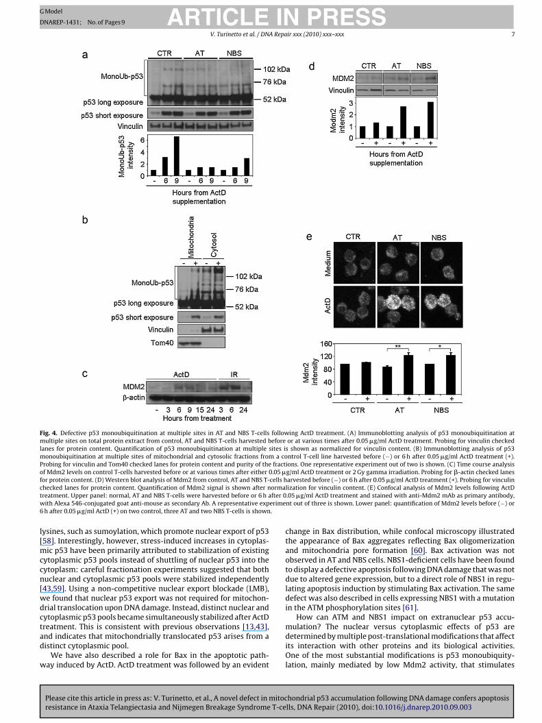
53 on one or few lysines is the preferred species that translocateso mitochondria in response to stress [17,43]. We determined thebiquitylation status of p53 in control, AT and NBS T-cells after ActDreatment. Long exposure of p53 immunoblot revealed an increasef (multi-)monoubiquitylated protein (6.7-fold at 9 h) in the con-
jugated goat anti-mouse as secondary Ab and with Green Fluorescent Mitotraker.tion. Cultured T-cells from one donor, one AT and one NBS patient were harvested
, with Alexa 546-conjugated goat anti-mouse as secondary Ab. One representative
trols, whereas it was only about 1.4- and 2.9-fold at 9 h in the AT andNBS cells, respectively (Fig. 4A). Since the monoubiquitylated frac-tion is a function of the total accumulated protein, we normalisedthis monoubiquitylated p53 form on the total p53 protein level andfound that whereas the monoubiquitylated fraction increased by2.3-fold at 9 h in the controls, it was only 0.35- and 0.71-fold (i.e., itdecreased with respect to total p53) in the AT and NBS cells, respec-tively (data not shown). This defective ubiquitylation may explainthe inability of AT and NBS T-cells to induce p53 mitochondriallocalization.
To demonstrate the importance of p53 monoubiquitination atmultiple sites in directing p53 mitochondrial localization, we deter-mined its levels in the extranuclear compartments. Low levels inuntreated cytoplasm increased significantly (2.5-fold) upon stress.Monoubiquitylated p53 was not found in untreated mitochondria,though some was detected following ActD treatment (Fig. 4B).
We then compared levels of Mdm2, the major E3 ubiquitin-protein ligase, following ActD and after 2 Gy IR (Fig. 2D). Inirradiated cells, Mdm2 upregulation occurred at high levels,
hondrial p53 accumulation following DNA damage confers apoptosislls, DNA Repair (2010), doi:10.1016/j.dnarep.2010.09.003
whereas it was reduced in ActD-treated cells: compared to ActD-treated cells, 6 h after IR Mdm2 increased by >2-fold (Fig. 4C).These differently induced levels may help to explain the differentresponses in ActD-treated and irradiated cells, as the low Mdm2level we observed after ActD could favour p53 monoubiquityla-
ARTICLE IN PRESSG Model
DNAREP-1431; No. of Pages 9
6 V. Turinetto et al. / DNA Repair xxx (2010) xxx–xxx
Fig. 3. Effect of the p53-inhibitors �-PFT and �-PFT, and of nuclear export blockage on the ActD-induced apoptosis. (A) T-cell lines from three healthy donors were treatedwith either 10 �M �-PFT or 30 �M �-PFT alone or in conjunction with 0.5 �g/ml ActD, and analyzed at 24 h by PI staining. Data are normalized to survival in medium alone(bars, mean ± SD of three independent experiment).(B) T-cell lines from three healthy donors were treated with either 10 �M �-PFT or 30 �M �-PFT alone or in conjunction with 0.5 �g/ml ActD, and analyzed at 24 h withanti-active caspase-3 antibody. Data are normalized to caspase activation with ActD alone (bars, mean ± SD of three independent experiment). (C) T-cell lines from twohealthy donors were treated with 10 nM LMB, alone or in conjunction with 0.5 �g/ml ActD, and analyzed at 24 h by PI staining. Data are normalized to survival in mediumalone (bars, mean ± SD). (D) T-cell lines from two healthy donors were treated with either 10 nM LMB, alone or in conjunction with 0.5 �g/ml ActD, and analyzed at 24 h witha tD aloT fter ta , and cT ative e
tfc2aI(mda
4
atdaAtt
ilDtphapamcs
nti-active caspase-3 antibody. Data are normalized to caspase activation with Ac-cells in the presence of LMB. Left panel: T-cells were harvested before (−) or 6 h analyzed for their total protein extract (left panel), nuclear fraction (central panel)om40 checked lanes for protein content and purity of the fractions. One represent
ion and extranuclear p53 activity. We finally analyzed Mdm2 levelollowing ActD in T-cells from AT and NBS patients. Compared toontrol T-cells, in AT and NBS T-cells the levels of Mdm2 increased.0- and 2.3-fold, respectively (Fig. 4D). Mdm2 expression was alsonalyzed by immunfluorescence staining and confocal microscopy.n the control T-cells Mdm2 increment was scarce 6 h after ActDFig. 4E), while conversely, in AT and NBS T-cells the Mdm2 incre-
ent was markedly higher at the same time point (Fig. 4E). Theseata help to explain the poor fraction of p53 monoubiquitinationt multiple sites observed in AT and NBS cells.
. Discussion
Our results disclose an undescribed defect in mitochondrial p53ccumulation in AT and NBS T-cells that makes them resistanto apoptosis following ActD treatment. This common defect mayepend on reciprocal post-translational regulation between ATMnd NBS1 [4,22–24,49], and should be listed among the features ofT and NBS cells. Conferring apoptosis resistance, this defect is par-
icularly significant as it may contribute to the genomic instabilityypical of AT and NBS cells.
Although acute sensitivity to IR and radiomimetic chemicalss considered to be a generalised feature of AT and NBS T-cells,oss of ATM may correlate with tumor resistance to some forms ofNA-damaging chemotherapy (DNA cross-linking agent cisplatin,
opoisomerase II inhibitor doxorubicin, fludarabine, cyclophos-hamide, chlorambucil) [50,51]. This chemoresistance phenotypeas been postulated as due to prevention of efficient execution ofp53-dependent apoptotic response: in p53 proficient cells, sup-
Please cite this article in press as: V. Turinetto, et al., A novel defect in mitocresistance in Ataxia Telangiectasia and Nijmegen Breakage Syndrome T-ce
ression of ATM protected tumors from being killed by genotoxicgents [52]. Defective apoptosis following DNA damaging treat-ents was also observed in NBS cells, and apoptosis resistance
ould not be ascribed to differences in p53 phosphorylation and/ortabilization [53]. ATM has also been shown to be the key to the
ne (bars, mean ± SD). (E) Immunoblotting analysis of p53 accumulation in controlreatment with 0.05 �g/ml ActD, alone or in conjunction with 10 nM LMB, and thenytosolic and mitochondrial fraction (left panel). Probing for vinculin, lamin-A andxperiment out of three is shown.
cascade of regulation of p53-mediated neuronal apoptosis follow-ing DNA damage: in ATM knockout mice, developing neurons failedto undergo apoptosis following treatment with various genotoxiccompounds [54–56] and ATM inactivation was antiapoptotic in dif-ferentiated neurons [57]. These data indicate that ATM (and NBS)functions as a molecular “binary switch” that channels the effectsof p53 signaling toward apoptosis by controlling its nuclear andcytoplasmic actions. It is tempting to speculate that the defect inmitochondrial p53 accumulation we have now described here inAT and NBS T-cells following ActD treatment may help to explainsome of the apoptotic defects previously observed in ATM- andNBS1-deficient cells.
Analysis of p53 expression revealed that treatment with ActDinduced high amounts of total p53 and the finding that p53 defi-cient Jurkat T-cells, as well as p53-silenced normal T-cells, wereresistant to ActD-induced apoptosis suggested that this pathwaywas non-redundant. Aside from nuclear stabilization, ActD causedcytoplasmic p53 accumulation and translocation to mitochondria.To ascertain the relative contribution of nuclear versus mitochon-drial p53 to ActD-induced apoptosis, we employed two distinctvariants of the chemical p53 inhibitor pifithrin. Pre-treatment of Tcells with �-PFT markedly decreased ActD-induced caspase-3 acti-vation, while inhibition of the transcriptional arm of p53 with �-PFTfailed to protect T-cells from ActD-induced apoptosis. These resultssuggest that the extranuclear p53 pool may prevail over its nuclearcounterpart in mediating cell death. Thus, the transcription inde-pendent p53 program was a major contributor to ActD-inducedapoptosis in human T-cells and this could be partly explained by thetranscriptional inhibitory activity of ActD, which would interferewith transactivating p53 activity.
hondrial p53 accumulation following DNA damage confers apoptosislls, DNA Repair (2010), doi:10.1016/j.dnarep.2010.09.003
The origin of mitochondrial translocated p53 is still debated[43,47]: monoubiquitination of p53 exposes a C-terminal nuclearexport sequence and promotes the dissociation of Mdm2, therebyallowing further post-translational modifications of the C-terminal
ARTICLE IN PRESSG Model
DNAREP-1431; No. of Pages 9
V. Turinetto et al. / DNA Repair xxx (2010) xxx–xxx 7
Fig. 4. Defective p53 monoubiquitination at multiple sites in AT and NBS T-cells following ActD treatment. (A) Immunoblotting analysis of p53 monoubiquitination atmultiple sites on total protein extract from control, AT and NBS T-cells harvested before or at various times after 0.05 �g/ml ActD treatment. Probing for vinculin checkedlanes for protein content. Quantification of p53 monoubiquitination at multiple sites is shown as normalized for vinculin content. (B) Immunoblotting analysis of p53monoubiquitination at multiple sites of mitochondrial and cytosolic fractions from a control T-cell line harvested before (−) or 6 h after 0.05 �g/ml ActD treatment (+).Probing for vinculin and Tom40 checked lanes for protein content and purity of the fractions. One representative experiment out of two is shown. (C) Time course analysisof Mdm2 levels on control T-cells harvested before or at various times after either 0.05 �g/ml ActD treatment or 2 Gy gamma irradiation. Probing for �-actin checked lanesfor protein content. (D) Western blot analysis of Mdm2 from control, AT and NBS T-cells harvested before (−) or 6 h after 0.05 �g/ml ActD treatment (+). Probing for vinculinc ormat fter 0w erime6 .
l[mccn[wdctad
w
hecked lanes for protein content. Quantification of Mdm2 signal is shown after nreatment. Upper panel: normal, AT and NBS T-cells were harvested before or 6 h aith Alexa 546-conjugated goat anti-mouse as secondary Ab. A representative exph after 0.05 �g/ml ActD (+) on two control, three AT and two NBS T-cells is shown
ysines, such as sumoylation, which promote nuclear export of p5358]. Interestingly, however, stress-induced increases in cytoplas-
ic p53 have been primarily attributed to stabilization of existingytoplasmic p53 pools instead of shuttling of nuclear p53 into theytoplasm: careful fractionation experiments suggested that bothuclear and cytoplasmic p53 pools were stabilized independently43,59]. Using a non-competitive nuclear export blockade (LMB),e found that nuclear p53 export was not required for mitochon-rial translocation upon DNA damage. Instead, distinct nuclear andytoplasmic p53 pools became simultaneously stabilized after ActDreatment. This is consistent with previous observations [13,43],
Please cite this article in press as: V. Turinetto, et al., A novel defect in mitocresistance in Ataxia Telangiectasia and Nijmegen Breakage Syndrome T-ce
nd indicates that mitochondrially translocated p53 arises from aistinct cytoplasmic pool.
We have also described a role for Bax in the apoptotic path-ay induced by ActD. ActD treatment was followed by an evident
lization for vinculin content. (E) Confocal analysis of Mdm2 levels following ActD.05 �g/ml ActD treatment and stained with anti-Mdm2 mAb as primary antibody,nt out of three is shown. Lower panel: quantification of Mdm2 levels before (−) or
change in Bax distribution, while confocal microscopy illustratedthe appearance of Bax aggregates reflecting Bax oligomerizationand mitochondria pore formation [60]. Bax activation was notobserved in AT and NBS cells. NBS1-deficient cells have been foundto display a defective apoptosis following DNA damage that was notdue to altered gene expression, but to a direct role of NBS1 in regu-lating apoptosis induction by stimulating Bax activation. The samedefect was also described in cells expressing NBS1 with a mutationin the ATM phosphorylation sites [61].
How can ATM and NBS1 impact on extranuclear p53 accu-mulation? The nuclear versus cytoplasmic effects of p53 are
hondrial p53 accumulation following DNA damage confers apoptosislls, DNA Repair (2010), doi:10.1016/j.dnarep.2010.09.003
determined by multiple post-translational modifications that affectits interaction with other proteins and its biological activities.One of the most substantial modifications is p53 monoubiquity-lation, mainly mediated by low Mdm2 activity, that stimulates
ING
D
8 A Repa
plapmfbiMtsIttMM[umtonrcscAtlillibNpta
b[beKbsct
adTpn
C
A
cMc
[
[
[
[
[
[[
[
[
[
[
[
[
[
[
[
[
[
[
[
ARTICLEModel
NAREP-1431; No. of Pages 9
V. Turinetto et al. / DN
53 arrival at the mitochondria; here, p53 is rapidly deubiquity-ated by mitochondrial HAUSP, thus generating the apoptoticallyctive non-ubiquitylated p53 [43,59]. Consistent with a rapid53 deubiquitylation was our observation of more abundantonoubiquitylated p53 in the cytosolic than in the mitochondrial
ractions of ActD-treated cells. The regulation of p53 ubiquitylationy Mdm2 is a rather complex process and ATM has different roles
n this process, direct and indirect. ATM directly phosphorylatesdm2, decreasing its activity and stability [62,63]; phosphoryla-
ion by ATM also inhibits Mdm2 RING domain oligomerization, andpecifically suppresses its action on p53 polyubiquitylation [64].n addition, ATM mediates indirect phosphorylation of Mdm2 viahe c-Alb kinase, which reduces the Mdm2-mediated ubiquityla-ion [65]. Mdm2 activity is in turn regulated by Mdmx, promoting
dm2’s activity as an E3 ligase of p53. ATM also phosphorylatesdmx contributing to ubiquitylation and degradation of Mdmx
66]. Finally, ATM phosphorylation of Mdm2 and Mdmx decreasesbiquitin-specific protease HAUSP’s binding to Mdm2/MdmX, pro-oting their proteasomal degradation [67]. Alteration of any of
hese events could favour changes in the ubiquitylation patternf p53. Indeed, our comparison of p53 ubiquitylation pattern inormal, AT and NBS T-cells following ActD treatment disclosed aeduced monoubiquitylated p53 fraction in patients’ T-cells. Thisonfirms that monoubiquitylation of p53 is regulated by ATM anduggests for the first time that it is also regulated by NBS1, andontributes to the p53-mediated apoptotic pathway activated byctD. Second, our finding that Mdm2 poorly accumulates in con-
rol T-cells treated with ActD treatment is consistent with a role forow Mdm2 level, through its promotion of p53 monoubiquitylation,n the apoptotic pathway thus induced. Third, the increased Mdm2evels in ActD-treated AT and NBS T-cells provide a mechanisticink to the apoptotic defect observed in patients’ T-cells, suggest-ng that altered Mdm2 regulation by ATM and, for the first time,y NBS1 is responsible for altered ubiquitylation of p53 in AT andBS cells. It is notable that the amount of Mdm2 in ActD-treatedatients’ T cells was comparable to that present in irradiated con-rol T cells, where p53 accumulated only in the nucleus and nopoptotis occurred.
Although other ubiquitin ligases, regulated directly or indirectlyy ATM after DNA damage, have been involved in p53 regulation68,69], they are only capable of mediating its polyubiquitylationy addressing it to proteasome-mediated degradation. The onlyxception is represented by MLS2, a new E3 ligase described byruse and Gu [70], which regulates p53 subcellular localizationy monoubiquitylation, without affecting p53 stability. The pos-ibility that this ubiquitin ligase or additional p53 modificationan also contribute to p53 localization and activity following ActDreatment cannot be ruled out at the moment.
In conclusion, we provide new insights linking the pro-apoptoticctivity of ActD with the mitochondrial translocation of p53 andependence of this phenomenon on functional ATM and Nbs1.hese data might help to explain some of the apoptotic defectsreviously observed in ATM- and NBS1-deficient cells, and suggestew roles for these two proteins in regulating apoptotic pathways.
onflict of interest
None declared.
cknowledgements
Please cite this article in press as: V. Turinetto, et al., A novel defect in mitocresistance in Ataxia Telangiectasia and Nijmegen Breakage Syndrome T-ce
The authors thank G. Del Sal, C.I.B., Trieste, Italy for carefulritical reading and suggestions and K. Chrzanowska, Children’semorial Health Institute, Warsaw, Poland, for providing biologi-
al samples from NBS patients. This work was partially supported
[
PRESSir xxx (2010) xxx–xxx
by grants from ‘Regione Piemonte’ (Ricerca Sanitaria Finaliz-zata 2008bis and 2009), ‘Fondazione Enrico, Umberto e LiviaBenassi’, Turin, Italy, and the Italian Association for Cancer Research(AIRC).
References
[1] P. Porcedda, V. Turinetto, E. Lantelme, E. Fontanella, K. Chrzanowska, R. Ragona,M. De Marchi, D. Delia, C. Giachino, Impaired elimination of DNA double-strandbreak-containing lymphocytes in ataxia telangiectasia and Nijmegen breakagesyndrome, DNA Repair 5 (2006) 904–913.
[2] S.P. Jackson, Sensing and repairing DNA double-strand breaks, Carcinogenesis23 (2002) 687–696.
[3] D.C. van Gent, J.H. Hoeijmakers, R. Kanaar, Chromosomal stability and the DNAdouble-stranded break connection, Nature Reviews Genetics 2 (2001) 196–206.
[4] Y. Shiloh, ATM and related protein kinases: safeguarding genome integrity,Nature Reviews 3 (2003) 155–168.
[5] K.L. Cann, G.G. Hicks, Regulation of the cellular DNA double-strand breakresponse, Biochemistry and Cell Biology (Biochimie et biologie cellulaire) 85(2007) 663–674.
[6] J.H. Hoeijmakers, DNA damage, aging, and cancer, The New England Journal ofMedicine 361 (2009) 1475–1485.
[7] C.J. Norbury, B. Zhivotovsky, DNA damage-induced apoptosis, Oncogene 23(2004) 2797–2808.
[8] Y. Aylon, M. Oren, Living with p53, dying of p53, Cell 130 (2007) 597–600.[9] K.H. Vousden, X. Lu, Live or let die: the cell’s response to p53, Nature Reviews
2 (2002) 594–604.10] T. Riley, E. Sontag, P. Chen, A. Levine, Transcriptional control of human p53-
regulated genes, Nature Reviews Molecular Cell Biology 9 (2008) 402–412.11] D.R. Green, G. Kroemer, Cytoplasmic functions of the tumour suppressor p53,
Nature 458 (2009) 1127–1130.12] M. Mihara, S. Erster, A. Zaika, O. Petrenko, T. Chittenden, P. Pancoska, U.M. Moll,
p53 has a direct apoptogenic role at the mitochondria, Molecular Cell 11 (2003)577–590.
13] J.E. Chipuk, T. Kuwana, L. Bouchier-Hayes, N.M. Droin, D.D. Newmeyer, M.Schuler, D.R. Green, Direct activation of Bax by p53 mediates mitochondrialmembrane permeabilization and apoptosis, Science (New York, NY) 303 (2004)1010–1014.
14] J.I. Leu, P. Dumont, M. Hafey, M.E. Murphy, D.L. George, Mitochondrial p53activates Bak and causes disruption of a Bak-Mcl1 complex, Nature Cell Biology6 (2004) 443–450.
15] J.P. Kruse, W. Gu, Modes of p53 regulation, Cell 137 (2009) 609–622.16] J.C. Marine, G. Lozano, Mdm2-mediated ubiquitylation: p53 and beyond, Cell
Death and Differentiation 17 (2010) 93–102.17] M. Li, C.L. Brooks, F. Wu-Baer, D. Chen, R. Baer, W. Gu, Mono- versus polyubiq-
uitination: differential control of p53 fate by Mdm2, Science (New York, NY)302 (2003) 1972–1975.
18] M.F. Lavin, Ataxia-telangiectasia: from a rare disorder to a paradigm for cellsignalling and cancer, Nature Reviews Molecular Cell Biology 9 (2008) 759–769.
19] S.G. Becker-Catania, R.A. Gatti, Ataxia-telangiectasia, Advances in ExperimentalMedicine and Biology 495 (2001) 191–198.
20] Y. Shiloh, Ataxia-telangiectasia and the Nijmegen breakage syndrome: relateddisorders but genes apart, Annual Review of Genetics 31 (1997) 635–662.
21] M. Digweed, K. Sperling, Nijmegen breakage syndrome: clinical manifesta-tion of defective response to DNA double-strand breaks, DNA Repair 3 (2004)1207–1217.
22] J.H. Lee, T.T. Paull, Direct activation of the ATM protein kinase by theMre11/Rad50/Nbs1 complex, Science (New York, NY) 304 (2004) 93–96.
23] J.H. Lee, T.T. Paull, ATM activation by DNA double-strand breaks through theMre11-Rad50-Nbs1 complex, Science (New York, NY) 308 (2005) 551–554.
24] S. Difilippantonio, A. Nussenzweig, The NBS1-ATM connection revisited, CellCycle (Georgetown, Tex) 6 (2007) 2366–2370.
25] H.H. Chun, R.A. Gatti, Ataxia-telangiectasia, an evolving phenotype, DNA Repair3 (2004) 1187–1196.
26] I. Kondratenko, O. Paschenko, A. Polyakov, A. Bologov, Nijmegen breakage syn-drome, Advances in Experimental Medicine and Biology 601 (2007) 61–67.
27] C. Knorr, T. Meyer, T. Janssen, J. Goehl, W. Hohenberger, Hyperthermic isolatedlimb perfusion (HILP) in malignant melanoma. Experience with 101 patients,European Journal of Surgical Oncology 32 (2006) 224–227.
28] D.M. Green, C.A. Cotton, M. Malogolowkin, N.E. Breslow, E. Perlman, J. Miser,M.L. Ritchey, P.R. Thomas, P.E. Grundy, G.J. D’Angio, J.B. Beckwith, R.C. Sham-berger, G.M. Haase, M. Donaldson, R. Weetman, M.J. Coppes, P. Shearer, P.Coccia, M. Kletzel, R. Macklis, G. Tomlinson, V. Huff, R. Newbury, D. Weeks,Treatment of Wilms tumor relapsing after initial treatment with vincristineand actinomycin D: a report from the National Wilms Tumor Study Group,Pediatric Blood & Cancer 48 (2007) 493–499.
29] M. Moschovi, G. Trimis, K. Stefanaki, J. Anastasopoulos, V. Syriopoulou, E. Koul-
hondrial p53 accumulation following DNA damage confers apoptosislls, DNA Repair (2010), doi:10.1016/j.dnarep.2010.09.003
touki, F. Tzortzatou-Stathopoulou, Favorable outcome of Ewing sarcoma familytumors to multiagent intensive preoperative chemotherapy: a single institu-tion experience, Journal of Surgical Oncology 89 (2005) 239–243.
30] K.M. Tewey, T.C. Rowe, L. Yang, B.D. Halligan, L.F. Liu, Adriamycin-induced DNAdamage mediated by mammalian DNA topoisomerase II, Science (New York,NY) 226 (1984) 466–468.
ING
D
A Repa
[
[
[
[
[
[
[
[
[
[
[
[
[
[
[
[
[
[
[
[
[
[
[
[
[
[
[
[
[
[
[
[
[
[
[
[
[
[
ARTICLEModel
NAREP-1431; No. of Pages 9
V. Turinetto et al. / DN
31] R.P. Perry, D.E. Kelley, Inhibition of RNA synthesis by actinomycin D: charac-teristic dose-response of different RNA species, Journal of Cellular Physiology76 (1970) 127–139.
32] M.J. Miller, Sensitivity of RNA synthesis to actinomycin D inhibition is depen-dent on the frequency of transcription: a mathematical model, Journal ofTheoretical Biology 129 (1987) 289–299.
33] Y. Sedletska, M.J. Giraud-Panis, J.M. Malinge, Cisplatin is a DNA-damaging anti-tumour compound triggering multifactorial biochemical responses in cancercells: importance of apoptotic pathways, Current Medicinal Chemistry 5 (2005)251–265.
34] J.A. Holden, DNA topoisomerases as anticancer drug targets: from the labora-tory to the clinic, Current Medicinal Chemistry 1 (2001) 1–25.
35] A. Motegi, Y. Murakawa, S. Takeda, The vital link between the ubiquitin-proteasome pathway and DNA repair: impact on cancer therapy, Cancer Letters283 (2009) 1–9.
36] V. Turinetto, P. Porcedda, L. Orlando, M. De Marchi, A. Amoroso,C. Giachino, The cyclin-dependent kinase inhibitor 5, 6-dichloro-1-beta-D-ribofuranosylbenzimidazole induces nongenotoxic, DNAreplication-independent apoptosis of normal and leukemic cells, regardless oftheir p53 status, BMC Cancer 9 (2009) 281.
37] K.K. Khanna, M.F. Lavin, Ionizing radiation and UV induction of p53 pro-tein by different pathways in ataxia-telangiectasia cells, Oncogene 8 (1993)3307–3312.
38] W. Jongmans, M. Vuillaume, K. Chrzanowska, D. Smeets, K. Sperling, J. Hall,Nijmegen breakage syndrome cells fail to induce the p53-mediated DNA dam-age response following exposure to ionizing radiation, Molecular and CellularBiology 17 (1997) 5016–5022.
39] N. Nasrin, M. Kunhi, M. Einspenner, S. al-Sedairy, M. Hannan, Reduced induc-tion of P53 protein by gamma-irradiation in ataxia telangiectasia cells withoutconstitutional mutations in exons 5,6, 7, and 8 of the p53 gene, Cancer Geneticsand Cytogenetics 77 (1994) 14–18.
40] E. Riballo, M. Kuhne, N. Rief, A. Doherty, G.C. Smith, M.J. Recio, C. Reis, K.Dahm, A. Fricke, A. Krempler, A.R. Parker, S.P. Jackson, A. Gennery, P.A. Jeggo,M. Lobrich, A pathway of double-strand break rejoining dependent upon ATM,Artemis, and proteins locating to gamma-H2AX foci, Molecular Cell 16 (2004)715–724.
41] P. Jeggo, M.F. Lavin, Cellular radiosensitivity: how much better do we under-stand it? International Journal of Radiation Biology 85 (2009) 1061–1081.
42] E. Lantelme, B. Palermo, L. Granziero, S. Mantovani, R. Campanelli, V. Monafo, A.Lanzavecchia, C. Giachino, Cutting edge: recombinase-activating gene expres-sion and V(D)J recombination in CD4 + CD3low mature T lymphocytes, Journalof Immunology 164 (2000) 3455–3459.
43] N.D. Marchenko, S. Wolff, S. Erster, K. Becker, U.M. Moll, Monoubiquityla-tion promotes mitochondrial p53 translocation, The EMBO Journal 26 (2007)923–934.
44] J. Cheng, M. Haas, Frequent mutations in the p53 tumor suppressor genein human leukemia T-cell lines, Molecular and Cellular Biology 10 (1990)5502–5509.
45] P.G. Komarov, E.A. Komarova, R.V. Kondratov, K. Christov-Tselkov, J.S. Coon,M.V. Chernov, A.V. Gudkov, A chemical inhibitor of p53 that protects micefrom the side effects of cancer therapy, Science (New York, NY) 285 (1999)1733–1737.
46] E. Strom, S. Sathe, P.G. Komarov, O.B. Chernova, I. Pavlovska, I. Shyshynova, D.A.Bosykh, L.G. Burdelya, R.M. Macklis, R. Skaliter, E.A. Komarova, A.V. Gudkov,Small-molecule inhibitor of p53 binding to mitochondria protects mice fromgamma radiation, Nature Chemical Biology 2 (2006) 474–479.
47] P. Dumont, J.I. Leu, A.C. Della Pietra III, D.L. George, M. Murphy, The codon72 polymorphic variants of p53 have markedly different apoptotic potential,Nature Genetics 33 (2003) 357–365.
48] T.R. Shirangi, A. Zaika, U.M. Moll, Nuclear degradation of p53 occurs duringdown-regulation of the p53 response after DNA damage, The FASEB Journal 16(2002) 420–422.
49] M.F. Lavin, S. Kozlov, N. Gueven, C. Peng, G. Birrell, P. Chen, S. Scott, Atm and cel-
Please cite this article in press as: V. Turinetto, et al., A novel defect in mitocresistance in Ataxia Telangiectasia and Nijmegen Breakage Syndrome T-ce
lular response to DNA damage, Advances in Experimental Medicine and Biology570 (2005) 457–476.
50] L. Ripolles, M. Ortega, F. Ortuno, A. Gonzalez, J. Losada, J. Ojanguren, J.A. Soler, J.Bergua, M.D. Coll, M.R. Caballin, Genetic abnormalities and clinical outcome inchronic lymphocytic leukemia, Cancer Genetics and Cytogenetics 171 (2006)57–64.
[
[
PRESSir xxx (2010) xxx–xxx 9
51] B. Austen, A. Skowronska, C. Baker, J.E. Powell, A. Gardiner, D. Oscier, A.Majid, M. Dyer, R. Siebert, A.M. Taylor, P.A. Moss, T. Stankovic, Mutation sta-tus of the residual ATM allele is an important determinant of the cellularresponse to chemotherapy and survival in patients with chronic lymphocyticleukemia containing an 11q deletion, Journal of Clinical Oncology 25 (2007)5448–5457.
52] H. Jiang, H.C. Reinhardt, J. Bartkova, J. Tommiska, C. Blomqvist, H. Nevan-linna, J. Bartek, M.B. Yaffe, M.T. Hemann, The combined status of ATM and p53link tumor development with therapeutic response, Genes & Development 23(2009) 1895–1909.
53] N. Thierfelder, I. Demuth, N. Burghardt, K. Schmelz, K. Sperling, K.H.Chrzanowska, E. Seemanova, M. Digweed, Extreme variation in apoptosiscapacity amongst lymphoid cells of Nijmegen breakage syndrome patients,European Journal of Cell Biology 87 (2008) 111–121.
54] M.R. Macleod, L. Ramage, A. McGregor, J.R. Seckl, Reduced, NMDA-inducedapoptosis in neurons lacking ataxia telangiectasia mutated protein, Neurore-port 14 (2003) 215–217.
55] Y. Lee, M.J. Chong, P.J. McKinnon, Ataxia telangiectasia mutated-dependentapoptosis after genotoxic stress in the developing nervous system is deter-mined by cellular differentiation status, Journal of Neuroscience 21 (2001)6687–6693.
56] E. Keramaris, A. Hirao, R.S. Slack, T.W. Mak, D.S. Park, Ataxia telangiectasia-mutated protein can regulate p53 and neuronal death independent of Chk2in response to DNA damage, The Journal of Biological Chemistry 278 (2003)37782–37789.
57] L.J. Martin, Z. Liu, J. Pipino, B. Chestnut, M.A. Landek, Molecular regulation ofDNA damage-induced apoptosis in neurons of cerebral cortex, Cereb Cortex 19(2009) 1273–1293.
58] S. Carter, O. Bischof, A. Dejean, K.H. Vousden, C-terminal modifications regulateMDM2 dissociation and nuclear export of p53, Nature Cell Biology 9 (2007)428–435.
59] N.D. Marchenko, U.M. Moll, The role of ubiquitination in the direct mitochon-drial death program of p53, Cell Cycle (Georgetown, Tex) 6 (2007) 1718–1723.
60] M.G. Annis, E.L. Soucie, P.J. Dlugosz, J.A. Cruz-Aguado, L.Z. Penn, B. Leber, D.W.Andrews, Bax forms multispanning monomers that oligomerize to permeabi-lize membranes during apoptosis, The EMBO Journal 24 (2005) 2096–2103.
61] K. Iijima, C. Muranaka, J. Kobayashi, S. Sakamoto, K. Komatsu, S. Matsuura,N. Kubota, H. Tauchi, NBS1 regulates a novel apoptotic pathway through Baxactivation, DNA Repair 7 (2008) 1705–1716.
62] R. Maya, M. Balass, S.T. Kim, D. Shkedy, J.F. Leal, O. Shifman, M. Moas, T.Buschmann, Z. Ronai, Y. Shiloh, M.B. Kastan, E. Katzir, M. Oren, ATM-dependentphosphorylation of Mdm2 on serine 395: role in p53 activation by DNA damage,Genes & Development 15 (2001) 1067–1077.
63] E. Meulmeester, Y. Pereg, Y. Shiloh, A.G. Jochemsen, ATM-mediated phosphory-lations inhibit Mdmx/Mdm2 stabilization by HAUSP in favor of p53 activation,Cell Cycle (Georgetown, Tex) 4 (2005) 1166–1170.
64] Q. Cheng, L. Chen, Z. Li, W.S. Lane, J. Chen, ATM activates p53 by regulat-ing MDM2 oligomerization and E3 processivity, The EMBO Journal 28 (2009)3857–3867.
65] Z. Goldberg, R. Vogt Sionov, M. Berger, Y. Zwang, R. Perets, R.A. Van Etten, M.Oren, Y. Taya, Y. Haupt, Tyrosine phosphorylation of Mdm2 by c-Abl: implica-tions for p53 regulation, The EMBO Journal 21 (2002) 3715–3727.
66] Y. Pereg, D. Shkedy, P. de Graaf, E. Meulmeester, M. Edelson-Averbukh, M. Salek,S. Biton, A.F. Teunisse, W.D. Lehmann, A.G. Jochemsen, Y. Shiloh, Phosphoryla-tion of Hdmx mediates its Hdm2- and ATM-dependent degradation in responseto DNA damage, Proceedings of the National Academy of Sciences of the UnitedStates of America 102 (2005) 5056–5061.
67] E. Meulmeester, M.M. Maurice, C. Boutell, A.F. Teunisse, H. Ovaa, T.E. Abraham,R.W. Dirks, A.G. Jochemsen, Loss of HAUSP-mediated deubiquitination con-tributes to DNA damage-induced destabilization of Hdmx and Hdm2, MolecularCell 18 (2005) 565–576.
68] D. Dornan, H. Shimizu, A. Mah, T. Dudhela, M. Eby, K. O’Rourke, S. Seshagiri,V.M. Dixit, ATM, engages autodegradation of the E3 ubiquitin ligase COP1 after
hondrial p53 accumulation following DNA damage confers apoptosislls, DNA Repair (2010), doi:10.1016/j.dnarep.2010.09.003
DNA damage, Science (New York, NY) 313 (2006) 1122–1126.69] Q. Li, S. Lin, X. Wang, G. Lian, Z. Lu, H. Guo, K. Ruan, Y. Wang, Z. Ye, J. Han, S.C.
Lin, Axin determines cell fate by controlling the p53 activation threshold afterDNA damage, Nature Cell Biology 11 (2009) 1128–1134.
70] J.P. Kruse, W. Gu, MSL2 promotes Mdm2-independent cytoplasmic localizationof p53, The Journal of Biological Chemistry 284 (2009) 3250–3263.