Review Article Ataxia Telangiectasia Mutated Protein Kinase
12
Review Article Ataxia Telangiectasia Mutated Protein Kinase: A Potential Master Puppeteer of Oxidative Stress-Induced Metabolic Recycling Marguerite Blignaut , Sarah Harries , Amanda Lochner , and Barbara Huisamen Centre for Cardio-Metabolic Research in Africa (CARMA), Division of Medical Physiology, Department of Biomedical Sciences, Faculty of Medicine and Health Sciences, Stellenbosch University, South Africa Correspondence should be addressed to Marguerite Blignaut; [email protected] Received 11 September 2020; Revised 15 February 2021; Accepted 28 February 2021; Published 1 April 2021 Academic Editor: Jon D. Lane Copyright © 2021 Marguerite Blignaut et al. This is an open access article distributed under the Creative Commons Attribution License, which permits unrestricted use, distribution, and reproduction in any medium, provided the original work is properly cited. Ataxia Telangiectasia Mutated protein kinase (ATM) has recently come to the fore as a regulatory protein fulfilling many roles in the fine balancing act of metabolic homeostasis. Best known for its role as a transducer of DNA damage repair, the activity of ATM in the cytosol is enjoying increasing attention, where it plays a central role in general cellular recycling (macroautophagy) as well as the targeted clearance (selective autophagy) of damaged mitochondria and peroxisomes in response to oxidative stress, independently of the DNA damage response. The importance of ATM activation by oxidative stress has also recently been highlighted in the clearance of protein aggregates, where the expression of a functional ATM construct that cannot be activated by oxidative stress resulted in widespread accumulation of protein aggregates. This review will discuss the role of ATM in general autophagy, mitophagy, and pexophagy as well as aggrephagy and crosstalk between oxidative stress as an activator of ATM and its potential role as a master regulator of these processes. 1. Introduction Ataxia Telangiectasia Mutated protein kinase (ATM) derives its name from the severe, recessive autosomal disease Ataxia- Telangiectasia (A-T). Although this neurodegenerative disease was initially identified in 1926 [1] and described as a clinical entity in 1958 [2], the gene and protein responsible for the disease were only characterized in the early 90’s [3–5]. Null mutations in the Atm gene that cause the loss of func- tional ATM, a 370 kDa protein, results in severe characteris- tic cerebral ataxia and dilated blood vessels present in the conjunctivae of the eyes, also known as telangiectasia [6]. Moreover, nonfunctional ATM has been associated with an increased risk for cancer, radiation sensitivity, endocrine disruption, progressive neurodegeneration, premature ageing, and chromosomal instability (most recently reviewed by Shiloh [7]). The degree of disease severity is dependent on the type of mutation in the Atm gene (single or bi-allelic) and heterozygous patients, which make up as much as 1.4-2% of the general population, also exhibit a high incidence of isch- aemic heart disease and insulin resistance [8, 9]. Constant oxidative stress is a common denominator in many of the A-T clinical and cellular phenotypes [10]. The loss of functional ATM results in prolonged activation of stress response pathways in the cerebellum but not in the cerebrum or liver [11]. More importantly, this suggests a cytoplasmic role for ATM. The protein resides predomi- nantly in the nucleus of dividing cells [12], where it acts as a transducer in the DNA damage response pathway (DDR), but ATM is mainly found in the cytoplasm of nondividing neuronal cells where it maintains basal metabolic flux [13]. In these cell types, ATM maintains autophagy, a catabolic process that delivers cytoplasmic components for degrada- tion to the lysosome, as well as redox homeostasis, rather than genomic stability and apoptosis. Moreover, it has been suggested that these divergent pathways could be a result of ATM’s subcellular localization, as well as different mecha- nisms of activation and cell survival outcomes [13]. The seminal study of Guo et al. [14] demonstrated for the first time that ATM can be activated in the cytosol in response to exogenous hydrogen peroxide (H₂O₂) independently of DNA damage response, through the formation of a reversible Hindawi Oxidative Medicine and Cellular Longevity Volume 2021, Article ID 8850708, 12 pages https://doi.org/10.1155/2021/8850708
-
Upload
khangminh22 -
Category
Documents
-
view
0 -
download
0
Transcript of Review Article Ataxia Telangiectasia Mutated Protein Kinase

Review ArticleAtaxia Telangiectasia Mutated Protein Kinase: A Potential MasterPuppeteer of Oxidative Stress-Induced Metabolic Recycling
Marguerite Blignaut , Sarah Harries , Amanda Lochner , and Barbara Huisamen
Centre for Cardio-Metabolic Research in Africa (CARMA), Division of Medical Physiology, Department of Biomedical Sciences,Faculty of Medicine and Health Sciences, Stellenbosch University, South Africa
Correspondence should be addressed to Marguerite Blignaut; [email protected]
Received 11 September 2020; Revised 15 February 2021; Accepted 28 February 2021; Published 1 April 2021
Academic Editor: Jon D. Lane
Copyright © 2021 Marguerite Blignaut et al. This is an open access article distributed under the Creative Commons AttributionLicense, which permits unrestricted use, distribution, and reproduction in anymedium, provided the original work is properly cited.
Ataxia Telangiectasia Mutated protein kinase (ATM) has recently come to the fore as a regulatory protein fulfilling many roles inthe fine balancing act of metabolic homeostasis. Best known for its role as a transducer of DNA damage repair, the activity of ATMin the cytosol is enjoying increasing attention, where it plays a central role in general cellular recycling (macroautophagy) as well asthe targeted clearance (selective autophagy) of damaged mitochondria and peroxisomes in response to oxidative stress,independently of the DNA damage response. The importance of ATM activation by oxidative stress has also recently beenhighlighted in the clearance of protein aggregates, where the expression of a functional ATM construct that cannot be activatedby oxidative stress resulted in widespread accumulation of protein aggregates. This review will discuss the role of ATM ingeneral autophagy, mitophagy, and pexophagy as well as aggrephagy and crosstalk between oxidative stress as an activator ofATM and its potential role as a master regulator of these processes.
1. Introduction
Ataxia Telangiectasia Mutated protein kinase (ATM) derivesits name from the severe, recessive autosomal disease Ataxia-Telangiectasia (A-T). Although this neurodegenerativedisease was initially identified in 1926 [1] and described asa clinical entity in 1958 [2], the gene and protein responsiblefor the disease were only characterized in the early 90’s [3–5].Null mutations in the Atm gene that cause the loss of func-tional ATM, a 370 kDa protein, results in severe characteris-tic cerebral ataxia and dilated blood vessels present in theconjunctivae of the eyes, also known as telangiectasia [6].Moreover, nonfunctional ATM has been associated with anincreased risk for cancer, radiation sensitivity, endocrinedisruption, progressive neurodegeneration, prematureageing, and chromosomal instability (most recently reviewedby Shiloh [7]). The degree of disease severity is dependent onthe type of mutation in the Atm gene (single or bi-allelic) andheterozygous patients, which make up as much as 1.4-2% ofthe general population, also exhibit a high incidence of isch-aemic heart disease and insulin resistance [8, 9].
Constant oxidative stress is a common denominator inmany of the A-T clinical and cellular phenotypes [10]. Theloss of functional ATM results in prolonged activation ofstress response pathways in the cerebellum but not in thecerebrum or liver [11]. More importantly, this suggests acytoplasmic role for ATM. The protein resides predomi-nantly in the nucleus of dividing cells [12], where it acts asa transducer in the DNA damage response pathway (DDR),but ATM is mainly found in the cytoplasm of nondividingneuronal cells where it maintains basal metabolic flux [13].In these cell types, ATM maintains autophagy, a catabolicprocess that delivers cytoplasmic components for degrada-tion to the lysosome, as well as redox homeostasis, ratherthan genomic stability and apoptosis. Moreover, it has beensuggested that these divergent pathways could be a result ofATM’s subcellular localization, as well as different mecha-nisms of activation and cell survival outcomes [13]. Theseminal study of Guo et al. [14] demonstrated for the firsttime that ATM can be activated in the cytosol in responseto exogenous hydrogen peroxide (H₂O₂) independently ofDNA damage response, through the formation of a reversible
HindawiOxidative Medicine and Cellular LongevityVolume 2021, Article ID 8850708, 12 pageshttps://doi.org/10.1155/2021/8850708

disulphide bond at the only cysteine site within the proteinkinase domain, Cys2991. Low levels of ROS are sufficient toactivate ATM at this residue, independently of the DNAdam-age response pathway [15], and these distinct activationmechanisms allow ATM to respond to different stresses aswell as control different cytoplasmic pathways [16]. Morerecently, studies showed that ATM can be activated by endog-enous ROS including peroxisomal reactive oxygen species(ROS) induced by clofibrate treatment [17] and mitochon-drial superoxide induced by low doses of the redox-cyclingchemical, menadione [18]. Both peroxisomal and mitochon-drial ROS activation of ATM increase autophagy throughthe activation of AMPK that results in mTOR suppressionin the cytosol [19]. Taken together, this suggests that ATMcan directly modulate the rate of autophagy in a ROS depen-dent manner [20] and will be discussed in further detail.
ATM acts as an important sensor of oxidative stress incells and regulates defences against redox stress [14] byrerouting of glycolysis to the pentose phosphate pathway(PPP) [21] (reviewed more extensively by Blignaut [22]).ATM also regulates mitochondrial biogenesis and DNA con-tent [23] and can lead to mitochondrial dysfunction whenabsent [24, 25]. Antioxidative treatment that targets themitochondria in the absence of ATM can decrease the meta-bolic syndrome, which supports the notion that A-T mightbe a mitochondrial disease [26, 27]. Importantly, ATM alsocontributes to glucose homeostasis [28] and is required forthe phosphorylation of the insulin-dependent protein kinase,Akt [29, 30].
This review will focus on crosstalk between ROS as anactivator of ATM and autophagy as a regulatory mechanismof protein aggregation and oxidative stress in the context ofnondividing cells.
2. ATM and Oxidative Stress
ATM is a relatively large protein of 370 kDA, consists ofapproximately 3056 residues and is part of the PI-3 kinase-like protein kinase (PIKKs) family [31]. The catalytic func-tion of ATM identifies with the mechanisms mostly foundin serine-threonine proteins that phosphorylate downstreamproteins on the hydroxyl group of the serine or threonineresidues [32].
The most common function of ATM is to respond todouble strand DNA breaks in the nucleus, where the proteinis autophosphorylated at Ser1996, followed by monomeriza-tion of the dimer, and activated in response to DNA damage[33–35]. Upon activation, ATM is responsible for the phos-phorylation and activation of downstream proteins, includ-ing the Mre11, Rad50, and Nbs1 complex (MRN complex),which aid in DNA repair [35].
Alternatively, ATM can be activated in response to oxida-tive stress and hypoxic conditions [36, 37], but the questionremained whether this can be achieved independently ofthe DDR pathway. This was answered in a groundbreakingstudy that reported the direct activation of ATM by hydrogenperoxide (H2O2) as an inducer of oxidative stress [14]. Thisstudy investigated ATM activation under oxidative stressconditions generated with H2O2 and double strand DNA
breaks (DSBs) with bleomycin, a well-known genotoxicagent, in human fibroblasts. Although p53 was phosphory-lated at Ser15 and Thr68 in response to H2O2 and bleomycin,in an ATM-specific manner, the histone variant, H2AX, as amarker of DNA repair, was only phosphorylated in responseto the latter treatment. Inhibition of ATM ablated thephosphorylation of the DNA damage-specific proteins p53,ATM, and Chk2 in the presence of H2O2, whilst activationof ATM by H2O2 was inhibited in the presence of the stronghydroxyl scavenger, N-acetylcysteine (NAC). They reportedthat oxidation resulted in a conformational change in ATMbut not the monomerization observed in response to DNAdamage. The study found that ATM forms a reversibledisulphide bond at the cysteine site, Cys2991, and mutationof this site from Cys2991 to Ala2991, resulted in a constructthat can be activated in the presence of DSBs but not oxida-tive stress. Although ATM contains several disulphide bonds,it is the covalent disulphide bond at Cys2991 through whichROS modulates its effects.
However, it should be noted that the interplay betweenoxidized ATM and DSB-activated ATM is complicated:Guo et al. [14] suggested that oxidative stress disrupts DNAbinding at the complex responsible for ATM recruitment tothe damaged site and can therefore inhibit ATM activationby DSBs, resulting in the oxidation of ATM under highROS conditions. A more recent study showed that excessendogenous ROS represses ATM-dependent homologousDNA repair in cells obtained from ataxia patients with oculo-motor apraxia type 3 (AOA3 cells) which has implicationsfor both neurodegeneration and tumorigenesis [38]. Irre-spective of the lack of consensus with regard to the oxidationof ATM under either high or low ROS conditions, many ofthe ATM substrates identified with proteomic analyses,implicate ATM in metabolic signalling pathways [39].
Under normal physiological conditions, ROS act assignalling intermediates in many cellular processes to induceredox homeostasis. On the other hand, elevated ROS levels,aptly described as oxidative stress, have been linked with over150 diseases, most notably atherosclerosis, diabetes, andcancer [19]. It has therefore been suggested that A-T might,in essence, be an oxidative stress disorder [40]. In order tounderstand how ATM contributes towards the maintenanceof basal metabolic flux and redox homeostasis, a short over-view of oxidants and their cellular targets is required.
Briefly, ROS derive from the reduction of molecular oxy-gen which most notably includes oxygen (O2
•-), hydroxyl(•OH), peroxyl (RO2•), and alkoxyl (RO•), as well as certainnonradicals that are either oxidizing agents or can be con-verted into radicals such as hypochlorous acid (HOCl), ozone(O3), single oxygen (ᶦO2), and H2O2 [41]. Metabolism ofnitric oxide (NO) results in the formation of reactive nitro-gen species (RNS) that can either contribute to oxidation,nitrosation, or nitration [42]. The enzymatic action of nitricoxide synthase (NOS) results in the formation of nitric oxide(NO) but can also produce O2
•- under the right circum-stances. A rapid reaction between NO and O2
•- results inthe formation of peroxynitrite (ONOOH) which is involvedin oxidation, nitrosation, and nitration. In the case of nitra-tion, nitrotyrosine can be formed and alter cell signalling
2 Oxidative Medicine and Cellular Longevity

pathways. For example, nitrite together with HOCl has beendetected in diseased human vascular tissue and drives theformation of artherogenic LDL which is implicated in athero-sclerosis [43].
There are numerous sources of endogenous ROS includ-ing the cytoplasm, where O2
•-, generated by either mitochon-dria or the NOX-family (nicotinamide adenine dinucleotidephosphate (NADPH) oxidases), is converted to H2O2, as wellas the production of H2O2 by the endoplasmic reticulum(ER) as a byproduct of protein oxidation and as an end prod-uct in several peroxisomal oxidation pathways including β-oxidation of long-chain fatty acids [44, 45]. NOX1, -2, -4,and -5 transport electrons across biological membranes inorder to reduce oxygen to superoxide and are expressedthroughout the cardiovascular system, brain, and cerebrovas-cular tissue (extensively reviewed by [41]). This protein fam-ily is one of the best known sources of cytoplasmic ROS,which in itself has been described as the cornerstone of cellu-lar signalling and disease pathophysiology [46–48]. Thebroad impact of ROS is made possible by the large numberof molecules that ROS can interact with, including smallorganic molecules, proteins, lipids, carbohydrates, andnucleic acids. These interactions can either destroy or irre-versibly change the function of the target molecule andaccordingly contribute towards pathogenesis [41].
Most redox reactions, however, occur through thereversible reduction and oxidation of crucial reactive cyste-ine residues that form thiolate anions at a physiologicalpH [49]. Oxidation of this residue, as is the case for ATMat Cys2991, results in a sulfenic residue (SOH), which isfurther modified to form an intramolecular disulphidebond. As mentioned previously, the addition of exogenousH2O2in vitro forms an active ATM dimer of twocovalently-linked monomers. Possible in vivo sources ofoxidants, that can reduce thiol and oxidizing disulphidebonds, are generated by the membrane bound NOX-familyof NADPH oxidases. These enzymes produce anions thatcan be dismutated into H2O2 which selectively re-entersthe cell through aquaporin channels [44].
NOX-4, which is located in close proximity of the nucleusin a wide range of human cells, produces ROS innately and iselevated in A-T cells [50]. Specific inhibition of both NOX-4and NOX-2 alleviates increased cancer risk in A-T null mice,whilst the inhibition of ATM increased NOX-4 expression innormal cells. NOX-4 is thus potentially a critical mediator ofROS and in the development of A-T.
Most recently, Zhang et al. [18] reported that ATM actsas a redox-sensor in response to endogenous mitochondrialROS (H2O2) and serves as a critical juncture in the regulationof carbohydrate metabolism. The study showed that glutathi-one production, which is also an endogenous antioxidant, isincreased in cells expressing an ATM Cys2291Ala mutantconstruct and suggests that this is an attempt to compensatefor a lower glucose flux through the PPP, thus decreasing theavailability of NADPH.
Taken together, ATM can be activated in response toexogenous (H2O2) and endogenous ROS (mitochondrial) aswell as through NADPH-oxidases, allowing it to respond asa redox-sensor for the PPP [16]. However, ATM has also
been shown to mediate autophagy in response to oxidativestress, which will be discussed in the following section.
3. ATM-Mediated Autophagy
Autophagy is a highly regulated catabolic process that liter-ally translates to “self-eating”; this general term describesthe delivery of cytoplasmic components, including parts ofthe cytosol and large protein complexes, within a doublemembrane vesicle (autophagosome) to the lysosome for deg-radation [51]. Basal physiological autophagy ensures cellularhomeostasis and protein recycling within all eukaryotic cells[52] but can also be stimulated in response to cellular stress,including but not limited to, oxidative stress, hypoxia, nutri-ent starvation, DNA damage, and protein aggregation [53].The ubiquitin-proteosome system (UPS) targets only individ-ual, short lived, or misfolded proteins for degradation, whilstautophagy recycles larger components such as damagedorganelles, excessive, or toxic byproducts and larger proteincomplexes and aggregates [54]. This diverse but specific deg-radation response is enabled by three types of autophagy,namely, macroautophagy, microautophagy, and chaperone-mediated autophagy, which differ with regard to their targetedsubstrate and sequestration mechanism [55]. This review willfocus on the role of ATM in ROS-induced macroautophagy.
The initiation of macroautophagy occurs in the cyto-plasm via the activation of adenosine 5′-monophosphate-(AMP-) activated protein kinase (AMPK) in response tonutrient starvation and hypoxia [56]. Moreover, AMPK acti-vation results in the inhibition of lipid and glycogen synthe-sis, whilst concurrently activating free fatty acid oxidationand glycolysis [56]. Moreover, the activation of AMPK phos-phorylates and activates TSC2 (tuberous sclerosis complex 2)resulting in the repression of mTOR complex 1 (mechanistictarget of rapamycin complex 1), which is a negative regulatorof autophagy [57]. AMPK activation can also phosphorylatethe mTOR-binding partner, raptor, and induces 14-3-3 bind-ing to raptor, which is required for the inhibition ofmTORC1 [58], as well as phosphorylate mTORC1 directlyat Thr2446 [59]. Once mTORC1 is repressed, unc-51-likekinase (ULK1) is dephosphorylated and consequently acti-vated. Under starvation conditions, AMPK promotesautophagy through the direct phosphorylation of ULK1 atSer317 and Ser777, whereas sufficient nutrients promotemTOR activity and prevents ULK1 activation through phos-phorylation at Ser757, consequently disrupting the interactionbetween ULK1 and AMPK [60]. ULK1, together with Atg1,form one of at least five core molecular components that isrequired for the formation of the autophagosome membrane[61]. The other core molecular components include theBeclin1/class III PI3K complex; the transmembrane proteins,Atg9 and vacuole membrane protein 1 (VMP1); and twoubiquitin-like protein conjugation systems, Atg12 andAtg8/LC3 [62]. Once the formation of a phagophore isinitiated, the vesicle expands sequentially and engulfs thecytosolic cargo, in either a selective or nonselective manner,to form the autophagosome [63]. The formation of theautophagosome is driven by the Atg (AuTophagy related)proteins and has been reviewed extensively [63, 64].
3Oxidative Medicine and Cellular Longevity

Selectivity of the targeted cargo is conferred by recep-tors that recognize and interact with lipidated ATG8 familyproteins, which are located on the concave side of thedeveloping autophagosome [65]. This interaction is enabledby LC3 interacting regions (LIR) that bind to the LIR dock-ing sites of ATG8 family proteins [65]. The ATG8 familyconsists of the LC3/GABARAP protein family and includesthe microtubule-associated protein 1 light chain 3(MAP1LC3A-B and C) or γ-aminobutyric acid (GABA) typeA receptor–associated protein (GABARAP and GABARAP-like 1 and -2) [66]. Of these proteins, the best-studied proteinis LC3B, which confers selectivity, together with theGABARAP proteins, for pexophagy and mitophagy throughinteraction with adapter proteins [66]. LC3B associates withthe forming autophagosomal membrane through the forma-tion of a covalent bond to phosphatidylethanol (PE) enabledby a ubiquitination-like sequence of enzymatic events whereATG7 acts as the LC3 activating enzyme and ATG 3 as theconjugating enzyme that transfer LC3 to PE to form lipidatedLC3-PE/LC3-II [67, 68].
Autophagy adapter proteins interact directly with theATG8 proteins and share the ability to interact simulta-neously with the autophagosome through interaction of theirLIR motif with LC3 as well as the cargo substrate, which isoften ubiquitylated [67]. These receptors include, amongstothers, p62/SQSTM1, BNIP3 (BCL2/adenovirus E1B19 kDa interacting protein), FUNDC1 (Fun14 domain con-taining 1), NBR1 (neighbour of BRCA1), NDP52 (nucleardot protein of 52 kDa), and optineuron, of which many havea ubiquitin-binding domain (UBD) that can interact withdifferent ubiquitin chain linkages associated with the targetedcargo and in doing so, provide selectivity [67]. Once thecargo is tethered to the forming autophagosome, LC3B andthe GABARAP subfamily promote the elongation and fusion(closure) of the autophagosome [66], which can then fusewith the lysosome to form an autolysosome and results inpH changes to occur in the lumen of the lysosome [69].The change in lysosomal pH is essential for successful proteindegradation as the hydrolyses responsible for cargo break-down are activated in an acidic environment [70]. Theprocess of autophagosome maturation, trafficking, and lyso-somal fusion as well as the proteins involved in this processhas recently been reviewed extensively [71].
In view of the oxidative stress induced activation of ATM,as well as the pathophysiology associated with elevated ROSin ATM-deficient cells, Alexander et al. [19] reported thatthe activation of oxidized ATM increases autophagy throughthe activation of TSC2 via the liver kinase B1 (LKB1)/AMPKpathway, resulting in the repression of mTORC1. Moreover,inhibition of mTORC1 with rapamycin results in the con-comitant improvement of ROS levels in ATM-/- mice. Theauthors found that low concentrations of H2O2 rapidlyinduced mTORC1 repression that could, in turn, be rescuedby the addition of NAC or pretreatment with catalase. Ofrelevance as well, is that chemical mitochondrial uncou-pling which depletes the antioxidant, glutathione, alsorepressed mTORC1 signalling, indicating that both exoge-nous and endogenous ROS activation of ATM can inducemTORC1 repression.
The same research group found that nitrosative stress(nitric oxide (NO)) also activates ATM and results in thephosphorylation of AMPK through LKB1, activation of theTSC2 complex and consequent repression of mTORC1[72]. ATM-mediated repression of mTORC1 decreasedphosphorylation of direct target proteins of mTORC1 suchas 4E-BP1 (4E-binding protein 1), S6K (ribosomal S6kinase), and ULK1 (Unc-51 like autophagy activatingkinase). Consequently, nitrosative stress-mediated activationof ATM can increase autophagy by decreasing mTORC1mediated phosphorylation of ULK1 at Ser757 and increasingULK1-phosphorylation at Ser317 by AMPK. However, theprecise mechanism through which NO activates ATM is stillunknown. Induction of autophagy by NO also resulted indecreased cell viability, which suggests a cytotoxic response.
LKB1 can be phosphorylated directly at Thr366 by activeATM in response to ionising radiation (IR) [73] as well asthrough oxidative stress as discussed above, consequentlyactivating AMPK directly to modulate apoptosis [74] orautophagy in the event of energetic stress.
On the other hand, one of the key roles of AMPK incardiac tissue is the response to hypoxia/ischaemia, whichis also under the direct control of LKB1; in the absence ofLKB1, mouse hearts show increased mTORC1 signallingand protein synthesis that can lead to hypertrophy [75].Interestingly, Emerling et al. [76] showed that the hypoxicactivation of AMPK in mouse fibroblasts is dependent onmitochondrial oxidative stress that is generated by the ETCand not the cytosolic adenosine monophosphate (AMP)/ade-nosine triphosphate (ATP) ratio. Although they did not eval-uate the role of ATM in their study, it supports the notionthat the oxidative activation of ATM, due to increased mito-chondrial dysfunction, can potentially mediate the activationof AMPK in response to hypoxic stress.
In a nutshell, autophagy is a catabolic process responsiblefor the degradation and recycling of damaged organelles andis central to the maintenance of cellular homeostasis. Theactivation of ATM through ROS and NO places ATMdirectly upstream of AMPK, which in turn, drives the inhibi-tion of mTORC1 and upregulation of autophagy throughULK1. This allows cells to eliminate damaged organelles thatcan drive increased oxidative stress and recycle these compo-nents to maintain nutrient and energy homeostasis, but thisprocess can also be mediated independently of ATM. ROS-induced autophagy can be induced by either O2
•- [77] orH2O2 [78] that is produced in response to either glucose ornutrient starvation and can cause mitochondrial energeticstress due to decreased ATP availability [52]. Both redoxbalance and ROS formation can be regulated by changes inthe autophagy rate and consequently either directly regulatemitochondrial homeostasis or indirectly regulate mitochon-drial function [79].
Key to effective autophagy, which is also responsible forthe degradation of ATM [80], is the fusion of the autophago-some to lysosomes, in order to form an autolysosome wherethe targeted content is degraded. Recent observations inATM-/- neurons showed upregulated autophagic flux oflysosomes with a more acidic pH and led to the finding thatthe ATPase, H+ transporting lysosomal V1 subunit A
4 Oxidative Medicine and Cellular Longevity

(ATP6V1A proton pump) is a target of ATM [80]. Theabsence of ATM results in the peri-nuclear accumulation oflysosomes which suggests that this could be due to a physicalinteraction between ATM and the retrograde transportmotor protein, dynein. Lysosomal dynein accumulates inATM -/- mouse brains indicating that ATM inhibits axonaltransport through dynein motor proteins. Similarly, thestudy found that the loss of ATM resulted in the impairedglucose uptake due to the inhibition of the translocationof the SLC2A4/GLUT4 (solute carrier family 4 (facilitatedglucose transporter) 4) to the plasma membrane, andincreased trafficking to lysosomes instead [80]. This obser-vation further supports previous reports that decreasedATM activity is associated with metabolic syndrome [81]and insulin resistance [82]. The importance of ATM inautophagy is highlighted by the accumulation of lysosomes,as well as increased oxidative stress in the cerebellum ofATM-null mice [83].
Increased oxidative stress and a weakened antioxidantdefence due to dysfunctional autophagy can induce cellulardamage and result in neuronal cell death, which is the majorcausative factor in the development of Parkinson’s disease,which predominantly affects aged individuals above 60[71]. Increasing age results in a decline in autophagy as wellas an increase in protein misfolding and oxidative stress[67] and can lead to the disruption of cellular homeostasis[68]. Similarly, age-associated decreases in ATM proteinlevels [84] may result in the development of metabolic syn-drome, lysosomal accumulation, and protein aggregationthat are associated with age-related neuronal diseases [85]and the development of cardiac dysfunction including fibro-sis and hypertrophy [86].
4. ATM and Aggrephagy
Protein aggregates develop when proteins are misfolded dueto mutations, incomplete translation, inappropriate proteinmodifications, oxidative stress, and ineffective assembly ofprotein complexes [87]. The accumulation of misfolded anddysfunctional protein aggregates, often due to oxidativestress [88, 89] or downregulated or disrupted autophagy[90], can be toxic to the cell and cause a disruption of cellularhomeostasis that is detrimental to cellular survival in manydiseases, and in particular, neurodegenerative diseases [91].Aggregation is driven by exposed hydrophobic patches inmisfolded proteins that sequester other proteins. Misfoldingcan be repaired by molecular chaperones, but if the damageis too great, the misfolded proteins are guided by chaperonecomplexes for degradation by either the ubiquitin-proteosome system (UPS) or the lysosome through chaper-one mediated autophagy or aggrephagy [87]. The latterprocess specifically refers to the selective sequestration ofprotein aggregates by macroautophagy and will be thediscussed further.
Protein aggregation is classically associated with neuro-degeneration but has been observed in nearly every cardio-metabolic disease [92]. The accumulation of proteins anddysfunctional organelles contributes to the development ofpathology in almost all tissues and thus requires a very fine
balance between apoptosis and autophagy [93]. There seemsto be synergistic roles for ATM and p53 with regard to theregulation of autophagy, where ATM regulates mitochon-drial homeostasis and oxidative stress in order to preventcells from undergoing apoptosis in response to nongenotoxicp53 activation [94]. Genetic or pharmacological loss of ATMkinase activity blocks autophagy and increases ROS, which issufficient to commit cells to apoptosis in response to Nutlin 3treatment, an inhibitor of the p53 E3 ubiquitin ligase MDM2that activates p53 [94].
Most recently, it has been shown that the loss of functionmutation that blocks ATM activation by oxidative stress, butnot genotoxic stress, results in widespread protein aggrega-tion, especially when cells are exposed to low levels of ROS,and includes polypeptides mainly implicated in DNAmetab-olism and gene expression [95]. This implicates a role forATM in protein homeostasis. Moreover, protein aggregationis very relevant to neurodegeneration, especially with regardto the loss of function of Purkinje neuronal cells, which is ahallmark of A-T [96].
Proteasomal degradation is also required for the mainte-nance of autophagy at physiological levels as is the case withULK1; it is specifically ubiquitinated by the E3 ligaseNEDD44 that marks it for proteasomal degradation, whilststill being actively translated and transcribed [75]. The tran-scription of ULK1 is in turn inhibited by mTOR during pro-longed autophagy and allows for the maintenance of ULK1protein at basal levels within the cell.
It is also possible that ATM can play a more active role inULK1 phosphorylation through p32. Although p32 was firstrecognized as a novel substrate of ATM in cardiac DNAdamage [97], it has recently been identified as a regulator ofULK1 stability [98]. In a study that investigated the cardio-toxicity and genotoxic effect of chemotherapeutic agents thatinduce cell death through the ATM-mediated phosphoryla-tion of p53, the protein, p32 (CIQBP/HABP1), was identifiedas an endogenous substrate in mouse hearts [97]. The proteinis phosphorylated at Ser148 by ATM in response to genotoxicstress, but the authors did not comment on the physiologicaleffect thereof [97]. However, p32 has been found to be essen-tial for maintaining the activity and stability of ULK1 [98].The study found that the ablation of p32 results in increasedproteolysis of ULK1, that consequently impaired starvation-induced autophagic flux as well as the clearance of damaged(uncoupled) mitochondria, and highlights the importance ofp32 for ULK1 activity. The phosphorylation of ULK1 byAMPK also regulates the translocation of ULK1 to mito-chondria in response to hypoxia [99] where it phosphory-lates the autophagy cargo receptor, FUNDC1 [100], andregulates mitophagy [101]. Although ATM was not investi-gated in this context, it is tempting to hypothesize that ATMcould influence ULK1 potentially through the phosphoryla-tion of p32 in the heart.
5. ATM Mediates Selective Autophagy
Constitutive autophagy plays a protective role in mito-chondrial rich cardiomyocytes, where accumulation ofabnormal proteins and organelles, especially mitochondria,
5Oxidative Medicine and Cellular Longevity

may directly cause cardiac dysfunction [102]. Mitophagy, aspecialized mechanism of autophagy that specifically aimsto degrade and maintain mitochondrial quality, is central tomaintaining cellular integrity and cellular homeostasis[103]. The process of mitophagy is known to decrease duringageing, thus resulting in mitochondrial dysfunction [104].
Classically, depolarized mitochondria initiate theaccumulation of (PTEN-) induced kinase 1 (PINK1) on theouter mitochondrial membrane [105]. PINK1 is degradedin healthy mitochondria but accumulate on the outer mito-chondrial membrane of damaged mitochondria that, in turn,drives the recruitment and translocation of Parkin [85] to themitochondria. Parkin is an E3 ubiquitin ligase which is phos-phorylated by PINK1, stimulating its translocation to themitochondria where it ubiquinates several outer mitochon-drial membrane proteins. This promotes further PINK1phosphorylation and the formation of ubiquitin chains thatlocalizes mitophagy receptors that contain UBDs to Parkin-ubiquitylated mitochondria, including p62 (SQSTM1),NBR1, and optineurin [106] that can attach to autophagoso-mal membranes and envelop the damaged mitochondria(reviewed by Nguyen et al. [107]) for degradation [108].
PINK1 also phosphorylates the fusion protein, mitofusin2 (Mfn2), which can serve as a mitochondrial receptor forParkin, promoting its ubiquitination [88]. The loss of Mfn2prevents the translocation of Parkin in depolarizedmitochondria and suppresses mitophagy, which drives theaccumulation of dysfunctional mitochondria and decreasedmitochondrial respiration in mouse cardiomyocytes [88].Alternatively, mitophagy can be mediated by mitophagyreceptors. Mitochondrial receptor-mediated autophagy ismediated by the pro-apoptotic proteins, BNIP3 and NIX,that localize to the outer mitochondrial membrane and actas receptors for targeting autophagosomes through directinteraction of conserved LC3-interacting regions (LIRs) withLC3 on the autophagosome, often in response to hypoxia[109] and in the absence of mitochondrial membrane perme-abilization [110]. FUNDCI is an outer mitochondrial mem-brane protein that has been implicated in hypoxia-mediatedmitophagy in mammalian cells [111]. Similar to BNIP3 andNIX, FUNDC1 acts as a receptor for the autophagosomalmembrane and interacts directly with LC3 through LIR.The serine/threonine protein phosphatase, PGAM5, dephos-phorylates FUNDC1 during hypoxia or mitochondrialmembrane depolarization and promotes interaction withLC3 with consequent mitophagy [112].
More recently, a direct link between ATM and PINK1/-Parkin recruitment was shown, as ATM was able to initiatethe accumulation of PINK1 and translocation of Parkin inthe presence of spermidine and lead (Pb) initiating mito-phagy [113, 114]. Spermidine is a natural polyamine involvedin several biological processes including cell proliferation andapoptosis and tends to decline with age [115]. Spermidinealso elicits mitochondrial depolarization that causes theformation of mitophagosomes and mitochondrial targetedlysosomes, which has been suggested to occur via ATM-dependent activation of the PINK1/Parkin mitophagypathway [114]. Spermidine-induced mitochondrial depolari-zation is abrogated in the presence of the chemical ATM
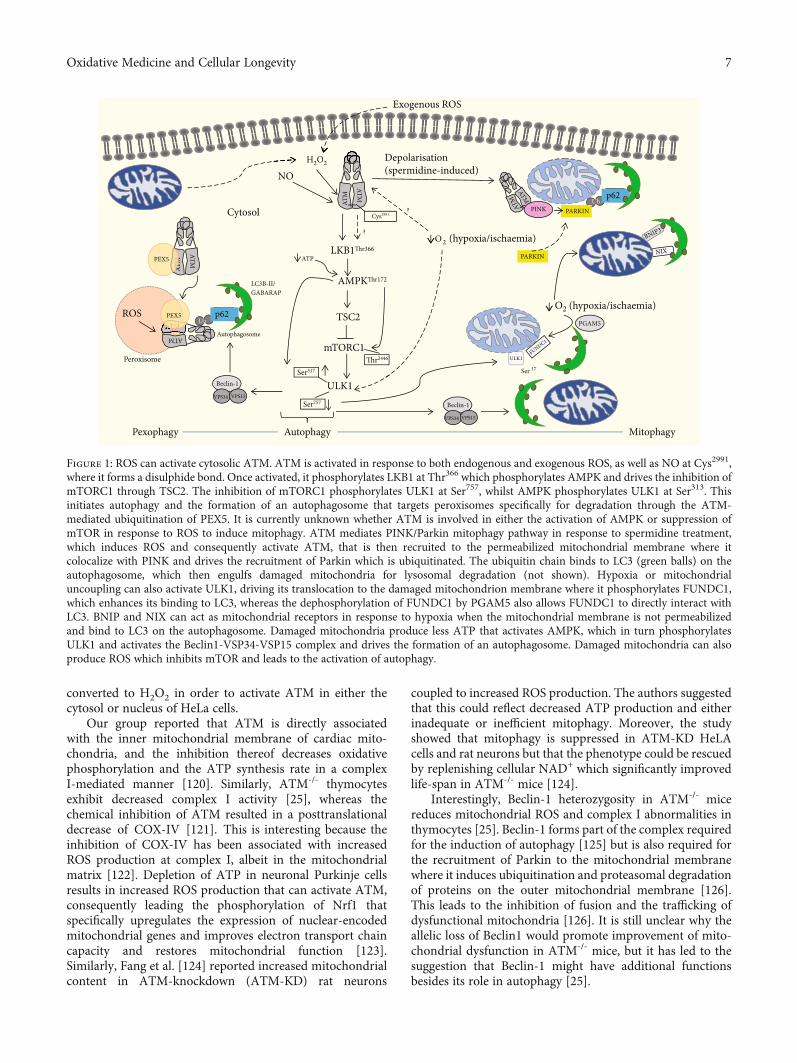
inhibitor, KU55933. Moreover, spermidine promotes thecolocalization of phosphorylated ATM and PINK1 on theouter mitochondrial membrane, which, together with thetranslocation of Parkin, can be blocked by the ATM inhib-itor. The authors suggest a model whereby activated ATMdrives PINK1 accumulation as well as Parkin translocationwith consequent mitophagy in response to spermidinetreatment (Figure 1).
ATM may therefore be central to mitophagy by directlyactivating the pathway or by indirectly activating autophagyin response to oxidative stress. Thus, if pathological ATMsignalling occurs, mitophagy could be affected, predisposingthe cell to mitochondrial oxidative stress [22]. ATM is alsoactivated by nitrosative stress and contribute to sustainedmitophagy of damaged mitochondria through the newlycharacterized ATM-denitrosylase S-nitrosoglutathionereductase (GSNOR) axis [116].
The chronic oxidative stress observed in A-T has led tothe suggestion that A-T might be a mitochondrial disease[40] and has also been linked with intrinsic mitochondrialdysfunction [24]. The latter study found that lymphoblastoidcells from A-T patients contain an increased population ofmitochondria with a decreased membrane potential, whencompared to control cells. Proteins with specific roles inmitochondrial DNA damage and/or ROS scavenging,including mnSOD, peroxiredoxin 3, and mitochondrialtopoisomerase, were also elevated in these cells. Indeed, thedecreased membrane potential translated into decreasedrespiratory activity in the A-T cells compared to the wild typecontrols. Concomitantly, the authors showed that the in vivoloss of ATM in mice resulted in mitochondrial dysfunction inthymocytes that was accompanied by increased mitochon-drial content and mitochondrial ROS due to a decrease inmitophagy. Interestingly, they observed a significant decreasein complex I activity as well as ATP production and anincrease in oxygen consumption. The study also found thatautophagy was not affected by the absence of ATM andsuggested that changes in mitochondrial dynamics such asfission and fusion could contribute towards defective mito-phagy. The authors concluded that the observed defects inthe absence of ATM suggest that ATMmight localize directlyto mitochondria. Fractionation studies in cells revealed thatthe mitochondrial fraction of HepG2 cells was enriched withATM and activated ATM in response to H2O2 treatment[117]. In contrast to previous observations that ATM associ-ated with the peroxisomal fraction [118], Morita et al. [117]detected almost no ATM in this fraction. This reverberateswith the suggestion [119] that both the cell type and cultureconditions of immortalized A-T cells can affect mitochon-drial homeostasis and autophagic responses which explainthe differences in mitochondrial content reported in A-Tdeficient cell lines.
Mitochondrial respiration inhibition can also lead toincreased mitochondrial ROS production. Treatment ofHeLa cells with either rotenone or Antimycin C failed toincrease mitochondrial hydrogen peroxide productionalthough it did increase mitochondrial superoxide produc-tion [18]. Superoxide itself failed to drive ATM dimerizationand suggested that mitochondrial superoxide must be
6 Oxidative Medicine and Cellular Longevity
converted to H2O2 in order to activate ATM in either thecytosol or nucleus of HeLa cells.
Our group reported that ATM is directly associatedwith the inner mitochondrial membrane of cardiac mito-chondria, and the inhibition thereof decreases oxidativephosphorylation and the ATP synthesis rate in a complexI-mediated manner [120]. Similarly, ATM-/- thymocytesexhibit decreased complex I activity [25], whereas thechemical inhibition of ATM resulted in a posttranslationaldecrease of COX-IV [121]. This is interesting because theinhibition of COX-IV has been associated with increasedROS production at complex I, albeit in the mitochondrialmatrix [122]. Depletion of ATP in neuronal Purkinje cellsresults in increased ROS production that can activate ATM,consequently leading the phosphorylation of Nrf1 thatspecifically upregulates the expression of nuclear-encodedmitochondrial genes and improves electron transport chaincapacity and restores mitochondrial function [123].Similarly, Fang et al. [124] reported increased mitochondrialcontent in ATM-knockdown (ATM-KD) rat neurons
coupled to increased ROS production. The authors suggestedthat this could reflect decreased ATP production and eitherinadequate or inefficient mitophagy. Moreover, the studyshowed that mitophagy is suppressed in ATM-KD HeLAcells and rat neurons but that the phenotype could be rescuedby replenishing cellular NAD+ which significantly improvedlife-span in ATM-/- mice [124].
Interestingly, Beclin-1 heterozygosity in ATM-/- micereduces mitochondrial ROS and complex I abnormalities inthymocytes [25]. Beclin-1 forms part of the complex requiredfor the induction of autophagy [125] but is also required forthe recruitment of Parkin to the mitochondrial membranewhere it induces ubiquitination and proteasomal degradationof proteins on the outer mitochondrial membrane [126].This leads to the inhibition of fusion and the trafficking ofdysfunctional mitochondria [126]. It is still unclear why theallelic loss of Beclin1 would promote improvement of mito-chondrial dysfunction in ATM-/- mice, but it has led to thesuggestion that Beclin-1 might have additional functionsbesides its role in autophagy [25].
Exogenous ROS
NO
ATM
ATM
TSC2
mTORC1
ULK1
Autophagy
ATM
ATM
ROS
ATMPEX5
ATM
PEX5
Autophagosome
p62Ub
Peroxisome
PINK
ATM
ATM
PARKIN
PARKIN
PGAM5
ULK1
Beclin-1
VPS34 VPS15
Beclin-1
VPS34 VPS15
Cytosol
p62Ub
H2O2
Cys2991
LKB1
AMPK
2446
Ser317 Ser 17
Ser757
O 2 (hypoxia/ischaemia)
O2 (hypoxia/ischaemia)
ATP
Depolarisation(spermidine-induced)
LC3B-II/ GABARAP
Pexophagy Mitophagy
NIX
FUNDC1
BNIP3
H2O2 Depolarisation
Figure 1: ROS can activate cytosolic ATM. ATM is activated in response to both endogenous and exogenous ROS, as well as NO at Cys2991,where it forms a disulphide bond. Once activated, it phosphorylates LKB1 at Thr366 which phosphorylates AMPK and drives the inhibition ofmTORC1 through TSC2. The inhibition of mTORC1 phosphorylates ULK1 at Ser757, whilst AMPK phosphorylates ULK1 at Ser313. Thisinitiates autophagy and the formation of an autophagosome that targets peroxisomes specifically for degradation through the ATM-mediated ubiquitination of PEX5. It is currently unknown whether ATM is involved in either the activation of AMPK or suppression ofmTOR in response to ROS to induce mitophagy. ATM mediates PINK/Parkin mitophagy pathway in response to spermidine treatment,which induces ROS and consequently activate ATM, that is then recruited to the permeabilized mitochondrial membrane where itcolocalize with PINK and drives the recruitment of Parkin which is ubiquitinated. The ubiquitin chain binds to LC3 (green balls) on theautophagosome, which then engulfs damaged mitochondria for lysosomal degradation (not shown). Hypoxia or mitochondrialuncoupling can also activate ULK1, driving its translocation to the damaged mitochondrion membrane where it phosphorylates FUNDC1,which enhances its binding to LC3, whereas the dephosphorylation of FUNDC1 by PGAM5 also allows FUNDC1 to directly interact withLC3. BNIP and NIX can act as mitochondrial receptors in response to hypoxia when the mitochondrial membrane is not permeabilizedand bind to LC3 on the autophagosome. Damaged mitochondria produce less ATP that activates AMPK, which in turn phosphorylatesULK1 and activates the Beclin1-VSP34-VSP15 complex and drives the formation of an autophagosome. Damaged mitochondria can alsoproduce ROS which inhibits mTOR and leads to the activation of autophagy.
7Oxidative Medicine and Cellular Longevity

Terminally differentiated cells such as cardiomyocytesand neuronal cells are dependent on the efficient removaland replacement of dysfunctional mitochondria to ensurecell survival and to maintain cellular homeostasis [111,127]. A decrease in ATP production and increased ROS pro-duction as indicators of mitochondrial dysfunction can resultin either the release of apoptotic proteins or the selectiveclearance of the damaged mitochondria. Mitophagy thusserves as an early cardioprotective response through theremoval of damaged mitochondria, and if this fails, apoptosiscan be induced in response to excessive oxidative stress [128].Moreover, reduced autophagy, together with the accumula-tion of dysfunctional mitochondria, has been associated withheart failure and aging [111].
Pexophagy is the targeted selective degradation of perox-isomes [129] and is another example of selective autophagy[130]. Peroxisomes utilise β-oxidation to reduce long-chainfatty acids into medium length fatty acids that can be shuttledto the mitochondria. These highly metabolic organellesgenerate ROS during β-oxidation and require homeostaticmaintenance to prevent oxidative stress. ATM binds to theperoxisome importer receptor, PEX5, in response to exces-sive ROS and mediates peroxisome-specific autophagy(pexophagy) by phosphorylating PEX5 at Ser141 and promot-ing mono-ubiquitylation at Lys209, whilst simultaneouslyinducing autophagy through the activation and phosphoryla-tion of TSC2 and ULK1 [17, 131, 132]. Ubiquitylation ofPEX5 is mediated by the complex PEX2-PEX10-PEX12 andis then recognized by the autophagy adapter proteins, p62and NBR1, which directs the autophagosome to the peroxi-somes for pexophagy [129].
Loss of function mutations in ATM, such as the ability tosense oxidative stress, can result in a reduction in mitochon-drial antioxidant defences, lead to the accumulation of ROSand oxidative damage to mitochondria and other cellularcomponents [18], as well as protein aggregation [95]. Selec-tive autophagy seems to be mainly mediated by ubiquitina-tion which is essential for conferring selectivity [133], as isthe case of ATM-mediated pexophagy. As previouslydiscussed, this also implies a potential role for ATM in aggre-phagy (degradation of damaged or misfolded proteins) whichis dependent on p62 ubiquitination [92].
6. Conclusion
This broad overview describes the apical protein, ATMprotein kinase, at the nexus of oxidative stress-inducedautophagy [14, 18] as well as nitrosative stress-inducedautophagy [72, 116], mitophagy [113, 114], and pexophagy[17, 132] mainly in the context of nondividing cells such ascardiomyocytes and neurons. Site-specific mutations thatrenders ATM insensitive to oxidative stress increase proteinaggregation [95], whilst loss of function increases peri-nuclear lysosomal accumulation [80] as well as mitochondrialoxidative stress [25] and dysfunction [24, 25, 120]. Cytoplas-mic ATM thus plays a central role in redox homeostasis andROS-mediated autophagy.
As a master regulator of DNA repair, activation of ATMby exogenous and endogenous oxidative stress, indepen-
dently of DNA strand breaks, only recently came to light[14, 18]. This finding paved the way to understanding thesevere neurodegeneration and associated protein aggregationobserved in A-T patients that is largely due to disruptedATM protein kinase functioning leading to disruptedautophagy, mitophagy, and pexophagy [80, 95]. Additionally,the regulation of ATM levels by autophagy [80] and the role ofATM in oxidative stress-mediated autophagy in an AMPK/m-TORC1dependentmannerwere discovered [13, 19, 132]. Sim-ilarly, ROS-induced pexophagy is modulated by ATMthrough the TSC2/AMPK/mTORC1 pathway in which thedisruption of this signalling pathway leads to interrupted cel-lular homeostasis causing pathologies linked to neurodegen-eration [17, 131, 134].
It has been suggested that the pathogenesis of A-T couldbe ascribed to excessive ROS and that A-T might therefore bean oxidative stress disease [40]. Several studies have investi-gated the effect of the absence or inhibition of ATM onmitochondrial function and found that ATM is innatelyassociated with the inner mitochondrial membrane andoxidative phosphorylation of cardiac mitochondria [120].In addition, the absence of functional ATM in the mitochon-dria of ATM-null thymocytes and fibroblasts was associatedwith decreased ATP production, increased ROS production[24, 25], and a decrease in mitophagy [119, 123].
Therefore, activation of ATM by oxidative stress andthe consequent maintenance of redox homeostasis throughautophagy, pexophagy, aggrephagy, and mitophagy placeATM at the centre of cross-talk between ROS andautophagy signalling.
Conflicts of Interest
The authors declare that they have no conflicts of interest.
Authors’ Contributions
M.B conceptualized and wrote the manuscript and createdFigure 1. SH contributed to the writing of the manuscript.B. H and A. L reviewed and modified the manuscript. Allauthors approved the final version of the manuscript.
Acknowledgments
This work was supported by the National Research Founda-tion South Africa (grant numbers CPRR160411161914 (BH),SFP180507326754 (MB)) and a Harry Crossley Foundationproject grant (SH).
References
[1] L. Syllaba and K. Henner, “Contribution a l’independance del’athetose double idiopathique et congenitale,” Revista deNeurologia, vol. 1, pp. 541–562, 1926.
[2] E. Boder and R. P. Sedgwick, “Ataxia-telangiectasia; a familialsyndrome of progressive cerebellar ataxia, oculocutaneoustelangiectasia and frequent pulmonary infection,” Pediatrics,vol. 21, no. 4, pp. 526–554, 1958.
8 Oxidative Medicine and Cellular Longevity

[3] R. A. Gatti, I. Berkel, E. Boder et al., “Localization of anataxia-telangiectasia gene to chromosome 11q22-23,”Nature,vol. 336, no. 6199, pp. 577–580, 1988.
[4] K. Savitsky, S. Sfez, D. A. Tagle et al., “The complete sequenceof the coding region of the ATM gene reveals similarity to cellcycle regulators in different species,”Humanmolecular genet-ics, vol. 4, pp. 2025–2032, 1995.
[5] Y. Ziv, A. Bar-Shira, I. Pecker et al., “Recombinant ATM pro-tein complements the cellular A-T phenotype,” Oncogene,vol. 15, no. 2, pp. 159–167, 1997.
[6] R. A. Gatti, S. Becker-Catania, H. H. Chun et al., “The path-ogenesis of ataxia-telangiectasia: learning from a Rosettastone,” Clinical Reviews in Allergy and Immunology, vol. 20,no. 1, pp. 87–108, 2001.
[7] Y. Shiloh, “The cerebellar degeneration in ataxia-telangiecta-sia: a case for genome instability,” DNA repair, vol. 95,p. 102950, 2020.
[8] Y. Su andM. Swift, “Mortality Rates among carriers of ataxia-telangiectasia mutant alleles,” Annals of internal medicine,vol. 133, no. 10, pp. 770–778, 2000.
[9] P. J. Connelly, N. Smith, R. Chadwick, A. R. Exley, J. M.Shneerson, and E. R. Pearson, “Recessive mutations in thecancer gene Ataxia Telangiectasia Mutated (ATM), at a locuspreviously associated with metformin response, cause dysgly-caemia and insulin resistance,” Diabetic Medicine, vol. 33,no. 3, pp. 371–375, 2016.
[10] A. Barzilai, G. Rotman, and Y. Shiloh, “ATM deficiency andoxidative stress: a new dimension of defective response toDNA damage,” DNA Repair, vol. 1, no. 1, pp. 3–25, 2002.
[11] D. J. Watters, “Oxidative stress in ataxia telangiectasia,”Redox report, vol. 8, pp. 23–29, 2013.
[12] Y. Shiloh, “ATM and related protein kinases: safeguardinggenome integrity,” Nature Reviews. Cancer, vol. 3, no. 3,pp. 155–168, 2003.
[13] A. Alexander and C. L. Walker, “Differential localization ofATM is correlated with activation of distinct downstream sig-naling pathways,” Cell Cycle, vol. 9, pp. 3685-3686, 2011.
[14] Z. Guo, S. Kozlov, M. F. Lavin, M. D. Person, and T. T. Paull,“ATM activation by oxidative stress,” Science, vol. 330,no. 6003, pp. 517–521, 2010.
[15] T. T. Paull, “Mechanisms of ATM activation,” Annual reviewof biochemistry, vol. 84, pp. 711–738, 2014.
[16] Y. Shiloh and Y. Ziv, “The ATM protein: the importance ofbeing active,” The Journal of Cell Biology, vol. 198, no. 3,pp. 273–275, 2012.
[17] J. Zhang, D. N. Tripathi, J. Jing et al., “ATM functions at theperoxisome to induce pexophagy in response to ROS,”Nature cell biology, vol. 17, pp. 1259–1269, 2015.
[18] Y. Zhang, J. H. Lee, T. T. Paull et al., “Mitochondrial redoxsensing by the kinase ATM maintains cellular antioxidantcapacity,” Science signaling, vol. 11, article eaaq0702, 2018.
[19] A. Alexander, S. L. Cai, J. Kim et al., “ATM signals to TSC2 inthe cytoplasm to regulate mTORC1 in response to ROS,” Pro-ceedings of the National Academy of Sciences, vol. 107, no. 9,pp. 4153–4158, 2010.
[20] A. Alexander and C. L. Walker, “The role of LKB1 andAMPK in cellular responses to stress and damage,” FEBS Let-ters, vol. 585, no. 7, pp. 952–957, 2011.
[21] C. Cosentino, D. Grieco, and V. Costanzo, “ATM activatesthe pentose phosphate pathway promoting anti-oxidant
defence and DNA repair,” The EMBO Journal, vol. 30,no. 3, pp. 546–555, 2011.
[22] M. Blignaut, An investigation into the role of ATM protein inmitochondrial defects associated with cardiovascular pathol-ogy resulting from insulin resistance, PhD Dissertation, Stel-lenbosch University, 2019.
[23] J. S. Eaton, Z. P. Lin, A. C. Sartorelli, N. D. Bonawitz, and G. S.Shadel, “Ataxia-telangiectasia mutated kinase regulates ribonu-cleotide reductase andmitochondrial homeostasis,”The Journalof Clinical Investigation, vol. 117, no. 9, pp. 2723–2734, 2007.
[24] M. Ambrose, J. V. Goldstine, and R. A. Gatti, “Intrinsic mito-chondrial dysfunction in ATM-deficient lymphoblastoidcells,” Human Molecular Genetics, vol. 16, no. 18, pp. 2154–2164, 2007.
[25] Y. A. Valentin-Vega, M. L. KH, J. Tait-Mulder et al., “Mito-chondrial dysfunction in ataxia-telangiectasia,” Blood,vol. 119, no. 6, pp. 1490–1500, 2012.
[26] J. R. Mercer, E. Yu, N. Figg et al., “The mitochondria-targetedantioxidant Mito Q decreases features of the metabolic syn-drome in ATM +/-/Apo E -/- mice,” Free Radical Biologyand Medicine, vol. 52, pp. 841–849, 2012.
[27] D. A. D’Souza, I. A. Parish, D. S. Krause, S. M. Kaech, andG. S. Shadel, “Reducing mitochondrial ROS improvesdisease-related pathology in a mouse model of ataxia-telangi-ectasia,” Molecular Therapy, vol. 21, pp. 42–48, 2012.
[28] M. Takagi, H. Uno, R. Nishi et al., “ATM regulates adipocytedifferentiation and contributes to glucose homeostasis,” Cellreports, vol. 10, pp. 957–967, 2015.
[29] J. G. Viniegra, N. Martínez, P. Modirassari et al., “Full activa-tion of PKB/Akt in response to insulin or ionizing radiation ismediated through ATM,” The Journal of Biological Chemis-try, vol. 280, no. 6, pp. 4029–4036, 2005.
[30] M. J. Halaby, J. C. Hibma, J. He, and D. Q. Yang, “ATM pro-tein kinase mediates full activation of Akt and regulates glu-cose transporter 4 translocation by insulin in muscle cells,”Cellular Signalling, vol. 20, no. 8, pp. 1555–1563, 2008.
[31] Y. Shiloh and Y. Ziv, “The ATMProtein kinase: regulating thecellular response to genotoxic stress, and more,” NaturereviewsMolecular cell biology, vol. 14, no. 4, pp. 197–210, 2013.
[32] Y. Shi, “Serine/threonine phosphatases: mechanism throughstructure,” Cell, vol. 139, no. 3, pp. 468–484, 2009.
[33] K. K. Khanna, M. F. Lavin, S. P. Jackson, and T. D. Mulhern,“ATM, a central controller of cellular responses to DNAdamage,” Cell Death and Differentiation, vol. 8, no. 11,pp. 1052–1065, 2001.
[34] C. J. Bakkenist and M. B. Kastan, “DNA damage activatesATM through intermolecular autophosphorylation anddimer dissociation,” Nature, vol. 421, no. 6922, pp. 499–506, 2003.
[35] A. Maréchal and L. Zou, “DNA damage sensing by the ATMand,” Cold Spring Harbor Perspectives in Biology, vol. 5,pp. 1–18, 2013.
[36] R. E. Shackelford, C. L. Innes, S. O. Sieber, A. N. Heinloth,S. A. Leadon, and R. S. Paules, “The ataxia telangiectasia geneproduct is required for oxidative stress-induced G1 and G2checkpoint function in human fibroblasts,” Journal of Biolog-ical Chemistry, vol. 276, pp. 21951–21959, 2001.
[37] Z. Bencokova, M. R. Kaufmann, I. M. Pires, P. S. Lecane, A. J.Giaccia, and E. M. Hammond, “ATM activation and signal-ing under hypoxic conditions,” Molecular and Cellular Biol-ogy, vol. 29, no. 2, pp. 526–537, 2009.
9Oxidative Medicine and Cellular Longevity

[38] J. Kobayashi, Y. Saito, M. Okui, N. Miwa, and K. Komatsu,“Increased oxidative stress in AOA3 cells disturbs ATM-dependent DNA damage responses,” Mutation Research/Ge-netic Toxicology and Environmental Mutagenesis, vol. 782,pp. 42–50, 2015.
[39] S. Ditch and T. T. Paull, “The ATM protein kinase and cellu-lar redox signaling: beyond the DNA damage response,”Trends in biochemical sciences, vol. 37, no. 1, pp. 15–22, 2012.
[40] M. Ambrose and R. A. Gatti, “Pathogenesis of ataxia-telangi-ectasia: the next generation of ATM functions,” Blood,vol. 121, no. 20, pp. 4036–4045, 2013.
[41] K. Bedard and K.-H. Krause, “The NOX family of ROS-generating NADPH oxidases: physiology and pathophysiol-ogy,” Physiological reviews, vol. 87, no. 1, pp. 245–313,2007.
[42] R. P. Patel, J. McAndrew, H. Sellak et al., “Biological aspectsof reactive nitrogen species,” Biochimica et Biophysica Acta(BBA)-Bioenergetics, vol. 1411, no. 2-3, pp. 385–400, 1999.
[43] E. A. Podrez, D. Schmitt, H. F. Hoff, and S. L. Hazen, “Mye-loperoxidase-generated reactive nitrogen species convertLDL into an atherogenic form in vitro,” The Journal of Clin-ical Investigation, vol. 103, no. 11, pp. 1547–1560, 1999.
[44] T. Finkel, “Signal transduction by reactive oxygen species,”The Journal of Cell Biology, vol. 194, no. 1, pp. 7–15, 2011.
[45] I. J. Lodhi and C. F. Semenkovich, “Peroxisomes: a nexus forlipid metabolism and cellular signaling,” Cell metabolism,vol. 19, no. 3, pp. 380–392, 2014.
[46] S. J. Forrester, D. S. Kikuchi, M. S. Hernandes, Q. Xu, andK. K. Griendling, “Reactive oxygen species in metabolic andinflammatory signaling,” Circulation research, vol. 122,no. 6, pp. 877–902, 2018.
[47] A. Tarafdar and G. Pula, “The role of NADPH oxidases andoxidative stress in neurodegenerative disorders,” Interna-tional Journal of Molecular Sciences, vol. 19, no. 12, p. 3824,2018.
[48] M. W. Ma, J. Wang, Q. Zhang et al., “NADPH oxidase inbrain injury and neurodegenerative disorders,” Molecularneurodegeneration, vol. 12, pp. 1–28, 2017.
[49] K. M. Holmström and T. Finkel, “Cellular mechanisms andphysiological consequences of redox-dependent signalling,”Nature reviews Molecular cell biology, vol. 15, no. 6,pp. 411–421, 2014.
[50] U. Weyemi, C. E. Redon, T. Aziz et al., “NADPH oxidase 4 isa critical mediator in ataxia telangiectasia disease,” Proceed-ings of the National Academy of Sciences, vol. 112, no. 7,pp. 2121–2126, 2015.
[51] N. Mizushima, “Autophagy: process and function,” Genes &Development, vol. 21, no. 22, pp. 2861–2873, 2007.
[52] G. Filomeni, D. De Zio, and F. Cecconi, “Oxidative stress andautophagy: the clash between damage and metabolic needs,”Cell Death &Differentiation, vol. 22, no. 3, pp. 377–388, 2015.
[53] G. Kroemer, G. Mariño, and B. Levine, “Autophagy and theintegrated stress response,” Molecular Cell, vol. 40, no. 2,pp. 280–293, 2010.
[54] D. J. Klionsky and P. Codogno, “The mechanism and physi-ological function of macroautophagy,” Journal of InnateImmunity, vol. 5, no. 5, pp. 427–433, 2013.
[55] D. Glick, S. Barth, and K. F. Macleod, “Autophagy : cellularand molecular mechanisms,” The Journal of Pathology,vol. 221, no. 1, pp. 3–12, 2010.
[56] J. Kim, G. Yang, Y. Kim, J. Kim, and J. Ha, “AMPK activators:mechanisms of action and physiological activities,” Experi-mental & molecular medicine, vol. 48, no. 4, p. e224, 2016.
[57] S. V. Kozlov, A. J. Waardenberg, K. Engholm-Keller, J. W.Arthur, M. E. Graham, and M. Lavin, “Reactive oxygen spe-cies (ROS)-activated ATM-dependent phosphorylation ofcytoplasmic substrates identified by large-scale phosphopro-teomics screen,” Molecular & Cellular Proteomics, vol. 15,no. 3, pp. 1032–1047, 2016.
[58] D. M. Gwinn, D. B. Shackelford, D. F. Egan et al., “AMPKphosphorylation of raptor mediates a metabolic checkpoint,”Molecular Cell, vol. 30, no. 2, pp. 214–226, 2008.
[59] S. W. Y. Cheng, L. G. D. Fryer, D. Carling, and P. R.Shepherd, “Thr2446 is a novel mammalian target of rapa-mycin (mTOR) phosphorylation site regulated by nutrientstatus,” Journal of Biological Chemistry, vol. 279, no. 16,pp. 15719–15722, 2004.
[60] Y. Kim, M. Kundu, B. Viollet, and K.-L. Guan, “AMPK andmTOR regulate autophagy through direct phosphorylationof Ulk1,” Nature Cell Biology, vol. 13, no. 2, pp. 132–141,2011.
[61] M. Zachari and I. G. Ganley, “The mammalian ULK1 com-plex and autophagy initiation,” Essays in Biochemistry,vol. 61, no. 6, pp. 585–596, 2017.
[62] Z. Yang and D. J. Klionsky, “Mammalian autophagy: coremolecular machinery and signaling regulation,” Currentopinion in cell biology, vol. 22, no. 2, pp. 124–131, 2010.
[63] N. T. Ktistakis and S. A. Tooze, “Digesting the expandingmechanisms of autophagy,” Trends in cell biology, vol. 26,no. 8, pp. 624–635, 2016.
[64] T. Nishimura and S. A. Tooze, “Emerging roles of ATG pro-teins and membrane lipids in autophagosome formation,”Cell Discovery, vol. 6, no. 1, 2020.
[65] T. Johansen and T. Lamark, “Selective autophagy: ATG8family proteins, LIR motifs and cargo receptors,” Journal ofmolecular biology, vol. 432, no. 1, pp. 80–103, 2020.
[66] M. B. E. Schaaf, T. G. Keulers, M. A. Vooijs, and K. M. A.Rouschop, “LC3/GABARAP family proteins: autophagy-(un)related functions,” The FASEB Journal, vol. 30, no. 12,pp. 3961–3978, 2016.
[67] P. Wild, D. G. McEwan, and I. Dikic, “The LC3 interactomeat a glance,” Journal of Cell Science, vol. 127, pp. 3–9, 2013.
[68] J. Dancourt and T. J. Melia, “Lipidation of the autophagyproteins LC3 and GABARAP is a membrane-curvaturedependent process,” Autophagy, vol. 10, no. 8, pp. 1470-1471, 2014.
[69] D. Colacurcio and R. Nixon, “Disorders of lysosomal acidifi-cation - the emerging role of v- ATPase in aging and neuro-degenerative disease,” Ageing Research Reviews, vol. 32,pp. 75–88, 2016.
[70] J. Y. Koh, H. N. Kim, J. J. Hwang, Y. H. Kim, and S. E. Park,“Lysosomal dysfunction in proteinopathic neurodegenerativedisorders: possible therapeutic roles of cAMP and zinc,”Molecular Brain, vol. 12, pp. 1–11, 2019.
[71] N. Jimenez-Moreno and J. D. Lane, “Autophagy and redoxhomeostasis in Parkinson’s: a crucial balancing act,” Oxida-tive Medicine and Cellular Longevity, vol. 2020, 38 pages,2020.
[72] D. N. Tripathi, R. Chowdhury, L. J. Trudel et al., “Reactivenitrogen species regulate autophagy through ATM-AMPK-TSC2-mediated suppression of mTORC1,” Proceedings of
10 Oxidative Medicine and Cellular Longevity
the National Academy of Sciences, vol. 110, no. 32, pp. E2950–E2957, 2013.
[73] G. P. Sapkota, M. Deak, A. Kieloch et al., “Ionizing radiationinduces ataxia telangiectasia mutated kinase (ATM)-medi-ated phosphorylation of LKB1/STK11 at Thr-366,” Biochem-ical Journal, vol. 516, pp. 507–516, 2002.
[74] R. J. Shaw, M. Kosmatka, N. Bardeesy et al., “The tumor sup-pressor LKB1 kinase directly activates AMP-activated kinaseand regulates apoptosis in response to energy stress,” Pro-ceedings of the National Academy of Sciences, vol. 101,no. 10, pp. 3329–3335, 2004.
[75] Y. Ikeda, K. Sato, D. R. Pimentel et al., “Cardiac-specific dele-tion of LKB1 leads to hypertrophy and dysfunction,” TheJournal of Biological Chemistry, vol. 284, no. 51, pp. 35839–35849, 2009.
[76] B. M. Emerling, F. Weinberg, C. Snyder et al., “Hypoxic acti-vation of AMPK is dependent on mitochondrial ROS butindependent of an increase in AMP/ATP ratio,” Free RadicalBiology and Medicine, vol. 46, pp. 1386–1391, 2009.
[77] Y. Chen, M. B. Azad, and S. B. Gibson, “Superoxide is themajor reactive oxygen species regulating autophagy,” CellDeath & Differentiation, vol. 16, no. 7, pp. 1040–1052,2009.
[78] G. Filomeni, E. Desideri, S. Cardaci, G. Rotilio, and M. R.Ciriolo, “Under the ROS: thiol network is the principalsuspect for autophagy commitment commitment,” Autoph-agy, vol. 6, pp. 999–1005, 2014.
[79] C. Garza-Lombó, A. Pappa, M. I. Panayiotidis, and R. Franco,“Redox homeostasis, oxidative stress and mitophagy,” Mito-chondrion, vol. 51, pp. 105–117, 2020.
[80] A. Cheng, K. H. Tse, H. M. Chow et al., “ATM loss disruptsthe autophagy-lysosomal pathway,” Autophagy, pp. 1–13,2020.
[81] J. G. Schneider, B. N. Finck, J. Ren et al., “ATM-dependentsuppression of stress signaling reduces vascular disease inmetabolic syndrome,” Cell Metabolism, vol. 4, no. 5,pp. 377–389, 2006.
[82] Y. Espach, A. Lochner, H. Strijdom, and B. Huisamen, “ATMprotein kinase signaling, type 2 diabetes and cardiovasculardisease,” Cardiovascular Drugs and Therapy, vol. 29, no. 1,pp. 51–58, 2015.
[83] C. Barlow, C. Ribaut-Barassin, T. A. Zwingman et al., “ATMis a cytoplasmic protein in mouse brain required to preventlysosomal accumulation,” Proceedings of the National Acad-emy of Sciences, vol. 97, no. 2, pp. 871–876, 2000.
[84] M. Qian, Z. Liu, L. Peng et al., “Boosting ATM activity allevi-ates aging and extends lifespan in a mouse model of proge-ria,” eLife, vol. 7, pp. 1–25, 2018.
[85] D. Carmona-Gutierrez, A. L. Hughes, F. Madeo, andC. Ruckenstuhl, “The crucial impact of lysosomes in agingand longevity,”Ageing research reviews, vol. 32, pp. 2–12, 2016.
[86] M. Abdellatif, S. Sedej, D. Carmona-Gutierrez, F. Madeo, andG. Kroemer, “Autophagy in cardiovascular aging,” Circula-tion Research, vol. 123, no. 7, pp. 803–824, 2018.
[87] T. Lamark and T. Johansen, “Aggrephagy : selective disposalof protein aggregates by macroautophagy,” InternationalJournal of Cell Biology, vol. 2012, 21 pages, 2012.
[88] H. Tsutsui, S. Kinugawa, and S. Matsushima, “Oxidativestress and heart failure,” American Journal of Physiology-Heart and Circulatory Physiology, vol. 301, pp. 2181–2190,2011.
[89] T. C. Squier, “Oxidative stress and protein aggregation duringbiological aging,” Experimental gerontology, vol. 36,pp. 1539–1550, 2001.
[90] A. Monaco and A. Fraldi, “Protein aggregation and dysfunc-tion of autophagy-lysosomal pathway: a vicious cycle in lyso-somal storage diseases,” Frontiers in Molecular Neuroscience,vol. 13, pp. 1–8, 2020.
[91] G. Merlini, V. Bellotti, A. Andreola et al., “Protein aggrega-tion,” Clinical Chemistry and Laboratory Medicine, vol. 39,pp. 1065–1075, 2001.
[92] T. D. Evans, I. Sergin, X. Zhang, and B. Razani, “Targetacquired: selective autophagy in cardiometabolic disease,”Science signaling, vol. 10, no. 468, p. eaag2298, 2017.
[93] S. Elmore, “Apoptosis: a review of programmed cell death,”Toxicologic Pathology, vol. 35, pp. 495–516, 2016.
[94] K. D. Sullivan, V. V. Palaniappan, and J. M. Espinosa, “ATMregulates cell fate choice upon p 53 activation by modulatingmitochondrial turnover and ROS levels,” Cell Cycle, vol. 14,no. 1, pp. 56–63, 2015.
[95] J. H. Lee, M. R. Mand, C. H. Kao et al., “ATM directs DNAdamage responses and proteostasis via genetically separablepathways,” Science signaling, vol. 11, no. 512, p. eaan5598,2018.
[96] C. Barlow, P. A. Dennery, M. K. Shigenaga et al., “Loss of theataxia–telangiectasia gene product causes oxidative damagein target organs,” Proceedings of the National Academy of Sci-ences, vol. 96, no. 17, pp. 9915–9919, 1999.
[97] H. Kato, S. Takashima, Y. Asano et al., “Identification of p 32as a novel substrate for ATM in heart,” Biochemical and Bio-physical Research Communications, vol. 366, no. 4, pp. 885–891, 2008.
[98] H. Jiao, G. Q. Su, W. Dong et al., “Chaperone-like protein p32regulates ULK1 stability and autophagy,” Cell Death & Differ-entiation, vol. 22, no. 11, pp. 1812–1823, 2015.
[99] W. Tian, W. Li, Y. Chen et al., “Phosphorylation of ULK1 byAMPK regulates translocation of ULK1 to mitochondria andmitophagy,” FEBS letters, vol. 589, no. 15, pp. 1847–1854,2015.
[100] A. L. Anding and E. H. Baehrecke, “Cleaning house : selectiveautophagy of organelles,” Developmental Cell, vol. 41, no. 1,pp. 10–22, 2017.
[101] W. Wu, W. Tian, Z. Hu et al., “ULK1 translocates to mito-chondria and phosphorylates FUNDC1 to regulate mito-phagy,” EMBO Reports, vol. 15, no. 5, pp. 566–575, 2014.
[102] K. Nishida, S. Kyoi, O. Yamaguchi, J. Sadoshima, andK. Otsu, “The role of autophagy in the heart,” Cell Deathand Differentiation, vol. 16, no. 1, pp. 31–38, 2009.
[103] W. X. Ding and X. M. Yin, “Mitophagy: mechanisms, patho-physiological roles, and analysis,” Biological Chemistry,vol. 393, no. 7, pp. 547–564, 2012.
[104] A. Diot, K. Morten, and J. Poulton, “Mitophagy plays a cen-tral role in mitochondrial ageing,” Mammalian Genome,vol. 27, no. 7-8, pp. 381–395, 2016.
[105] A. Roberta, M. Gottlieb, and A. Thomas, “Mitophagy andmitochondrial quality control mechanisms in the heart,” Cur-rent pathobiology reports, vol. 5, pp. 161–169, 2017.
[106] A. Hamacher-Brady and N. R. Brady, “Mitophagy programs :mechanisms and physiological implications of mitochondrialtargeting by autophagy,” Cellular and molecular life sciences,vol. 73, pp. 775–795, 2016.
11Oxidative Medicine and Cellular Longevity

[107] T. N. Nguyen, B. S. Padman, and M. Lazarou, “Decipheringthe molecular signals of PINK1/Parkin mitophagy,” Trendsin cell biology, vol. 26, no. 10, pp. 733–744, 2016.
[108] S. Sciarretta, Y. Maejima, D. Zablocki, and J. Sadoshima, “Therole of autophagy in the heart,” Annual Review of Physiology,vol. 80, no. 1, pp. 1–26, 2018.
[109] J. Zhang and P. A. Ney, “Role of BNIP3 and NIX in cell death,autophagy, and mitophagy,” Cell Death & Differentiation,vol. 16, no. 7, pp. 939–946, 2009.
[110] S. Rikka, M. N. Quinsay, R. L. Thomas et al., “Bnip3 impairsmitochondrial bioenergetics and stimulates mitochondrialturnover,” Cell Death & Differentiation, vol. 18, no. 4,pp. 721–731, 2011.
[111] A. G. Moyzis, J. Sadoshima, and Å. B. Gustafsson, “Mendinga broken heart: the role of mitophagy in cardioprotection,”American Journal of Physiology-Heart and Circulatory Physi-ology, vol. 308, no. 3, pp. H183–H192, 2015.
[112] H. Wei, L. Liu, and Q. Chen, “Selective removal of mitochon-dria via mitophagy: distinct pathways for different mitochon-drial stresses,” Biochimica et Biophysica Acta (BBA)-Molecular Cell Research, vol. 1853, pp. 2784–2790, 2015.
[113] X. Gu, Y. Qi, Z. Feng, L. Ma, K. Gao, and Y. Zhang, “Lead(Pb) induced ATM-dependent mitophagy via PINK1/Parkinpathway,” Toxicology letters, vol. 291, pp. 92–100, 2018.
[114] Y. Qi, Q. Qiu, X. Gu, Y. Tian, and Y. Zhang, “ATM mediatesspermidine-induced mitophagy via PINK1 and Parkin regu-lation in human fibroblasts,” Scientific reports, vol. 6,p. 24700, 2016.
[115] T. Eisenberg, H. Knauer, A. Schauer et al., “Induction ofautophagy by spermidine promotes longevity,” Nature cellbiology, vol. 11, no. 11, pp. 1305–1314, 2009.
[116] C. Cirotti, S. Rizza, P. Giglio et al., “Redox activation of ATMenhances GSNOR translation to sustain mitophagy and toler-ance to oxidative stress,” EMBO reports, vol. 22, articlee50500, 2020.
[117] A. Morita, K. Tanimoto, T. Murakami, T. Morinaga, andY. Hosoi, “Mitochondria are required for ATM activationby extranuclear oxidative stress in cultured human hepato-blastoma cell line Hep G2 cells,” Biochemical and biophysicalresearch communications, vol. 443, no. 4, pp. 1286–1290,2014.
[118] D. Watters, P. Kedar, K. Spring et al., “Localization of a por-tion of extranuclear ATM to peroxisomes,” The Journal ofBiological Chemistry, vol. 274, no. 48, pp. 34277–34282, 1999.
[119] Y. A. Valentin-Vega and M. B. Kastan, “A new role for ATM:regulating mitochondrial function and mitophagy,” Autoph-agy, vol. 8, pp. 840-841, 2014.
[120] M. Blignaut, B. Loos, S. W. Botchway, A. W. Parker, andB. Huisamen, “Ataxia-telangiectasia mutated is located incardiac mitochondria and impacts oxidative phosphoryla-tion,” Scientific Reports, vol. 9, no. 1, p. 4782, 2019.
[121] A. Y. Patel, T. M. McDonald, L. D. Spears, J. K. Ching, andJ. S. Fisher, “Ataxia telangiectasia mutated influences cyto-chrome _c_ oxidase activity,” Biochemical and biophysicalresearch communications, vol. 405, no. 4, pp. 599–603, 2011.
[122] Q. Chen, E. J. Vazquez, S. Moghaddas, C. L. Hoppel, and E. J.Lesnefsky, “Production of reactive oxygen species by mito-chondria,” The Journal of Biological Chemistry, vol. 278,no. 38, pp. 36027–36031, 2003.
[123] H. M. Chow, A. Cheng, X. Song, M. R. Swerdel, R. P. Hart,and K. Herrup, “ATM is activated by ATP depletion and
modulates mitochondrial function through NRF1,” The Jour-nal of Cell Biology, vol. 218, no. 3, pp. 909–928, 2019.
[124] E. F. Fang, H. Kassahun, D. L. Croteau et al., “NAD+ replen-ishment improves lifespan and healthspan in ataxia telangiec-tasia models via mitophagy and DNA repair,” CellMetabolism, vol. 24, no. 4, pp. 566–581, 2016.
[125] S. Kobayashi and Q. Liang, “Autophagy and mitophagy indiabetic cardiomyopathy,” Biochimica et Biophysica Acta(BBA)-Molecular Basis of Disease, vol. 1852, no. 2, pp. 252–261, 2015.
[126] V. Choubey, M. Cagalinec, J. Liiv et al., “BECN1 is involved inthe initiation of mitophagy: it facilitates PARK2 translocationto mitochondria,” Autophagy, vol. 10, no. 6, pp. 1105–1119,2014.
[127] Y. Wang, N. Liu, and B. Lu, “Mechanisms and roles of mito-phagy in neurodegenerative diseases,” CNS Neuroscience &Therapeutics, vol. 25, no. 7, pp. 859–875, 2019.
[128] D. A. Kubli and Å. B. Gustafsson, “Mitochondria and mito-phagy: the yin and yang of cell death control,” CirculationResearch, vol. 111, no. 9, pp. 1208–1221, 2012.
[129] K. Germain and P. K. Kim, “Pexophagy : a model for selectiveautophagy,” International journal of molecular sciences, vol. ‘,pp. 1–27, 2020.
[130] C. He and D. J. Klionsky, “Regulation mechanisms and sig-naling pathways of autophagy,” Annual Review of Genetics,vol. 43, no. 1, pp. 67–93, 2009.
[131] D. N. Tripathi, J. Zhang, J. Jing, R. Dere, and C. L. Walker, “Anew role for ATM in selective autophagy of peroxisomes(pexophagy),” Autophagy, vol. 12, no. 4, pp. 711-712, 2016.
[132] J. Zhang, J. Kim, A. Alexander et al., “A tuberous sclerosiscomplex signalling node at the peroxisome regulatesmTORC1 and autophagy in response to ROS,” Nature CellBiology, vol. 15, no. 10, pp. 1186–1196, 2013.
[133] C. Kraft, M. Peter, and K. Hofmann, “Selective autophagy:ubiquitin-mediated recognition and beyond,” Nature cellbiology, vol. 12, no. 9, pp. 836–841, 2010.
[134] D. S. Jo and D. H. Cho, “Peroxisomal dysfunction in neuro-degenerative diseases,” Archives of pharmacal research,vol. 42, no. 5, pp. 393–406, 2019.
12 Oxidative Medicine and Cellular Longevity