CEP41 is mutated in Joubert syndrome and is required for tubulin glutamylation at the cilium
Upload
independentCategory
view
0download
0
eScholarship provides open access, scholarly publishingservices to the University of California and delivers a dynamicresearch platform to scholars worldwide.
Lawrence Berkeley National LaboratoryLawrence Berkeley National Laboratory
Peer Reviewed
Title:Inhibition of TGFbeta1 Signaling Attenutates ATM Activity in Response to Genotoxic Stress
Author:Kirshner, JuliaJobling, Michael F.Pajares, Maria JoseRavani, Shraddha A.Glick, Adam B.Lavin, Martin J.Koslov, SergeiShiloh, YosefBarcellos-Hoff, Mary Helen
Publication Date:09-15-2006
Publication Info:Lawrence Berkeley National Laboratory
Permalink:http://escholarship.org/uc/item/3476t6kx

Inhibition of TGFβ1 Signaling Attenuates ATM Activity in Response to Genotoxic Stress
Julia Kirshner.*1, Michael F. Jobling, *1, Maria Jose Pajares1, Shraddha A. Ravani1, Adam B.
Glick2, Martin J. Lavin3, Sergei Koslov3, Yosef Shiloh4 & Mary Helen Barcellos-Hoff1,5
*Authors contributed equally.
1 Life Sciences Division, Lawrence Berkeley National Laboratory, Berkeley, CA 947202 Laboratory of Cellular Carcinogenesis and Tumor Promotion National Cancer Institute, Bethesda,
MD 208923 The Queensland Institute of Medical Research, Royal Brisbane Hospital, Herston, Queensland
4029, Australia.4 Department of Human Genetics, Sackler School of Medicine, Tel Aviv University, Ramat Aviv
69978 Israel
5 Corresponding Author: Mary Helen Barcellos-Hoff
Life Sciences Division
1 Cyclotron Road, Bldg. 977
Berkeley, CA 94720
(510) 495-2535 FAX
(510) 486-6371 PHONE
Financial Support: Support was provided by NASA program Biomedical Research and
Countermeasures Ground Research in Radiation Health to MHBH, grant number T6275-W, DOD
DAMD17-01-0291 postdoctoral fellowship to MFJ, and the Low Dose Radiation Program of the
DOE Office of Biological Effects Research to MHBH and the Office of Health and Environmental
Research, Health Effects Division, United States Department of Energy (contract no.03-76SF00098).
Total Word Count: 6945
Abstract: 202
Text:4062
Figures: 6
Keywords: DNA damage; epithelial; cytokine
Abbreviations: Transforming growth factor β1, TGFβ; Ataxia telangiectasia mutated, ATM; ionizing
radiation, IR; mammary epithelial cells, MEC; TGFβ receptor type I, TβRI; radiation-induced foci,
RIF
Running Title: TGFβ Mediates DNA Damage Responses

Kirshner et al. 2
ABSTRACT
Ionizing radiation causes DNA damage that elicits a cellular program of damage control coordinated
by the kinase activity of ataxia telangiectasia mutated protein (ATM). Transforming growth factor
β1 (TGFβ), which is activated by radiation, is a potent and pleiotropic mediator of physiological and
pathological processes. Here we show that TGFβ inhibition impedes the canonical cellular DNA
damage stress response. Irradiated Tgfβ1 null murine epithelial cells or human epithelial cells treated
with a small molecule inhibitor of TGFβ type I receptor kinase exhibit decreased phosphorylation
of Chk2, Rad17 and p53, reduced γH2AX radiation-induced foci, and increased radiosensitivity
compared to TGFβ competent cells. We determined that loss of TGFβ signaling in epithelial cells
truncated ATM autophosphorylation and significantly reduced its kinase activity, without affecting
protein abundance. Addition of TGFβ restored functional ATM and downstream DNA damage
responses. These data reveal a heretofore undetected critical link between the microenvironment and
ATM that directs epithelial cell stress responses, cell fate and tissue integrity. Thus, TGFβ1, in
addition to its role in homoeostatic growth control, plays a complex role in regulating responses to
genotoxic stress, the failure of which would contribute to the development of cancer; conversely,
inhibiting TGFβ may be used to advantage in cancer therapy.

Kirshner et al. 3
Introduction
An orchestrated response to DNA damage in multicellular organisms is important for rapid
restoration of homeostasis and long-term prevention of cancer but how signaling is regulated across
tissues is unknown. Little is known about the influence of extracellular signaling from growth factors
on the cellular response to DNA damage, however, transforming growth factor β1 (TGFβ) is rapidly
and persistently activated in tissues following IR (1). TGFβ coordinates responses to a great variety
of other stimuli by regulating cell proliferation, differentiation, and apoptosis (reviewed in (2, 3)),
is involved in many aspects of development, growth regulation and is known as a classical tumor
suppressor (reviewed in (3)). We previously showed that epithelial tissues of Tgfβ1 null embryos
fail to undergo either apoptosis or inhibition of cell cycle in response to IR, suggesting a surprising
requirement for its activity in the responses to DNA damage (4). Neither the point at which TGFβ1
impacts the genotoxic stress program or specific mechanisms of action have been identified.
Independent of its control of proliferation and differentiation, studies by Glick and others have
implicated TGFβ in maintenance of genomic stability. Tgfβ1 null cells are genomically unstable (5),
cannot repair alkylating damage (6) and fail to apoptose or undergo cell cycle inhibition after
ionizing radiation (IR) exposure in vivo (4). Tgfβ1 null keratinocytes exhibit increased frequency
of gene amplification as marked by N-phosphonoacetyl-L-aspartate (PALA) resistance (5). Tgfβ1
null keratinocytes transduced with v-rasHa develop aneuploidy at higher frequencies, have more
chromosomal abnormalities than the wild-type controls and undergo spontaneous malignant
transformation more frequently and with shorter latency than wild-type counterparts (7-9). The fact
that TGFβ treatment of Tgfβ1 null cells inhibits PALA resistance, reduces the percentage of
Kirshner et al. 4
aneuploid metaphases, and decreases the number of spontaneous chromosome breaks indicates an
ongoing process rather than selection of genomically unstable subpopulation. TGFβ is rapidly and
persistently activated in vivo by ionizing radiation (IR) (10-14). Radiation- induced apoptosis and
cell cycle arrest in epithelial cells in vivo are decreased in a Tgfβ1 gene dosage dependent fashion
(4). Similarly, the mammary epithelium of Tgfβ1 heterozygous mice or animals treated with TGFβ
neutralizing antibodies fails to undergo an apoptotic response and exhibits diminished
phosphorylation of p53 in response to IR. Although these data suggest that TGFβ plays a direct role
in the DNA damage response, the mechanism by which TGFβ signaling impacts the DNA damage
response program has not been identified.
We postulated that TGFβ provides a microenvironment signal to ensure coordinated epithelial fate
decisions and restoration of homeostasis. The primary transducer of genotoxic stress caused by IR
is the nuclear protein kinase ataxia telangiectasia mutated (ATM) (15-18). ATM is a
phosphoinositide 3-kinase related serine/threonine kinase that mediates DNA damage responses to
initiate, recruit and activate a complex program of checkpoints for cell cycle, apoptosis and genomic
integrity (see reviews (19, 20)). Mutations in human ATM lead to ataxia-telangiectasia, which is
characterized by genomic instability, cellular radiation sensitivity and increased cancer. ATM is
activated in response to double strand breaks caused by ionizing radiation and in turn, phosphorylates
numerous substrates, thereby modulating cell fate decisions. We show here both TGFβ1 depletion
by genetic knockout in mouse cells and inhibition of TGFβ signaling in human cells compromise
ATM kinase activity and autophosphorylation, leading to reduced phosphorylation of critical DNA
damage transducers, abrogation of the cell cycle block and increased radiosensitivity. Linking ATM

Kirshner et al. 5
to TGFβ ensures that cell fate decisions are functionally connected to tissue damage, which is a
novel mechanism for maintaining homeostasis, but failure of this control would greatly accelerate
neoplastic potential.
Materials and methods
Cell culture. C57bl/129SV or Balb/c Tgfβ1 +/+ and +/- primary mammary MECs were cultured in
serum-free medium as described (21). Serum was removed 24 hr after culture initiation. Balb/c
Tgfβ1 wildtype and null primary keratinocytes cultured from neonates gave rise to two independently
derived, spontaneously immortalized keratinocyte cell lines of each genotype (5). These cell lines,
H01 and H04 from Tgfβ1 heterozygote and K01 and K03 from Tgfβ1 null cultures, were used in
these experiments and showed similar responses. Cells were plated in calcium-free EMEM medium
containing 8% chelexed FBS and 0.2mM Ca2+, then passaged in serum with 0.05 mM Ca2+, which
was changed every third day until confluence . Twelve to eighteen hours prior to irradiation, the
medium was replaced with 8% serum-replacement medium (KnockoutSR, Gibco) to remove
exogenous sources of TGFβ. MCF10A cells (purchased from ATCC) were cultured under serum
free conditions in MEGM medium (Cambrex) supplemented with 0.1ug/ml of cholera toxin
(Calbiochem).
Unless otherwise noted, confluent, growth arrested cells were used in experiments. Cells were
exposed to a 5 Gy dose unless otherwise noted of 250 KVp X-ray or Co60 γ-radiation in air at room
temperature. Control cells were sham-irradiated. In some experiments, cells were treated as
indicated with 500 pg/ml recombinant TGFβ1 (R&D Systems, Minneapolis, MN). In other

Kirshner et al. 6
experiments, TβRI kinase inhibitor (240nM, Calbiochem, Cat. No. 616451) was added to confluent
cultures. To relieve the TGFβRI kinase inhibitor block, medium was replaced with fresh medium
and cells were cultured for an additional 48hr prior to irradiation.
Protein analysis. Primary cells and immortalized cell lines were isolated at indicated time points
and lysed in buffer containing 50mM Tris (pH 7.5), 50mM glycerophosphate, 150mM NaCl, 10%
glycerol, 1% Tween-20, 1mM of PMSF, 100 µM DTT, 10 µM NaVa, and 1mM NaF. Protein
samples collected at times indicated post-IR were stored at -80 BC prior to separation using 4-15%
SDS-PAGE gel. Proteins were transferred to Immobilon P (Millipore) PVDF membrane and
incubated with primary antibodies, washed, incubated with goat anti-mouse Alexa680 (Molecular
Probes cat. #A-21058) or goat anti-rabbit Dye800 (Rockland cat. #611-132-122) secondary
antibodies, subsequently washed at room temperature. Membranes were scanned on the Odyssey
Infrared Imaging System (LiCor). Target proteins were normalized to β-actin for loading and to the
irradiated wildtype response for genotype comparisons; mean and standard error were determined
from three or more independent experiments.
Antibodies. Antibodies to p53 serine 18, p53 serine 23, Rad17 serine 645, and total Rad17 were
purchased from Cell Signaling Technology (Beverly, MA). Total p53 was detected using monoclonal
G59-12 antibody purchased from Pharmigen. ATM was immunoprecipitated using anti-ATM
antibodies (exon 53) from Bethyl (Montgomery, TX), and immunoblotted using ATM 2C1
antibodies from Genetex (San Antonio, TX). β-actin monoclonal clone EC-15 was from Sigma (St.
Louis, MO). Monoclonal antibody clone 10H11.E12 and rabbit polyclonal antibodies to
phosphorylated serine 1981 of ATM were purchased from Rockland Antibodies (Gilbertsville, PA).

Kirshner et al. 7
Sheep anti-phospho serine 1981 ATM antibody was produced using KLH-phospho serine 1981 ATM
peptide and affinity purified. Monoclonal γH2AX antibody was from Upstate Cell Signaling (Lake
Placid NY) and affinity purified rabbit anti-53BP1 antibody (BL181) was purchased from Bethyl
Labs (Montgomery, TX).
ATM kinase assay. The ATM kinase assay was performed on fresh cell extracts using the
GST-p531-44 substrate as described in (22). A-T human fibroblasts, purchased from Coriell Institute
and cultured as recommended by supplier, were included as negative controls.
Immunofluorescence. Immunostaining to detect indicated target protein RIF was performed and
imaged using cells cultured on LabTek 8-well chamber slides as reported (23). After treatment, cells
were fixed using 2% parafomaldehyde for 5 min at room temperature followed by 100% MeOH for
20 min at -20°C. Negative controls were incubated without primary antibodies. Nuclei were
counterstained with DAPI (4',6-Diamidino-2-Phenylindole) using 0.5 µg/ml. Representative false
color images are shown. In some experiments, nuclear fluorescence of 50-150 cells from 4 random
fields were quantified by defining the DAPI-stained nucleus as the region of interest and integrating
the mean intensity per nucleus. The mean intensity + S.E.M. was determined for each experimental
condition.
Flow Cytometry. Asynchronously growing cells were pulsed for 30min with 10 µM BrdU (Sigma)
at the indicated time after irradiation. Trypsinized cells were fixed in 70% ethanol for 24hr and
stained with 50 µg/ml propidium iodide (Molecular Probes) and analyzed on Beckton Dickinson
FACScan to determine cell cycle distribution.

Kirshner et al. 8
Colony Assay. MCF10A cells were grown to confluence as described above, treated with 240 nM
of TβRI kinase inhibitor for 48 hr before irradiation using 250 KVp X-ray (0.61 Gy/min) . Cells
were trypsinized 3 hrs later. Cells were plated in triplicate at 3 dilutions into 6-well plates and
colonies were allowed to grow before fixing and staining. Colonies containing >50 cells were
counted. To determine percent survival, colony forming efficiency was determined, averaged, and
normalized to those of the non-irradiated control. The mean survival + s.e.m. was calculated for
three replicate plates. The data shown are representative of three experiments.
Results
TGFβ dependence of the radiation response is cell-intrinsic
We have shown that p53 phosphorylation and apoptosis are significantly decreased in irradiated
Tgfβ1 heterozygote compared to wildtype mouse mammary gland (4). To determine whether chronic
TGFβ depletion in vivo had fundamentally (i.e. irreversibly) altered the radiation response of Tgfβ1
heterozygote epithelial cells, or if it actually mediates the execution of the radiation response, we
examined primary cultures of murine mammary epithelial cells (MEC). Irradiated Tgfβ1
heterozygote MEC cultures showed significantly reduced phosphorylation of p53 compared to
irradiated wildtype cells; p53 phosphorylation at serine 18 was reduced by 54% and at serine 23 by
63% relative to wildtype cell cultures (Figure 1A). Total MEC protein extracts were immunoblotted
to confirm that p53 levels were similar between genotypes. IR-induced phosphorylations of p53
increase stability and transcriptional activity to induce downstream effector genes that mediate cell
cycle delay and apoptosis, as well as initiating apoptosis directly (24, 25). Consistent with decreased
p53 phosphorylation, caspase 3 cleavage, which is a marker of apoptosis, peaked at 2 hr post-IR in

Kirshner et al. 9
wildtype MEC, but was not detected in Tgfβ1 heterozygote up to 4 hr post-IR (Figure 1B). We then
asked whether this phenotype was reverted by supplementation of the serum-free culture with TGFβ
(500 pg/ml). TGFβ treatment restored the ability of irradiated heterozygote MEC to phosphorylate
p53 (Figure 1A) and induce caspase cleavage (Figure 1C). These data indicated that the TGFβ
dependence of appropriate signaling and cell fate decisions are cell intrinsic and are compromised
when TGFβ is limited.
IR-induced phosphorylation of genotoxic stress response proteins and subsequent cell cycle
arrest are severely compromised in Tgfβ1 null keratinocytes.
To further characterize the nature and extent of the molecular defects in epithelial response to IR,
we used two heterozygote and two null Tgfβ1 keratinocyte cell lines that spontaneously immortalized
from primary cultures (5). Tgfβ1 heterozygote cells are competent to produce some TGFβ, albeit
greatly reduced compared to wildtype (26), while null cell lines depend on, and are responsive to,
exogenous sources of TGFβ such as serum. Therefore serum was eliminated by growing cell
cultures to confluence before changing the media to a serum-free formulation, which also ensured
that cell cycle distribution was consistent during subsequent experiments.
The prototype DNA damage response induced by IR is mobilized by the highly cytotoxic double
strand break (27). The mechanism that allows this rapid dissemination of the damage alarm is based
on a signal transduction pathway that begins with sensor/activator proteins that sense the damage or
possibly the chromatin alterations that follow damage induction. These proteins play a major role
in the activation of the transducers, which further convey the signal to multiple downstream effectors
(28). Thus we examined the abundance and phosphorylation status of p53, Chk2, and Rad17 as a

Kirshner et al. 10
function of time post-IR. Unirradiated cells of either genotype showed similar levels of total p53,
Chk2, and Rad17 protein (Figure 2A). Irradiation of Tgfβ1 heterozygote cell lines induced prolonged
phosphorylation of p53 serine 18 and Rad17 serine 645 that were maximal at 4 hr, while
phosphorylation of p53 serine 23 and Chk2 threonine 68 were maximally induced at 1 hr post-IR and
undetectable at later time points. In comparison, null genotype keratinocytes were hypo-
phosphorylated in response to IR. Phosphorylation of p53 serine 18 was 30% in Tgfβ1 null cells
relative to heterozygote cells post IR at 15 min and considerably reduced at later time points. p53
serine 23 phosphorylation was undetectable at any time in Tgfβ1 null keratinocyte cells. p53 serine
23 is phosphorylated by Chk2 (29), which requires phosphorylation at threonine 68 for its kinase
activity (30). Compared to heterozygote cells Chk2 threonine 68 phosphorylation was also
significantly reduced in null keratinocyte cells. Phosphorylation of Rad17 at serine 645, which is
necessary for the DNA damage-induced activation of cell-cycle checkpoints (31), was markedly
decreased in Tgfβ1 null cells relative to heterozygote cell lines.
A hallmark of the DNA damage response is the activation of cell cycle checkpoints, which
temporarily halt the cell cycle until the damage is repaired (32). Reduced phosphorylation of Rad17,
Chk2 and p53 in response to DNA damage should compromise cell cycle checkpoints in cycling
cells (reviewed in (33)). Because TGFβ regulates cell cycle, and radiation response is a function of
cell cycle phase, we used cells grown to fed-confluence so that cell cycle differences were not a
factor. However, experiments using asynchronous cultures showed that molecular responses were
also compromised in proliferating Tgfβ1 null cells (data not shown). Therefore, we examined cell
cycle distribution in response to IR in exponentially growing cells. As expected the percent of S-
phase cells in Tgfβ1 heterozygote cells at 5hr post irradiation was reduced from 32% to 22%

Kirshner et al. 11
(Students t-test, p value<0.005) and the percent of cells in G2 increased nearly 3-fold. In contrast,
irradiated Tgfβ1 null cells did not undergo a significant change in cell cycle distribution. The lack
of cell cycle arrest in this Tgfβ1 null cell line is comparable to the absence of DNA synthesis block
observed in epithelial tissues of irradiated Tgfβ1 null embryos (4).
Nuclear γH2AX radiation-induced foci (RIF) are an early event elicited by DNA double strand
breaks (34). γH2AX RIF formed readily in Tgfβ1heterozygote cells, but were significantly reduced
in irradiated Tgfβ1 null cells (Figure 2B). In contrast, 53BP1, which binds to epitopes in methylated
lysine 79 of histone H3 (35), formed RIF in both irradiated Tgfβ1 genotypes (Figure 2C). 53BP1 RIF
confirm the presence of DNA damage caused by IR. The reduced phosphorylation of p53, Rad17,
Chk2 and H2AX suggests that the necessary kinase is compromised.
Atm kinase activity and autophosphorylation are markedly reduced in Tgfβ1 null cells
ATM is a serine/threonine protein kinase required for the rapid response to IR-induced DNA double
strand breaks (36). ATM can directly phosphorylate p53 serine 18, Rad17 serine 645, Chk2 threonine
68 and H2AX in response to IR and thus is a candidate for the defective DNA damage response of
Tgfβ1 null cells. To test this hypothesis, Atm kinase activity was measured in Tgfβ1 null and
heterozygote keratinocyte cell lines prior to and 1 hr postr using a p53 GST-substrate in vitro kinase
assay (Figure 3A). The level of substrate phosphorylation by Atm immunoprecipitated from
irradiated Tgfβ1 null keratinocytes was 30% that of Tgfβ1 heterozygote keratinocytes (Figure 3B).
We determined that Atm protein levels were unaffected by Tgfβ1 gene status as measured by
immunoblotting total Atm protein in cell extracts of null versus heterozygote keratinocyte cell lines
(Figure 3C), null versus wildtype primary keratinocytes or heterozygote versus wildtype mammary
Kirshner et al. 12
primary mammary epithelial cells (not shown). These data indicate that Atm activity, rather than
abundance, is affected by TGFβ depletion.
Following radiation exposure, the Atm dimer undergoes auto-phosphorylation (22, 37, 38).
Bakkenkist and Kastan showed that ATM autophosphorylation at serine 1981 is involved in the
dissociation of inactive dimer or higher order multimers and the initiation of kinase activity and
correlates with its activity (37). Atm serine 1981 phosphorylation was clearly evident in Tgfβ1
heterozygote cells from 15 min through 4 hr post-IR. In contrast, Tgfβ1 null keratinocytes showed
minimal ability to undergo Atm autophosphorylation at serine 1981 immediately following
irradiation, and did not recover up to 4 hr post-IR (Figure 3C). Since Bakkenkist and Kastan showed
that ATM autophosphorylation occurs within 2 minutes of DNA damage, these data, in conjunction
with compromised substrate phosphorylation and failure of cell cycle arrest, suggest that Atm
activation in these cells is absent rather than delayed.
ATM is also involved in the response to UV irradiation (39). UV causes the formation of
cyclobutane pyrimidine dimers and formation of single stranded breaks as the cell attempts to repair
the damage. Both UV and IR elicit p53 phosphorylation. UV irradiated Tgfβ1 heterozygote cells
showed prominent p53 serine 18 phosphorylation at 1 and 3 hr post UV irradiation, but Tgfβ1 null
cells did not exhibit detectable p53 phosphorylation (Figure 3D). Likewise Atm 1981P was readily
observed at 1 and 3 hr post UV irradiation in Tgfβ1 heterozygote cells, although absent in Tgfβ1 null
keratinocytes. Furthermore, asynchronous cultures of Tgfβ1 heterozygote cells underwent cell cycle
arrest in G2 after UV (15 J/m2), while Tgfβ1 null cells did not undergo a significant change in cell
cycle distribution (data not shown). Thus, TGFβ1 depletion broadly compromises the genotoxic

Kirshner et al. 13
stress response to physical damage caused by IR and UV.
A fraction of activated ATM binds to the DSB sites (37, 40). Many of ATM substrates are
phosphorylated by the chromatin-bound fraction of this kinase (41), such as Chk2, occur at DNA
double strand breaks (reviewed in (32). Upon IR exposure, Tgfβ1 heterozygote cells exhibited
formation of Atm serine 1981P nuclear RIF (Figure 4A). Consistent with the biochemical data,
neither diffuse nor punctate phosphorylated serine 1981 Atm was detectable in Tgfβ1 null cells. The
average fluorescence intensity of Atm serine 1981P did not change in irradiated null cells (53.5 ±
9.1 vs 51.9 ± 11.6 s.e.m. irradiated) while the fluorescence relative to sham-irradiated heterozygote
cells increased 4.6-fold at 1 hr (mean fluorescence intensity 35.31 ± 6.4 vs 164.3 ± 22.4 s.e.m.
irradiated). The difference between genotypes did not alter up to 4 hr post-IR (data not shown).
According to current models (18), recruitment of both the ATM monomer and the ATM substrates
are mediated by several proteins, including the MRN complex, MDC1, and 53BP1. As shown in
Figure 2C, 53BP1 RIF formation is intact. We determined Nbs-1 and Mre-11 protein levels of were
unaffected by immunoblotting (data not shown). Finally, we determined that Tgfβ1 heterozygote
mammary epithelium irradiated in vivo exhibited reduced phosphorylated ATM immunoreactivity
compared to tissue from wildtype mice (Figure 4B), consistent with our previous observation of
reduced p53 phosphorylation. Together, the localization and biochemical data indicate that Atm
activation and autophosphorylation fail to respond to IR-induced DNA damage in TGFβ
compromised murine epithelial cells.
These data suggest Atm damage responses are a function of TGFβ availability; if so addition of
TGFβ should be sufficient to restore the program. TGFβ treatment for 4 hr or more did not induce

Kirshner et al. 14
Atm serine 1981 in unirradiated cells but restored Atm serine 1981 autophosphorylation in response
to IR as determined by immunoblotting (Figure 5A). IR-induced Atm serine 1981 RIF were also
restored indicating fully functional activation (Figure 5B). Restitution of autophosphorylation
correlated with function as shown by restoration of p53 serine 18 phosphorylation (Figure 4A) and
nuclear γH2AX after IR (Figure 5C). Thus, treatment with TGFβ is sufficient restore Atm
autophosphorylation and activity in the Tgfβ1 null keratinocytes, indicating that TGFβ is an essential
regulator of this pathway.
TßR1 kinase inhibitor abrogates genotoxic stress responses in human epithelial cells.
To test whether the DNA damage response of human epithelial cells is mediated by TGFβ, MCF10A
human mammary epithelial cells were treated with a small molecule inhibitor of TGFβ type I
receptor kinase (42). Control experiments showed that TGFβ type I receptor (TβRI) kinase inhibitor
treatment released MCF10A cells from TGFβ growth inhibition and blocked phosphorylation of
Smad 2 (data not shown). When growth arrested MCF10A cells were irradiated following a 48 hr
exposure to the TβRI kinase inhibitor, phosphorylated p53, Chk2 and Rad17 were significantly
reduced compared to irradiated cells treated with vehicle (Figure 6A). Consistent with this, ATM
serine 1981 phosphorylation was decreased by more than 50%. Releasing the cells from the inhibitor
by re-feeding with fresh media for 48 hr prior to IR exposure, restored the DNA damage-induced
phosphorylations. Furthermore, TβRI kinase inhibitor blocked the induction of ATM serine1981
(Figure 6B) and γH2AX RIF in irradiated MCF10A cells (Figure 6C). As in murine cells, 53BP1
RIF were unaffected (data not shown).
AT cells are very radiosensitive and deletion of ATM leads to radiation hypersensitivity as measured
Kirshner et al. 15
by clonogenic survival (43). We determined that clonogenic survival following a graded IR dose
response was significantly decreased by TGFβ inhibition relative to cells irradiated without inhibitor
treatment (Figure 6D). The survival of MCF10A cells irradiated with 2 Gy following treatment with
TβRI kinase inhibitor decreased by 35% compared to vehicle treated controls (36.1+1.9 S.E.M. vs
56.0 +2.0 S.E.M., n=3 experiments). Tgfβ1 null keratinocytes were also more radiosensitive as
measured by clonogenic survival (data not shown).
Discussion
TGFβ is a key extracellular player for initiating and integrating multiple cellular responses to tissue
response to IR and other types of damage (44). Our studies demonstrate that the activation of the
ATM-mediated genotoxic stress program in mouse and human epithelial cells is severely
compromised by loss of TGFβ1 signaling. Rather than affecting kinase or substrate abundance, our
data point to modulation of ATM kinase activation by one or more TGFβ transcriptional targets.
Decreased Atm activity and Ser1981 autophosphorylation suggests that inhibition of TGFβ signaling
affects its ability to initiate the damage response. This conclusion is supported by compromised
substrate phosphorylation (i.e. p53, chk2 and Rad17) as well as by the absence of γH2AX foci in
irradiated Tgfβ1 heterozygote cells and human cells treated with small molecule inhibitor of the type
I receptor kinase. The abrogated genotoxic stress signaling by Atm in cultured cells and
compromised autophosphorylation observed in irradiated Tgfβ1 heterozygote mammary gland
(Figure 5) provides a mechanism to explain our previous observation that both apoptosis and cell
cycle arrest are absent in the skin or liver of irradiated Tgfβ1 null embryos (4).
Our study reveals a novel functional link between TGFβ signaling and the ATM mediated molecular

Kirshner et al. 16
cascades that dictate epithelial cell fate. Notably, TGFβ treatment of null keratinocytes prior to
irradiation was essential for restoration of the DNA damage response, indicating that TGFβ signaling
primes cells to respond to DNA damage either by assisting in the recruitment of ATM to the site of
DNA damage or by facilitating ATM activation. This would suggest that an additional signal is
required for these processes in epithelial cells or that one of the proteins normally involved in ATM
activation is missing or defective. Alternatively TGFβ may directly or indirectly suppress an
inhibitory function of the activation process, although, at this time, there is no known inhibitor of
ATM activation. In the absence of TGFβ production or signaling, this function is dominant in both
human and mouse epithelial cells.
An interesting question raised by these studies is why normal epithelial cells should require an
extracellular factor in order to respond to DNA damage? The coupling of intracellular response
mediated by ATM and extracellular signaling by TGFβ would ensure an integrated tissue response
to damage and restoration of homeostasis, which is a novel mechanism for preventing cancer (45).
Unlike fibroblasts and lymphoid cells, epithelial cells function in large part as an integrated unit,
which if breached make the organism susceptible to a wide range of pathologies. By ensuring that
extracellular and intracellular signaling in the response to DNA damage are intrinsically and
reciprocally intertwined, then organisms can maintain homeostatic coordination of cellular events
within an epithelium.
Importantly these studies suggest an additional mechanism by which early escape from TGFβ
signaling could contribute to the development of cancer. Preneoplastic lesions exhibit high levels
of DNA damage response proteins, which is postulated to increase the potential for genomic

Kirshner et al. 17
instability (46). Our studies suggest that escape from TGFβ1, in addition to releasing epithelial cells
from growth control, would compromise responses to genotoxic stress, thus priming cells to become
unstable. Consistent with this, keratinocytes from Tgfβ1 null mice exhibit a 100-1000 fold greater
genomic instability measured by gene amplification than wildtype cells (5). TGFβ has been
considered a canonical tumor suppressor of epithelial tissues (reviewed in (3, 47, 48); our studies
indicate that TGFβ acts to maintain homeostasis in an even more comprehensive manner than
previously recognized. Furthermore, current development of TGFβ inhibitors for use in cancer
therapy, potentially in combination with DNA damaging agents, may well provide therapeutic
advantage, as is demonstrated by increased radiosensitivity in our model systems.
Acknowledgments
The authors wish to thank Ms. Yalda Afshar and Rosie Chau for experimental assistance and Mr.
William Chou for preparation of figures.

Kirshner et al. 18
References
1. Barcellos-Hoff, M. H., Derynck, R., Tsang, M. L.-S., and Weatherbee, J. A. Transforming
growth factor-β activation in irradiated murine mammary gland. J Clin Invest, 93: 892-899,
1994.
2. Siegel, P. M. and Massague, J. Cytostatic and apoptotic actions of TGF-beta in homeostasis
and cancer. Nat Rev Cancer, 3: 807-821, 2003.
3. Derynck, R., Ackhurst, R. J., and Balmain, A. TGF-β signaling in tumor suppression and
cancer progression. Nature Genet, 29: 117-129, 2001.
4. Ewan, K. B., Henshall-Powell, R. L., Ravani, S. A., Pajares, M. J., Arteaga, C., Warters, R.,
Akhurst, R. J., and Barcellos-Hoff, M. H. Transforming Growth Factor-β1 Mediates Cellular
Response to DNA Damage in Situ. Cancer Res, 62: 5627-5631, 2002.
5. Glick, A. B., Weinberg, W. C., Wu, I. H., Quan, W., and Yuspa, S. H. Transforming growth
factor beta 1 suppresses genomic instability independent of a G1 arrest, p53, and Rb. Cancer
Res, 56: 3645-3650, 1996.
6. Yamada, H., Vijayachandra, K., Penner, C., and Glick, A. Increased sensitivity of
transforming growth factor (TGF) beta 1 null cells to alkylating agents reveals a novel link
between TGFbeta signaling and O(6)-methylguanine methyltransferase promoter
hypermethylation. J Biol Chem, 276: 19052-19058, 2001.
7. Glick, A., Popescu, N., Alexander, V., Ueno, H., Bottinger, E., and Yuspa, S. H. Defects in

Kirshner et al. 19
transforming growth factor-beta signaling cooperate with a Ras oncogene to cause rapid
aneuploidy and malignant transformation of mouse keratinocytes. Proc Natl Acad Sci USA,
96: 14949-14954, 1999.
8. Glick, A. B., Lee, M. M., Darwiche, N., Kulkarni, A. B., Karlsson, S., and Yuspa, S. H.
Targeted deletion of the TGF-β1 gene causes rapid progression to squamous cell carcinoma.
Genes Dev, 8: 2429-2440, 1994.
9. Glick, A. B., Kulkarni, A. B., Tennenbaum, T., Hennings, H., Flanders, K. C., O'Reilly, M.,
Sporn, M. B., Karlsson, S., and Yuspa, S. H. Loss of expression of transforming growth
factor β in skin and skin tumors is associated with hyperproliferation and a high risk for
malignant conversion. Proc Natl Acad Sci USA, 90: 6076-6080, 1993.
10. Barcellos-Hoff, M. H. Radiation-induced transforming growth factor β and subsequent
extracellular matrix reorganization in murine mammary gland. Cancer Res, 53: 3880-3886,
1993.
11. Barcellos-Hoff, M. H. and Dix, T. A. Redox-mediated activation of latent transforming
growth factor-β1. Molec Endocrin, 10: 1077-1083, 1996.
12. Martin, M., Lefaix, J.-L., Pinion, P., Crechet, C., and Daburon, F. Temporal modulation of
TGF-β1 and β-actin gene expression in pig skin and muscular fibrosis after ionizing
radiation. Radiat. Res., 134: 63-70, 1993.
13. Martin, M., Vozenin, M. C., Gault, N., Crechet, F., Pfarr, C. M., and Lefaix, J. L.

Kirshner et al. 20
Coactivation of AP-1 activity and TGF-beta1 gene expression in the stress response of
normal skin cells to ionizing radiation. Oncogene, 15: 981-989, 1997.
14. Ehrhart, E. J., Carroll, A., Segarini, P., Tsang, M. L.-S., and Barcellos-Hoff, M. H. Latent
transforming growth factor-β activation in situ: Quantitative and functional evidence
following low dose irradiation. FASEB J, 11: 991-1002, 1997.
15. Kurz, E. U. and Lees-Miller, S. P. DNA damage-induced activation of ATM and ATM-
dependent signaling pathways. DNA Repair (Amst), 3: 889-900, 2004.
16. Shiloh, Y. ATM and related protein kinases: safeguarding genome integrity. Nat Rev Cancer,
3: 155-168, 2003.
17. Shiloh, Y. ATM and ATR: networking cellular responses to DNA damage. Current Opinion
in Genetics & Development, 11: 71-77, 2001.
18. Kastan, M. B. and Bartek, J. Cell-cycle checkpoints and cancer. Nature, 432: 316-323, 2004.
19. Abraham, R. T. Checkpoint signaling: epigenetic events sound the DNA strand-breaks alarm
to the ATM protein kinase. Bioessays, 25: 627-630, 2003.
20. Kastan, M. B., Lim, D. S., Kim, S. T., Xu, B., and Canman, C. Multiple signaling pathways
involving ATM. Cold Spring Harb Symp Quant Biol, 65: 521-526, 2000.
21. Barcellos-Hoff, M. H., Aggeler, J., Ram, T. G., and Bissell, M. J. Functional differentiation
and alveolar morphogenesis of primary mammary epithelial cells cultures on reconstituted

Kirshner et al. 21
basement membrane. Development, 105: 223-235, 1989.
22. Kozlov, S., Gueven, N., Keating, K., Ramsay, J., and Lavin, M. F. ATP Activates Ataxia-
Telangiectasia Mutated (ATM) in Vitro. Importance of Autophosphorylation. J. Biol. Chem.,
278: 9309-9317, 2003.
23. Costes, S. V., Boissiere, A., Ravani, S. A., Romano, R., Parvin, B., and Barcellos-Hoff, M.
H. Imaging features that discriminate between high and low LET radiation-induced foci in
human fibroblasts. Radiat Res, 165: 505-515, 2006.
24. Appella, E. and Anderson, C. W. Post-translational modifications and activation of p53 by
genotoxic stresses. European Journal of Biochemistry, 268: 2764-2772, 2001.
25. Meek, D. W. The p53 response to DNA damage. DNA Repair (Amst), 3: 1049-1056, 2004.
26. Ewan, K. B., Shyamala, G., Ravani, S. A., Tang, Y., Akhurst, R. J., Wakefield, L., and
Barcellos-Hoff, M. H. Latent TGF-β activation in mammary gland: Regulation by ovarian
hormones affects ductal and alveolar proliferation. Am J Path, 160: 2081-2093, 2002.
27. Bassing, C. H. and Alt, F. W. The cellular response to general and programmed DNA double
strand breaks. DNA Repair (Amst), 3: 781-796, 2004.
28. Bakkenist, C. J. and Kastan, M. B. Initiating cellular stress responses. Cell, 118: 9-17, 2004.
29. Keramaris, E., Hirao, A., Slack, R. S., Mak, T. W., and Park, D. S. Ataxia Telangiectasia-
mutated Protein Can Regulate p53 and Neuronal Death Independent of Chk2 in Response

Kirshner et al. 22
to DNA Damage. J. Biol. Chem., 278: 37782-37789, 2003.
30. Matsuoka, S., Rotman, G., Ogawa, A., Shiloh, Y., Tamai, K., and Elledge, S. J. Ataxia
telangiectasia-mutated phosphorylates Chk2 in vivo and in vitro. PNAS, 97: 10389-10394,
2000.
31. Bao, S., Tibbetts, R. S., Brumbaugh, K. M., Fang, Y., Richardson, D. A., Ali, A., Chen, S.
M., Abraham, R. T., and Wang, X. F. ATR/ATM-mediated phosphorylation of human Rad17
is required for genotoxic stress responses. Nature, 21: 969-974, 2001.
32. Lukas, J., Lukas, C., and Bartek, J. Mammalian cell cycle checkpoints: signalling pathways
and their organization in space and time. DNA Repair (Amst), 3: 997-1007, 2004.
33. Sancar, A., Lindsey-Boltz, L. A., Unsal-Kacmaz, K., and Linn, S. Molecular Mechanisms
of Mammalian DNA Repair and the DNA Damage Checkpoints. Annual Review of
Biochemistry, 73: 39-85, 2004.
34. Burma, S., Chen, B. P., Murphy, M., Kurimasa, A., and Chen, D. J. ATM Phosphorylates
Histone H2AX in Response to DNA Double-strand Breaks. J. Biol. Chem., 276: 42462-
42467, 2001.
35. Huyen, Y., Zgheib, O., Ditullio, R. A., Jr., Gorgoulis, V. G., Zacharatos, P., Petty, T. J.,
Sheston, E. A., Mellert, H. S., Stavridi, E. S., and Halazonetis, T. D. Methylated lysine 79
of histone H3 targets 53BP1 to DNA double-strand breaks. Nature, 432: 406-411, 2004.
36. Shiloh, Y. ATM: Sounding the double-strand break alarm. Cold Spring Harb Symp Quant

Kirshner et al. 23
Biol, 65: 527-533, 2000.
37. Bakkenist, C. J. and Kastan, M. B. DNA damage activates ATM through intermolecular
autophosphorylation and dimer dissociation. Nature, 421, 2003.
38. Kozlov, S. V., Graham, M. E., Peng, C., Chen, P., Robinson, P. J., and Lavin, M. F.
Involvement of novel autophosphorylation sites in ATM activation. EMBO J, 25: 3504-
3514, 2006.
39. Jazayeri, A., Falck, J., Lukas, C., Bartek, J., Smith, G. C. M., Lukas, J., and Jackson, S. P.
ATM- and cell cycle-dependent regulation of ATR in response to DNA double-strand breaks.
8: 37-45, 2006.
40. Andegeko, Y., Moyal, L., Mittelman, L., Tsarfaty, I., Shiloh, Y., and Rotman, G. Nuclear
Retention of ATM at Sites of DNA Double Strand Breaks. J. Biol. Chem., 276: 38224-
38230, 2001.
41. Lukas, C., Falck, J., Bartkova, J., Bartek, J., and Lukas, J. Distinct spatiotemporal dynamics
of mammalian checkpoint regulators induced by DNA damage. Nat Cell Biol, 5: 255-260,
2003.
42. Sawyer, J. S., Anderson, B. D., Beight, D. W., Campbell, R. M., Jones, M. L., Herron, D. K.,
Lampe, J. W., McCowan, J. R., McMillen, W. T., Mort, N., Parsons, S., Smith, E. C. R.,
Vieth, M., Weir, L. C., Yan, L., Zhang, F., and Yingling, J. M. Synthesis and Activity of
New Aryl- and Heteroaryl-Substituted Pyrazole Inhibitors of the Transforming Growth

Kirshner et al. 24
Factor- Type I Receptor Kinase Domain. J Med Chem, 46: 3953 - 3956, 2003.
43. Xu, Y. and Baltimore, D. Dual roles of ATM in the cellular response to radiation and in cell
growth control. Genes Dev, 10: 2401-2410, 1996.
44. Barcellos-Hoff, M. H. How tissues respond to damage at the cellular level: orchestration by
transforming growth factor-{beta} (TGF-{beta}). BJR Suppl, 27: 123-127, 2005.
45. Barcellos-Hoff, M. H. Integrative radiation carcinogenesis: interactions between cell and
tissue responses to DNA damage. Semin Cancer Biol, 15: 138-148, 2005.
46. Bartkova, J., Horejsi, Z., Koed, K., Kramer, A., Tort, F., Zieger, K., Guldberg, P., Sehested,
M., Nesland, J. M., Lukas, C., Orntoft, T., Lukas, J., and Bartek, J. DNA damage response
as a candidate anti-cancer barrier in early human tumorigenesis. Nature, 434: 864-870, 2005.
47. Massague, J., Blain, S. W., and Lo, R. S. TGF-β signaling in growth control, cancer, and
heritable disorders. Cell, 103: 295–309, 2000.
48. Roberts, A. B. and Wakefield, L. M. The two faces of transforming growth factor β in
carcinogenesis. PNAS, 100: 8621-8623, 2003.

Kirshner et al. 25
Figures
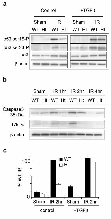
Figure 1. p53 is hypophosphorylated and caspase cleavage are compromised in irradiated
Tgfβ1 heterozygote primary MEC and are restored by TGFβ treatment.
A. Representative immunoblots of p53 phosphorylation at serine 18 and serine 23, and total p53 in
sham and irradiated wildtype (WT) and heterozygote (HT) mammary MEC in serum-free culture
treated with or without TGFβ prior to IR. β-actin is shown as protein loading control. B.
Representative immunoblots of total caspase 3 and cleavage product in wildtype and heterozygote
mammary MEC following IR. β-actin is shown as protein loading control. C. Densitometric
analysis of 17kDa cleavage fragment normalized to total caspase-3 demonstrates restoration of
caspase cleavage by TGFβ treatment prior to IR.

Kirshner et al. 26
Figure 2. IR- induced phosphorylation of DNA damage response proteins and cell cycle arrest
are compromised in Tgfβ1 null keratinocyte cell lines.
A. Immunoblots of p53 phosphorylation at serine 18 and serine 23, Rad17 serine 645
phosphorylation, and Chk2 threonine 685 of sham and irradiated Tgfβ1 null and heterozygote
keratinocyte cell lines as a function of time after IR Total proteins and β actin are shown. B.
Immunofluorescence detection of phosphorylated γH2AX (green) RIF formation in DAPI stained
nuclei (blue) of sham and irradiated Tgfβ1 null and heterozygote keratinocytes. γH2AX RIF
formation at 30 min post-IR (2 Gy) is evident in heterozygote keratinocytes but is barely detectable
in irradiated null keratinocytes. C. Immunolocalization of 53BP1 (green) in DAPI stained nuclei
(blue). Sham-irradiated Tgfβ1 heterozygote and null keratinocytes showed diffuse nuclear
immunoreactivity. 53BP1 formed distinct RIF at 30 min post-IR (2 Gy) throughout the nuclei of
both Tgfβ1 heterozygote and null keratinocyte cells.

Kirshner et al. 27
Figure 3. ATM kinase activity and autophosphorylation are markedly reduced in TGFβ1
compromised cells.
A. Representative kinase assay of immortalized Tgfβ1 null compared to heterozygote keratinocyte
cell lines. Irradiated ataxia telangiectasia (A-T) fibroblasts were included as a negative control.
ATM immunoblotting after immunoprecipitation shows similar ATM loading for each genotype and
treatment. Kinase activity was dramatically reduced in null versus heterozygote cells. B.
Quantitation by densitometry of the kinase activity normalized to ATM protein immunoprecipitation
of Tgfβ1 null and heterozygote cell lines (mean+s.e.m., n=3 experiments). C. Dual immunoblot using
infrared antibodies shows ATM serine 1981 autophosphorylation (green/yellow) and total ATM (red)
as a function of time post-IR. Heterozygote keratinocytes show rapid and persistent ATM
autophosphorylation that is lacking in null keratinocyte cells. β-actin is shown as protein loading
control. D. Dual immunoblot analysis of Tgfβ1 heterozygote and knockout keratinocyte cell extracts
shows that phosphorylation of p53 serine18 and ATM serine 1981 are diminished in Tgfβ1 null cells
at 1hr post UV and do not recover by 3 hr. γ-tublin is shown as protein loading control.
Kirshner et al. 28
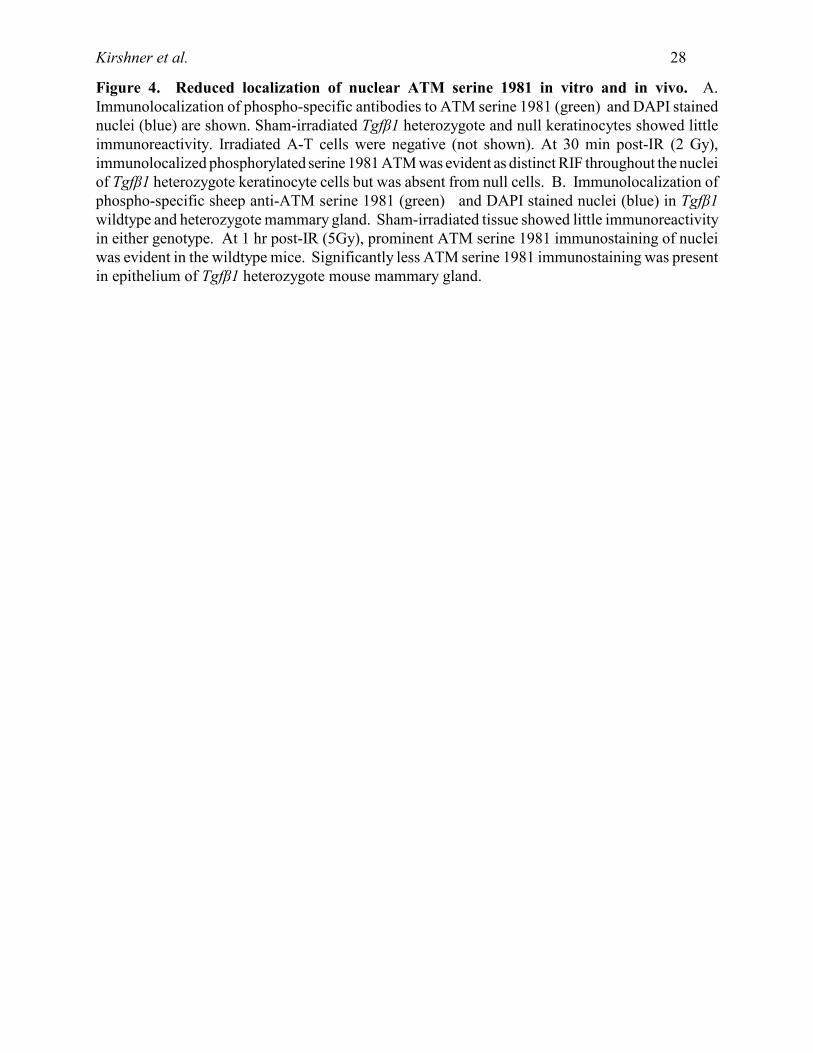
Figure 4. Reduced localization of nuclear ATM serine 1981 in vitro and in vivo. A.
Immunolocalization of phospho-specific antibodies to ATM serine 1981 (green) and DAPI stained
nuclei (blue) are shown. Sham-irradiated Tgfβ1 heterozygote and null keratinocytes showed little
immunoreactivity. Irradiated A-T cells were negative (not shown). At 30 min post-IR (2 Gy),
immunolocalized phosphorylated serine 1981 ATM was evident as distinct RIF throughout the nuclei
of Tgfβ1 heterozygote keratinocyte cells but was absent from null cells. B. Immunolocalization of
phospho-specific sheep anti-ATM serine 1981 (green) and DAPI stained nuclei (blue) in Tgfβ1
wildtype and heterozygote mammary gland. Sham-irradiated tissue showed little immunoreactivity
in either genotype. At 1 hr post-IR (5Gy), prominent ATM serine 1981 immunostaining of nuclei
was evident in the wildtype mice. Significantly less ATM serine 1981 immunostaining was present
in epithelium of Tgfβ1 heterozygote mouse mammary gland.

Kirshner et al. 29
Figure 5. TGFβ treatment restores ATM autophosphorylation, localization, and function in
Tgfβ1 null keratinocytes.
A. Immunoblot of ATM serine 1981 phosphorylation, p53 serine 18 and β actin loading control for
null keratinocytes. Treatment with TGFβ for 4 hr prior to IR (5Gy) significantly increased ATM
serine 1981 autophosphorylation and p53 serine 18 phosphorylation. B. Immunolocalization of
phospho-specific antibodies to ATM serine 1981 (green) and DAPI stained nuclei (blue) of null
keratinocyte cells 30 min after irradiation (2 Gy). RIF formation was restored in TGFβ treated null
keratinocyte cells. C. Histograms of the mean intensity per nucleus of γH2AX immunoreactivity
of Tgfβ1 null cells cultured in the presence or absence of TGFβ1 before being irradiated. γH2AX
immunoreactivity was not induced by TGFβ treatment alone. TGFβ1 exposure prior to irradiation
restored γH2AX in the majority of irradiated Tgfβ1 null cells.

Kirshner et al. 30
Figure 6. TβR1 kinase small molecule inhibitor decreases the DNA damage response in human
epithelial cells. A. Confluent MCF10A human mammary epithelial cells cultured in serum-free
medium for 96 hr were treated as follows: Control (lane 1, white bar), irradiated (lane 2, black bar),
treated for the final 48 hr with TβR1 kinase inhibitor and irradiated (lane 3, light grey bar), treated
for 48 hr with TβR1 kinase inhibitor and refed with fresh medium without inhibitor and irradiated
(lane 4, dark grey bar). Cultures were harvested 1 hr after irradiation (5Gy) for immunoblotting.
Quantitation of phosphorylation-specific antibodies using the LiCor Odyssey was normalized to the
respective total protein. TβR1 kinase inhibitor treatment did not change the abundance or
phosphorylation status of proteins in the absence of irradiation (not shown). B, C: RIF formation
in MCF10A cells treated for 24 hr with and without 240nM TβRI kinase inhibitor and sham or
irradiated (2 Gy). Forty minutes post-IR cells were fixed and stained with phospho-specific
antibodies to either γH2AX (B, green fluorescence) or phosphorylated ATM serine 1981 (C, green
fluorescence). Nuclei are DAPI stained (blue). Treatment with TβRI kinase inhibitor impedes
formation of nuclear RIF of both γH2AX and phosphorylated ATM serine 1981. The formation of
53BP1 nuclear RIF was unaffected (not shown). D. Colony forming efficiency of irradiated
MCF10A control cells (black) and treated for 48 hr with 240nM TβRI kinase inhibitor (red).
Inhibition of TGFβ decreased clonogenic survival compared to controls as a function of radiation
dose (ANOVA, p<0.0001).


Copyright © 2022 FDOKUMEN