Blood outgrowth endothelial cells from Hereditary Haemorrhagic Telangiectasia patients reveal...
14
Blood outgrowth endothelial cells from Hereditary Haemorrhagic Telangiectasia patients reveal abnormalities compatible with vascular lesions Africa Fernandez-L a,1 , Francisco Sanz-Rodriguez b,1 , Roberto Zarrabeitia c , Alfonso Pe ´rez-Molino c , Robert P. Hebbel d , Julia Nguyen d , Carmelo Bernabe ´u a, * ,1 , Luisa-Maria Botella a,1 a Centro de Investigaciones Biologicas, CSIC, Ramiro de Maeztu, 9. Madrid 28040, Spain b Departamento de Biologı ´a, Universidad Auto ´noma, Madrid, Spain c Departamento de Medicina Interna, Hospital Sierrallana, Torrelavega, Santander, Spain d Vascular Biology Centre, MMC 480,420, Delaware St, SE Minneapolis, MN 55455, USA Received 24 January 2005; received in revised form 26 May 2005; accepted 15 June 2005 Time for primary review 14 days Available online 5 July 2005 Abstract Objective: Hereditary haemorrhagic telangiectasia (HHT) is originated by mutations in endoglin (HHT1 ) and ALK1 (HHT2) genes. The purpose of this work was to isolate and characterize circulating endothelial cells from HHT patients. Methods: Pure primary cultures of blood outgrowth endothelial cells (BOECs) were obtained from 50 ml of peripheral blood by selection on collagen plates with endothelial medium. Results: The amount of endoglin in HHT1 – BOECs is half the controls, but HHT2 – BOECs are also endoglin-deficient. Since the TGF-h/ ALK1 pathway activates the endoglin promoter activity, these results suggest the involvement of ALK1 in endoglin gene expression. Endothelial TGF-h pathways, mediated by ALK1 and ALK5, are impaired in HHT cells. HHT – BOECs show disorganized and depolymerized actin fibers, as compared to the organized stress fibers of healthy – BOECs. Functionally, HHT– BOECs have impaired tube formation, in contrast with the cord-like structures derived from normal donors. Conclusions: Decreased endoglin expression, impaired TGF-h signalling, disorganized cytoskeleton, and failure to form cord-like structures are common characteristics of endothelial cells from HHT patients. These features may lead to fragility of small vessels and bleeding characteristic of the HHT vascular dysplasia and to a disrupted and abnormal angiogenesis, which may explain the clinical symptoms associated with this disease. D 2005 European Society of Cardiology. Published by Elsevier B.V. All rights reserved. Keywords: Endothelial receptors; Endothelial function; Signal transduction; Angiogenesis; Remodelling This article is referred to in the Editorial by R. Hirschberg (pages 180 – 182) in this issue. 1. Introduction Hereditary haemorrhagic telangiectasia (HHT) is an autosomal dominant, multisystemic vascular dysplasia, characterized by recurrent haemorrhages [1,2]. Mutations in endoglin (ENG; chromosome 9) [3] and ALK1 (chromo- some 12) [4] are responsible for HHT1 and HHT2 , respectively. Endoglin and ALK1 play important roles in 0008-6363/$ - see front matter D 2005 European Society of Cardiology. Published by Elsevier B.V. All rights reserved. doi:10.1016/j.cardiores.2005.06.009 * Correspondence author. Tel.: +34 91 8373112x4246; fax: +34 91 5360432. E-mail address: [email protected] (C. Bernabe ´u). 1 These authors contributed equally to this work. Cardiovascular Research 68 (2005) 235 – 248 www.elsevier.com/locate/cardiores by guest on September 12, 2014 http://cardiovascres.oxfordjournals.org/ Downloaded from
-
Upload
independent -
Category
Documents
-
view
0 -
download
0
Transcript of Blood outgrowth endothelial cells from Hereditary Haemorrhagic Telangiectasia patients reveal...
www.elsevier.com/locate/cardiores
Cardiovascular Research
http://cardiovD
ownloaded from
Blood outgrowth endothelial cells from Hereditary
Haemorrhagic Telangiectasia patients reveal abnormalities
compatible with vascular lesions
Africa Fernandez-L a,1, Francisco Sanz-Rodriguez b,1, Roberto Zarrabeitia c,
Alfonso Perez-Molino c, Robert P. Hebbel d, Julia Nguyen d,
Carmelo Bernabeu a,*,1, Luisa-Maria Botella a,1
aCentro de Investigaciones Biologicas, CSIC, Ramiro de Maeztu, 9. Madrid 28040, SpainbDepartamento de Biologıa, Universidad Autonoma, Madrid, Spain
cDepartamento de Medicina Interna, Hospital Sierrallana, Torrelavega, Santander, SpaindVascular Biology Centre, MMC 480,420, Delaware St, SE Minneapolis, MN 55455, USA
Received 24 January 2005; received in revised form 26 May 2005; accepted 15 June 2005
Time for primary review 14 days
Available online 5 July 2005
by guest on September 12, 2014
ascres.oxfordjournals.org/
Abstract
Objective: Hereditary haemorrhagic telangiectasia (HHT) is originated by mutations in endoglin (HHT1) and ALK1 (HHT2) genes. The
purpose of this work was to isolate and characterize circulating endothelial cells from HHT patients.
Methods: Pure primary cultures of blood outgrowth endothelial cells (BOECs) were obtained from 50 ml of peripheral blood by selection on
collagen plates with endothelial medium.
Results: The amount of endoglin in HHT1 –BOECs is half the controls, but HHT2 –BOECs are also endoglin-deficient. Since the TGF-h/ALK1 pathway activates the endoglin promoter activity, these results suggest the involvement of ALK1 in endoglin gene expression.
Endothelial TGF-h pathways, mediated by ALK1 and ALK5, are impaired in HHT cells. HHT–BOECs show disorganized and
depolymerized actin fibers, as compared to the organized stress fibers of healthy–BOECs. Functionally, HHT–BOECs have impaired tube
formation, in contrast with the cord-like structures derived from normal donors.
Conclusions: Decreased endoglin expression, impaired TGF-h signalling, disorganized cytoskeleton, and failure to form cord-like structures
are common characteristics of endothelial cells from HHT patients. These features may lead to fragility of small vessels and bleeding
characteristic of the HHT vascular dysplasia and to a disrupted and abnormal angiogenesis, which may explain the clinical symptoms
associated with this disease.
D 2005 European Society of Cardiology. Published by Elsevier B.V. All rights reserved.
Keywords: Endothelial receptors; Endothelial function; Signal transduction; Angiogenesis; Remodelling
This article is referred to in the Editorial by R.
Hirschberg (pages 180–182) in this issue.
0008-6363/$ - see front matter D 2005 European Society of Cardiology. Publishe
doi:10.1016/j.cardiores.2005.06.009
* Correspondence author. Tel.: +34 91 8373112x4246; fax: +34 91
5360432.
E-mail address: [email protected] (C. Bernabeu).1 These authors contributed equally to this work.
1. Introduction
Hereditary haemorrhagic telangiectasia (HHT) is an
autosomal dominant, multisystemic vascular dysplasia,
characterized by recurrent haemorrhages [1,2]. Mutations
in endoglin (ENG; chromosome 9) [3] and ALK1 (chromo-
some 12) [4] are responsible for HHT1 and HHT2,
respectively. Endoglin and ALK1 play important roles in
68 (2005) 235 – 248
d by Elsevier B.V. All rights reserved.
A. Fernandez-L et al. / Cardiovascular Research 68 (2005) 235–248236
by guest on September 12, 2014
http://cardiovascres.oxfordjournals.org/D
ownloaded from
cardiovascular development, angiogenesis and vascular
remodelling [5–9].
Endoglin and ALK1 are components of the TGF-hreceptor complex primarily expressed in endothelial cells
(ECs) [9–11], where they collaborate with each other in
TGF-h signalling [12,13]. TGF-h is a multifunctional
cytokine member of a large superfamily of proteins
including activins and bone morphogenetic proteins [14].
Cellular responses triggered by TGF-h are elicited via type I
and type II serine/threonine kinase receptors and down-
stream by the Smad family of signal transducers [15]. Type I
receptors act downstream from type II and determine the
signalling specificity in the receptor complex. Since ECs
express two different type I receptors, ALK1 and ALK5,
TGF-h may activate two different pathways. ALK1 pro-
motes Smad1 and Smad5 phosphorylation stimulating cell
proliferation and migration, whereas activation of ALK5
promotes phosphorylation of Smad2 and Smad3, and
stimulates inhibition of proliferation and extracellular matrix
synthesis [16]. Endoglin interacts with ThRII, ALK1 and
ALK5 [13,17] and cooperates with ALK1 in the endothelial
TGF-h signal transduction favouring cell proliferation
[12,13]. Endoglin binds different members of the TGF-hsuperfamily in the presence of the signalling receptors types
I and II [18,19], and counteracts the TGF-h-induced growth
inhibition of ECs [20].
The diagnosis of HHT remains at the clinical level,
according to the consensus criteria of Curacao [21]. These
include epistaxis (spontaneous, recurrent nose bleeds),
telangiectasias (multiple at the mucosa level), visceral
lesions (in lung, brain, liver, or spinal cord) and family
history. HHT diagnosis is definite if 3 criteria are present.
Haploinsufficiency is generally accepted as the molecular
basis for HHT, since many of the mutated endoglin and
ALK1 proteins are not expressed at the cell surface
[1,22,23], although it is not excluded that some missense
mutations, affecting the ALK1 kinase domain, could be
expressed [24].
The endothelial expression of ENG and ALK1 makes
difficult the measurement of protein levels in HHT patients
due to difficulties in isolating this cell type. Only in cases of
pregnancies from HHT families, endothelial cells from the
umbilical vein (HUVECs) of affected newborns were
available for direct studies [23,25]. However, this is not
the best model, as they are derived from newborns, and
HHT penetrance [26], as endoglin deficiency in activated
monocytes [27], are age-dependent.
Blood outgrowth endothelial cells (BOECs) [28,29] are
functionally mature ECs [30] able to be incorporated in vivo
into the vascular endothelium and successfully used in gene
therapy [29]. In this paper we report for the first time the
characterization of pure cultures of BOECs from Spanish
HHT patients with clinical diagnosis and identified muta-
tions in ENG (HHT1) or ALK1 (HHT2). HHT–BOECs
were characterized and used to investigate the molecular
basis of the disease.
2. Materials and methods
2.1. Patient samples and DNA sequencing
Informed consent was obtained from individuals partic-
ipating in the study. Controls were healthy donors, as
certified by annual medical surveys. The investigation
conforms with the principles outlined in the Declaration of
Helsinki (Cardiovascular Research 1997;35:2–4). HHT
diagnoses were based on Curacao criteria. Genomic DNA
was isolated from peripheral blood lymphocytes using Qia-
amp Mini kit (Qiagen). The fifteen exons from endoglin and
the nine exons from ALK1 were PCR-amplified using
HotMaster Polymerase (Eppendorf), reported primers [22–
24] and sequenced with a cycle sequencing protocol
(Applied Biosystems, USA).
2.2. Plasmids and site directed mutagenesis
GAL4-Smad1 and GAL4-Smad3 plasmids were gener-
ously provided by Drs. Nakayama and Derynck, respec-
tively. TGF-h-responsive constructs (CAGA)12-Luc,
p(BRE)2-Luc, were kindly provided by Dr. ten Dijke.
Endoglin promoter construct pCD105 (�450 /+350) and
vector encoding human endoglin have been described
[17,31]. Vectors encoding wild type ALK1 (WT), kinase
deficient ALK1 (K229R), and constitutively active ALK1
kinase (Q201D) in pcDNA3-HASL plasmid were generous
gifts from Dr. Miyazono (University of Tokyo, Japan). The
reporter vector for the ALK5 promoter, pTGFbeta type I
receptor (�1422 /�65)-Luc, was a kind gift of Dr. Kojima
(RIKEN Wako, Saitama, Japan). Site directed mutagenesis
of pcDNA3.1 ALK1 (WT) was made by PCR to substitute
1120 C by T yielding ALK1 (R374W).
2.3. Cell cultures and tube formation assay
BOECs were grown from 50 ml peripheral blood,
culturing buffy coat mononuclear cells on collagen I
coated wells using EGM/EBM-2 medium (Clonetics), as
described [28,29]. Briefly, cells from mononuclear layers
were pelleted and resuspended in 5 ml of medium. Cells
were centrifuged again and the pellet washed twice in
culture medium, before resuspension in 5 ml of medium
and plating on collagen coated P-6 wells. Cells were
incubated in 5% CO2 at 37 -C and medium was replaced
daily for the first week and thereafter medium was
changed every two days. BOECs were established as pure
endothelial cultures, being the only surviving cell type
covering the wells after 30–45 days of growth. For
characterization and functional studies, BOECs from 2nd
to 4th passage were used. For tube formation assays, cells
were plated in EBM/EGM-2 culture medium on matrigel
(Becton Dickinson) and incubated at 37 -C, as indicated
by the manufacturer. Human umbilical vein ECs
(HUVECs) were cultured in EBM/EGM-2. Human primary
A. Fernandez-L et al. / Cardiovascular Research 68 (2005) 235–248 237
by guest on September 12, 2014
http://cardiovascres.oxfordjournals.org/D
ownloaded from
fibroblasts and 293 T cell line (kidney epithelium) were
cultured in DMEM medium.
2.4. Western blot analysis
Cells were lysed on ice for 30 min in 1% SDS. Lysates
were centrifuged at 14,000 �g for 5 min. Aliquots of
cleared cell lysates were boiled in SDS sample buffer and
analyzed in 7.5% SDS-PAGE under non-reducing condi-
tions. Proteins were electrotransferred to nitrocellulose
membranes, followed by immunodetection with anti-endo-
glin (P4A4), anti-ALK1 (MAB370, R&D; sc-402 Santa
Cruz), or anti-HLA class I (W6/32; Sigma). Secondary
antibodies were horseradish peroxidase conjugates from
Dako. Membranes were developed by chemiluminescence
(ECL, Supersignal, Pierce).
2.5. Reverse transcription-PCR
RNA was extracted from 106 cells using RNAeasy kit
(Qiagen). RNA was reverse-transcribed to cDNA using
random hexamers and AMV reverse transcriptase. PCRs
with oligonucleotides for ALK5, ThRII [32] or GAPDHwere
performed using the HotMaster Taq polymerase (Eppendorf).
2.6. Immunofluorescent microscopy
Cells grown onto glass coverslips coated with collagen
type I were fixed with 3.5% formaldehyde in PBS, washed
and blocked with 2% BSA in PBS for 1 h at 4 -C. Cells wereincubated for 1 h at 4 -C with anti-endoglin (P4A4), anti-
PECAM1 (Clone HC1/6; Chemicon) anti-ALK1 (MAB370,
R&D), anti-von Willebrand Factor (vWF, sc-7154, Santa
Cruz) and VE-cadherin (sc-9989, Santa Cruz) antibodies. For
staining with vWF, cells were permeabilized with 100 Ag/ml
l-a-lysophosphatidylcholine, followed by incubation with
Alexa-488 green-conjugated anti-rabbit/mouse IgG antibody
(Molecular Probes). For staining actin filaments, cells were
fixed, stained, and permeabilized in a single step by addition
of 5 units/ml Alexa-546 phalloidin (Molecular Probes), 100
Ag/ml l-a-lysophosphatidylcholine, and 3.5% formaldehyde
in cold PBS. Coverslips were mounted with Mowiol 44-88
(Sigma) and observed with a spectral confocal microscope
(Leica Microsystems). When required, BOECs were trans-
fected, prior to actin staining, with expression vectors for
green fluorescence protein (pEGFP), endoglin (pCMV5-
ENG), or with a vector encoding a siRNA endoglin specific
sequence (pSUPER-Endo), as described [34].
2.7. Flow cytometry
BOECs were incubated with mAb P4A4 (anti-endoglin),
anti-vWF (permeabilized cells as above), anti-ALK1
(MAB370, R&D), anti-Flk-1/KDR (MAB3572, R and D),
anti-Flt-1 (MAB321, R&D), anti-fibroblast surface protein
(1B10, Sigma) or an isotype-matched control for 30 min at 4
-C in cold PBS with human IgG, followed by secondary
FITC-conjugated anti-IgG. Samples were analyzed on a
Coulter Epics XL flow cytometer. Antigen measures are
represented by the expression index, which is the result of
multiplying the percentage of cells expressing antigen
(positive cells) by the mean fluorescence intensity of the
whole population (positive and negative).
2.8. Cell transfection and reporter assays
Transient transfections were made with Superfect (Qia-
gen) according to the manufacturer’s recommendations. For
GAL4 transactivation assays, GAL4-Smad1 or GAL4-
Smad3 plasmids were cotransfected with the GAL4-lucifer-
ase reporter pFr5-Luc (Stratagene). Reporter assays with
TGF-h responsive promoter constructs (CAGA)12-luc,
p(BRE)2 and pCD105(�450 /+350), and with the ALK5
promoter construct (�1422 /�65)-Luc were performed as
described [13,32]. After transfection, cells were incubated
with 10 ng/ml TGF-h1 (Preprotech) for 24 h (reporters
(CAGA)12-luc, GAL4-Smad3, and GAL4-Smad2) or with 1
ng/ml TGF-h1 for 3 h (reporters p(BRE)2, GAL4-Smad1,
and ALK5/�1422 /�65-Luc). Relative luciferase units were
measured in a TD20 /20 luminometer (Promega, Madison,
WI). Samples were cotransfected with the SV40-h-galacto-sidase expression vector to correct for transfection efficiency.
Measures of h-galactosidase activity were performed using
Galacto-light (Tropix). Transfections were made in duplicate
and repeated at least in three independent experiments.
Representative experiments are shown in the figures.
3. Results
3.1. Culture and characterization of ECs from peripheral
blood
EC cultures were established using peripheral blood from
either control donors or HHT patients. On average, it took
between 30–45 days to get a confluent pure endothelial
culture of about 105 cells since plating the mononuclear
layer. Distinguishable clusters of ECs appeared after 9–20
days, depending on the sample. Five independent control
BOECs and a total of 11 HHT–BOEC cultures (from 8
different families /mutations) were obtained. ECs were
characterized by morphology and staining with endothelial
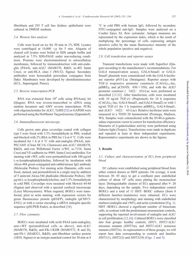
markers (endoglin and vWF), and actin cytoskeleton (Fig. 1).
HHT–BOECs showed a significant number of binucleate
cells, in contrast with the predominant mononuclear controls,
supporting the reported involvement of endoglin and ALK1
in cell proliferation [12,16]. Cultured BOECs were classified
into four groups: healthy controls, HHT1, HHT2/ALK1
missense mutants (HHT2m), and HHT2/ALK1 nonsense
mutants (HHT2n). As representative of these groups, we will
report here data corresponding to controls and families
HHT1(1), HHT2(2) and HHT2(10) (Figs. 2 and 3).
A. Fernandez-L et al. / Cardiovascular Research 68 (2005) 235–248238
http:/D
ownloaded from
Healthy BOECs had the characteristic endothelial cobble-
stone shape at confluence (Fig. 1A). Five different cultures
of BOECs were derived from HHT1 patients. Two of them,
belonged to father and son of family #1 where DNA
sequencing revealed a nonsense mutation in c.511C>T
leading to a stop codon (R171X) (Fig. 2). The other three
belonged to families #4 and #23 (Fig. 1A), each one with a
new ENG mutation (Fernandez-L et al., in preparation). The
HHT1 cells were bigger and rather round-shaped (Fig. 1A)
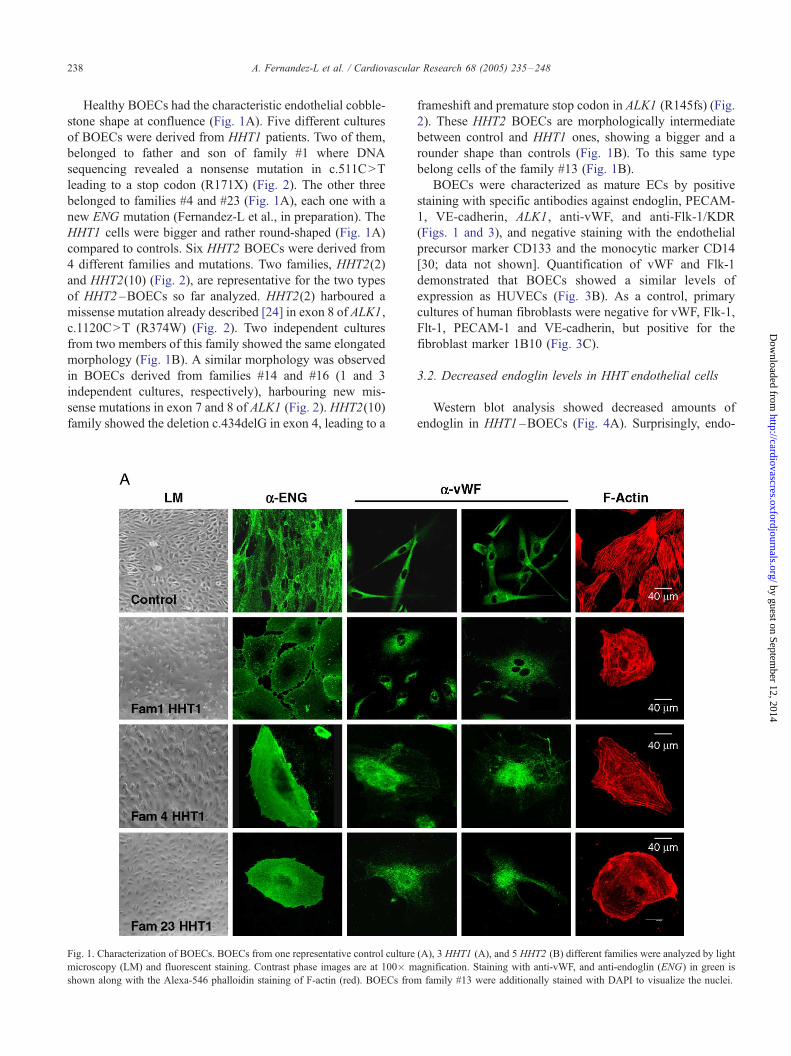
compared to controls. Six HHT2 BOECs were derived from
4 different families and mutations. Two families, HHT2(2)
and HHT2(10) (Fig. 2), are representative for the two types
of HHT2 –BOECs so far analyzed. HHT2(2) harboured a
missense mutation already described [24] in exon 8 of ALK1,
c.1120C>T (R374W) (Fig. 2). Two independent cultures
from two members of this family showed the same elongated
morphology (Fig. 1B). A similar morphology was observed
in BOECs derived from families #14 and #16 (1 and 3
independent cultures, respectively), harbouring new mis-
sense mutations in exon 7 and 8 of ALK1 (Fig. 2). HHT2(10)
family showed the deletion c.434delG in exon 4, leading to a
Fig. 1. Characterization of BOECs. BOECs from one representative control culture
microscopy (LM) and fluorescent staining. Contrast phase images are at 100� m
shown along with the Alexa-546 phalloidin staining of F-actin (red). BOECs from
frameshift and premature stop codon in ALK1 (R145fs) (Fig.
2). These HHT2 BOECs are morphologically intermediate
between control and HHT1 ones, showing a bigger and a
rounder shape than controls (Fig. 1B). To this same type
belong cells of the family #13 (Fig. 1B).
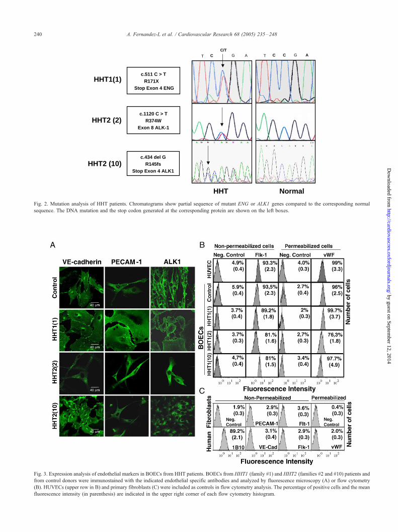
BOECs were characterized as mature ECs by positive
staining with specific antibodies against endoglin, PECAM-
1, VE-cadherin, ALK1, anti-vWF, and anti-Flk-1/KDR
(Figs. 1 and 3), and negative staining with the endothelial
precursor marker CD133 and the monocytic marker CD14
[30; data not shown]. Quantification of vWF and Flk-1
demonstrated that BOECs showed a similar levels of
expression as HUVECs (Fig. 3B). As a control, primary
cultures of human fibroblasts were negative for vWF, Flk-1,
Flt-1, PECAM-1 and VE-cadherin, but positive for the
fibroblast marker 1B10 (Fig. 3C).
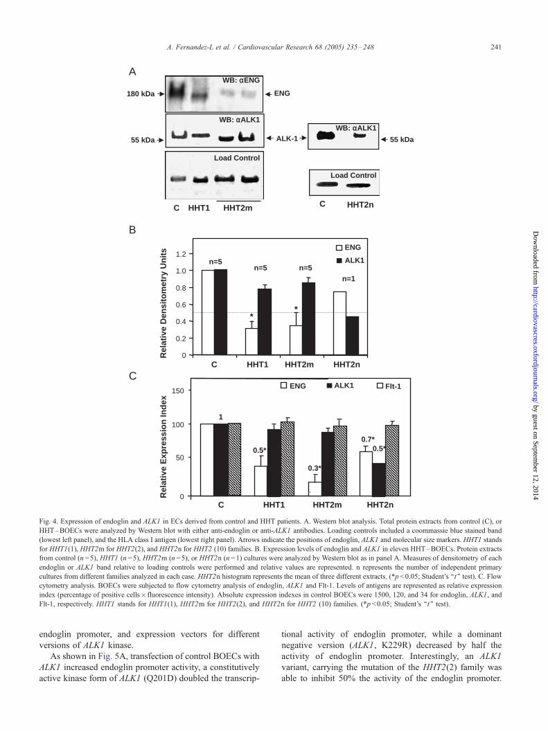
3.2. Decreased endoglin levels in HHT endothelial cells
Western blot analysis showed decreased amounts of
endoglin in HHT1 –BOECs (Fig. 4A). Surprisingly, endo-
(A), 3 HHT1 (A), and 5 HHT2 (B) different families were analyzed by light
agnification. Staining with anti-vWF, and anti-endoglin (ENG) in green is
family #13 were additionally stained with DAPI to visualize the nuclei.
by guest on September 12, 2014
/cardiovascres.oxfordjournals.org/
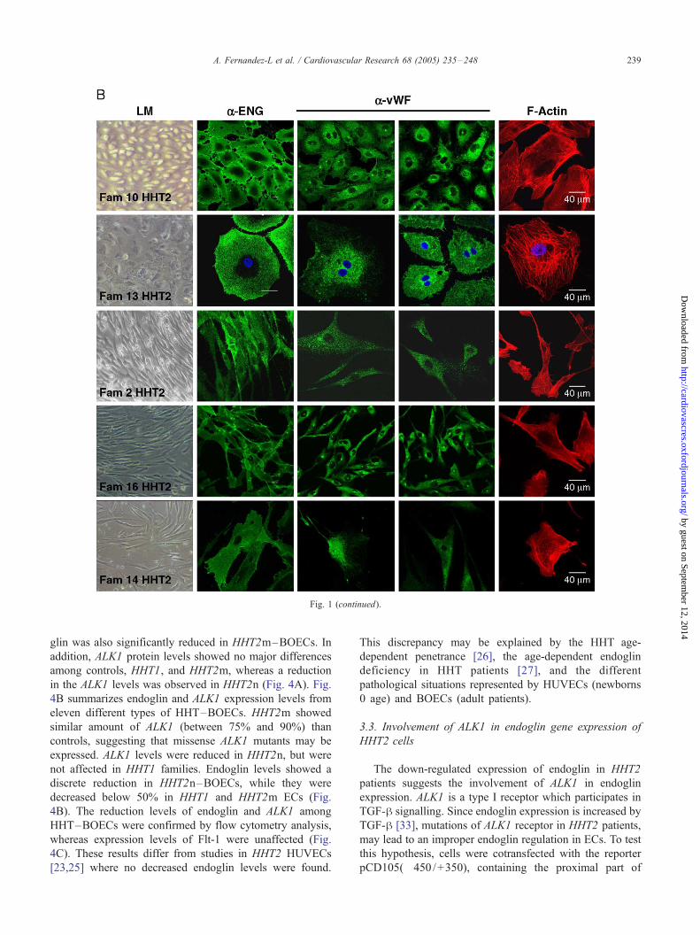
Fig. 1 (continued).
A. Fernandez-L et al. / Cardiovascular Research 68 (2005) 235–248 239
by guest on September 12, 2014
http://cardiovascres.oxfordjournals.org/D
ownloaded from
glin was also significantly reduced in HHT2m–BOECs. In
addition, ALK1 protein levels showed no major differences
among controls, HHT1, and HHT2m, whereas a reduction
in the ALK1 levels was observed in HHT2n (Fig. 4A). Fig.
4B summarizes endoglin and ALK1 expression levels from
eleven different types of HHT–BOECs. HHT2m showed
similar amount of ALK1 (between 75% and 90%) than
controls, suggesting that missense ALK1 mutants may be
expressed. ALK1 levels were reduced in HHT2n, but were
not affected in HHT1 families. Endoglin levels showed a
discrete reduction in HHT2n–BOECs, while they were
decreased below 50% in HHT1 and HHT2m ECs (Fig.
4B). The reduction levels of endoglin and ALK1 among
HHT–BOECs were confirmed by flow cytometry analysis,
whereas expression levels of Flt-1 were unaffected (Fig.
4C). These results differ from studies in HHT2 HUVECs
[23,25] where no decreased endoglin levels were found.
This discrepancy may be explained by the HHT age-
dependent penetrance [26], the age-dependent endoglin
deficiency in HHT patients [27], and the different
pathological situations represented by HUVECs (newborns
0 age) and BOECs (adult patients).
3.3. Involvement of ALK1 in endoglin gene expression of
HHT2 cells
The down-regulated expression of endoglin in HHT2
patients suggests the involvement of ALK1 in endoglin
expression. ALK1 is a type I receptor which participates in
TGF-h signalling. Since endoglin expression is increased by
TGF-h [33], mutations of ALK1 receptor in HHT2 patients,
may lead to an improper endoglin regulation in ECs. To test
this hypothesis, cells were cotransfected with the reporter
pCD105(�450 /+350), containing the proximal part of
HHT2 (2)c.1120 C > T
R374WExon 8 ALK-1
HHT2 (10)c.434 del G
R145fsStop Exon 4 ALK1
CT AGC/T
HHT1(1)
CT AGC
HHT Normal
c.511 C > TR171X
Stop Exon 4 ENG
Fig. 2. Mutation analysis of HHT patients. Chromatograms show partial sequence of mutant ENG or ALK1 genes compared to the corresponding normal
sequence. The DNA mutation and the stop codon generated at the corresponding protein are shown on the left boxes.
Fig. 3. Expression analysis of endothelial markers in BOECs from HHT patients. BOECs from HHT1 (family #1) and HHT2 (families #2 and #10) patients and
from control donors were immunostained with the indicated endothelial specific antibodies and analyzed by fluorescence microscopy (A) or flow cytometry
(B). HUVECs (upper row in B) and primary fibroblasts (C) were included as controls in flow cytometry analysis. The percentage of positive cells and the mean
fluorescence intensity (in parenthesis) are indicated in the upper right corner of each flow cytometry histogram.
A. Fernandez-L et al. / Cardiovascular Research 68 (2005) 235–248240
by guest on September 12, 2014
http://cardiovascres.oxfordjournals.org/D
ownloaded from
A
B
0
50
100
150
Rel
ativ
e E
xpre
ssio
n In
dex
0.5*
1
ALK-155 kDa
WB: αALK1
55 kDa
Load Control
ENG
WB: αALK1
Load Control
WB: αENG
C
180 kDa
C HHT1 HHT2m HHT2n
Rel
ativ
e D
ensi
tom
etry
Un
its ENG
ALK1
0.2
0.4
0.6
0.8
1.2
0
1.0 n=5 n=5n=1
n=5
**
C
C HHT1 HHT2m HHT2n
C HHT1 HHT2n
0.3*
HHT2m
0.7*0.5*
ENG ALK1 Flt-1
Fig. 4. Expression of endoglin and ALK1 in ECs derived from control and HHT patients. A. Western blot analysis. Total protein extracts from control (C), or
HHT–BOECs were analyzed by Western blot with either anti-endoglin or anti-ALK1 antibodies. Loading controls included a coommassie blue stained band
(lowest left panel), and the HLA class I antigen (lowest right panel). Arrows indicate the positions of endoglin, ALK1 and molecular size markers. HHT1 stands
for HHT1(1), HHT2m for HHT2(2), and HHT2n for HHT2 (10) families. B. Expression levels of endoglin and ALK1 in eleven HHT–BOECs. Protein extracts
from control (n =5), HHT1 (n =5), HHT2m (n =5), or HHT2n (n =1) cultures were analyzed by Western blot as in panel A. Measures of densitometry of each
endoglin or ALK1 band relative to loading controls were performed and relative values are represented. n represents the number of independent primary
cultures from different families analyzed in each case. HHT2n histogram represents the mean of three different extracts. (*p <0.05; Student’s ‘‘t’’ test). C. Flow
cytometry analysis. BOECs were subjected to flow cytometry analysis of endoglin, ALK1 and Flt-1. Levels of antigens are represented as relative expression
index (percentage of positive cells� fluorescence intensity). Absolute expression indexes in control BOECs were 1500, 120, and 34 for endoglin, ALK1, and
Flt-1, respectively. HHT1 stands for HHT1(1), HHT2m for HHT2(2), and HHT2n for HHT2 (10) families. (*p <0.05; Student’s ‘‘t’’ test).
A. Fernandez-L et al. / Cardiovascular Research 68 (2005) 235–248 241
by guest on September 12, 2014
http://cardiovascres.oxfordjournals.org/D
ownloaded from
endoglin promoter, and expression vectors for different
versions of ALK1 kinase.
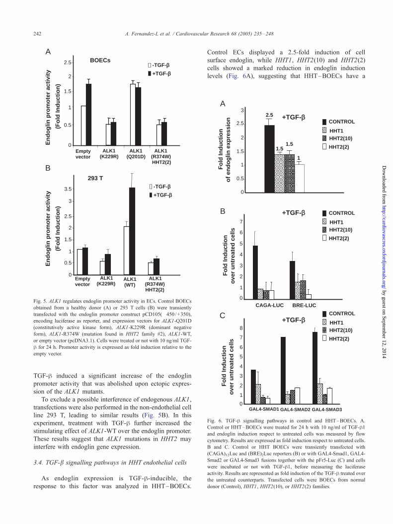
As shown in Fig. 5A, transfection of control BOECs with
ALK1 increased endoglin promoter activity, a constitutively
active kinase form of ALK1 (Q201D) doubled the transcrip-
tional activity of endoglin promoter, while a dominant
negative version (ALK1, K229R) decreased by half the
activity of endoglin promoter. Interestingly, an ALK1
variant, carrying the mutation of the HHT2(2) family was
able to inhibit 50% the activity of the endoglin promoter.
0
0.5
1
1.5
2
2.5
ALK1(K229R)
En
do
glin
pro
mo
ter
acti
vity
(Fo
ld In
du
ctio
n)
Emptyvector
ALK1(Q201D)
ALK1(R374W)HHT2(2)
3
3.5
0
0.5
1
1.5
2
2.5
ALK1(WT)
ALK1 (R374W)HHT2(2)
ALK1 (K229R)
-TGF-β+TGF-β
Emptyvector
A
B
En
do
glin
pro
mo
ter
acti
vity
(Fo
ld In
du
ctio
n)
-TGF-β+TGF-β
BOECs
293 T
Fig. 5. ALK1 regulates endoglin promoter activity in ECs. Control BOECs
obtained from a healthy donor (A) or 293 T cells (B) were transiently
transfected with the endoglin promoter construct pCD105(�450/+350),
encoding luciferase as reporter, and expression vectors for ALK1-Q201D
(constitutively active kinase form), ALK1-K229R (dominant negative
form), ALK1-R374W (mutation found in HHT2 family #2), ALK1-WT,
or empty vector (pcDNA3.1). Cells were treated or not with 10 ng/ml TGF-
h for 24 h. Promoter activity is expressed as fold induction relative to the
empty vector.
CAGA-LUC BRE-LUC
1
2
3
4
5
6
7
0
CONTROL
HHT1HHT2(10)
HHT2(2)
Fo
ld In
du
ctio
no
ver
un
trea
ted
cel
ls
A
C
GAL4-SMAD1 GAL4-SMAD2 GAL4-SMAD30
1
2
3
4
5
6
7
8
CONTROL
HHT1HHT2(10)
HHT2(2)
B
0
0.5
1
1.5
2
2.5
32.5
1.51.5
1
CONTROL
HHT1HHT2(10)
HHT2(2)
Fo
ld In
du
ctio
no
ver
un
trea
ted
cel
ls
Fo
ld In
du
ctio
no
f en
do
glin
exp
ress
ion +TGF-β
+TGF-β
+TGF-β
Fig. 6. TGF-h signalling pathways in control and HHT–BOECs. A.
Control or HHT–BOECs were treated for 24 h with 10 ng/ml of TGF-h1and endoglin induction respect to untreated cells was measured by flow
cytometry. Results are expressed as fold induction respect to untreated cells.
B and C. Control or HHT BOECs were transiently transfected with
(CAGA)12Luc and (BRE)2Luc reporters (B) or with GAL4-Smad1, GAL4-
Smad2 or GAL4-Smad3 fusions together with the pFr5-Luc (C) and cells
were incubated or not with TGF-h1, before measuring the luciferase
activity. Results are represented as fold induction of the TGF-h treated over
the untreated counterparts. Transfected cells were BOECs from normal
donor (Control), HHT1, HHT2(10), or HHT2(2) families.
A. Fernandez-L et al. / Cardiovascular Research 68 (2005) 235–248242
by guest on September 12, 2014
http://cardiovascres.oxfordjournals.org/D
ownloaded from
TGF-h induced a significant increase of the endoglin
promoter activity that was abolished upon ectopic expres-
sion of the ALK1 mutants.
To exclude a possible interference of endogenous ALK1,
transfections were also performed in the non-endothelial cell
line 293 T, leading to similar results (Fig. 5B). In this
experiment, treatment with TGF-h further increased the
stimulating effect of ALK1-WT over the endoglin promoter.
These results suggest that ALK1 mutations in HHT2 may
interfere with endoglin gene expression.
3.4. TGF-b signalling pathways in HHT endothelial cells
As endoglin expression is TGF-h-inducible, the
response to this factor was analyzed in HHT–BOECs.
Control ECs displayed a 2.5-fold induction of cell
surface endoglin, while HHT1, HHT2(10) and HHT2(2)
cells showed a marked reduction in endoglin induction
levels (Fig. 6A), suggesting that HHT–BOECs have a

A. Fernandez-L et al. / Cardiovascular Research 68 (2005) 235–248 243
Dow
nloa
deficient TGF-h signalling. ECs coexpress two different
TGF-h type-I receptors, the ubiquitous ALK5, and ALK1,
which is more specific for ECs [7,11,16,32]. Since
endoglin cooperates with TGF-h/ALK1 pathway, but
interferes with TGF-h/ALK5 route [12,13], these path-
ways were studied in HHT BOECs. The reporters
(CAGA)12-Luc and (BRE)2-Luc were used to monitor
ALK5 and ALK1 dependent signalling (Fig. 6B). In
addition, since ALK1 and ALK5 signal via Smad1/5 and
Smad2/3, respectively, GAL4 fusion constructs of
Smad1, Smad2 and Smad3 were used to assess TGF-
h/receptor I (ALK1 or ALK5)-dependent signalling in
these cells (Fig. 6C).
As shown in Fig. 6, the reporter activities of
(CAGA)12-Luc and (BRE)2-Luc were stimulated from 4-
to 5-fold upon TGF-h treatment in control BOECs.
Similarly, GAL4-Smad1, GAL4-Smad2 and GAL4-Smad3
Den
sito
met
ry o
f
AL
K-5
vs
GA
PD
H
GAPDH
ALK-5
A
C HHT1(1) HHT2(10) HHT2(2)
C HHT1(1) HHT2(10) HHT2(2)
C HHT1(1) HHT2(10) HHT2(2)
C HHT1(1) HHT2(10) HHT2(2)
00.20.40.60.8
11.2
B
GAPDH
TβRII
0
0.2
0.4
0.6
0.8
1
1.2
Den
sito
met
ry o
f
TβR
II vs
GA
PD
H
Fig. 7. ALK5 expression is downregulated in HHT–BOECs. A and B. Semiquan
HHT BOECs. RNA from control, HHT1(1), HHT2(2) and HHT2(10) BOECs,
primers for ALK5, ThRII and GAPDH for 25 cycles. Bands were quantified by d
three independent sets of experiments are shown. C and D. Effect of ALK1 and e
were cotransfected with a luciferase reporter for ALK5 promoter, and expression v
negative ALK1 mutant (K229R), endoglin (ENG), or empty vector (pcDNA3), as
luciferase activity was normalized by h-galactosidase measures.
were transactivated in response to TGF-h, from 3-fold in
GAL4-Smad1, to 7–8-fold in Gal4-Smad2 and GAL4-
Smad3. Thus, both ALK1/TGF-h and ALK5/TGF-hpathways are active in ECs from normal donors. By
contrast, TGF-h pathways were seriously affected in
HHT–BOECs. As expected from the endoglin/ALK1
cooperation [12,13], the TGF-h response of the ALK1-
dependent reporters was impaired in HHT cells. Thus, the
transactivation activity of GAL4-Smad1 and the (BRE)2-
Luc reporter activity in the presence of TGF-h were
severely reduced (from 4- to 1.5-fold) in HHT1-and
HHT2–BOECs (Fig. 6). Surprisingly, the ALK5-depend-
ent TGF-h induction levels of (CAGA)12-Luc reporter or
GAL4-Smad3 transactivation were significantly reduced
in HHT1-and HHT2–BOECs (Fig. 6). Altogether the
results show that the responses of HHT–BOECs to TGF-
h are severely compromised.
C
01020
3040
506070
80
90R
LU
Control +TGF-β1
0
10
20
30
40
50
60
RL
U
Control
+TGF-β1
pcDNA.3
ALK1 WT
ALK1 Q20
1D
ALK1 K22
9RENG
pcDNA.3
ALK1 WT
ALK1 Q20
1D
ALK1 K22
9RENG
D
titative RT-PCR of ALK5 (A) or ThRII (B) versus GAPDH in control and
was retrotranscribed and the resulting cDNA was amplified using specific
ensitometry as shown in the histograms. Representative experiments out of
ndoglin on ALK5 promoter activity. Control BOECs (C) or 293 T cells (D)
ectors for wild type ALK1, constitutively active ALK1 (Q201D), dominant
indicated. After transfection, cells were treated or not with TGF-h1, and the
by guest on September 12, 2014
http://cardiovascres.oxfordjournals.org/ded from
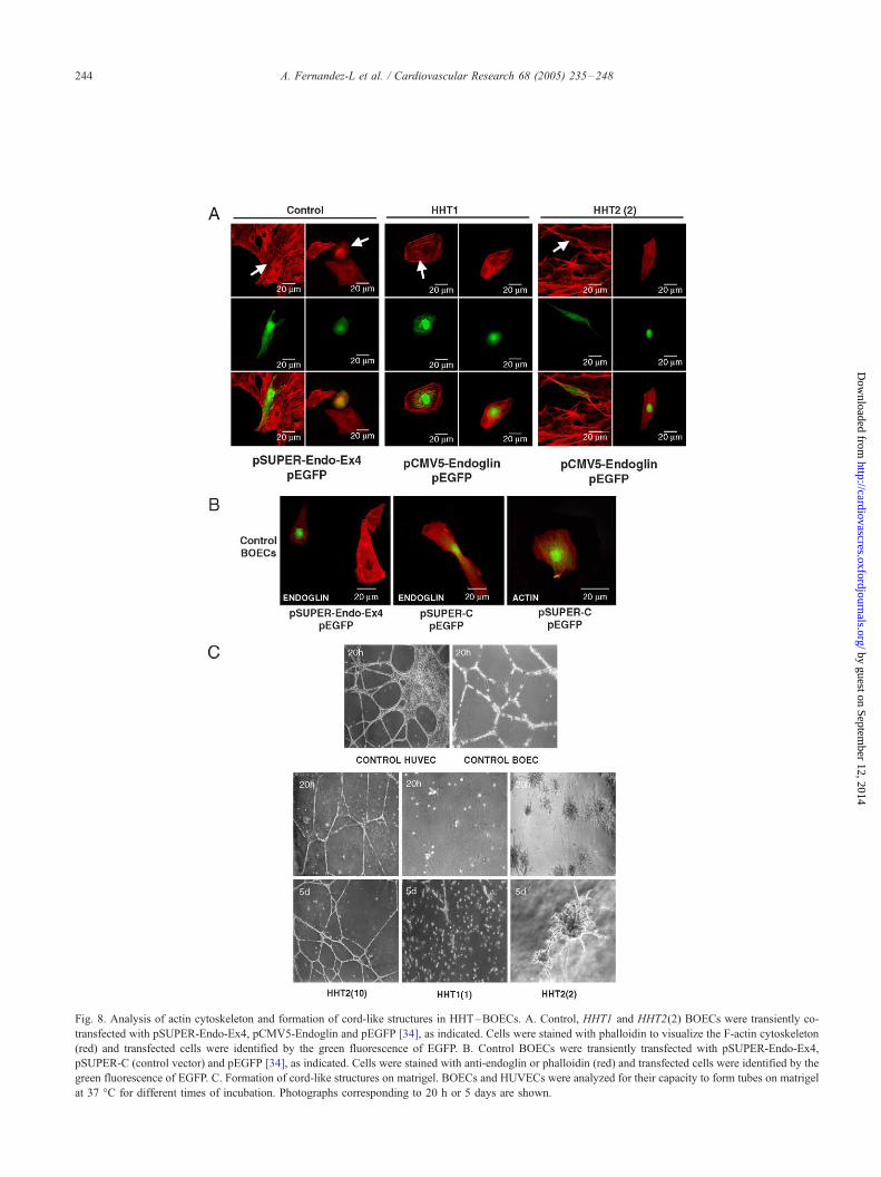
Fig. 8. Analysis of actin cytoskeleton and formation of cord-like structures in HHT–BOECs. A. Control, HHT1 and HHT2(2) BOECs were transiently co-
transfected with pSUPER-Endo-Ex4, pCMV5-Endoglin and pEGFP [34], as indicated. Cells were stained with phalloidin to visualize the F-actin cytoskeleton
(red) and transfected cells were identified by the green fluorescence of EGFP. B. Control BOECs were transiently transfected with pSUPER-Endo-Ex4,
pSUPER-C (control vector) and pEGFP [34], as indicated. Cells were stained with anti-endoglin or phalloidin (red) and transfected cells were identified by the
green fluorescence of EGFP. C. Formation of cord-like structures on matrigel. BOECs and HUVECs were analyzed for their capacity to form tubes on matrigel
at 37 -C for different times of incubation. Photographs corresponding to 20 h or 5 days are shown.
A. Fernandez-L et al. / Cardiovascular Research 68 (2005) 235–248244
by guest on September 12, 2014
http://cardiovascres.oxfordjournals.org/D
ownloaded from
A. Fernandez-L et al. / Cardiovascular Research 68 (2005) 235–248 245
by guest on September 12, 2014
http://cardiovascres.oxfordjournals.org/D
ownloaded from
3.5. ALK5 expression is reduced in HHT ECs and regulated
by ALK1 and endoglin
Since the ALK5 pathway is disrupted in HHT–BOECs
and a previous report [12] have shown a significant
reduction in the levels of ALK5 in eng+ /eng� mouse
embryonic ECs, we hypothesized that ALK5 might be
downregulated as a cell adaptation to compensate for
decreased endoglin/ALK1 expression. Therefore, we exam-
ined ALK5 expression by RT-PCR. Fig. 7A shows a
substantial decrease (80%) of ALK5 expression in endoglin
mutant HHT1 compared to control BOECs. Similarly, a
reduction of ALK5 was observed when ALK1 was mutated
in families HHT2(2) and HHT2(10). These results are ALK5
specific and are not due to a general decrease of the TGF-hsignalling components since the ThRII transcript levels
were not affected (Fig. 7B).
Next, we explored the mechanism leading to the down-
regulation of ALK5 expression in HHT–BOECs. The
possibility of a transcriptional regulatory crosstalk between
ALK1/endoglin and ALK5 pathways was assessed by
transient cotransfections of control BOECs with an ALK5
promoter construct and different expression vectors for
ALK1 and endoglin. As shown in Fig. 7C, cotransfection of
wild type ALK1, constitutively active ALK1 (Q201D), or
wild type endoglin, stimulate the ALK5 promoter activity by
3-, 5-, or 5.2-fold, respectively. By contrast, a dominant
negative ALK1 (K229R) decreased ALK5 promoter activity
almost to basal levels, even after TGF-h treatment. More-
over, TGF-h significantly enhanced the transactivation
activity of cells transfected with wild type ALK1 or
endoglin. Similar results were obtained with the non-
endothelial cell line 293 T (Fig. 7D).
Altogether, these results suggest that ECs keep a fine
tuning between ALK1/endoglin and ALK5 levels, and that
mutations in either endoglin (HHT1), or ALK1 (HHT2)
downregulate ALK5 gene expression to maintain a physio-
logically adaptative balance betweenALK1 and ALK5 routes.
3.6. HHT BOECs show changes in cytoskeleton and tube
formation
The abnormal shape of HHT–BOECs compared to
controls (Figs. 1 and 3), suggests that actin cytoskeleton
may be affected. While control ECs showed a highly
organized cytoskeleton with stress fibers crossing the entire
cell, HHT1-and HHT2 –BOECs had a disorganized actin
structure (Fig. 1). Most of the F-actin was disorganized and
depolymerized at many points giving rise to very intrincated
patterns with different foci of actin polymerization in
HHT1 –BOECs. HHT2(2) BOECs also showed a poor
organization of the actin cytoskeleton, with extense areas
of depolymerization. HHT2(10) BOECs showed a combined
situation: some cells displayed a completely normal F-actin
cytoskeleton, while others had patches of F-actin depolyme-
rization. Since HHT1 and HHT2 BOECs have in common a
significant decrease in the amount of endoglin, these results
are compatible with the reported role of endoglin in the
organization of actin cytoskeleton via its interaction with the
zyxin family of proteins [34]. Supporting this view, endoglin
suppression by siRNA in control BOECs leads to a
disruption of the actin cytoskeleton (Fig. 8A). Conversely,
overexpression of endoglin in HHT1 BOECs significantly
restored the actin network. However, overexpression of
endoglin in HHT2 BOECs resulted only in a slight recovery,
visualized by some stress fibres, suggesting that endoglin
expression was not sufficient to reverse the adaptation
process induced by ALK1 mutations. As expected, control
BOECs transfected with pSUPER-Endo-Ex4 showed a
decreased expression of endoglin compared to untransfected
cells or to cells transfected with an irrelevant vector
(pSUPER-C); also, transfection with pSUPER-C did not
affect endoglin F-actin cytoskeleton (Fig. 8B).
The TGF-h/ALK1/endoglin pathway induces prolifera-
tion, migration and tube formation of ECs [12,16]. To gain
more insight into possible functional problems of HHT–
BOECs, we investigated their behaviour when building up
cord-like structures in vitro. HHT1 and HHT2 cells were
plated on matrigel, using normal donor BOECs and
HUVECs as controls (Fig. 8C). While normal donor
HUVECs and BOECs formed robust cord-like structures
in less than 24 h, HHT–BOECs displayed deficient tube
formation, but the pattern varied depending on the type of
mutation. HHT1 –BOECs did not give rise to clear cord-like
structures and only after 5 days, a few short threads were
visible. HHT2n cells, represented by family 10, formed a
weak and thin tube-network after 20 h, but involving much
less surface than control BOECs. In the case of HHT2m,
represented by family 2, no tubes were formed, but instead
big clusters of cells showing visible sprouting were quite
abundant and, occasionally, were interconnected by short
tubes after long time of incubation. Similar results were
obtained with BOECs from different members of HHT2m
famiIies #14 and #16 (data not shown). Interestingly, when
comparing HHT2(2) vs. HHT2(10), the lower capacity to
form tubes was associated with a lower endoglin expression
levels suggesting that endoglin is involved in tube for-
mation. As endoglin and ALK1 collaborate in the TGF-h/ALK1/endoglin pathway leading to the formation of cord-
like structures, it could be hypothesized that levels of
endoglin or ALK1 activity below a critical threshold might
hamper the tube formation in HHT–BOECs.
4. Discussion
This study represents the first molecular characterization
of ECs from adult HHT patients, primary targets of the
vascular disorder. The development of pure EC cultures from
patients with clinical symptoms is a direct attempt to
understand the status of HHT endothelial function. We have
characterized and studied 5 different cultures from healthy
A. Fernandez-L et al. / Cardiovascular Research 68 (2005) 235–248246
http://cardiovascres.oxfordjourD
ownloaded from
donors and 11 different cultures from HHT patients. HHT
samples belonged to three types of mutations: ENG nonsense
(5 independent cultures), ALK1 nonsense (one culture), and
ALK1 missense (5 independent cultures). Common features
to these HHT cells were positive staining for endothelial
markers, endoglin deficiency, reduced ALK5 expression and
TGF-h signalling, disorganization of the F-actin cytoskele-
ton, and abnormal formation of cord-like structures.
As expected, HHT1 –BOECs were endoglin haploinsuf-
ficient but, surprisingly, HHT2m and HHT2n cells were also
endoglin-deficient. This is in agreement with the upregula-
tion of endoglin observed upon ALK1 overexpression [35].
In addition, we have previously reported endoglin deficient
upregulation in activated monocytes of HHT2 patients [27],
suggesting that ALK1 may modulate endoglin promoter
activity during the monocyte–macrophage transition. The
present work not only confirms this role for ALK1, but also
demonstrates that ALK1 mutations have an impact on the
endoglin promoter activity when transfected in control
BOECs (Fig. 5).
HHT–BOECs showed impaired TGF-h signalling. The
decreased TGF-h/ALK1 pathway could be predicted for
HHT1 and HHT2 cells since endoglin cooperates with ALK1
in ECs [12,13]. On the other hand, HHT1-and HHT2–
BOECs exhibited a downregulation of TGF-h /ALK5 signal-
ling. In the case of HHT1, the ALK5 downregulated
activities agree with the decreased expression of ALK5
observed in eng+ /eng� mouse embryonic ECs [12] and in
EndoglinALK-1 and ALK-5 in equilibrium
SmadSignaling
Fragile cy
Endoglin
Non-HHT HHT-
End
ALK5ALK1
End
AALK1
EndogALK-5
TGF-βAdapta
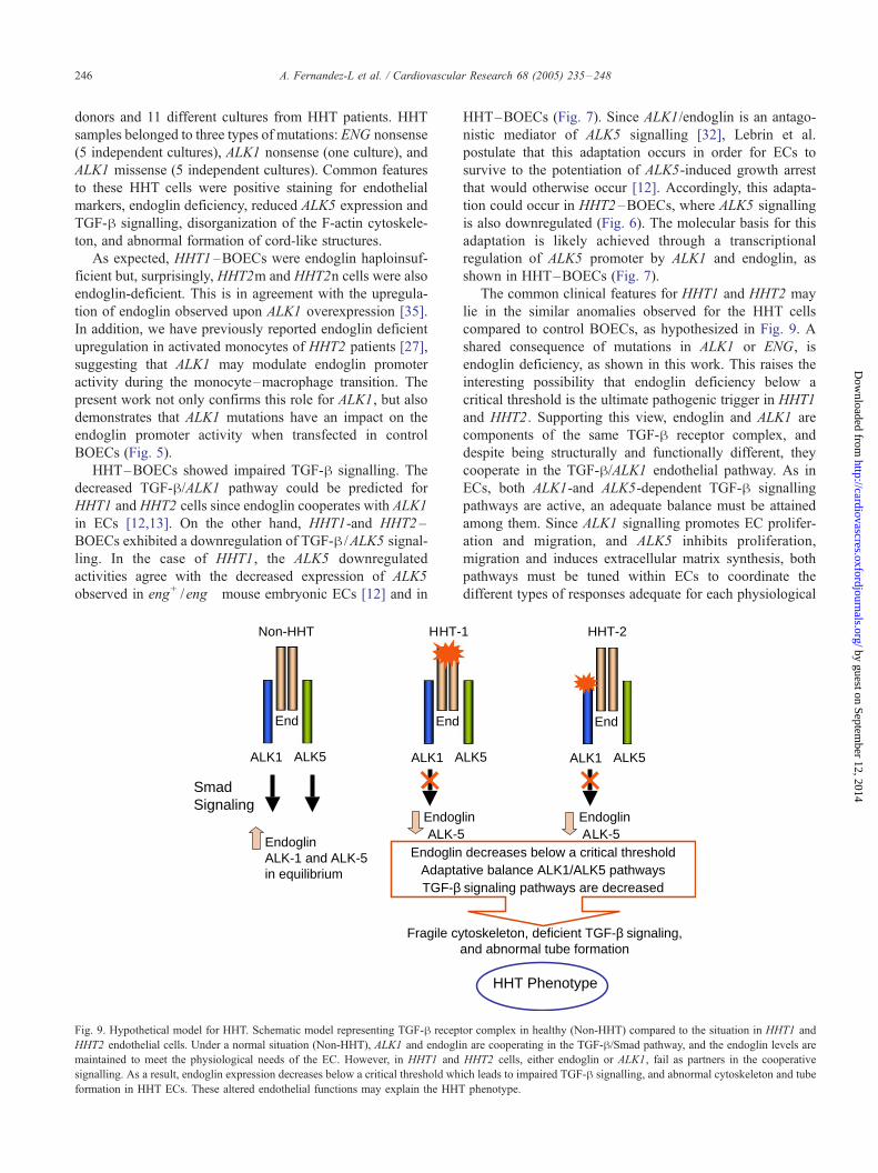
Fig. 9. Hypothetical model for HHT. Schematic model representing TGF-h recep
HHT2 endothelial cells. Under a normal situation (Non-HHT), ALK1 and endogli
maintained to meet the physiological needs of the EC. However, in HHT1 and
signalling. As a result, endoglin expression decreases below a critical threshold wh
formation in HHT ECs. These altered endothelial functions may explain the HHT
HHT–BOECs (Fig. 7). Since ALK1/endoglin is an antago-
nistic mediator of ALK5 signalling [32], Lebrin et al.
postulate that this adaptation occurs in order for ECs to
survive to the potentiation of ALK5-induced growth arrest
that would otherwise occur [12]. Accordingly, this adapta-
tion could occur in HHT2 –BOECs, where ALK5 signalling
is also downregulated (Fig. 6). The molecular basis for this
adaptation is likely achieved through a transcriptional
regulation of ALK5 promoter by ALK1 and endoglin, as
shown in HHT–BOECs (Fig. 7).
The common clinical features for HHT1 and HHT2 may
lie in the similar anomalies observed for the HHT cells
compared to control BOECs, as hypothesized in Fig. 9. A
shared consequence of mutations in ALK1 or ENG, is
endoglin deficiency, as shown in this work. This raises the
interesting possibility that endoglin deficiency below a
critical threshold is the ultimate pathogenic trigger in HHT1
and HHT2. Supporting this view, endoglin and ALK1 are
components of the same TGF-h receptor complex, and
despite being structurally and functionally different, they
cooperate in the TGF-h/ALK1 endothelial pathway. As in
ECs, both ALK1-and ALK5-dependent TGF-h signalling
pathways are active, an adequate balance must be attained
among them. Since ALK1 signalling promotes EC prolifer-
ation and migration, and ALK5 inhibits proliferation,
migration and induces extracellular matrix synthesis, both
pathways must be tuned within ECs to coordinate the
different types of responses adequate for each physiological
HHT Phenotype
toskeleton, deficient TGF-β signaling,and abnormal tube formation
decreases below a critical threshold
1 HHT-2
LK5
End
ALK5ALK1
lin EndoglinALK-5
signaling pathways are decreasedtive balance ALK1/ALK5 pathways
tor complex in healthy (Non-HHT) compared to the situation in HHT1 and
n are cooperating in the TGF-h/Smad pathway, and the endoglin levels are
HHT2 cells, either endoglin or ALK1, fail as partners in the cooperative
ich leads to impaired TGF-h signalling, and abnormal cytoskeleton and tube
phenotype.
by guest on September 12, 2014
nals.org/
A. Fernandez-L et al. / Cardiovascular Research 68 (2005) 235–248 247
http://cardiovascres.oxfordjournalsD
ownloaded from
situation. Since ALK1 pathway is decreased in HHT–
BOECs, a correcting mechanism to downregulate ALK5
levels is probably induced to avoid the ALK5-dependent
inhibition of cell proliferation. This regulation seems to be
based on direct signals from ALK1 and endoglin to the
ALK5 promoter. Further experiments in this line of
investigation may help to elucidate the complicated network
of regulatory interactions which ultimately lead to a fine
tuning among the signalling components in the TGF-hpathway. In HHT, this tuning requires a physiological
adaptation of ECs presumably reached during the differ-
entiation from precursor to mature EC.
A direct consequence of ALK1/TGF-h deficient signal-
ling in HHT–BOECs is the decreased capacity to form
cord-like structures during angiogenesis [12], which may
affect the organization of the capillary network. In addition,
the abnormal actin cytoskeleton of HHT–BOECs may be
also related to endoglin decreased levels. In fact, endoglin
cytoplasmic domain interacts with ZRP1, present at the
points of actin polymerization [34]. Accordingly, the
decrease in endoglin levels would disrupt the actin polymer-
ization sites. A disorganized cytoskeleton is prone to cell
breaking with changes in shear stress and blood pressure.
This might lead to vessel haemorrhages and eventual
disappearance of the capillary network, as reported for the
HHT vascular disorder. Finally, although our data suggest
that BOECs constitute a novel and interesting cellular model
to study the basis of HHT, it will be important to confirm
these findings in mature vessel ECs from HHT patients.
by guest on September 12, 2014
.org/
Acknowledgements
Authors are indebted to Drs. Michelle Letarte and Ursula
Cymerman for suggestions and advice on sequencing
methods, Dr. Benilde Jimenez for human fibroblasts, Car-
men Langa for technical assistance, Dr. Carmelo Morales
for data on HHT families, Ma Victoria Gomez Espana and
Marıa Jesus Borquez for blood extractions, and to all the
volunteers and HHT patients for their collaboration. Drs. R.
Hebbel and J. Nguyen are funded by the National Institutes
of Health USA (HL71269). This work was supported by
grants from Ministerio de Educacion y Ciencia (SAF2004-
01390), Fondo de Investigacion Sanitaria (PI020200) and
HHT Foundation International to CB. Africa Fernandez-L is
a predoctoral fellow of I3P Program from Ministerio de
Educacion y Ciencia.
References
[1] Marchuk DA, Lux A. Hereditary hemorrhagic telangiectasia. In:
Scriver CR, Beaudet AL, Sly WS, Valle D, editors. The metabolic and
molecular bases of inherited disease, IV, 8th ed. McGraw-Hill Medical
Publishing Division; 2001. p. 5419–31.
[2] van den Driesche S, Mummery CL, Westermann CJ. Hereditary
hemorrhagic telangiectasia: an update on transforming growth factor
beta signalling in vasculogenesis and angiogenesis. Cardiovasc Res
2003;58:20–31.
[3] McAllister KA, Grogg KM, Johnson DW, Gallione CJ, Baldwin MA,
Jackson CE, et al. Endoglin, a TGF-beta binding protein of endothelial
cells, is the gene for hereditary haemorrhagic telangiectasia type 1. Nat
Genet 1994;8:345–51.
[4] Johnson DW, Berg JN, Baldwin MA, Gallione CJ, Marondel I, Yoon
SJ, et al. Mutations in the activin receptor-like kinase 1 gene in
hereditary haemorrhagic telangiectasia type 2. Nat Genet 1996;13:
189–95.
[5] Li DY, Sorensen LK, Brooke BS, Urness LD, Davis EC, Taylor DG, et
al. Defective angiogenesis in mice lacking endoglin. Science
1999;284:1534–7.
[6] Bourdeau A, Dumont DJ, Letarte M. A murine model of hereditary
hemorrhagic telangiectasia. J Clin Invest 1999;104:1343–51.
[7] Oh SP, Seki T, Goss KA, Imamura T, Yi Y, Donahoe PK, et al. Activin
receptor like kinase I modulates transforming growth factor beta 1
signalling in the regulation of angiogenesis. Proc Natl Acad Sci
2000;97:2626–31.
[8] Arthur HM, Ure J, Smith AJ, Renforth G, Wilson DI, Torsney E, et al.
Endoglin, an ancillary TGFbeta receptor, is required for extraem-
bryonic angiogenesis and plays a key role in heart development. Dev
Biol 2000;217:42–53.
[9] Duff SE, Li C, Garland JM, Kumar S. CD105 is important for
angiogenesis: evidence and potential applications. FASEB J 2003;17:
984–92.
[10] Gougos A, Letarte M. Primary structure of endoglin, an RGD-
containing glycoprotein of human endothelial cells. J Biol Chem
1990;265:8361–4.
[11] Seki T, Yun J, Oh SP. Arterial endothelium-specific activin receptor-
like kinase 1 expression suggests its role in arterialization and vascular
remodeling. Circ Res 2003;93:682–9.
[12] Lebrin F, Goumans MJ, Jonker L, Carvalho R, Valdimarsdottir G,
Thorikay M, et al. Endoglin promotes endothelial cell proliferation
and TGF-h /ALK1 signal transduction. EMBO J 2004;23:4018–28.
[13] Blanco FJ, Santibanez JF, Guerrero-Esteo M, Langa C, Vary
CPH, Bernabeu C. Interaction and functional interplay between
endoglin and ALK1, two components of the endothelial trans-
forming growth factor-h receptor complex. J Cell Physiol 2005;
204:574–84.
[14] Piek E, Heldin CH, ten Dijke P. Specificity, diversity and regulation in
TGF-h superfamily signaling. FASEB J 1999;13:2105–24.
[15] Shi Y, Massague J. Mechanisms of TGF-h signaling from cell
membrane to the nucleus. Cell 2003;113:685–700.
[16] Goumans MJ, Valdimarsdottir G, Itoh S, Rosendahl A, Sideras P, ten
Dijke P. Balancing the activation state of the endothelium via two
distinct TGF-h type I receptors. EMBO J 2002;21:1743–53.
[17] Guerrero-Esteo M, Sanchez-Elsner T, Letamendia A, Bernabeu C.
Extracellular and cytoplasmic domains of endoglin interact with the
transforming growth factor-beta receptors I and II. J Biol Chem
2002;277:29197–209.
[18] Barbara NP, Wrana JL, Letarte M. Endoglin is an accessory protein
that interacts with the signaling receptor complex of multiple members
of the transforming growth factor-beta superfamily. J Biol Chem
199;274:584-94.
[19] Letamendia A, Lastres P, Botella LM, Raab U, Langa C, Velasco B, et
al. Role of endoglin in cellular responses to transforming growth
factor-beta. A comparative study with betaglycan. J Biol Chem
1998;273:33011–9.
[20] Li C, Hampson IN, Hampson L, Kumar P, Bernabeu C, Kumar S, et
al. CD105 antagonizes the inhibitory signaling of transforming
growth factor beta1 on human vascular endothelial cells. FASEB J
2000;14:55–64.
[21] Shovlin CL, Guttmacher AE, Buscarini E, Faughnan ME, Hyland RH,
Westermann CJ, et al. Diagnostic criteria for hereditary hemorrhagic
telangiectasia (Rendu–Osler–Weber syndrome). Am J Med Genet
2000;91:66–8.
A. Fernandez-L et al. / Cardiovascular Research 68 (2005) 235–248248
http://cD
ownloaded from
[22] Pece N, Vera S, Cymerman U, White RJ, Wrana JL, Letarte M. Mutant
endoglin in hereditary hemorrhagic telangiectasia type 1 is transiently
expressed intracellularly and is not a dominant negative. J Clin Invest
1997;100:2568–79.
[23] Abdalla SA, Pece-Barbara N, Vera S, Tapia E, Paez E, Bernabeu C, et
al. Analysis of ALK1 and endoglin in newborns from families with
hereditary hemorrhagic telangiectasia type 2. Hum Mol Genet 2000;9:
1227–37.
[24] Abdalla SA, Geisthoff UW, Bonneau D, Plauchu H, McDonald J,
Kennedy S, et al. Visceral manifestations in hereditary haemorrhagic
telangiectasia type 2. J Med Genet 2003;40:494–502.
[25] Cymerman U, Vera S, Pece-Barbara N, Bourdeau A, White RI Jr,
Dunn J, et al. Identification of hereditary hemorrhagic telangiectasia
type 1 in newborns by protein expression and mutation analysis of
endoglin. Pediatr Res 2000;47:24–35.
[26] Plauchu H, de Chadarevian JP, Bideau A, Robert JM. Age-related
clinical profile of hereditary hemorrhagic telangiectasia in an
epidemiologically recruited population. Am J Med Genet 1989;32:
291–7.
[27] Sanz-Rodriguez F, Fernandez-L A, Zarrabeitia R, Perez-Molino A,
Ramırez JR, Coto E, et al. Mutation analysis in Spanish patients with
hereditary hemorrhagic telangiectasia. Deficient endoglin upregulation
in activated monocytes. Clin Chem 2004;279:32858–68.
[28] Lin Y, Weisdorf DJ, Solovey A, Hebbel RP. Origins of circulating
endothelial cells and endothelial outgrowth from blood. J Clin Invest
2000;105:71–7.
[29] Lin Y, Chang L, Solovey A, Healey JF, Lollar P, Hebbel RP. Use of
blood outgrowth endothelial cells for gene therapy for hemophilia A.
Blood 2002;99:457–62.
[30] Blann AD, Woywodt A, Bertolini F, Bull TM, Buyon JP, Clancy RM,
et al. Circulating endothelial cells. Biomarker of vascular disease.
Thromb Haemost 2005;93:228–35.
[31] Sanchez-Elsner T, Botella LM, Velasco B, Langa C, Bernabeu C.
Endoglin expression is regulated by transcriptional cooperation
between the hypoxia and transforming growth factor-beta pathways.
J Biol Chem 2002;277:43799–808.
[32] Goumans MJ, Valdimarsdottir G, Itoh S, Lebrin F, Larsson J,
Mummery C, et al. Activin receptor-like kinase (ALK)1 is an
antagonistic mediator of lateral TGFbeta /ALK5 signaling. Mol Cell
2003;12:817–28.
[33] Botella LM, Sanchez-Elsner T, Rius C, Corbi A, Bernabeu C.
Identification of a critical Sp1 site within the endoglin promoter and
its involvement in the transforming growth factor-beta stimulation. J
Biol Chem 2001;276:34486–94.
[34] Sanz-Rodriguez F, Guerrero-Esteo M, Botella LM, Banville D, Vary
CP, Bernabeu C. Endoglin regulates cytoskeletal organization through
binding to ZRP-1, a member of the Lim family of proteins. J Biol
Chem 2004;279:32858–68.
[35] Ota T, Fujii M, Sugizaki T, Ishii M, Miyazawa K, Aburatani H, et al.
Targets of transcriptional regulation by two distinct type I receptors for
transforming growth factor-h in human umbilical vein endothelial
cells. J Cell Physiol 2002;193:299–318.
ard
by guest on September 12, 2014iovascres.oxfordjournals.org/