Neurite Outgrowth on a DNA Crosslinked Hydrogel with Tunable Stiffnesses
Upload
independentCategory
view
0download
0
Ganglioside Inhibition of Neurite Outgrowth Requires NogoReceptor FunctionIDENTIFICATION OF INTERACTION SITES AND DEVELOPMENT OF NOVEL ANTAGONISTS*□S
Received for publication, March 14, 2008 Published, JBC Papers in Press, April 14, 2008, DOI 10.1074/jbc.M802067200
Gareth Williams‡, Andrew Wood§, Emma-Jane Williams‡, Ying Gao§, Mary L. Mercado§, Alan Katz§,Diane Joseph-McCarthy§, Brian Bates§, Huai-Ping Ling§, Ann Aulabaugh§, Joe Zaccardi§, Yuhong Xie§,Menelas N. Pangalos§, Frank S. Walsh§, and Patrick Doherty‡1
From the ‡Wolfson Centre for Age-Related Diseases, King’s College London, London SE1 1UL, United Kingdom and§Neuroscience Discovery, Wyeth Research, Princeton, New Jersey 08543
Gangliosides are key players in neuronal inhibition, with anti-body-mediated clustering of gangliosides blocking neurite out-growth in cultures and axonal regeneration post injury. In thisstudy we show that the ganglioside GT1b can form a complexwith the Nogo-66 receptor NgR1. The interaction is shown byanalytical ultracentrifugation sedimentation and ismediated bythe sialic acidmoiety onGT1b,withmutations in FRGmotifs onNgR1 attenuating the interaction. One FRG motif was devel-oped into a cyclic peptide (N-AcCLQKFRGSSC-NH2) antago-nist ofGT1b, reversing theGT1b antibody inhibition of cerebel-lar granule cell neurite outgrowth. Interestingly, the peptidealso antagonizes neurite outgrowth inhibition mediated by sol-uble forms of themyelin-associated glycoprotein (MAG). Struc-ture function analysis of the peptide point to the conserved FRGtriplet being the minimal functional motif, and mutationswithin this motif inhibit NgR1 binding to both GT1b andMAG.Finally, using gene ablation, we show that the cerebellar neuronresponse to GT1b antibodies and soluble MAG is indeeddependent on NgR1 function. The results suggest that ganglio-sides inhibit neurite outgrowth by interacting with FRG motifsin theNgR1 and that this interaction can also facilitate the bind-ing of MAG to the NgR1. Furthermore, the results point to arational strategy for developing novel ganglioside antagonists.
Antibodies that bind to the gangliosideGT1b2 inhibit neuriteoutgrowth (1–3). Furthermore, passive immunization withanti-ganglioside antibodies directly inhibits axonal regenera-tion after injury in mice (4). A considerable body of evidencealso suggests that autoimmune anti-ganglioside antibodiesmight contribute to the poor prognosis of some patients withGuillain-Barre syndrome (5) and other peripheral neuropathies(6). A better understanding of themechanisms whereby gangli-
oside antibodies inhibits neurite outgrowth and the develop-ment of agents to circumvent thismight lead to novel therapeu-tic opportunities for some peripheral neuropathies.The myelin-associated glycoprotein (MAG) can inhibit neu-
rite outgrowth (7, 8). The response to soluble MAG and GT1bantibodies are not obviously different when compared side byside (1).MAG can bind directly to gangliosides including GT1b(9), and the presence of complex gangliosides in neurons isrequired for MAG function (10). This suggests that in somecircumstances MAG inhibits neurite outgrowth by binding toand activating a GT1b receptor complex in an antibody-likemanner. In support, the inhibitory response to soluble MAGand ganglioside antibodies requires activation of RhoA (Rashomolog gene family A) (1, 3). It follows that insights into howanti-ganglioside antibodies inhibit neurite outgrowth might begleaned from understanding how MAG inhibits neurite out-growth and vice versa.MAGcan bind to a receptor complex in neurons that contain
the Nogo-66 receptor NgR1 (11–14), the p75 neurotrophinreceptor (p75NTR) (15, 16), and Lingo-1 (17). The p75NTR
receptor is the key signaling component of the complex, andGT1b also appears to be associated with the complex as anti-bodies to GT1b can immunoprecipitate p75NTR from neurons(3, 18). These data suggest that GT1b could facilitate the inter-action between MAG and the NgR1, and indeed enzymes thatremove sialic acid fromcomplexgangliosides inhibit solubleMAGbinding toNgR1 andNgR2 in cells (19) and inhibitMAG function(20). Recent studies using independently generated lines of micethat lack the NgR1 have clearly shown that the ability of solubleMAG to induce growth cone collapse from dorsal root ganglionneurons is dependent on this receptor (21, 22). However, the laterstudy also provided conclusive evidence that the NgR1 is notrequired for the function of substrate boundMAG.In the present study we have used analytical ultracentrifuga-
tion sedimentation to demonstrate that GT1b can form higherorder complexeswith theNgR1. The binding required the pres-ence of sialic acid on the ganglioside and was inhibited whenany one of three independent FRG motifs in the NgR1 wasmutated. One NgR1 sequence that contains an FRG motif(LQKFRGSS) lends itself well to the design of a cyclic peptidemimetic (N-Ac-CLQKFRGSSC-NH2). This mimetic peptideprevented GT1b antibodies from inhibiting neurite outgrowth.These data suggest that the inhibitory activity of anti-GT1b
* The costs of publication of this article were defrayed in part by the paymentof page charges. This article must therefore be hereby marked “advertise-ment” in accordance with 18 U.S.C. Section 1734 solely to indicate this fact.
□S The on-line version of this article (available at http://www.jbc.org) containssupplemental Fig. 1.
1 To whom correspondence should be addressed: The Wolfson Centre forAge-Related Diseases King’s College London, Wolfson Wing HodgkinBldg., London SE1 1UL, UK. Tel.: 0207-848-6811; Fax: 0207-848-6816;E-mail: [email protected].
2 The abbreviations used are: GT1b, trisialoganglioside1b; NgR, nogo recep-tor; MAG, myelin-associated glycoprotein; GM1, monosialoganglioside 1;CHO, Chinese hamster ovary; AP, alkaline phosphatase.
THE JOURNAL OF BIOLOGICAL CHEMISTRY VOL. 283, NO. 24, pp. 16641–16652, June 13, 2008© 2008 by The American Society for Biochemistry and Molecular Biology, Inc. Printed in the U.S.A.
JUNE 13, 2008 • VOLUME 283 • NUMBER 24 JOURNAL OF BIOLOGICAL CHEMISTRY 16641
by guest on August 26, 2016
http://ww
w.jbc.org/
Dow
nloaded from
by guest on August 26, 2016
http://ww
w.jbc.org/
Dow
nloaded from
by guest on August 26, 2016
http://ww
w.jbc.org/
Dow
nloaded from
by guest on August 26, 2016
http://ww
w.jbc.org/
Dow
nloaded from

antibodies is dependent on NgR1 function. In support we showthat GT1b antibodies do not inhibit neurite outgrowth fromneurons isolated frommice that have theNgR1 gene geneticallyablated from the germline.The same NgR1 peptide that inhibited the GT1b antibody
response also antagonized the response stimulated by solubleMAG, with alanine scanning identifying the FRG sequence as thefunctional motif within the peptide. These data suggest that insome circumstances soluble MAG can inhibit neurite outgrowththrough the ganglioside/NgR1 pathway. In support, mutations ofthe FRGmotif that inhibit GT1b binding to the NgR1 also inhibitMAGbinding to the receptor, and the inhibitory activity of solubleMAG was significant attenuated in neurons that do not expresstheNgR1.However, it is also clear that substrate-boundMAGcaninhibit neurite outgrowth in the absence of NgR1 function (22)and, in accord the NgR1 derived inhibitory peptides identified inthis study, are not expected to, and indeeddonot, inhibit the func-tion of substrate-boundMAG.
EXPERIMENTAL PROCEDURES
Neurite Outgrowth Assays—Cerebellar granule neurons iso-lated from post-natal day 2–3 rats or from day 3–5 mice werecultured over monolayers of 3T3 cells essentially as previouslydescribed (23). Monolayers were established by seeding�80,000 3T3 cells into individual chambers of an 8-chambertissue culture slide coatedwith poly-L-lysine and fibronectin. Ingeneral, co-cultures were established by removing the mediafrom the monolayers and seeding �6000 dissociated cerebellarneurons into each well in SATOmedium (Dulbecco’s modifiedEagle’s medium supplemented with 0.062 mg/liter progester-one, 16.1 mg/liter putrescine, 0.4 mg/liter thyroxine, 0.039mg/liter selenium, 0.337 mg/liter triiodothyronine, 10 mg/literinsulin (bovine pancreas), 100 mg/liter transferrin (human))supplemented with 2% fetal calf serum. However, in someexperiments the co-cultures were maintained in neurobasalmedium � B27 � 1% L-glutamate � 1% penicillin/streptomy-cin � 25 mM KCl. Monolayers were established for 24 h beforethe addition of the neurons, and the cultures weremaintained for�23–27 h before fixation with 4% paraformaldehyde. In general,the neurons were stained with a GAP-43 antibody, and the meanlength of the longest neurite per cell wasmeasured for�120–150neurons, again as previously described (23). However, in someexperiments neurons were labeled using TUJ1 (anti-�III tubulin)followed by anti-mouse IgG-Alexa488, and nuclei were labeledwith Hoechst. Mean total neurite length was calculated using theNeuronal Profiling bioapplicationon aCellomicsArrayScan. Sim-ilar results were obtained using bothmethods.For neurite outgrowth on substrate-bound MAG, 96-well
plates were coated with a thin layer of nitrocellulose beforeincubating with 1 �g/ml MAG(d1–5) at 4 °C overnight. Wellswere subsequently coated with 17 �g/ml of poly-D-lysine(Sigma) followed by incubation in Dulbecco’s modified Eagle’smedium containing 10% fetal calf serum. Rat cerebellar granuleneurons were dissociated and seeded at a density of 1 � 104cells perwell. Cellswere cultured for 18–20hbefore being fixedwith 4% paraformaldehyde and stained with a neuronal specificanti-�III-tubulin antibody. The average of total neurite lengthsfrom each neuron was measured automatically by the Meta-
Xpress Neurite Outgrowth module (Molecular Devices) fromat least 200 neurons per well, in triplicate wells per experiment.Results were repeated independently more than three times.Immunoprecipitations and Western Blots—Chinese hamster
ovary (CHO) K1 cells (100 mm dishes) were transfected withp75NTR and various mutants of the NgR1. The cells were har-vested after 24 h and lysed in 1ml of radioimmune precipitationassay buffer (Sigma) supplemented with Complete proteaseinhibitor mixture (Roche Applied Science). After centrifuga-tion at 14,000 � g for 15 min, the supernatants were collected,and a protein assay (Bio-Rad) was performed. Protein lysates(0.5 mg) were preincubated with protein G-Sepharose beads(GEHealthcare) at 4 °C for 1 h, then incubatedwith 2�g of goatanti human NgR antibody (R&D Systems) plus proteinG-Sepharose at 4 °C overnight. The beads were washed threetimes with radioimmune precipitation assay buffer and boiledin Laemmli sample buffer (Bio-Rad). Supernatants were sub-jected to 4–12% NuPAGE (Invitrogen), transferred onto nitro-cellulose membrane (Bio-Rad), and probed with antibodies totheNgR andp75NTR (Promega).Western blot imageswere ana-lyzed on a Storm gel imaging system using ImageQuant soft-ware (GE Healthcare).Whole brains from adult 129/lex, NgR1 knock-out, and NgR2
knock-out mice were homogenized and sonicated in radioim-mune precipitation assay buffer (Sigma). Supernatants were col-lected after centrifugation, and a protein assay (Bio-Rad) was pre-formed. Protein lysates were subjected to 4–12% NuPAGE(Invitrogen), transferredontonitrocellulosemembrane (Bio-Rad),and probed with antibodies to NgR1 or NgR2 (R&D Systems).Western blot images (see supplemental Fig. 1b) were scanned andanalyzed by Odyssey infrared imaging system (Li-Cor).Construction of NgR1 Mutants—Human NgR1 point
mutants were constructed using the QuikChange XL site-di-rected mutagenesis kit (Stratagene) following the manufactur-er’s recommended protocol. Thewild type humanNgR1 cDNA(IMAGE:2121045 3) in a mammalian expression vector wasused as a template to construct all the described mutants.Receptor Binding Assay—COS-7 cells were co-transfected
with either wild type or mutant NgR1 constructs along with acytomegalovirus-�-gangliosidase plasmid (pCMVb, BD Bio-sciences) as a transfection control. Transfectionwas performedin six-well plates using Lipofectamine 2000 (Invitrogen) follow-ing the manufacturer’s protocol. The next day cells weretrypsinized and seeded at 30,000 cells per well in poly-lysine-coated 96-well plates (BD Biosciences). At that time, two sisterplates were established, one of which was used in the bindingassay, and the other was used to correct for transfection effi-ciency by measuring �-galactosidase activity (see below).Remaining cells were separately plated and assayed for surfaceexpression of the NgR1 proteins by immunocytochemistry andfor total NgR1 protein levels by Western blotting. All mutantproteins were expressed on the cell surface and produced incomparable amounts to the wild type protein (data not shown).The next day wells were rinsed once with Hanks’ balanced saltsolution containing 0.5 mg/ml bovine serum albumin, 0.1%NaN3, 20 mMHEPES, pH 7.0, at room temperature followed byincubation with 100 �l of fusion protein MAG-alkaline phos-phatase (AP) diluted to a final concentration of 10 �g/ml in 20
Overcoming Ganglioside and MAG Inhibition with FRG Peptides
16642 JOURNAL OF BIOLOGICAL CHEMISTRY VOLUME 283 • NUMBER 24 • JUNE 13, 2008
by guest on August 26, 2016
http://ww
w.jbc.org/
Dow
nloaded from

mM HEPES, pH 7.0, for 90 min. After this, wells were washed 6times with gentle shaking in 20 mM Hepes, pH 7.0, at roomtemperature, 5 min each wash. Cells were then fixed with ace-tone-formaldehyde (60-3%, in 20mMHEPES, pH 7.0) for 15 s atroom temperature then washed 3 times for 5 min each withHanks’ balanced salt solution. Binding of AP-tagged ligandswas then measured using the Great EscAPe SEAP kit (BD Bio-sciences) following themanufacturer’s recommendedprotocol.Briefly, after aspirating Hanks’ balanced salt solution, a 60-�ldilution buffer was added to each well, and the plates weresealed and then incubated at 65 °C for 90 min. Plates werecooled on ice, and then 60 �l of assay buffer was added per welland incubated at room temperature for 5min. Sixty microlitersof diluted chemiluminescent alkaline phosphatase substratewas then added per well, incubated 10 min at room tempera-ture, and then read on a LMAXII luminometer (MolecularDevices). Absolute binding numbers were corrected by sub-tracting average binding values obtained from mock-trans-fected controls. Binding was further corrected for sample-to-sample variations in transfection efficiency by normalizing to�-galactosidase activity.�-Galactosidase activity wasmeasuredusing the luminescent �-galactosidase detection kit II (BD Bio-sciences) following themanufacturer’s recommendedprotocol.Three independent binding experiments were conducted withat least six replicates per experiment. Background was sub-tracted, and �-galactosidase-corrected binding values wereexpressed relative to the wild type receptor.Preparation of AP-tagged Fusion Proteins—A fusion protein
(Nogo66-AP) containing an N-terminal human placental APand a C-terminal Nogo66 domain was constructed by ligatingnucleotides encoding amino acids 1055–1120 of humanNogoA(reticulon-4, NP_065393) to sequences encoding amino acids23–511 of AP (NM_001632). This fusion was further modifiedby changing amino acid 47 of the Nogo66 sequence from cys-teine to valine and introducing six consecutive histidine resi-dues at the C terminus. The coding sequence was inserted intoa mammalian expression vector and transiently transfectedinto HEK293GT cells (Invitrogen) using Lipofectamine 2000(Invitrogen). The next day serum-free medium (Free Style 293,Invitrogen) was added, and cells were incubated a further 48 hbefore collection of crude conditioned medium. Nogo66-APconcentration was determined bymeasuring alkaline phospha-tase activity and by Western blotting for alkaline phosphatase.A stableCHOcell line expressing a fusion protein containing anN-terminal human myelin-associated glycoprotein (humanMAG; NM_002361; amino acids 1–516) and a C-terminal APdomain (amino acids 23–511) bearing six C-terminal histidineresidues was created (referred to asMAG-AP). Cells were incu-bated in serum-free medium for 48 h; the conditioned mediumwas collected, and the fusion protein was purified usingTALON cobalt affinity chromatography (Clontech) followingthe manufacturer’s protocol. MAG-AP concentration wasdetermined by measuring alkaline phosphatase activity and byWestern blotting for alkaline phosphatase and MAG.Analytical Ultracentrifugation—Sedimentation velocity exper-
iments were performed on a Beckman XLI/XLA analytical ultra-centrifuge.NgR1(310)-fc (0.21 or 0.38�M final) was added to gan-glioside at increasing ganglioside concentrations from 0 to 48�M.
Mutant protein used in the sedimentation velocity experimentscorresponded to the column fraction of greatest purity based onSDS gel analysis. Wild type or mutant NgR(310)-Fc was addedto Tris-buffered saline (TBS) buffer or TBS buffer containingGT1b to a final concentration of 16–30 �g/ml protein and 0 or22 �M GT1b in a microcentrifuge tube. The solution, 400 �l,was loaded into 2-channel (1.2-cm path length) carbon-Eponcenterpieces in anAn-50-Ti rotor. Scanswere recorded at 20 °Cwith a rotor speed of 35,000 rpm, and the signal was detected at230 nm with a spacing of 0.006 in the continuous mode. Sedi-mentation profiles were analyzed by the program Sedfit (24) toobtain the sedimentation coefficient distributions. The solventdensity (1.006) and partial specific volume (0.72) were calcu-lated using the program Sednterp (25).Neuraminidase Treatment—CHO parental cells and NgR1
stable cells were seeded at 30,000 cells per well in 96-well platesthe night before the assay. Various concentrations of Vibriocholerae neuraminidase (Roche Applied Science) in growthmedium (Dulbecco’s modified eagle medium containing 10%fetal bovine serum) were incubated with cells for 1 h at 37 °C.Medium was replaced with affinity-purified MAG-AP orNogo66-AP in Hanks’ balanced salt solution supplementedwith 1% fetal bovine serum and 20 nM HEPES and incubated atroom temperature for 90 min. Cells were then washed fourtimes with supplemented Hanks’ balanced salt solution. Alto-Phos (0.6 mg/ml) (Promega) was added to indicate of boundligands. After a 30-min incubation at room temperature, theplates were read at emission/excitation wavelength of 400nm/505 nm by FlexStationII384 (Molecular Devices).Reagents—Synthetic peptides were all obtained from a com-
mercial supplier (Multiple Peptide Systems). All peptides werepurified to the highest grade by reverse-phase high perform-ance liquid chromatography and obtained at the highest level ofpurity (�97%). With all peptides there was no indication ofhigher molecular weight species. Where peptide sequences areunderlined, this denotes a peptide that has been cyclized via adisulfide bond between the given cysteine residues. All peptideswere acetylated and amide-blocked. Recombinant MAG-Fcchimera was obtained from R&D Systems and used at a finalconcentration of 5–25 �g/ml. The monoclonal antibody toGT1b (clone GMR5) was obtained from Seikagaku Americaand was used at a final concentration of 20 �g/ml. All reagentswere diluted into the co-culture media and in general added tothe cultures just before plating of the neurons. GT1b and GM1were a kind gift of Dr. Gino Toffano. Asialo-GM1was obtainedfrom Sigma. The recombinant NgR1(310)-Fc chimera and theextracellular portion of MAG(d1–5) were expressed and puri-fied in-house. Pharmacological reagents were obtained fromCalbiochem and/or Sigma.Generation and Characterization of NgR1 and NgR2 Knock-
out Mice—Targeting vectors (see supplemental Fig. 1a) wereintroduced into mouse embryonic stem cells of the 129SvBrdbackground via electroporation. Homologously targeted inte-grants were selected in G418 (0.4 g/liter) and identified bySouthern blotting using probes external to the targeting vectoron both the 5� and 3� sides. Retention of the 5�-most loxP site inthe targeted cells was determined by PCR of genomic DNAusing the following primers: for NgR1 allele, 5�-GGTCTAGG-
Overcoming Ganglioside and MAG Inhibition with FRG Peptides
JUNE 13, 2008 • VOLUME 283 • NUMBER 24 JOURNAL OF BIOLOGICAL CHEMISTRY 16643
by guest on August 26, 2016
http://ww
w.jbc.org/
Dow
nloaded from

GATGCATCTCAG and 5�-ACATCTGAAGGCCTTCTGG;for the NgR2 allele, 5�-GTTGGTGGGGTTCTTGTCTCAGGand 5�-GGCCTGGCCCCCTCCCTTCAC. Embryonic stemcells were used to establishmouse lines and animals bearing theNgR1f and NgR2f alleles were crossed to Prm-cre transgenicmice (26). The Prm-cre transgene directs expression of crerecombinase in the male germline. Therefore, males harboringthe transgene along with theNgR1f orNgR2f allele transmit thenon-functional NgR1d or NgR2d alleles, respectively, to theiroffspring. Cre-mediated recombination of the floxed (f) allelesto the deleted (d) alleles was detected by PCR of genomic DNAusing the following primers: for NgR1, 5�-GGTCTAGGGATG-CATCTCAG and 5�-GTGGTCTGTGTGGCTCCTGC; forNgR2, 5�-GTTGGTGGGGTTCTTGTCTCAGG and 5�-CCCC-CCTGCCCCAGCTACGC. All alleles were maintained on the129SvBrd background.
RESULTS
BindingMotifs on theNgR1—There are two published crystalstructures of the NgR1, Protein Data Bank accessions 1OZN(27) and 1P8T (28), but currently no ligand-receptor complexstructure has been solved. Detailed mutagenesis studies haverecentlymapped the residues critical for the binding ofMAG tothe receptor (29), and these are illustrated in Fig. 1a. Smallligand binding sites show up as cavities and can be revealed bythe clustering of a small probe under the influence of a van derWaals potential. In Fig. 1bwe show the two lowest energy clus-ters for a probe with van der Waals radius of 3.5 Å. The poten-tial binding pockets lie on the convex side of the protein, andinterestingly, both neighbor FRG triplet motifs that can befound in the other NgRs (discussed in detail below). Sialic acid
residues on gangliosides and possi-bly other glycoconjugates binddirectly to an FRG motif in MAGitself (30). These observations haveled us to develop the hypothesis thatgangliosides can interact with FRGmotifs in the NgR and that thisinteraction might facilitate MAGbinding to the receptor.Binding of MAG, but Not Nogo66,
to NgR1 Is Partially Sensitive toNeuraminidase—In neurons solu-ble MAG binds to the NgR1 andNgR2 in a sialic acid-dependentmanner (19). In the present studywe confirmed the neuraminidasesensitivity of MAG binding to theNgR1 expressed in CHO cells. Thedata show that over a wide range ofconcentrations (2.5–20 �g/ml) thespecific binding of the MAG-APfusion protein to NgR1-expressingcells is partially inhibited (55%) bytreating the CHO cells with neura-minidase. The effect was dependentupon the concentration of neura-minidase, and even at the highest
concentration Nogo66-AP binding remained completely unaf-fected (Fig. 2). These data suggest that MAG binding to theNgR1 is dependent at least in part on sialic acid binding to thereceptor.Effects of Loop 2 and Additional FRG Mutations on GT1b
Binding to the NgR1—GT1b is a sialic acid-containing ganglio-side that has previously been reported to be a key component ofthe MAG receptor complex (18, 31). We have tested whetherGT1b can bind directly to the ectodomain of the NgR1 usinganalytical ultracentrifugation. In the absence of GT1b, thedimeric NgR1(310)-Fc migrates with a sedimentation coeffi-cient of �6.5 S (Fig. 3). In the presence of low �M concentra-tions of GT1b, the 6.5 S species decreases, and additional peakswith higher sedimentation coefficients appear in a dose-dependent manner (Fig. 3a). In this assay, GM1 can also inter-act with NgR1 (Fig. 3b), which suggests that the interactionmight be dependent on sialic acid. In support, no change insedimentation coefficient of the NgR1(310)-Fc is observed inthe presence of asialo-GM1. No effect was observed upon theaddition of 22 �M GT1b to anti-hNgR AF 1208 antibody fromR&D, which provides additional evidence that the interactionof GT1b with NgR1 is specific (not shown).We next determined if the binding of GT1b to the NgR1 was
sensitive to mutation of the FRGmotifs. Importantly, based onthe relative ratios of the �6.7- and �11-S peaks, it can be esti-mated that mutation of the arginine 279 to an aspartic acidreduced binding to�56% that of wild typeNgR, suggesting thissite plays a role inmediating the interaction (Fig. 3d). Mutationof arginine 151 (Fig. 3e) or arginine 199 (Fig. 3f) also reducedGT1b binding to 49 and 33% that of wild type, respectively.These data suggest that all three FRG sites might be important
FIGURE 1. Identification of putative functional motifs on the NgR1. The protein binding site of the NgR1 isshown in a. The concave face of NgR1 is shown in space-filled mode, and the residues critical for protein binding(MAG, OMGP, Nogo66) are shown in red. Two putative small ligand binding cavities are shown on the convexface of NgR1 in b. Clusters of energy minima for a simple 3.55-Å diameter van der Waals probe define two smallpockets (in proximity to the red and yellow spheres in b). The three occurrences of the FRG motif are alsohighlighted in (b) (red, green, and yellow patches), with the 198FRG200 and 278FRG280 peptides seen to be neigh-boring the predicted small molecule binding pockets. In c, a ribbon diagram of the NgR is shown to highlightthose parts of the receptor (with corresponding sequences) that are obviously amenable to a cyclic peptidemimetic approach for determining functional significance of the highlighted loops. The figures were gener-ated with DS Viewer from Accelrys.
Overcoming Ganglioside and MAG Inhibition with FRG Peptides
16644 JOURNAL OF BIOLOGICAL CHEMISTRY VOLUME 283 • NUMBER 24 • JUNE 13, 2008
by guest on August 26, 2016
http://ww
w.jbc.org/
Dow
nloaded from

in facilitating GT1b binding to NgR1. In all three instances thesedimentation coefficient curves can be seen to be qualitativelydifferent to the curve seen with the wild type NgR1 construct.Whereas a higher migrating species (�11 S) becomes the dom-inant species in the presence of GT1b with the wild type recep-tor, lower migrating species remain dominant with all threemutated receptors (Fig. 3, d–f).A FRG-containing Mimetic of an NgR1 Loop Inhibits the
Function of a GT1b Antibody—In general antibodies that bindto neurons do not inhibit neurite outgrowth (including anti-bodies to neural cell adhesion molecule, N-cadherin, L1, andthe fibroblast growth factor receptor, and THY-1, e.g. see Refs.32 and 33). However, antibodies that cluster GT1b inhibit neu-rite outgrowth,most likely by clusteringGT1bwith consequentclustering and activation of an inhibitory molecule complex
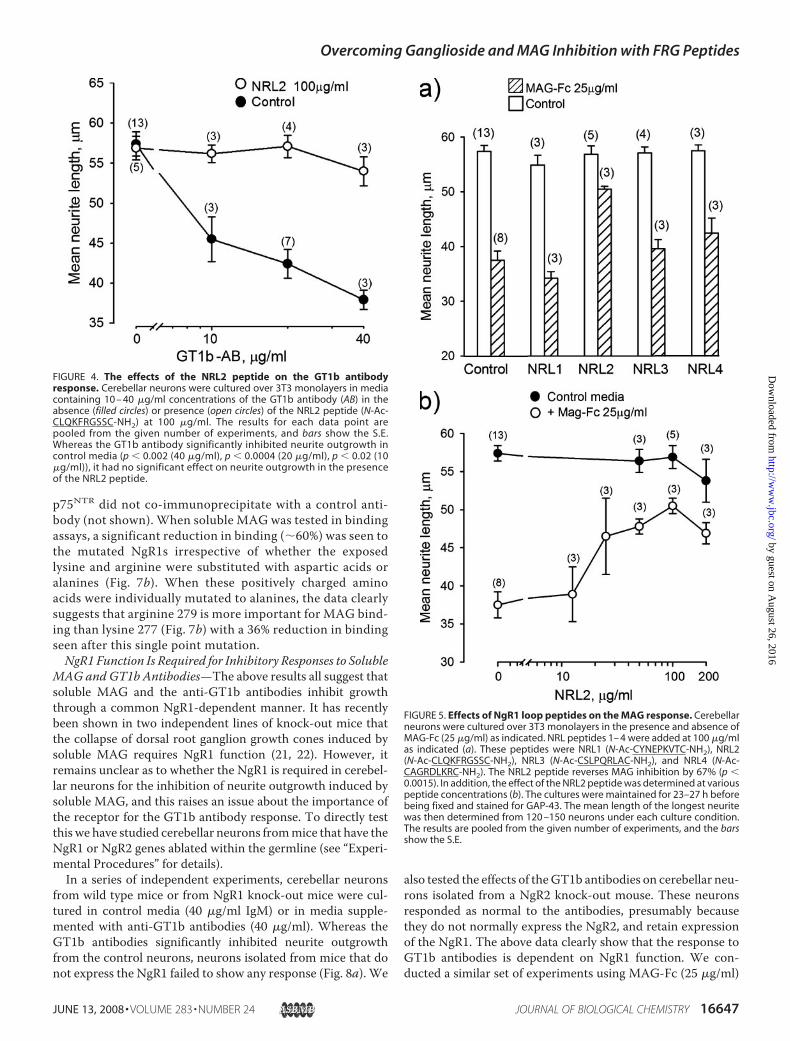
(1–3).One of the FRGmotifs implicated inGT1b binding to theNgR1 is part of an exposed loop that lends itself well to thedesign of a cyclic peptide mimetic (see Fig. 1c). In the presentstudy post-natal day 2/3 cerebellar neurons were cultured overmonolayers of 3T3 fibroblasts for �23 h in the presence andabsence of a GT1b antibody. As previously reported, the anti-body inhibits neurite outgrowth in a dose-dependent mannerwith a robust inhibition seen at 40 �g/ml (Fig. 4). When theantibody was added in the presence of the 100 �g/ml cyclicpeptide (N-Ac-CLQKFRGSSC-NH2) that mimicked the FRGmotif-containing loop (theNRL2 peptide), it failed to inhibit neu-rite outgrowth when tested at up to 40 �g/ml (Fig. 4). By showingthat a NgR1-derived peptide can inhibit the GT1b antibodyresponse, the data further substantiate the hypothesis that theGT1b antibody response might rely on GT1b binding to the FRGmotifs in the NgR1 (see also effect of NgR1 ablation; see Fig. 8).Effects of the NRL2 Peptide on MAG Inhibition of Neurite
Outgrowth—Awide range of Fc chimeras that bind to neuronsdo not inhibit neurite outgrowth (32, 34, 35). In contrast, asoluble MAG-Fc chimera inhibits neurite outgrowth in a man-ner that can depend upon both ganglioside and NgR function(see the Introduction). In the present study theMAG-Fc inhib-ited neurite outgrowth from post-natal day 2/3 cerebellar neu-rons in a dose-dependentmanner (not shown)with robust inhi-bition seen at 25 �g/ml (Fig. 5a). The NRL2 peptide again hadno effect on basal neurite outgrowth, but it was striking that theMAG-Fc fails to substantially inhibit neurite outgrowth whenthis peptide is present in the growth media (Fig. 5a). As a con-trol we tested cyclic versions of the three other exposed NgRloops (see Fig. 1c for details) for their effects on neurite out-growth. These peptides were coded NRL1 (N-Ac-CYNEP-KVTC-NH2), NRL3 (N-Ac-CSLPQRLAC-NH2), and NRL4(N-Ac-CAGRDLKRC-NH2). When tested at 100 �g/ml,these peptides had no effect on basal neurite outgrowth or onthe suppressed neurite outgrowth seen in the presence of theMAG-Fc. We next looked at the dose-response curve for theNRL2 peptide. It can be seen to have no significant effect onneurite outgrowth in control media when tested at up to 200�g/ml. In contrast, the peptide promotes neurite outgrowthin a dose-dependent manner in the presence of the MAG-Fcwith the response reaching a plateau at around 50 �g/ml(�45 �M) (Fig. 5b).Identification of Key Functional Amino Acids in the NRL2
Sequence—Structural analysis of the NgR1 shows that themostconspicuous amino acids within the loop corresponding to theNRL2 peptide are the positively charged lysine and arginine;both are highly solvent-exposed, with their side chains clearlyavailable for binding (not shown). Of the surrounding aminoacids, the phenylalanine is buried in the structure but mightplay a role in stabilizing the local region. The glycine and serineare partially solvent-exposed but look less likely to mediate abinding interaction. Based on this analysis, we designed twosmall peptides that both have the exposed lysine and argininewithin them. These were the NRL2a (N-Ac-CKFRGSC-NH2)andNRL2b (N-Ac-CQKFRGC-NH2) peptides, and it should benoted that they contain a common four-amino acid motif(KFRG). Both peptides had no effect on neurite outgrowth incontrol media (not shown), with their ability to promote
FIGURE 2. Effect of neuraminidase treatment on MAG-AP and Nogo66-APbinding to NgR1-expressing cells. Parental CHO cells or CHO cells stablyexpressing NgR1 were treated with V. cholerae neuraminidase at various con-centrations (5–100 milliunits/ml) for 1 h. Binding of MAG-AP or Nogo66-AP tocells was performed after the treatment. The top panel shows that neuramin-idase treatment (20 milliunits/ml) (dashed line) decreased MAG-AP binding toNgR1 stable cells (circle) (p � 0.008 for 20 �g/ml MAG-AP), whereas littlebinding was detected for parental CHO cells (diamond). The bottom panelshows relative binding of MAG-AP (10 �g/ml) (diamond) or Nogo66-AP (circle)to NgR1-cells after neuraminidase treatments. Only MAG-AP, but notNogo66-AP binding to NgR1, was affected by neuraminidase treatment. RFU,relative fluorescence units.
Overcoming Ganglioside and MAG Inhibition with FRG Peptides
JUNE 13, 2008 • VOLUME 283 • NUMBER 24 JOURNAL OF BIOLOGICAL CHEMISTRY 16645
by guest on August 26, 2016
http://ww
w.jbc.org/
Dow
nloaded from

growth in the presence of theMAG-Fc shown in Fig. 6a.Withinthe inhibitory environment, both peptides promote neuriteoutgrowthwith significant effects seen at 25�g/ml (30�M) andmaximal effects seen at 50 �g/ml (60 �M). At this higher con-centration the inhibitory activity of theMAG-Fcwas effectivelyabolished. This suggests that the functional activity within theNRL2 peptide sequence resides within the KFRG motif. Toidentify key amino acids within this short region, we designedfour peptides (A1–A4) with individual alanine substitutionswithin the KFRG sequence of the NRL2b peptide. When testedat 100 �g/ml, peptides with alanine substitutions in positions 1and 2 were as effective as the parental peptide in inhibiting theMAG response (Fig. 6b). However, when tested over a range ofconcentrations, substitution at position 1 had no obvious effecton the efficacy of the peptide, whereas substitution at position 2is seen to reduce efficacy by a factor of �2 (Fig. 6, c and d). Incontrast, substitutions at position 3 or 4 rendered the peptidesineffective at inhibiting the MAG response when tested at 100�g/ml (Fig. 6b). Also, a linear version of theQKFRGpeptide didnot inhibit the MAG response (LNRL2b, Fig. 6b). These data
demonstrate that to be functional,the QKFRG motif needs to be con-strained by a disulfide bond and thatsingle mutations to any amino acidwithin the FRG motif compromisesactivity of the peptide.To determine whether a rela-
tively metabolically stable peptidewould retain biological activity, wecyclized the NgR1 sequence via astable peptide bond (homodeticcyclization), and we replaced theamino acids by their chiral partners.Specifically, we replaced the L-typeamino acids of the original peptideby non-native D-type amino acids.The peptide sequence is reversed toensure that the side chain orienta-tions are preserved. Such peptidesare referred to as retro-inverso pep-tides. Explicitly, the sequence of thehomodetic retro-inverso peptide(hriNRL2) is c[sGrfkq], where c[ ]refers to homodetic cyclization, andthe lowercase letters refer to D-typeamino acids. Note that glycine hasno chirality because it has no sidechain. When tested in the MAG-Fcassay, this peptide can be seen toretain full efficacy in inhibiting theMAG response (Fig. 6e).Neuraminidase inhibits the func-
tion of soluble but not substrate-bound MAG (20, 36). We interpretthis as suggesting that solubleMAGrequires a sialic acid containing co-receptor formaximal efficacy. Inter-estingly, we have been unable to
inhibit the function of substrate-bound MAG with any of theNRL2 peptides, and this is shown for the hriNRL2 peptide inFig. 6f. In this example the hriNRL2 peptide has no significanteffect on neurite outgrowth when tested at up to 200 �g/ml onthe suppressed growth that is seen on the MAG substrate. TheNRL2 peptides do not promote growth over substrate-boundmyelin (data not shown), confirming that they do not have non-specific effects on neurite outgrowth.Effects of Loop 2 Mutations on Ligand Binding to the NgR1—
The above data suggest that the 277KFRG280 motif in loop 2 inthe NgR1 plays an important role in the context of solublebut not substrate-bound MAG function. Given that thelysine 277 and arginine 279 are positively charged and highlysolvent-exposed, the effects of mutating both residues tonegatively charged aspartic acids or neutral alanines wasdetermined. In both instances the mutations had no obviouseffect on the level of expression of the NgR1 (Fig. 7a), andbased on co-immunoprecipitation, a normal interactionbetween the mutated NgR1 constructs and p75NTR, presum-ably in the cell membrane, was apparent (Fig. 7a). The
FIGURE 3. GT1b interacts with the NgR1 in an FRG-dependent manner. Sedimentation coefficient distribu-tion, c (S), plots of NgR(310)-Fc as a function of increasing GT1b (a), GM1 (b), and asialo-GM1 (c) is shown. Theeffects of GT1b (22 �M) on sedimentation of NgR1 constructs containing single point mutations was alsodetermined and shown for mutants R279E (d), R151E (e), and R199E (f).
Overcoming Ganglioside and MAG Inhibition with FRG Peptides
16646 JOURNAL OF BIOLOGICAL CHEMISTRY VOLUME 283 • NUMBER 24 • JUNE 13, 2008
by guest on August 26, 2016
http://ww
w.jbc.org/
Dow
nloaded from
p75NTR did not co-immunoprecipitate with a control anti-body (not shown). When soluble MAG was tested in bindingassays, a significant reduction in binding (�60%) was seen tothe mutated NgR1s irrespective of whether the exposedlysine and arginine were substituted with aspartic acids oralanines (Fig. 7b). When these positively charged aminoacids were individually mutated to alanines, the data clearlysuggests that arginine 279 is more important for MAG bind-ing than lysine 277 (Fig. 7b) with a 36% reduction in bindingseen after this single point mutation.NgR1 Function Is Required for Inhibitory Responses to Soluble
MAG and GT1b Antibodies—The above results all suggest thatsoluble MAG and the anti-GT1b antibodies inhibit growththrough a common NgR1-dependent manner. It has recentlybeen shown in two independent lines of knock-out mice thatthe collapse of dorsal root ganglion growth cones induced bysoluble MAG requires NgR1 function (21, 22). However, itremains unclear as to whether the NgR1 is required in cerebel-lar neurons for the inhibition of neurite outgrowth induced bysoluble MAG, and this raises an issue about the importance ofthe receptor for the GT1b antibody response. To directly testthis we have studied cerebellar neurons frommice that have theNgR1 or NgR2 genes ablated within the germline (see “Experi-mental Procedures” for details).In a series of independent experiments, cerebellar neurons
from wild type mice or from NgR1 knock-out mice were cul-tured in control media (40 �g/ml IgM) or in media supple-mented with anti-GT1b antibodies (40 �g/ml). Whereas theGT1b antibodies significantly inhibited neurite outgrowthfrom the control neurons, neurons isolated from mice that donot express the NgR1 failed to show any response (Fig. 8a). We
also tested the effects of theGT1b antibodies on cerebellar neu-rons isolated from a NgR2 knock-out mouse. These neuronsresponded as normal to the antibodies, presumably becausethey do not normally express the NgR2, and retain expressionof the NgR1. The above data clearly show that the response toGT1b antibodies is dependent on NgR1 function. We con-ducted a similar set of experiments using MAG-Fc (25 �g/ml)
FIGURE 4. The effects of the NRL2 peptide on the GT1b antibodyresponse. Cerebellar neurons were cultured over 3T3 monolayers in mediacontaining 10 – 40 �g/ml concentrations of the GT1b antibody (AB) in theabsence (filled circles) or presence (open circles) of the NRL2 peptide (N-Ac-CLQKFRGSSC-NH2) at 100 �g/ml. The results for each data point arepooled from the given number of experiments, and bars show the S.E.Whereas the GT1b antibody significantly inhibited neurite outgrowth incontrol media (p � 0.002 (40 �g/ml), p � 0.0004 (20 �g/ml), p � 0.02 (10�g/ml)), it had no significant effect on neurite outgrowth in the presenceof the NRL2 peptide.
FIGURE 5. Effects of NgR1 loop peptides on the MAG response. Cerebellarneurons were cultured over 3T3 monolayers in the presence and absence ofMAG-Fc (25 �g/ml) as indicated. NRL peptides 1– 4 were added at 100 �g/mlas indicated (a). These peptides were NRL1 (N-Ac-CYNEPKVTC-NH2), NRL2(N-Ac-CLQKFRGSSC-NH2), NRL3 (N-Ac-CSLPQRLAC-NH2), and NRL4 (N-Ac-CAGRDLKRC-NH2). The NRL2 peptide reverses MAG inhibition by 67% (p �0.0015). In addition, the effect of the NRL2 peptide was determined at variouspeptide concentrations (b). The cultures were maintained for 23–27 h beforebeing fixed and stained for GAP-43. The mean length of the longest neuritewas then determined from 120 –150 neurons under each culture condition.The results are pooled from the given number of experiments, and the barsshow the S.E.
Overcoming Ganglioside and MAG Inhibition with FRG Peptides
JUNE 13, 2008 • VOLUME 283 • NUMBER 24 JOURNAL OF BIOLOGICAL CHEMISTRY 16647
by guest on August 26, 2016
http://ww
w.jbc.org/
Dow
nloaded from
to inhibit neurite outgrowth. Again the results clearly demon-strate that a significant component of soluble MAG function isalso dependent onNgR1 function in these neurons. In this con-
text the inhibition of neurite out-growth induced by soluble MAGwas significantly (p � 0.5%) greaterin neurons isolated from wild typeand NgR2 knock-out mice as com-pared with neurons from the NgR1knock-out mice (Fig. 8b).GT1b Antibodies and Soluble
MAG Activate the Same SignalingCascade—The above data suggestthat GT1b antibodies and solubleMAG inhibit neurite outgrowth byactivating the same receptor com-plex in cerebellar granule cells. Anumber of drugs have been identi-fied that can inhibit the response toMAG; these include a PKC inhibitor(Go6976), two secretase inhibitors(inhibitor X and inhibitor IX) thatact at the level of the receptor com-plex by inhibiting the intramem-brane proteolysis of p75NTR, ordrugs that act further downstreamincluding the Rho kinase inhibitorY27632 and the epidermal growthfactor receptor inhibitor AG1478(for review, see Ref. 37). If the GT1bantibody is activating the MAGreceptor complex in cerebellar neu-rons, then the above drugs shouldinhibit the response.We find that allof the drugs discussed above thathave been reported to inhibit MAGresponses also inhibit the GT1bantibody response (Fig. 9).
DISCUSSION
In trying to understand how anti-bodies to gangliosides might inhibitneurite outgrowth in culture andaxonal regeneration in vivo, wewereguided by studies on gangliosideinteractions withMAG. In this con-text, arginine 118 is part of an FRGmotif in MAG that recognizes sialicacid residues on gangliosides (1, 36).This begs the question as towhethergangliosides might be able to inter-act with FRG motifs in other mole-cules. Interestingly, the NgR1, a keycomponent of a receptor complexthat inhibits neurite outgrowth,contains no less than three con-served FRG motifs.Using sedimentation assays, we
have been able to obtain evidence that GT1b can indeed inter-act with the NgR1, albeit at low �M concentrations. At theseconcentrations GT1b forms micelles that migrate with a sedi-
FIGURE 6. Peptides containing the KFRG motif circumvent the MAG response. In a, cerebellar neurons werecultured over 3T3 monolayers in media supplemented with MAG-Fc at 20 �g/ml. The media were further supple-mented with the NRL2a (N-Ac-CKFRGSC-NH2) or NRL2b (N-Ac-CQKFRGC-NH2) peptides as indicated. In b cerebellarneurons were cultured over 3T3 monolayers in control media (black bars) or media supplemented with the MAG-Fcat 25 �g/ml (blank bars) in the absence and presence of the NRL2b peptide, four peptides with alanine substitutionswithin the NRL2b sequence (coded A1-A4 as indicated), or a linear version of the NRL2b peptide (coded LNR2b). Thepeptides were present at 100 �g/ml, and the results are pooled form the given number of experiments. PeptidesNRL2b (p � 0.013), A1 (p � 0.005), and A2 (p � 0.012) are seen to reverse the MAG response. In c– e, cerebellarneurons were cultured over 3T3 monolayers in control media (filled circles) or media supplemented with the MAG-Fcat 25 �g/ml (blank circles) in the presence of increasing concentrations of the NRL2bA1, NRL2bA2, or hriNRL2 (p �0.02 (100 �g/ml)) peptides as indicated. In the above instances the cultures were maintained for 23 h before beingfixed and stained for GAP-43. The mean length of the longest neurite was then determined from 120–150 neuronsunder each culture condition. The results are pooled from between 3 and 5 independent experiments, and barsshow the S.E. In f, neurons were cultured over a control substrate or a MAG-coated substrate in the presence ofincreasing concentrations of the hriNRL2 peptide for �18 h (see “Experimental Procedures” for full details). Eachdata point is the mean of three independent cultures from a single representative experiment.
Overcoming Ganglioside and MAG Inhibition with FRG Peptides
16648 JOURNAL OF BIOLOGICAL CHEMISTRY VOLUME 283 • NUMBER 24 • JUNE 13, 2008
by guest on August 26, 2016
http://ww
w.jbc.org/
Dow
nloaded from

mentation coefficient of �4.5 corresponding to �10–12 mol-ecules per micelle (38). This would account for the relativelylarge shift in the sedimentation coefficient of the NgR1 that isinduced, in a dose-dependent manner, by GT1b. This wouldappear to be a sialic acid-dependent binding as the same shiftcan be induced by the much simpler GM1 ganglioside. Thedemonstration that asialo-GM1 does not induce a shift is con-sistent with the binding beingmediated directly by sialic acid. Itis perhaps worth noting that gangliosides are present in neuro-nal membranes at high concentrations (39), and productiveinteractions with neuronal receptors in the same membraneneed not necessarily involve high affinity interactions. How-ever, additional evidence, perhaps including co-crystals ofGT1b with the NgR1, will be required for a full understandingof this potential interaction. It is perhaps also worth noting thatin this binding assay there are no spatial restrictions in placeand no competingmolecular interactions. One or both of these
might play a role in promoting and/or restricting the interac-tion of individual gangliosides with the NgR in neuronal mem-branes. This might explain why GT1b antibodies can inhibitneurite outgrowth in an NgR-dependent manner, whereasGM1 antibodies do not (1).Nonetheless, several lines of evidence speak to the specificity
of the GT1b/NgR1 interaction. In this context, asialo-GM1 didnot interact with the NgR1, and this is to be expected for an
FIGURE 7. Interaction of p75NTR and MAG with NgR1 mutants. NgR1 con-structs with single mutations K277A or R279A and double mutations K277D/R279D or K277A/R279A were generated and expressed alone or togetherwith p75NTR in CHO-K1 cells. In a, immunoprecipitates (IP) by the NgR1 anti-body have been Western-blotted (WB) with an antibody to NgR1 itself or withan antibody to p75NTR as indicated. All NgR1 mutants showed no significantloss of binding activity to p75NTR. b shows the relative binding of a MAG-APfusion proteins to wild type (WT) and mutated NgR1 constructs (expressed inCOS-7 cells) as indicated as determined in three experiments. For mutantK277D/R279D, MAG-AP binding was reduced by 57% as compared with wildtype (p � 0.008). For K277A/R279A, MAG-AP binding was reduced by 58% ascompared with wild type (p � 0.002). For R279A MAG-AP binding wasreduced by 36% as compared with wild type (p � 0.02).
FIGURE 8. The effects of NgR1 and NgR2 gene ablation on the MAG andGT1b antibody responses. Cerebellar granule cells were isolated from wildtype (WT), NgR1 knock-out, or NgR2 knock-out mice and cultured over 3T3monolayers. In a, the neurons were cultured in control media containing 40�g/ml IgM or media containing the anti-GT1b antibody at 40 �g/ml. In b, theneurons were cultured in control media containing an irrelevant Fc fusionprotein or the MAG-Fc at 25 �g/ml. In all cases the cells were fixed after 24 h in4% paraformaldehyde. Neurons were labeled using TUJ1 (anti-�III tubulin)followed by anti-mouse IgG-Alexa488, and the nuclei were labeled withHoechst. Mean total neurite length was calculated using the Neuronal Profil-ing bioapplication on a Cellomics ArrayScan. Total neurite lengths are pre-sented as % of the control media value for each genotype. Bars show S.E.*, p � 0.05, Student’s t test.
Overcoming Ganglioside and MAG Inhibition with FRG Peptides
JUNE 13, 2008 • VOLUME 283 • NUMBER 24 JOURNAL OF BIOLOGICAL CHEMISTRY 16649
by guest on August 26, 2016
http://ww
w.jbc.org/
Dow
nloaded from
interaction that was predicted to be sialic acid-dependent.However, perhaps more importantly, single point mutationwithin the three FRG motifs in the NgR1 each substantiallyreduced the interaction. In each case Arg3Glu mutationsresulted in less binding determined at close to saturation con-centrations of GT1b. This suggests that GT1b can interact withat least three spatially distinct sites on theNgR1.Whereas com-plex formation was reduced by �50% when Arg-151 or R279were mutated, it was reduced by �70% when Arg-199 wasmutated. Of the three NgR1 FRG motifs, two are conserved inNgR2 (Arg-151 and Arg-199), and two are conserved in NgR3(Arg-199 and Arg-279). The conservation of the site aroundArg-199 in all three receptorsmight argue for amore importantfunction for thismotif, and indeed,mutation at this site had themost dramatic effect on GT1b binding. Arg-199 also has threeneighboring arginines (196, 223, and 175) arranged in a clusterthat may play a key role in forming the site of a binding pocketfor GT1b and/or another sialic acid containing glycoconjugate.The above data clearly implicate NgR1 FRGmotifs as candi-
date binding sites for the sialic acid moiety on gangliosides.However, some residual GT1b�NgR1 complex formation(�30%) could still be seen after mutating Arg-199, with a sim-ilar level seen after mutation of the arginines in all three FRGmotifs (data not shown). This suggests that the residual GT1bbinding might involve additional sites. Nonetheless, it is alsopossible that residual binding might reflect a lower affinityinteraction with one or more of the mutated FRG sites. Theprecedence for this comes from studies on MAG itself wheremutation of arginine 118 (within an FRGmotif) has been inter-
preted as reducing the affinity, rather than abolishing the bind-ing, of sialic acid to the site (1).An independent way to test if FRG motifs are important for
ganglioside function is to test if FRG peptides can function asganglioside antagonists. We have as yet been unable to inhibitGT1b binding to the NgR1 with FRG motif peptides; howeverthis is perhaps not surprising given the general low affinity ofpeptides and the fact that the peptide is being tested in a bindingassaywhere the ligand is presented at relatively high concentra-tions in a multivalent form. In this context inhibition of a bio-logical response with a small peptide is usually more sensitiveand more pertinent, given that the peptide is being tested inmore physiologically relevant conditions. Of the three FRGmotifs present in the NgR1, one is contained in an exposedN-terminal loop that lends itself well to a strategy for making acyclic peptidemimetic of the loop. In this context, constraininga loop sequence by a disulfide bond often holds the mimetic ina configuration that shares structural overlapwith the sequencein the native protein structure (23, 40, 41). A constrained cyclicpeptide mimetic of the FRG-containing NgR1 loop sequencedid in fact function as a GT1b antagonist in that it fully inhib-ited the GT1b antibody response. Therefore, two independentlines of investigation support the hypothesis that GT1b caninteract with the NgR1 and perhaps other NgRs by interactingwith FRG motifs.Irrespective of the precise role of FRG motifs and even con-
sidering the possibility of indirect interactions between GT1band the NgR1, the above data lend support to the hypothesisthat gangliosides can inhibit neurite outgrowth in an NgR1-de-pendent manner. We have directly tested this hypothesis bydetermining the effects of the GT1b antibodies on neurons iso-lated from mice that have the NgR1 gene ablated from thegermline. The results clearly show that the GT1b antibodyresponse is absolutely dependent on NgR1 function in cerebel-lar neurons. Thus, we have not only identified anNgR1-derivedpeptide that can inhibit the ganglioside response, but we haveobtained direct evidence that the NgR1 is required for theresponse.Having made clear headway in understanding how antibod-
ies to gangliosides inhibit neurite outgrowth, the questionremains as towhat extent antibodies to gangliosides and solubleMAG activate common or independent receptor complexes incerebellar neurons. The data in the present study point to aconsiderable overlap in that we have shown that both responsesrequire NgR1 function, and both responses are dependent onactivation of a common signaling cascade as judged by inhibi-tion with a common set of pharmacological reagents. There area number of models that could account for this, with perhapsthe key question being whether MAG binding to the NgR1 isdependent or not upon ganglioside interactions with thereceptor. In this context it is clear that MAG binding to theNgR1 is partly dependent on sialic acid binding to the recep-tor; however, a protein-protein interaction site that canmediate a direct interaction has also clearly been mapped onthe receptor (Fig. 1a).A number of observations suggest that the ability of soluble
MAG to elicit the inhibitory response is facilitated by a gangli-oside/NgR interaction. This includes the fact that solubleMAG
FIGURE 9. The effects of various pharmacological agents on the GT1bantibody response. Neurons from post-natal day 2/3 rats were cultured incontrol media (blank bars) or media supplemented with the anti-GT1b anti-body (AB) at 20 �g/ml (gray bars) in control media or media further supple-mented as indicated with secretase inhibitors (inhibitor X and IX), an epider-mal growth factor receptor inhibitor (AG1478), a Rho kinase inhibitor(Y27632), or a protein kinase C inhibitor (Go6976). The cultures were main-tained for 23 h before being fixed and stained for GAP-43. The meanlength of the longest neurite was then determined from 120 –150 neuronsunder each culture condition. The results are from a single representativeexperiment where all of the inhibitors were tested side by side. Similarresults were obtained with each inhibitor in at least two additional exper-iments. *, p � 0.02.
Overcoming Ganglioside and MAG Inhibition with FRG Peptides
16650 JOURNAL OF BIOLOGICAL CHEMISTRY VOLUME 283 • NUMBER 24 • JUNE 13, 2008
by guest on August 26, 2016
http://ww
w.jbc.org/
Dow
nloaded from

does not inhibit neurite outgrowth in the presence of an agentthat blocks the synthesis of complex gangliosides (10). In addi-tion, in the present studywe have identified ganglioside bindingsites on the NgR1 and shown that a peptide mimetic of one ofthese not only inhibits the GT1b antibody response but alsoinhibits the soluble MAG response. The use of short peptidesand alanine scanning identified the FRG triplet as the minimalfunctional motif within the peptide. Moreover, we have shownthat mutation of the ganglioside binding site (which is spatiallydistinct from the direct binding site for MAG) substantially(�60%) inhibitsMAGbinding to theNgR1 in cells. As a controlwe have shown that themutations have no obvious effect on theinteraction between the NgR1 with itself or with p75NTR. Also,the same mutations had no significant effect on the binding ofsoluble Nogo66-AP to the receptor.3
MAG binding to the exposed FRG motif-containing loopcould be direct and/or indirect. A number of observations sug-gest that it is likely to be indirect. First,mutationswithin the sitereduced rather than completely inhibited MAG binding. Sec-ond, a much more extensive mutagenesis study has mapped adirect MAG binding site to a different face on the receptor (seeFig. 1a). Finally, we have demonstrated in a direct binding assaythat gangliosides can bind to the same FRG region of the recep-tor, and gangliosides are already well established as beingrequired for MAG binding to cells (see Introduction).Gangliosides can bind to FRG motifs in MAG itself and to
FRG motifs in the NgR1. Because gangliosides such as GT1bhave two branches with terminal sialic acids, one possibility isthat they facilitate soluble MAG binding to the NgR by cross-linking both molecules via their shared FRG motifs, and insome circumstances this might be sufficient for activation ofthe NgR1 complex by MAG. Alternatively, a MAG/gangliosideinteraction might facilitate a direct protein-protein interactionbetweenMAG and the NgR1. Irrespective of the mechanism, itmight be of some value that small FRG motif-containing pep-tides can disrupt the interaction, possibly by interfering withthe ganglioside/NgR interaction or theMAG/ganglioside inter-action or both. However, it is also clear MAG and at least twoother myelin molecules (OMgp and Nogo-66) can inhibit neu-rite outgrowth in a NgR-independent manner, particularlywhen these molecules are substrate-bound (22). Based on thisfact, we would not expect the NgR1-derived peptides to inhibitresponses to substrate-bound MAG, and this was indeed thecase. For these reasons the FRG peptides are unlikely to offertherapeutic opportunities in circumstances where myelininhibits regeneration.However, a recent study showed that pas-sive immunization with anti-ganglioside antibodies directlyinhibits axonal regeneration after axonal injury in mice (4). Aconsiderable body of evidence also exists suggesting that auto-immune, anti-ganglioside antibodies might contribute to thepoor prognosis of some patients with peripheral neuropathies(6). The results obtained in the present study might be of valuein considering therapeutic opportunities for peripheral neu-
ropathies where antibodies to gangliosides might play a patho-logic role.
REFERENCES1. Vinson, M., Strijbos, P. J., Rowles, A., Facci, L., Moore, S. E., Simmons,
D. L., and Walsh, F. S. (2001) J. Biol. Chem. 276, 20280–202852. Williams, G., Williams, E. J., Maison, P., Pangalos, M. N., Walsh, F. S., and
Doherty, P. (2005) J. Biol. Chem. 280, 5862–58693. Fujitani, M., Kawai, H., Proia, R. L., Kashiwagi, A., Yasuda, H., and Ya-
mashita, T. (2005) J. Neurochem. 94, 15–214. Lehmann, H. C., Lopez, P. H., Zhang, G., Ngyuen, T., Zhang, J., Kieseier,
B. C., Mori, S., and Sheikh, K. A. (2007) J. Neurosci. 27, 27–345. Walsh, F. S., Cronin, M., Koblar, S., Doherty, P., Winer, J., Leon, A., and
Hughes, R. A. (1991) J. Neuroimmunol. 34, 43–516. Willison, H. J., and Yuki, N. (2002) Brain 125, 2591–26257. McKerracher, L., David, S., Jackson, D. L., Kottis, V., Dunn, R. J., and
Braun, P. E. (1994) Neuron 13, 805–8118. Mukhopadhyay, G., Doherty, P., Walsh, F. S., Crocker, P. R., and Filbin,
M. T. (1994) Neuron 13, 757–7679. Collins, B. E., Yang, L. J., Mukhopadhyay, G., Filbin, M. T., Kiso, M., Ha-
segawa, A., and Schnaar, R. L. (1997) J. Biol. Chem. 272, 1248–125510. Mehta, N. R., Lopez, P. H., Vyas, A. A., and Schnaar, R. L. (2007) J. Biol.
Chem. 282, 27875–2788611. Fournier, A. E., GrandPre, T., and Strittmatter, S. M. (2001) Nature 409,
341–34612. Domeniconi, M., Cao, Z., Spencer, T., Sivasankaran, R., Wang, K., Niku-
lina, E., Kimura, N., Cai, H., Deng, K., Gao, Y., He, Z., and Filbin,M. (2002)Neuron 35, 283–290
13. Liu, B. P., Fournier, A., GrandPre, T., and Strittmatter, S.M. (2002) Science297, 1190–1193
14. Wang, K. C., Koprivica, V., Kim, J. A., Sivasankaran, R., Guo, Y., Neve,R. L., and He, Z. (2002) Nature 417, 941–944
15. Wang, K. C., Kim, J. A., Sivasankaran, R., Segal, R., and He, Z. (2002)Nature 420, 74–78
16. Wong, S. T., Henley, J. R., Kanning, K. C., Huang, K. H., Bothwell, M., andPoo, M. M. (2002) Nat. Neurosci. 5, 1302–1308
17. Mi, S., Lee, X., Shao, Z., Thill, G., Ji, B., Relton, J., Levesque,M., Allaire, N.,Perrin, S., Sands, B., Crowell, T., Cate, R. L., McCoy, J. M., and Pepinsky,R. B. (2004) Nat. Neurosci. 7, 221–228
18. Yamashita, T., Higuchi, H., and Tohyama, M. (2002) J. Cell Biol. 157,565–570
19. Venkatesh, K., Chivatakarn, O., Lee, H., Joshi, P. S., Kantor, D. B., New-man, B. A., Mage, R., Rader, C., and Giger, R. J. (2005) J. Neurosci. 25,808–822
20. DeBellard,M. E., Tang, S.,Mukhopadhyay, G., Shen, Y. J., and Filbin,M. T.(1996)Mol. Cell. Neurosci. 7, 89–101
21. Kim, J. E., Liu, B. P., Park, J. H., and Strittmatter, S. M. (2004) Neuron 44,439–451
22. Chivatakarn, O., Kaneko, S., He, Z., Tessier-Lavigne, M., and Giger, R. J.(2007) J. Neurosci. 27, 7117–7124
23. Williams, E., Williams, G., Gour, B. J., Blaschuk, O. W., and Doherty, P.(2000) J. Biol. Chem. 275, 4007–4012
24. Schuck, P. (2000) Biophys. J. 78, 1606–161925. Laue, T. M., Shah, B. D., Ridgeway, T. M., and Pelletier, S. L. (1992) in
Analytical Ultracentrifugation in Biochemistry and Polymer Science (Har-ding, S. E., Rowe, A. J., and Horton, J., eds) pp. 90–125, Royal Society ofChemistry, Cambridge, UK
26. O’Gorman, S., Dagenais, N. A., Qian, M., and Marchuk, Y. (1997) Proc.Natl. Acad. Sci. U. S. A. 94, 14602–14607
27. He, X. L., Bazan, J. F., McDermott, G., Park, J. B., Wang, K., Tessier-Lavigne, M., He, Z., and Garcia, K. C. (2003) Neuron 38, 177–185
28. Barton, W. A., Liu, B. P., Tzvetkova, D., Jeffrey, P. D., Fournier, A. E., Sah,D., Cate, R., Strittmatter, S. M., and Nikolov, D. B. (2003) EMBO J. 22,3291–3302
29. Lauren, J., Hu, F., Chin, J., Liao, J., Airaksinen,M. S., and Strittmatter, S.M.(2007) J. Biol. Chem. 282, 5715–5725
30. Tang, S.,Woodhall, R.W., Shen,Y. J., deBellard,M.E., Saffell, J. L.,Doherty, P.,
3 G. Williams, A. Wood, E.-J. Williams, Y. Gao, M. L. Mercado, A. Katz, D. Joseph-McCarthy, B. Bates, H.-P. Ling, A. Aulabaugh, J. Zaccardi, Y. Xie, M. N. Pan-galos, F. S. Walsh, and P. Doherty, unpublished observations.
Overcoming Ganglioside and MAG Inhibition with FRG Peptides
JUNE 13, 2008 • VOLUME 283 • NUMBER 24 JOURNAL OF BIOLOGICAL CHEMISTRY 16651
by guest on August 26, 2016
http://ww
w.jbc.org/
Dow
nloaded from

Walsh, F. S., and Filbin, M. T. (1997)Mol. Cell. Neurosci. 9, 333–34631. Vyas, A. A., Patel, H. V., Fromholt, S. E., Heffer-Lauc, M., Vyas, K. A.,
Dang, J., Schachner, M., and Schnaar, R. L. (2002) Proc. Natl. Acad. Sci.U. S. A. 99, 8412–8417
32. Williams, E. J., Furness, J., Walsh, F. S., and Doherty, P. (1994)Neuron 13,583–594
33. Doherty, P., Singh, A., Rimon, G., Bolsover, S. R., and Walsh, F. S. (1993)J. Cell Biol. 122, 181–189
34. Meiri, K. F., Saffell, J. L.,Walsh, F. S., andDoherty, P. (1998) J. Neurosci. 18,10429–10437
35. Doherty, P., Williams, E., and Walsh, F. S. (1995) Neuron 14, 57–66
36. Tang, S., Shen, Y. J., DeBellard, M. E., Mukhopadhyay, G., Salzer, J. L.,Crocker, P. R., and Filbin, M. T. (1997) J. Cell Biol. 138, 1355–1366
37. Chaudhry, N., and Filbin, M. T. (2006) J. Cereb. Blood Flow Metab. 27,1096–1107
38. Formisano, S., Johnson,M. L., Lee, G., Aloj, S.M., and Edelhoch, H. (1979)Biochemistry 18, 1119–1124
39. Wang, B., Miller, J. B., McNeil, Y., and McVeagh, P. (1998) Comp. Bio-chem. Physiol. 119, 435–439
40. Hruby, V. J. (2002) Nat. Rev. Drug Discov. 1, 847–85841. Williams, E. J., Williams, G., Howell, F. V., Skaper, S. D., Walsh, F. S., and
Doherty, P. (2001) J. Biol. Chem. 276, 43879–43886
Overcoming Ganglioside and MAG Inhibition with FRG Peptides
16652 JOURNAL OF BIOLOGICAL CHEMISTRY VOLUME 283 • NUMBER 24 • JUNE 13, 2008
by guest on August 26, 2016
http://ww
w.jbc.org/
Dow
nloaded from
"
Suppl. Figure 1a! NgR1 and NgR2 ES cell targeting strategy. #$%" &'( #$)* $+',-./ 01%2/12%+0 3.14 .55201%&1.6+ %+01%./1.,' 0.1+0 &%+ .'(./&1+( .' 14+ 277+% 5.'+0 ,8 9 &'( : %+07+/1.6+5;< =>,'0 &%+ .'(./&1+( ?; 0,5.( ?5&/@ ?,>+0 &'( 5&?+55+( &0 +" &'( +* A+>,'0 " &'( *B< C%,?+0 8,% (+1+/1.,' ,8 4,-,5,$,20 .'1+$%&1.,' &%+ .'(./&1+( ?; $%+; ?,>+0 ?+5,3 14+ 5.'+< D.E+ ,8 ?&'(0 (+1+/1+( .' D,214+%' ?5,1 ?; 7%,?+0 &81+% 14+ .'(./&1+( %+01%./1.,' (.$+010 &%+ 04,3'< #+,F%& 0+5+/1.,' /&00+11+ .0 85&'@+( ?; 8%1 0.1+0 A$%+; ?,>+0B &%+ 04,3' 8,% +&/4 1&%$+1.'$ 6+/1,%< G,>C 0.1+0 85&'@.'$ /%.1./&5 +>,'0 &'( 0+5+/1.,' /&00+11+ &%+ .'(./&1+( ?; ?5&/@ 1%.&'$5+0< #,1+ 14&1 .' &'.-&50 /&%%;.'$ &77%,7%.&1+5; 1&%$+1+( &55+5+0H /%+I-+(.&1+( %+/,-?.'&1.,' 3.55 %+0251 .' (+5+1.,' ,8 +>,'* 8,% ?,14 #$)" &'( #$%* /%+&1.'$ '255 &55+5+0<

Suppl. Figure 1b! Western blot analysis for NgR1 and NgR2. #$)" &'( #$)* AJB 7%,1+.' +>7%+00.,' .' &(251 ?%&.' 5;0&1+0 ,8 3.5(I1;7+ AKLBH #$)"I(+8./.+'1 A#$)" MNB &'( #$)*I(+8./.+'1 A#$)* MNB -./+< C%,1+.' +>7%+00.,' ,8 #$)" ,% #$)* .0 /,-75+1+5; 5,01 .' .10 %+07+/1.6+ (+8./.+'1 -./+<
*

Zaccardi, Yuhong Xie, Menelas N. Pangalos, Frank S. Walsh and Patrick DohertyAlan Katz, Diane Joseph-McCarthy, Brian Bates, Huai-Ping Ling, Ann Aulabaugh, Joe Gareth Williams, Andrew Wood, Emma-Jane Williams, Ying Gao, Mary L. Mercado,
NOVEL ANTAGONISTSIDENTIFICATION OF INTERACTION SITES AND DEVELOPMENT OF
Ganglioside Inhibition of Neurite Outgrowth Requires Nogo Receptor Function:
doi: 10.1074/jbc.M802067200 originally published online April 14, 20082008, 283:16641-16652.J. Biol. Chem.
10.1074/jbc.M802067200Access the most updated version of this article at doi:
Alerts:
When a correction for this article is posted•
When this article is cited•
to choose from all of JBC's e-mail alertsClick here
Supplemental material:
http://www.jbc.org/content/suppl/2008/04/16/M802067200.DC1.html
http://www.jbc.org/content/283/24/16641.full.html#ref-list-1
This article cites 40 references, 19 of which can be accessed free at
by guest on August 26, 2016
http://ww
w.jbc.org/
Dow
nloaded from
Copyright © 2022 FDOKUMEN