Suppressor of Cytokine Signalling6 Promotes Neurite Outgrowth via JAK2/STAT5-Mediated Signalling...
15
Suppressor of Cytokine Signalling-6 Promotes Neurite Outgrowth via JAK2/STAT5-Mediated Signalling Pathway, Involving Negative Feedback Inhibition Sakshi Gupta 1 , Kanchan Mishra 1 , Avadhesha Surolia 2,3 *, Kakoli Banerjee 1 * 1 Eukaryotic Gene Expression Laboratory, National Institute of Immunology, New Delhi, India, 2 Centre for Molecular Medicine, National Institute of Immunology, New Delhi, India, 3 Molecular Biophysics Unit, Indian Institute of Sciences, Bangalore, India Abstract Background: Suppressors of cytokine signalling (SOCS) protein family are key regulators of cellular responses to cytokines and play an important role in the nervous system. The SOCS6 protein, a less extensively studied SOCS family member, has been shown to induce insulin resistance in the retina and promote survival of the retinal neurons. But no reports are available about the role of SOCS6 in neuritogenesis. In this study, we examined the role of SOCS6 in neurite outgrowth and neuronal cell signalling. Methodology/Principal Findings: The effect of SOCS6 in neural stem cells differentiation was studied in neural stem cells and PC12 cell line. Highly elevated levels of SOCS6 were found upon neural cell differentiation both at the mRNA and protein level. Furthermore, SOCS6 over-expression lead to increase in neurite outgrowth and degree of branching, whereas SOCS6 knockdown with specific siRNAs, lead to a significant decrease in neurite initiation and extension. Insulin-like growth factor-1 (IGF-1) stimulation which enhanced neurite outgrowth of neural cells resulted in further enhancement of SOCS6 expression. Jak/Stat (Janus Kinase/Signal Transducer And Activator Of Transcription) pathway was found to be involved in the SOCS6 mediated neurite outgrowth. Bioinformatics study revealed presence of putative Stat binding sites in the SOCS6 promoter region. Transcription factors Stat5a and Stat5b were involved in SOCS6 gene upregulation leading to neuronal differentiation. Following differentiation, SOCS6 was found to form a ternary complex with IGFR (Insulin Like Growth Factor- 1 Receptor) and JAK2 which acted in a negative feedback loop to inhibit pStat5 activation. Conclusion/Significance: The current paradigm for the first time states that SOCS6, a SOCS family member, plays an important role in the process of neuronal differentiation. These findings define a novel molecular mechanism for Jak2/Stat5 mediated SOCS6 signalling. Citation: Gupta S, Mishra K, Surolia A, Banerjee K (2011) Suppressor of Cytokine Signalling-6 Promotes Neurite Outgrowth via JAK2/STAT5-Mediated Signalling Pathway, Involving Negative Feedback Inhibition. PLoS ONE 6(11): e26674. doi:10.1371/journal.pone.0026674 Editor: Ramin Homayouni, University of Memphis, United States of America Received July 29, 2011; Accepted September 30, 2011; Published November 17, 2011 Copyright: ß 2011 Gupta et al. This is an open-access article distributed under the terms of the Creative Commons Attribution License, which permits unrestricted use, distribution, and reproduction in any medium, provided the original author and source are credited. Funding: This work was supported by Department of Biotechnology, Government of India and National Institute of Immunology, New Delhi, India. Dr. Gupta was also supported by a Council of Scientific and Industrial Research Fellowship, India. The funders had no role in study design, data collection and analysis, decision to publish, or preparation of the manuscript. Competing Interests: The authors have declared that no competing interests exist. * E-mail: [email protected] (KB); [email protected] (AS) Introduction Nervous system function depends on the complex architecture of neuronal networks and this complexity arises from the morphological intricacy that neurons acquire during the course of differentiation [1]. This process of differentiation is regulated by a variety of signalling mechanisms, including growth factors, cytokines, transcription factors and soluble as well as membrane- bound receptors [2] Though several molecules involved in this signalling are now known, how extracellular signals regulate changes in the cytoskeletal arrangement are just beginning to be elucidated. The ‘‘Suppressors of Cytokine Signalling’’ (SOCS) proteins have been shown to be involved in this process of neuronal differentiation [3,4]. The SOCS family consists of eight members, CIS (Cytokine Inducible SH2-Containing Protein) and the SOCS 1–7 proteins [5,6]. The SOCS members are localized in the cytoplasm, where they interact with their target proteins [7,8]. It has been shown that SOCS1, SOCS2 and SOCS3 are all expressed in the nervous system throughout development [9]. SOCS1 regulates the interferon gamma mediated sensory neuron survival [10]. SOCS2 is involved in the neuronal differentiation by inhibiting the growth hormone (GH) signalling and induces neurite-outgrowth by regulation of epidermal growth factor receptor activation [3,11,12]. SOCS3 over- expression inhibits astrogliogenesis and promotes maintenance of neural stem cells (NSC) [13,14]. We have previously shown that SOCS3 is activated by IGF-1 and is also involved in neuronal cell survival and differentiation [15]. In vitro studies have implicated insulin-like growth factor-1 (IGF-1) in neuronal differentiation [16]. Mice, carrying a null mutation in the IGF-1 gene display a decrease in cortical thickness while the ventricular zone is enlarged, suggesting that absence of IGF-1 leads to anomaly in the differentiation of stem cells into neurons [17,18]. Likewise, transgenic mice overexpressing IGF-1 show an enlarged cortex [19]. PLoS ONE | www.plosone.org 1 November 2011 | Volume 6 | Issue 11 | e26674
-
Upload
independent -
Category
Documents
-
view
2 -
download
0
Transcript of Suppressor of Cytokine Signalling6 Promotes Neurite Outgrowth via JAK2/STAT5-Mediated Signalling...
Suppressor of Cytokine Signalling-6 Promotes NeuriteOutgrowth via JAK2/STAT5-Mediated SignallingPathway, Involving Negative Feedback InhibitionSakshi Gupta1, Kanchan Mishra1, Avadhesha Surolia2,3*, Kakoli Banerjee1*
1 Eukaryotic Gene Expression Laboratory, National Institute of Immunology, New Delhi, India, 2 Centre for Molecular Medicine, National Institute of Immunology, New
Delhi, India, 3 Molecular Biophysics Unit, Indian Institute of Sciences, Bangalore, India
Abstract
Background: Suppressors of cytokine signalling (SOCS) protein family are key regulators of cellular responses to cytokinesand play an important role in the nervous system. The SOCS6 protein, a less extensively studied SOCS family member, hasbeen shown to induce insulin resistance in the retina and promote survival of the retinal neurons. But no reports areavailable about the role of SOCS6 in neuritogenesis. In this study, we examined the role of SOCS6 in neurite outgrowth andneuronal cell signalling.
Methodology/Principal Findings: The effect of SOCS6 in neural stem cells differentiation was studied in neural stem cellsand PC12 cell line. Highly elevated levels of SOCS6 were found upon neural cell differentiation both at the mRNA andprotein level. Furthermore, SOCS6 over-expression lead to increase in neurite outgrowth and degree of branching, whereasSOCS6 knockdown with specific siRNAs, lead to a significant decrease in neurite initiation and extension. Insulin-like growthfactor-1 (IGF-1) stimulation which enhanced neurite outgrowth of neural cells resulted in further enhancement of SOCS6expression. Jak/Stat (Janus Kinase/Signal Transducer And Activator Of Transcription) pathway was found to be involved inthe SOCS6 mediated neurite outgrowth. Bioinformatics study revealed presence of putative Stat binding sites in the SOCS6promoter region. Transcription factors Stat5a and Stat5b were involved in SOCS6 gene upregulation leading to neuronaldifferentiation. Following differentiation, SOCS6 was found to form a ternary complex with IGFR (Insulin Like Growth Factor-1 Receptor) and JAK2 which acted in a negative feedback loop to inhibit pStat5 activation.
Conclusion/Significance: The current paradigm for the first time states that SOCS6, a SOCS family member, plays animportant role in the process of neuronal differentiation. These findings define a novel molecular mechanism for Jak2/Stat5mediated SOCS6 signalling.
Citation: Gupta S, Mishra K, Surolia A, Banerjee K (2011) Suppressor of Cytokine Signalling-6 Promotes Neurite Outgrowth via JAK2/STAT5-Mediated SignallingPathway, Involving Negative Feedback Inhibition. PLoS ONE 6(11): e26674. doi:10.1371/journal.pone.0026674
Editor: Ramin Homayouni, University of Memphis, United States of America
Received July 29, 2011; Accepted September 30, 2011; Published November 17, 2011
Copyright: � 2011 Gupta et al. This is an open-access article distributed under the terms of the Creative Commons Attribution License, which permitsunrestricted use, distribution, and reproduction in any medium, provided the original author and source are credited.
Funding: This work was supported by Department of Biotechnology, Government of India and National Institute of Immunology, New Delhi, India. Dr. Gupta wasalso supported by a Council of Scientific and Industrial Research Fellowship, India. The funders had no role in study design, data collection and analysis, decisionto publish, or preparation of the manuscript.
Competing Interests: The authors have declared that no competing interests exist.
* E-mail: [email protected] (KB); [email protected] (AS)
Introduction
Nervous system function depends on the complex architecture
of neuronal networks and this complexity arises from the
morphological intricacy that neurons acquire during the course
of differentiation [1]. This process of differentiation is regulated by
a variety of signalling mechanisms, including growth factors,
cytokines, transcription factors and soluble as well as membrane-
bound receptors [2] Though several molecules involved in this
signalling are now known, how extracellular signals regulate
changes in the cytoskeletal arrangement are just beginning to be
elucidated. The ‘‘Suppressors of Cytokine Signalling’’ (SOCS)
proteins have been shown to be involved in this process of
neuronal differentiation [3,4].
The SOCS family consists of eight members, CIS (Cytokine
Inducible SH2-Containing Protein) and the SOCS 1–7 proteins
[5,6]. The SOCS members are localized in the cytoplasm, where
they interact with their target proteins [7,8]. It has been shown that
SOCS1, SOCS2 and SOCS3 are all expressed in the nervous system
throughout development [9]. SOCS1 regulates the interferon
gamma mediated sensory neuron survival [10]. SOCS2 is involved
in the neuronal differentiation by inhibiting the growth hormone
(GH) signalling and induces neurite-outgrowth by regulation of
epidermal growth factor receptor activation [3,11,12]. SOCS3 over-
expression inhibits astrogliogenesis and promotes maintenance of
neural stem cells (NSC) [13,14]. We have previously shown that
SOCS3 is activated by IGF-1 and is also involved in neuronal cell
survival and differentiation [15]. In vitro studies have implicated
insulin-like growth factor-1 (IGF-1) in neuronal differentiation [16].
Mice, carrying a null mutation in the IGF-1 gene display a decrease
in cortical thickness while the ventricular zone is enlarged, suggesting
that absence of IGF-1 leads to anomaly in the differentiation of stem
cells into neurons [17,18]. Likewise, transgenic mice overexpressing
IGF-1 show an enlarged cortex [19].
PLoS ONE | www.plosone.org 1 November 2011 | Volume 6 | Issue 11 | e26674
The SOCS6 protein is a less extensively studied SOCS family
member. It has been shown to induce insulin resistance in the
retina and promote survival of the retinal neurons [20]. Though it
does not inhibit signalling via growth hormone, leukaemia
inhibitory factor, or prolactin, it is known to impair the Insulin
Receptor signalling and is involved in the proteasome mediated
degradation [21–28]. Out of all the SOCS family members,
SOCS6 has a unique addition of 300 amino acids to its N-terminal
region, but the role of this addition remains unclear. Thus the
SOCS6 protein might be expected to function differently than the
other SOCS members. In this study, we have described a novel
role of SOCS6 in neuronal differentiation. We have identified the
transcription factors that mediate SOCS6 upregulation in the
signalling pathway leading to neurite differentiation.
Materials and Methods
Ethics statementAnimal procedures were approved by the National Institute of
Immunology’s Institutional Animal Ethics Committee.
The Ethics Approval ID number is: IAEC 237/10.
Reagents and plasmidsIGF-1, IL-6 (Interleukin), TNF-a (Tumor Necrosis Factor-a),
mEGF (Murine Epidermal Growth Factor) and bFGF (Basic
Fibroblast Growth Factor) were purchased from PeprotechAsia/
Cytolab (New Jersey, USA) and PMSF, glutamine and penicillin-
streptomycin from Sigma-Aldrich (St. Louis, Missouri, USA).
Antibodies against Stat5, SOCS6, pY20 (phosphor-Tyrosine),
IGFR, Jak2 and GAPDH were from Santa Cruz Biotechnology
(Santa Cruz, California). Antibody against phospho-Stat5 was
from Cell Signalling technologies (Danver, MA). Anti-mouse-HRP
and anti-rabbit-HRP were from GE Healthcare (Buckingham-
shire, UK). Dulbecco’s modified Eagle’s medium (DMEM),
neurobasal media, trypsin-EDTA, B27 supplement, NGF, fetal
bovine serum, horse serum, Glutamax, antibiotics and antimycot-
ics agents were from Invitrogen (NY, USA). Protein G Separose
beads were procured from GE Healthcare (Buckinghamshire,
UK). Protease inhibitors were from Roche Molecular Systems
(Alameda, CA, USA). Inhibitors tyrphostin AG490 was from
Sigma-Aldrich (St. Louis, Missouri, USA). All other fine chemicals
were from Sigma-Aldrich (St. Louis, Missouri, USA). b-galacto-
sidase plasmid was generously provided by Dr S Sengupta, (NII,
India), and STAT5A-pRK5, STAT5B- pRK5 and their dominant
negatives aSTAT5A- pRK5 and aSTAT5B- pRK5 plasmids were
a gift from Dr. James Ihle (Howard Hughes Medical Institute,
Memphis, Tennessee).
AnimalsTimed mated Sprague Dawley (Charles River, Sulzfeld,
Germany) rats were bred and culled as done previously [15].
Neocortical tissue was dissected from embryonic day 14–16 rat
brains and processed as before [15].
Cell culturesThe protocol for neurosphere culture followed was adapted
from a procedure described previously [29]. Neurospheres were
prepared from embryonic day 14–16 (E14–16) rat embryo cortex
and sub-ventricular zone (SVZ) of day 2 rat pups (P2). Cortices
were dissected in neural basal medium supplemented with B27,
glutamine, Glutamax, pen-strep and growth factors bFGF and
mEGF and the cells were seeded in T-25 flasks and were grown as
neurospheres at 37uC in a humidified atmosphere with 5% CO2
(Thermo Scientific) so as to obtain neurospheres.
For differentiation, neurospheres were mechanically dissociated
and plated on the poly-lysine coated plates without growth factors
and cultured for 4–8 days at 37uC in a humidified atmosphere
with 5% CO2. IGF-1 was used at 20 ng/ml for neurospheres
stimulation while preparing lysate.
PC12 (rat pheochromocytoma) cells were cultured in DMEM
medium with 5% fetal bovine serum, 10% horse serum, antibiotic,
and antimycotic agents (complete growth medium). Cells were
differentiated by removal of serum and addition of 50 ng/ml
nerve growth factor (NGF; Invitrogen) for 48 hours. The cells
were grown in a humidified incubator at 37uC with 5% CO2. IGF-
1 was used at 50 ng/ml for cells stimulation while preparing lysate.
Construction of the cDNA for expression of rat SOCS6Total RNA was isolated from PC12 cells. Total cDNA was
prepared using High-Capacity cDNA Archive Kit (Applied
Biosystems) as per the manufacturer’s instructions. Forward
SOCS6 Primer: 59 CGGAATTCATGAAGAAAATCAGTCT-
GAA 39; Reverse SOCS6 Primer: 59 CGGAATTCTCAG-
TAGTGCTTCTCCTGCA 39. The amplified products were
cloned into EGFP C1 vector from Clontech.
Semi-quantitative RT-PCRTotal RNA from neurospheres and differentiated cells were
isolated and equal amounts of RNA were reverse trasnscribed into
cDNA using High-Capacity cDNA Archive Kit (Applied Biosys-
tems) as per the manufacturer’s instructions. The following RT-
PCR conditions were employed: SOCS6: 98uC- 1 min, 54.4uC-
1 min 30 sec, and 72uC- 2 min; GAPDH: 94uC- 1 min, 62uC-
1 min, and 72uC- 1 min. Primers used for SOCS6 were the same
as those used for cloning rat SOCS6 cDNA. To ensure that the
PCR products fall within the linear range, cycle dependency was
carried out.
Transient transfectionsPC12 cells were grown to 60% confluence on collagen l coated
dishes. DNA was transfected using Lipofectamine 2000 (Invitro-
gen, NY, USA) as per manufacturer’s instructions in OPTI-MEM
medium (Invitrogen, NY, USA). The cells were incubated for
6 hours and subsequently the medium was replaced with complete
growth medium. The cells were stimulated after 24 hours to
48 hours post transfection.
Amaxa TransfectionsNeuropspheres were grown to in 60 mm dishes. Empty vector
EGFP C1, rat SOCS6- EGFP C1 were transfected into the cells
using Amaxa nucleofection kit 2000 (Amaxa Corp.) as per the
manufacturer’s instructions.
Immunoblot analysisCells were plated at a density of 16105 cells/mL in 60 mm
culture dishes and incubated for 24 hours. The cells were serum
starved for 12 to 14 hours prior to stimulation with IGF-1 in
serum free medium. The media was then aspirated and the cells
were washed with ice cold PBS. Subsequently the cells were lysed
in ice-cold RIPA buffer containing 100 mg/ml phenylmethylsul-
phonyl fluoride and 16 protease inhibitor cocktail (Roche, Basel,
Switzerland). 50 mg of protein samples were electrophoresed on
denaturing SDS-PAGE (Polyacrylamide Gel Electrophoresis) gels
and transferred to Immobilon-P membranes (Millipore Corp.,
Bedford, MA) and probed with antibodies. Immunoreactivity was
revealed with horseradish peroxidase-conjugated anti-rabbit or
anti-mouse secondary antibodies (GE Healthcare) and enhanced
chemiluminescence reagents (GE Healthcare) [15].
SOCS6 Promotes Neuritic Outgrowth
PLoS ONE | www.plosone.org 2 November 2011 | Volume 6 | Issue 11 | e26674

ImmunoprecipitationCells were plated and subsequently treated as described
previously and lysed in RIPA buffer. About 300 mg of lysate was
incubated with 1–2 mg of antibodies at 4uC overnight on an end-
to-end shaker. Subsequently, antigen-antibody complexes were
incubated with 50 ml of protein G-Sepharose beads (GE
healthcare) for 2 hours with end-to-end shaking. After washing
with lysis buffer 3 times, beads were finally resuspended in 50 ml of
sample buffer and blotted as described previously.
Small interfering RNA–mediated silencingPC12 cells were cultured for 2–3 days to 60–70% confluency
and transfected with siRNA oligonucleotides pool (Dharmacon,
Lafayette, CO) using lipofectamine 2000 (Invitrogen Life technol-
ogies), according to manufacturer’s instructions. Cells were
maintained in medium for 4 days before stimulation with agonists.
As control, cells received an equal amount of labeled control
oligonucleotides (green, non-targeting oligonucleotides from
Dharmacon). The effect of antisense oligos was determined by
immunoblot analysis and morphological studies.
Evaluation of neuritogenesisNumber of neurites per cell was counted. Neurite length was
measured in randomly chosen cells, in at least n = 3 experiments,
essentially as previously described [11]. Cells were visualized and
images of all neurons in random fields were captured using a
Nikon TE2000 microscope fitted with a CCD camera and
appropriate excitation/emission filters. Adobe photoshop and
software was used for preparation of images. Neurite lengths were
measured by tracing individual neurites using Leica IM50 software
(Leica Microsystems Imaging Solutions, Cambridge, UK). Aver-
age number of neurite was measured by summing the total
number of neurites per differentiated neuron.
Luciferase assay1500 bp upstream of ATG of SOCS6 gene was amplified and
cloned into pGL3 basic vector from Promega (USA). The
chimeric construct was then co-transfected along with pcDNA3.1
Stat1, pcDNA3.1-Stat3, pcDNA3.1- Stat5a, pcDNA3.1- Stat5b,
or pcDNA3.1- Stat6 into PC12 cells. As a control for luciferase
expression, pGL3 basic vector from Promega (USA) was used.
Total amount of DNA/well for all transfections was kept constant
at 1.8 mg by co-transfecting the pcDNA3.1 vector. Uniformity of
transfection efficiency was achieved by co-transfections with
equal quantities of b-Galactosidase vector. After 24 hours, cells
were collected and lysed in passive lysis buffer from Promega
(USA) followed by b-Galactosidase assay. The luciferase activity
in the lysate was measured using a luminometer (Packard
LumoCount) Luciferase activity was normalized against b-
galactosidase activity to normalize for the variation in transfec-
tion efficiency.
Nuclear and Cytoplasmic Protein ExtractionAn extraction kit from Geno-technology (St. Louis, MO) was
used to extract proteins from the nucleus and cytoplasm as per the
manufacturer’s instructions. Briefly, the cells were spun down after
treatment, washed with PBS, and resuspended in cytoplasmic
extraction buffer. Subsequently, the cells were passed through the
narrow mouth tip and centrifuged. The supernatant contained
cytoplasmic protein. The pellet was washed several times in the
same buffer. Subsequently, nuclear extraction buffer was added
and incubated on ice. After centrifugation, the supernatant
contained nuclear extract.
Electrophoretic mobility shift assay (EMSA)Nuclear and cytoplasmic proteins were isolated as described
previously using an extraction kit from Geno-technology (St.
Louis, Missouri, USA). To 0.5 mg labeled double stranded oligo-
DNA (26105 cpm),15 mg of nuclear extracts was added and
incubated in a 20 ml volume of binding reaction (200 mM
HEPES, 4 mM DTT, 50% glycerol, 0.5 mg poly dI:dC, for
30 min at room temperature). In competition experiments, prior
to the addition of radioactive probes, 100 fold excess amount of
unlabeled competitors were added to the binding reaction and
incubated with nuclear extract for 10 min on ice. The binding
reaction was then allowed to proceed for 30 minutes at room
temperature. All binding mixtures were separated, using 0.56TBE buffer as the running buffer, at 150 V for 3.5 hours on 4–6%
gradient TBE gels. The gels were dried, and analyzed by
phosphoimager (Fuji FLA-5000).
ImmunocytochemistryPC12 cells grown in multiwell Lab-Tek slides were allowed to
differentiate for 2 days with NGF (50 ng/ml). Cells were fixed in
3.7% paraformaldehyde, permeabilized in 0.1% Triton, blocked
and incubated overnight at 4uC with mouse anti-SOCS6 and
rabbit anti-IGFR antibodies. Cells were probed with Alexafluor
594 (Invitrogen) anti-mouse and Alexafluor 488 (Invitrogen) anti-
rabbit antibodies. Cells were visualized using Zeiss Axio Imager
fluorescence microscope, and images were processed using Adobe
Photoshop or Axiovision software.
Statistical analysisData were expressed as means 6 S.E. Results were analyzed for
statistical significance using t test or by ANOVA followed by a
Bonferroni Comparison Post Hoc test. All error bars were
expressed as SD, with *p,0.05was considered statistically
significant difference, **p,0.01 was considered statistically very
significant difference and ***p,0.001 was considered statistically
extremely significant difference. All the experiments were
independently repeated 3 times with similar results.
Results
IGF-1 enhances neurite-outgrowthPreviously, we have shown that cytokine TNF-a was inhibitory
to primary cortical neurons and cell lines, whereas IGF-1
enhanced growth and could rescue cells from TNF-a mediated
cell death [15]. In this study the action of exogenous IGF-1 on the
differentiation of cortex-derived rat foetal neural stem cells was
explored. All experiments were performed in the absence of B-27
as it contained insulin which has similar mechanism of action as
IGF-1. Neural stem cells (NSCs) were allowed to differentiate
without growth factors, in the presence of IGF-1 for 4 days.
Increased neurite-outgrowth was observed in IGF-1 treated cells as
compared to control (Figure 1A). IGF-1 treated cells had
significantly longer length of primary and secondary neurites as
well as more number of neurites per cell as compared to the
control cells (Figure 1B and 1C). Thus IGF-1 promoted neurite-
outgrowth as well as branching of foetal NSCs where increased
neurite-outgrowth is indicative of differentiation.
SOCS6 is triggered by differentiation and furtherenhanced by IGF-1
In order to explore the effect of growth factor/cytokine
mediated signalling on SOCS family members, neurospheres
from cortex of embryonic day 14 (E14) rat embryos were cultured
SOCS6 Promotes Neuritic Outgrowth
PLoS ONE | www.plosone.org 3 November 2011 | Volume 6 | Issue 11 | e26674

and stimulated with TNF-a, IL-6 and IGF-1. The cells were
stimulated for 3 hours and immunoblotting was performed using
various anti-SOCS antibodies (Figure S1). Among the various
SOCS tested, SOCS3 showed a modest increase with IGF-1 and
IL-6 whereas SOCS6 expression was found to be upregulated
maximally by IGF-1 (Figure S1 and Figure 2A). There was a
40% increase in SOCS6 levels following IGF-1 stimulation as
compared to about 10% with IL-6 (Figure 2A). In order to find out
whether SOCS6 expression was restricted only to embryonic
cortical stem cells, neurospheres of young pups at postnatal day 2
(P2) were cultured from the sub-ventricular zone; a region where
maximum number of NSCs are found in newborn pups. SOCS6
levels were higher at P2 compared to E14 and showed a higher
sensitivity to IGF-1 at P2, indicating an increasing requirement of
SOCS6 during development (Figure 2B).
Temporal upregulation of SOCS6 upon differentiationUpregulation of SOCS6 at P2 stage was indicative of SOCS6
requirement during differentiation. To explore the role of SOCS6
in neural differentiation, neurospheres generated from the embry-
onic rat-cortex were differentiated following withdrawal of growth
factors for 4 and 8 days and then stimulated with IGF-1. The level of
SOCS6 in differentiated cells was compared with neurospheres in
the presence or absence of IGF-1 by immunoblotting. A 400 fold
increase in SOCS6 levels were observed following 4 days of
differentiation (Figure 3A). Expression pattern was temporal with
levels peaking on day 4 of differentiation and reducing thereby.
Thus for all subsequent experiments 4 days differentiated NSCs
were used. SOCS6 levels were further enhanced by about 45%
upon IGF-1 stimulation (Figure 3A). To understand the contribu-
tion of specific neural cell types towards increase in SOCS6, stem
cells were differentiated into various neural subtypes, stained with
lineage specific antibody markers and counted (Figure S2).
Neurons were about 40%, astrocytes about 50% and oligodendro-
cytes were about 9% after 4 days of differentiation and the
difference between neurons and astrocytes was statistically insignif-
icant. There was a marginal increase in the number of astrocytes
after 8 days of differentiation. These numbers were very similar to a
previous study [30]. Thus the decrease in SOCS6 after 4 days was
not due to decrease of any specific neural cell subtype.
To check if SOCS6 expression pattern was similar in non-stem
cells, PC12 cells were differentiated for 1, 2 and 3 days with NGF.
PC12 has been previously used as an instructive model for
studying the underlying mechanisms of neuronal differentiation in
response to NGF [31]. SOCS6 levels were elevated in NGF
differentiated PC12 cells and further enhanced by about 45%
upon IGF-1 stimulation, as compared to undifferentiated cells.
SOCS6 expression pattern was again temporally regulated with
maximum levels after 2 days of NGF treatment and reducing
thereafter (Figure 3B). Thus all further experiments were
performed using 2 days differentiated PC12 cells.
In order to see if the increase in SOCS6 expression occurred at
transcriptional level, neurospheres and neurospheres differentiated
for 4 days were stimulated with IGF-1 and the levels of SOCS6
mRNA was determined by reverse transcriptase PCR. An increase
in the mRNA levels of SOCS6 was observed in differentiated cells,
Figure 1. IGF-1 enhances neurite-outgrowth. (A) E14 neurospheres were allowed to differentiate in the presence or absence of IGF-1. (B, C)Analysis of average of primary and secondary neurite length and average number of neurites per cell in NSCs differentiated in presence and absenceof IGF-1. Neurite length was measured in randomly chosen cells (at least 8–10 different fields and approx 5 cells per field) by tracing individualneurites (as described in experimental procedures) and results are expressed as (B) average of total primary and secondary neurite lengths or (C)average number of neurites per cell. Statistical significance of the difference was determined using ANOVA. The result shows the mean 6 S.E. of n = 3combined experiments (***p,0.001, *p,0.05).doi:10.1371/journal.pone.0026674.g001
SOCS6 Promotes Neuritic Outgrowth
PLoS ONE | www.plosone.org 4 November 2011 | Volume 6 | Issue 11 | e26674

which was further enhanced by IGF-1 stimulation (Figure 3C).
These results indicated that increase in SOCS6 expression in
differentiated cells occurred at transcriptional level.
Figure 2. IGF-1 stimulation enhances SOCS6 levels. (A) Primaryneurospheres were generated from cortex of E14 rat embryos andstimulated with TNF-a (100 pg/ml), IL-6 (20 ng/ml) or IGF-1 (20 ng/ml)for three hours. The cell lysate was immunoblotted with anti-SOCS6antibody. The same membrane was stripped and reprobed with anti-GAPDH antibody for protein loading control. (B) Primary neurosphereswere generated from cortex of E14 and sub ventricular zone of pups atpostnatal day two (P2) and stimulated with/without IGF-1 for 3 hours.The cell lysate was immunoblotted with anti-SOCS6 antibody. The samemembrane was stripped and reprobed with anti-GAPDH antibody forprotein loading control. The densitometry data shown was normalizedwith the untreated control (taken as 100%). The result shows the mean6 S.E. of n = 3 combined experiments (***p,0.001, **p,0.01).doi:10.1371/journal.pone.0026674.g002
Figure 3. Temporal increase in SOCS6 levels followingdifferentiation. (A) Western-blot analysis of SOCS6 expression inE14 neurospheres and neurospheres upon differentiation at day 4 andday 8 with/without 3 hours of IGF-1 (20 ng/ml) stimulation. The samemembrane was stripped and reprobed with anti-GAPDH antibody forprotein loading control. (B). Western-blot analysis of SOCS6 expressionin undifferentiated PC12 cells and PC12 cells differentiated with NGF(50 ng/ml) for 1, 2 or 3 days and stimulated with/without IGF-1 for3 hours. The same membrane was stripped and reprobed with anti-GAPDH antibody for protein loading control. (C) E14 neurospheres orneurospheres upon 4 days of differentiation were stimulated with/without IGF-1. Total RNA was isolated and RT-PCR was performed usingSOCS6 specific primers and GAPDH primers on the same sample.(M = Marker; C = Control). The result shows the mean 6 S.E. of n = 3combined experiments (***p,0.001, **p,0.01). The densitometryshown below was normalized with the untreated control (taken as100%).doi:10.1371/journal.pone.0026674.g003
SOCS6 Promotes Neuritic Outgrowth
PLoS ONE | www.plosone.org 5 November 2011 | Volume 6 | Issue 11 | e26674
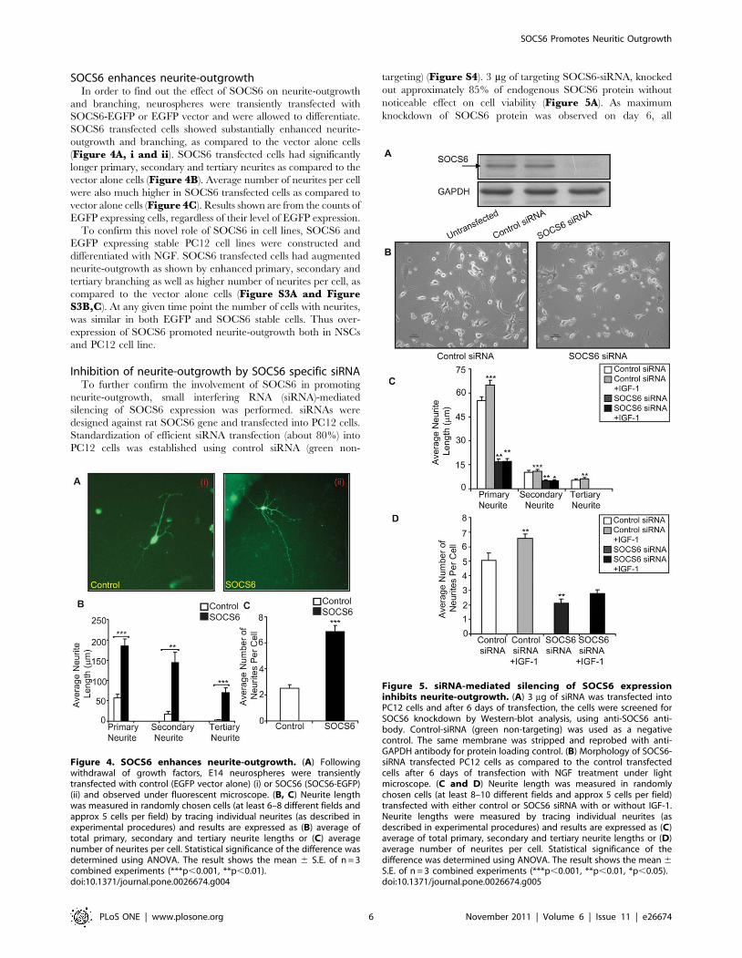
SOCS6 enhances neurite-outgrowthIn order to find out the effect of SOCS6 on neurite-outgrowth
and branching, neurospheres were transiently transfected with
SOCS6-EGFP or EGFP vector and were allowed to differentiate.
SOCS6 transfected cells showed substantially enhanced neurite-
outgrowth and branching, as compared to the vector alone cells
(Figure 4A, i and ii). SOCS6 transfected cells had significantly
longer primary, secondary and tertiary neurites as compared to the
vector alone cells (Figure 4B). Average number of neurites per cell
were also much higher in SOCS6 transfected cells as compared to
vector alone cells (Figure 4C). Results shown are from the counts of
EGFP expressing cells, regardless of their level of EGFP expression.
To confirm this novel role of SOCS6 in cell lines, SOCS6 and
EGFP expressing stable PC12 cell lines were constructed and
differentiated with NGF. SOCS6 transfected cells had augmented
neurite-outgrowth as shown by enhanced primary, secondary and
tertiary branching as well as higher number of neurites per cell, as
compared to the vector alone cells (Figure S3A and FigureS3B,C). At any given time point the number of cells with neurites,
was similar in both EGFP and SOCS6 stable cells. Thus over-
expression of SOCS6 promoted neurite-outgrowth both in NSCs
and PC12 cell line.
Inhibition of neurite-outgrowth by SOCS6 specific siRNATo further confirm the involvement of SOCS6 in promoting
neurite-outgrowth, small interfering RNA (siRNA)-mediated
silencing of SOCS6 expression was performed. siRNAs were
designed against rat SOCS6 gene and transfected into PC12 cells.
Standardization of efficient siRNA transfection (about 80%) into
PC12 cells was established using control siRNA (green non-
targeting) (Figure S4). 3 mg of targeting SOCS6-siRNA, knocked
out approximately 85% of endogenous SOCS6 protein without
noticeable effect on cell viability (Figure 5A). As maximum
knockdown of SOCS6 protein was observed on day 6, all
Figure 4. SOCS6 enhances neurite-outgrowth. (A) Followingwithdrawal of growth factors, E14 neurospheres were transientlytransfected with control (EGFP vector alone) (i) or SOCS6 (SOCS6-EGFP)(ii) and observed under fluorescent microscope. (B, C) Neurite lengthwas measured in randomly chosen cells (at least 6–8 different fields andapprox 5 cells per field) by tracing individual neurites (as described inexperimental procedures) and results are expressed as (B) average oftotal primary, secondary and tertiary neurite lengths or (C) averagenumber of neurites per cell. Statistical significance of the difference wasdetermined using ANOVA. The result shows the mean 6 S.E. of n = 3combined experiments (***p,0.001, **p,0.01).doi:10.1371/journal.pone.0026674.g004
Figure 5. siRNA-mediated silencing of SOCS6 expressioninhibits neurite-outgrowth. (A) 3 mg of siRNA was transfected intoPC12 cells and after 6 days of transfection, the cells were screened forSOCS6 knockdown by Western-blot analysis, using anti-SOCS6 anti-body. Control-siRNA (green non-targeting) was used as a negativecontrol. The same membrane was stripped and reprobed with anti-GAPDH antibody for protein loading control. (B) Morphology of SOCS6-siRNA transfected PC12 cells as compared to the control transfectedcells after 6 days of transfection with NGF treatment under lightmicroscope. (C and D) Neurite length was measured in randomlychosen cells (at least 8–10 different fields and approx 5 cells per field)transfected with either control or SOCS6 siRNA with or without IGF-1.Neurite lengths were measured by tracing individual neurites (asdescribed in experimental procedures) and results are expressed as (C)average of total primary, secondary and tertiary neurite lengths or (D)average number of neurites per cell. Statistical significance of thedifference was determined using ANOVA. The result shows the mean 6S.E. of n = 3 combined experiments (***p,0.001, **p,0.01, *p,0.05).doi:10.1371/journal.pone.0026674.g005
SOCS6 Promotes Neuritic Outgrowth
PLoS ONE | www.plosone.org 6 November 2011 | Volume 6 | Issue 11 | e26674

experiments were performed after 6 days of transfection. Control
siRNA had no effect on SOCS6 levels confirming the specificity of
SOCS6 siRNA (Figure 5A).
In order to investigate the effect of SOCS6 knockdown on
neurite-outgrowth, SOCS6-siRNA or control siRNA transfected
PC12 cells were differentiated in the presence of NGF. The cells
transfected with control siRNA had usual branching pattern and
neurite length. In contrast, SOCS6-siRNA transfected cells had
fewer branches and smaller neurites (Figure 5B). The length of
primary neurites in SOCS6-siRNA transfected neurons was
reduced with very few secondary and tertiary neurite branching
as compared to the cells with control siRNA (Figure 5C). Average
number of neurites per cell was also much less in SOCS6-siRNA
transfected cells as compared to control (Figure 5D). IGF-1
stimulation lead to increase in neurite-outgrowth just like in NSC,
but its effect was attenuated in SOCS6 depleted cells, indicative of
involvement of SOCS6 in IGF-1 mediated differentiation. This
data confirms that SOCS6 was involved in neurite-outgrowth
which is indicative of differentiation.
Jak/Stat pathway is involved in SOCS6 upregulationSince SOCS signalling has been shown to involve Jak/Stat
pathway, AG490, a pharmacologic inhibitor for the Jak/Stat
pathway [32], was used to ascertain its involvement in SOCS6
signalling. When NSCs as well as PC12 cells were treated with
50 mM of AG490, very little neurite-outgrowth and branching was
observed in the treated cells as compared to the control cells
(Figure 6A and Figure S5A,B). In both NSCs and PC12 cells,
the percentage of cells undergoing differentiation was reduced to
almost 20% as compared to the control cells (Figure 6B, FigureS5A,B). The fewer cells seen in AG490 treated NSCs were due to
the fact that the undifferentiated cells did not spread out but
remain close to the neurospheres. The length of primary and
secondary neurites in the AG490 treated cells was reduced as
compared to the untreated control cells (Figure 6C and FigureS5C,D).). Average number of neurites per cell was also much less
in cells cultured in presence of AG490 as compared to the control
cells (Figure 6D and Figure S5C,D). Since 50 mM of AG490
very mildly reduced cell survival (of PC12 cells) as measured by the
MTT assay (87.9%610.3 of controls) (Figure S5E), the AG490
inhibitory effect on neurite-outgrowth was unlikely to be a
consequence of non-specific cytotoxic effects of AG490. IGF-1
was unable to rescue the inhibitory effects of AG490, indicating
that IGF-1 actions were via the Jak/Stat pathway (Figure S5Cand S5C,D). In order to look for effects of Jak/Stat pathway on
SOCS6 expression, NSCs were treated with AG490 with or
without IGF-1 stimulation. SOCS6 expression was muted in the
presence of the inhibitor (Figure 6E), which indicated that
SOCS6 was downstream of Jak/Stat. IGF-1 enhanced SOCS6
expression marginally in the presence of the inhibitor, indicating
involvement of alternative pathways of SOCS6 stimulation by
IGF-1. In order to rule out non-specific action of AG490 on other
kinase pathways which could also have an impact on neural cell
differentiation, effect of AG490 was checked on erk and PI3 kinase
pathways. No non-specific effects were observed in neural stem
cells (data not shown). Elevated levels of pStat5 levels were
observed in undifferentiated cells as compared to the differentiated
cells (Figure 6F). IGF-1 was able to substantially enhance pStat5
levels in neuropsheres, but only marginally in differentiated cells,
indicating that Jak/Stat5 pathway was more active in neuro-
spheres with its involvement diminishing following differentiation
(Figure 6F). AG490 which has been previously shown to diminish
Stat3 activation also inhibited the activation of Stat5 in NSCs
(Figure 6G).
To confirm the involvement of Stat5, PC12 cells were
transiently transfected with Stat5a, Stat5b, dominant-negative
Stat5a (dnStat5a) and dominant-negative Stat5b (dnStat5b)
constructs and were allowed to differentiate in the presence of
NGF. Stat5a and Stat5b transfected cells showed enhanced
neurite-outgrowth and branching, as compared to the vector
alone cells (Figure S6). In contrast, the cells transfected with
dnStat5a and dnStat5b had branching pattern and neurite length
comparable to that of control, indicating that Stat5 activated
SOCS6 which in turn promoted neurite outgrowth (Figure 6H).
Together, these data suggest that (a) Jak/Stat was a major pathway
for SOCS6 mediated neurite-outgrowth, and (b) differentiation
cues inhibit Stat5 activation.
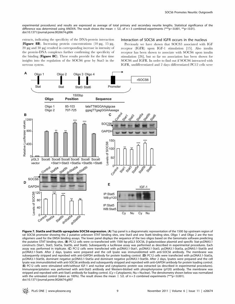
Stat5 acts as a transcription factor for SOCS6 promoterTo identify the specific Stat(s) involved in SOCS6 upregula-
tion, we selected the 1500 bp TATA less promoter region
immediately upstream of SOCS6 start codon and identified
various putative Stat binding sites using the Genomatix bioinfor-
matics tool (Figure 7A). The 1500 bp upstream region was
cloned into pGL3-Basic luciferase expression vector and trans-
fected into PC12 cells either alone or along with various
pcDNA3.1- Stat constructs. b-galactosidase plasmid was co-
transfected in all cases to normalize the transfection efficiency.
Although this 1500 bp genomic region of SOCS6 showed high
levels of baseline promoter activity indicating the presence of
other factors that could be binding to this region, it was
significantly enhanced by Stat5a and Stat5b but not by Stat1,
Stat3 or Stat6 (Figure 7B). Thus, Stat5 was a likely
transactivating factor for SOCS6 gene activation.
To confirm the role of Stat5 in SOCS6 upregulation, PC12 cells
were transfected with Stat1, Stat3, Stat5a, Stat5b and Stat6 and
SOCS6 expression was checked 24 hours post-transfection.
SOCS6 expression levels were highest in Stat5a and Stat5b
transfected PC12 cells (Figure 7C). To confirm this, dnStat5a and
dnStat5b were transfected into PC12 cells. Western-blot analysis
showed enhanced levels of SOCS6 in Stat5a and Stat5b
transfected PC12 cells whereas dominant negative mutants of
Stat5a and Stat5b significantly blocked this activation to almost
basal control levels (Figure 7D).
Stat5 has been shown to translocate to the nucleus upon
stimulation, a response that is inhibited by AG490 [33]. In order
to demonstrate the nuclear translocation of activated Stat5 in
response to IGF-1 stimulation, PC12 cells were stimulated with
or without IGF-1. The nuclear and cytoplasmic fractions were
extracted and analyzed for levels of pStat5. pStat5 presence was
much more pronounced in both cytoplasmic and nuclear
fraction of IGF-1 stimulated PC12 cells with higher levels in
the nucleus (Figure 7E). This indicated that upon IGF-1
stimulation, there was activation and translocation of pStat5 into
the nucleus.
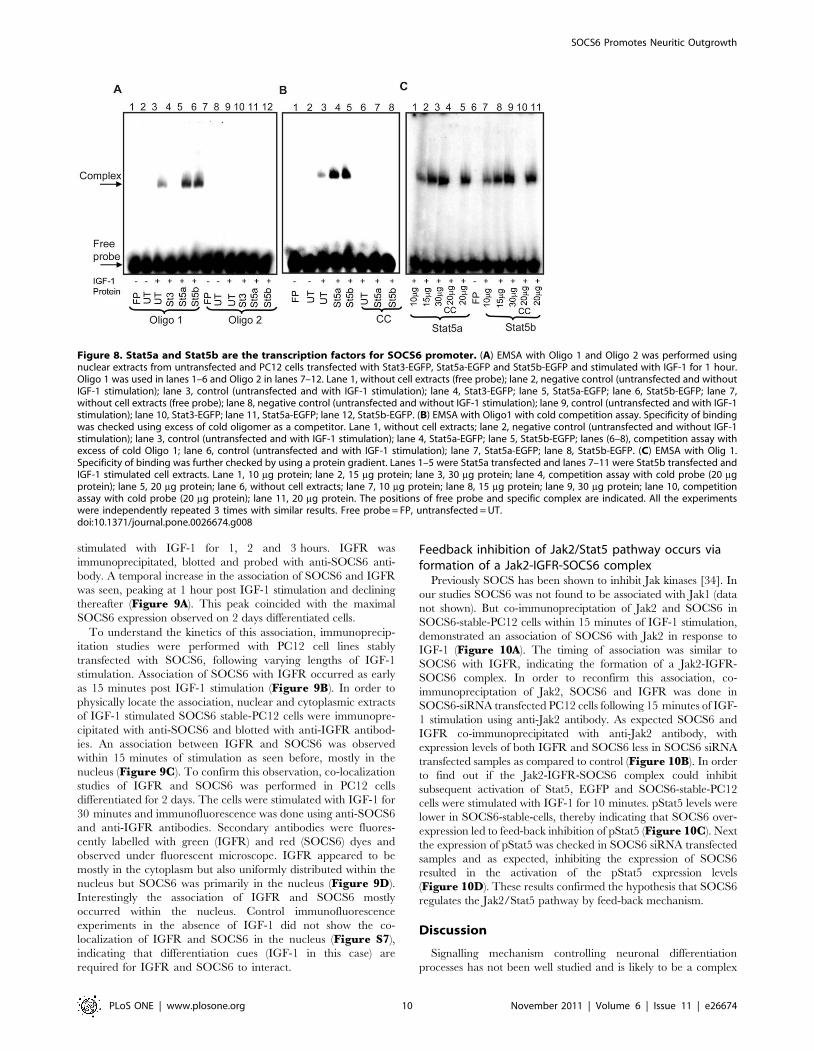
In order to confirm the actual Stat5 binding sequence in the
SOCS6 regulatory sequence, EMSA was performed using
oligonucleotides Oligo 1 and Oligo 2 (sequence shown in
Figure 7A) representing both the putative STAT binding sites
(Figure 8A). Nuclear extracts prepared from IGF-1 stimulated
PC12 cells transfected with Stat5a-EGFP and Stat5b-EGFP, were
used for binding studies. Extracts from both Stat5a and Stat5b
showed mobility shift with Oligo 1 and no binding was observed
with Oligo 2, demonstrating the specificity of binding to Oligo 1
STAT binding sequence (Figure 8A). Cold competition experi-
ments using 100 fold excess of cold oligomer was able to
specifically compete out both Stat5a and Stat5b transfected
SOCS6 Promotes Neuritic Outgrowth
PLoS ONE | www.plosone.org 7 November 2011 | Volume 6 | Issue 11 | e26674

Figure 6. Jak/Stat pathway is involved in neural stem cell differentiation. (A) E14 Neurospheres were allowed to differentiate in the absenceor presence of 50 mM AG490 (Jak2/Stat3 inhibitor) for 4 days and the cells were observed under light microscope. (B) Number of cells with neuritesper field was counted. An average of 13 fields was taken. The untreated control was taken as 100%. (C and D) Neurite length was measured inrandomly chosen cells by tracing individual neurites (as described in experimental procedures) and results are expressed as (C) average of totalprimary and secondary neurite lengths. The untreated control was taken as 100%. (D) Average number of neurites per cell. (E) Untreated or AG490treated neurospheres were stimulated with/without IGF-1. The cell lysate was immunoblotted with anti-SOCS6 antibody. The membrane was thenstripped and reprobed with anti-GAPDH antibody. (F) E14 Neurospheres and neurospheres upon differentiation were stimulated with/without IGF-1for 10 minutes. Stat5 was immunoprecipitated from 300 mg protein extract and Western-blotted using anti-phosphotyrosine antibody (pY20). Themembrane was then stripped and reprobed with anti-Stat5 antibody. (G) Untreated or AG490 treated neurospheres were stimulated with/withoutIGF-1 for 10 minutes. Stat5 was immunoprecipitated from 300 mg protein extract and Western-blotted using anti-phosphotyrosine antibody (pY20).The membrane was then stripped and reprobed with anti-Stat5 antibody. (H) PC12 cells, transfected with Stat5a- pcDNA3.1, Stat5b- pcDNA3.1,dominant negative Stat5a- pcDNA3.1 and dominant negative Stat5b- pcDNA3.1 were allowed to differentiate in the presence of NGF and the cellswere observed under light microscope. Neurite length was measured in randomly chosen cells by tracing individual neurites (as described in
SOCS6 Promotes Neuritic Outgrowth
PLoS ONE | www.plosone.org 8 November 2011 | Volume 6 | Issue 11 | e26674
extracts, indicating the specificity of the DNA-protein interaction
(Figure 8B). Increasing protein concentrations (10 mg, 15 mg,
20 mg and 30 mg) resulted in corresponding increase in intensity of
the protein-DNA complexes further confirming the specificity of
the binding (Figure 8C). These results provide for the first time
insights into the regulation of the SOCS6 gene by Stat5 in the
nervous system.
Interaction of SOCS6 and IGFR occurs in the nucleusPreviously we have shown that SOCS3 associated with IGF
receptor (IGFR) upon IGF-1 stimulation [15]. Also insulin
receptor has been shown to associate with SOCS6 upon insulin
stimulation [26], but so far no association has been shown for
SOCS6 and IGFR. In order to find out if SOCS6 interacted with
IGFR, undifferentiated and 2 days differentiated PC12 cells were
experimental procedures) and results are expressed as average of total primary and secondary neurite lengths. Statistical significance of thedifference was determined using ANOVA. The result shows the mean 6 S.E. of n = 3 combined experiments (***p,0.001, **p,0.01).doi:10.1371/journal.pone.0026674.g006
Figure 7. Stat5a and Stat5b upregulate SOCS6 expression. (A) Top panel is a diagrammatic representation of the 1500 bp upstream region ofrat SOCS6 promoter showing the 2 putative unknown STAT binding sites, one Stat3 and one Stat6 binding sites. Oligo 1 and Oligo 2 are the twooligomers used for the EMSA binding assays. The lower panel displays the sequence of the two oligos based on the Genomatix software predictingthe putative STAT binding sites. (B) PC12 cells were co-transfected with 1500 bp-pGL3 SOCS6, b-galactosidase plasmid and specific Stat-pcDNA3.1constructs (Stat1, Stat3, Stat5a, Stat5b, and Stat6). Subsequently a luciferase assay was performed as described in experimental procedures. Eachassay was performed in triplicate. (C) PC12 cells were transfected with pcDNA3.1-Stat1, pcDNA3.1-Stat3, pcDNA3.1-Stat5a, pcDNA3.1-Stat5b andpcDNA3.1-Stat6. After 2 days, lysates were prepared and the cell lysate was immunoblotted with anti-SOCS6 antibody. The membrane wassubsequently stripped and reprobed with anti-GAPDH antibody for protein loading control. (D) PC12 cells were transfected with pcDNA3.1-Stat5a,pcDNA3.1-Stat5b, dominant negative pcDNA3.1-Stat5a and dominant negative pcDNA3.1-Stat5b. After 2 days, lysates were prepared and the celllysate was immunoblotted with anti-SOCS6 antibody and subsequently stripped and reprobed with anti-GAPDH antibody for protein loading control.(E) PC12 cells were stimulated with/without IGF-1 and nuclear and cytoplasmic protein was extracted (as described in experimental procedures).Immunoprecipitation was performed with anti-Stat5 antibody and Western-blotted with phosphotyrosine (pY20) antibody. The membrane wasstripped and reprobed with anti-Stat5 antibody for loading control. (Cy = Cytoplasmic; Nu = Nuclear). The densitometry shown below was normalizedwith the untreated control (taken as 100%). The result shows the mean 6 S.E. of n = 3 combined experiments (***p,0.001).doi:10.1371/journal.pone.0026674.g007
SOCS6 Promotes Neuritic Outgrowth
PLoS ONE | www.plosone.org 9 November 2011 | Volume 6 | Issue 11 | e26674
stimulated with IGF-1 for 1, 2 and 3 hours. IGFR was
immunoprecipitated, blotted and probed with anti-SOCS6 anti-
body. A temporal increase in the association of SOCS6 and IGFR
was seen, peaking at 1 hour post IGF-1 stimulation and declining
thereafter (Figure 9A). This peak coincided with the maximal
SOCS6 expression observed on 2 days differentiated cells.
To understand the kinetics of this association, immunoprecip-
itation studies were performed with PC12 cell lines stably
transfected with SOCS6, following varying lengths of IGF-1
stimulation. Association of SOCS6 with IGFR occurred as early
as 15 minutes post IGF-1 stimulation (Figure 9B). In order to
physically locate the association, nuclear and cytoplasmic extracts
of IGF-1 stimulated SOCS6 stable-PC12 cells were immunopre-
cipitated with anti-SOCS6 and blotted with anti-IGFR antibod-
ies. An association between IGFR and SOCS6 was observed
within 15 minutes of stimulation as seen before, mostly in the
nucleus (Figure 9C). To confirm this observation, co-localization
studies of IGFR and SOCS6 was performed in PC12 cells
differentiated for 2 days. The cells were stimulated with IGF-1 for
30 minutes and immunofluorescence was done using anti-SOCS6
and anti-IGFR antibodies. Secondary antibodies were fluores-
cently labelled with green (IGFR) and red (SOCS6) dyes and
observed under fluorescent microscope. IGFR appeared to be
mostly in the cytoplasm but also uniformly distributed within the
nucleus but SOCS6 was primarily in the nucleus (Figure 9D).
Interestingly the association of IGFR and SOCS6 mostly
occurred within the nucleus. Control immunofluorescence
experiments in the absence of IGF-1 did not show the co-
localization of IGFR and SOCS6 in the nucleus (Figure S7),
indicating that differentiation cues (IGF-1 in this case) are
required for IGFR and SOCS6 to interact.
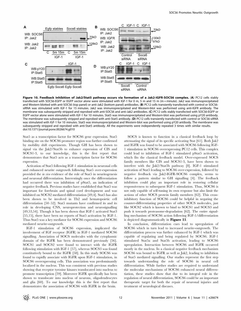
Feedback inhibition of Jak2/Stat5 pathway occurs viaformation of a Jak2-IGFR-SOCS6 complex
Previously SOCS has been shown to inhibit Jak kinases [34]. In
our studies SOCS6 was not found to be associated with Jak1 (data
not shown). But co-immunopreciptation of Jak2 and SOCS6 in
SOCS6-stable-PC12 cells within 15 minutes of IGF-1 stimulation,
demonstrated an association of SOCS6 with Jak2 in response to
IGF-1 (Figure 10A). The timing of association was similar to
SOCS6 with IGFR, indicating the formation of a Jak2-IGFR-
SOCS6 complex. In order to reconfirm this association, co-
immunopreciptation of Jak2, SOCS6 and IGFR was done in
SOCS6-siRNA transfected PC12 cells following 15 minutes of IGF-
1 stimulation using anti-Jak2 antibody. As expected SOCS6 and
IGFR co-immunoprecipitated with anti-Jak2 antibody, with
expression levels of both IGFR and SOCS6 less in SOCS6 siRNA
transfected samples as compared to control (Figure 10B). In order
to find out if the Jak2-IGFR-SOCS6 complex could inhibit
subsequent activation of Stat5, EGFP and SOCS6-stable-PC12
cells were stimulated with IGF-1 for 10 minutes. pStat5 levels were
lower in SOCS6-stable-cells, thereby indicating that SOCS6 over-
expression led to feed-back inhibition of pStat5 (Figure 10C). Next
the expression of pStat5 was checked in SOCS6 siRNA transfected
samples and as expected, inhibiting the expression of SOCS6
resulted in the activation of the pStat5 expression levels
(Figure 10D). These results confirmed the hypothesis that SOCS6
regulates the Jak2/Stat5 pathway by feed-back mechanism.
Discussion
Signalling mechanism controlling neuronal differentiation
processes has not been well studied and is likely to be a complex
Figure 8. Stat5a and Stat5b are the transcription factors for SOCS6 promoter. (A) EMSA with Oligo 1 and Oligo 2 was performed usingnuclear extracts from untransfected and PC12 cells transfected with Stat3-EGFP, Stat5a-EGFP and Stat5b-EGFP and stimulated with IGF-1 for 1 hour.Oligo 1 was used in lanes 1–6 and Oligo 2 in lanes 7–12. Lane 1, without cell extracts (free probe); lane 2, negative control (untransfected and withoutIGF-1 stimulation); lane 3, control (untransfected and with IGF-1 stimulation); lane 4, Stat3-EGFP; lane 5, Stat5a-EGFP; lane 6, Stat5b-EGFP; lane 7,without cell extracts (free probe); lane 8, negative control (untransfected and without IGF-1 stimulation); lane 9, control (untransfected and with IGF-1stimulation); lane 10, Stat3-EGFP; lane 11, Stat5a-EGFP; lane 12, Stat5b-EGFP. (B) EMSA with Oligo1 with cold competition assay. Specificity of bindingwas checked using excess of cold oligomer as a competitor. Lane 1, without cell extracts; lane 2, negative control (untransfected and without IGF-1stimulation); lane 3, control (untransfected and with IGF-1 stimulation); lane 4, Stat5a-EGFP; lane 5, Stat5b-EGFP; lanes (6–8), competition assay withexcess of cold Oligo 1; lane 6, control (untransfected and with IGF-1 stimulation); lane 7, Stat5a-EGFP; lane 8, Stat5b-EGFP. (C) EMSA with Olig 1.Specificity of binding was further checked by using a protein gradient. Lanes 1–5 were Stat5a transfected and lanes 7–11 were Stat5b transfected andIGF-1 stimulated cell extracts. Lane 1, 10 mg protein; lane 2, 15 mg protein; lane 3, 30 mg protein; lane 4, competition assay with cold probe (20 mgprotein); lane 5, 20 mg protein; lane 6, without cell extracts; lane 7, 10 mg protein; lane 8, 15 mg protein; lane 9, 30 mg protein; lane 10, competitionassay with cold probe (20 mg protein); lane 11, 20 mg protein. The positions of free probe and specific complex are indicated. All the experimentswere independently repeated 3 times with similar results. Free probe = FP, untransfected = UT.doi:10.1371/journal.pone.0026674.g008
SOCS6 Promotes Neuritic Outgrowth
PLoS ONE | www.plosone.org 10 November 2011 | Volume 6 | Issue 11 | e26674

process, requiring interplay of many signalling events. We have
previously shown that IGF-1 upregulated the expression of
SOCS3, which further promoted neurite-outgrowth in primary
cortical neurons [15]. In this study, our focus was to identify if any
other members of the SOCS family responsive to IGF-1, were
involved in neural stem cell differentiation. Our initial studies
demonstrated that IGF-1 enhanced the neurite-outgrowth and
differentiation of foetal NSCs as shown previously in other cell
types [35–37]. In neurospheres, SOCS6 levels increased by 30%
following IGF-1 stimulation. Interestingly SOCS6 jumped 400
fold following differentiation cues, both at the transcriptional as
well as translational levels. IGF-1 further enhanced SOCS6 levels
by about 30–40%. Though SOCS6 has been previously shown to
be involved in regulation of glucose metabolism, previous studies
on SOCS6 knockouts [24] and SOCS6 transgenics [25] have not
explored its role in neuronal differentiation or brain development.
This is the first time the involvement of SOCS6 in neuronal
differentiation has been demonstrated.
NGF treated PC12 cells which have previously been shown to
cease proliferation, extend neurites and acquire a number of
properties characteristic of sympathetic neurons [38], showed a
temporal increase of SOCS6 levels following differentiation cues.
Previously only SOCS2 has been shown to regulate NSC
differentiation following growth hormone stimulation [39]. NSCs
transiently transfected with SOCS6-EGFP plasmids as well as
PC12 cell line stably expressing SOCS6 under differentiating
conditions, showed increased numbers of neurites, enhanced
neurite-outgrowth and longer neurites, all of which are known
indicators of differentiation [40]. Unlike an earlier study, no
apoptotic effects were seen in SOCS6 overexpressing NSCs and
PC12 cells [41]. Further, SOCS6 silencing inhibited neurite
initiation and branching, confirming the role of SOCS6 in
differentiation.
Since we had previously seen the involvement of Jak/Stat
pathway in SOCS3 mediated signalling [15], attempts were made
to find out if SOCS6 signalling was also mediated via the Jak/Stat
pathway. AG490 a potent inhibitor of Jak/Stat [32], dampened
the SOCS6 expression in both NSCs and PC12 cells. Jak/Stat
pathway was found to be involved in enhancing the neurite length,
number of neurites and branching per cell. This is consistent with
the earlier reports where Jak/Stat pathway has been shown to be
involved in regulating differentiation of multipotent NSCs into
astrocytes and SOCS mediated signalling [42–45]. Further, IGF-1
which enhanced SOCS6 expression was unable to rescue the
inhibitory effects of AG490, indicating the involvement of Jak/Stat
pathway in IGF-1 mediated signalling. Silencing SOCS6 rendered
IGF-1 incapable of exerting its effects, which confirmed that IGF-
1 action is mediated via SOCS6. Once SOCS6 was activated
following differentiation cues, IGF-1 was not required for SOCS6
mediated neurite-outgrowth.
Since the role of Stats as transcription activators of SOCS gene
has been reported previously [46–48] search for Stat binding sites
in the SOCS6 gene promoter revealed various putative Stat
binding sites. Increased SOCS6 expression following overexpres-
sion of Stat5a and Stat5b, which was inhibited in the presence of
dominant–negative Stat5a and Stat5b, was indicative of the role of
Figure 9. SOCS6 associates with IGFR upon IGF-1 stimulation.(A) Undifferentiated or NGF differentiated PC12 cells were stimulatedwith/without IGF-1 for 1, 2 or 3 hours. Using 300 mg of cell lysate, IGFRwas pulled down and Western-blotted with anti-SOCS6 antibody. Themembrane was stripped and reprobed with anti-IGFR antibody forloading control. (B) SOCS6 stable PC12 cells were stimulated with/without IGF-1 for 5, 15, and 30 minutes. IGFR was immunoprecipitatedand Western-blotted with anti-SOCS6 antibody. The membrane wasstripped and reprobed with anti-IGFR antibody for loading control. (C)PC12 cells were differentiated for 2 days with NGF and then stimulatedwith/without IGF-1 for 15, 30 m or 1 hr (m = minutes and hr = hours)and nuclear and cytoplasmic protein was extracted (as described inexperimental procedures). Using 300 mg of cell lysate, SOCS6 was pulleddown and Western-blotted with anti-IGFR antibody. The membranewas stripped and reprobed with anti-SOCS6 antibody for loadingcontrol. (D) PC12 cells were allowed to differentiate for 2 days with NGF
and stimulated with IGF-1 for 30 minutes. The cells were then fixed andpermeabilized. After primary antibody treatment (anti-SOCS6, anti-IGFRand anti-SOCS6+anti-IGFR), SOCS6 was stained with Alexafluor 594 (red)and IGFR was stained with Alexafluor 488 (green). The cells werevisualized under fluorescent microscope. DAPI staining shows thelocation of the nucleus.doi:10.1371/journal.pone.0026674.g009
SOCS6 Promotes Neuritic Outgrowth
PLoS ONE | www.plosone.org 11 November 2011 | Volume 6 | Issue 11 | e26674
Stat5 as a transcription factor for SOCS6 gene expression. Stat5
binding site on the SOCS6 promoter region was further confirmed
by mobility shift experiments. Though GH has been shown to
signal via the Jak2/Stat5b to enhance expression of CIS and
SOCS1-3, to our knowledge, this is the first report that
demonstrates that Stat5 acts as a transcription factor for SOCS6
expression.
Activation of Stat5 following IGF-1 stimulation in neuronal cells
and enhanced neurite outgrowth following Stat5 over-expression
provided the in vivo evidence of the role of Stat5 in neuritogenesis
and neuronal differentiation. Interestingly once the differentiation
had occurred there was inhibition of pStat5, an indication of
negative feedback. Previous studies have established that Stat5 was
important for forebrain and spinal cord development and was
inhibited on SOCS2 mediated differentiation [49,4]. Stat5 has also
been shown to be involved in Th2 and hematopoietic cell
differentiation [50–52]. Stat5 mutants have confirmed its and its
role in developing CNS, neuroprotection and neurosignalling
[49,53,54]. Though it has been shown that IGF-1 activated Stat3
[55,15], there have been no reports of Stat5 activation by IGF-1.
Thus Stat5 was a key mediator for SOCS6 expression and SOCS6
mediated neurite-outgrowth.
IGF-1 stimulation of SOCS6 expression, implicated the
involvement of IGF receptor (IGFR) in IGF-1 mediated SOCS6
signalling. Association of SOCS molecules with the cytoplasmic
domain of the IGFR has been demonstrated previously [56].
SOCS1 and SOCS2 were found to interact with the IGFR
following stimulation with IGF-1 [57], whereas SOCS3 was found
constitutively bound to the IGFR [58]. In this study SOCS6 was
found to rapidly associate with IGFR upon IGF-1 stimulation, in
SOCS6 overexpressing cells. This association was predominantly
localized in the nucleus. This was consistent with previous studies
showing that receptor tyrosine kinases translocated into nucleus to
promote transcription [59]. Moreover IGFR specifically has been
shown to translocate into nucleus of neurons, oligodendrocytes
and glia [60]. To our knowledge this is the first report that
demonstrates the association of SOCS6 with IGFR in the brain.
SOCS is known to function in a classical feedback loop by
attenuating the signal of its specific activating Stat [61]. Both Jak2
and IGFR was found to be associated with SOCS6 following IGF-
1 stimulation in SOCS6 overexpressing PC12 cells. This complex
could lead to inhibition of IGF-1 stimulated pStat5 activation,
which fits the classical feedback model. Over-expressed SOCS
family members like CIS and SOCS1-3, have been shown to
interfere with the Jak2/Stat5b pathway [8]. IGF-1 stimulated
activation of Stat5 leading to SOCS6 over-expression, followed by
negative feedback via Jak2-IGFR-SOCS6 complex, seems to
follow a pattern similar to GH signalling [4]. The feedback
inhibition could play an important role in restoring cellular
responsiveness to subsequent IGF-1 stimulation. Thus, SOCS6 is
not only capable of self-tuning its own response but also limit the
actions of other SOCS protein family members [62]. This cross-
inhibitory function of SOCS6 could be helpful in negating the
counter-differentiating properties of other SOCS molecules, just
like SOCS2 which is known to bind to SOCS1 and SOCS3 and
push it towards proteasome-degradation [62]. The entire signal-
ling mechanism of SOCS6 action following IGF-1/differentiation
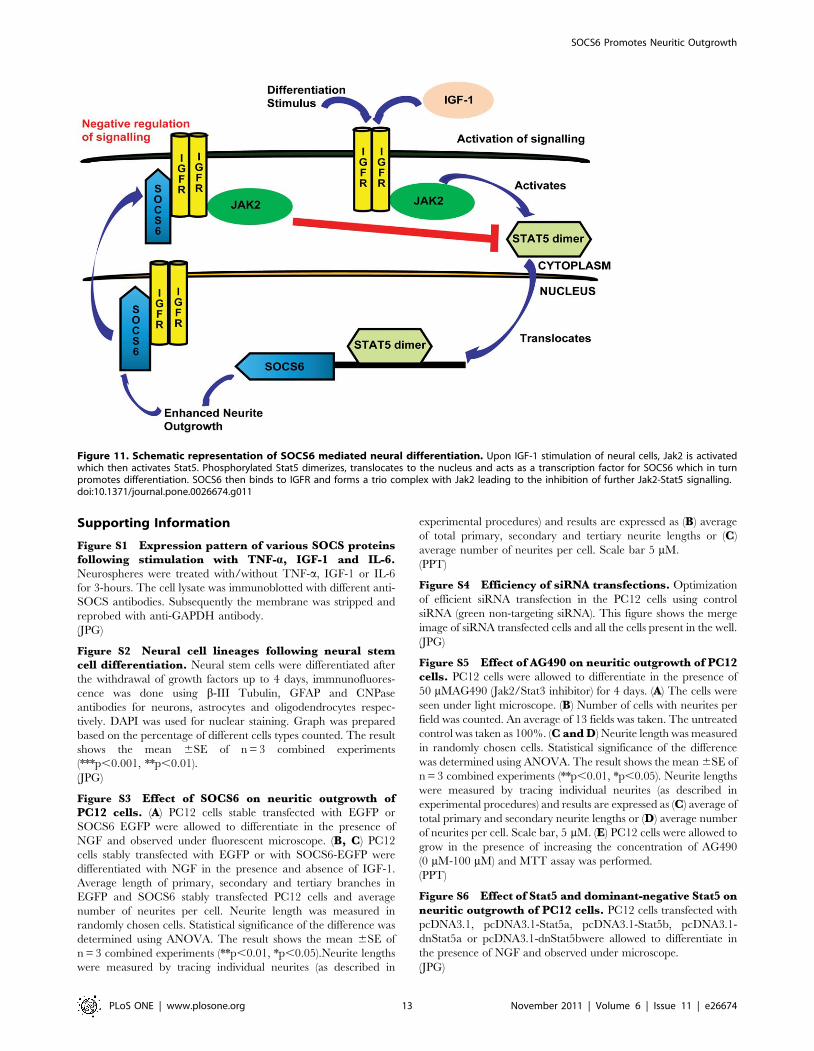
is depicted diagrammatically in Figure 11.
In conclusion, differentiation cues lead to upregulation of
SOCS6 which in turn lead to increased neurite-outgrowth. The
differentiation process was further enhanced by IGF-1 which was
capable of regulating and being regulated by SOCS6. IGF-1
stimulated Stat5a and Stat5b activation, leading to SOCS6
upregulation. Interaction between SOCS6 and IGFR occurred
mostly in the nucleus. In a classical negative feedback mechanism
SOCS6 was bound to IGFR as well as Jak2, leading to inhibition
of Stat5 mediated signalling. Our studies represent the first step
towards understanding the role of SOCS6 in neural cell
differentiation. While further studies are required to understand
the molecular mechanisms of SOCS6 enhanced neural differen-
tiation, these studies show that due to its integral role in the
regulation of neural differentiation, SOCS6 could be an important
therapeutic target for both the repair of neuronal injuries and
treatment of neurological diseases.
Figure 10. Feedback inhibition of Jak2/Stat5 pathway occurs via formation of a Jak2-IGFR-SOCS6 complex. (A) PC12 cells stablytransfected with SOCS6-EGFP or EGFP vector alone were stimulated with IGF-1 for 0 m, 5 m and 15 m (m = minute). Jak2 was immunoprecipitatedand Western-blotted with anti-SOCS6 (top panel) or anti-Jak2 (bottom panel) antibodies. (B) PC12 cells transiently transfected with control or SOCS6-siRNA was stimulated with IGF-1 for 15 minutes. Jak2 was immunoprecipitated and Western-blot was performed using anti-IGFR antibody. Themembrane was subsequently stripped and reprobed with anti-SOCS6 and anti-Jak2 antibodies. (C) PC12 cells stably transfected with SOCS6-EGFP orEGFP vector alone were stimulated with IGF-1 for 10 minutes. Stat5 was immunoprecipitated and Western-blot was performed using pY20 antibody.The membrane was subsequently stripped and reprobed with anti-Stat5 antibody. (D) PC12 cells transiently transfected with control or SOCS6-siRNAwas stimulated with IGF-1 for 10 minutes. Stat5 was immunoprecipitated and Western-blot was performed using pY20 antibody. The membrane wassubsequently stripped and reprobed with anti-Stat5 antibody. All the experiments were independently repeated 3 times with similar results.doi:10.1371/journal.pone.0026674.g010
SOCS6 Promotes Neuritic Outgrowth
PLoS ONE | www.plosone.org 12 November 2011 | Volume 6 | Issue 11 | e26674
Supporting Information
Figure S1 Expression pattern of various SOCS proteinsfollowing stimulation with TNF-a, IGF-1 and IL-6.Neurospheres were treated with/without TNF-a, IGF-1 or IL-6
for 3-hours. The cell lysate was immunoblotted with different anti-
SOCS antibodies. Subsequently the membrane was stripped and
reprobed with anti-GAPDH antibody.
(JPG)
Figure S2 Neural cell lineages following neural stemcell differentiation. Neural stem cells were differentiated after
the withdrawal of growth factors up to 4 days, immnunofluores-
cence was done using b-III Tubulin, GFAP and CNPase
antibodies for neurons, astrocytes and oligodendrocytes respec-
tively. DAPI was used for nuclear staining. Graph was prepared
based on the percentage of different cells types counted. The result
shows the mean 6SE of n = 3 combined experiments
(***p,0.001, **p,0.01).
(JPG)
Figure S3 Effect of SOCS6 on neuritic outgrowth ofPC12 cells. (A) PC12 cells stable transfected with EGFP or
SOCS6 EGFP were allowed to differentiate in the presence of
NGF and observed under fluorescent microscope. (B, C) PC12
cells stably transfected with EGFP or with SOCS6-EGFP were
differentiated with NGF in the presence and absence of IGF-1.
Average length of primary, secondary and tertiary branches in
EGFP and SOCS6 stably transfected PC12 cells and average
number of neurites per cell. Neurite length was measured in
randomly chosen cells. Statistical significance of the difference was
determined using ANOVA. The result shows the mean 6SE of
n = 3 combined experiments (**p,0.01, *p,0.05).Neurite lengths
were measured by tracing individual neurites (as described in
experimental procedures) and results are expressed as (B) average
of total primary, secondary and tertiary neurite lengths or (C)
average number of neurites per cell. Scale bar 5 mM.
(PPT)
Figure S4 Efficiency of siRNA transfections. Optimization
of efficient siRNA transfection in the PC12 cells using control
siRNA (green non-targeting siRNA). This figure shows the merge
image of siRNA transfected cells and all the cells present in the well.
(JPG)
Figure S5 Effect of AG490 on neuritic outgrowth of PC12cells. PC12 cells were allowed to differentiate in the presence of
50 mMAG490 (Jak2/Stat3 inhibitor) for 4 days. (A) The cells were
seen under light microscope. (B) Number of cells with neurites per
field was counted. An average of 13 fields was taken. The untreated
control was taken as 100%. (C and D) Neurite length was measured
in randomly chosen cells. Statistical significance of the difference
was determined using ANOVA. The result shows the mean 6SE of
n = 3 combined experiments (**p,0.01, *p,0.05). Neurite lengths
were measured by tracing individual neurites (as described in
experimental procedures) and results are expressed as (C) average of
total primary and secondary neurite lengths or (D) average number
of neurites per cell. Scale bar, 5 mM. (E) PC12 cells were allowed to
grow in the presence of increasing the concentration of AG490
(0 mM-100 mM) and MTT assay was performed.
(PPT)
Figure S6 Effect of Stat5 and dominant-negative Stat5 onneuritic outgrowth of PC12 cells. PC12 cells transfected with
pcDNA3.1, pcDNA3.1-Stat5a, pcDNA3.1-Stat5b, pcDNA3.1-
dnStat5a or pcDNA3.1-dnStat5bwere allowed to differentiate in
the presence of NGF and observed under microscope.
(JPG)
Figure 11. Schematic representation of SOCS6 mediated neural differentiation. Upon IGF-1 stimulation of neural cells, Jak2 is activatedwhich then activates Stat5. Phosphorylated Stat5 dimerizes, translocates to the nucleus and acts as a transcription factor for SOCS6 which in turnpromotes differentiation. SOCS6 then binds to IGFR and forms a trio complex with Jak2 leading to the inhibition of further Jak2-Stat5 signalling.doi:10.1371/journal.pone.0026674.g011
SOCS6 Promotes Neuritic Outgrowth
PLoS ONE | www.plosone.org 13 November 2011 | Volume 6 | Issue 11 | e26674
Figure S7 Background immuno-staining of PC12 cells inthe absence of IGF-1 stimulation. PC12 cells were allowed to
differentiate for 2 days with NGF. The cells were then fixed and
permeabilized. After primary antibody treatment (anti-SOCS6,
anti-IGFR and anti-SOCS6+anti-IGFR), SOCS6 was stained with
Alexafluor594 (red) and IGFR was stained withAlexafluor488
(green). The cells were visualized under fluorescent microscope.
DAPI staining shows the location of the nucleus.
(JPG)
Acknowledgements
We thank Dr. S Iyenger, NBRC for her help in neurite count studies and
Dr. Agam P. Singh and Dr. Sandeep Saxena, NII for their help on
manuscript preparation.
Author Contributions
Conceived and designed the experiments: AS KB. Performed the
experiments: SG KM. Analyzed the data: SG KB. Wrote the paper: SG
KB. Contributed towards manuscript preparation: AS KM.
References
1. da Silva JS, Dotti CG (2002) Breaking the neuronal sphere: regulation of the
actin cytoskeleton in neuritogenesis. Nat Rev Neurosci 3: 694–704.
2. Goldshmit Y, Galea MP, Wise G, Bartlett PF, Turnley AM (2004) Axonalregeneration and lack of astrocytic gliosis in EphA4-deficient mice. J Neurosci
24: 10064–10073.
3. Goldshmit Y, Greenhalgh CJ, Turnley AM (2004) Suppressor of cytokine
signalling-2 and epidermal growth factor regulate neurite outgrowth of corticalneurons. Eur J Neurosci 20: 2260–22660.
4. Goldshmit Y, Walters CE, Scott HJ, Greenhalgh CJ, Turnley AM (2004)
SOCS2 induces neurite outgrowth by regulation of epidermal growth factor
receptor activation. J Biol Chem 279: 16349–16355.
5. Krebs DL, Hilton DJ (2000) SOCS: physiological suppressors of cytokinesignalling. J Cell Sci 113: 2813–2819.
6. Wormald S, Hilton DJ (2004) Inhibitors of cytokine signal transduction. J Biol
Chem 279: 821–824.
7. Hansen JA, Lindberg K, Hilton DJ, Nielsen JH, Billestrup N (1999) Mechanism
of inhibition of growth hormone receptor signalling by suppressor of cytokinesignalling proteins. Mol Endocrinol 13: 1832–1843.
8. Ram PA, Waxman DJ (1999) SOCS/CIS protein inhibition of growth hormone-
stimulated STAT5 signalling by multiple mechanisms. J Biol Chem 274:35553–35561.
9. Polizzotto MN, Bartlett PF, Turnley AM (2000) Expression of ‘‘suppressor of
cytokine signalling’’ (SOCS) genes in the developing and adult mouse nervous
system. J Comp Neurol 423: 348–358.
10. Turnley AM, Starr R, Bartlett PF (2001) SOCS1 regulates interferon-gammamediated sensory neuron survival. Neuroreport 12: 3443–3445.
11. Scott HJ, Stebbing MJ, Walters CE, McLenachan S, Ransome MI, et al. (2006)
Differential effects of SOCS2 on neuronal differentiation and morphology. Brain
Res 1067: 138–145.
12. Turnley AM, Faux CH, Rietze RL, Coonan JR, Bartlett PF (2002) Suppressor ofcytokine signalling 2 regulates neuronal differentiation by inhibiting growth
hormone signalling. Nat Neurosci 5: 1155–1162.
13. Cao F, Hata R, Zhu P, Ma YJ, Tanaka J, et al. (2006) Over-expression of
SOCS3 inhibits astrogliogenesis and promotes maintenance of neural stem cells.J Neurochem 98: 459–470.
14. Zhu P, Hata R, Cao F, Gu F, Hanakawa Y, et al. (2008) Ramified microglial
cells promote astrogliogenesis and maintenance of neural stem cells throughactivation of Stat3 function. FASEB J 22: 3866–3877.
15. Yadav A, Kalita A, Dhillon S, Banerjee K (2005) JAK/STAT3 pathway is
involved in survival of neurons in response to insulin-like growth factor and
negatively regulated by suppressor of cytokine signalling-3. J Biol Chem 280:31830–31840.
16. Drago J, Murphy M, Carroll SM, Harvey RP, Bartlett PF (1991) Fibroblast
growth factor-mediated proliferation of central nervous system precursors
depends on endogenous production of insulin-like growth factor I. Proc NatlAcad Sci U S A 88: 2199–2203.
17. Bondy CA (1991) Transient IGF-I gene expression during the maturation of
functionally related central projection neurons. J Neurosci 11: 3442–3455.
18. Liu JL, Grinberg A, Westphal H, Sauer B, Accili D, et al. (1998) Insulin-likegrowth factor-I affects perinatal lethality and postnatal development in a gene
dosage-dependent manner: manipulation using the Cre/loxP system in
transgenic mice. Mol Endocrinol 12: 1452–1462.
19. Ye P, Xing Y, Dai Z, D’Ercole AJ (1996) In vivo actions of insulin-like growthfactor-I (IGF-I) on cerebellum development in transgenic mice: evidence that
IGF-I increases proliferation of granule cell progenitors. Brain Res Dev BrainRes 95: 44–54.
20. Liu X, Mameza MG, Lee YS, Eseonu CI, Yu CR, et al. (2008) Suppressors ofcytokine-signalling proteins induce insulin resistance in the retina and promote
survival of retinal cells. Diabetes 57: 1651–1658.
21. Masuhara M, Sakamoto H, Matsumoto A, Suzuki R, Yasukawa H, et al. (1997)Cloning and characterization of novel CIS family genes. Biochem Biophys Res
Commun 239: 439–446.
22. Nicholson SE, Willson TA, Farley A, Starr R, Zhang JG, et al. (1999)
Mutational analyses of the SOCS proteins suggest a dual domain requirementbut distinct mechanisms for inhibition of LIF and IL-6 signal transduction.
EMBO J 18: 375–385.
23. Howard JK, Flier JS (2006) Attenuation of leptin and insulin signalling by SOCS
proteins. Trends Endocrinol Metab 17: 365–371.
24. Krebs DL, Uren RT, Metcalf D, Rakar S, Zhang JG, et al. (2002) SOCS-6 binds
to insulin receptor substrate 4, and mice lacking the SOCS-6 gene exhibit mild
growth retardation. Mol Cell Biol 22: 4567–4578.
25. Li L, Gronning LM, Anderson PO, Li S, Edvardsen K, et al. (2004) Insulin
induces SOCS-6 expression and its binding to the p85 monomer of
phosphoinositide 3-kinase, resulting in improvement in glucose metabolism.
J Biol Chem 279: 34107–34114.
26. Mooney RA, Senn J, Cameron S, Inamdar N, Boivin LM, et al. (2001)
Suppressors of cytokine signalling-1 and -6 associate with and inhibit the insulin
receptor. A potential mechanism for cytokine-mediated insulin resistance. J Biol
Chem 276: 25889–25893.
27. Bayle J, Lopez S, Iwai K, Dubreuil P, De Sepulveda P (2006) The E3 ubiquitin
ligase HOIL-1 induces the polyubiquitination and degradation of SOCS6
associated proteins. FEBS Lett 580: 2609–2614.
28. Hwang MN, Ha TH, Park J, Shim J, Lee H, et al. (2007) Increased SOCS6
stability with PMA requires its N-terminal region and the Erk pathway via
Pkcdelta activation. Biochem Biophys Res Commun 354: 184–189.
29. Balasubramaniyan V, Timmer N, Kust B, Boddeke E, Copray S (2004)
Transient expression of Olig1 initiates the differentiation of neural stem cells into
oligodendrocyte progenitor cells. Stem Cells 22: 878–882.
30. Shanley DK, Sullivan AM (2006) Alterations in cellular phenotypes differen-
tiating from embryonic rat brain neurosphere cultures by immunoselection of
neuronal progenitors. Brain Res 1067: 85–94.
31. Vaudry D, Stork PJ, Lazarovici P, Eiden LE (2002) Signalling pathways for
PC12 cell differentiation: making the right connections. Science 296:
1648–1649.
32. Wang LH, Kirken RA, Erwin RA, Yu CR, Farrar WL (1999) JAK3, STAT, and
MAPK signalling pathways as novel molecular targets for the tyrphostin AG-490
regulation of IL-2-mediated T cell response. J Immunol 162: 3897–3904.
33. Nakasato M, Shirakura Y, Ooga M, Iwatsuki M, Ito M, et al. (2006)
Involvement of the STAT5 signalling pathway in the regulation of mouse
preimplantation development. Biol Reprod 75: 508–517.
34. Yasukawa H, Misawa H, Sakamoto H, Masuhara M, Sasaki A, et al. (1999) The
JAK-binding protein JAB inhibits Janus tyrosine kinase activity through binding
in the activation loop. EMBO J 18: 1309–1320.
35. Brooker GJ, Kalloniatis M, Russo VC, Murphy M, Werther GA, et al. (2000)
Endogenous IGF-1 regulates the neuronal differentiation of adult stem cells.
J Neurosci Res 59: 332–341.
36. Arsenijevic Y, Weiss S, Schneider B, Aebischer P (2001) Insulin-like growth
factor-I is necessary for neural stem cell proliferation and demonstrates distinct
actions of epidermal growth factor and fibroblast growth factor-2. J Neurosci 21:
7194–7202.
37. Ning H, Lin G, Fandel T, Banie L, Lue TF, et al. (2008) Insulin growth factor
signalling mediates neuron-like differentiation of adipose-tissue-derived stem
cells. Differentiation 76: 488–494.
38. Greene LA, Tischler AS (1976) Establishment of a noradrenergic clonal line of
rat adrenal pheochromocytoma cells which respond to nerve growth factor. Proc
Natl Acad Sci U S A 73: 2424–2428.
39. Turnley AM (2005) Role of SOCS2 in growth hormone actions. Trends
Endocrinol Metab 16: 53–58.
40. Nikolic M, Dudek H, Kwon YT, Ramos YF, Tsai LH (1996) The cdk5/p35
kinase is essential for neurite outgrowth during neuronal differentiation. Genes
Dev 10: 816–825.
41. Lai RH, Hsiao YW, Wang MJ, Lin HY, Wu CW, et al. (2010) SOCS6, down-
regulated in gastric cancer, inhibits cell proliferation and colony formation.
Cancer Lett 288: 75–85.
42. Bonni A, Sun Y, Nadal-Vicens M, Bhatt A, Frank DA, et al. (1997) Regulation
of gliogenesis in the central nervous system by the JAK-STAT signalling
pathway. Science 278: 477–483.
43. Narita M, Miyatake M, Shibasaki M, Shindo K, Nakamura A, et al. (2006)
Direct evidence of astrocytic modulation in the development of rewarding effects
induced by drugs of abuse. Neuropsychopharmacology 31: 2476–2488.
44. O’Sullivan LA, Liongue C, Lewis RS, Stephenson SE, Ward AC (2007)
Cytokine receptor signalling through the Jak-Stat-Socs pathway in disease. Mol
Immunol 44: 2497–2506.
45. Murray PJ (2007) The JAK-STAT signalling pathway: input and output
integration. J Immunol 178: 2623–2629.
SOCS6 Promotes Neuritic Outgrowth
PLoS ONE | www.plosone.org 14 November 2011 | Volume 6 | Issue 11 | e26674
46. Croker BA, Kiu H, Nicholson SE (2008) SOCS regulation of the JAK/STAT
signalling pathway. Semin Cell Dev Biol 19: 414–422.47. Lee CK, Raz R, Gimeno R, Gertner R, Wistinghausen B, et al. (2002) STAT3 is
a negative regulator of granulopoiesis but is not required for G-CSF-dependent
differentiation. Immunity 17: 63–72.48. Hwang MN, Kim KS, Choi YW, Jou I, Yoon S (2007) PMA activates Stat3 in
the Jak/Stat pathway and induces SOCS5 in rat brain astrocytes. Mol Cells 23:94–99.
49. Markham K, Schuurmans C, Weiss S (2007) STAT5A/B activity is required in
the developing forebrain and spinal cord. Mol Cell Neurosci 35: 272–282.50. Zhu J, Cote-Sierra J, Guo L, Paul WE (2003) Stat5 Activation Plays a Critical
Role in Th2 Differentiation. Immunity 19: 739–748.51. Nosaka T, Kawashima T, Misawa K, Ikuta K, Mui ALF, et al. (1999) STAT5 as
a molecular regulator of proliferation, differentiation and apoptosis inhematopoietic cells. EMBO J 18: 4754–4765.
52. Kyba M, Perlingeiro RC, Hoover RR, Lu CW, Pierce J, et al. (2003) Enhanced
hematopoietic differentiation of embryonic stem cells conditionally expressingStat5. Proc Natl Acad Sci U S A 100 Suppl 1: 11904–10.
53. Byts N, Samoylenko A, Fasshauer T, Ivanisevic M, Hennighausen L, et al.(2008) Essential role for Stat5 in the neurotrophic but not in the neuroprotective
effect of erythropoietin. Cell Death and Differentiation 15: 783–792.
54. Lee J-Y, Muenzberg H, Gavrilova O, Reed JA, Berryman D, et al. (2008) Loss ofCytokine-STAT5 Signaling in the CNS and Pituitary Gland Alters Energy
Balance and Leads to Obesity. PLoS ONE 3(2): e1639.
55. Zong CS, Chan J, Levy DE, Horvath C, Sadowski HB, et al. (2000) Mechanism
of STAT3 activation by insulin-like growth factor I receptor. J Biol Chem 275:15099–15105.
56. Himpe E, Kooijman R (2009) Insulin-like growth factor-I receptor signal
transduction and the Janus Kinase/Signal Transducer and Activator ofTranscription (JAK-STAT) pathway. Biofactors 35: 76–81.
57. Dey BR, Spence SL, Nissley P, Furlanetto RW (1998) Interaction of humansuppressor of cytokine signalling (SOCS)-2 with the insulin-like growth factor-I
receptor. J Biol Chem 273: 24095–24101.
58. Dey BR, Furlanetto RW, Nissley P (2000) Suppressor of cytokine signalling(SOCS)-3 protein interacts with the insulin-like growth factor-I receptor.
Biochem Biophys Res Commun 278: 38–43.59. Garcia-Segura LM, Rodriguez JR, Torres-Aleman I (1997) Localization of the
insulin-like growth factor I receptor in the cerebellum and hypothalamus of adultrats: an electron microscopic study. J Neurocytology 26: 479–490.
60. Sehat B, Tofigh A, Lin Y, Trocme E, Liljedahl U, et al. (2010) SUMOylation
Mediates the Nuclear Translocation and Signaling of the IGF-1 receptor. SciSignal 3: ra10.
61. Naka T, Narazaki M, Hirata M, Matsumoto T, Minamoto S, et al. (1997)Structure and function of a new STAT-induced STAT inhibitor. Nature 387:
924–929.
62. Piessevaux J, Lavens D, Montoye T, Wauman J, Catteeuw D, et al. (2006)Functional cross-modulation between SOCS proteins can stimulate cytokine
signalling. J Biol Chem 281: 32953–32966.
SOCS6 Promotes Neuritic Outgrowth
PLoS ONE | www.plosone.org 15 November 2011 | Volume 6 | Issue 11 | e26674