Phenotypic variations and new mutations in JAK2 V617F–negative polycythemia vera, erythrocytosis,...
15
Phenotypic variations and new mutations in JAK2 V617F-negative polycythemia vera, erythrocytosis and idiopathic myelofibrosis Donna M. Williams, Ann H. Kim, Ophelia Rogers, Jerry L. Spivak, and Alison R. Moliterno Johns Hopkins University School of Medicine Abstract (1) Objective—The chronic myeloproliferative disorders (MPD), polycythemia vera (PV), essential thrombocytosis (ET) and idiopathic myelofibrosis (IMF), are characterized by a spectrum of clinical features and linked by common genetic lesions in JAK2 and MPL. However, the clinical phenotypes in genetically undefined MPD patients are similar to those patients with JAK2 and MPL lesions. We, therefore, sought to determine whether there were JAK2 or MPL lesions in a well- defined, JAK2 V617F-negative MPD cohort, and to determine if clinical associations could be identified based on variations identified in these genes. (2) Methods—We examined the JAK2 and MPL genes in JAK2 V617F-negative PV, IMF and idiopathic erythrocytosis (ERT) patients for sequence variations. (3) Results—We identified two previously unrecognized JAK2 mutations and three previously unrecognized MPL mutations in JAK2 V617F-negative PV, erythrocytosis and IMF patients. We identified JAK2 exon 12 lesions in 30% of JAK2 V617F-negative PV patients, and either JAK2 V617F or JAK2 exon 12 lesions in 9% of ERT patients In IMF, in addition to the MPL gene mutation, W515K, we identified three additional mutations: 204P and 2 intervening sequence transitions: IVS 11/12 and 10/11. (4) Conclusions—While the clinical phenotype of JAK2 exon 12 lesions in the MPD was predominantly erythroid, there was significant disease spectrum overlap between JAK2 V617F and JAK2 exon 12 mutations. By contrast, MPL gene mutations were not associated with erythrocytosis, but segregated primarily with the phenotypes of thrombocytosis, extramedullary disease, myelofibrosis and osteosclerosis. Introduction Polycythemia vera (PV), idiopathic myelofibrosis (IMF) and essential thrombocytosis (ET) are clonal myeloproliferative disorders whose shared features include origin in a multipotent hematopoietic progenitor cell(1), cytogenetic abnormalities of chromosomes 1,8,9,13 and 20 (2), growth factor-independent in vitro hematopoietic colony formation(3) and epigenetic abnormalities, such as increased granulocyte PRV-1 mRNA expression(4) and impaired megakaryocyte and platelet thrombopoietin receptor protein (Mpl) expression(5). Taken together, these shared phenotypic and genotypic features suggest that PV, IMF and ET are pathogenetically related. The discovery that the same JAK2 activating mutation (V617F) was common to all three disorders supported this contention(6-9). Corresponding author: Alison R. Moliterno MD, Johns Hopkins University School of Medicine, Ross Research 1025, 720 Rutland Avenue, Baltimore MD 21205, 410-614-6360, 410-955-0185 (FAX), [email protected]. Publisher's Disclaimer: This is a PDF file of an unedited manuscript that has been accepted for publication. As a service to our customers we are providing this early version of the manuscript. The manuscript will undergo copyediting, typesetting, and review of the resulting proof before it is published in its final citable form. Please note that during the production process errors may be discovered which could affect the content, and all legal disclaimers that apply to the journal pertain. NIH Public Access Author Manuscript Exp Hematol. Author manuscript; available in PMC 2008 November 1. Published in final edited form as: Exp Hematol. 2007 November ; 35(11): 1641–1646. NIH-PA Author Manuscript NIH-PA Author Manuscript NIH-PA Author Manuscript
Transcript of Phenotypic variations and new mutations in JAK2 V617F–negative polycythemia vera, erythrocytosis,...

Phenotypic variations and new mutations in JAK2 V617F-negativepolycythemia vera, erythrocytosis and idiopathic myelofibrosis
Donna M. Williams, Ann H. Kim, Ophelia Rogers, Jerry L. Spivak, and Alison R. MoliternoJohns Hopkins University School of Medicine
Abstract(1) Objective—The chronic myeloproliferative disorders (MPD), polycythemia vera (PV),essential thrombocytosis (ET) and idiopathic myelofibrosis (IMF), are characterized by a spectrumof clinical features and linked by common genetic lesions in JAK2 and MPL. However, the clinicalphenotypes in genetically undefined MPD patients are similar to those patients with JAK2 and MPLlesions. We, therefore, sought to determine whether there were JAK2 or MPL lesions in a well-defined, JAK2 V617F-negative MPD cohort, and to determine if clinical associations could beidentified based on variations identified in these genes.
(2) Methods—We examined the JAK2 and MPL genes in JAK2 V617F-negative PV, IMF andidiopathic erythrocytosis (ERT) patients for sequence variations.
(3) Results—We identified two previously unrecognized JAK2 mutations and three previouslyunrecognized MPL mutations in JAK2 V617F-negative PV, erythrocytosis and IMF patients. Weidentified JAK2 exon 12 lesions in 30% of JAK2 V617F-negative PV patients, and either JAK2V617F or JAK2 exon 12 lesions in 9% of ERT patients In IMF, in addition to the MPL gene mutation,W515K, we identified three additional mutations: 204P and 2 intervening sequence transitions: IVS11/12 and 10/11.
(4) Conclusions—While the clinical phenotype of JAK2 exon 12 lesions in the MPD waspredominantly erythroid, there was significant disease spectrum overlap between JAK2 V617F andJAK2 exon 12 mutations. By contrast, MPL gene mutations were not associated with erythrocytosis,but segregated primarily with the phenotypes of thrombocytosis, extramedullary disease,myelofibrosis and osteosclerosis.
IntroductionPolycythemia vera (PV), idiopathic myelofibrosis (IMF) and essential thrombocytosis (ET)are clonal myeloproliferative disorders whose shared features include origin in a multipotenthematopoietic progenitor cell(1), cytogenetic abnormalities of chromosomes 1,8,9,13 and 20(2), growth factor-independent in vitro hematopoietic colony formation(3) and epigeneticabnormalities, such as increased granulocyte PRV-1 mRNA expression(4) and impairedmegakaryocyte and platelet thrombopoietin receptor protein (Mpl) expression(5). Takentogether, these shared phenotypic and genotypic features suggest that PV, IMF and ET arepathogenetically related. The discovery that the same JAK2 activating mutation (V617F) wascommon to all three disorders supported this contention(6-9).
Corresponding author: Alison R. Moliterno MD, Johns Hopkins University School of Medicine, Ross Research 1025, 720 RutlandAvenue, Baltimore MD 21205, 410-614-6360, 410-955-0185 (FAX), [email protected]'s Disclaimer: This is a PDF file of an unedited manuscript that has been accepted for publication. As a service to our customerswe are providing this early version of the manuscript. The manuscript will undergo copyediting, typesetting, and review of the resultingproof before it is published in its final citable form. Please note that during the production process errors may be discovered which couldaffect the content, and all legal disclaimers that apply to the journal pertain.
NIH Public AccessAuthor ManuscriptExp Hematol. Author manuscript; available in PMC 2008 November 1.
Published in final edited form as:Exp Hematol. 2007 November ; 35(11): 1641–1646.
NIH
-PA Author Manuscript
NIH
-PA Author Manuscript
NIH
-PA Author Manuscript
However, up to 10% of patients meeting the clinical criteria for PV do not express JAK2 V617F(6). Recently, a few of these patients were found to have mutations in JAK2 exon 12(10) andthe majority primarily had an erythrocytosis phenotype in contrast to the panmyelopathy thatis characteristic of JAK2 V617F-positive PV patients. Importantly, in a few JAK2 V617F-negative IMF patients, new activating MPL gene mutations have been found(11-13), althoughthe molecular basis of the bulk of these patients remains unknown.
To investigate these issues further, we examined the JAK2 gene in 157 PV patients and 44erythrocytosis patients and sequenced the MPL gene in 32 JAK2 V617F-negative IMF patients.We report here two previously unrecognized JAK2 exon 12 mutations and 3 unrecognizedMPL gene mutations together with an analysis of the corresponding genotype-phenotypecorrelations.
MethodsThe study was approved by our Institutional Review Board, and written informed consent wasobtained from all patients. The diagnosis of PV was based on the criteria of the PolycythemiaVera Study Group and included an elevated red cell mass(14). The diagnosis of IMF was basedon the Italian Consensus Conference clinical criteria(15). Acquired idiopathic erythrocytosiswas defined by the presence of an increased red cell mass and a low or normal serumerythropoietin level in the absence of a definable stimulus. The patients recruited to this studywere evaluated in the Johns Hopkins Center for the Chronic Myeloproliferative Disorders; allblood samples were obtained between 2005 and 2007. Clinical data including cell counts,family history and spleen size were extracted from the medical record at the time blood wascollected for analysis.
JAK2 sequence analysisGenomic DNA was prepared from density gradient purified granulocytes using the PuregeneCell kit (Gentra Systems, Minneapolis). Quantitative JAK2 V617F allele percentage wasperformed as previously described on granulocyte genomic DNA using an allele-specific,quantitative real-time PCR assay sensitive to 10% of either the wild-type or mutant JAK2 allele(16). Exon 12 of the JAK2 gene was amplified with the following primers (Forward: 5′–CTCCTCTTTGGAGCAATTCA -3′; Reverse: 5′– AACATCTAACACAAGGTTGGCATA-3′) using Qiagen Taq PCR Master Mix Kit (Qiagen, Santa Clarita CA) under the followingconditions: 35 cycles of denaturation at 94º C for 1 minute, annealing at 58º C for 1 minuteand extension at 72º C for 1.5 minutes. PCR products were cloned using the TOPO TA CloningKit for Sequencing (Invitrogen, Carlsbad, CA). Mutations identified from abnormalelectropherograms from directly sequenced PCR products were verified by cloning andplasmid sequence analysis. Sequence analysis was performed using the fluorescent dideoxyterminator method of cycle sequencing on an Applied Biosystems Division 3700 DNAanalyzer.
MPL sequence analysisMPL genomic DNA was amplified with the primers shown in Table 1 using the conditionsdescribed above. PCR products were purified from the reaction mixture using the QIAquickPCR purification kit (Qiagen, Santa Clarita CA) and sequenced as above.
Statistical AnalysisWe used the t-test for continuous variables and the chi-square test for categorical variables asappropriate. P-values less than 0.05 were considered significant. Statistical calculations wereperformed using SigmaStat.
Williams et al. Page 2
Exp Hematol. Author manuscript; available in PMC 2008 November 1.
NIH
-PA Author Manuscript
NIH
-PA Author Manuscript
NIH
-PA Author Manuscript

ResultsJAK2 lesions in a JAK2 V617F-negative PV cohort
In a group of 157 subjects fulfilling the diagnostic criteria of PV, 147 (94%) were JAK2 V617F-positive. Table 2 compares the clinical features of the 10 JAK2 V617F-negative PV patientswith the 147 JAK2 V617F-positive subjects. There were no significant differences in medianage at presentation, disease duration, family history of an MPD, gender, antecedent history ofET, or prevalence of splenomegaly between JAK2 V617F-positive and -negative PV patients.However, in keeping with previous reports(17), both leukocyte and platelet counts were higherin the JAK2 V617F-positive patients, although only the difference in white cell count reachedstatistical significance in this cohort. We sequenced exon 12 of the JAK2 gene in these 10JAK2 V617F-negative PV patients and identified two new JAK2 lesions, R541-E543delinsKand H538-K539delinsL (Figure 2), in three of them. Clinical data from these three patients arepresented in Table 3A. Erythrocytosis and splenomegaly were the common denominators forclinical phenotype in this small group of patients.
JAK2 lesions in an idiopathic erythrocytosis cohortSince the phenotype of the patients with JAK2 exon 12 lesions in this JAK2 V617F-negativePV cohort and those previously described(10) appeared to be primarily erythrocytosis, wescreened 44 subjects with idiopathic erythrocytosis for both JAK2 V617F and JAK2 exon 12lesions. We found 4 subjects harboring JAK2 mutations: 2 with JAK2 V617F (quantitativeJAK2 V617F allele percentages of 34 and 59%), one with JAK2 N542-E543del and one withJAK2 R541-E543delinsK. Clinical data from these three patients are presented in Table 3B.None had any hematological abnormality except erythrocytosis.
Given the small numbers of patients identified with JAK2 exon 12 lesions, we pooled allreported patient clinical data from JAK2 exon 12 lesion subjects(10) with the clinical data fromthis cohort for comparison with JAK2-lesion negative erythrocytosis patients (ERT) (Table4A) or JAK2 V617F-positive PV or erythrocytosis patients (Table 4B). Aside from a femalepredominance in the JAK2 lesion-positive erythrocytosis group, there were no significantdifferences in platelet count, white cell count, splenomegaly prevalence or age at diagnosis inthe JAK2 exon 12 lesion-positive subjects from the combined cohort compared with the 40JAK2 lesion-negative erythrocytosis patients (Table 4A). We then compared the clinicalfeatures of all the reported JAK2 exon 12 lesion patients, whether classified as PV orerythrocytosis, to all of our JAK2 V617F-positive PV or erythrocytosis patients, and foundthat the JAK2 V617F-positive subjects had significantly higher white cell and platelet countscompared to the JAK2 exon 12-positive subjects (Table 4B).
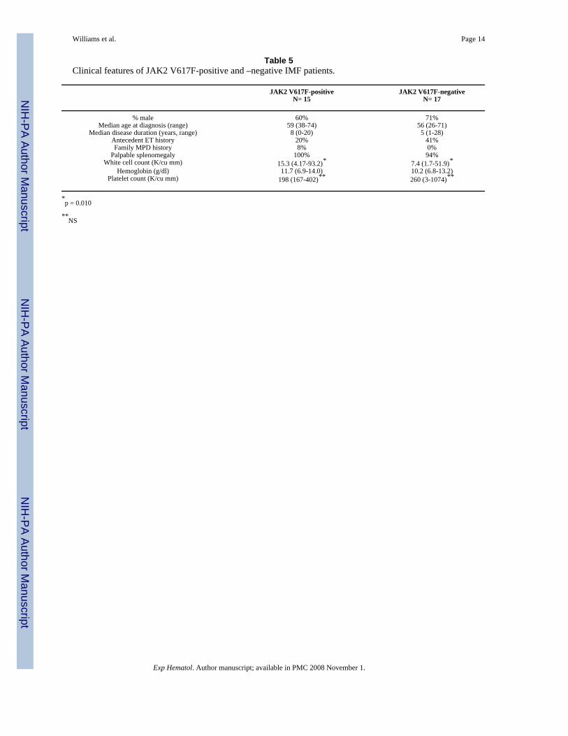
Clinical phenotype in a JAK2 V617F-negative IMF cohortIn a group of 32 subjects fulfilling the diagnostic criteria of IMF, 15 (47%) were JAK2 V617F-positive. Table 5 compares the clinical features of the 17 JAK2 V617F-negative IMF patientsto the 15 JAK2 V617F-positive IMF patients. There were no significant differences in medianage at presentation, disease duration, family history of an MPD, gender or prevalence ofsplenomegaly between JAK2 V617F-positive and -negative IMF patients. In both groups therewas a male predominance, and there was a higher incidence of antecedent ET in the JAK2V617F-negative group, and a significantly higher white cell count in the JAK2 V617F-positivegroup (p = 0.010).
MPL sequence variation in a JAK2 V617F-negative IMF cohortWe sequenced all the exons, the flanking intervening sequences and the 5′-proximaluntranslated region of the MPL gene in the 17 JAK2 V617F-negative IMF patients presented
Williams et al. Page 3
Exp Hematol. Author manuscript; available in PMC 2008 November 1.
NIH
-PA Author Manuscript
NIH
-PA Author Manuscript
NIH
-PA Author Manuscript

in Table 5. In one IMF patient, we identified a two base substitution (TG11534-5AA; Genbankaccession number GI:89161185) in exon 10 the MPL gene which results in a tryptophan tolysine substitution at amino acid 515 (W515K); the wild-type sequence was not identified fromeither peripheral blood CD34+ cells or neutrophils isolated from this patient. In a second IMFpatient, we identified a nucleotide transition (T1686C), which results in a serine to prolinesubstitution at amino acid 240 (S204P). In two other IMF patients, we identified heterozygousnucleotide transitions in intervening sequences of the splice recognition sites of exons 10(C11561T) and 11 (G14503A); these transitions were not identified in the public SNPdatabases. We directly sequenced exon 4, exon 10 and the intervening sequences of exon 10and 11 in an additional 13 JAK2V617F-positive IMF patients and in 9 of the 10 JAK2 V617F-negative PV patients listed in Table 2; no other sequence variation was identified. Clinical datafrom the four IMF patients with MPL sequence variation identified is presented in Table 6 andlocations of MPL sequence variation in the MPD are presented in Figure 2. Although patientnumbers are small, no distinct clinical differences were characterized the JAK2 V617f-negativepatients with MPL gene mutations.
DiscussionThe World Health Organization (WHO) currently classifies PV, IMF and ET together withchronic myelogenous leukemia, chronic eosinophilic leukemia, chronic neutrophilic leukemia,the hypereosinophilic syndrome and unclassifiable chronic myeloproliferative disease underthe rubric of the chronic myeloproliferative disorders(18). However, PV, IMF and ET havemore in common phenotypically with each other than with these other disorders and this wasconfirmed genotypically by the discovery of the JAK2 V617F mutation(6), the expression ofwhich is largely confined to PV, IMF and ET (19).
Extensive studies of JAK2 V617F have indicated that its frequency within PV, IMF and ET isvariable, being most common in PV. The relationship of JAK2 V617F and erythrocytosis, thedefining feature of PV, is supported by the observation of heightened erythropoietin sensitivityof homozygous V617F clones compared to heterozygous V617F clones(20), by higherhemoglobin concentrations in JAK2 V617F positive compared to JAK2 V617F-negative ETpatients(21), by mouse gene transfection experiments(8) and more recently by theidentification of JAK2 exon 12 lesions, which associate primarily with an erythrocytosisphenotype in both man and the mouse(10).
The data presented here and more extensive studies of MPL 515K/L mutations(12;13) placepatients with the JAK2 exon12, and MPL 515K/L/S204P lesions together with JAK2 V617Fin the spectrum of the chronic myeloproliferative disorders. While some patients with JAK2V617F and JAK2 exon 12 lesions are clinically similar, on average the JAK2 exon 12 patientshave significantly lower white cell and platelet counts than JAK2 V617F patients. In addition,JAK2 V617F, but not JAK2 exon 12 mutations, are associated with either thrombocytosis ormyelofibrosis phenotypes that are clinically indistinguishable from those associated withMPL515K/L. To date, JAK2 exon 12 lesions do not appear to associate with the ET phenotype,and in only rare, longstanding cases (patient PV3, Table 3A), do they associate with significantextramedullary disease. Thus, although the number of patients currently identified with thesemutations is small, so far the phenotypes generated by MPL and JAK2 exon 12 lesions do notappear to intersect, while JAK2 V617F, MPL gene mutations and Mpl epigenetic lesions do(Figure 3). When possible, a meta-analysis of larger groups of patients with JAK2 and MPLmutant alleles will be instrumental in further defining the associated clinical phenotypes.
We propose that the phenotypic variability associated with JAK2 V617F, extending fromerythrocytosis alone as described here and previously(22) to idiopathic myelofibrosis, can beexplained to some extent by variable JAK2 V617F allele burdens in MPD patients(16;23;24).
Williams et al. Page 4
Exp Hematol. Author manuscript; available in PMC 2008 November 1.
NIH
-PA Author Manuscript
NIH
-PA Author Manuscript
NIH
-PA Author Manuscript

The JAK2 V617F allele burden alone, however, cannot entirely account for the overlap ofphenotypes associated with JAK2 V617F. Beyond the JAK2 V617F clonal burden, the datasuggest that other genetic or epigenetic changes in MPL and other genes are probably importantmodifiers of JAK2 V617F-associated disease, and MPD generating in and of themselves.
Acknowledgements
We thank Linda M. Scott, Ph.D. (Cambridge, U. K.) for sharing data on JAK2 exon 12 mutations. This work wassupported by NIH research grants NHLBI RO1- HL082995 and NCI P01CA108671, a Department of Defense grant,MPO48019, and the Myeloproliferative Disorders Foundation.
Reference List1. Adamson JW, Fialkow PJ, Murphy S, Prchal JF, Steinmann L. Polycythemia vera: stem-cell and
probable clonal origin of the disease. N Engl J Med 1976 October 21;295(17):913–6. [PubMed:967201]
2. Najfeld V, Montella L, Scalise A, Fruchtman S. Exploring polycythaemia vera with fluorescence insitu hybridization: additional cryptic 9p is the most frequent abnormality detected. Br J Haematol 2002November;119(2):558–66. [PubMed: 12406101]
3. Prchal JF, Axelrad AA. Letter: Bone-marrow responses in polycythemia vera. N Engl J Med 1974June 13;290(24):1382. [PubMed: 4827655]
4. Temerinac S, Klippel S, Strunck E, Roder S, Lubbert M, Lange W, Azemar M, Meinhardt G, SchaeferHE, Pahl HL. Cloning of PRV-1, a novel member of the uPAR receptor superfamily, which isoverexpressed in polycythemia rubra vera. Blood 2000 April 15;95(8):2569–76. [PubMed: 10753836]
5. Moliterno AR, Hankins WD, Spivak JL. Impaired expression of the thrombopoietin receptor byplatelets from patients with polycythemia vera. N Engl J Med 1998 February 26;338(9):572–80.[PubMed: 9475764]
6. Baxter EJ, Scott LM, Campbell PJ, East C, Fourouclas N, Swanton S, Vassiliou GS, Bench AJ, BoydEM, Curtin N, Scott MA, Erber WN, Green AR. Acquired mutation of the tyrosine kinase JAK2 inhuman myeloproliferative disorders. Lancet 2005 March 19;365(9464):1054–61. [PubMed:15781101]
7. Levine RL, Wadleigh M, Cools J, Ebert BL, Wernig G, Huntly BJ, Boggon TJ, Wlodarska I, Clark JJ,Moore S, Adelsperger J, Koo S, Lee JC, Gabriel S, Mercher T, D'Andrea A, Frohling S, Dohner K,Marynen P, Vandenberghe P, Mesa RA, Tefferi A, Griffin JD, Eck MJ, Sellers WR, et al. Activatingmutation in the tyrosine kinase JAK2 in polycythemia vera, essential thrombocythemia, and myeloidmetaplasia with myelofibrosis. Cancer Cell 2005 April;7(4):387–97. [PubMed: 15837627]
8. James C, Ugo V, Le Couedic JP, Staerk J, Delhommeau F, Lacout C, Garcon L, Raslova H, Berger R,naceur-Griscelli A, Villeval JL, Constantinescu SN, Casadevall N, Vainchenker W. A unique clonalJAK2 mutation leading to constitutive signalling causes polycythaemia vera. Nature 2005 April 28;434(7037):1144–8. [PubMed: 15793561]
9. Kralovics R, Passamonti F, Buser AS, Teo SS, Tiedt R, Passweg JR, Tichelli A, Cazzola M, SkodaRC. A gain-of-function mutation of JAK2 in myeloproliferative disorders. N Engl J Med 2005 April28;352(17):1779–90. [PubMed: 15858187]
10. Scott LM, Tong W, Levine RL, Scott MA, Beer PA, Stratton MR, Futreal PA, Erber WN, McMullinMF, Harrison CN, Warren AJ, Gilliland DG, Lodish HF, Green AR. JAK2 exon 12 mutations inpolycythemia vera and idiopathic erythrocytosis. N Engl J Med 2007 February 1;356(5):459–68.[PubMed: 17267906]
11. Pikman Y, Lee BH, Mercher T, McDowell E, Ebert BL, Gozo M, Cuker A, Wernig G, Moore S,Galinsky I, DeAngelo DJ, Clark JJ, Lee SJ, Golub TR, Wadleigh M, Gilliland DG, Levine RL.MPLW515L is a novel somatic activating mutation in myelofibrosis with myeloid metaplasia. PLoSMed 2006 July;3(7):e270. [PubMed: 16834459]
12. Pardanani AD, Levine RL, Lasho T, Pikman Y, Mesa RA, Wadleigh M, Steensma DP, Elliott MA,Wolanskyj AP, Hogan WJ, McClure RF, Litzow MR, Gilliland DG, Tefferi A. MPL515 mutationsin myeloproliferative and other myeloid disorders: a study of 1182 patients. Blood 2006 November15;108(10):3472–6. [PubMed: 16868251]
Williams et al. Page 5
Exp Hematol. Author manuscript; available in PMC 2008 November 1.
NIH
-PA Author Manuscript
NIH
-PA Author Manuscript
NIH
-PA Author Manuscript

13. Guglielmelli P, Pancrazzi A, Bergamaschi G, Rosti V, Villani L, Antonioli E, Bosi A, Barosi G,Vannucchi AM. Anaemia characterises patients with myelofibrosis harbouring Mpl mutation. Br JHaematol 2007 May;137(3):244–7. [PubMed: 17408465]
14. Berk PD, Goldberg JD, Donovan PB, Fruchtman SM, Berlin NI, Wasserman LR. Therapeuticrecommendations in polycythemia vera based on Polycythemia Vera Study Group protocols. SeminHematol 1986 April;23(2):132–43. [PubMed: 3704665]
15. Barosi G. Myelofibrosis with myeloid metaplasia: diagnostic definition and prognostic classificationfor clinical studies and treatment guidelines. J Clin Oncol 1999 September;17(9):2954–70. [PubMed:10561375]
16. Moliterno AR, Williams DM, Rogers O, Spivak JL. Molecular mimicry in the chronicmyeloproliferative disorders: reciprocity between quantitative JAK2 V617F and Mpl expression.Blood 2006 December 1;108(12):3913–5. [PubMed: 16912229]
17. Tefferi A, Lasho TL, Schwager SM, Strand JS, Elliott M, Mesa R, Li CY, Wadleigh M, Lee SJ,Gilliland DG. The clinical phenotype of wild-type, heterozygous, and homozygous JAK2V617F inpolycythemia vera. Cancer 2006 February 1;106(3):631–5. [PubMed: 16369984]
18. Vardiman JW, Harris NL, Brunning RD. The World Health Organization (WHO) classification ofthe myeloid neoplasms. Blood 2002 October 1;100(7):2292–302. [PubMed: 12239137]
19. Jones AV, Kreil S, Zoi K, Waghorn K, Curtis C, Zhang L, Score J, Seear R, Chase AJ, Grand FH,White H, Zoi C, Loukopoulos D, Terpos E, Vervessou EC, Schultheis B, Emig M, Ernst T, LengfelderE, Hehlmann R, Hochhaus A, Oscier D, Silver RT, Reiter A, Cross NC. Widespread occurrence ofthe JAK2 V617F mutation in chronic myeloproliferative disorders. Blood 2005 September 15;106(6):2162–8. [PubMed: 15920007]
20. Dupont S, Masse A, James C, Teyssandier I, Lecluse Y, Larbret F, Ugo V, Saulnier P, Koscielny S,Le Couedic JP, Casadevall N, Vainchenker W, Delhommeau F. The JAK2 V617F mutation triggerserythropoietin hypersensitivity and terminal erythroid amplification in primary cells from patientswith polycythemia vera. Blood. 2007 March 27;
21. Campbell PJ, Scott LM, Buck G, Wheatley K, East CL, Marsden JT, Duffy A, Boyd EM, Bench AJ,Scott MA, Vassiliou GS, Milligan DW, Smith SR, Erber WN, Bareford D, Wilkins BS, Reilly JT,Harrison CN, Green AR. Definition of subtypes of essential thrombocythaemia and relation topolycythaemia vera based on JAK2 V617F mutation status: a prospective study. Lancet 2005December 3;366(9501):1945–53. [PubMed: 16325696]
22. Percy MJ, Jones FG, Green AR, Reilly JT, McMullin MF. The incidence of the JAK2 V617F mutationin patients with idiopathic erythrocytosis. Haematologica 2006 March;91(3):413–4. [PubMed:16503548]
23. Passamonti F, Vanelli L, Malabarba L, Rumi E, Pungolino E, Malcovati L, Pascutto C, Morra E,Lazzarino M, Cazzola M. Clinical utility of the absolute number of circulating CD34-positive cellsin patients with chronic myeloproliferative disorders. Haematologica 2003 October;88(10):1123–9.[PubMed: 14555308]
24. Vannucchi AM, Antonioli E, Guglielmelli P, Rambaldi A, Barosi G, Marchioli R, Marfisi RM, FinazziG, Guerini V, Fabris F, Randi ML, De S V, Caberlon S, Tafuri A, Ruggeri M, Specchia G, Liso V,Rossi E, Pogliani E, Gugliotta L, Bosi A, Barbui T. Clinical profile of homozygous JAK2V617Fmutation in patients with polycythemia vera or essential thrombocythemia. Blood. 2007 March 22;
25. Moliterno AR, Williams DM, Gutierrez-Alamillo LI, Salvatori R, Ingersoll RG, Spivak JL. MplBaltimore: A Thrombopoietin Receptor Polymorphism Associated with Thrombocytosis. Proc NatlAcad Sci U S A. 2004
26. Ding J, Komatsu H, Wakita A, Kato-Uranishi M, Ito M, Satoh A, Tsuboi K, Nitta M, Miyazaki H,Iida S, Ueda R. Familial essential thrombocythemia associated with a dominant-positive activatingmutation of the c-MPL gene, which encodes for the receptor for thrombopoietin. Blood 2004 June1;103(11):4198–200. [PubMed: 14764528]
Williams et al. Page 6
Exp Hematol. Author manuscript; available in PMC 2008 November 1.
NIH
-PA Author Manuscript
NIH
-PA Author Manuscript
NIH
-PA Author Manuscript

Figure 1.Somatic mutations of JAK2 exon 12 identified in JAK2 V617F-negative PV and idiopathicerythrocytosis patients. Unique mutations identified from this cohort are indicated by theasterisks. Mutations not described in this study are from Scott et al(10).
Williams et al. Page 7
Exp Hematol. Author manuscript; available in PMC 2008 November 1.
NIH
-PA Author Manuscript
NIH
-PA Author Manuscript
NIH
-PA Author Manuscript

Figure 2.MPL sequence variation associated with the chronic myeloproliferative disorders, K39N(25),S505N(26), W515K/L(11;12) as previously described.
Williams et al. Page 8
Exp Hematol. Author manuscript; available in PMC 2008 November 1.
NIH
-PA Author Manuscript
NIH
-PA Author Manuscript
NIH
-PA Author Manuscript
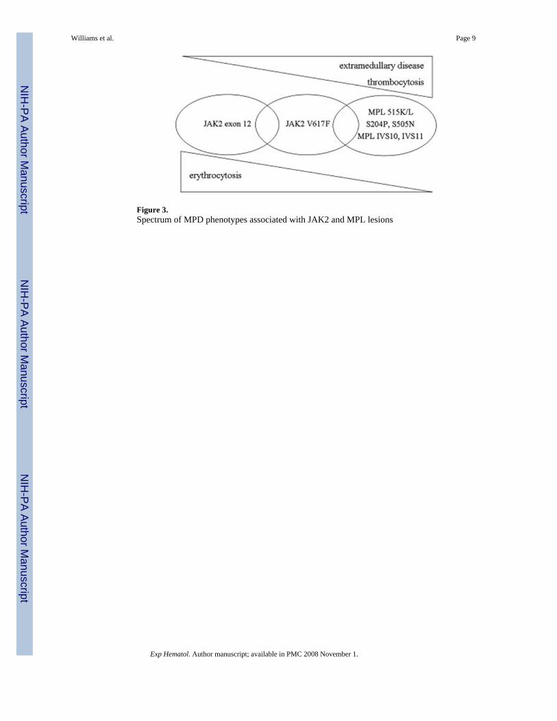
Figure 3.Spectrum of MPD phenotypes associated with JAK2 and MPL lesions
Williams et al. Page 9
Exp Hematol. Author manuscript; available in PMC 2008 November 1.
NIH
-PA Author Manuscript
NIH
-PA Author Manuscript
NIH
-PA Author Manuscript

NIH
-PA Author Manuscript
NIH
-PA Author Manuscript
NIH
-PA Author Manuscript
Williams et al. Page 10
Table 1Primer pairs to amplify MPL regions
Target Exon Forward (5′ to 3′) Reverse (5′ to 3′)
5′ flank B AGCCATCTCCAATCTGAGCA GAGATGAGGAAACGGAGATACA5′ flan A CAGCAGATTAGCCTCCCAAG CATCTTCTCCGCCACTGTGT
1-3 CTGAAGGGAGGATGGGCTA GTGACAGGAGGATGGCTCTT4 AGTCCAGAGGCTGAGCCATA AGGTCTGGAATCCCCAAAGT
5-6 CCACAGAGATGCTGTGCAAAT TTCTGCAAGATTGTGGCTCA7-8 AGGCCATCGTTCTTGTAGGA TCCCAAAGACAAAGGGAAGA9-10 GGATTTGGGTCAAACAGACG GCGGTATAGTGGGCGTGTTA11-12 CCATGGCTCAGTCTGCTTCT ACTGGGAGTGAGGAGCCTGT
Exp Hematol. Author manuscript; available in PMC 2008 November 1.

NIH
-PA Author Manuscript
NIH
-PA Author Manuscript
NIH
-PA Author Manuscript
Williams et al. Page 11
Table 2Clinical features of JAK2 V617F-positive and –negative PV patients.
JAK2 V617F-positiveN= 147
JAK2 V617F-negativeN= 10
% female 61% 60%Median age at diagnosis (range) 49 (18-74) 50 (18-67)
Median disease duration (years, range) 8 (1-28) 6 (3-54)Antecedent ET history 21% 20%Family MPD history 8% 20%
Palpable splenomegaly 43% 50%White cell count (K/cu mm) 11.9 (3.6-98.4)* 8.1 (4.5-20.2)*
Platelet count (K/cu mm) 507 (18-1320)** 427 (87-682)**
*p <0.001
**NS
Exp Hematol. Author manuscript; available in PMC 2008 November 1.

NIH
-PA Author Manuscript
NIH
-PA Author Manuscript
NIH
-PA Author Manuscript
Williams et al. Page 12Ta
ble
3C
linic
al fe
atur
es o
f pat
ient
s with
JAK
2 m
utat
ions
A. S
ubje
cts c
lass
ified
as P
V
Patie
ntJA
K2
lesi
onA
ge a
t dia
gnos
is/G
ende
rD
isea
se d
urat
ion
(yea
rs)
Whi
te c
ell
coun
t (K
/cu
mm
)Pl
atel
et c
ount
(K/c
u m
m)
Palp
able
sple
nom
egal
y
PV1
R54
1-E5
43de
linsK
61/M
16.
757
91
cmPV
2R
541-
E543
delin
sK50
/F5
7.8
303
2 cm
PV3
H53
8-K
539d
elin
sL18
/M54
11.0
60m
assi
ve
B. S
ubje
cts c
lass
ified
as i
diop
athi
c er
ythr
ocyt
osis
Patie
ntJA
K2
lesi
onA
ge a
t dia
gnos
is/G
ende
rD
isea
se d
urat
ion
(yea
rs)
Whi
te c
ell c
ount
(K/c
u m
m)
Plat
elet
cou
nt (K
/cu
mm
)Pa
lpab
le sp
leno
meg
aly
ERT1
N54
2-E5
43de
l54
/F15
8.5
257
none
ERT2
R54
1-E5
43de
linsK
15/M
15.
829
1no
neER
T3JA
K2
V61
7F41
/F1
5.7
227
none
ERT4
JAK
2 V
617F
82/F
106.
911
1no
ne
Exp Hematol. Author manuscript; available in PMC 2008 November 1.

NIH
-PA Author Manuscript
NIH
-PA Author Manuscript
NIH
-PA Author Manuscript
Williams et al. Page 13
Table 4Clinical features of patients with erythrocytosis
A. Subjects classified as idiopathic erythrocytosisJAK2 lesion-negative ERT
N= 40JAK2 lesion-positive ERT
N= 8*
% female 35% 75%Median age at diagnosis (range) 49 (18-74) 41(15-82)
Palpable splenomegaly 0% 0%White cell count (K/cu mm) 6.9 (4.2-14.7) 6.4 (5.1-8.6)
Platelet count (K/cu mm) 227 (109-753) 285 (111-308)
B. Subjects with erythrocytosis and a JAK2 lesionJAK2 V617F-positive PV or ERT
N= 149JAK2 exon12 lesions PV or ERT
N= 15*
% female 61% 53%Median age at diagnosis (range) 49 (18-74) 50 (15-62)
Palpable splenomegaly 43% 40%White cell count (K/cu mm) 11.9 (3.6-98.4)§ 7.8 (6.7-12.1)§
Platelet count (K/cu mm) 507 (18-1320)# 298 (60-579)#
*includes erythrocytosis patients from Scott et al(10).
*includes PV and erythrocytosis patients from Scott et al(10)
§p=0.001
#p=0.017
Exp Hematol. Author manuscript; available in PMC 2008 November 1.
NIH
-PA Author Manuscript
NIH
-PA Author Manuscript
NIH
-PA Author Manuscript
Williams et al. Page 14
Table 5Clinical features of JAK2 V617F-positive and –negative IMF patients.
JAK2 V617F-positiveN= 15
JAK2 V617F-negativeN= 17
% male 60% 71%Median age at diagnosis (range) 59 (38-74) 56 (26-71)
Median disease duration (years, range) 8 (0-20) 5 (1-28)Antecedent ET history 20% 41%Family MPD history 8% 0%
Palpable splenomegaly 100% 94%White cell count (K/cu mm) 15.3 (4.17-93.2)* 7.4 (1.7-51.9)*
Hemoglobin (g/dl) 11.7 (6.9-14.0) 10.2 (6.8-13.2)Platelet count (K/cu mm) 198 (167-402)** 260 (3-1074)**
*p = 0.010
**NS
Exp Hematol. Author manuscript; available in PMC 2008 November 1.

NIH
-PA Author Manuscript
NIH
-PA Author Manuscript
NIH
-PA Author Manuscript
Williams et al. Page 15Ta
ble
6C
linic
al fe
atur
es o
f IM
F pa
tient
s with
MPL
sequ
ence
var
iatio
n
Patie
ntM
PL le
sion
Age
at d
iagn
osis
/G
ende
rD
isea
se d
urat
ion
(yea
rs)
Whi
tece
llco
unt
(K/c
um
m)
Hem
oglo
bin
(g/
dl)
Plat
elet
coun
t (K
/cu
mm
)B
one
mar
row
Palp
able
sple
nom
egal
y
IMF1
W51
5K26
/M23
8.6*
12.1
209
oste
oscl
eros
isM
assi
veIM
F2S2
04P
42/M
111
.1*
12.8
368
oste
oscl
eros
isM
oder
ate
IMF3
IVS
11/1
271
/F3
51.9
*8.
824
oste
oscl
eros
isM
assi
veIM
F4IV
S 10
/11
35/M
312.
8*6.
847
oste
oscl
eros
isM
assi
ve
* Whi
te c
ell d
iffer
entia
l lef
t-shi
fted
to th
e m
yelo
cyte
stag
e
Exp Hematol. Author manuscript; available in PMC 2008 November 1.