Autoinhibition of Jak2 tyrosine kinase is dependent on specific regions in its pseudokinase domain

MOLECULAR AND CELLULAR BIOLOGY, June 2004, p. 5510–5520 Vol. 24, No. 120270-7306/04/$08.00�0 DOI: 10.1128/MCB.24.12.5510–5520.2004Copyright © 2004, American Society for Microbiology. All Rights Reserved.
Impaired Alveologenesis and Maintenance of Secretory MammaryEpithelial Cells in Jak2 Conditional Knockout MiceKay-Uwe Wagner,1* Andrea Krempler,1† Aleata A. Triplett,1 Yongyue Qi,1
Nicholas M. George,1 Jianqiong Zhu,2 and Hallgeir Rui2
Eppley Institute for Research in Cancer and Allied Diseases and Department of Pathology and Microbiology,University of Nebraska Medical Center, Omaha, Nebraska 68198-6805,1 and Department of Oncology,
Lombardi Comprehensive Cancer Center, Georgetown University Medical Center,Washington, D.C. 20057-14692
Received 29 December 2003/Returned for modification 21 February 2004/Accepted 20 March 2004
Jak2 is a hormone-receptor-coupled kinase that mediates the tyrosine phosphorylation and activation ofsignal transducers and activators of transcription (Stat). The biological relevance of Jak2-Stat signaling inhormone-responsive adult tissues is difficult to investigate since Jak2 deficiency leads to embryonic lethality.We generated Jak2 conditional knockout mice to study essential functions of Jak2 during mammary glanddevelopment. The mouse mammary tumor virus-Cre-mediated excision of the first coding exon resulted in aJak2 null mutation that uncouples signaling from the prolactin receptor (PRL-R) to its downstream mediatorStat5 in the presence of normal and supraphysiological levels of PRL. Jak2-deficient females were unable tolactate as a result of impaired alveologenesis. Unlike Stat5a knockouts, multiple gestation cycles could notreverse the Jak2-deficient phenotype, suggesting that neither other components of the PRL-R signaling cascadenor other growth factors and their signal transducers were able to compensate for the loss of Jak2 function toactivate Stat5 in vivo. A comparative analysis of Jak2-deficient mammary glands with transplants fromStat5a/b knockouts revealed that Jak2 deficiency also impairs the pregnancy-induced branching morphogen-esis. Jak2 conditional mutants therefore resemble PRL-R knockouts more closely, which suggested that Jak2deficiency might affect additional PRL-R downstream mediators other than Stat5a and Stat5b. To addresswhether Jak2 is required for the maintenance of PRL-responsive, differentiating alveolar cells, we utilized atransgenic strain that expresses Cre recombinase under regulatory elements of the whey acidic protein gene(Wap). The Wap-Cre-mediated excision of Jak2 resulted in a negative selection of differentiated alveolar cells,suggesting that Jak2 is required not only for the proliferation and differentiation of alveolar cells but also fortheir maintenance during lactation.
Prolactin (PRL), in synergy with other peptide and steroidhormones as well as local growth factors, controls the prolif-eration and differentiation of mammary epithelial cells (12, 13,38). PRL seems to play a central role in this process, as it actsas a mitogen and a morphogen (15, 32). Upon dimerization ofits receptor, PRL is suggested to signal through Janus kinases(Jak) and signal transducers and activators of transcription(Stat), in particular Jak2, Stat5a, and Stat5b (14, 34), as well asRas/mitogen-activated protein kinase (6, 8), phosphatidylino-sitol 3-kinase–AKT, and the phospholipase C-protein kinase Cpathway (10, 26). Loss-of-function studies with mouse modelsprovide new insights into biologically relevant functions ofindividual components of the PRL receptor (PRL-R) signalingcascade. Surprisingly, the lack of PRL (15), the PRL-R (32),and Stat5a (25, 37) exhibited an unexpected level of specificityduring mammogenesis. Indispensable functions of these pro-teins are restricted to pregnancy-associated changes in themammary gland, and all three knockout models (PRL�/�,PRL-R�/�, and Stat5a�/� mice) lack alveolar proliferation
and differentiation. Based on this line of evidence, it is appar-ent that Stat5a mediates essential, biologically relevant func-tions of PRL signaling in normal mammary epithelial cells. Itis known from studies using in vitro model systems that uponligand-induced aggregation of the receptor, Jak2 is stably re-cruited to the receptor complex. Jak2 then undergoes auto-phosphorylation and phosphorylates the PRL-R on varioustyrosine residues, thus generating binding sites for several SH2domain-bearing signaling molecules including Stat5a andStat5b (reviewed in reference 14). The subsequent phosphor-ylation of Stat5a and Stat5b by Jak2 triggers their homo- andheterodimerization and translocation into the nucleus, wherethey bind to specific DNA sequence motifs and activate tran-scription of target genes such as �-casein and the whey acidicprotein gene (Wap).
Although the biological relevance of Stat5 activation in re-sponse to PRL signaling has been demonstrated with knockoutmice, the enzymatic coupling between the PRL-R and Stat5 invivo and biologically relevant functions of Jak2 are difficult toelucidate due to the prenatal lethality of Jak2 conventionalknockouts (29, 33). Pivotal studies with conventional knockoutmice revealed that the biological functions of Jak2 are pleio-tropic, which is consistent with its suggested coupling to mul-tiple cytokine receptors in specific target cells (reviewed inreference 20). Specifically, Jak2 is suggested to mediate signalsthrough a variety of single-chain receptors for ligands such as
* Corresponding author. Mailing address: Eppley Institute for Re-search in Cancer and Allied Diseases, University of Nebraska MedicalCenter, 986805 Nebraska Medical Center, Rm. 8009, Omaha, NE68198-6805. Phone: (402) 559-3288. Fax: (402) 559-4651. E-mail:[email protected].
† Present address: Department of Medical Biochemistry and Mo-lecular Biology, University of the Saarland, Homburg 66424, Germany.
5510

PRL, growth hormone (GH), erythropoietin (EPO), andthrombopoietin as well as to the multichain interleukin 3 re-ceptor family (e.g., interleukin 3 receptor and granulocyte-macrophage colony-stimulating factor receptor) and membersof the gp130 receptor family. To define a role for Jak2 duringmammary development, Shillingford and coworkers (35) re-cently reported the first successful attempt to transplant mam-mary gland anlagen from Jak2-deficient embryos into wild-typerecipients. These initial studies revealed that Jak2-deficientmammary epithelial cells showed a reduced proliferation indexin animals treated with estrogen and progesterone, and thelack of Jak2 impaired the specification of secretory epithelialcells. Since alveolar precursors do not proliferate, it is evidentthat this transplantation model lacks the entire alveolar lin-eage. Consequently, transplantation models do not allow theexamination of essential functions of Jak2 in differentiatingepithelial subtypes during pregnancy and lactation. Moreover,in transplanted epithelia of postpartum females, differentiatedcells immediately inactivate Stat5, upregulate Stat3, and initi-ate proapoptotic signaling pathways (e.g., expression of Baxand Bcl-xs) due to the lack of a nipple and, consequently, milkstasis (22).
To identify essential functions of Jak2 at defined stages ofmammary development, we have generated two types of mam-ma-specific knockout mice on the basis of the Cre-lox system.The mouse mammary tumor virus (MMTV)-Cre-mediated ex-cision of the first coding exon resulted in a Jak2 null mutation,which is incapable of activating Stat5 in mammary epithelialcells in response to PRL signaling. Like in the transplantmodel, the MMTV-Cre-mediated deletion of Jak2 from ductalprogenitors had no effect on ductal elongation and branchingmorphogenesis in nulliparous females. Mutants were, however,unable to lactate as a result of impaired alveolar specificationduring early gestation. Unlike the Stat5a knockout (23), mul-tiple gestation cycles did not reverse this abnormal phenotype,suggesting that neither other components of the PRL-R sig-naling cascade nor other growth factors and their signal trans-ducers, such as Src and ErbB family members (18, 31), wereable to compensate for the loss of Jak2 function to activateStat5 in vivo. To study the function of Jak2 specifically in thealveolar lineage, we utilized Wap-Cre transgenic mice to deletethe Jak2 gene specifically from PRL-responsive, differentiatingalveolar cells during the last phase of pregnancy and lactation.This approach revealed a strong negative selection of differ-entiated alveolar cells, suggesting that Jak2 is required not onlyfor the proliferation and initiation of differentiation of imma-ture alveolar cells but also for their maintenance at the differ-entiated stage during lactation.
MATERIALS AND METHODS
Mouse models and genotyping protocols. Technical details about the cloningand sequencing of the Jak2 genomic locus (GenBank accession numberAY157991), the construction of the targeting construct, and the production ofgenetically engineered mice with two Jak2 conditional knockout alleles (Jak2fl/fl
or Jak2tm1Kuw) or the derived null alleles will be described elsewhere (A. Krem-pler, submitted for publication). The PCR protocols for genotyping Wap-Cre,MMTV-Cre, and Rosa-lacZ mice have been described previously (36, 39, 41).The Stat5a/b double knockout mice (37) were a kind gift from James N. Ihle (St.Jude Children’s Research Hospital, Memphis, Tenn.) to K.U.W. Athymic nudemice (NCr strain, National Cancer Institute) were used for transplantation stud-
ies. All animals used in the studies were treated humanely and in accordance withfederal guidelines and institutional policies.
Transplantation method. The surgical procedures for clearing the fat pad of3-week-old female mice and the method of implanting tissue fragments and cellsuspensions have been described recently in great detail (43). Briefly, randomfragments (�1.0 mm3) of mammary epithelium were taken from nulliparousStat5a/b�/� females and implanted into the cleared fat pad of 3-week-old recip-ients (athymic NCr-nu mice). The recipients were kept as nulliparous virgins for12 weeks to provide sufficient time for the transplanted epithelium to form aductal tree. Subsequently, the transplant-carrying females were bred with aheterozygous athymic NCr-nu male, and mammary glands were taken from therecipients immediately after delivering the young. The mammary glands wereprepared as whole mounts and stained as described below.
PRL injections into mice. Ovine PRL (NIDDK-oPRL-21, AFP-10692C) waskindly provided by A. F. Parlow under the sponsorship of the National Hormoneand Pituitary Program, National Institute of Diabetes and Digestive and KidneyDiseases (National Institutes of Health, Bethesda, Md.). Nulliparous Jak2-defi-cient mice and their wild-type controls were injected intraperitoneally with ap-proximately 100 �g of PRL (5 �g per g of body weight) or plain saline solution.Mammary glands were taken 30 min later and fixed overnight at 4°C in 10%buffered formalin. The next day, the tissues were dehydrated, sectioned, andstained with an anti-phospho-STAT5a/b antibody.
Carmine alum and X-Gal staining of mammary whole mounts. Whole mountswere prepared and stained in carmine alum as described previously (42). Astandard X-Gal (5-bromo-4-chloro-3-indolyl-�-D-galactopyranoside) stainingprocedure (39) was performed to detect �-galactosidase activity in mammarygland whole mounts. Tissues were incubated overnight at 30°C in X-Gal stainingbuffer containing a final concentration of 1 mg of X-Gal/ml. X-Gal-stainedmammary whole mounts were postfixed in 10% buffered formalin (Fisher Sci-entific Company), dehydrated to 100% ethanol, and placed overnight in Histo-Clear (National Diagnostics, Atlanta, Ga.) before microscopic analysis. For theexamination of tissue sections, mammary glands were dehydrated following theX-Gal procedure, embedded in paraffin, and sectioned.
Immunohistochemistry. For bromodeoxyuridine labeling, TUNEL (terminaldeoxynucleotidyltransferase-mediated dUTP-biotin nick end labeling) assay, orgeneral immunohistochemistry, mammary glands were fixed overnight at 4°C in10% buffered formalin (Fisher Scientific Company). Paraffin sections were pro-cessed and stained with anti-Stat5 and anti-phospho-Stat5a/b (Tyr694/Tyr699)antibodies as described previously (30). The anti-Stat5a-specific antibody was akind gift from Lothar Hennighausen (National Institutes of Health). The anti-bromodeoxyuridine mouse monoclonal antibody was a component of the cellproliferation kit from Amersham Pharmacia Biotech. Biotinylated or AlexaFluor 488-conjugated secondary antibodies were used to detect the phosphory-lated and/or inactive form of Stat5. The labeling of apoptotic nuclei with Chro-maTide Alexa Fluor 488-5-dUTP was performed as described previously (19).Slides were counterstained briefly with DAPI (4�,6�-diamidino-2-phenylindole)or hematoxylin to visualize nuclei. Bright-field and fluorescence images of his-tological slides were taken on a Nikon Labophot microscope equipped with aNikon Coolpix 990 camera as well as fluorescein isothiocyanate and DAPI filtersets.
Southern blot of recombined tissues. Genomic DNA from tissues was pre-pared by using standard phenol-chloroform extraction. Fifteen micrograms wasdigested with EcoRI at 37°C overnight and separated on a 0.8% agarose gel. TheDNA was denatured and blotted onto a nylon membrane (Genescreen plus;NEN) and hybridized with a 32P-labeled probe. The 3� external probe, approx-imately 643 bp in size, was generated by PCR using the 1724-1725 primer pair(5�-CCA GGT TCA TAC ATC TCA AAA CC-3� and 5�-GTC ACA GTA GTCCTT TGT CAG G-3�). Membranes were washed in 0.1� SSC (1� SSC is 0.15 MNaCl plus 0.015 M sodium citrate) buffer containing sodium dodecyl sulfate andexposed for 16 h to a Kodak XOMAT-AR film.
Cell culture. Mouse embryonic fibroblasts (MEFs) were derived from 12.5-day-old embryos and grown at 37°C with 5% CO2 until confluence in 75-cm2
culture flasks in Dulbecco’s modified Eagle’s medium (Biofluids, Rockville, Md.)containing 10% fetal calf serum (Atlanta Biologicals, Norcross, Ga.), 2 mML-glutamine, 0.1 mM nonessential amino acids, 10 �g of gentamicin/ml, andpenicillin-streptomycin (50 IU/ml and 50 �g/ml, respectively). Cells were starvedin serum-free Dulbecco’s modified Eagle’s medium for 16 h and then incubatedwith or without ovine 20 nM GH for 15 min at 37°C. Cells were harvested, frozenon dry ice, and stored at �80°C before being used in protein immunoblottingexperiments.
Protein analysis. Embryos (12.5 days old) were pooled into groups of threeand homogenized on ice in a Triton X-100-based lysis buffer with proteaseinhibitors and processed as described previously (30). Frozen cell pellets from
VOL. 24, 2004 CONDITIONAL KNOCKOUT OF Jak2 5511

MEFs were lysed directly in the same buffer. Clarified tissue lysates correspond-ing to 2.0 mg of total protein were immunoprecipitated with 4 �g of a Jak2polyclonal rabbit antibody (Upstate Biotechnology, Lake Placid, N.Y.) per ml.Clarified cell lysates were immunoprecipitated with 4 �g of either the Jak2antibody or a panStat5a/b antibody (AX55; Advantex Bioreagents, Conroe, Tex.)per ml. Immunoprecipitates or whole-cell extracts were resolved by sodiumdodecyl sulfate–7.5% polyacrylamide gel electrophoresis and immunoblottedwith either phosphoStat5a/b (Y694/9) antibody (0.5 �g/ml; AX1; AdvantexBioreagents), panStat5a/b antibody (2 �g/ml), phosphotyrosine antibody (1 �g/ml; 4G10; Upstate Biotechnology), or Jak2 antibody (1:3,000). Horseradish per-oxidase-coupled secondary antibodies and enhanced chemiluminescence (Am-ersham, Piscataway, N.J.) were used for detection.
RESULTS
The conditional deletion of Jak2 results in a true null mu-tation. We generated conditional knockout mice in which thefirst coding exon of Jak2 can be deleted through Cre-mediated
recombination (Fig. 1A). A detailed description of the con-struction of the targeting vector and phenotypic consequencesof the deletion of Jak2 in the germ line will be describedelsewhere (Krempler et al., submitted). In brief, the Jak2 con-ditional knockout allele contains a floxed PGK-neo selectablemarker approximately 600 bp upstream of the first coding exon(exon 2). A third loxP site was placed 500 bp downstream ofthis exon. Homozygous mutant mice that carried two floxedJak2 alleles developed normally until adulthood. Both malesand females were fertile, and they exhibited no phenotypicabnormalities, suggesting that the insertion of the selectablemarker upstream of the first coding exon did not affect thetranscriptional regulation of Jak2. Next, we generated knock-out mice that lack Jak2 completely in all organs (Jak2�/�) toassess phenotypic abnormalities that were reported previously
FIG. 1. Conditional knockout of the Jak2 gene. (A) The targeted insertion of loxP sites around the first coding exon (exon 2) created aconditional knockout allele of Jak2 that can be excised upon Cre-mediated recombination. The deletion of Jak2 can be monitored by usingSouthern blot analysis with a 3� external probe following an EcoRI (R) restriction digest. Alternatively, a PCR assay with the 1786-1787 primerset can be employed to verify the presence of a Jak2 null allele in cells expressing Cre. (B) Immunoprecipitation and Western blot analysis of Jak2derived from embryos carrying two wild-type or two knockout alleles. (C) Immunoprecipitation (IP) and Western blot analysis to monitor theactivation of Stat5 and autophosphorylation of Jak2 in GH-treated MEFs that are heterozygous or homozygous knockouts. Note that Jak2deficiency (Jak2�/�) prevents the GH-mediated tyrosine phosphorylation (PY) of Stat5.
5512 WAGNER ET AL. MOL. CELL. BIOL.

in Jak2 conventional knockout mice with targeted deletions ofexons 2 or 3 (29, 33). The Cre-mediated deletion of Jak2 in thegerm line of mutant mice resulted in embryonic lethality atmidgestation due to a lack of EPO signaling and defectivedefinitive erythropoiesis (Krempler et al., submitted). Thesephenotypic data suggested that the conditional deletion of thefirst coding exon results in a true null mutation of Jak2 in cellsexpressing Cre recombinase.
To verify the absence of the Jak2 protein in cells with twonull alleles, we established primary MEF cultures from anemicembryos at day 12.5 of gestation and their normal-lookingwild-type or heterozygous littermates. The presence of twoJak2 null alleles (Jak2�/�) in retarded embryos and derivedfibroblasts was verified by PCR and Southern blot analysis(data not shown). The immunoprecipitation and Western blotanalysis demonstrated that the Jak2 protein was not expressedin Jak2�/� MEFs (Fig. 1B). We also did not detect any smallerprotein variants of Jak2 in these knockout cells (data notshown), suggesting that other downstream ATG codons wereinsufficient to initiate translation and that the Jak2 null alleledoes not produce a residual protein with limited functionality.The phenotypic resemblance of the mutant mice with othertargeted Jak2 mutations (29, 33) supported this conclusion.
Jak2-deficient (Jak2�/�) fibroblasts multiplied normally invitro, suggesting that MEFs do not depend on GH-mediatedactivation of Stat5. Heterozygous control cells (Jak2�/�) donot have active Stat5 (Fig. 1C), but GH-receptor-mediatedautophosphorylation of Jak2 and activation of Stat5 can beinduced in control cells immediately after adding GH into thegrowth medium. In contrast, Jak2-deficient MEFs lack theGH-induced activation of Stat5, suggesting that the functionalinhibition of Jak2 is sufficient to decouple GH from its down-stream mediators Stat5a and Stat5b. In summary, the pheno-typic examination of Jak2-deficient embryos and the biochem-ical analysis of derived MEFs clearly demonstrate that aknockout of Jak2 is sufficient to suppress EPO and GH signal-ing through their corresponding single-chain receptors.
The MMTV-Cre-mediated deletion of Jak2 results in im-paired mammary gland development in virgin, pregnant, andpostpartum females. We generated conditional knockout micethat carry an MMTV-Cre transgene in the Jak2 homozygousfloxed background (MMTV-Cre Jak2fl/fl) to address importantbiological functions of Jak2 during mammary gland develop-ment. The MMTV-Cre transgene is expressed in all majorepithelial subtypes (i.e., luminal and myoepithelial cells) of theprepubescent mammary gland (40). Mutant mice lacking Jak2somatically in MMTV-Cre target tissues develop normally andare fertile. Jak2-deficient dams were, however, unable to lac-tate (n � 17). Similar to the Jak2�/� transplantation model byShillingford et al. (35), the MMTV-Cre-mediated deletion ofJak2 in nulliparous females does not cause phenotypic abnor-malities during ductal elongation and the formation of second-ary side branches (Fig. 2A and B). A careful analysis of theconditional knockouts, however, revealed an important differ-ence in the architecture of virgin mammary glands that was notreported previously. Lobular units that usually reside on ter-minal ducts of tertiary side branches of mature virgins (Fig. 2A,arrow) were absent in nulliparous Jak2 conditional knockouts(Fig. 2B). This finding suggests that Jak2-mediated signaltransduction plays an important role in the origination of al-
veolar precursors in response to the concerted action of hor-mones during the normal estrus cycle. Due to the lack ofalveolar precursors, the development of secretory lobules isseverely impaired in pregnant and postpartum MMTV-CreJak2fl/fl females (Fig. 2C and D). During early pregnancy (day7.5 of gestation), the proliferation index is significantly de-creased in Jak2 conditional knockouts compared to in theirlittermate wild-type controls (4 versus 11%; n 3,000). Shil-lingford et al. demonstrated previously that after the adminis-tration of estrogen and progesterone into nulliparous females,the multiplication of epithelial cells is affected in Jak2-deficienttransplants. Based on our observations of the conditionalknockout, the difference in the proliferation index is likely aconsequence of the lack of the entire alveolar lineage sinceprogesterone is a mitogen for this specific epithelial subtype.To verify the lack of alveolar specification in the Jak2 condi-tional knockout, we analyzed the expression of ductal versusalveolar markers, in particular Nkcc1 (Slc12a2) and Npt2b(Slc34a2), in Jak2-deficient mammary glands. Indeed, Jak2�/�
epithelia did not undergo alveolar specification during preg-nancy (data not shown). In addition, we examined the func-tional differentiation and expression of late milk protein geneWap, which is known to be regulated by PRL signaling andStat5a activation (25, 27). While the Wap protein was highlyabundant within the lumen and secretory alveolar cells in post-partum wild-type controls, Jak2-deficient females failed to syn-thesize this milk protein, suggesting that in addition to alveolarspecification, mammary epithelial cells do not undergo termi-nal differentiation in the absence of Jak2 (data not shown).
It is a common feature of many genetically engineeredmouse strains that lactation can be restored after multiplepregnancies. For instance, PRL-R heterozygous mutants andStat5a knockout mice develop normal secretory alveoli aftermultiple pregnancy cycles (23, 32). We recently proposed auniversal mechanism for the selective amplification of alveolarcells expressing differentiation markers while retaining charac-teristics of stem-progenitor cells in the remodeled mammarygland (39). In analogy to these studies, we monitored Jak2-deficient dams during multiple pregnancy cycles for their abil-ity to produce milk to determine whether Jak2 deficiency canbe compensated by accompanying signaling pathways that havebeen implicated in Jak2-independent activation of Stat5 (18,31). Mice that exhibited a lack of lactogenesis as single-parousdams did not resume milk production after four to seven preg-nancies (n � 6), suggesting that mammary epithelial stem cellsor specific precursors of the alveolar lineage were unable toactivate alternative signal transduction cascades to bypass thelack of Jak2.
Jak2-deficient mammary epithelial cells fail to activateStat5. The Jak2-mediated phosphorylation of Stat5a andStat5b triggers their dimerization and translocation into thenucleus, where they activate the transcription of target genessuch as Csnb (�-casein) and Wap (9, 24). We could demon-strate that in cultured fibroblasts, a null mutation of Jak2 wasable to abolish the GH-induced phosphorylation of Stat5 (seeabove). To verify that Jak2 deficiency results in a lack of Stat5activation in vivo, we examined the localization and phosphor-ylation of Stat5 using immunohistochemistry on histologicalsections of mammary tissues derived from conditional knock-outs and their littermate controls. At parturition, the vast ma-
VOL. 24, 2004 CONDITIONAL KNOCKOUT OF Jak2 5513
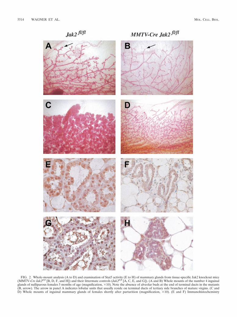
FIG. 2. Whole-mount analysis (A to D) and examination of Stat5 activity (E to H) of mammary glands from tissue-specific Jak2 knockout mice(MMTV-Cre Jak2fl/fl [B, D, F, and H]) and their littermate controls (Jak2fl/fl [A, C, E, and G]). (A and B) Whole mounts of the number 4 inguinalglands of nulliparous females 5 months of age (magnification, �10). Note the absence of alveolar buds at the end of terminal ducts in the mutants(B, arrow). The arrow in panel A indicates lobular units that usually reside on terminal ducts of tertiary side branches of mature virgins. (C andD) Whole mounts of inguinal mammary glands of females shortly after parturition (magnification, �10). (E and F) Immunohistochemistry
5514 WAGNER ET AL. MOL. CELL. BIOL.
jority of the Stat5a protein is localized in the nucleus of luminalepithelial cells in the wild-type controls (Fig. 2E). In contrast,Stat5a was confined to the cytoplasm in mammary epithelia ofJak2 knockout mice (Fig. 2F). Using a phosphorylation-spe-cific antibody against Stat5, we observed that the nuclear pro-tein was phosphorylated in the controls but not in Jak2-defi-cient dams (Fig. 2G and H). In conclusion, these observationssuggest that other receptor tyrosine kinases are unable to ac-tivate Stat5 under normal physiological conditions in mam-mary epithelia lacking Jak2.
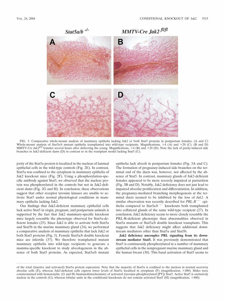
Our findings that Jak2-deficient mammary epithelial cellslack active Stat5 in virgin, pregnant, and postpartum animals issupported by the fact that Jak2 mammary-specific knockoutmice largely resemble the phenotype observed for Stat5a-de-ficient females (25). Since Jak2 is able to activate both Stat5aand Stat5b in the murine mammary gland (24), we performeda comparative analysis of mammary epithelia that lack Jak2 orboth Stat5 proteins (Fig. 3). Female Stat5a/b double knockoutmice are infertile (37). We therefore transplanted mutantmammary epithelia into wild-type recipients to generate amamma-specific knockout to study alveologenesis in the ab-sence of both Stat5 proteins. As expected, Stat5a/b mutant
epithelia lack alveoli in postpartum females (Fig. 3A and C).The formation of pregnancy-induced side branches on the ter-minal end of the ducts was, however, not affected by the ab-sence of Stat5. In contrast, mammary glands of Jak2-deficientfemales appeared to be more severely impaired at parturition(Fig. 3B and D). Notably, Jak2 deficiency does not just lead toimpaired alveolar proliferation and differentiation. In addition,the pregnancy-mediated branching morphogenesis at the ter-minal ducts seemed to be inhibited by the loss of Jak2. Asimilar observation was recently described for PRL-R�/� epi-thelia compared to Stat5a/b�/� knockouts both transplantedinto collateral glands of the same wild-type recipient (27). Inconclusion, Jak2 deficiency seems to more closely resemble thePRL-R-deficient phenotype than abnormalities observed inStat5a mutants or Stat5a/b double knockout transplants. Thissuggests that Jak2 deficiency might affect additional down-stream mediators other than Stat5a and Stat5b.
Jak2 deficiency uncouples PRL signaling from its down-stream mediator Stat5. It was previously demonstrated thatStat5 is continuously phosphorylated in a number of mammaryepithelial cells in the nonpregnant murine mammary gland andthe human breast (30). This basal activation of Stat5 seems to
FIG. 3. Comparative whole-mount analysis of mammary epithelia lacking Jak2 or both Stat5 proteins in postpartum females. (A and C)Whole-mount analysis of Stat5a/b mutant epithelia transplanted into wild-type recipients. Magnifications, �4 (A) and �20 (C). (B and D)MMTV-Cre Jak2fl/fl females several hours after delivering the young. Magnifications, �4 (B) and �20 (D). Note the lack of parity-induced sidebranches in Jak2-deficient dams (D) in contrast to in the transplant model lacking Stat5 (C).
of the total (inactive and activated) Stat5a protein expression. Note that the majority of Stat5a is confined to the nucleus in normal secretoryalveolar cells (E), whereas Jak2-deficient cells express lower levels of Stat5a localized in cytoplasm (F) (magnification, �400). Slides werecounterstained with hematoxylin. (G and H) Immunohistochemistry of activated (tyrosine-phosphorylated [PY]) Stat5. Active Stat5 is exclusivelynuclear in the controls (G), whereas lobular units in the conditional knockouts do not contain activated Stat5 (H) (magnification, �400).
VOL. 24, 2004 CONDITIONAL KNOCKOUT OF Jak2 5515

be controlled by hormones of the pituitary gland, in particular,by PRL. Phosphorylated Stat5 was not observed in hypophy-sectomized females and in nulliparous PRL-R knockouts. Al-though a large amount of the Stat5a protein is generally con-
fined to the cytoplasm (Fig. 4A), phosphorylated Stat5 can beclearly detected in a significant number of nuclei in luminalepithelial cells of nonpregnant wild-type females (Fig. 4E).Consistent with our previous studies of hypophysectomized
FIG. 4. Lack of PRL-induced phosphorylation and nuclear translocation of Stat5 in mammary epithelial cells of nulliparous Jak2 conditionalknockouts (MMTV-Cre Jak2fl/fl [B, D, F, and H]) compared to nonpregnant controls (Jak2fl/fl [A, C, E, and G]). (A to D) Immunofluorescencestaining of the total (inactive and activated) Stat5a protein in nonpregnant females injected with ovine PRL (oPRL) (� oPRL [C and D]) or saline(� oPRL [A and B]). Note that the majority of the Stat5a protein is confined to the nucleus in wild-type cells after administration ofsupraphysiological levels of oPRL (C), whereas Jak2-deficient mice exhibit no major changes in the nuclear transport of Stat5 (D). Magnification,�400. (E to H) Immunohistochemistry of activated (tyrosine-phosphorylated [PY]) Stat5 in nonpregnant females injected with oPRL (G and H)or saline (E and F). Active Stat5 can be detected in a number of nuclei in luminal epithelial cells of nonpregnant wild-type females (E), whereasJak2-deficient mice have a significantly reduced basal activation of Stat5 (F). A virtually uniform activation of Stat5 is evident in nulliparous femalesafter administration of supraphysiological levels of oPRL (G). In contrast, Jak2-deficient mice have a significantly reduced PRL-inducedphosphorylation and nuclear translocation of Stat5 after administration of oPRL (H). Magnification, �400.
5516 WAGNER ET AL. MOL. CELL. BIOL.
virgins and PRL-R mutants, the deletion of Jak2 results in asignificant inhibition of the basal Stat5 activation. NuclearStat5 is virtually absent in MMTV-Cre Jak2fl/fl animals (Fig. 4Band F). The administration of supraphysiological levels ofovine PRL induced a nearly uniform phosphorylation and nu-clear translocation of the bulk of the Stat5 protein in nonpreg-nant wild-type controls (Fig. 4C and G). In contrast, Stat5remained cytoplasmic or perinuclear in the majority of luminalepithelial cells in the Jak2 mutants injected with the sameamount of PRL (Fig. 4D and H). In conclusion, the results ofthe comparative analysis and the PRL injection study confirmthat (i) the basal activation of Stat5 in nulliparous females ismediated by PRL via Jak2 and (ii) Jak2 deficiency disruptsPRL-R signaling in luminal epithelial cells. Based on the se-verity of the phenotype observed in the Jak2 mutants com-pared to that of the Stat5a/b null transplants, a knockout ofJak2 might curtail PRL-R downstream targets other thanStat5.
Jak2 is required for PRL-mediated maintenance of func-tionally differentiated alveolar cells. The MMTV-Cre-medi-ated mamma-specific knockout of Jak2 in nulliparous, preg-nant, and postpartum females revealed that alveolar proliferation,specification, and differentiation are critically dependent on Jak2.We then wanted to determine whether Jak2 is also required forthe maintenance and survival of differentiated, secretory alveolarcells. Declining circulating levels of PRL in combination with anactivation of proapoptotic factors induced by milk stasis are re-sponsible for the collapse of alveolar structures during the firstphase of involution at the end of lactation (22, 42). The dephos-phorylation of Stat5 and activation of Stat3 are two molecularevents that seem to stimulate the programmed cell death of ter-minally differentiated cells (reviewed in reference 13). To exam-ine whether Jak2-Stat5 signaling is required for the PRL-medi-ated survival and maintenance of differentiated mammaryepithelial cells, we specifically deleted Jak2 from this secretoryepithelial subpopulation during late pregnancy and lactation.
The experimental design for this study is illustrated in Fig.5A. As discussed earlier, Wap is a PRL-induced milk proteingene whose expression is largely confined to secretory alveolarcells. Since Wap is highly upregulated during the last phase ofpregnancy, the expression of this gene is generally applied as aterminal differentiation marker for mammary epithelial cells.We developed transgenic mice that express Cre under thecontrol of the Wap gene promoter (Wap-Cre) to target thisrecombinase specifically to PRL-responsive, differentiatingmammary epithelial cells (41). A careful examination of theWap-Cre expression profile by using a Cre-lox reporter strain,Rosa-lox-Stop-lox-LacZ (36), revealed that Cre expression andactivation of the reporter coincide with the upregulation of theendogenous Wap locus during the second half of pregnancy(39). In this context, it is important that adequate levels of Wap(and therefore Wap-Cre expression) require the formation of aproper three-dimensional structure of an alveolus during earlypregnancy (4). Primary ducts and large side branches do notexpress Wap-Cre (39). According to our experimental design,the Wap-Cre-mediated excision of Jak2 (Wap-Cre Jak2fl/fl)should disrupt the enduring Jak2-Stat5 signaling cascade indifferentiated alveolar cells, i.e., an epithelial cell populationthat is highly dependent on the presence of Jak2 from theearliest stages of pregnancy, when these cells emerge from
undifferentiated alveolar progenitors (see phenotypic abnor-malities in the MMTV-Cre Jak2fl/fl model). This strategy per-mitted us to evaluate the role of Jak2 in the maintenance andsurvival of cells in this epithelial compartment. Because wehypothesized that a loss of Jak2 in differentiated alveolar cellswould lead to cell death and a progressive negative selection ofJak2-deficient cells, we bred the Rosa-lox-Stop-lox-LacZ re-porter (herein called Rosa-LacZ) into the Jak2 conditionalknockout mice (Wap-Cre Jak2fl/fl Rosa-LacZ). The X-Gal stain-ing technique of mammary gland whole mounts allowed us toreadily monitor Cre expression and thereby the loss of Jak2, aswell as any cell selection that might occur as a consequence ofJak2 deficiency.
Indeed, the prediction that Jak2 is critical for the survivaland maintenance of differentiated alveolar cells was supportedby our findings in the Wap-Cre Jak2fl/fl Rosa-LacZ model.While numerous alveolar cells expressed �-galactosidase in theWap-Cre Jak2�/� Rosa-LacZ controls (Fig. 5B, upper panel),the number of X-Gal-positive cells significantly declined whenJak2 was excised from differentiating cells (Fig. 5B, lowerpanel, arrow). These observations suggest that continuousPRL signaling through Jak2-Stat5 is required for the mainte-nance of secretory alveolar cells. To verify this assumption, wemonitored the induction of apoptosis in cells activating Wap-Cre and Rosa-LacZ (i.e., cells lacking Jak2) during early stagesof alveolar differentiation (Fig. 5C). Unlike in postpartum fe-males (Fig. 5B), Wap-Cre Jak2fl/fl Rosa-LacZ mice possess nu-merous X-Gal-positive cells at day 16.5 of pregnancy (Fig. 5C,panel a). X-Gal-positive cells that are confined to differentiat-ing alveoli (Fig. 5C, panel b) undergo apoptosis as determinedby a TUNEL (terminal deoxynucleotidyltransferase-mediateddUTP-biotin nick end labeling) assay (Fig. 5C, panels c and d).The negative selection against differentiated cells lacking Jak2was further verified by using a Southern blot assay to deter-mine the amount of recombined cells during an entire lactationperiod (Fig. 5D). While mammary tissues contained someJak2-deficient cells during the first few days of lactation (Fig.5D, lanes 2 to 4), these cells were completely absent during theremainder of the lactation period (Fig. 5D, lanes 5 and 6). Thenegative selection of Jak2-deficient, differentiating cellsseemed to not affect undifferentiated alveolar progenitors thatdo not express Wap-Cre in this model. A new generation ofJak2 knockout cells was generated with each subsequent preg-nancy (Fig. 5D, lane 7). In summary, this set of experimentswith Wap-Cre-mediated conditional knockout mice conclu-sively determined that Jak2 is critical for the PRL-mediatedsurvival and maintenance of differentiated alveolar cells in themammary glands of pregnant and lactating females.
DISCUSSION
Jak2 is indispensable for the PRL-R-mediated activation ofStat5. We generated the first conditional knockout mousemodel that allows the targeted disruption of Jak2-Stat5 signal-ing from any given cell type at virtually any stage of develop-ment. The Cre-mediated excision of the first coding exon ofJak2 from the germ line resulted in mutant embryos with iden-tical phenotypes described previously (29, 33), suggesting thatthe conditional knockout approach creates a true null allele ofJak2 in cells where Cre recombinase is active. Using an
VOL. 24, 2004 CONDITIONAL KNOCKOUT OF Jak2 5517

FIG. 5. Wap-Cre-mediated deletion of Jak2 in differentiating alveolar cells during pregnancy and lactation. (A) Experimental design. Crerecombinase is under the control of the promoter of the Wap gene, which is a PRL target gene that is predominantly expressed in secretory alveolarcells during late pregnancy and lactation. Therefore, Jak2 is deleted in these cells after they initiate the differentiation program. P, (tyrosine)phosphorylation; ORF, open reading frame. (B) The inclusion of a Rosa-lox-Stop-lox-LacZ (i.e., Rosa-LacZ) reporter transgene in this experi-mental design permits the detection of Cre-expressing (i.e., Jak2-deficient) cells in mammary whole mounts stained with X-Gal. Note that many
5518 WAGNER ET AL. MOL. CELL. BIOL.
MMTV-Cre-mediated inactivation of Jak2 in the mammarygland, we determined nonredundant functions of this gene foralveolar proliferation and differentiation in virgin, pregnant,and lactating females. The exogenous administration of PRLinto the conditional knockouts and their controls verifies thepivotal role of Jak2 for the enzymatic coupling of PRL-Rsignaling and Stat5 activation in vivo. The results of our studiesof the Jak2 conditional knockout model imply that growthfactor receptors and their signal transducers, in particular Srcand ErbB family members, do not activate Stat5 in a Jak2-independent manner in normal mammary epithelial cells aswas previously proposed (18, 31).
While some phenotypic consequences of Jak2 deficiencyduring pregnancy have been observed previously in a mam-mary anlagen transplant model (35), there are several newobservations and consequential implications that we obtainedby analyzing Jak2 conditional knockout mice. First, a defi-ciency of Jak2 resulted in blunted tertiary ducts lacking lobularunits that are typically found in wild-type, mature virgins. Avery similar phenotype was observed in PRL-R knockout mice(2). Since MMTV-Cre Jak2fl/fl females are fertile, it is unlikelythat the lack of lobular units in nulliparous mutants can beattributed simply to impaired ovarian function. Second, thecomparative analysis of postpartum mammary glands fromJak2 mutants and epithelial transplants lacking Stat5a andStat5b also revealed a difference in the pregnancy-inducedductal morphogenesis. The phenotype in the Jak2 conditionalknockout appears, therefore, to be more similar to abnormal-ities observed in PRL-R�/� epithelial transplants engraftedinto wild-type mice (27), suggesting that Jak2-coupled PRLsignaling targets other downstream mediators in addition toStat5. Jak2 phosphorylates the receptor, thereby creating dock-ing sites for other SH2-domain proteins such as Src, Fyn, andTec that might signal in a Stat5-independent manner (1, 5, 21).The defect in the formation of terminal side branches duringpregnancy is likely to be intrinsic to Jak2�/� mammary epithe-lia since the conditional knockouts have normal levels of PRL,normal ovarian function (i.e., full-term pregnancy), and, con-sequently, adequate steroid hormone synthesis. PRL, on theother hand, has been demonstrated to increase estrogen re-ceptor expression in mammary epithelial cells in vitro (7). Theestrogen receptor is known to increase both progesterone re-ceptor (PR) and PRL-R expression (11, 28). PR signaling isindispensable for the formation of small side branches in ter-minal duct lobular units and alveologenesis (3, 16). The dis-ruption of PRL-R signaling in the Jak2 conditional knockoutsmight therefore affect its own feedback mechanism and lead toreduced PRL-R expression and reduced PR signaling andtherefore impaired ductal branching morphogenesis duringpregnancy, when levels of progesterone in serum are normally
elevated. The Jak2 conditional knockout will be a very valuabletool to address this aspect of a synergistic function of PR andPRL-R signaling in much greater detail.
PRL-R–Jak2 signaling supports the survival of secretoryepithelial cells. Unlike transplantation models, Cre-lox-basedconditional knockouts permit an inhibition of a target gene atdefined stages of development. We utilized this distinct featureto delete Jak2 specifically from differentiating alveolar cellsthat initiated a differentiation program in response to preg-nancy hormones, in particular PRL, to examine the importanceof PRL-R–Jak2 signaling for cell survival. The Wap-Cre-medi-ated excision of Jak2 from PRL-responsive, differentiating al-veolar cells had a profound negative impact on their survival.The vast majority of Jak2-deficient cells died during late preg-nancy and the first week of lactation. The Jak2-deficient cellswere progressively replaced by alveolar cells that did not ex-press the Wap-Cre transgene (i.e., cells with functional, unre-combined Jak2 alleles). There is evidence from previously pub-lished observations to suggest that lactogenic hormones andStat5 activation are important for the survival of secretoryepithelial cells during lactation. While lactogenic hormone lev-els, including PRL levels, decline after the weaning of theyoung, cell-autonomous, proapoptotic pathways become activein response to milk stasis (22, 42). Shortly after the weaning ofthe litter, Stat5 is readily dephosphorylated. The deactivationof Stat5 is, however, not just the result of reduced levels ofPRL in circulation. Three different experimental designs thatinduce milk stasis in the presence of a normal suckling stimulusand therefore high levels of lactogenic hormones have demon-strated that cell-autonomous pathways are able to modulatePRL-R signaling and Stat5 activation, (i) the sealed nippleexperiment, (ii) the transplantation method, and (iii) oxytocinknockouts with an impaired milk let-down reflex (22, 42). Is theactivation of Stat5, however, truly a prerequisite for the sur-vival of PRL-responsive, secretory epithelial cells? The resultsobtained from the Jak2 conditional knockout and recently pub-lished data on a Stat5 overexpression model show that thismight indeed be the case. Mice expressing a dominant-activeform of Stat5 exhibited a higher proliferation index, delayedinvolution, and a higher susceptibility to mammary tumorigen-esis (17). Clearly, the Wap-Cre-mediated deletion model ofJak2 supports the hypothesis of Stat5 as a survival factor fordifferentiated alveolar cells. More specifically, the deletion ofJak2 disrupts the enzymatic coupling between the PRL-R andStat5 in the presence of elevated circulating levels of PRLduring pregnancy and lactation. This experimental designmight therefore recapitulate the decoupling of extracellularpeptide hormones, in particular PRL, from intracellular pro-cesses, which is an essential step during the first phase ofinvolution.
alveolar cells express �-galactosidase in lactating dams that are the wild type for Jak2 (B, upper panel), whereas these cells are absent in postpartumWap-Cre Jak2fl/fl females (B, lower panel). Magnification, �20. (C) X-Gal staining of a whole mount (a) or a histological section of differentiatingalveolar cells (b) derived from a Wap-Cre Jak2fl/fl Rosa-LacZ female at day 16.5 of pregnancy. X-Gal-positive (i.e., Jak2�/�) cells were stained withDAPI (4�,6�-diamidino-2-phenylindole) (c) and labeled with ChromaTide Alexa Fluor 488-5-dUTP (d) to detect apoptotic cells. Magnification,�400. (D) Southern blot on genomic DNA derived from mammary tissues of lactating Wap-Cre Jak2fl/fl dams (lanes 2 to 6) and a Jak2fl/fl control(lane 1) to monitor the negative selection of differentiated cells lacking Jak2. The schematic of the Southern blot strategy to distinguish betweenthe floxed allele (�8 kb) and the null allele (�4 kb) is illustrated in Fig. 1. An intermediate band of �5.5 kb represents a partial recombined allelelacking the coding exon. Note that Jak2-deficient cells (complete or partially deleted alleles of Jak2) are negatively selected during the lactationperiod. *, second lactation period.
VOL. 24, 2004 CONDITIONAL KNOCKOUT OF Jak2 5519
ACKNOWLEDGMENTS
This work was supported in part by the Public Health Service grantsCA93797 (to K.U.W.) and CA101841 (to H.R. and K.U.W.) from theNational Cancer Institute. H.R. receives a Public Health Service grantfrom the National Institutes of Health (DK052013). A.K. received astipend from the Deutsche Forschungsgemeinschaft (DFG, KR 2107/1-1). Support provided to K.U.W. by the Nebraska Cancer and Smok-ing Disease Research Program (NE DHHS LB595) and Cattlemen’sBall of Nebraska, Inc., was imperative to finance the generation of theJak2-deficient animal model.
We thank James Ihle (St. Jude Children’s Research Hospital) forproviding Stat5a/b double knockout mice. We also thank Lothar Hen-nighausen and Jim Turner (National Institutes of Health) as well asJurg Biber (University of Zurich, Zurich, Switzerland) for contributingvarious antibodies (Nkcc1 and Npt2b) to this study.
REFERENCES
1. Berlanga, J. J., J. A. F. Vara, J. Martin-Perez, and J. P. Garcia-Ruiz. 1995.Prolactin receptor is associated with c-src kinase in rat liver. Mol. Endocri-nol. 9:1461–1467.
2. Brisken, C., S. Kaur, T. E. Chavarria, N. Binart, R. L. Sutherland, R. A.Weinberg, P. A. Kelly, and C. J. Ormandy. 1999. Prolactin controls mam-mary gland development via direct and indirect mechanisms. Dev. Biol.210:96–106.
3. Brisken, C., S. Park, T. Vass, J. P. Lydon, B. W. O’Malley, and R. A.Weinberg. 1998. A paracrine role for the epithelial progesterone receptor inmammary gland development. Proc. Natl. Acad. Sci. USA 95:5076–5081.
4. Chen, L. H., and M. J. Bissell. 1989. A novel regulatory mechanism for wheyacidic protein gene expression. Cell Regul. 1:45–54.
5. Clevenger, C. V., and M. V. Medaglia. 1994. The protein tyrosine kinaseP59fyn is associated with prolactin (PRL) receptor and is activated by PRLstimulation of T-lymphocytes. Mol. Endocrinol. 8:674–681.
6. Das, R., and B. K. Vonderhaar. 1997. Prolactin as a mitogen in mammarycells. J. Mammary Gland Biol. Neoplasia 2:29–39.
7. Edery, M., W. Imagawa, L. Larson, and S. Nandi. 1985. Regulation ofestrogen and progesterone receptor levels in mouse mammary epithelialcells grown in serum-free collagen gel cultures. Endocrinology 116:105–112.
8. Erwin, R. A., R. A. Kirken, M. G. Malabarba, W. L. Farrar, and H. Rui. 1995.Prolactin activates Ras via signaling proteins SHC, growth factor receptorbound 2, and son of sevenless. Endocrinology 136:3512–3518.
9. Gouilleux, F., H. Wakao, M. Mundt, and B. Groner. 1994. Prolactin inducesphosphorylation of Tyr694 of Stat5 (MGF), a prerequisite for DNA bindingand induction of transcription. EMBO J. 13:4361–4369.
10. Grimley, P. M., F. Dong, and H. Rui. 1999. Stat5a and Stat5b: fraternal twinsof signal transduction and transcriptional activation. Cytokine Growth Fac-tor Rev. 10:131–157.
11. Haslam, S. Z., and G. Shyamala. 1979. Effect of oestradiol on progesteronereceptors in normal mammary glands and its relationship with lactation.Biochem. J. 182:127–131.
12. Hennighausen, L., and G. W. Robinson. 1998. Think globally, act locally: themaking of a mouse mammary gland. Genes Dev. 12:449–455.
13. Hennighausen, L., and G. W. Robinson. 2001. Signaling pathways in mam-mary gland development. Dev. Cell 1:467–475.
14. Hennighausen, L., G. W. Robinson, K. U. Wagner, and W. Liu. 1997. Pro-lactin signaling in mammary gland development. J. Biol. Chem. 272:7567–7569.
15. Horseman, N. D., W. Zhao, E. Montecino-Rodriguez, M. Tanaka, K. Na-kashima, S. J. Engle, F. Smith, E. Markoff, and K. Dorshkind. 1997. De-fective mammopoiesis, but normal hematopoiesis, in mice with a targeteddisruption of the prolactin gene. EMBO J. 16:6926–6935.
16. Humphreys, R. C., J. Lydon, B. W. O’Malley, and J. M. Rosen. 1997. Mam-mary gland development is mediated by both stromal and epithelial proges-terone receptors. Mol. Endocrinol. 11:801–811.
17. Iavnilovitch, E., B. Groner, and I. Barash. 2002. Overexpression and forcedactivation of stat5 in mammary gland of transgenic mice promotes cellularproliferation, enhances differentiation, and delays postlactational apoptosis.Mol. Cancer Res. 1:32–47.
18. Jones, F. E., T. Welte, X. Y. Fu, and D. F. Stern. 1999. ErbB4 signaling in themammary gland is required for lobuloalveolar development and Stat5 acti-vation during lactation. J. Cell Biol. 147:77–88.
19. Kingsley-Kallesen, M., S. S. Mukhopadhyay, S. L. Wyszomierski, S. Schan-ler, G. Schutz, and J. M. Rosen. 2002. The mineralocorticoid receptor maycompensate for the loss of the glucocorticoid receptor at specific stages ofmammary gland development. Mol. Endocrinol. 16:2008–2018.
20. Kisseleva, T., S. Bhattacharya, J. Braunstein, and C. W. Schindler. 2002.
Signaling through the JAK/STAT pathway, recent advances and future chal-lenges. Gene 285:1–24.
21. Kline, J. B., D. J. Moore, and C. V. Clevenger. 2001. Activation and associ-ation of the Tec tyrosine kinase with the human prolactin receptor: mappingof a Tec/Vav1-receptor binding site. Mol. Endocrinol. 15:832–841.
22. Li, M., X. Liu, G. Robinson, U. Bar-Peled, K. U. Wagner, W. S. Young, L.Hennighausen, and P. A. Furth. 1997. Mammary-derived signals activateprogrammed cell death during the first stage of mammary gland involution.Proc. Natl. Acad. Sci. USA 94:3425–3430.
23. Liu, X., M. I. Gallego, G. H. Smith, G. W. Robinson, and L. Hennighausen.1998. Functional release of Stat5a-null mammary tissue through the activa-tion of compensating signals including Stat5b. Cell Growth Differ. 9:795–803.
24. Liu, X., G. W. Robinson, and L. Hennighausen. 1996. Activation of Stat5aand Stat5b by tyrosine phosphorylation is tightly linked to mammary glanddifferentiation. Mol. Endocrinol. 10:1496–1506.
25. Liu, X., G. W. Robinson, K. U. Wagner, L. Garrett, A. Wynshaw-Boris, andL. Hennighausen. 1997. Stat5a is mandatory for adult mammary gland de-velopment and lactogenesis. Genes Dev. 11:179–186.
26. Llovera, M., P. Touraine, P. A. Kelly, and V. Goffin. 2000. Involvement ofprolactin in breast cancer: redefining the molecular targets. Exp. Gerontol.35:41–51.
27. Miyoshi, K., J. M. Shillingford, G. H. Smith, S. L. Grimm, K. U. Wagner, T.Oka, J. M. Rosen, G. W. Robinson, and L. Hennighausen. 2001. Signaltransducer and activator of transcription (Stat) 5 controls the proliferationand differentiation of mammary alveolar epithelium. J. Cell Biol. 155:531–542.
28. Mizoguchi, Y., J. Y. Kim, J. Enami, and S. Sakai. 1997. The regulation of theprolactin receptor gene expression in the mammary gland of early pregnantmouse. Endocr. J. 44:53–58.
29. Neubauer, H., A. Cumano, M. Muller, H. Wu, U. Huffstadt, and K. Pfeffer.1998. Jak2 deficiency defines an essential developmental checkpoint in de-finitive hematopoiesis. Cell 93:397–409.
30. Nevalainen, M. T., J. Xie, L. Bubendorf, K. U. Wagner, and H. Rui. 2002.Basal activation of transcription factor signal transducer and activator oftranscription (stat5) in nonpregnant mouse and human breast epithelium.Mol. Endocrinol. 16:1108–1124.
31. Olayioye, M. A., I. Beuvink, K. Horsch, J. M. Daly, and N. E. Hynes. 1999.ErbB receptor-induced activation of stat transcription factors is mediated bySrc tyrosine kinases. J. Biol. Chem. 274:17209–17218.
32. Ormandy, C. J., A. Camus, J. Barra, D. Damotte, B. Lucas, H. Buteau, M.Edery, N. Brousse, C. Babinet, N. Binart, and P. A. Kelly. 1997. Null muta-tion of the prolactin receptor gene produces multiple reproductive defects inthe mouse. Genes Dev. 11:167–178.
33. Parganas, E., D. Wang, D. Stravopodis, D. J. Topham, J. C. Marine, S.Teglund, E. F. Vanin, S. Bodner, O. R. Colamonici, J. M. van Deursen, G.Grosveld, and J. N. Ihle. 1998. Jak2 is essential for signaling through avariety of cytokine receptors. Cell 93:385–395.
34. Rui, H., R. A. Kirken, and W. L. Farrar. 1994. Activation of receptor-associated tyrosine kinase JAK2 by prolactin. J. Biol. Chem. 269:5364–5368.
35. Shillingford, J. M., K. Miyoshi, G. W. Robinson, S. L. Grimm, J. M. Rosen,H. Neubauer, K. Pfeffer, and L. Hennighausen. 2002. Jak2 is an essentialtyrosine kinase involved in pregnancy-mediated development of mammarysecretory epithelium. Mol. Endocrinol. 16:563–570.
36. Soriano, P. 1999. Generalized lacZ expression with the ROSA26 Cre re-porter strain. Nat. Genet. 21:70–71.
37. Teglund, S., C. McKay, E. Schuetz, J. M. van Deursen, D. Stravopodis, D.Wang, M. Brown, S. Bodner, G. Grosveld, and J. N. Ihle. 1998. Stat5a andStat5b proteins have essential and nonessential, or redundant, roles in cyto-kine responses. Cell 93:841–850.
38. Topper, Y. J., and C. S. Freeman. 1980. Multiple hormone interactions in thedevelopmental biology of the mammary gland. Physiol. Rev. 60:1049–1106.
39. Wagner, K. U., C. A. Boulanger, M. D. Henry, M. Sgagias, L. Hennighausen,and G. H. Smith. 2002. An adjunct mammary epithelial cell population inparous females: its role in functional adaptation and tissue renewal. Devel-opment 129:1377–1386.
40. Wagner, K. U., K. McAllister, T. Ward, B. Davis, R. Wiseman, and L.Hennighausen. 2001. Spatial and temporal expression of the Cre gene underthe control of the MMTV-LTR in different lines of transgenic mice. Trans-genic Res. 10:545–553.
41. Wagner, K. U., R. J. Wall, L. St. Onge, P. Gruss, A. Wynshaw-Boris, L.Garrett, M. Li, P. A. Furth, and L. Hennighausen. 1997. Cre-mediated genedeletion in the mammary gland. Nucleic Acids Res. 25:4323–4330.
42. Wagner, K. U., W. S. Young, X. Liu, E. I. Ginns, M. Li, P. A. Furth, and L.Hennighausen. 1997. Oxytocin and milk removal are required for post-partum mammary-gland development. Genes Funct. 1:233–244.
43. Young, L. J. T. 2000. The cleared mammary fat pad and the transplantationof mammary gland morphological structures and cells, p. 67–74. In M. M. Ipand B. B. Ash, Methods in mammary gland biology and breast cancer.Kluwer Academic/Plenum Publishers, New York, N.Y.
5520 WAGNER ET AL. MOL. CELL. BIOL.
Copyright © 2022 FDOKUMEN