Characterisation of Necator americanus excretory/secretory ...
Upload
independentCategory
view
4download
0
Biochemistry 1981,
polymerization of these proteins is characterized by both a critical concentration and a kinetic lag period.
It is apparent from Figures 2, 3, and 5 that a simple kinetic formulation of clathrin basket formation does not apply to the polymerization reaction, While the dependence of initial rates on concentration appears to be closer to a first- than to a second-order process, the reaction as a function of time fits neither of these two orders. The analysis of the data as a function of time needs a mechanism more complex than a fmt- or second-order process, presumably involving consecutive or competitive second-order processes.
References Blitz, A. L., Fine, R. E., & Toselli, P. A. (1977) J. Cell Biol.
Bloom, W. S., Schook, W., Feageson, E., Ores, C., & Puszkin,
Brown, M. S., & Goldstein, J. L. (1979) Proc. Natl. Acad.
Camerini-Otero, R. D., & Day, L. A. (1978) Biopolymers 17,
Gaskin, F., Cantor, C. R., & Shelanski, M. L. (1974) J. Mol.
Goldstein, J. L., Anderson, R. G. W., & Brown, M. (1979)
Hofrichter, J., Ross, P. D., & Easton, W. A. (1974) Proc. Natl.
Keen, J. H., Willingham, M. C., & Pastan, I. (1979) Cell
Laemmli, U. K. (1970) Nature (London) 227,680-685. Maxfield, F. R., Schlessinger, J., Schechter, Y., Pastan, I.,
75, 135-147.
S. (1980) Biochim. Biophys. Acta 598, 447-455.
Sci. U.S.A. 76, 3330-3337.
224 1-2249.
Biol. 89, 737-758.
Nature (London) 279, 695-685.
Acad. Sci. U.S.A. 71, 4864-4868.
(Cambridge, Mass.) 16, 303-3 12.
20,4135-4140 4135
& Willingham, M. C. (1978) Cell (Cambridge, Mass.) 14,
Nandi, P. K., Pretorius, H. T., Lippoidt, R. E., Johnson, M. L., & Edelhoch, H. (1980) Biochemistry 19, 5917-5921.
Ockleford, C. D., & Whyte, A. (1977) J. Cell Sci. 25,
Pearse, B. M. F. (1975) J. Mol. Biol. 97, 93-98. Pearse, B. M. F. (1976) Proc. Natl. Acad. Sci. U.S.A. 73,
Pearse, B. M. F. (1978) J. Mol. Biol. 126, 803-812. Pecora, R., & Aragon, S. R. (1974) Chem. Phys. Lipids 13,
1-10, Powell, R. E. (1953) in Kinetics and Mechanism (Frost, A.
A., & Pearson, R. G., Eds.) pp 8-26, Wiley, New York. Pretorius, H. T., Nandi, P. K., Lippoldt, R. E., Johnson, M.
L., Keen, J. H., Pastan, I., & Edelhoch, H. (1981) Bio- chemistry 20, 2777-2783.
Rothman, J. E., Bursztyn-Pettegrew, H., & Fine, R. E. (1980) J. Cell Biol. 86, 162-171.
Schlessinger, J., Schechter, Y., Willingham, M. C., & Pastan, I. (1978) Proc. Natl. Acad. Sci. U.S.A. 75, 2659-2663.
Schook, W.;Puszkin, S., Bloom, W., Ures, C., & Kochwa, S. (1979) Proc. Natl. Acad. Sci. U.S.A. 76, 116-120.
von Hippel, P. H., & Schleich, T. (1969) in Structure and Stability of Biological Macromolecules (Timasheff, S. N., & Fasman, G. D., Eds.) pp 417-574, Marcel Dekker, New York.
Woods, J. W., Woodward, M. P., & Roth, T. F. (1978) J. Cell Sci. 30, 87-97.
Woodward, M. P., & Roth, T. F. (1979) J. Supramol. Struct.
805-8 10.
293-3 12.
1255-1259.
11,237-250.
Isolation and Partial Characterization of Secretory Protein I from Bovine Parathyroid Glandsf David V. Cohn,* Jeremiah J. Morrissey, James W. Hamilton, Robert E. Shofstall, Fred L. Smardo, and Luke L. H. Chu
ABSTRACT: Secretory protein I, a protein that is cosecreted with parathormone, has been isolated from bovine parathyroid tissue. The purification procedure was aided by the inclusion in the starting material of fresh tissue that had been incubated with radioactive amino acids to label the newly formed se- cretory protein I. The isolation of the secretory protein I was then followed by locating the radioactive species. Later, pu- rification was also followed by radioimmunoassay. The pro- cedures included salt fractionation, gel filtration, and two steps of ion-exchange chromatography, yielding a 96-fold purifi- cation of secretory protein I. The final product contained two species that were shown to be related by comparison of their
I n vitro study of the parathyroid indicates that the tissue secretes at least two major proteins that are rapidly synthesized
+ From the Research LaGratories, Veterans Administration Medical Center, Kansas City, Missouri 64128, and the University of Kansas Medical Center, Kansas City, Kansas 66103. Received December 17, 1980. Supported in part by National Institutes of Health Grant AM 18323.
*Correspondence should be addressed to this author at the Veterans Administration Medical Center, Kansas City, MO 64128.
0006-296018 110420-41 35$01.25/0
tryptic peptides and the release of only a single major residue at each step of the Edman degradation. On the basis of amino acid analysis, secretory protein I contains about 30% acidic amino acid residues, contributing to an isoelectric point of 4.5, and has a minimum molecular weight of about 70000. It contains 2.6% carbohydrate. A radioimmunoassay was es- tablished for secretory protein I. A partial amino acid se- quence spanning the first 32 residues of the amino-terminal region was obtained. This portion of the structure appeared to be unrelated to those of the known parathyroid hormonal peptides.
(Kemper et al., 1974; Morrissey et al., 1978). The synthesis, metabolism, and secretion of one of them, namely, para- thormone, have been the subject of intensive study (Habener, 1979; Cohn & MacGregor, 1981). A cohesive picture of its processing has emerged. In general, the hormone is synthesized on rough endoplasmic reticulum as a large precursor molecule that is converted to the native form during its passage through the cisternae of the endoplasmic reticulum and the Golgi complex prior to secretion (MacGregor et al., 1976; Chu et
0 1981 American Chemical Society

4136 BIOCHEMISTRY C O H N ET A L .
al., 1977; Habener et al., 1979). Similar intracellular pro- cessing has been described for a number of other peptide hormones (Steiner et al., 1980).
The second major secreted protein, secretory protein I' (SP-I),2 is much larger than parathormone. It is synthesized at a rate equal to, if not faster than, that of the hormone but unlike parathormone contains carbohydrates (Morrissey et al., 1978). Its secretion is suppressed by high levels of calcium and magnesium ions in the incubation medium as is the se- cretion of parathormone (Kemper et al., 1974; Morrissey & Cohn, 1978). It coexists in the same granules as parathormone (Ravazzola et al., 1978). It resides in two intracellular pools that are distinguished by the age of the protein contained within; only the older pool responds to dibutyryl cyclic AMP or @-adrenergic agents (Morrissey & Cohn, 1979a). These similarities suggest that SP-I is closely associated with para- thormone during its intracellular processing. The multiple forms of SP-I that have been reported to exist in the two intracellular pools (Morrissey et al., 1980b) and the addition of carbohydrates during or just prior to secretion (Morrissey et al., 1980a) have led to the speculation that SP-I is involved in the maturation of the secretory granules or in the secretory process per se (Morrissey & Cohn, 1979b).
Hitherto, the study of SP-I was primarily based on its measurement as a radioactive species following incubation of parathyroid tissue with radioactive amino acids or carbohy- drates. In order to have sufficient material available for chemical and structural studies and for other purposes such as generating antibodies for immunological identification and quantitation, it became necessary to isolate SP-I. The present report describes its purification and provides some data on its chemical and physical properties.
Materials and Methods Isolation of Crude SP-I. For each isolation, 10 g of fresh
bovine parathyroid slices was incubated for 90 min at 37 OC in 40 mL of Krebs-Ringer supplemented buffer (Morrissey & Cohn, 1978) containing 2% fetal calf serum and 1 mCi of [35S]methionine alone or with 2 mCi of [3H]leucine (New England Nuclear, Boston, MA). All of the subsequent iso- lation and purification procedures, unless otherwise specified, were performed between 0 and 4 OC. The tissue was homo- genized in 150 mL of ice-cold acetone and filtered. The homogenization was repeated twice. The tissue residue and 90 g of nonincubated tissue were mixed and homogenized in 1 L of 8 M urea, 0.1 M HC1, and 0.2 M cysteine. The hom- ogenate was stirred overnight and centrifuged. Acetone and glacial acetic acid, 1 L of each, were added to the supernatant fluid and stirred for 60 min. An additional 3 L of acetone and 32 mL of 1 M NaCl were added. The mixture was stirred for an additional 60 min and centrif~ged.~ The precipitate was washed twice with 1 L of 90% ethanol and once with 1 L of acetone. Each time the solution was separated from the
' The same protein has been termed parathyroid secretory protein by Kemper et al. (1974).
Abbreviations used: cyclic AMP, adenosine cyclic 3',5'-phosphate; DEAE-cellulose, diethylaminoethylcellulose; DMAA, dimethylallyl- amine; EDTA, ethylenediaminetetraacetic acid; PTH, parathormone; proPTH, proparathormone; NaDodSO.,, sodium dodecyl sulfate; SP-I, secretory protein I; C13CCOOH, trichloroacetic acid.
The procedure of urea-HCl-cysteine homogenization up to this point is part of a standard procedure for the isolation of parathormone (Ham- ilton et al., 1971; Cohn et al., 1972). In this procedure, the supernatant fluid at this step could be further treated with ether, acetic acid, and trichloroacetic acid to yield a crude parathormone preparation (C13CCOOH powder).
insoluble residue by centrifugation. This acetone-acetic acid precipitate was dried to a tine powder by lyophilization. The powder was extracted with 400 mL of a 0.1 M Tris buffer, pH 8.2, containing 10 mM EDTA and 2.5 mM phenyl- methanesulfonyl fluoride to inhibit potential proteolysis. After centrifugation, the sediment was reextracted with 200 mL of the same Tris buffer. The extracts were pooled and made 50% saturated with respect to ammonium sulfate by the addition of the solid salt. After the mixture was stirred for 60 min, the precipitate was collected by centrifugation. It was termed crude SP-I.
We also used a simplified procedure for obtaining crude SP-I. A 10-g sample of freshly incubated radioactive tissue prepared as above and 90 g of nonincubated tissue were homogenized with acetone at 0 OC and filtered. Dry ice was added during the homogenization to maintain the temperature. The precipitate was washed with ice-cold ether and air-dried. This acetone powder was then treated in an identical fashion as was the acetone-acetic acid precipitate described above. Both methods yielded qualitatively similar results. Despite the latter procedure being simpler, we employed the former method for most of our isolations since it allowed us to obtain parathormone as byprod~ct .~
Purification of SP-I. The crude SP-I was dissolved in 0.15 M ammonium acetate buffer, pH 8.2, and chromatographed on a Sepharose 4B column (90 X 3 cm) that was equilibrated and eluted with the same buffer. The major peak of protein and radioactivity was pooled, diluted with H20 to reduce the salt concentration, and chromatographed on a DEAE-cellulose column (25 X 1.6 cm). The column was eluted with a salt gradient of 0.1-0.5 M ammonium acetate, pH 8.2. A single major peak of protein and radioactivity emerged. The eluates comprising this peak were pooled, diluted with H20, applied to a second DEAE-cellulose column (24 X 0.8 cm), and eluted with a gradient of 0.1-0.4 M ammonium acetate, pH 6.2. The major peak of protein and radioactivity that emerged from this column represented the final product.
Analysis of Purified SP-I. ( a ) Amino Acid Analysis. Samples of purified SP-I were hydrolyzed either in 6 N HC1 at 110 OC for 20 h or in p-toluenesulfonic acid containing 1.7% indole (Liu & Chang, 1971) for 25,48, and 72 h. The amino acids in these hydrolysates were analyzed in a Beckman Model 12 1 MB analyzer.
(b) Sequence Analysis. Analysis of the amino acid sequence was performed by the Edman degradative procedure in a Beckman Model 89OC sequencer with cold trap by using either a DMAA program as described earlier (MacGregor et al., 1978) or a dilute Quadrol program (Beckman No. 121078). The resulting phenylthiohydantoin derivatives were identified both by high-pressure liquid chromatography with a Waters Associates apparatus employing a Cl8 reverse-phase column and by back-hydrolysis (for those residues not destroyed by the hydrolytic procedure) and analysis of the products in the amino acid analyzer. In some cases, when [35S]methionine- labeled SP-I was the sample, each thiazolinone derivative was assayed for 35S radioactivity. In this case, detection of me- thionine was based on recovery of 3sS and by analyses of the products from back-hydrolysis as mentioned above (Huang et al., 1975).
( c ) Gel Electrophoresis. Separation of radioactive proteins was accomplished in the acrylamide gel system of Reisfeld et al. (1972) as modified (Morrissey & Cohn, 1978) or in a system containing 0.1% NaDodS04 (Laemmli, 1970). The gels were stained with Coomassie Blue R-250, dried, and autoradiographed.

P A R A T H Y R O I D SECRETORY P R O T E I N V O L . 2 0 , N O . 1 4 , 1 9 8 1 4137
(d) Separation of Tryptic Peptides of SP-I. A 100-pg sample of purified SP-I was subjected to electrophoresis on disc gels with the NaDodSO, system. The gels were sliced and extracted with 0.5 M acetic acid. Portions of the extracts were counted for radioactivity. The portions that corresponded to the doublet bands described below were pooled separately, lyophilized, and digested with 10 pg of trypsin for 6 h at 37 OC. The tryptic digests were lyophilized, and the peptides subsequently were separated by ion-exchange chromatography on sulfopropyl-Sephadex (MacGregor et al., 1978).
Radioimmunoassay of SP-I. Antibodies were generated in three rabbits by intradermal injection of 100 pg of purified SP-I in Freund’s complete adjuvant at multiple dorsal sites. Three booster injections were given 1 month apart. The serum that contained the highest titer from one rabbit (R-2) was used as our antiserum., 12’I-Labeled SP-I was prepared according to the method of Hunter & Greenwood (1962). Protein A (Pansorbin) (Calbiochem-Behring, La Jolla, CA) was used in the radioimmunoassay to absorb y-globulin (O’Keefe & Bennet, 1980). Each assay tube contained in 0.3 mL of buffer about 7000 cpm of 1251-labeled SP-I, antiserum R-2 complexed with protein A at 1 : 10 000 dilution, and different amounts of SP-I or unknown sample. The ingredients of the buffer were 44 mM sodium barbital, 65 mM sodium acetate, 0.1% Triton X-100, 0.5 mM EDTA, 0.2 mM phenylmethanesulfonyl fluoride, 0.2 mM e-aminocaproic acid, 0.01% sodium azide, 10 pg/mL soybean trypsin inhibitor, and 2 mg/mL bovine serum albumin. After 72 h of incubation at 4 OC, 0.1 mL of 0.25% protein A in the buffer was added to each tube followed by 3.5 mL of 2 M urea, 0.1 M glycine, and 0.1% Triton X-100. The tubes were centrifuged for 30 min at 3500 rpm. The supernatant fluid was aspirated, and the radioactivity re- maining in the precipitate was counted in a y counter and termed B. The counts from tubes that did not contain SP-I were termed Bo. B/Bo was calculated for each sample, and the amount of SP-I was estimated from a standard curve obtained in each assay.
Other Analyses and Methods. Hexoses were determined by the method of Dubois et al. (1956) with the phenolsulfuric acid reaction. Total carbohydrate was estimated after the samples were deaminated by the procedure of Lee & Mont- gomery (1961). The isoelectric point of purified SP-I was determined by focusing on slab gels with a pH gradient be- tween 3 and 10. Radioactivity in different samples was measured in toluene-based scintillation fluid containing Triton X-100 in a Packard liquid scintillation spectrometer employing internal standardization. The amount of protein in various samples was determined by the method of Lowry et al. (1951) against bovine serum albumin as a reference standard. All other reagents and chemicals were obtained from various suppliers and were of the highest quality available.
Results and Discussion SP-I was first identified as a radioisotopic species of defined
mobility in gel electrophoretograms of incubation medium of parathyroid tissue (Kemper et al., 1974; Morrissey et al., 1978). Since there is no known biological activity for which one can assay, the isolation of SP-I initially required that radioactive SP-I be added to the tissue as a reference. This was accomplished by the inclusion of radioactive amino acid labeled parathyroid tissue since SP-I is a major radioactive protein in such tissues, particularly over short incubation pe-
‘ D. V. Cohn, J. J. Morrissey, R. E. Shofstall, and L. L. Chu, un- published experiments.
Table I: Isolation of Secretory Protein I SP-I
proteins SP-Ia recov- SP-I/ fraction (mg) (mg) cry(%) protein
(1) homogenate 8095.0 81.4 100.0 0.01 (2) extract 670.0 23.7 29.1 0.04 (3) ammonium sulfate 112.0 21.8 26.8 0.20
(4) Sepharose peak S 25.5 11.8 14.5 0.46 (5) DEAE-cellulose, 10.8 9.0 11.1 0.83
(6) DEAE-cellulose, 7.3 7.0 8.6 0.96
precipitate
pH 8.2, SP-I peak
pH 6.2, combined fractions
a Determined by radioimmunoassay using fraction B (Figure 3) from the second DEAE-cellulose column (PH 6.2) as standard.
FIGURE 1: Gel fitration of crude SP-I. The precipitate resulting from 50% ammonium sulfate treatment was dissolved in 16 mL of 0.1 M ammonium acetate, pH 8.2, and applied to a Sepharose 4B column (90 X 3 cm) that was equilibrated and eluted with the same buffer. Fractions of 6 mL were collected. Arrows marked T, A, and C indicate positions where thyroglobulin, bovine serum albumin, and cytochrome c markers, res ctively, were eluted. The arrow marked S indicates the peak of raEctivity. Fractions 47-58 (indicated by the horizontal bar under peak S) were pooled and subjected to the subsequent fractionation.
r i d (Morrissey et al., 1978). The data presented below show that comigration of the radioactive marker and the isolated protein and the identical location of methionine residues in the sequences of both assured us that the radioactive marker and the isolated protein were the same. After antibody became available, we were able to monitor the isolation both by the radioactive SP-I and by radioimmunoassay.
Most of the SP-I was precipitated in the initial purification step by acetone-acetic acid. The protein could be extracted from the acetone-acetic acid precipitate by Tris buffer and salted out at 50% ammonium sulfate. Table I summarizes a complete isolation. Overall, a 96-fold purification was achieved, based on total immunoactive protein in the final product. Since the antiserum was prepared against SP-I pu- rified by this procedure and the same SP-I preparation was used in the assay, contaminants in the final purified product might also interact in the assay, giving rise to a somewhat arbitrary index of purity.
Figure 1 depicts the elution pattern from the Sepharose 4B column of the dissolved ammonium sulfate precipitate (fraction 3, Table I). There was one broad radioactivity peak (S) that coeluted with the major protein peak. The molecular size of this radioactive species was estimated to be about 300000, based on calibration of the same column with molecular weight standards. Subsequent purification of the central region of this peak (fraction 4, Table I) on DEAE-cellulose yielded a single peak of radioactivity and protein (Figure 2). Re- chromatography of the central region of this peak (fraction 5 , Table I) on a second DEAE-cellulose column with a lower

C O H N er AL.
FR-
m m 2 DEAEallulosc d w n n chmnatography cf p k S IraC(i0n. The pmled franion S fmm the Sephamre 4 8 column was diluted with an equal volume of H,O and chromatographed on a DEAE-cellulose column (25 X 1.6 cm) with a gradient of 0.14.5 M ammonium acelaic, pH 8.2. Fractions of 12 mL were collected. Fractions 59-69 (indicated by the horizontal bar and denoted SP-I) were p l e d for further purification.
FRACTION
FIGURE 3: DEAEccllulow mlumn chromatography of SP-I. The p l e d SP-I fraction fmm the previous mlumn (Figure 2) was diluted with 2 volumes of H,O and chromatographed on a second DEAE- cellulose column (24 X 0.8 cm) with a nradicnt of 0.14.4 M am- monium acelate, pH 6.2. FraCU& of 10 mL were collected. Fractions 55-60 (A). 61-66 (e). and 67-77 (C).
p H (6.2) and a flatter salt gradient yielded a broader and polydisperse pcak of protein and radioactivity (Figure 3). Analysis of amino acid composition of the hydrolysates of protein from the leading (A). central (B). and trailing (C) regions of the peak, howwer. yielded almost identical data (Table 11). indicating that any contaminants still remaining in the final product either were minor or coeluted with the major protein throughout the peak. Fractions B a n d C were individually subjected to isoelectric focusing over the pH range 3-9. Both samples yielded a single symmetrical peak a t p H 4.5. The mult of isoelectric focusing for fraction B is provided in Figure 4.
In contrast to the results of isoelactric focusing, the auto- radiogram of s a m p l s from the pH 6.2 DEAEsellulose column (Figure 3) revealed that each fraction of the peak contained two major bands. In addition, the early fractions contained some minor bands apparently of smaller molecular weight (Figure 5). T h e pattern of bands was coincident with and looked identical with that revealed by Coomassie Blue staining of the gel. The two major bands (estimated molecular weights of 72000 and 70000) were observed in every preparation and were p r s e n t w e n in the first acetontacet ic acid precipitate. On the basis of Coomassie Blue staining and autoradiography,
Table I I of DIACaUulou 1 ractlonr of SP-Io
Comparison of Amino Acid C'omp~~lliun
7" C O r n W I l t l O "
amino acid fraction A fraction B fraction C
AS% 8.2 7.8 7.8 Thr 2.6 2.3 2.3 Ser 8.8 8.9 9. I GLx 24.0 24.6 24.4 Pro 9.1 8.8 7.9 GIY 8.4 8.7 9.4 AG 8.1 7.9 8.0 C", N D ~ ND N D ~, Val Met IlC Leu Tyr Phe LYS
3.0 2.6 0.9 6.5 0.6 1.2 9.3
ND
2.8 2.7 0.8 6.7 0.7 1.4 8.4
ND
3.0 2.9 0.8 6.8 0.7 1.2 8.5
NU
~~ ~
0 1:ractions A-C are those indicated in Figure 3. The sampler (6 Mg each) were hydrolyzed far 20 h in 6 N IICI. Not detcr- mined.
3 - * . J
m m 4 : Lu~~l&fowingcfSP-I. Thesample,30~,wasaapplkd IO a cylindrical gel and focused for 18 h. Each fraction consisted of a I-mm section of the gel.
.94 x103
.60 io3
55 60 65 70 75 FIGURE 5: Autoradiogram of SP-I on a NaDodS0,acrylamidc gel. Dillerent fractions (indicated by number) from the second DEAE- cellulow column (Figure 3) were subjected to electrophoresis on a NaDalSO.acrylamide gel. The gel was s t a i d with Coomassic Blue and autoradiographed. The stained bands were coincident with and looked identical with the dark bands on the autoradiogram. The bars on the right side indicate the migratory positions of the molecular weight markers: phosphorylase. 94 X IO'; bovine serum albumin, 68 X IO'; ovalbumin. 45 X IO'.
P A R A T H Y R O I D S E C R E T O R Y P R O T E I N
FRACTION
(b)3H-PROTEIN
FRACTION
FIGURE 6: Sulfopropyl-Sephadex column chromatography of tryptic digests of SP-I bands. Lyophilized tryptic digests of SP-I were dissolved in 6 mL of 0.01 M ammonium acetate and chromatographed on sulfopropyl-Sephadex columns (40 X 0.6 cm) with a gradient of 0.014.25 M anmoNum acetate. Fractions of 1.5 mL were collected, and each fraction was assayed for 35S and 'H. The profile of "S radioactivity is plotted in (A), that of 'H radioactivity, in (B). The recovery of radioactivity from the column was about 70%.
the 70000-dalton species appeared to represent about two- thirds of the total protein in the final product. The minor bands, on the other hand, appeared to vary in intensity from preparation to preparation. They could represent contaminants or degradative products generated during the final stage of isolation.
We suspected that the two major bands were multiple species of SP-I since several different species of preSP-I were detected in cell-free translation products of bovine parathyroid mRNA (Majzoub et al., 1979) and definitive multiple forms of SP-I possibly differing in carbohydrate content were re- ported for porcine tissue (Morrissey et al., 1980b). That they were indeed related was supported by the following evidence: The two proteins (labeled with ['?3]rnethionine and ['HI- leucine) were separated by disc gel electrophoresis, eluted, and subjected to tryptic digestion. The elution patterns derived from both proteins were similar (Figure 6A,B). Furthermore, Edman degradation of the purified SP-I yielded only one major amino acid residue at each cycle of cleavage (Figure 7), in- dicating that the amineterminal sequences for the two proteins were identical (providing that the amino terminus of one of the proteins was not blocked). For these reasons, the final DEAE-cellulose fractions (A, B, and C) were pooled and considered purified SP-I (fraction 6, Table I).
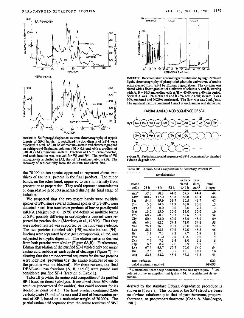
Table I11 provides the amino acid composition of the purified SP-I based on timed hydrolysis. It contained about 30% acidic residues (uncorrected for amides) that could account for its isoelectric point of 4.5. The final product contained 2.6% carbohydrate (7 mol of hexose and 3 mol of hexosamine per mol of SP-I, based on a molecular weight of 70000). The partial amino acid sequence from the amino terminus of SP-I
VOL. 2 0 , NO. 1 4 , 1 9 8 1 4139
0 1020301 RETENTION TIME (min)
FIGURE 7: Representative chromatograms obtained by high-pressure liquid chromatography of phenylthiohydantoin derivatives of amino acids cleaved from SP-I by Fdman degradation. The column was eluted with a linear gradient of a mixture of solvents A and B, starting with A/B = 95:5 and ending with A/B = 4060, over a 4O-min period. Solvent A was 10% methanol and 0.25% acetic acid; solvent B was 90% methanol and 0.025% acetic acid. The flow rate was 2 mL/min. The standard mixture contained 1 nmol of each amino acid derivative.
P W L AMINO ACID SEWENCE OF SP-1
1 5 10
15 20
25 30
FIGURE 8: Partial amino acid sequence of SP-I determined by standard Edman degradation.
Table 111: Amino Acid ComDosition of Secretory Protein I"
nmol/fraction extrap resi-
amino olated d u e j mol acids 25 h 48 h 12 h to 0 h mol integer
AsxC 52.3 58.2 Glxc 180.2 111.0 Ser 54.4 49.9 Thr 15.6 14.8 CY s 2.8 0.0 Met 13.0 13.0 Pro 68.1 68.2 Gly 60.4 58.0 Ala 66.0 62.5 Val 26.1 26.1 Leu 58.9 58.3 Ile 1.1 1.7 Phe 11.2 11.0 T Y ~ 1.7 1.5 TIP 8.5 8.2 Lys 67.4 65.1 His 12.5 12.1 A r g 52.6 52.2
total residues: calcd minimum mol wt:
44.0 51.5 44.4 44 153.8 186.0 143.4 144 39.7 60.5 46.1 47 11.9 16.8 13.0 13 0.0 3.0 2.3 3 10.0 13.0 10.0 10 59.2 69.6 53.7 54 50.6 63.5 48.9 49 54.5 11.0 54.8 55 23.1 28.0 21.6 22 50.9 59.0 45.5 46 1.2 1.7 5.9 6 9.6 11.6 9.0 9 6.4 8.0 6.1 6 7.0 8.9 6.9 7 51.1 10.0 54.0 54 10.5 13.1 10.1 10 45.4 53.5 41.3 41
620 69 000
Determined from the p-toluenesulfonic acid hydrolysis. Cal- culated on the assumption that lysine = 54. mined.
Amides not deter-
~~
derived by the standard Edman degradation procedure is shown in Figure 8. This portion of the SP-I structure bears no obvious relationship to that of parathormone, propara- thormone, or pre-proparathormone (Cohn & MacGregor, 1981).

4140 B I O C H E M IS T R Y C O H N E T A L .
Tx’”- 1 I
4 1
RESIDUE
FIGURE 9: Microsequence analysis of SP-I. [’%I Methionine-labeled SP-I (83 OOO dpm) was subjected to 32 cycles of Edman degradation. Aliquots of the thiazoliione derivative at each cycle were assayed for radioactivity.
1 d 1 , # L < 8 8 l , , I , 8 8 / 1 I 1 , l t d
01 1 10 100 1000 PROTEIN, ng
FIGURE 10: Radioimmunoassay of SP-I. In curves labeled proPTH and PTH, different amounts of the hormonal peptides in place of SP-I were. tested. The radioimmunoassay was performed as described under Materials and Methods.
Figure 9 provides additional data confirming that the ra- dioactive SP-I was chemically identical with the bulk-purified SP-I. [35S]Methionine-labeled SP-I was subjected to Edman degradation. Radioactive methionine was detected at residues 7, 15, and 32 in accord with the analyses of the bulk protein. That the radioactivity was indeed in methionine residues was confirmed by back-hydrolysis.
Figure 10 shows the radioimmunoassay curve of SP-I. The linear region ranges from 0.15 to 20 ng of SP-I. Parathormone and proparathormone at levels as high as 500 ng did not react in this assay. The measurement of SP-I was unaffected by the presence of parathormone or proparathormone (data not shown).
The differences in apparent molecular weights (300 OOO vs. 70000) for the SP-I species estimated by gel filtration and NaDodS04 gel electrophoresis, respectively, are in accord with the conclusion of Kemper et al. (1974) that SP-I exists as polymers in aqueous solution. If this is so, these polymers are not formed by covalent linkage since we observed identical mobility of the SP-I species in NaDodS04 gels whether or not the samples were reduced by mercaptoethanol prior to analysis. On the other hand, polymers might not exist at all. Their early elution during gel filtration could be a result of their being in highly extended forms in aqueous medium. Indeed, an anomalous migration of parathormone itself has been described which has been attributed to a high degree of asymmetry of
the molecule (Cohn et al., 1974; Fiskin et al., 1977). The availability of purified SP-I and its antibodies will now
allow more definitive answers to these questions and a more complete search for its physiological function.
References Chu, L. L. H., MacGregor, R. R., & Cohn, D. V. (1977) J .
Cohn, D. V., & MacGregor, R. R. (1981) Endocr. Rev. 2 , l . Cohn, D. V., MacGregor, R. R., Chu, L. L. H., Kimmel, J.
R., & Hamilton, J. W. (1972) Proc. Natl. Acad. Sci. U.S.A. 69, 1521.
Cohn, D. V., MacGregor, R. R., Sinha, D., Huang, D. W. Y., Edelhoch, H., & Hamilton, J. W. (1974) Arch. Biochem. Biophys. 164, 669.
Dubois, M., Gilles, K. A., Hamilton, J. K., Rebers, P. A., & Smith, F. (1956) Anal. Chem. 28, 350.
Fiskin, A. M., Cohn, D. V., & Peterson, G. S. (1977) J . Biol. Chem. 252, 826 1 .
Habener, J. F. (1979) in Endocrinology (DeGroot, J., Ed.) Vol. 2, p 599, Grune & Stratten, New York.
Habener, J. F., Rosenblatt, M., Dee, P. C., & Potts, J. T., Jr. (1979) J . Biol. Chem. 254, 10596.
Hamilton, J. W., Spierto, F. W., MacGregor, R. R., & Cohn, D. V. (1971) J . Biol. Chem. 246, 3224.
Huang, W.-Y., Chu, L. L. H., Hamilton, J. W., McGregor, D. H., & Cohn, D. V. (1975) Arch. Biochem. Biophys. 166, 67.
Hunter, W. M., & Greenwood, F. C. (1962) Nature (London) 194, 495.
Kemper, B., Habener, J. F., Rich, A., & Potts, J. To, Jr. (1974) Science (Washington, D.C.) 184, 167.
Laemmli, U. K. (1970) Nature (London) 227, 680. Lee, Y. C., & Montgomery, R. (1961) Arch. Biochem. Bio-
Liu, T.-Y., & Chang, Y. H. (1971) J. Biol. Chem. 246,2842. Lowry, 0. H., Rosebrough, N. J., Farr, A. L., & Randall, R.
J. (1951) J . Biol. Chem. 193, 265. MacGregor, R. R., Chu, L. L. H., & Cohn, D. V. (1976) J .
Biol. Chem. 251, 6711. MacGregor, R. R., Hamilton, J. W., & Cohn, D. V. (1978)
J . Biol. Chem. 253, 2012. Majzoub, J. A., Kronenberg, H. M., Potts, J. T., Jr., Rich,
A., & Habener, J. F. (1979) J. Biol. Chem. 254, 7449. Morrissey, J. J., & Cohn, D. V. (1978) Endocrinology
(Baltimore) 103, 208 1 . Morrissey, J. J., & Cohn, D. V. (1979a) J . Cell Biol. 82, 93. Morrissey, J. J., & Cohn, D. V. (1979b) J. Cell Biol. 83, 521. Morrissey, J. J., Hamilton, J. W., & Cohn, D. V. (1978)
Biochem. Biophys. Res. Commun. 82, 1279. Morrissey, J. J., Hamilton, J. W., & Cohn, D. V. (1980a)
Endocrinology (Baltimore) 106, 187. Morrissey, J. J., Shofstall, R. E., Hamilton, J. W., & Cohn,
D. V. (1980b) Proc. Nail. Acad. Sci. U.S.A. 77, 6406. O’Keefe, E., & Bennet, V. (1980) J . Biol. Chem. 255, 561. Ravazzola, M., Orci, L., Habener, J. F., & Potts, J. T., Jr.
(1 978) Lancet No. 8085, 37 1. Reisfeld, R. A., Lewis, U. J., & Williams, D. E. (1972) Nature
(London) 195, 281. Steiner, D. F., Quinn, P. S. , Chan, S. J., Marsh, J., & Tager,
H. S . (1980) in Precursor Processing in the Biosynthesis of Proteins (Zimmerman, M., Mumford, R. A., & Steiner, D. F., Eds) p 1, Academic Sciences, New York.
Cell Biol. 72, 1 .
phys. 93, 292.
Copyright © 2022 FDOKUMEN