Indian Primary Hyperparathyroidism Patients with Parathyroid Carcinoma do not Differ in...
11
Indian Primary Hyperparathyroidism Patients with Parathyroid Carcinoma do not Differ in Clinicoinvestigative Characteristics from Those with Benign Parathyroid Pathology Gaurav Agarwal, MS, DNB, PDC, 1 Kaushal K. Prasad, MD, PDC, 2 Dilip K Kar, MS, PDC, 1 Narendra Krishnani, MD, 2 Rakesh Pandey, MD, 2 Saroj K. Mishra, MS, FACS 1 1 Department of Endocrine Surgery, Sanjay Gandhi Postgraduate Institute of Medical Sciences, Raebareli Road, Lucknow 226014, India 2 Department of Pathology, Sanjay Gandhi Postgraduate Institute of Medical Sciences, Raebareli Road, Lucknow 226014, India Abstract Introduction: No foolproof preoperative diagnostic indicators of parathyroid carcinoma (PC) exist in absence of nonskeletal metastases. Palpable parathyroid tumor, advanced skeletal and renal manifestations, and very high serum calcium and parathyroid hormone levels are considered strong predictors. Most of these features are common in Indian primary hyperparathyroidism (PHPT) patients although only few have PC. The aim of this study was to identify dependable clinicoinvestigative predictors of PC in Indian PHPT patients. Materials and Methods: Clinical, biochemical, radiological, and densitometric attributes of 100 PHPT patients who underwent successful parathyroidectomy (1990–2004) were studied. Various parameters of patient groups with parathyroid adenoma (n = 84), primary hyperplasia (n = 12), and carcinoma (n = 4) were compared using ANOVA, with P value < 0.05 considered significant. Results: Mean age of patients was 37.4 years, with no difference in the 3 groups (P = 0.92). Patients in 3 groups had comparably severe bone disease; 36 had coexistent renal disease. Two patients with PC and 27 (32%) with adenoma had palpable parathyroid tumor. None of the bio- chemical parameters predicted malignant pathology. Mean tumor weight (milligram) in carcinoma patients (15,080 – 5,638.02) was significantly higher than those with adenoma (5,724 – 1,257.9) (P = 0.002). Postoperative course and recovery in carcinoma patients were similar to those with adenoma. In follow-up (mean: 33 months), none of the adenoma patients were found to have persistent/recurrent PHPT attributable to missed PC. Conclusion: Indian patients with parathyroid adenoma, hyperplasia, and carcinoma were not found to differ in their clinical, biochemical, and pathological characteristics except for significantly higher tumor weight in the carcinoma group. P arathyroid carcinoma (PC) is a rare cause for pri- mary hyperparathyroidism (PHPT), accounting for less than 2% of all PHPT cases, 1–3 although the rela- tive incidence is reported at 4%–8% from developing Correspondence to: Gaurav Agarwal, MS, DNB, PDC, Associate Professor Department of Endocrine Surgery, Sanjay Gandhi Post- graduate Institute of Medical Sciences, Raebareli Road, Lucknow 226014, India, e-mail: gaurav@ sgpgi.ac.in Ó 2006 by the Socie ´te ´ Internationale de Chirurgie World J Surg (2006) 30: 732–742 Published Online: 17 April 2006 DOI: 10.1007/s00268-005-0366-5
-
Upload
independent -
Category
Documents
-
view
3 -
download
0
Transcript of Indian Primary Hyperparathyroidism Patients with Parathyroid Carcinoma do not Differ in...

Indian Primary Hyperparathyroidism Patientswith Parathyroid Carcinoma do not Differ inClinicoinvestigative Characteristics from Thosewith Benign Parathyroid PathologyGaurav Agarwal, MS, DNB, PDC,1 Kaushal K. Prasad, MD, PDC,2 Dilip K Kar, MS, PDC,1
Narendra Krishnani, MD,2 Rakesh Pandey, MD,2 Saroj K. Mishra, MS, FACS1
1Department of Endocrine Surgery, Sanjay Gandhi Postgraduate Institute of Medical Sciences, Raebareli Road, Lucknow
226014, India2Department of Pathology, Sanjay Gandhi Postgraduate Institute of Medical Sciences, Raebareli Road, Lucknow 226014,
India
Abstract
Introduction: No foolproof preoperative diagnostic indicators of parathyroid carcinoma (PC) exist in
absence of nonskeletal metastases. Palpable parathyroid tumor, advanced skeletal and renal
manifestations, and very high serum calcium and parathyroid hormone levels are considered
strong predictors. Most of these features are common in Indian primary hyperparathyroidism
(PHPT) patients although only few have PC. The aim of this study was to identify dependable
clinicoinvestigative predictors of PC in Indian PHPT patients.
Materials and Methods: Clinical, biochemical, radiological, and densitometric attributes of 100
PHPT patients who underwent successful parathyroidectomy (1990–2004) were studied. Various
parameters of patient groups with parathyroid adenoma (n = 84), primary hyperplasia (n = 12),
and carcinoma (n = 4) were compared using ANOVA, with P value < 0.05 considered significant.
Results: Mean age of patients was 37.4 years, with no difference in the 3 groups (P = 0.92).
Patients in 3 groups had comparably severe bone disease; 36 had coexistent renal disease. Two
patients with PC and 27 (32%) with adenoma had palpable parathyroid tumor. None of the bio-
chemical parameters predicted malignant pathology. Mean tumor weight (milligram) in carcinoma
patients (15,080 – 5,638.02) was significantly higher than those with adenoma (5,724 – 1,257.9)
(P = 0.002). Postoperative course and recovery in carcinoma patients were similar to those with
adenoma. In follow-up (mean: 33 months), none of the adenoma patients were found to have
persistent/recurrent PHPT attributable to missed PC.
Conclusion: Indian patients with parathyroid adenoma, hyperplasia, and carcinoma were not found
to differ in their clinical, biochemical, and pathological characteristics except for significantly higher
tumor weight in the carcinoma group.
Parathyroid carcinoma (PC) is a rare cause for pri-
mary hyperparathyroidism (PHPT), accounting for
less than 2% of all PHPT cases,1–3 although the rela-
tive incidence is reported at 4%–8% from developing
Correspondence to: Gaurav Agarwal, MS, DNB, PDC, AssociateProfessor Department of Endocrine Surgery, Sanjay Gandhi Post-graduate Institute of Medical Sciences, Raebareli Road, Lucknow226014, India, e-mail: gaurav@ sgpgi.ac.in
� 2006 by the Societe Internationale de Chirurgie World J Surg (2006) 30: 732–742
Published Online: 17 April 2006 DOI: 10.1007/s00268-005-0366-5

countries, as also in early reports from the developed
world.4–7 In absence of apparent nonskeletal metastatic
disease, it is difficult to establish the diagnosis of PC, and
the diagnosis is rarely evident preoperatively.8 Radical
surgical excision of a PC along with any infiltrated struc-
tures and metastatic lymph nodes provides the best, and
perhaps the only chance of long-term cure in these pa-
tients.9 In order to cure hyperparathyroidism and achieve
adequate oncological control of this disease, it is imper-
ative that the condition be identified before or during the
operation and dealt with appropriately at the time of the
primary surgery.1,9,10
Various clinical and investigative features, such as
palpable parathyroid tumor, symptomatic skeletal mani-
festations, osteitis fibrosa cystica (OFC) (especially when
these coexist with renal manifestations), very high serum
calcium (s-Ca) and parathyroid hormone (s-PTH) levels,
and large tumors are considered predictive of the pres-
ence of PC.1,9,10 However, no foolproof preoperative
diagnostic criteria are known. Even on histology, diag-
nosis may at times be difficult although a set of criteria
proposed by Shantz and Castleman 11are widely
followed.
Indian PHPT patients present at a relatively young
age with symptomatic disease. The majority of patients
have OFC, extreme osteopenia (syndrome of disap-
pearing bones), and some are crippled due to fractures,
brown tumors, and muscle weakness (parathyroid crip-
ples). Renal manifestations are common and are often
present simultaneously with bone disease. Many have a
palpable parathyroid tumor and extremely high s-PTH
levels.4,5 In spite of these clinical, biochemical, radiolog-
ical, and pathological features commonly used to indicate
possible presence of malignant parathyroid tumors, fewer
than 5% of Indian PHPT patients are diagnosed with PC.
Doubts may thus be raised about the applicability of these
criteria for prediction of parathyroid malignancy. The
current study aimed at comparing clinical and laboratory
parameters of Indian PHPT patients with benign and
malignant parathyroid tumors in order to identify predic-
tive features of PC.
MATERIALS AND METHODS
Primary hyperparathyroidism patients managed at the
Department of Endocrine Surgery, Sanjay Gandhi Post-
graduate Institute of Medical Sciences (SGPGIMS),
Lucknow, India from 1990 to 2004 were the subjects of
this study. The patients were divided into groups A
(parathyroid adenoma, n = 84), B (primary parathyroid
hyperplasia, n = 12), and C (parathyroid carcinoma,
n = 4). The diagnosis of parathyroid adenoma, hyper-
plasia, or carcinoma were made on the basis of para-
thyroid imaging, operative findings, and gross and
microscopic pathology reported by two pathologists
independently using established criteria,12–14 and follow-
up.
Parathyroid adenoma diagnosis was based on locali-
zation of a single pathological parathyroid gland (PTG) on99mTc-MIBI scanning (or Tl-Tc subtraction scans before
1999) and high-resolution ultrasonography and finding of
a parathyroid tumor in the predicted location during fo-
cused or minimally invasive parathyroidectomy, with
gross and microscopic pathology consistent with an
adenoma. In earlier years of the study, when preoperative
parathyroid imaging was not routinely employed, and in a
few recent patients with inconclusive parathyroid imaging,
diagnosis of parathyroid adenoma was based on the
finding of a single pathological and three normal PTGs at
bilateral neck exploration and if the removed pathological
PTG had gross and microscopic features consistent with
an adenoma. The diagnosis of double adenoma and
primary parathyroid hyperplasia were based on findings
at bilateral neck exploration and gross and microscopic
pathology. Operative findings of large, grayish-white,
firm-to-hard tumor with infiltration into surrounding mus-
cles and visceral structures were considered indicative of
PC, and such patients were treated with an en bloc
resection of parathyroid tumor together with infiltrated
structures.
Histopathological diagnosis was based on weight and
histological findings of the excised parathyroid tissues.
Hyperplasia was diagnosed when histopathological cri-
teria of adenoma were absent and multiple glands were
enlarged (weight: >59 mg) and had abnormal microscopic
appearance with decreased fat-cell content together with
pathologic arrangement and reduced cytoplasmic fat
content of the parenchymal cells.12,13 Diagnosis of PC
was made on the basis of gross and microscopic
pathology.11,14 Following parathyroidectomy, patients
were followed up for a minimum of 1 year with periodic s-
Ca and intact PTH estimations and appropriate imaging
so as to ensure they did not have metastatic disease,
which would have changed the diagnosis from adenoma
or hyperplasia to carcinoma.
Information on clinical, biochemical, radiological, den-
sitometric, and pathological features of patients in groups
A, B, and C were derived from a prospectively main-
tained parathyroid data base at the Department of
Endocrine Surgery. Biochemical evaluation included se-
rial estimation of s-Ca, inorganic phosphorus (s-iP),
Agarwal et al.: Hyperparathyroidism in Indians 733

alkaline phosphatase (s-ALP), creatinine, s-iPTH, and
25-hydroxy vitamin D (s-25-OH-D). Calcium and creati-
nine excretions in 24-hour urine collections were also
sampled. X-rays of the hands, skull, pelvis, spine, and
any fracture or brown tumor sites of patients in groups A,
B, and C were compared. Bone mineral densitometry
(BMD) using dual-energy X-ray absorptiometry (DEXA)
was carried out before and after parathyroidectomy and
during follow-up. Weight of pathological parathyroid tis-
sue in the 3 groups was compared. Values are presented
as mean – standard error (SE). Values of various bio-
chemical indices, clinical symptoms, and PTG weight
were compared using one-way analysis of variance
(ANOVA), with P value <0.05 considered significant.
RESULTS
Mean patient age was 37.4 – 7.04 years (range: 13–66
years), with no significant difference between the 3
groups (P = 0.92). There were 73 females and 27 males
(male/female ratio: 1:3). Comparison of the clinical attri-
butes of patients in the 3 groups is provided in Table 1.
None of the patients were truly asymptomatic; all but 3
presented with symptoms attributable to PHPT and its
complications. Hypercalcemia was detected in 3 patients
during evaluation for unrelated symptoms in 1 and on
periodic health check-ups in 2. On careful scrutiny,
however, all 3 were found to have musculoskeletal
symptoms attributable to PHPT together with osteopenia
and low BMD. Six patients presented with hypercalcemic
crisis, of whom 4 were found to have a parathyroid ade-
noma and 1 each PC and hyperplasia. The average
duration of symptoms in all 100 patients was 4.0 years
(range: 6 months–26 years), with no significant difference
between the 3 groups.
Skeletal manifestations were the commonest clinical
presentation, with all patients having bone pains, and 60
proximal muscle weakness. As a result of osteopenia,
pathological fractures, and proximal muscle weakness, 27
patients were confined to bed. Though the proportion of
such ‘‘parathyroid cripples’’ in the PC group was higher,
this difference was not statistically significant (P value
0.078). Renal involvement was present in 36 patients: 14
had nephrocalcinosis, 27 nephrolithiasis, and 5 had both.
Concomitant skeletal and renal manifestations occurred
with comparable frequency in the 3 patient groups. Psy-
chiatric manifestations in 38 and gastrointestinal symp-
toms in 14 occurred with comparable frequency in the 3
groups. Parathyroid tumors were palpable in the neck in
29 of the 88 patients with PHPT due to parathyroid tu-
mors. These included, besides two (50%) patients with
PC, 27 (32%) parathyroid adenoma patients as well, (no
significant difference, P-value = 0.238).
Table 2 depicts biochemical features in PHPT patients
in the 3 groups. The mean s-Ca and s-iP were not sig-
nificantly different in the 3 groups (P = 0.634 and 0.329,
respectively), nor were the mean s-ALP and s-iPTH lev-
els (P = 0.083 and 0.154, respectively). The s-25-OH-D
levels were also not significantly different in the 3 groups
(P = 0.662). PHPT patients with adenoma, hyperplasia,
and carcinoma had equally severe bone disease, as
evidenced by comparable BMDs and z-scores assessed
by DEXA (Table 3). Forty-nine patients had brown tu-
mors, and 56 had pathological fractures, and these were
not predictive of type of parathyroid pathology. Compar-
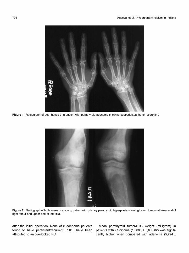
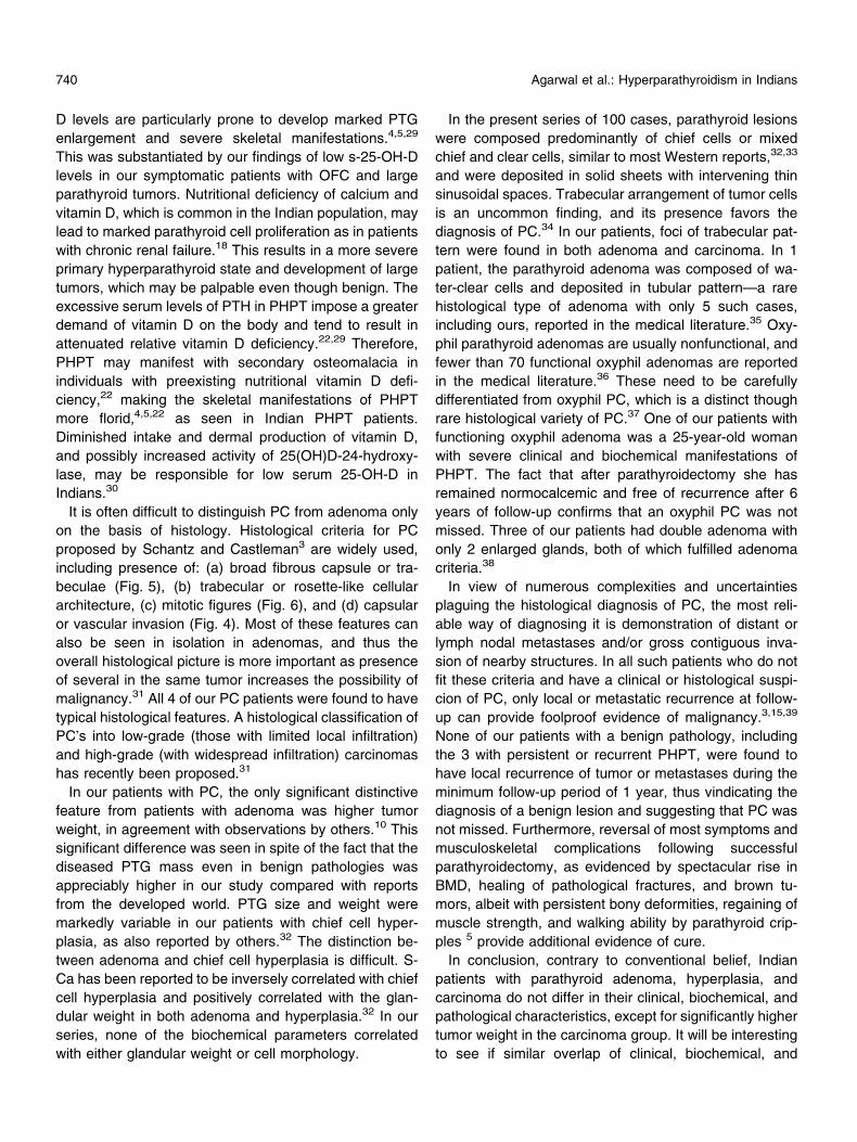
ison of qualitative radiological features did not reveal
significant difference between the 3 groups (Figs. 1 and
Table 1.Clinical features of primary hyperparathyroidism patients with parathyroid adenoma, hyperplasia, and carcinoma
Group Aa Group Bb Group Cc P-value
Age (years): mean (range) 36.8 (14–66) 38.9 (13–50) 37.0 (22–56) 0.92Male: female ratio 1:4 1:3 1:3 0.311Duration of symptoms (years) 3.5 (0.5–26) 8.0 (3–20) 2.5 (0.5–6) 0.154Bone pain 84 (100%) 12 (100%) 4 (100%) 0.590Proximal muscle weakness 49 (58.3%) 7 (58.3%) 4 (100%) 0.065Crippling 20 (23.8%) 4 (33.3%) 3 (75%) 0.078Concomitant skeletal and renal manifestations
(renal stone/nephrocalcinosis)30 (35.7%) 4 (33.3%) 2 (50%) 0.076
Behavioral disorders 33 (39.2%) 5 (41.6%) 2 (50%) 0.192Gastrointestinal symptoms 11 (13.1%) 2 (16.7%) 1 (25%) 0.188Palpable parathyroid tumor/ gland 27 (32.1%) None 2 (50%) 0.238
aParathyroid adenoma (n = 84).bPrimary parathyroid hyperplasia (n = 12).cParathyroid carcinoma (n = 4).dSignificant P value < 0.05.
734 Agarwal et al.: Hyperparathyroidism in Indians

2). Of the 7 patients with ‘‘disappearing bones’’ syndrome
(Figure 3), 5 had adenoma and 2 had carcinoma.
Forty-two patients were scheduled to have a minimally
invasive or focused parathyroidectomy and the rest a
bilateral neck exploration. Of the 42 undergoing minimally
invasive parathyroidectomy, 4 had the procedure con-
verted to bilateral exploration due to suspicion of multi-
gland disease in 3 and failure to find a tumor in the
location predicted on preoperative parathyroid imaging in
1. During the operation, presence of a large, hard, gray-
ish-white tumor invading the thyroid lobe, strap muscles,
or other surrounding structures led to a suspicion of PC in
6 patients. These were subjected to en bloc resection of
parathyroid tumor along with infiltrated structures. Two of
these were, however, diagnosed as adenomas based on
histology. In follow-up of 3 and 4.5 years, both these
patients have remained free of tumor recurrence, thus
vindicating the diagnoses of adenoma.
Postoperative hypocalcemia occurred in 94 patients
and was managed with calcium carbonate and calcitriol
supplements, with 83 requiring intravenous calcium infu-
sion. One patient had persistent hyperparathyroidism
attributed to a missed parathyroid adenoma at the initial
operation. During follow-up (mean: 33 months; range: 6–
136 months), 2 patients with parathyroid adenoma pre-
sented with recurrent hyperparathyroidism, one due to a
missed second adenoma and the other due to asym-
metrical parathyroid hyperplasia. Three of the 4 PC pa-
tients had local recurrence and/or metastases during
follow-up. One had intracranial metastases and suc-
cumbed to neurological and metabolic consequences
after 39 months of follow-up. Another died after 24
months with end-stage renal disease a few months after
detection of recurrence in the contralateral thyroid lobe
and neck nodes confirmed on cytology. Two patients are
alive and free of disease at 16 months and 9 years of
follow-up although the latter had a recurrent but asymp-
tomatic subcutaneous nodule not resulting in recurrent
hyperparathyroidism along the fine-needle aspiration
cytology (FNAC) track of the primary tumor 64 months
Table 3.Radiological features and parathyroid weights of primary hyperparathyroidism patients with parathyroid adenoma, hyperplasia, and
carcinoma
Group Aa Group Bb Group Cc P-valued
Mean BMD at distal radius (gm/cm2)/ z-score 0.426 / )3.8 0.542 / )3.3 0.388 / )4.4 0.121Radiological features of osteitis fibrosa cystica 72 (85.7%) 9 (75%) 4 (100%) 0.34Pathological fractures 48 (57.1%) 5 (41.7%) 3 (75%) 0.183Brown tumors 39 (46.4%) 6 (50%) 4 (100%) 0.067Tumor/PTG weight (mg) mean – S.D. 5,724 – 1,257.9 3,108e – 1,129.3 15,080 – 5,638.02 0.02*
BMD: bone mineral density; PTG: parathyroid gland.aParathyroid adenoma (n = 84).bPrimary parathyroid hyperplasia (n = 12).cParathyroid carcinoma (n = 4).dSignificant P value < 0.05.eCombined. PTG weight of the excised, abnormal parathyroid tissue.*Statistically significant difference.
Table 2.Biochemical features in primary hyperparathyroidism patients with parathyroid adenoma, hyperplasia, and carcinoma
Group Aa Group Bb Group Cc P-valued
Serum calcium (mean, mg/dl) (Normal range: 8.2–10.5) 12.6 12.3 13.2 0.634Serum inorganic phosphorus (mean, mg/dl) (Normal range: 3.5–5.5) 2.8 2.3 2.1 0.329Serum alkaline phosphatase (mean, IU/l) (Normal <135) 1789.3 2872.4 673.8 0.083Serum parathyroid hormone (mean, pg/ml) (Normal range: 9–55) 683 728 922 0.154Serum 25-OH vitamin D (mean, ng/dl) (Normal range: 9–45) 11.6 7.5 12.1 0.662
aParathyroid adenoma (n = 84).bPrimary parathyroid hyperplasia (n = 12).cParathyroid carcinoma (n = 4).dSignificant P value <0.05.
Agarwal et al.: Hyperparathyroidism in Indians 735
after the initial operation. None of 3 adenoma patients
found to have persistent/recurrent PHPT have been
attributed to an overlooked PC.
Mean parathyroid tumor/PTG weight (milligram) in
patients with carcinoma (15,080 – 5,638.02) was signifi-
cantly higher when compared with adenoma (5,724 –
Figure 1. Radiograph of both hands of a patient with parathyroid adenoma showing subperiosteal bone resorption.
Figure 2. Radiograph of both knees of a young patient with primary parathyroid hyperplasia showing brown tumors at lower end ofright femur and upper end of left tibia.
736 Agarwal et al.: Hyperparathyroidism in Indians

1,257.9) and hyperplasia (3,310 – 655.84) (P = 0.002).
Histopathologically, in all cases of parathyroid hyperpla-
sia and carcinoma, the cells were predominantly dis-
posed in solid sheets with intervening thin sinusoidal
spaces. In 66 (78.6%) cases with parathyroid adenoma,
the cells were predominantly disposed in solid sheets with
intervening thin sinusoidal spaces. In 3 (3.6%) adenomas,
cells were disposed in sheets and tubular pattern while in
15 (17.8%), they were predominantly disposed in tubular
pattern. In one case of adenoma, the cells in a few foci
were disposed in follicular pattern with intrafollicular
amyloid deposition. Eighty-one (96.4%) adenomas were
composed of either chief cells or mixed chief and clear
cells. One adenoma was composed of water-clear cells
and 2 of oxyphilic cells. One case with functioning oxy-
philic parathyroid adenoma manifested with severe clini-
cal manifestation of PHPT but had a rather small (950
mg) and noninvasive parathyroid tumor. Three patients
had synchronous double adenomas with only 2 enlarged
glands, both of which fulfilled adenoma criteria. The
remaining 2 parathyroids were visually normal, and a
biopsy from one of those did not show features sugges-
tive of hyperplasia. All cases of parathyroid hyperplasia
and carcinoma were histologically composed of either
chief cells or mixed chief and clear cells. Vascular and
capsular invasion (Fig. 4), thick fibrous septae (Fig. 5),
and characteristic cellular features such as nuclear aty-
pia, vesicular nuclei, macro nucleoli, and frequent mitotic
figures (Fig. 6) were seen in all 4 cases of PC. In 2 cases,
local tumor infiltration was also present. Diagnosis of PC
was further substantiated by occurrence of metastatic
and locally or regionally recurrent disease in 3 patients
while diagnosis was quite apparent from gross contigu-
ous invasion of neck structures in the fourth. None of the
biochemical parameters correlated with either glandular
weight or cell morphology. Glandular weight and size
were markedly variable in chief cell hyperplasia.
DISCUSSION
The clinical, biochemical, radiological, and pathological
features of 84 Indian PHPT patients with parathyroid
adenoma, 12 with primary parathyroid hyperplasia, and 4
with PC were not significantly different. These findings are
contrary to a common belief that patients with parathyroid
cancer have different clinical and biochemical features
than those with a benign parathyroid pathology.3,9,10
Carcinoma of the parathyroid is a rare cause of PHPT,
constituting less than 2% of all PHPT cases.1–3 The
proportion of PHPT patients with PC is reported higher in
series from developing countries and in earlier studies
from certain developed countries.4–7 Most reports have
shown no gender predilection for PC,1,3,15 in contrast to a
definite female preponderance of adenomas. Our study,
as have a few others 16, found a similar preponderance of
Figure 3. Radiographs of pelvis and upper femurs of a patient with parathyroid carcinoma. Extreme bone destruction, browntumors, cysts, and fractures have caused much of the skeletal framework to become invisible: ‘‘disappearing bones’’ syndrome.
Agarwal et al.: Hyperparathyroidism in Indians 737

women even in patients with PC. PCs are reported to
occur about a decade earlier than adenomas, with a
mean age of approximately 45 years for PC compared to
55 years for adenomas.1,3,15 In our experience, the
average age of PHPT patients with benign or malignant
pathology were similar, reflecting an overall young age of
Indian PHPT patients.
Diagnosis of PC is often difficult to establish and is only
rarely evident preoperatively.8,17 This rare condition is
diagnosed by a combination of clinical, biochemical, and
pathological features attributed to this disease. Clinical
features such as symptomatic skeletal manifestations of
PHPT in young patients, especially when present simul-
taneously with renal manifestations, and a palpable
parathyroid mass are considered indicative of presence
of PC.9 Extreme hypercalcemia, with s-Ca levels >14 mg/
dl, and very high s-PTH levels have also been seen as
warning signs of malignant parathyroid pathology in
PHPT patients.8–10 In our patients, s-Ca levels were only
moderately elevated in a majority, including those with
PC, and s-PTH levels were extremely high irrespective of
the parathyroid pathology. Of the 6 patients who pre-
sented with hypercalcemic crisis, only one was found to
have PC. Presentation with hypercalcemic crisis in 25%,
i.e., 1 of 4 PC patients, in contrast to only 4/84 (5%)
parathyroid adenoma and 1/12 (8.3%) of hyperplasia
Figure 4. Photomicrograph showing nests of bland-looking cells traversed by delicate vasculature and a focus of capsular invasionin a case with parathyroid carcinoma. H & E, 200·.
Figure. 5. Photomicrograph of a parathyroid carcinoma showing thick fibrous band traversing the tumor. H & E, 200·.
738 Agarwal et al.: Hyperparathyroidism in Indians

patients, indicates a greater degree of hypercalcemia and
perhaps more rapidly rising serum calcium in patients
with PC. However, the small number of patients with
hypercalcemic crisis in the PC group limits a valuable
comparison.
Presence of severe osteopenia on BMD and radiolog-
ical features of OFC, brown tumors, or cysts and patho-
logical fractures are often considered to be associated
with parathyroid cancer. However, the extent of bone loss
and nature of skeletal disease in our patients with PC and
benign pathology was quite comparable. At operation,
presence of a large, gray-white parathyroid tumor with
invasion into the surrounding strap muscles, thyroid lobe,
and other structures and high tumor weight have often
been considered suggestive of a malignant parathyroid
neoplasm.1,9,10 Similar operative findings led to intraop-
erative suspicion and en bloc radical excision of the
parathyroid tumor in 6 patients, of which 2 were eventu-
ally found to be large adenomas. The benign nature of
disease in both these patients has been confirmed by
long-term relapse-free course during follow-up.
The basis of such clinical and investigative features
being considered diagnostic or indicative of PC and as
distinctive from parathyroid adenomas is observations in
a relatively small number of PC patients and a huge
number of asymptomatic PHPT patients with small ade-
nomas in industrially developed countries. The picture in
the Indian PHPT patients with PC closely resembles that
of PC patients reported from developed countries. How-
ever, the majority of Indian PHPT patients with benign
parathyroid pathology have similar symptoms, advanced
bone and renal disease, and large but clearly noninvasive
benign tumors, making preoperative reliable prediction of
parathyroid cancers almost impossible.
The reasons for little difference between Indian patients
with parathyroid adenoma, primary hyperplasia, and
carcinoma may be manifold. Firstly, unlike Western pa-
tients where most are diagnosed at asymptomatic stages,
often on basis of biochemical survey,18 almost all PHPT
patients in India are diagnosed at a relatively late symp-
tomatic stage and are rarely asymptomatic.4,5,19–21 Indian
PHPT patients present at a relatively young age with OFC
and extreme osteopenia (‘‘disappearing bones’’ syn-
drome). Some are crippled due to fractures, brown tu-
mors, and muscle weakness (parathyroid cripples). Renal
manifestations are common, and as skeletal manifesta-
tions are ubiquitous, the two are often present simulta-
neously. Many have palpable parathyroid tumor and
extremely high serum PTH levels.4,5 Reports from other
developing countries have indicated presence of similar
clinical picture of PHPT.6,22–28 Possibly, only severe
cases are diagnosed, after a relatively long duration of
illness due to relative lack of awareness of the disease in
India and other developing countries. In spite of such
clinical, biochemical, radiological, and pathological fea-
tures that are conventionally considered to indicate
presence of malignant tumors, fewer than 5% of Indian
PHPT patients are diagnosed with PC.
Secondly, nutritional and environmental factors may
play a role in pathogenesis and progression of parathy-
roid tumors.4,5,21,22,29 Vitamin D status influences the
clinical expression of PHPT, and patients with low vitamin
Figure 6. Photomicrograph showing nuclear atypia with vesicular nuclei, mitotic figures, presence of macronuclei, and moderateamount of pale cytoplasm in section from parathyroid carcinoma. H & E, 400·.
Agarwal et al.: Hyperparathyroidism in Indians 739
D levels are particularly prone to develop marked PTG
enlargement and severe skeletal manifestations.4,5,29
This was substantiated by our findings of low s-25-OH-D
levels in our symptomatic patients with OFC and large
parathyroid tumors. Nutritional deficiency of calcium and
vitamin D, which is common in the Indian population, may
lead to marked parathyroid cell proliferation as in patients
with chronic renal failure.18 This results in a more severe
primary hyperparathyroid state and development of large
tumors, which may be palpable even though benign. The
excessive serum levels of PTH in PHPT impose a greater
demand of vitamin D on the body and tend to result in
attenuated relative vitamin D deficiency.22,29 Therefore,
PHPT may manifest with secondary osteomalacia in
individuals with preexisting nutritional vitamin D defi-
ciency,22 making the skeletal manifestations of PHPT
more florid,4,5,22 as seen in Indian PHPT patients.
Diminished intake and dermal production of vitamin D,
and possibly increased activity of 25(OH)D-24-hydroxy-
lase, may be responsible for low serum 25-OH-D in
Indians.30
It is often difficult to distinguish PC from adenoma only
on the basis of histology. Histological criteria for PC
proposed by Schantz and Castleman3 are widely used,
including presence of: (a) broad fibrous capsule or tra-
beculae (Fig. 5), (b) trabecular or rosette-like cellular
architecture, (c) mitotic figures (Fig. 6), and (d) capsular
or vascular invasion (Fig. 4). Most of these features can
also be seen in isolation in adenomas, and thus the
overall histological picture is more important as presence
of several in the same tumor increases the possibility of
malignancy.31 All 4 of our PC patients were found to have
typical histological features. A histological classification of
PC�s into low-grade (those with limited local infiltration)
and high-grade (with widespread infiltration) carcinomas
has recently been proposed.31
In our patients with PC, the only significant distinctive
feature from patients with adenoma was higher tumor
weight, in agreement with observations by others.10 This
significant difference was seen in spite of the fact that the
diseased PTG mass even in benign pathologies was
appreciably higher in our study compared with reports
from the developed world. PTG size and weight were
markedly variable in our patients with chief cell hyper-
plasia, as also reported by others.32 The distinction be-
tween adenoma and chief cell hyperplasia is difficult. S-
Ca has been reported to be inversely correlated with chief
cell hyperplasia and positively correlated with the glan-
dular weight in both adenoma and hyperplasia.32 In our
series, none of the biochemical parameters correlated
with either glandular weight or cell morphology.
In the present series of 100 cases, parathyroid lesions
were composed predominantly of chief cells or mixed
chief and clear cells, similar to most Western reports,32,33
and were deposited in solid sheets with intervening thin
sinusoidal spaces. Trabecular arrangement of tumor cells
is an uncommon finding, and its presence favors the
diagnosis of PC.34 In our patients, foci of trabecular pat-
tern were found in both adenoma and carcinoma. In 1
patient, the parathyroid adenoma was composed of wa-
ter-clear cells and deposited in tubular pattern—a rare
histological type of adenoma with only 5 such cases,
including ours, reported in the medical literature.35 Oxy-
phil parathyroid adenomas are usually nonfunctional, and
fewer than 70 functional oxyphil adenomas are reported
in the medical literature.36 These need to be carefully
differentiated from oxyphil PC, which is a distinct though
rare histological variety of PC.37 One of our patients with
functioning oxyphil adenoma was a 25-year-old woman
with severe clinical and biochemical manifestations of
PHPT. The fact that after parathyroidectomy she has
remained normocalcemic and free of recurrence after 6
years of follow-up confirms that an oxyphil PC was not
missed. Three of our patients had double adenoma with
only 2 enlarged glands, both of which fulfilled adenoma
criteria.38
In view of numerous complexities and uncertainties
plaguing the histological diagnosis of PC, the most reli-
able way of diagnosing it is demonstration of distant or
lymph nodal metastases and/or gross contiguous inva-
sion of nearby structures. In all such patients who do not
fit these criteria and have a clinical or histological suspi-
cion of PC, only local or metastatic recurrence at follow-
up can provide foolproof evidence of malignancy.3,15,39
None of our patients with a benign pathology, including
the 3 with persistent or recurrent PHPT, were found to
have local recurrence of tumor or metastases during the
minimum follow-up period of 1 year, thus vindicating the
diagnosis of a benign lesion and suggesting that PC was
not missed. Furthermore, reversal of most symptoms and
musculoskeletal complications following successful
parathyroidectomy, as evidenced by spectacular rise in
BMD, healing of pathological fractures, and brown tu-
mors, albeit with persistent bony deformities, regaining of
muscle strength, and walking ability by parathyroid crip-
ples 5 provide additional evidence of cure.
In conclusion, contrary to conventional belief, Indian
patients with parathyroid adenoma, hyperplasia, and
carcinoma do not differ in their clinical, biochemical, and
pathological characteristics, except for significantly higher
tumor weight in the carcinoma group. It will be interesting
to see if similar overlap of clinical, biochemical, and
740 Agarwal et al.: Hyperparathyroidism in Indians

pathological features in parathyroid adenoma, primary
hyperplasia, and PC remains when a higher proportion of
asymptomatic PHPT patients are studied. At present,
lack of features for differentiation of various parathyroid
pathologies makes preoperative prediction of PC nearly
impossible.
REFERENCES
1. Wang CA, Gaz RD. Natural history of parathyroid carci-
noma. Diagnosis, treatment and results. Am J Surg
1985;149:522–527.
2. Obara T, Fujimoto Y. Diagnosis and treatment of patients
with parathyroid carcinoma: An update and review. World J
Surg 1991;15:738–744.
3. DeLellis RA. Parathyroid carcinoma: an overview. Adv Anat
Pathol. 2005;12(2):53–61.
4. Mishra SK, Agarwal G, Kar DK, et al. Unique clinical char-
acteristics of primary hyperparathyroidism in India. Br J
Surg 2001;88:708–714.
5. Agarwal G, Mishra SK, Kar DK, et al. Recovery pattern of
patients with osteitis fibrosa cystica in primary hyperpara-
thyroidism after successful parathyroidectomy. Surgery.
2002;132(6):1075–1083.
6. Biyabani SR, Talati J. Bone and renal stone disease in
patients operated for primary hyperparathyroidism in Paki-
stan: is the pattern of disease different from the west? J Pak
Med Assoc 1999;49(8):194–198.
7. Fujimoto Y, Obara T, Ito Y, et al. Surgical treatment of ten
cases of parathyroid carcinoma: importance of an initial en
bloc tumor resection. World J Surg 1984;8(3):392–400.
8. Iacobone M, Lumachi F, Favia G. Up-to-date on parathyroid
carcinoma: analysis of an experience of 19 cases. J Surg
Oncol 2004;88(4):223–228.
9. Kebebew E. Parathyroid carcinoma. Curr Treat Options
Oncol 2001;2(4):347–354.
10. Robert JH, Trombetti A, Garcia A, et al. Primary hyper-
parathyroidism: can parathyroid carcinoma be anticipated
on clinical and biochemical grounds? Report of nine cases
and review of the literature. Ann Surg Oncol 2005;
12(7):526–532.
11. Schantz A, Castleman B. Parathyroid carcinoma: a study of
70 cases. Cancer 1973;31:600–605.
12. Akerstrom G, Grimelius L, Johansson H, et al. The paren-
chymal cell mass in normal human parathyroid glands. Acta
Pathol Microbiol Scand 1981;89:367–375.
13. Grimelius L, et al. Anatomy and histopathology of human
parathyroid glands. Pathol Annu 1981;16:1–24.
14. Anderson BJ, Samaan NA, Vassilopoulou-Sellin R, et al.
Parathyroid carcinoma: features and difficulties in diagnosis
& management. Surgery 1983;94:906–915.
15. Shane E, Bilezikian JP. Parathyroid carcinoma: a review of
62 patients. Endocr Rev 1982;3:218–226.
16. Shortell CK, Andrus CH, Phillips CK, Jr. et al. Carcinoma of
the parathyroid gland: a 30-year experience. Surgery
1991;110(4):704–708.
17. Fujimoto Y, Obara T. How to recognize and treat parathy-
roid carcinoma [Review]. Surg Clin North Am 1987;67:
343–357.
18. Akerstrom G. Non-Familial primary hyperparathyroidism.
Semin in Surgical Oncol 1997;13:104–113.
19. Bhansali A, Masoodi SR, Reddy KS, et al. Primary hyper-
parathyroidism in north India: a description of 52 cases. Ann
Saudi Med 2005;25(1):29–35.
20. Kapur MM, Agrawal MS, Gupta A, et al. Clinical and bio-
chemical features of primary hyperparathyroidism. Indian J
Med Res 1985;81:607–612.
21. Harinarayan CV, Gupta N, Kochupillai N. Vitamin D status
in primary hyperparathyroidism in India. Clin Endocrinol
1995;43:351–358.
22. Ingemansson SG, Hugosson CH, Woodhouse NJY. Vitamin
D deficiency and hyperparathyroidism with severe bone
disease. World J Surg 1988;12:517–521.
23. Chan SP, Hew FL, Jayaram G, et al. A case report of
primary hyperparathyroidism with severe bony involve-
ment and nephrolithiasis. Ann Acad Med Singapore
2001;30(1):66–70.
24. Velazquez D, Gamino R, Reza-Alberran A, et al. Clinical
characteristics and course of severe hypercalcemia caused
by primary hyperparathyroidism in surgically treated pa-
tients. Rev Invest Clin 2000;52(6):618–426.
25. Deshmukh RG, Alsagoff SA, Krishnan S, et al. Primary
hyperparathyroidism presenting with pathological fracture. J
R Coll Surg Edinb 1998;43(6):424–427.
26. Bandeira F, Caldas G, Freese E, et al. Relationship
between serum vitamin D status and clinical manifestations
of primary hyperparathyroidism. Endocr Pract 2002;
8(4):266–270.
27. Sawa TE, Safar SB. Pathological fracture: a common pre-
sentation of primary hyperparathyroidism in Iraq. Eur J Surg
1996;162(10):777–781.
28. Lopez JM, Sapunar J, Campusano C, et al. Changes in the
clinical presentation of primary hyperparathyroidism. Anal-
ysis of 84 cases (Spanish). Rev Med Chil 1993;121(3):265–
272.
29. Kleeman CR, Norris K, Coburn JW. Is the clinical expres-
sion of primary hyperparathyroidism a function of long-term
vitamin D status of the patient? [Review]. Mineral Electro-
lyte Metab 1987;13:305–310.
30. Awumey EM, Mitra DA, Hollis BW, et al. Vitamin D
metabolism is altered in Asian Indians in the Southern
United States: A clinical research center study. J Clin
Endocrinol Metab 1998;83(1):169–173.
31. Kameyama K, Takami H. Proposal for the histological
classification of parathyroid carcinoma. Endocr Pathol
2005;16(1):49–52.
32. Wallfelt C, Ljunghall S, Bergstrom R, et al. Clinical char-
acteristics and surgical treatment of sporadic primary
Agarwal et al.: Hyperparathyroidism in Indians 741

hyperparathyroidism with emphasis on chief cell hyperpla-
sia. Surgery 1990;107:13–19.
33. Akerstrom G, Rudberg C, Grimelius L, et al. Histologic
parathyroid abnormalities in autopsy series. Human Pathol
1986;17:520–527.
34. van Heerden JA, Weiland LH, ReMine WH, et al. Cancer of
the parathyroid glands. Arch Surg 1979;114:475–480.
35. Prasad KK, Agarwal G, Krishnani N. Water-clear cell ade-
noma of the parathyroid gland: a rare entity. Indian J Pathol
Microbiol 2004;47(1):39–40.
36. Prasad KK, Agarwal G, Mishra SK, et al. Oxyphilic cell
adenoma of parathyroid resulting in primary hyperpara-
thyroidism and Osteitis fibrosa cystica. Indian J Pathol
Microbiol 2006 (In Press).
37. Erickson LA, Jin L, Papotti M, et al. Oxyphil parathyroid
carcinoma: clinicopathologic & immunohistochemical
study of 10 cases. Am J Surg Pathol 2002;26(3):344–
349.
38. Harness JK, Ramsburg SR, Nishiyama RH, et al. Multiple
adenomas of the parathyroids: do they exist? Arch Surg
1979;114:468–474.
39. Sandelin K, Auer G, Bondeson L, et al. Prognostic factors in
parathyroid cancer: a review of 95 cases. World J Surg
1992;16(4):724–731.
742 Agarwal et al.: Hyperparathyroidism in Indians