Fetal and Postnatal Development of the Adrenal Glands in ...
Upload
khangminh22Category
view
1download
0
Endocrine block-Anatomy-Lecture 2
Anatomy & Embryology of Thyroid and Parathyroid
glands
Editing file

● Describe the shape, position, relations and structure of the thyroid gland.
● List the blood supply & lymphatic drainage of the thyroid gland.
● List the nerves endanger with thyroidectomy operation.
● Describe the shape, position, blood supply & lymphatic drainage of the parathyroid glands.
● Describe the development of the thyroid & parathyroid glands.
● Describe the most common congenital anomalies of the thyroid gland.
At the end of the lecture, students should be able to:
ObjectivesColor guide :Only in boys slides in GreenOnly in girls slides in Purpleimportant in RedNotes in Grey
● Prevertebral layer.● Surrounds prevertebral
muscles
● Pretracheal layer.● Surrounds the trachea and
thyroid gland
● Investing layer.● surrounds sternocleidomastoid
anterolaterally and trapezius posterolaterally
3
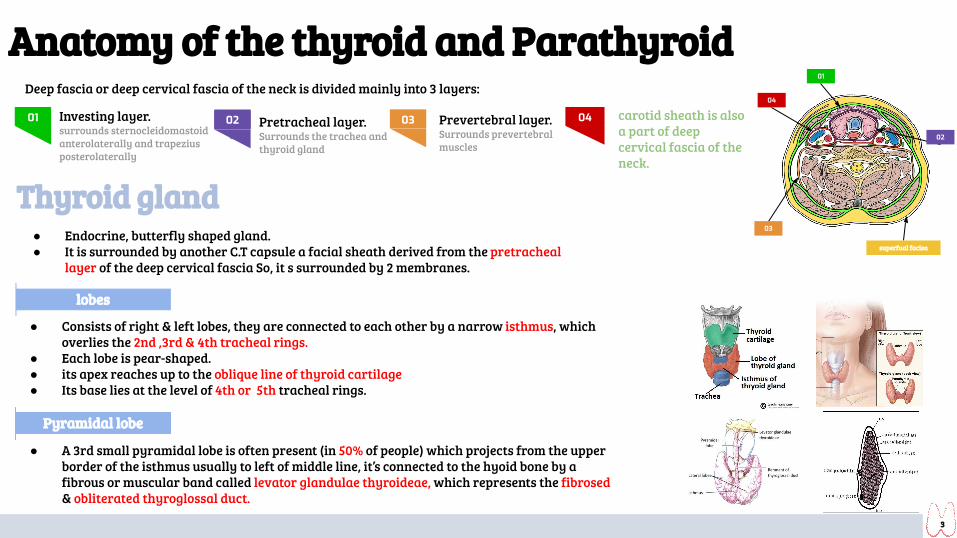
Anatomy of the thyroid and ParathyroidDeep fascia or deep cervical fascia of the neck is divided mainly into 3 layers:
01 02 03 04 carotid sheath is also a part of deep cervical fascia of the neck.
03
01
04
02
superfual facisa
Thyroid gland ● Endocrine, butterfly shaped gland.● It is surrounded by another C.T capsule a facial sheath derived from the pretracheal
layer of the deep cervical fascia So, it s surrounded by 2 membranes.
lobes
● Consists of right & left lobes, they are connected to each other by a narrow isthmus, which overlies the 2nd ,3rd & 4th tracheal rings.
● Each lobe is pear-shaped.● its apex reaches up to the oblique line of thyroid cartilage● Its base lies at the level of 4th or 5th tracheal rings.
Pyramidal lobe
● A 3rd small pyramidal lobe is often present (in 50% of people) which projects from the upper border of the isthmus usually to left of middle line, it’s connected to the hyoid bone by a fibrous or muscular band called levator glandulae thyroideae, which represents the fibrosed & obliterated thyroglossal duct.

4
Thyroid gland: RelationsSurfaces
Anterolaterally Posteriorly Medially (Above) Medially (Below)
1. Sternothyroid2. Sternohyoid3. Superior belly of
omohyoid 4. Sternomastoid
1. Carotid sheath & its contents.
1. Larynx 2. pharynx . 3. Cricothyroid
muscle 4. external laryngeal
nerves
1. Trachea 2. esophagus. 3. Recurrent laryngeal
nerve in between.4. Cricothyroid muscle 5. external laryngeal
nerves
border
posterior
● Rounded ● Related to the superior & inferior
parathyroid glands.● Also related to the anastomosis between
superior & inferior thyroid arteries.
5
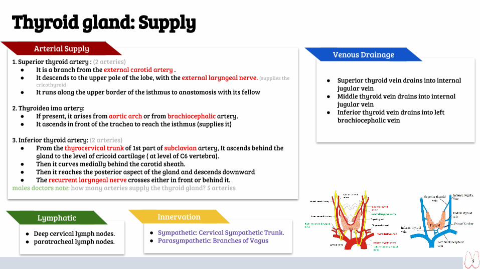
● Superior thyroid vein drains into internal jugular vein
● Middle thyroid vein drains into internal jugular vein
● Inferior thyroid vein drains into left brachiocephalic vein
1. Superior thyroid artery : (2 arteries)● It is a branch from the external carotid artery .● It descends to the upper pole of the lobe, with the external laryngeal nerve. (supplies the
cricothyroid● It runs along the upper border of the isthmus to anastomosis with its fellow
2. Thyroidea ima artery:● If present, it arises from aortic arch or from brachiocephalic artery.● It ascends in front of the trachea to reach the isthmus (supplies it)
3. Inferior thyroid artery: (2 arteries)● From the thyrocervical trunk of 1st part of subclavian artery, It ascends behind the
gland to the level of cricoid cartilage ( at level of C6 vertebra).● Then it curves medially behind the carotid sheath. ● Then it reaches the posterior aspect of the gland and descends downward● The recurrent laryngeal nerve crosses either in front or behind it.
males doctors note: how many arteries supply the thyroid gland? 5 arteries
Arterial SupplyVenous Drainage
Thyroid gland: Supply
● Deep cervical lymph nodes.● paratracheal lymph nodes.
Lymphatic
● Sympathetic: Cervical Sympathetic Trunk.● Parasympathetic: Branches of Vagus
Innervation
6
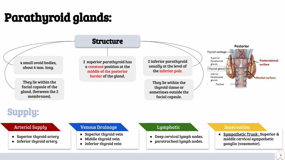
Structure
They lie within the facial capsule of the
gland, (between the 2membranes).
4 small ovoid bodies, about 6 mm. long.
2 superior parathyroid has a constant position at the
middle of the posterior border of the gland.
2 inferior parathyroid usually at the level of
the inferior pole.
They lie within the thyroid tissue or
sometimes outside the facial capsule.
Parathyroid glands:
Supply:
● Superior thyroid vein● Middle thyroid vein● Inferior thyroid vein
Venous Drainage
● Deep cervical lymph nodes.● paratracheal lymph nodes.
Lymphatic ● Sympathetic Trunk : Superior &
middle cervical sympathetic ganglia (vasomotor).
Innervation
● Superior thyroid artery● Inferior thyroid artery
Arterial Supply
7
Clinical notes 1
2
3
4
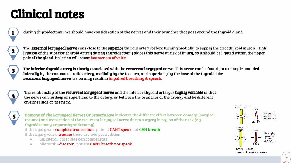
during thyroidectomy, we should have consideration of the nerves and their branches that pass around the thyroid gland
The External laryngeal nerve runs close to the superior thyroid artery before turning medially to supply the cricothyroid muscle. High ligation of the superior thyroid artery during thyroidectomy places this nerve at risk of injury, so it should be ligated within the upper pole of the gland. Its lesion will cause hoarseness of voice.
The inferior thyroid artery is closely associated with the recurrent laryngeal nerve. This nerve can be found , in a triangle bounded laterally by the common carotid artery, medially by the trachea, and superiorly by the base of the thyroid lobe.recurrent laryngeal nerve lesion may result in impaired breathing & speech.
The relationship of the recurrent laryngeal nerve and the inferior thyroid artery is highly variable in that the nerve can lie deep or superficial to the artery, or between the branches of the artery, and be different on either side of the neck.
Damage Of The Laryngeal Nerves Or Semon’s Law indicates the different effect between damage (surgical trauma) and transection of the recurrent laryngeal nerve due to surgery in region of the neck (e.g. thyroidectomy or parathyroidectomy).if the injury was complete transection : patient CANT speak but CAN breathif the injury was a trauma there are two possibilities:
● unilateral: other side can compensate ● bilateral: =disaster , patient CANT breath nor speak
5
8
Embryology of the thyroid and ParathyroidPharyngeal Apparatus
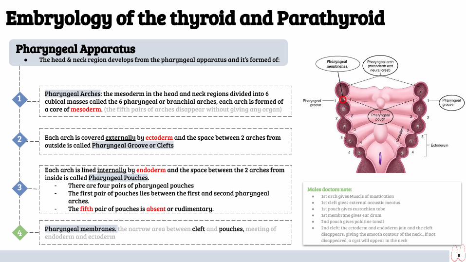
● The head & neck region develops from the pharyngeal apparatus and it’s formed of:
Pharyngeal Arches: the mesoderm in the head and neck regions divided into 6 cubical masses called the 6 pharyngeal or branchial arches, each arch is formed of a core of mesoderm. (the fifth pairs of arches disappear without giving any organ)
Each arch is covered externally by ectoderm and the space between 2 arches from outside is called Pharyngeal Groove or Clefts
Each arch is lined internally by endoderm and the space between the 2 arches from inside is called Pharyngeal Pouches.
- There are four pairs of pharyngeal pouches- The first pair of pouches lies between the first and second pharyngeal
arches.- The fifth pair of pouches is absent or rudimentary.
1
2
3
Pharyngeal membranes. the narrow area between cleft and pouches, meeting of endoderm and ectoderm
Males doctors note:● 1st arch gives Muscle of mastication● 1st cleft gives external acoustic meatus● 1st pouch gives eustachian tube● 1st membrane gives ear drum● 2nd pouch gives palatine tonsil● 2nd cleft: the ectoderm and endoderm join and the cleft
disappears, giving the smooth contour of the neck., If not disappeared, a cyst will appear in the neck
Pharyngeal membranes.
4

9
Development of Parathyroid glands
● The ventral part of the 3rd pouch gives the thymus gland primordium ● The ventral part of the 4th pouch forms what is called Ultimopharyngeal body (an
embryological structure that gives rise to parafollicular cells of thyroid that secrete calcitonin hormone which lowers the blood calcium level).
● As the thymus primordium develops, it descends downward to the thorax, behind the sternum in superior mediastinum.
● It draws the inferior parathyroid bud to a lower level than the superior parathyroid.● Both parathyroid glands lie behind the thyroid gland.
Each of the 3rd & 4th pharyngeal pouch develops into dorsal and ventral parts.
By the 6th week : ● The dorsal part of the 3rd pouch develops into inferior parathyroid bud,● The dorsal part of the 4th pouch develops into the superior parathyroid bud.
1
2
3
4
10
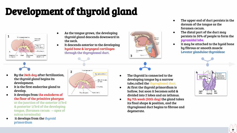
● By the 24th day after fertilization, the thyroid gland begins its development.
● It is the first endocrine gland to develop.
● it develops from the endoderm of the floor of the primitive pharynx at the junction of the anterior 2/3rd & posterior 1/3rd of the developing tongue, (foramen cecum → apex of sulcus terminalis)
● it develops from the thyroid primordium
● As the tongue grows, the developing thyroid gland descends downward in the neck.
● It descends anterior to the developing hyoid bone & laryngeal cartilages through the thyroglossal duct.
● The upper end of duct persists in the dorsum of the tongue as the foramen cecum.
● The distal part of the duct may persists in 50% of people to form the pyramidal lobe.
● it may be attached to the hyoid bone by fibrous or smooth muscle Levator glandulae thyroideae
● The thyroid is connected to the developing tongue by a narrow tube,called the thyroglossal duct.
● At first the thyroid primordium is hollow, but soon it becomes solid & divided into 2 lobes and an isthmus.
● By 7th week (50th day) the gland takes its final shape & position, and the thyroglossal duct begins to fibrose and degenerate.
Picture
J
G
Picture
1
2 4
3
Development of thyroid gland

11
Congenital Anomalies of Thyroid gland1 Cervical thyroglossal duct cyst
Normally, thyroglossal duct undergo fibrosis and degenerated after the thyroid gland takes its final position. In case of the thyroglossal cyst, the duct does not completely close, forming a cyst. When the cyst get infected, it may rupture and open to the external environment forming a sinus.Most of thyroglossal duct cysts are located just anterior & inferior to hyoid bone.
Pic A: showing the possible locations of thyroglossal duct cysts through the broken line indicating the course of the duct. A thyroglossal duct sinus is illustrated.Pic B: illustrating lingual & cervical thyroglossal duct cysts.
Ectopic thyroid tissue
Normally:- The thyroid glands develops high up close to foramen
cecum of the developing tongue.- Then it descends along the thyroglossal duct to reach its
final position by the 7th week.Ectopic:
- Descent of the thyroid could be arrested at any point, or extends down behind the sternum (lead to formation of a retrosternal goiter) in the thorax.
6
Agenesis of thyroid gland
Thyroid gland formed but doesn't function well
Thyroid gland didn’t form
uncomplete margination of thyroid gland, which lead to form other small gland usually in the posterior of the tongue
2
3
4
5
Thyroglossal duct didn’t fibrous
Persistence of thyroglossal duct
Accessory thyroid tissue
Congenital hypothyroidism
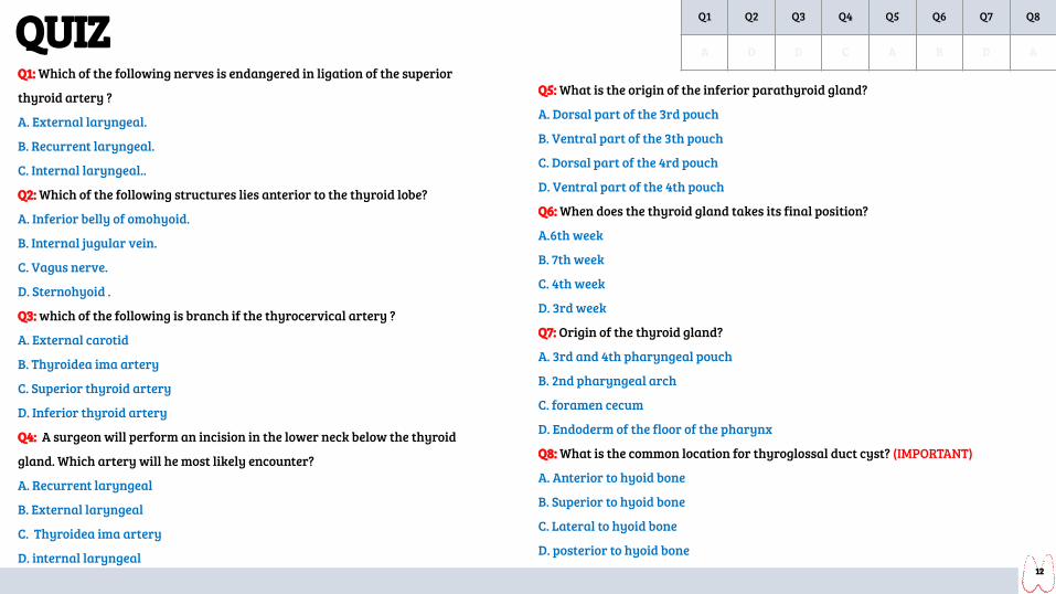
QUIZQ1: Which of the following nerves is endangered in ligation of the superior
thyroid artery ?
A. External laryngeal.
B. Recurrent laryngeal.
C. Internal laryngeal..
Q2: Which of the following structures lies anterior to the thyroid lobe?
A. Inferior belly of omohyoid.
B. Internal jugular vein.
C. Vagus nerve.
D. Sternohyoid .
Q3: which of the following is branch if the thyrocervical artery ?
A. External carotid
B. Thyroidea ima artery
C. Superior thyroid artery
D. Inferior thyroid artery
Q4: A surgeon will perform an incision in the lower neck below the thyroid
gland. Which artery will he most likely encounter?
A. Recurrent laryngeal
B. External laryngeal
C. Thyroidea ima artery
D. internal laryngeal
Q5: What is the origin of the inferior parathyroid gland?
A. Dorsal part of the 3rd pouch
B. Ventral part of the 3th pouch
C. Dorsal part of the 4rd pouch
D. Ventral part of the 4th pouch
Q6: When does the thyroid gland takes its final position?
A.6th week
B. 7th week
C. 4th week
D. 3rd week
Q7: Origin of the thyroid gland?
A. 3rd and 4th pharyngeal pouch
B. 2nd pharyngeal arch
C. foramen cecum
D. Endoderm of the floor of the pharynx
Q8: What is the common location for thyroglossal duct cyst? (IMPORTANT)
A. Anterior to hyoid bone
B. Superior to hyoid bone
C. Lateral to hyoid bone
D. posterior to hyoid bone12
Q1 Q2 Q3 Q4 Q5 Q6 Q7 Q8
A D D C A B D A

Members board
● Abdulrahman Shadid ● Ateen Almutairi
Girls team :
● Ajeed Al Rashoud● Taif Alotaibi● Noura Al Turki● Amirah Al-Zahrani● Alhanouf Al-haluli● Sara Al-Abdulkarem● Renad Al Haqbani● Nouf Al Humaidhi● Jude Al Khalifah● Nouf Al Hussaini● Danah Al Halees● Rema Al Mutawa● Maha Al Nahdi ● Razan Al zohaifi ● Ghalia Alnufaei
Team leaders
Editing file
Boys team:
● Mohammed Al-huqbani● Salman Alagla● Ziyad Al-jofan● Ali Aldawood● Khalid Nagshabandi● Sameh nuser● Abdullah Basamh● Alwaleed Alsaleh● Mohaned Makkawi● Abdullah Alghamdi
Contact us:
● A huge thanks for pharma’s queen: May Babeer
Copyright © 2022 FDOKUMEN