The ultrastructural aspects of neoplastic myoepithelial cell in pleomorphic adenomas of salivary...
13
369 J. Cell. Mol. Med. Vol 8, No 2, 2004 pp. 369-381 The salivary gland pleomorphic adenoma is a benign epithelial neoplasm, histologically charac- terized by a great diversity of morphological aspects. According to literature data, the pleomor- phic adenoma represents 45-74 % of all the sali- vary gland tumors [1-4]. At the Armed Forces Institute of Pathology (AFIP) the pleomorphic adenomas represent 60 % of the benign tumors from all the salivary gland sites: 61 % of the major gland tumors and 54 % of the minor gland tumors [5]. The ultrastructural aspects of neoplastic myoepithelial cell in pleomorphic adenomas of salivary glands C. Margaritescu a *, M. Raica b , Maria Florescu a , Cristiana Simionescu a , M. Surpateanu c , F. Jaubert d , Fl. Bogdan e a Department of Pathology, Emergency County Hospital, No. 1, University of Medicine and Pharmacy, Craiova, Romania b Department of Cytology and Histology, "Victor Babes" University of Medicine and Pharmacy, Timisoara, Romania c Department of Oral Maxilla Facial Surgery, Emergency County Hospital, No. 1, University of Medicine and Pharmacy, Craiova, Romania d Department of Pathology, Rene Descartes, University Paris V, Faculty of Medicine Necker Enfants Malades, France e Centre of Research (Centre for Microscopic Morphology and Immunology Studies), University of Medicine and Pharmacy, Craiova, Romania Received: April 5, 2004; Accepted: May 29, 2004 Abstract The purpose of this study has been to establish the major ultrastructural aspects of the myoepithelial cell and the myoepithelial-like cells proliferated in the pleomorphic adenomas of salivary glands. Thus, twelve benign pleomor- phic adenomas of salivary glands have been studied by electron-microscopy transmission techniques. Our analysis has proved the proliferation of two major cellular populations, one of ductal type and one of myoepithelial type, which tried to reproduce the tubulo-acinar cytoarchitecture from the normal salivary glands. We have also noticed the key role of the so-called ‘modified’ myoepithelial cells from the periphery of the proliferating epithelial units in the genesis of the myxoid and chondromyxoid tumoral stromal areas. All these ultrastructural aspects have explained the great histological diversity of these salivary gland neoplasms as well as the key role of the myoepithelial cell in its histogenesis. Keywords: pleomorphic adenomas • myoepithelial cell • ultrastructural * Correspondence to: Univ. Assist. Dr. C. MARGARITESCU Pathology Department, University of Medecine and Pharmacy, 2 Petru Rares str., Craiova, 200349, Romania. Tel.: +40 740 152 550, E-mail: [email protected], [email protected] Introduction
Transcript of The ultrastructural aspects of neoplastic myoepithelial cell in pleomorphic adenomas of salivary...

369
J. Cell. Mol. Med. Vol 8, No 2, 2004 pp. 369-381
The salivary gland pleomorphic adenoma is abenign epithelial neoplasm, histologically charac-terized by a great diversity of morphological
aspects. According to literature data, the pleomor-phic adenoma represents 45-74 % of all the sali-vary gland tumors [1-4]. At the Armed ForcesInstitute of Pathology (AFIP) the pleomorphicadenomas represent 60 % of the benign tumorsfrom all the salivary gland sites: 61 % of themajor gland tumors and 54 % of the minor glandtumors [5].
The ultrastructural aspects of neoplastic myoepithelial cellin pleomorphic adenomas of salivary glands
C. Margaritescu a *, M. Raica b, Maria Florescu a, Cristiana Simionescu a, M. Surpateanu c, F. Jaubert d, Fl. Bogdan e
a Department of Pathology, Emergency County Hospital, No. 1, University of Medicine and Pharmacy, Craiova, Romania
b Department of Cytology and Histology, "Victor Babes" University of Medicine and Pharmacy, Timisoara, Romania
c Department of Oral Maxilla Facial Surgery, Emergency County Hospital, No. 1, University of Medicine and Pharmacy, Craiova, Romania
d Department of Pathology, Rene Descartes, University Paris V, Faculty of Medicine Necker Enfants Malades, Francee Centre of Research (Centre for Microscopic Morphology and Immunology Studies),
University of Medicine and Pharmacy, Craiova, Romania
Received: April 5, 2004; Accepted: May 29, 2004
Abstract
The purpose of this study has been to establish the major ultrastructural aspects of the myoepithelial cell and themyoepithelial-like cells proliferated in the pleomorphic adenomas of salivary glands. Thus, twelve benign pleomor-phic adenomas of salivary glands have been studied by electron-microscopy transmission techniques. Our analysishas proved the proliferation of two major cellular populations, one of ductal type and one of myoepithelial type,which tried to reproduce the tubulo-acinar cytoarchitecture from the normal salivary glands. We have also noticedthe key role of the so-called ‘modified’ myoepithelial cells from the periphery of the proliferating epithelial units inthe genesis of the myxoid and chondromyxoid tumoral stromal areas. All these ultrastructural aspects have explainedthe great histological diversity of these salivary gland neoplasms as well as the key role of the myoepithelial cell inits histogenesis.
Keywords: pleomorphic adenomas • myoepithelial cell • ultrastructural
* Correspondence to: Univ. Assist. Dr. C. MARGARITESCUPathology Department, University of Medecine and Pharmacy,2 Petru Rares str., Craiova, 200349, Romania.Tel.: +40 740 152 550,E-mail: [email protected], [email protected]
Introduction

Its structural pleomorphism is given both by theepithelial component, as a result of the cytologicaldifferentiations and the growing patterns, and bythe stromal component because of its rich morpho-logical and quantitative diversity [5-6].
The basic cellular components of this kind oftumor have been represented by two distinctive cel-lular populations: a luminal one mainly by the duc-tal luminal cell and an abluminal one (the myoep-ithelial cell and the myoepithelial-like cells derivedfrom it). The cytoarchitectural organization of thesecells may achieve a lot of growing patterns such as:tubular, insular, trabecular, fascicular, micro- andmacrocysts, pseudoangiomatous, cribriform, basa-loid, sarcomatoid or similar to the Verocay corpus-cles from schwannoma [5-8]. The stroma of thesetumors may have the following aspects: myxoid,chondroid, chondromyxoid, hyaline, fibrous, scle-ro-hyaline or even osseous. These aspects are usu-ally associated with the predominance of one ofthem in variable proportion [5-6, 9-21].
The myoepithelial cells are the result of thedifferentiation of the pluripotent cells from thesalivary ducts during the tenth week of the embry-onic life, but in adult life they may proceed fromthe reserve cells of the intercalary and striatedsalivary ducts [22]. Data from literature haveproved the involvement of the myoepithelial cellin the histogenesis of the pleomorphic adenomaand of other monomorphic adenomas [23-30].The myoepithelial cells represent the major cellu-lar population of this kind of tumor, beinginvolved in the genesis of the stromal areas aswell, especially due to its ability to transdifferen-tiate into cells of the mesenchymal line. There hasalso been quoted in literature the existence ofmany types of myoepithelial-like cells or the so-called ‘modified’ myoepithelial cells derivedfrom the neoplastic myoepithelial cell as a resultof metaplasia, dedifferentiation and transdifferen-tiation processes [5-6, 23-24, 31-35]. Thus, therehave been described:• „myxoid” cells (stellate cells placed in stromal
areas with myxoid and chondromyxoid predom-inance) ;
• „myoid” cells (cells with fusiform morphology,mostly placed in the fascicular proliferatingareas) ;
• „hyaline” cells (polyhedral cells, with obviouscellular limits, abundant, hyaline, eosinophilic
cytoplasm, round nucleus with dispersed chro-matin and obvious nucleolus) ;
• clear cells (polyhedral cells, with indistinct cellboundaries, abundant, clear cytoplasm and cen-tral or lightly eccentrically nucleus, with finegranular chromatin and hardly visible nucleo-lus).There have been reported few ultrastructural
studies of the salivary gland pleomorphic adeno-mas. The case series have generally been small, onan average of seven cases [13, 25, 27-28, 30, 36-41]. All these studies have indicated the prolifera-tion of the epithelial cells with ductal and myoep-ithelial differentiations in these tumors. It has alsobeen demonstrated the involvement of the myoep-ithelial cell in the genesis of the tumoral stromalareas [23-30].
Materials and methods
There were selected twelve cases of pleomorphic adeno-mas for ultrastructural investigation. The surgical speci-mens were provided by the Oral Maxilla Facial SurgeryDepartment of the Clinical County Hospital fromCraiova, mostly from 15- 65 year-old females (8 cases),with prevalent parotid localization (7 cases). Theparotidectomy pieces were routinely fixed with 10 percent buffered formalin and sent to the laboratory ofcytology and pathology of the same hospital. They wereprocessed by the classical histopathological techniquewith paraffin embedding and stained with Hematoxylin-Eosin (HE.), Hematoxylin Eosin Safranin (HES),Trichromic Masson and Periodic Acid Schiff-BlueAlcian (PAS- AA).
The electron microscope processing took place in thelaboratory for histological, histopathological andimmunohistochemical techniques from „Victor Babes”University of Medicine and Pharmacy, Timisoara. Weused Reynolds electron-microscopic methods. Fromparaffin blocks there were cut out samples of 1 mm3
thick, which were deparaffinized, re-hydrated andwashed. The postfixation took place in osmium tetroxide1% aqueous solution, at +4°C for an hour. After havingbeen washed, the specimens were dehydrated in gradedalcohols and embedded in epoxy resin - Epon 812. Thesemi-thin cut sections (0.5-1 µm thick) were obtainedwith Leica- Ultracut R ultramicrotom (glass knife) andstained with blue toluidine 1%. The fine sections (about
370
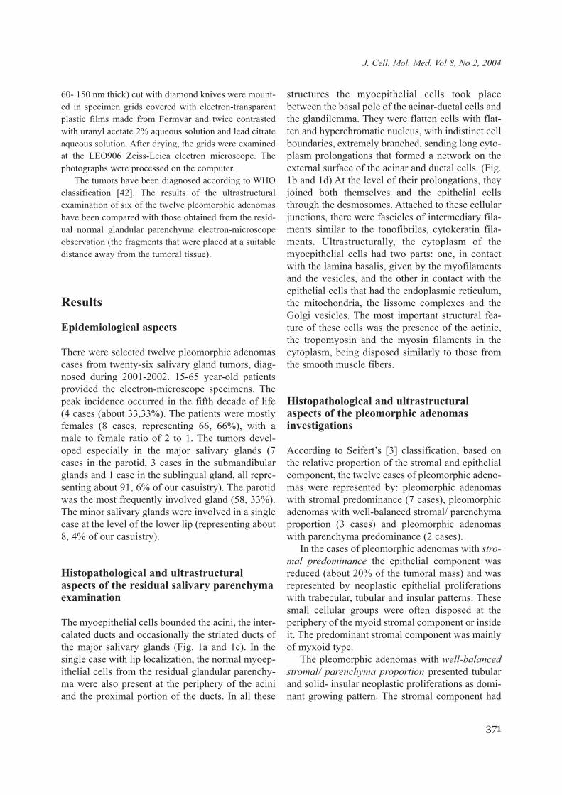
60- 150 nm thick) cut with diamond knives were mount-ed in specimen grids covered with electron-transparentplastic films made from Formvar and twice contrastedwith uranyl acetate 2% aqueous solution and lead citrateaqueous solution. After drying, the grids were examinedat the LEO906 Zeiss-Leica electron microscope. Thephotographs were processed on the computer.
The tumors have been diagnosed according to WHOclassification [42]. The results of the ultrastructuralexamination of six of the twelve pleomorphic adenomashave been compared with those obtained from the resid-ual normal glandular parenchyma electron-microscopeobservation (the fragments that were placed at a suitabledistance away from the tumoral tissue).
Results
Epidemiological aspects
There were selected twelve pleomorphic adenomascases from twenty-six salivary gland tumors, diag-nosed during 2001-2002. 15-65 year-old patientsprovided the electron-microscope specimens. Thepeak incidence occurred in the fifth decade of life(4 cases (about 33,33%). The patients were mostlyfemales (8 cases, representing 66, 66%), with amale to female ratio of 2 to 1. The tumors devel-oped especially in the major salivary glands (7cases in the parotid, 3 cases in the submandibularglands and 1 case in the sublingual gland, all repre-senting about 91, 6% of our casuistry). The parotidwas the most frequently involved gland (58, 33%).The minor salivary glands were involved in a singlecase at the level of the lower lip (representing about8, 4% of our casuistry).
Histopathological and ultrastructuralaspects of the residual salivary parenchymaexamination
The myoepithelial cells bounded the acini, the inter-calated ducts and occasionally the striated ducts ofthe major salivary glands (Fig. 1a and 1c). In thesingle case with lip localization, the normal myoep-ithelial cells from the residual glandular parenchy-ma were also present at the periphery of the aciniand the proximal portion of the ducts. In all these
structures the myoepithelial cells took placebetween the basal pole of the acinar-ductal cells andthe glandilemma. They were flatten cells with flat-ten and hyperchromatic nucleus, with indistinct cellboundaries, extremely branched, sending long cyto-plasm prolongations that formed a network on theexternal surface of the acinar and ductal cells. (Fig.1b and 1d) At the level of their prolongations, theyjoined both themselves and the epithelial cellsthrough the desmosomes. Attached to these cellularjunctions, there were fascicles of intermediary fila-ments similar to the tonofibriles, cytokeratin fila-ments. Ultrastructurally, the cytoplasm of themyoepithelial cells had two parts: one, in contactwith the lamina basalis, given by the myofilamentsand the vesicles, and the other in contact with theepithelial cells that had the endoplasmic reticulum,the mitochondria, the lissome complexes and theGolgi vesicles. The most important structural fea-ture of these cells was the presence of the actinic,the tropomyosin and the myosin filaments in thecytoplasm, being disposed similarly to those fromthe smooth muscle fibers.
Histopathological and ultrastructuralaspects of the pleomorphic adenomasinvestigations
According to Seifert’s [3] classification, based onthe relative proportion of the stromal and epithelialcomponent, the twelve cases of pleomorphic adeno-mas were represented by: pleomorphic adenomaswith stromal predominance (7 cases), pleomorphicadenomas with well-balanced stromal/ parenchymaproportion (3 cases) and pleomorphic adenomaswith parenchyma predominance (2 cases).
In the cases of pleomorphic adenomas with stro-mal predominance the epithelial component wasreduced (about 20% of the tumoral mass) and wasrepresented by neoplastic epithelial proliferationswith trabecular, tubular and insular patterns. Thesesmall cellular groups were often disposed at theperiphery of the myoid stromal component or insideit. The predominant stromal component was mainlyof myxoid type.
The pleomorphic adenomas with well-balancedstromal/ parenchyma proportion presented tubularand solid- insular neoplastic proliferations as domi-nant growing pattern. The stromal component had
371
J. Cell. Mol. Med. Vol 8, No 2, 2004

372
Fig. 1 a. Histological aspects of the remainder parotid parenchyma: serous acini (AS), striated duct (*-lumen),periacinal and periductal myoepithelial cells (arrows), HE, X100; b. Ultrastructural aspects of the remainder parotidparenchyma: serous acini- lumen (L), serous acinar cells (CA), and periacinal myoepithelial cells (CM), cytoplasmicprocess (arrow). X8000; c. Histological aspects of the remainder submandibular salivary gland parenchyma: serousacini (AS), striated ducts (*-lumen), periacinal and periductal myoepithelial cells (arrows). HES,X100; d. Ultrastructural aspects of the remainder submandibular salivary gland parenchyma: striated duct- lumen(*), columnar ductal cells (CD), periductal myoepithelial cells (arrows). X4800; e. Histopathological aspects of aparotid pleomorphic adenoma: abortive ductal structures (asterisks) exteriorly outline by a single row of myoepithe-lial cells (arrow heads). HE, X200; f. Ultrastructural aspects of an parotid pleomorphic adenoma: abortive ductalstructure outlined to inner by a row of slow columnar ductal cells (CD) and external by a row of myoepithelial cells(CM), that keep contact with rests of basal membrane (arrows heads). X9000.

mainly myxoid, chondro-myxoid and chondroiddifferentiations, more often associated.
In the structure of the pleomorphic adenomaswith parenchyma predominance the most encoun-tered patterns were the tubular and the insular ones.The stromal component was reduced (no more than20-30% of the tumoral mass) and mostly of myxoidtype.
Both histopathologically and ultrastructurally,we have found in the structure of these tumors a lotof cellular differentiation, such as: luminal cells,abluminal cells, ‘myxoid’ cells, ‘myoid’ cells andchondroid cells.
The major neoplastic cellular populations wererepresented by the luminal and abluminal cellswhich proliferated in a great diversity of growingpatterns (tubular, insular, trabecular, fascicular,cystic, pseudoangiomatous, cribriform et al.) moreoften associated. The most encountered cytoarchi-tectural model was the tubular one. It was presentin all the investigated cases, but in different pro-portion from case to case, prevailing especially inthose two cases of pleomorphic adenomas withparenchymatous predominance. In these cases theneoplastic tubular structures were often denselydisposed, side by side, separated by minimal quan-tities of myxoid and fibro-hyaline stroma. In all theother cases the tubular structures were found insmall number with clump or isolated distribution inthe stromal myxoid areas.
In four cases we have noticed tubular neoplasticproliferations outlined by two cellular rows: theinternal one made up of ductal cells and the exter-nal one composed of myoepithelial cells, attachedto a material similar to the basal membrane bystructures of hemidesmosomes type. Thus, thereresulted tubular neoplastic structures with cytoar-chitectonics similar to the striated ducts from thenormal salivary glands (Fig. 1e and 1f).
The other tubular neoplastic proliferations wereexternally outlined by many cellular rows ofmyoepithelial type with concentric muff dispositionor irradiating ‘sun-like rays’. Ultrastructurally,these cells usually had the morphology of a ‘nor-mal’ myoepithelial cell, being flatten, with numer-ous cytoplasmic processes, flatten and hyper-chromic nucleus. Some of these myoepithelial cellspresented a lot of cytoplasmic inclusions, represent-ed by glycogen aggregates (the ultrastructural fea-ture consisted of irregularly shaped 15 to 30 nm
particles - beta glycogen particles), lipid droplets(empty cytoplasmic vacuoles) and mucigenousgranules with variable degree of extraction.
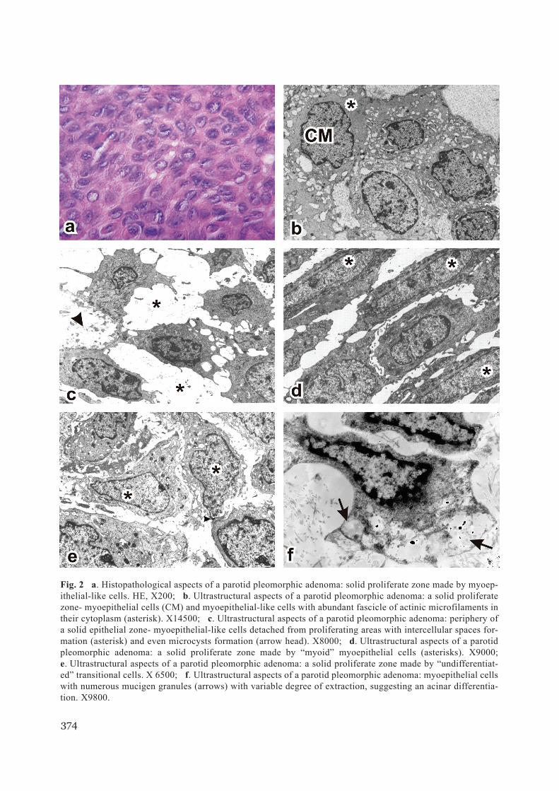
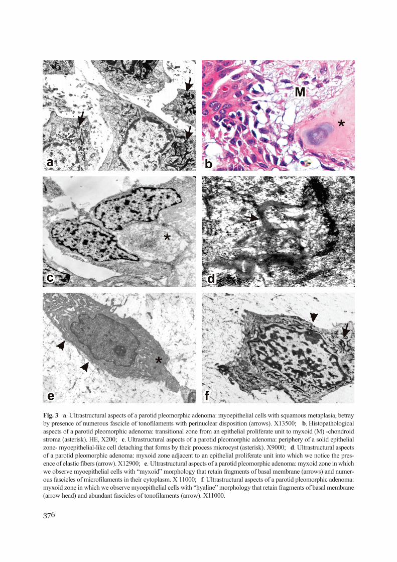
The second pattern of the epithelial proliferationwas the solid-insular one, where the cells with ultra-structural aspects of myoepithelial type weregrouped, being linked by desmosomes. (Fig. 2a and2b). Similar to the neoplastic epithelial prolifera-tions with tubular pattern, the myoepithelial cellsbegan to detach at their periphery. Ultrastructurally,they also less resembled the normal myoepithelialcells. The most peripherical proliferating cells hadmore irregular forms, elongated cytoplasmic pro-cesses, being in close relationship with the solidneoplastic proliferations. Focally, there could beobserved microvilli and rests of basal membrane(Fig. 2c).
The examination of some of the solid neoplasticepithelial areas from a pleomorphic adenoma withepithelial predominance has revealed the existenceof a solid area mainly made up of cells with theultrastructural feature of the ‘myoid’ myoepithelialcells type. These cells had a fusiform morphology,flat and hyperchromic nucleus, abundant 6 nm fila-ment fascicles with longitudinal disposition, paral-lel to the ductal axis, which here and there present-ed electron-dense plasma membrane associatedplaques, perinuclear tonofilaments fascicles anddesmosomes junctions between them (Fig. 2d).
In another solid tumoral area of a pleomorphicadenoma with well-balanced stroma/parenchymaratio we have noticed a compact neoplastic epithe-lial proliferation made up of cells which ultrastruc-turally could not be categorized either as luminal ormyoepithelial cells. They looked like the modifiedmyoepithelial cells that underwent a mesenchymaltransdifferentiation process. The myoepithelial ori-gin of these cells has been suggested by the pres-ence on limited areas of the basal lamina fragmentson the plasmalemma. They remained in contact bythe desmosomes (Fig. 2e).
In one of the two cases of pleomorphic adeno-mas with parenchyma predominance we havenoticed solitary cells in the compact proliferatingzones. Besides the ultrastructural aspects of themyoepithelial cell, they had abundant mucigenousgranules with variable degree of extraction, whichmay explain the possible acinar differentiation ofthese cells (Fig. 2f). These mucigen granules hadvariable size (0.7-1.8 µm) and electron density;
373
J. Cell. Mol. Med. Vol 8, No 2, 2004
374
Fig. 2 a. Histopathological aspects of a parotid pleomorphic adenoma: solid proliferate zone made by myoep-ithelial-like cells. HE, X200; b. Ultrastructural aspects of a parotid pleomorphic adenoma: a solid proliferatezone- myoepithelial cells (CM) and myoepithelial-like cells with abundant fascicle of actinic microfilaments intheir cytoplasm (asterisk). X14500; c. Ultrastructural aspects of a parotid pleomorphic adenoma: periphery ofa solid epithelial zone- myoepithelial-like cells detached from proliferating areas with intercellular spaces for-mation (asterisk) and even microcysts formation (arrow head). X8000; d. Ultrastructural aspects of a parotidpleomorphic adenoma: a solid proliferate zone made by “myoid” myoepithelial cells (asterisks). X9000;e. Ultrastructural aspects of a parotid pleomorphic adenoma: a solid proliferate zone made by “undifferentiat-ed” transitional cells. X 6500; f. Ultrastructural aspects of a parotid pleomorphic adenoma: myoepithelial cellswith numerous mucigen granules (arrows) with variable degree of extraction, suggesting an acinar differentia-tion. X9800.
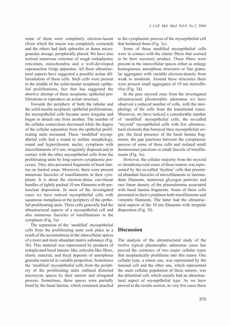
some of them were completely electron-lucent(from which the mucin was completely extracted)and the others had dark spherules or dense micro-granules storage, peripherally placed. We have alsonoticed numerous cisternae of rough endoplasmicreticulum, mitochondria and a well-developedsupranuclear Golgi apparatus. All these ultrastruc-tural aspects have suggested a possible acinar dif-ferentiation of these cells. Such cells were presentin the middle of the solid-insular neoplastic epithe-lial proliferations, fact that has suggested theabortive attempt of these neoplastic epithelial pro-liferations to reproduce an acinar structure.
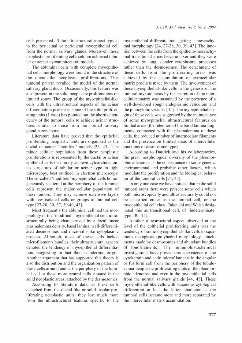
Towards the periphery of both the tubular andthe solid-insular neoplastic epithelial proliferations,the myoepithelial cells became more irregular andbegan to detach one from another. The number ofthe cellular connections decreased while the degreeof the cellular separation from the epithelial prolif-erating units increased. These ‘modified’ myoep-ithelial cells had a round to stellate morphology,round and hyperchromic nuclei, cytoplasm withmicrofilaments of 6 nm, irregularly disposed and incontact with the other myoepithelial cells from theproliferating units by long narrow cytoplasmic pro-cesses. They also presented fragments of basal lam-ina on limited areas. Moreover, there were presentnumerous fascicles of tonofilaments in their cyto-plasm. It is about the electron-dense curvilinearbundles of tightly packed 10 nm filaments with per-inuclear disposition. In most of the investigatedcases we have noticed myoepithelial cells withsquamous metaplasia at the periphery of the epithe-lial proliferating units. These cells generally had theultrastructural aspects of a myoepithelial cell andalso numerous fascicles of tonofilaments in thecytoplasm (Fig. 3a).
The separation of the ‘modified’ myoepithelialcells from the proliferating units took place as aresult of the accumulation in the intercellular spacesof a more and more abundant matrix substance (Fig.3b). This material was represented by products ofreduplicated basal lamina- like, reticulin-like fibers,elastic material, and focal deposits of amorphousgranular material in variable proportion. Sometimesthe ‘modified’ myoepithelial cells from the periph-ery of the proliferating units outlined distortedmicrocysts spaces by their narrow and elongatedprocess. Sometimes, these spaces were partiallylined by the basal lamina, which remained attached
to the cytoplasmic process of the myoepithelial cellthat bordered them (Fig. 3c).
Some of these modified myoepithelial cellswere in contact with the elastic fibers that seemedto be their secretory product. These fibers werepresent in the intercellular spaces either as enlargehomogenous, amorphous structures or fine granu-lar aggregates with variable electron-density fromweak to moderate. Around these structures therewere present small aggregates of 10 nm microfib-riles (Fig. 3d).
In the pure myxoid zone from the investigatedultrastructural pleomorphic adenomas we haveobserved a reduced number of cells, with the mor-phology of the cells from the transitional zones.Moreover, we have noticed a considerable numberof ‘modified’ myoepithelial cells, the so-called“myxoid” myoepithelial cells with few ultrastruc-tural elements that betrayed their myoepithelial ori-gin: the focal presence of the basal lamina frag-ments, the gap junctions between the cytoplasmicprocess of some of these cells and isolated smalldesmosomes junctions or small fascicle of tonofila-ments (Fig. 3e).
However, the cellular majority from the myxoidor chondromyxoid zones of these tumors was repre-sented by the so-called ‘hyaline’ cells that present-ed abundant fascicles of microfilaments or interme-diate filaments, numerous glycogen particles andrare linear density of the plasmalemma associatedwith basal lamina fragments. Some of these cellspresented in their cytoplasm both tonofilaments andvimentin filaments. The latter had the ultrastruc-tural aspects of the 10 nm filaments with irregulardisposition (Fig. 3f).
Discussion
The analysis of the ultrastructural study of thetwelve typical pleomorphic adenomas cases hasproved the existence of two major cellular typesthat neoplastically proliferate into this tumor. Onecellular type, a minor one, was represented by theluminal cell and the other one, which representedthe main cellular population of these tumors, wasthe abluminal cell, which usually had an ultrastruc-tural aspect of myoepithelial type. As we haveproved in the results section, in very few cases these
375
J. Cell. Mol. Med. Vol 8, No 2, 2004
376
Fig. 3 a. Ultrastructural aspects of a parotid pleomorphic adenoma: myoepithelial cells with squamous metaplasia, betrayby presence of numerous fascicle of tonofilaments with perinuclear disposition (arrows). X13500; b. Histopathologicalaspects of a parotid pleomorphic adenoma: transitional zone from an epithelial proliferate unit to myxoid (M) -chondroidstroma (asterisk). HE, X200; c. Ultrastructural aspects of a parotid pleomorphic adenoma: periphery of a solid epithelialzone- myoepithelial-like cell detaching that forms by their process microcyst (asterisk). X9000; d. Ultrastructural aspectsof a parotid pleomorphic adenoma: myxoid zone adjacent to an epithelial proliferate unit into which we notice the pres-ence of elastic fibers (arrow). X12900; e. Ultrastructural aspects of a parotid pleomorphic adenoma: myxoid zone in whichwe observe myoepithelial cells with “myxoid” morphology that retain fragments of basal membrane (arrows) and numer-ous fascicles of microfilaments in their cytoplasm. X 11000; f. Ultrastructural aspects of a parotid pleomorphic adenoma:myxoid zone in which we observe myoepithelial cells with “hyaline” morphology that retain fragments of basal membrane(arrow head) and abundant fascicles of tonofilaments (arrow). X11000.
cells presented all the ultrastructural aspect typicalto the periacinal or periductal myoepithelial cellfrom the normal salivary glands. Moreover, theseneoplastic proliferating cells seldom achieved tubu-lar or acinar cytoarchitectural models.
The abluminal cells with complete myoepithe-lial cells morphology were found in the structure ofthe ductal-like neoplastic proliferations. Thistumoral pattern recalled the model of the normalsalivary gland ducts. Occasionally, this feature wasalso present in the solid neoplastic proliferations onlimited zones. The group of the myoepithelial-likecells with the ultrastructural aspects of the acinardifferentiation present in some of the solid prolifer-ating units (1 case) has pointed out the abortive ten-dency of the tumoral cells to achieve acinar struc-tures similar to those from the normal salivarygland parenchyma.
Literature data have proved that the epithelialproliferating neoplastic units are organized as theductal or acinar ‘modified’ models [25, 43]. Theminor cellular population from these neoplasticproliferations is represented by the ductal or acinarepithelial cells that rarely achieve cytoarchitecton-ics structures of tubular or acinar type in lightmicroscopy, best outlined in electron microscopy.The so-called ‘modified’ myoepithelial cells homo-geneously scattered at the periphery of the luminalcells represent the major cellular population ofthese tumors. They may achieve extensive layerswith few isolated cells or groups of luminal celltype [27-28, 30, 37, 39-40, 43].
Most frequently the abluminal cell had the mor-phology of the ‘modified” myoepithelial cell, ultra-structurally being characterized by a focal linearplasmalemma density, basal lamina, well-differenti-ated desmosomes and microvilli-like cytoplasmicprocess. Although, most of these cells lackedmicrofilaments bundles, their ultrastructural aspectsdenoted the tendency of myoepithelial differentia-tion, suggesting in fact their ectodermic origin.Another argument that has supported this theory isalso the distribution and the organization pattern ofthese cells around and at the periphery of the lumi-nal cell or those more central cells situated in thesolid neoplastic areas, attached by the desmosomes.
According to literature data, as these cellsdetached from the ductal-like or solid-insular pro-liferating neoplastic units, they lost much morefrom the ultrastructural features specific to the
myoepithelial differentiation, getting a mesenchy-mal morphology [24, 27-28, 30, 39, 43]. The junc-tion between the cells from the epithelio-mesenchy-mal transitional areas became laxer and they wereachieved by long, slender cytoplasmic processesrather than the desmosomes. The detachment ofthese cells from the proliferating areas wasachieved by the accumulation of extracellularmatrix products made by them. The involvement ofthese myoepithelial-like cells in the genesis of thetumoral myxoid areas by the secretion of the inter-cellular matrix was sustained by the presence of awell-developed rough endoplasmic reticulum andthe pinocytotic vesicles [41]. The myoepithelial ori-gin of these cells was suggested by the maintenanceof some myoepithelial ultrastructural features onlimited areas (the retention of the basal lamina frag-ments, connected with the plasmalemma of thesecells, the reduced number of intermediate filamentsand the presence on limited areas of intercellularjunctions of desmosome type).
According to Dardick and his collaboratories,the great morphological diversity of the pleomor-phic adenomas is the consequence of some genetic,environmental and probably other factors, whichmodulate the proliferation and the biological behav-ior of the tumoral cells [24, 43].
In only one case we have noticed that in the solidtumoral areas there were present some cells whichboth microscopically and ultrastructurally could notbe classified either as the luminal cell, or themyoepithelial cell class. Takeuchi and Welsh desig-nated this as transitional cell, of ‘indeterminate’type [30, 41].
Another ultrastructural aspect observed at thelevel of the epithelial proliferating units was thetendency of some myoepithelial-like cells to squa-mous metaplasia (polyhedral morphology, attach-ments made by desmosomes and abundant bundlesof tonofilaments). The immunohistochemicalinvestigations have proved this coexistence of thecytokeratin and actin microfilaments in the angularor fusiform cell from the periphery of the tubulo-acinar neoplastic proliferating units of the pleomor-phic adenomas and even in the myoepithelial cellsfrom the normal salivary glands [44, 45]. Thesemyoepithelial-like cells with squamous cytologicaldifferentiation lost the latter character as thetumoral cells became more and more separated bythe intercellular matrix accumulation.
377
J. Cell. Mol. Med. Vol 8, No 2, 2004
The myoepithelial origin of the cells from themyxoid stromal areas has been proved by the main-tenance of their connection with the cells from theperiphery of the ductal-acinar neoplastic units.These junctions were achieved by a very long, slen-der cytoplasmic process. More literature data havesustained the existence of some small cellulargroups in the myxoid stromal areas that presentbundles of tonofilaments, desmosomes and smallfragments of basal lamina in contact with their plas-malemma [27-28, 30, 39, 43]. The stromal areaswith myxoid differentiation played a central role inthe genesis of the chondroid stromal areas of thistumor. Literature data have revealed isolated oreven group presence of pure myoepithelial cells orsquamous metaplastic cells in the myxoid andchondroid stromal areas [27-28, 30, 39, 43].
It has been suggested the possible epithelial ori-gin of the chondroid areas by the close associationof these cells with the modified myoepithelial cel-lular groups and by the presence of some tonofila-ments fascicles in their cytoplasm [38, 43; 46-47].
Kusafuka and his collaboratories have reportedthat the genesis of the chondroid stromal areas areassociated with the overexpression of the bone mor-phogenetic proteins (BMPs) by the neoplasticmyoepithelial cells [48]. This protein belongs to thetransforming growing factor (TGF)-β superfamilywhich interferes with the regulation of the mes-enchymal tissue formation. The TGF-β2 isexpressed by the neoplastic ductal epithelial cells,while TGF-β3 is expressed by the neoplasticmyoepithelial cells from the solid neoplastic epithe-lial proliferating areas, the metaplastic squamouscells and the luminal cells from the tubular neoplas-tic proliferating areas [48]. TGF-β is involved in thecontrol of the luminal and myoepithelial neoplasticcell differentiation [48]. Moreover, Devlin andSloan have shown that only the ductal cells and notin the neoplastic myoepithelial cells expressed thecartilage-derived retinoic acid-sensitive protein(CD-RAP), a recently described immunomarkerinvolved in the morphogenesis and the develop-ment of the salivary glands [49].
According to Dardick and his collaboratories,the histogenetic model of these tumors is based onthe myoepithelial cell capacity from the proliferat-ing units of ductal-like type or solid-insular prolif-erating areas to progressive transdifferentiation,squamous metaplasia and detachment, as the extra-
cellular matrix products accumulate. These process-es take place simultaneously in different areas, sug-gesting the great histological variability of thistumor.
Anderson and his collaboratories observed thatthe modifications of the immunohistochemical pro-file (from vimentin, keratin and actin positive to thecoexpression of vimentin and glial fibrillary acidprotein (GFAP)) at the level of the cells from theperiphery of the epithelial proliferating units andfrom the epithelial-mesenchymal transitional areas.Ultrastructurally, these correspond to a progressiveloss of myoepithelial features, reflecting theirengagement in the chondroid differentiation line[50].
According to Regezi and Eversole, the stem cellor the reserve cell from the intercalated ducts hasthe key role in the tumoral genesis of the salivaryglands [26, 29]. Batsakis issues a hypothesisaccording to which the salivary gland tumors andthe salivary parenchyma regeneration may recapit-ulate the embryonic stages of the salivary glanddevelopment [51-52].
Until now, neither the presence of the reservecells in the structure of the intercalated ducts of thesalivary glands, nor their functional involvement inthe acinar or excretory ducts regeneration havebeen proved. After an experiment on rats, Barkaand Boshell achieved salivary gland hyperplasia,thus proving the regenerative potential of the acinarcells and implicitly of the myoepithelial cells [53-54]. These facts do not necessarily mean that a sim-ilar process may also take place in the neoplasticproliferations.
According to the latest investigations (using thedouble immunostaining procedure for Ki67 and α-actin or other subtypes of cytokeratins) the regener-ative potential owns not only the myoepithelialcells but also the basal cells, intercalary ductal cells,acinar cells and oxyphil cells. The regeneration ofthe acinar and intercalary duct cells issues indepen-dently from those of myoepithelial cells or basalcells. The oxyphil regeneration and the majority ofthe epithelial metaplasia from the salivary glandsderive from the basal cell of the striated ducts,which present a great capacity of pluripotentialmorphogenetic differentiation [55].
In an experiment made by Bockman regardingthe pancreas carcinogenesis, it was proved the exis-tence of a gradual dedifferentiation process from
378
the acinar units to tubular structures [25].Moreover, the absence of the myoepithelial cellsfrom the pancreas and their presence only in thesalivary glands, the sweat glands and the breastexplained the pleomorphic adenoma developmentonly in the latter structures and not in the pancreas.
Thomas Aigner and his collaboratories haveproved that the pleomorphic adenomas representthe in vivo model epithelial-mesenchymal transdif-ferentiation process in adult. Although the epithe-lial-mesenchymal transdifferentiation process wasconsidered normal only in the fetal period, in theprocess of palate, neural crest, metanephric and car-diac development, they may also take place afterbirth [57]. Thus, such process may be observed interatocarcinomas, mesotheliomas, carcinomas with(pseudo) sarcomatous differentiation and chordo-mas of the spinal axis [58-59].
In the pleomorphic adenomas case, the detection(by in situ hybridization technique) in the ductalcells of the genes involved in the aggregan coreprotein mRNA synthesis, has demonstrated theexpression of the mesenchymal genes in the cellswith epithelial origin [23]. Thus, the onset of themesenchymal gene expression in the ductal cellsleads to the secretion of an abundant extracellularmatrix rich in glycosaminoglycan that generates themyxoid matrix formation. This fact suggests thatthe neoplastic ductal cells were involved in the gen-esis of the myxoid stromal areas from the pleomor-phic adenomas [23].
Conclusion
The neoplastic myoepithelial cell represents amajor component of the neoplastic cellular popula-tion of the pleomorphic adenomas. It has a key rolein the genesis of these tumors, being capable ofdedifferentiation, metaplasia and transdifferentia-tion. The electron-microscope study has confirmedthe involvement of the neoplastic myoepithelial cellfrom the pleomorphic adenomas in the epithelial-mesenchymal transdifferentiation process. Accor-dingly, these cells detached from the ductal-like orsolid-insular proliferating neoplastic units, lostmuch of the ultrastructural features specific to themyoepithelial differentiation, getting a mesenchy-mal morphology. These cells produce glycosamino-
glycans and intercellular fibrillary materials gener-ating the myxoid stromal tumoral areas. The endstage of this epithelial-mesenchymal transdifferen-tiation process is represented by gradual formationof chondroid stromal areas with typical chondro-cyte lacunae from myxoid areas. According to thetype, amount and distribution of the matrix productsof these myoepithelial-like cells from the peripheryof the tubular-solid proliferating areas, we mayexplain the great variety of histological aspects ofthe stroma in this tumor type.
The great diversity of cytological differentia-tions present in pleomorphic adenomas can beexplained by metaplastic potential of the neoplas-tic myoepithelial cells. We observe that somemyoepithelial-like cells from neoplastic prolifera-tions present the ultrastructural aspects of acinardifferentiation. This fact points out the abortivebehaviour of the tumoral cells to achieve acinarstructures similar those from the normal salivarygland parenchyma.
References
1. Eneroth CM., Salivary gland tumors in the parotid gland,submandibular gland, and the palate region, Cancer, 27:1415-1418, 1971
2. Eveson JW, Cawson RA., Salivary gland tumours. Areview of 2410 cases with particular reference to histolog-ical types, site, age and sex distribution, J Pathol, 146: 51-58, 1985
3. Seifert G, Miehlke A, Haubrich J, Chilla R., Diseases ofthe salivary glands: diagnosis, pathology, treatment, facialnerve surgery. Georg Thieme Verlag, Stuttgart, 1986
4. Spiro RH., Salivary neoplasms: overview of a 35-yearexperience with 2, 807 patients. Head Neck Surg, 8: 177-84, 1986
5. Ellis GL, Auclair PL. Mixed tumor (pleomorphic adeno-ma). In: Tumors of the salivary glands. Atlas of TumorPathology, 3rd Series, Fascicle 17, Washington, D.C.:Armed Forces Institute of Pathology, 1996, Chapter 4,Section 4. 1: pp. 39-57
6. Cheuk W., Chan JKC: Salivary gland tumors In:Christopher D. M. Fletcher eds: Diagnostic histopathologyof tumors, Churchill Livingstone, London, Volume I,2000, Chapter 7: 231-312
7. Gary L. E. And Auclair P. L. Tumors of the salivaryglands AFIP, Fascicle II, Washington D. C., 1996
8. Lam PW, Chan JK, Sin VC Nasal pleomorphic adenomawith skeletal muscle differentation potential misdiagnosisas rabdomyosarcoma, Hum Pathol, 28: 1299-1302, 1997
9. Atula T, Klemi PJ, Donath K, Happonen RP, JoensuuH, Grenman R. Basal cell adenocarcinoma of the parotid
379
J. Cell. Mol. Med. Vol 8, No 2, 2004
gland: a case report and review of the literature. J.Laryngol Otol., 107: 862-864, 1993
10. Batsakis JG, Luna MA. Basaloid salivary carcinoma.Ann. Otol. Rhinol. Laryngol., 100: 785-787, 1991
11. Batsakis JG, Luna MA, el-Naggar AK. Basaloidmonomorphic adenomas. Ann. Otol. Rhinol. Laryngol.,100: 687-690, 1991
12. Chen KT. Carcinoma arising in monomorphic adenomaof the salivary gland. Am. J. Otolaryngol., 6: 39-41, 1985.
13. David R, Buchner A. Elastosis in benign and malignantsalivary gland tumors. A histochemical and ultrastructuralstudy. Cancer, 45: 2301-2310, 1980
14. David R, Buchner A. Tannic acid- glutaraldehyde fixativeand pleomorphic adenomas of the salivary gland: an ultra-structural study. J. Oral. Pathol., 11: 26-38, 1982
15. Fantasia JE, Neville BW. Basal cell adenomas of theminor salivary glands. A clinicopathologic study of seven-teen new cases and a review of the literature. Oral. Surg.Oral. Med. Oral. Pathol., 50: 433-440, 1980
16. Harrison JD, Auger DW: Mucosubstance histochemistryof pleomorphic adenoma of parotid and submandibularsalivary glands of man. Light and electron microscopy.Histochem. J., 23: 293-302, 1991
17. Nakanishi K, Kawai T, Suzuki M, Shinmei M:Glycosaminoglycans in pleomorphic adenoma and ade-noid cytic carcinoma of the salivary gland. Arch. Pathol.Lab. Med., 114: 1227-1231, 1990
18. Nara Y, Takeuchi J, Yoshida K, Fukatsu T, Nagasaka T,Kawaguchi T, Meng N, Kikuchi H, Nakashima N:Immunohistochemical characterisation of extracellularmatrix components of salivary gland tumours. Br. J.Cancer, 64: 307-314, 1991
19. Neville BW, Damm DD, Allen CM, Bouquot JE:Salivary glands tumors in Oral & Maxillofacial Pathology,WB Saunders, Salivary gland pathology, 2002, Chapter11, pp. 410-432
20. Nikai H, Ogawa I, Ijuhin N, Yamasaki A, Takata T,Elbardaie A. Ultrastructural cytochemical demonstrationof elastin in the matrix of salivary gland tumors. ActaPathol. Jpn., 33: 1171-81, 1983
21. Ogawa I, Nikai H, Takata T, Miyauchi M, Ito H, IjuhinN. The cellular composition of basal cell adenoma of theparotid gland: an immunohistochemical analysis. OralSurg. Oral Med. Oral Pathol., 70: 619-626, 1990
22. Tandler B, Denning CR, Mandel ID, Kutscher AH. -Ultrastructure of human labial salivary glands.Myoepitelium and ducts. Morphol., 130: 227-246, 1970
23. Aigner T., Neureiter D, Volker U., Belke J, Kirchner T.,Epithelial-mesenchymal transdifferentiation and extracel-lular matrix gene expression in pleomorphic adenomas ofthe parotid salivary gland. J. Pathol., 186: 178-185, 1998
24. Dardick I, van Nostrand AWP, Phillips MJ:Histogenesis of salivary gland pleomorphic adenoma(mixed tumors) with an evaluation of the role of themyoepithelial cell. Hum. Pathol., 13: 62-64, 1982
25. Erlandson RA, Cardon- Cardo C, Higgins PJ.Histogenesis of benign pleomorphic adenoma (mixedtumor) of the major salivary glands. An ultrastructural andimmunohistochemical study. Am. J. Surg. Pathol., 8: 803-820, 1984
26. Eversole LR: Histogenetic classification of salivary glandtumors Arch. Pathol. 92:433, 1980
27. Hubner G, Klein HJ, Kleisasser O et al.: Role of myoep-ithelial cells in the development of salivary gland tumors.Cancer, : 27: 1225, 1971
28. Mylius EA: The identification and the role of the myoep-ithelial cell in salivary gland tumors. Acta Pathol.Microbiol. Scand. ; 50 (suppl 139) :1-10, 1960
29. Regezi JA, Batsakis JG: Histogenesis of salivary glandneoplasms. Otolaryngol. Clin. North. Am., 10: 297, 1977
30. Welsh RA, Mayer AT: Mixed tumors of human salivaryglands; histogenesis. Arch. Pathol., 85: 433, 1968
31. Marius Raica, Cristian Suciu, Cezar Geynes, Hystologyfor pathologists: The histological peculiar features of sali-vary glands, Romanian J. Path., Vol 6, 1&2: 98-113, 2002
32. Morinaga S, Nakajima T, Shimosato Y. Normal and neo-plastic myoepithelial cells in salivary glands: an immuno-histochemical study. Hum. Pathol., 18:1218-1226, 1987
33. Ogawa Y., Toyosawa S., Ishida T., Ijuhin N. : Keratin 14immunoreactive cells in pleomorphic adenomas and ade-noid cystic carcinomas of salivary glands Wirchows Arch.,437: 58-68, 2000
34. Fujita Y., Yoshida T., Sakakura Y., Sakakura T.,Reconstruction of pleomorphic adenoma of the salivaryglands in the three-dimensional collagen gel matrix cul-ture, Virchows Arch., 434:137-143, 1999
35. Ogawa Y., Kishino M., Atsumi Y., Kimoto M., FukudaY., Ishido T., Ijuhin N. : Plasmacytoid cells in salivary-gland pleomorphic adenomas: evidence of luminal celldifferentiation, Virchows Arch.,, 443: 625-634, 2003
36. Chisholm DM, Waterhouse JP, Kraucunas W, et al.: Aquantitative ultrastructural study of the pleomorphic ade-noma (mixed tumor) of the human minor salivary glands.Cancer, 34: 1631, 1974
37. Deppisch LM, Toker C: Mixed tumor of the parotid glandan ultrastructural study. Cancer, 24: 174, 1969
38. Doyle LE, Lynn JA, Panopio IT, et al.: Ultrastructural ofthe chondroid region of the benign mixed tumor of sali-vary gland. Cancer, 22: 225, 1968
39. Eneroth CM, Wersall J: Fine structure of the epithelialcells in mixed tumors of the parotid gland. Ann. Otol., 22:75:95, 1966
40. Oota K, Takahashi N: Electron microscopic studies onthe so-called benign mixed tumor of the salivary gland.Gann., 49: 234, 1958
41. Takeuchi J, Sobue M, Yoshida M, Esaki T, Katok Y:Pleomorphic adenoma of the salivary gland. With specialreference to histochemical and electron microscopic stud-ies and biochemical analysis of glycosaminoglycans invivo and in vitro. Cancer, 36: 1771-1789, 1975
42. Seifert G, Sobin LH. Histological typing of salivary glandtumours. World Health Organization international histo-logical classification of tumours. 2nd ed. New York:Springer-Verlag 1991
43. Dardick I., van Nostrand A.W.P., Jeans M.T., RippsteinP., Edwards V., Pleomorphic adenoma, I: ultrastructuralorganization of epithelial regions, Hum. Pathol., 14: 780-797, 1983
44. Castelitz J, Osborn M, Seifert G et al.: Intermediate-sized filament proteins (prekeratin, vimentin, desmin) in
380

normal parotid gland and parotid gland tumors: imunoflu-orescence study. Virchows Arch. [Pathol. Anat.], 393: 273,1981
45. Franke WW, Schmid E, Osborn M, Weber K. Differentintermediate- sized filaments distinguished by immunoflu-orescence microscopy. Proc. Natl. Acad. Sci. USA, 75:5034, 1978
46. Mills Se, Cooper Ph An ultrastructural study of cartilagi-nos zones and surrounding epithelium in mixed tumors ofsalivary glands and skin. Lab. Invest; 44: 6-12, 1981
47. Varela-Duran J, Diaz-Flores L, Varela-Nunez R:Ultrastructure of chondroid siringoma. Cancer, 44: 148,1979
48. Kusafuka K., Yamaguchi A., KayanoT., Takemura T.Immunohistochemical localization of members of thetransforming growth factor (TGF)-β superfamily in nor-mal human salivary glands and pleomorphic adenomas. J.Oral. Pathol. Med., 30: 413-420, 2001
49. Devlin H and Sloan P. Immunolocalisation of cartilage-derived retinoic acid-sensitive protein in pleomorphic ade-noma of the parotid salivary glands. J. Oral. Pathol. Med.,30: 87-90, 2001
50. Anderson C, Knibbs DR, Abbott SJ et al. Glial fibrillaryacidic protein expression in pleomorphic adenoma of sali-vary gland: An immunoelectron microscopic study.Ultrastruct. Pathol., 14: 263-271, 1990
51. Batsakis JG. Salivary gland neoplasia: an outcome ofmodified morphogenesis for salivary gland neoplasm.Oral. Surg. 55: 374-381, 1983
52. Batsakis JG, Brannon RB, Scuibba JJ: Monomorphicadenomas of major salivary gland: a histologic study of 96tumours. Clin. Otolaryngol., 6: 129- 135, 1981
53. Barka T, Induced cell proliferation: effect of isopro-terenol, Exp. Cell Res. 37: 662, 1965
54. Boshell JL, Pennington C: Histological observation onthe effects of isoproterenol on regenerating submandibularglands of the rat. Cell Tissue Res., 213: 411, 1980
55. Ihrler S, Zietz C., Sendelhofert A., Lang S.,Blasenbreu-Voght S., Lohrs U., A morphogenetic con-cept of salivary duct regeneration and metaplasia,Virchows Arch., 440: 519-526, 2002
56. Bockman DE: Cells of origin of pancreatic cancer: exper-imental animal tumors related to human pancreas. Cancer,47: 1528, 1981
57. Hay ED. Epithelial-mesnchymal transitions. Semin. Dev.Biol., 1: 347-356, 1990
58. Jeffrey PB, Biava CG, Davis RL, Chondroid chordomaAm. J. Clin. Pathol.,, 103: 271-279, 1995
59. Zeng L, Fleury-Feith, Monnet I, Boutin C, Bignon J,Jaurand MC, Immunonocytochemical characterization ofcell lines from human malignant mesotheliomas. Hum.Pathol., 25: 227-234, 1994
381
J. Cell. Mol. Med. Vol 8, No 2, 2004