Insight of Embryology and Development Biology
181
Insight of Embryology and Development Biology Lec. Dr.Ruwaidah F. Khaleel
-
Upload
khangminh22 -
Category
Documents
-
view
4 -
download
0
Transcript of Insight of Embryology and Development Biology

Insight of Embryology and Development Biology
Lec. Dr.Ruwaidah F. Khaleel

• The study of the early development called the Embryology.
• So the simplest definition of embryology is: -
• (The study of development of an organism from fertilization to birth)
• The survival of each species of viruses, bacteria, fungi, plants or animals requires that its individual members multiply to produce new individuals to replace that one killed by the natural death in old age or by predators, parasites, environmental pollutions or others factors.
• The multiplication of new species is achieved by two basic process, called asexual reproduction, and sexual reproduction

• In the asexual reproduction, the progeny arises from single existing organism which splits, buds or fragments from the body of the organism, to give rise to two or more off springs, all of which have hereditary traits identical to those of the parental organisms.
• In the sexual reproduction, the progeny arises from the fusion of the two genetically identical gametes (sperms & eggs) in the multicellular animals; each of them is derived from different sex of the organisms with genome different from the parent, the resultant cell called fertilized egg or zygote.

• Both the asexual and sexual reproduction which constitute two different aspects of development are :
Ontogenetic development Phylogenetic development• The Ontogenetic development: - is the
historical or evolutionary development of species, which includes the embryogenesis period, can be defined as Embryogenesis, that mean, development of a new individual from the zygote and the blastogenesis, that mean, development of a new individual from an a sexual reproduction.
• The different in the concept between the embryogenesis and the development is that the development is a wider term than the embryogenesis, because its include all the changes that undergo in the organism from the fertilized egg until death. (In another meaning = the life history' of the organism).

Most animals have two distinct periods in their life history (development):-
1. Embryonic period or called Pre-natal periodThis represents the period in the egg or inside the body of the mother.2. Postembryonic period or called Post-natal periodThis period extend from hatching or birth up to the natural death of the organism due to aging. Thus, all the developmental events and changes which occur from the time of fertilization of the egg through Formation of the embryo or fetus until the hatching or birth belong to the prenatal or embryonic period

• These events are included in embryogenesis and the science which studies the embryogenesis is called Embryology.
• The field of biology which deals with the study of those embryogenetic and blastogenetic process by which organism undergoes progressive and orderly changes in structure and function during their entire life history, unlike embryology, does not restricted itself to the study of the development of embryo (i.e., embryogenesis) but deals with all aspects (genetic, biochemical, physiological and morphological) of the entire developmental period of the individual organism.

The embryogenesis of an animal species includes the following stages or phases:
1. Gametogenesis• This stage is started from
the time of development and specialization of haploid male & female sex cells, from diploid somatic cells of each parent during a process called gametogenesis. Gametogenesis includes spermatogenesis in male tests and oogenesis in female ovaries.

2. Fertilization
• This second phase of embryogenesis is fertilization, which is the union of spermatozoon (sperm) and the ovumand then activation of egg to begin the embryogenesis of a new individual.

3. Cleavage• During third phase of embryogenesis, the cleavage, no growth of egg
occur, but rate of synthesis of some macromolecule such as DNA, protein, glycogen, fats and yolk, so the fertilized egg undergoes repeated mitotic cell divisions and produces a compact mass of cell, called morula.
• The morula soon get arranged in hollow spherical mass, called blastula, with a layer of blastocyts called blastomeres surrounding a fluid-filled central space or cavity, and called the blastocoel. The process of conversion of a fertilized egg into multicellular blastula called blastulation.

4. Gastrulation• In this phase, there are an extensive movements and
rearrangements of cells of blastula, transforming the one-layered thick embryo (blastula) into a two or three-layered thick embryo, called Gastrula.
• From the gastrula the three primary germ layers are formed, the outer layer called ectoderm, the middle layer called mesoderm, and the inner layer called endoderm; from these three germ layers various organs of animal body are formed.

• In the middle center of the gastrula found a central cavity called the archenteron, which is lined by endoderm and communicates to the outer environment by an opening called blastopore.
• During later stages of the development, the archenteron becomes the cavity of the alimentary canal, while the blastopore becomes mouth opening in all invertebrates, except echinoderms and hemichordates, therefore these animals in embryology called porotostomia.
• But in the other groups of animals includes echinoderms,hemichordatesand all vertebrates the blastopore becomes the anal opening ' (anus), therefore called duterostomia.

5. Organogenesis• During the fifth phase of embryogenesis, the
organogenesis or named the formation of the organs.
• The continuous growing of the masses of the three germ layers will split up into smaller groups of cells, called primary organ rudiments, each of which is destined to produce a certain organ or part of the adult body.

6. Growth• The simplest definition of the growth in embryology is
the increase in the body mass of the animal.
• This increase in the body mass is achieved by synthesis of new nuclear material and cytoplasm and cell multiplication, thus during growth period the organ rudiments of the embryo begin to grow and greatly increase their volume.

7. Differentiation
• The differentiation refers to the events by which parts become different from one another and also different from their origin.

8. Morphogenesis • The morphogenesis refers to that tissues and
organs are take a special shape and size that make the special form of the organism body.
• The form of an organism depends on two main factors:-
The form of the cells (differentiation) The position of the different cells• Communicates between the growing cells
physically and chemically are the important factors in the morphogenesis process, morphogens are chemicals produced during development to determine the morphogenesis and differentiation of cells, tissues or organs.
• The morphogens become active when the embryo become multicellular (2-cell stage of egg) theses morphogens like the hormones chemically.

9. Metamorphosis• The growth, differentiation and morphogenesis transform the embryo
into a young animal. • After hatching from the egg morphological features are similar to the
adult animal except in being of sexually immature, so this will called juvenile stage or may found different types of organs which serve the young animal during certain period and disappear later, this is called larval stage.
• The young larva later passes through an important development process. So the Metamorphosis means:
A change in the form and often habits of an animal during normal development after the embryonic stage.
The metamorphosis causes morphological and physiological changes in feature under hormone control the transforms into an animal similar to the adult.

Branches of Embryology• Modern embryology is truly a scientific cross road, a meeting place of
many sciences as physics, chemistry, zoology, botany, microbiology, genetics, etc. however modern embryology has following branches:
1. Descriptive embryologyFor centuries observation and descriptive of different embryonic stages of the ontogenetic development of a species depend on notices.2. Comparative embryologyComparative study is made between the embryology of most animals types.3. Experimental embryology’This is youngest and most modern field in embryology, study the fertilization, cleavage, and gastrulation in the vivo.4. Chemical embryologyStudy the embryology phases in molecular terms.

Cell cycle and Cell division
• Cell Cycleeach cell capable of division passes through a cycle called interphase cycle or cell cycle which can be defined as a sequence of events happening from the one nuclear division to the beginning of the next.
• The total time of the cell cycle in the animal cells varies with the species and many physical conditions like temperature, and the type of the cell.

The cell cycle divided into 4 phases:-1- G1 phase
• Is the first gap or growth stage of the cycle begins immediately following cell division, in which the
Nucleus and cytoplasm are enlarged. Chromatin is fully extended not recognized as
chromosomes. This time is considered the time of the active
synthesis of RNA and protein and reactive all the metabolic pathways that slowed during the cell division.
• G1 is very variable in its duration; it may occupy 30-50% of the total time of the cell cycle or may be lacking because the rapid cell division like in the mammalian embryos, differentiated somatic cells that no longer divide like in neurons are arrested usually in the G1 stage is often referred to as GO phase.

2. S phase• Called the synthetic phase, Replication of DNA and synthesis of
histones occur. DNA molecule of each chromosome
becomes doubled in the amount. So that each chromosome will carry double
set of the genes or two DNA molecules (2n).
At the end of this phase each chromosome is composed of two sister chromatids with one centromere.
• Cells in early embryos are completely locate in S phase because the short generation time have, and these cells have no G1 and G2 like the prokaryotic cells, this means that total genome DNA is replicated 100 times faster in early embryos than in late embryos or in adults tissues.

3- G2 phase• This is the second gap, or called the growth phase represent
the less time of the cell cycle, the previous process continues in this time.
• At the end the cell now is ready to inter the mitotic division or M phase.
The cell cycle in mature cell

Induction• The embryonic development is regulated by
signals of many types in many ways, one of the most important embryonic signals is the process of induction, and the simple meaning of induction is an effect of one embryonic tissue on another.
• Inductive interaction occurs throughout much of embryonic development and even into postnatal life.
• In vertebrates, the first major inductive event involves the induction of mesoderm in the embryo, the mesoderm induce the nervous system during gastrulation so it's called primary induction, shortly after gastrulation the nervous system itself induce other structures (sensory organs) this induction is called secondary induction and the tissues produced from the secondary inductions sometimes induce the formation of other structures therefore this called tertiary induction.

Differentiation• Differentiation is the process by which a cell becomes specialized and
the final product is called a differential cell.• There are many ways of differentiation:1. Biochemical, ways, the differentiated cells choose one or more
specialized synthetic pathways, for example, the synthesis of hemoglobin by erythrocytes.
2. Functional ways, the development may progress towards certain function, like the ability of the muscle fibers to contraction.

Cell death (programmed cell death -apoptosis)
• The death of the cells, play a vital role in the development of the embryos the cell death is one of the components of the development although many findings refer to its role during postembryonic events, such as the desorption of the tail in the tadpoles, separation of digits of the hands and foot in the avian and mammalian embryos.

• The mechanism of the apoptosis appears to be genetically, hormones sometimes play an important role in stimulating the death of the cells.
• The primitive female genital ducts (Mullerian) regress in the presence of the male gonad and its secretions, also the motor nerve cells that fail to make functional contact with the muscle fiber the death is the fate.

Appoptosis

Regulation & Regeneration• During early development of the entire organs of specific
systems,
• Most vertebrate’s embryos have supernatural ability to recognize whether the structure is intact,
• If part is lost or damaged by accident the loss is recognized and stimulate the repetitive process,
• If this occurs before differentiation of the structure the restoration of the missing material is called regulation.

• Regulation is the basis for the development of the identical twins. In mammals, twinning usually results from the subdivision of the embryos during early stages of cleavage, each half of the embryo is able to compensate the lost tissues and develop into normal individual, in some cases the separation of the two embryos is incomplete, this result in the formation of conjoined twins.
• In the late embryo or in the postnatal life a missing structure can be replaced, if differentiation of this structure has occurred, the process of the replacement is called regeneration

Twin in human

Thank you
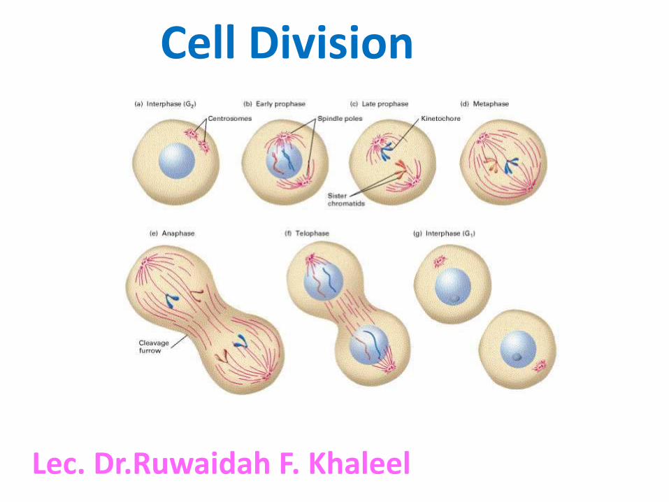
Cell Division
Lec. Dr.Ruwaidah F. Khaleel

• Growth and development of any organism depend in large part on:
Multiplication, Enlargement Differentiation of its cells beginning with the zygote. • Division of nucleated cells consists of two division, Nuclear division (karyokinesis) Cytoplasmic division (cytokinesis).• The nuclear division in turn consists of two types: o Mitosis , mitosis is associated with nuclear division of the somatic
cellso Meiosis, the meiosis occurs in the gametes.

• Mitosis• Mitosis (mitos=fiber) begins at the end point of the interphase, it is
important for growth, replacement of cells lost during normal development or under certain cases like wounds healing.
• The mitosis is rapid during the early development but it slows down with age because all cells divide but later as the embryo grows
• DNA replication slows down to allow the cells to become differentiate, irreversibly some cells lose the ability to divide after stop the genes shut off as in the nerve cells and skeletal muscles,
Some cells are functionally differentiated and non-dividing but never lose mitotic potential, like the liver cells can resume the mitosis when they are isolated in nutrient culture medium or exposed to strong stimuli as wounds or some cuts in the organ, also there are some cells like the bone marrow cells and epithelium cells in skin or in the digestive system remains mitotically active all life span of the organism.

There are 4 steps of mitosis:-
1. Prophase (pro= before)
• Appearance of chromosomes like the thin thread-like
• Cells become spherical
• Regress the nucleolus
• Appear the spindle fibers (mitotic apparatus)
• Regress the nuclear membrane

• The chromosomes composed of 2 coiled filaments called the chromatids result from the replication of the DNA during-the S phase in the cell_cycle, bind together by the centromere (each chromosome have 4 thread-chromatids with one centromere) therefore they are called the sister chromatids, and the chromosomes called the daughter chromosomes, these chromatids become shorter and thicker.
• The cells have 2 pairs of centrioles (duplicated in the interphase) migrate to the opposite site of the cell

2. Metaphase (meta= between)
• The centromeres occupy the middle plane of the cytoplasm (metaphase plate)
• the sister chromatids are still held together by connecting chromatin fibers at the centromere

3. Anaphase (ana= back)
• The chromatids separate and migrate towards poles
• The centromere divide

4. Telophase (telo=end)• The end of the daughter chromosomes migration
marks the beginning of the Telophase and the Telophase is terminated after reorganization of the two new nuclei and their entry into G1
The events here are reverse to the prophase New nuclear membrane are formedReform the nucleoli Long, thick and slender chromosomes Cytokinesis
(cytoplasmic division)

Cytokinesis (cytoplasmic divisions)
• A furrow will appear in the surface of the cell.
• Slowly cutting the cell into 2 daughter cells.


Meiosis• Meiosis (meioum=to
diminish) is a universal process restricted in sexual reproduction of animals, the numbers of the chromosomes is reduced by half, and one cell will give 4 haploid cells,
• So the meiosis consists of two divisions:
Reduction division
Equational division

• The meiosis includes 3 minor processes:-
1. Reduction the number2. Crossing over or called
recombination3. Random arrangement of the
homologous chromosomes . Although the timing of meiosis
is very different between male and female, the basic chromosomal events are the same in the two sexes. Therefore we can put a simple definition of it
Meiosis: - is a specialized of cell division that occurs only in the germ cells (oogonia & spermatozoa) in which the chromosome number is reduced from diploid (2n) to haploid (n)

Mechanism of the meiosis• A meiocyte is any diploid cell that is undergoes
meiosis. In animals the meiocyte such as spermatogonia in males and oogonia in females give rise to gametes, these cells undergo several mitosis to give another type of cells, primary spermatocytes, and primary oocytes respectively, but then will shift from mitosis , to meiosis during G2 phase

• The meiocyte before inter the meiosis must have 4 times (4n) content of the amount of DNA thus the primary spermatocytes and primary oocytes are tetraploid (4n) amount of the DNA, after the meiosis 1 the amount of the DNA become diploid (2n), while after the meiosis2 only the haploid (n) amount of DNA is present.
• Thus the meiosis like the mitosis when the cell duplicates the amount of DNA during prophase, but contrast to the mitosis, this duplication is followed by two divisions.

Stages of meiosis
Meiosis requires two cell divisions:
1. Meiosis I (reduction division)
2. Meiosis II (equatorial division)

1-Meiosis I (reduction division)• At the following some important events that occur during this
stage:-• During early development the germ cell is a diploid chromosomes
(46) with two copy of DNA (diploid ,2n)• The DNA content is duplicated (2n 4n) while the chromosomes
number (46) remain unchanged, so the cell is diploid,4n, once the DNA replicates, each chromosome consists of two parallel strands of chromatids joined together at a structure called centromere. Each chromatid contains a single DNA-molecule (which itself double strand).
• The meiosis I; consists of five phases like the mitosis.

1. Prophase I
• This phase is longest phase of meiosis, in human female it persists in each primary oocyte from a fetal age of 12-16 weeks until reach ovulation if it reaches. The prophase I divided into five stages:
• (leptoten, zygote, pachyten, diploten, Diakinesis)
• 1- Leptoten (lepto=thin)• The nucleus enlarges• The chromatin material condense• The centriole duplicate

2. Zygote (zygon= joining)
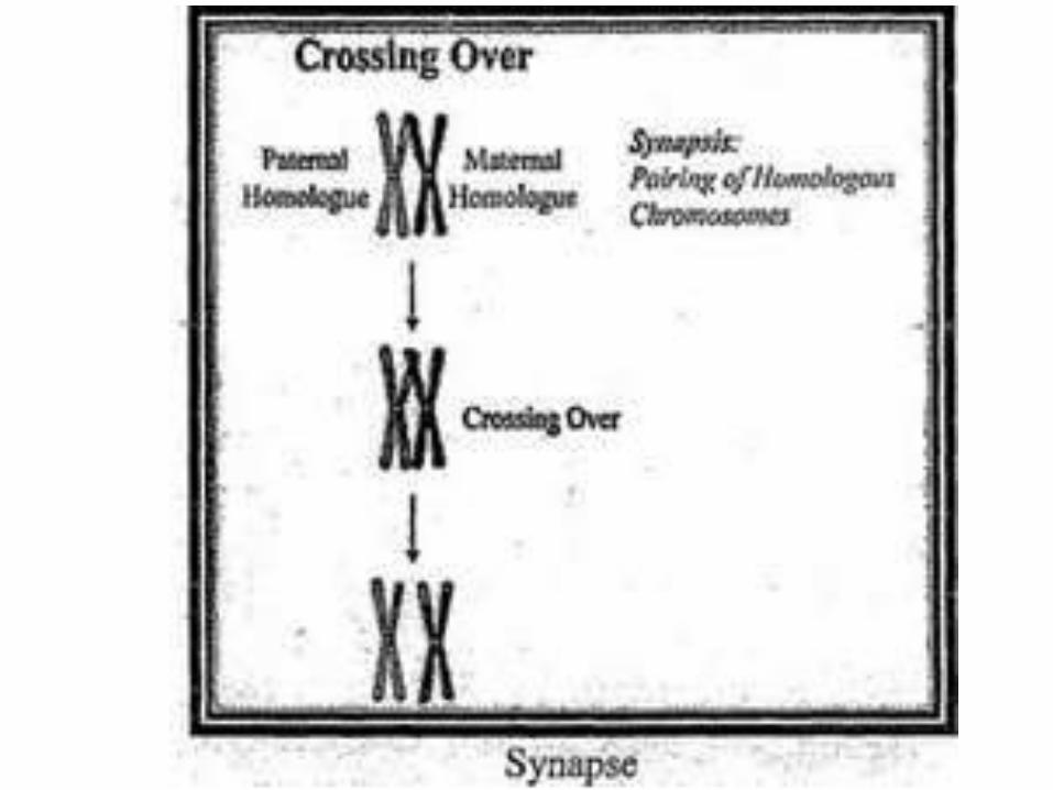
• Pair each of the homologous chromosomes nears each other (one comes from mother and the other from the father), this called the synapse, and the paired chromosomes are called bivalent.

3. Pachyten (pachus = thick)• Each homologous chromosome of
each bivalent splits longitudinally into 2 chromatids attached to the centromere, each bivalent now has 4 strands (4 chromatids) therefore called tetrad, and each one of the 4 chromatids called sister chromatids, exchange some genes or portions of chromosomes between two non-sister chromatids by a process called crossing over or genetic recombination.
• This result in a genetic diversity of the future gametes and prevents the children from being clones of their parents.

4. Diploten (Diploos=double)
• Beginning the separation of the paired chromosomes but not completely because the homologous chromosomes remain united by their points of will form a joint X- like structure called, chaismata (2 chromosomes = 4 chromatids with 2 centromeres).
• This stage is taking long time, in human female during fifth week of gestation the oocyte of the embryo, remain in diploten until many years until ovulation occur

5- Diakinesis (dia=across)
• This is the final stage of the prophase 1
• Chromosomes separate
• Disappear of the nucleolus
• Degenerate the nuclear membrane

b. Metaphase I
• Arrival the synapsed chromosome pairs at the equator of the cell
• Some homologous chromosomes still bind by chaismata

c. Anaphase I• This phase is similar to anaphase in mitosis
except that each chromosome consists of two chromatids that remain held together
• Complete disjunction occur in the homologous chromosomes
• Homologous chromosomes migrate to opposite poles
• The centromere that hold the sister chromatids do not divide as in mitosis, therefore .each two chromatids of one chromosome will move together
• The daughter cells are haploid but 2n (1 chromosomes with 2-sister chromatids), and the important thing here, that these 2 sister chromatids are not genetically identical. This is unlike in the mitosis)

d. Telophase• 1 double-strands chromosomes of each of the
homologous chromosomes is distributed to each daughter cell, so the resulting cells contain the same amount of DNA as the parent germ cells but half chromosomes (haploid with 2n)
• Formation of nuclear and nucleolus membrane
• The resultant cells are 2 daughter cells, each nucleus is haploid (double amount of DNA but one chromosome from each homologous chromosome.

e. Cytokinesis (division of the cytoplasm)
• The cytoplasm of the 2 daughter cells undergoes division to result 2 cells


2-Meiosis ll (equatorial division) At the following some important events that occur during this stage:-• No DNA replication will happen.• The Meiosis II is similar to those in the mitosis and consists of 5 phases also.
(Prophase, Metaphase, Anaphase, Telophase, and Cytokinesis).• The centromeres replicate in the prophase II• Each double-stranded chromosome condenses during prophase II and line up
during the metaphase II.• In the anaphase the double-stranded chromosome pull a part into a side of
the cell• The cytoplasm division (cytokinesis) occurs and the 2 daughter cell will form
with-one-strand chromosome and half copy of DNA (i.e., haploid, I n)• The final result of one germ cell division those 4 daughter cells with half
number of chromosomes and half copy of DNA molecule. So theses 4 cells called haploid I n)


Item
Description
Function
Takes place in
Types of cells
Number of divisions
Produces
Crossing over
Number of chromosomes
Pairing of Homologs
Centromeres Split
Mitosis Miosis

Thank you

Gametogenesis
Lec.Dr.Ruwaidah F. KhaleelLec.Dr.Ruwaidah F. Khaleel

Gametogenesis• At fertilization, the maternal and paternal gametes are united
forming the zygote. • The maternal gamete, the oocyte is the largest non-motile cell
of the body, while the paternal gamete, the spermatozoon has ability for motion and penetration of the oocyte.
• The cells which produce the gametes during the embryological period are called the Primordial germ cells (PGCs)
• The gametogenesis: is the process by which the maternal and paternal gametes are produced from the primordial germ cells to form mature sex cells called the gametes.
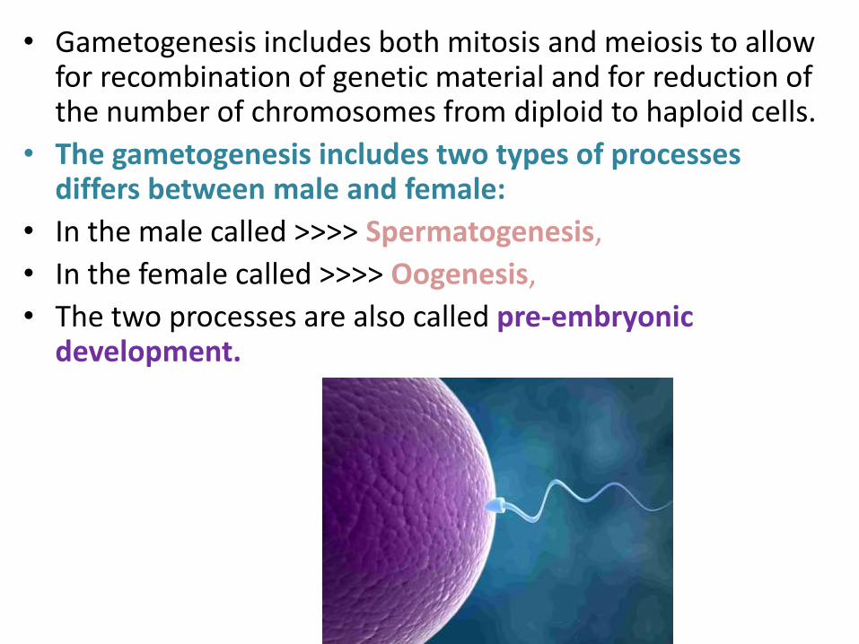
• Gametogenesis includes both mitosis and meiosis to allow for recombination of genetic material and for reduction of the number of chromosomes from diploid to haploid cells.
• The gametogenesis includes two types of processes differs between male and female:
• In the male called >>>> Spermatogenesis,
• In the female called >>>> Oogenesis,
• The two processes are also called pre-embryonic development.

Primordial Germ Cells (PGCs)• Although much of the early history of the germ cells is still
unknown, the cells that give rise to the gametes are recognized at early stage in development.
• PGC of birds, reptiles, and mammals arise in the epiblast of the embryo and then go temporary in extraembryonictissues before return to the body of the embryo.

• In frog and number of invertebrate species, germ cells can be recognized very early in the life of the animal, sometimes in the vegetal pole cytoplasm of the zygote as specific cells during the cleavage stages.
PGC
Animal pole
Vegetal pole

• The PGCs of the birds arerecognized in the, germinal crescent which is located beyond the head region of the embryo,

• In the mammals the PGCs can be found in the posterior wall of the yolk sac near the origin of the allantois.
So many studies proved that the site of the PGCs in various reptiles, birds, and mammals is from an extra-gonadal origin.

• In most vertebrates, mitosis in the PGC is arrested after their until reach the genital region, but this is differ in the mammals, there is no stop in the mitotic activity of the PGC during migration to the genital area.

• Like all normal somatic cells (non-germ cells) the PGCs contain 23pairs of chromosomes (46 chromosomes). One chromosome of each pair is obtained from the maternal gamete and the other from the paternal gamete, (22 pair) consists of homologous chromosomes called autosomes, the remaining 2 chromosomes (1 pair) are called sex chromosomes.'
• In most vertebrates, mitosis in the PGC is arrested after their until reach the genital region, but this is differ in the mammals, there is no stop in the mitotic activity of the PGC during migration to the genital area.


• In males, PGCs remain dormant in the testes from 6th Week of human embryonic development until puberty.
• At puberty, seminiferous tubules mature by the male hormones that stimulate the differentiation of the PGCs into spermatogonia by mitosis.
• In contrast in the female, the PGCs, undergo a few more mitotic divisions and invested by some somatic cells from the stroma of the ovaries before 5th Week of the human embryonic development the PGCs transform to theoogonia, and by the 5th month of the fetal development all oogonia inter meiosis I, they are called primary oocytes, the primary oocytes still arrest in the ovaries until sexual maturity

• The development of the spermatozoa takes place in the male gonads, the testes, in all vertebrates the testes are mixed gland
Exocrine organ, produce sperms Endocrine gland, producing male
hormones.• The spermatogenesis takes place
continuously from puberty until death, At birth, the germ cells in the infant male can be recognized in the sex cords of the testis, as large, pale cells surrounded by supporting cells.
• The supporting cells called the Sertoli cells; originate from the surface of epithelium of the seminiferous tubules, the sertoli cells have physical and chemical support to the spermatocytes.
• At puberty, the testes begin to secret greatly increased amounts of the steroid . hormones, testosterone, among effects of this hormone; the PGCs will resume the development and divide several times by mitosis, and then differentiate into Spermatogonia.


• Spermatogonia locate under the basement membrane of the seminiferous tubule.
• The spermatogenesis is a continuous process; it can be divided into two stages:
1- Spermatocytogenesis 2- Spermiogenesis1. Spermatocytogenesis: - the process which the primordial germ
cells (PGCs) produce the spermatozoa during several stages of mitosis.
2. Spermiogenesis: - the process which includes the re-structuring of the spermatids to spermatozoa (sperms) without any cell division only morphological changes. Because a sperm or spermatozoon is very active and motile, therefore it requires a degree of specialization, head and tail of the sperm are formed in this process.

Spermatocytogenesis• PGCs (46,2n) move from the wall of the yolk sac arrive in the testes
and remain dormant until birth, • At puberty the testes differentiate during mitosis into Spermatogonia
(46,2n) which are located peripherally in the seminiferous tubules, these cells are active mitotically all the life.
• The DNA replication is occur in mitosis convert the Spermatogoniafrom 2n to 4n the resultant cells called the Primary spermatocytes (46, 4n)
• Primary spermatocytes (46, 4n) inter the meiosis I and divided into two daughter cells called Secondary spermatocytes (23, 2n),
• Each of the secondary , spermatocytes inter the meiosis ll to form four haploid spermatids (23,In).
Commonly in the human the meiosis I last for several weeks, whereas the meiosis ll is completed in about 8 hours.
• Through this series of division, cytokinesis is incomplete; leaving all cells still connected through thin cytoplasmic bridges, and connects with sertoli cells, which support them physically and metabolically.
• Although the spermatids no longer divide, they undergo a transformation to specialized spermatozoa; this is done by the spermiogenesis process.
• PGCs (46,2n) move from the wall of the yolk sac arrive in the testes and remain dormant until birth,
• At puberty the testes differentiate during mitosis into Spermatogonia(46,2n) which are located peripherally in the seminiferous tubules, these cells are active mitotically all the life.
• The DNA replication is occur in mitosis convert the Spermatogoniafrom 2n to 4n the resultant cells called the Primary spermatocytes (46, 4n)
• Primary spermatocytes (46, 4n) inter the meiosis I and divided into two daughter cells called Secondary spermatocytes (23, 2n),
• Each of the secondary , spermatocytes inter the meiosis ll to form four haploid spermatids (23,In).
Commonly in the human the meiosis I last for several weeks, whereas the meiosis ll is completed in about 8 hours.
• Through this series of division, cytokinesis is incomplete; leaving all cells still connected through thin cytoplasmic bridges, and connects with sertoli cells, which support them physically and metabolically.
• Although the spermatids no longer divide, they undergo a transformation to specialized spermatozoa; this is done by the spermiogenesis process.


Spermiogenesis• The Spermiogenesis is the second process of the
spermatogenesis mechanisms, it consists of series of morphological changes that responsible of transformation the spermatids into mature spermatozoa (sperms) (23,In).
• Because the sperm or spermatozoon is very active and mobile cells, so it must be supported with great amount of energy and activation, therefore there is need to a high degree of specialization must occur in the sperm cells before release from the testes.

After the end of the meiosis II, The nucleus begins to lose fluid. Decrease its size The nucleus loses its RNA molecules, nucleolus and most proteins, Only the haploid copy The chromatin compress until forming the head of the sperm, At the apical end of the developing sperm head, there is a Golgi
complex form the pro-acrosomal granules, contains hydrolytic enzymes that are released; during fertilization.
The centriole develop to be a point of the flagellum.

After the meiosis II The two centrioles move through the cytoplasm of the
spermatid just behind the nucleus, These centrioles give rise to the axial filament of the flagellum
of the sperms, which later differentiate into tail, Mitochondria begin to form a spiral form called the
mitochondria helix as a source of energy, During development the rest of the cytoplasm will sloughed off
and phagocytized by the sertoli cells

All these events make the mature spermatozoa leaves all the non-essential parts, at the end the mature
spermatozoa will consists of
1. Head: containing nucleus and acrosome
2. A neck: containing the centriole
3. A middle piece: containing the proximal part of the flagellum and mitochondria helix
4. The tail: highly specialized flagellum

Oogenesis
Dr. Ruwaidah F. Khaleel

Oogenesis• Is a period of growth and
maturation of the egg occurring in the female gonads (ovaries).
• The female gametes is larger than most somatic cells and non-motile,because in all animal egg have many vital roles like:
It has a nucleus containing half of the chromosomal content, (haploid number of the chromosomes),
It has to supply all cytoplasm to the embryo after fertilization
Also it has supply food store to the embryo to develop.
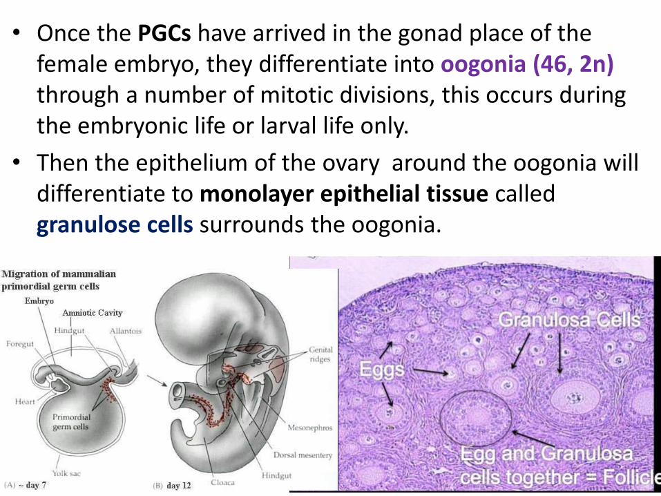
• Once the PGCs have arrived in the gonad place of the female embryo, they differentiate into oogonia (46, 2n) through a number of mitotic divisions, this occurs during the embryonic life or larval life only.
• Then the epithelium of the ovary around the oogonia will differentiate to monolayer epithelial tissue called granulose cells surrounds the oogonia.
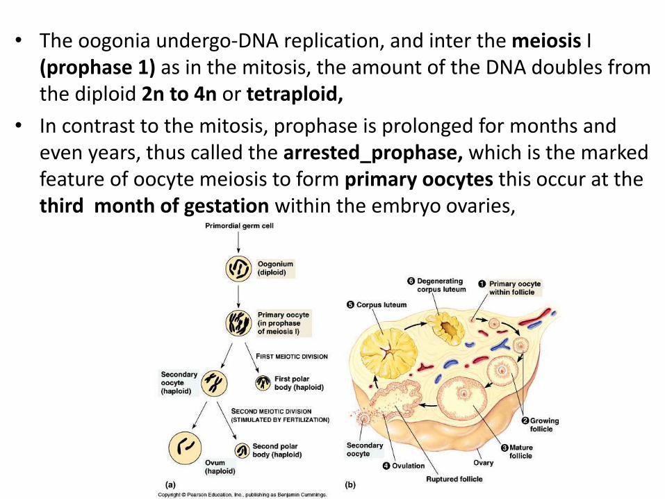
• The oogonia undergo-DNA replication, and inter the meiosis I (prophase 1) as in the mitosis, the amount of the DNA doubles from the diploid 2n to 4n or tetraploid,
• In contrast to the mitosis, prophase is prolonged for months and even years, thus called the arrested_prophase, which is the marked feature of oocyte meiosis to form primary oocytes this occur at the third month of gestation within the embryo ovaries,

• The primary oocytes which are surrounded by a single-layer of squamous epithelial cells derived from the somatic support cells in the ovary that enclosed the oogonia tightly, The primary oocytes with this capsule of epithelial cells, called primordial follicle.
• Once primary oocyte stop dividing the cells enter a prolonged (resting phase). Stay in the ovary during prophase I (diploten) for long year until reach the puberty. Then each one of primary oocyte will complete the meiosis I.

Development of the ovarian follicles • Before puberty and without hormonal control of the
pituitary gland, the follicular epithelium of a small group of primordial follicles thickness, converting the single-layered follicular epithelium from a layer of squamous cells to cuboidal cells, these follicles is now called primary follicles

• The oocytes with the follicle cells secrete a thin layer of cellular material composed of only a few types of glycoproteins, into surface of the oocyte that are deposited as an outer layer this layer will name (Zona pellucida), the zona pellucida appears to form a complete physical barrier between the follicle cells and the oocyte, this layer is penetrated by projections from the follicle cells to contact with the oocyte membrane to exchange the metabolites and mechanical support

Just before puberty, some of the growing follicles stop to develop and degenerate, whereas a few continue to enlarge in response to rising levels Follicular Stimulating Hormone (FSH).
This hormone stimulates the follicle cell to:
Resume the growth and complete the meiosis I
Form two daughter cells non equal in size, a secondary follicle (23,2n) and first polar body , which degenerate later,

• Follicle cells then produce a fluid between the cells and form a cavity called the Antrum, • At the same time the connective tissue of the ovary (stroma) around the secondary follicles
will differentiate to two layers: The inner, Theca internae The outer, Theca externa, The theca interna is responsible for synthesis and produce the steroids hormones.


• After the meiosis I was complete, the secondary follicle is ready to inter the meiosis II, and stay in the metaphase II until few time before ovulation,
If the fertilization is occur the secondary follicle complete the following stages of the meiosis II after leaving the ovary and reach the uterus to form a mature oocyte (23,In) and the second polar body
If fertilization not occur it will degenerate and out through the menstrual cycle

Amount and distribution of yolk and types of eggs
• Yolk, the major nutritional reserve of the oocytes during the growth and development and after the fertilization to provide the embryo with nutrition, the yolk varies greatly in amount and distribution in different animal groups

Based on the amount of the yolk, the eggs can be classified into the following:-
1. Microlecithal (oligolecital) eggs
• These are small sized eggs which contain a very small amount of yolk distributed in the cytoplasm (ooplasm), this type found in certain marine invertebrate, ex: Hydra, and some chordates like Amphioxus,cephalochrdates, tunicates, and the eutherian mammals, in the mammals this type of egg called also Alecithal egg.

2. Mesolecithal eggs
• This type of eggs contain moderate amount of yolk, found in annelid worms, molluscus, petromyzon, lung fish and amphibian

3. Macrolecithal (megalecithal) eggs
• These eggs contain enormous amount of yolk, found in insects, chondrichthyes, reptiles, birds and monotrems
monotrems
based on the distribution of the yolk
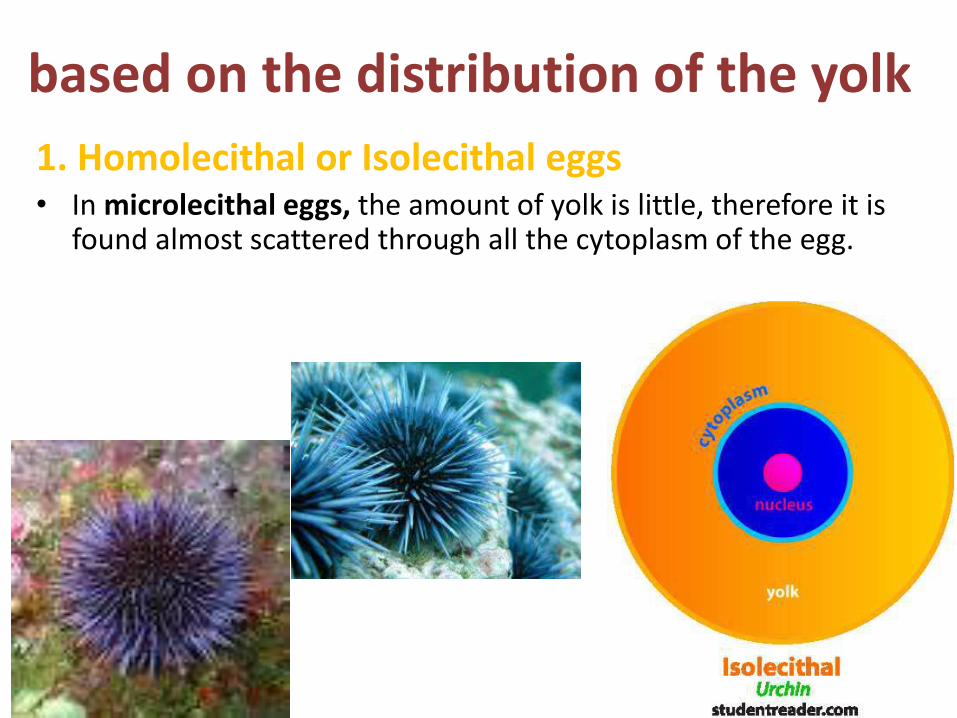
1. Homolecithal or Isolecithal eggs• In microlecithal eggs, the amount of yolk is little, therefore it is
found almost scattered through all the cytoplasm of the egg.

2. Telolecithal eggs• This type of egg has a polarized distribution of yolk in the ooplasm due to the
gravity of the yolk, it concentrated more in one side than in the other.
• The side that contain no or very small amounts of yolk is called the animal pole,
• The side where the yolk concentrated more called vegetal pole
This type found in the amphibian, petromyzon, cartilaginous and bony fishes, reptiles, and birds
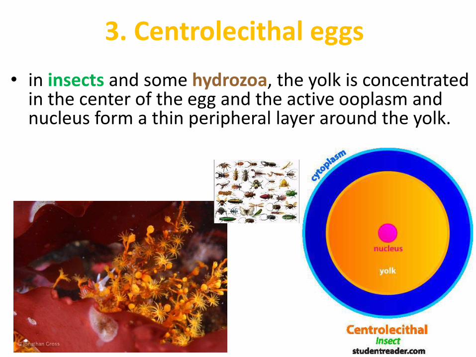
3. Centrolecithal eggs
• in insects and some hydrozoa, the yolk is concentrated in the center of the egg and the active ooplasm and nucleus form a thin peripheral layer around the yolk.

Spermatogenesis Oogenesis1. Occur in the testes 1. Occur in ovaries
2- The reserve food material depend on the sertoli cells
2-Yolk is the reserve food
3- The yield of the meiosis is four functional spermatozoan equal in size
3- The yield of the meiosis is four cellsNot equal in size, one big active oocytes and
three small inert polar bodies
4- After complete the meiosis thespermatozoon need to differentiation to be
functional
4-After complete the meiosis theoocytes ready to start development without
differentiation
5- The meiosis 1 begin after reach the puberty 5- The meiosis 1 begin during the embryological development.
6-The meiosis continue to the last stages during puberty
6-The meiosis stop in the prophase I, and resume after puberty
7-The spermatogenesis continue until death 7-Oogenesis stop after many years
8-The meiosis II complete inside the testes after puberty
8-The meiosis II begin at puberty reach the metaphase II and stop again, resume its action
after fertilization.


Lec. Dr. Ruwaidah F. Khaleel

Formation of egg membrane
Sponges Coelenterates
• In most animals, except the sponges and coelenterates, oocyte maturation is not completed until additional structures, called egg membranes,
• Egg membranes are produced outside the plasma membranes of egg and vary in different animal groups and sometime reflect the adaptations by the animals.

There are several ways of classifying the egg membranes, but the simplest way is according to their
origin as follow:-
1. Primary egg membranes
• These membrane are formed in the ovary between the egg plasma membrane and follicle cells they are formed either by ovum of follicle cells,

The type of egg membranes includes:-a- Vitelline membrane
• In fact it is not a membrane, but a non-cellular transparent layer of mucoprotein out of egg give the physical support and elasticity.
• The vitelline membrane is a structure directly adjacent to the outer surface of the plasma membrane of an ovum
• Vitelline membrane has different names for example, in amphibian and birds is contact to the ooplasm until the fertilization then will separate therefore it's called the fertilized membrane, in fish called chorion, and in the reptiles and mammals called zona pellucida

1) Primary egg membranesthese membrane are formed in the ovary
a) Vitelline membrane
Amphibian and bird >>> contact to ooplasmuntil the fertilization then will separate there for is called the fertilized membrane

Fish >>> chorion

Reptile and mammels >>> zona pellucida

b) Jelly envelope
In echinoderms and many egg of marine invertebrates, the primary egg envelopes is much thicker structure like jelly coat

2) Secondary egg membrane
Secreted outside the primary egg membrane by follicle cells. It occurs in the form of chitinous shell around egg in insects and cyclostomes and called chorionThis chorion different from chorion fish that formed by oocyte and contains proteins and polysaccharides

No secondary
membranes are found in = amphibians, reptiles, birds and even mammals eggs
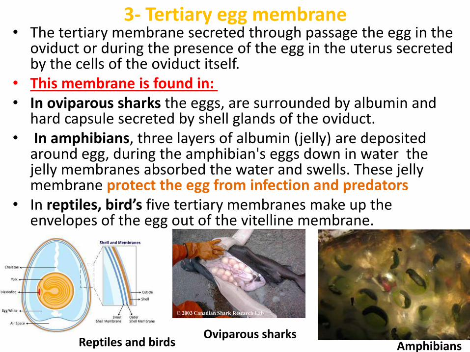
3- Tertiary egg membrane• The tertiary membrane secreted through passage the egg in the
oviduct or during the presence of the egg in the uterus secreted by the cells of the oviduct itself.
• This membrane is found in:• In oviparous sharks the eggs, are surrounded by albumin and
hard capsule secreted by shell glands of the oviduct.• In amphibians, three layers of albumin (jelly) are deposited
around egg, during the amphibian's eggs down in water the jelly membranes absorbed the water and swells. These jelly membrane protect the egg from infection and predators
• In reptiles, bird’s five tertiary membranes make up the envelopes of the egg out of the vitelline membrane.
Oviparous sharksAmphibiansReptiles and birds

3) Tertiary egg membrane
Oviparous sharks
Amphibians
Reptiles and birds

Ovulation and transportation of ova
viviparus OviparusFemale reproductive organ
• The ovulation and transportation of the ova are done by ovaries and accessory reproductive organs of the female.
• The female accessory reproductive organs include organs which assist in the transportation of mature eggs from the place of origin to the exterior in the oviparous animals' likefish, amphibians, or to female reproductive organ such in uterus as in the viviparous animals

Time and regulation of ovulation(1Environmental control
The time of ovulation is directly related to the breeding cycle of animal, three methods that control the ovulation in the animals
Many factors that effect on the ovulation in animals like Light Temperature Rain fallmost animal time their breeding and ovulation with the good seasons, for example, when rats and mice were kept in dark all day they changed the time of ovulation from the midnight to near noon and continuous exposure to light Resulted in inhibition of ovulation, Sexual photoperiodicityIs phenomenon in which the light controls the ovulatory or sexual cycle of female

2) Hormonal control
The function of the ovary and ovulation in vertebrates are regulated by, hormones of pituitary gland. In mammals the function of the ovary are under three gonadotropic hormones of pituitary Gonadotropic hormones of pituitary
FSH (Follicle Stimulating Hormone)>> induce growth of the follicles
LH (Luteinizing hormones)>> act with FSH on maturation of the follicles and ovulation and formation of corpus luteumProlactin>> responsible for milk secretion

3) Neural control The nervous system of vertebrates has indirect effect on the ovulation through the pituitary gland
Pituitary gland
Secreted pituitary hormones neurohormones(under control of hypothalamus)
Chemicals(releasing factors)
Stimulate pituitary gland to release hormones (FSH,LH)
secrete

Ovulation• Is the process of releasing the ova from the ovaries• The ovulation occurs by different method in different vertebrates. The female reproductive or breeding activities of almost all animals are
cyclic the reproductive cycles are seasonal, in most vertebrates, the
reproductive cycles is yearly, like sheep, while in higher primates including the human, which can breed at any
time.• The female mammal that follows an annual reproductive season, passes
through several shorter sexual cycles during her breeding season, when, they to more receptive of the males and when ovulation occur spontaneously these seasonal reproductive cycles called estrous cycles

Ovulation
Estrous cyclesSeasonal ovulation changes in females because the physiological and histological events in the female reproductive system in some animals makes the females are more receptive of the maleswhile in the primates including human females the sexual cycle and changes in the endometrium of the uterus, all are hormonally control occur monthly

Mechanism of ovulation
In the days immediately Before ovulation secondary follicle growsUnder (FSH,LH)
LH increase
Increase activity of collagenase enzyme
Digestion of collagen fibers surrounding the follicle in the ovary
Increase prostaglandin level
Local muscular concentrations in ovary wall
Facilitate release of oocyte with its surrounding the granulosa cell
After ovulation, some of the granulosa cells remain in the ovary with the effect of the LH hormone both the granuloga cells and the theca interna of the ovary develop yellowish pigment mass of cells called corpus luteum, this mass begins secrete the prostaglandin hormone
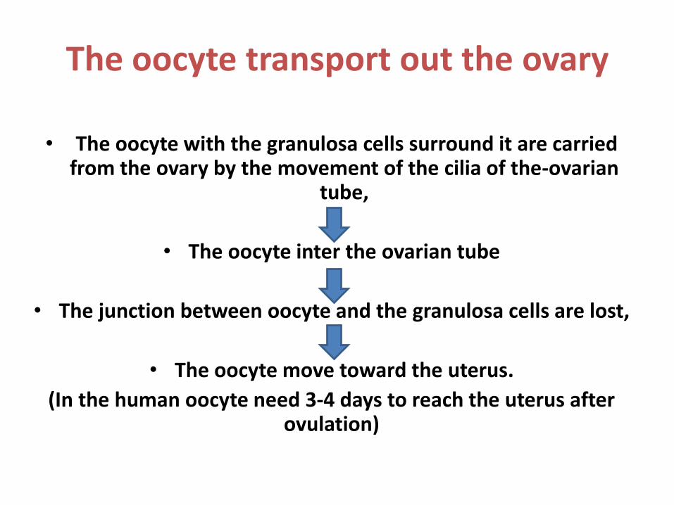
The oocyte transport out the ovary
• The oocyte with the granulosa cells surround it are carried from the ovary by the movement of the cilia of the-ovarian
tube,
• The oocyte inter the ovarian tube
• The junction between oocyte and the granulosa cells are lost,
• The oocyte move toward the uterus.
(In the human oocyte need 3-4 days to reach the uterus after ovulation)

If fertilization not occurs
the corpus luteum reaches its maximums development
then shrinks and degenerate after 14 days
transform to corpus albicans
the oocyte will rupture and degenerate

If fertilization is occur
• The corpus-luteum remain active and produces and secrets the prostaglandin for 4 months
• This due to the human chorionic gonadotropin (hCG) that produced by the embryo cells, this hormone
called also the pregnant hormone, keep the corpus leuteum active during this period


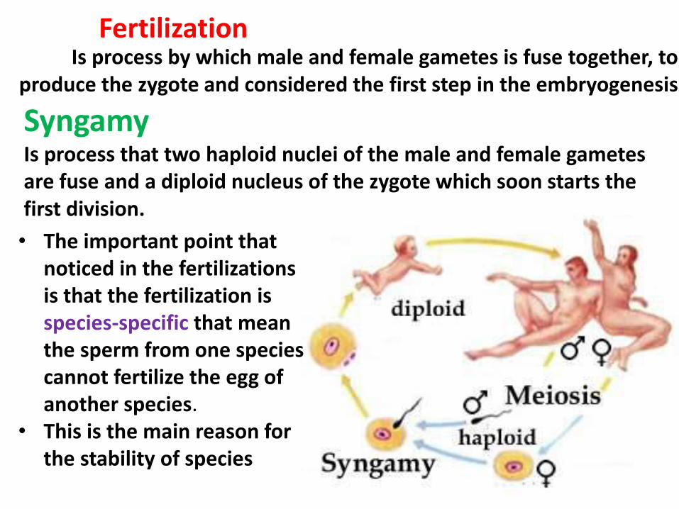
FertilizationIs process by which male and female gametes is fuse together, to
produce the zygote and considered the first step in the embryogenesis
SyngamyIs process that two haploid nuclei of the male and female gametes are fuse and a diploid nucleus of the zygote which soon starts the first division.
• The important point that noticed in the fertilizations is that the fertilization is species-specific that mean the sperm from one species cannot fertilize the egg of another species.
• This is the main reason for the stability of species
Generally in most animals the process of fertilization requires the following factors to occur successfully
1. In most animals the fertilization must occur in a fluid medium. example Sea water in oviparous animals, or body-fluid in viviparous animals.
2. The life-span of gametes is short, therefore in the most water invertebrates, fish, and amphibian, must be fertilized not more few minutes, while eggs within, the body of the female have a life-span longer example. Human eggs can be fertilized for at least 24h. after ovulation
Also the sperms have the same aspect, for example: the human sperms can be viable in the female genital tract about 24h
3. In the normal fertilization, the number of the sperms must be more than the eggs number

• Before fertilization the spermatozoa are not able to fertilize the oocyte immediately after inter the female genital tract, because, the spermatozoa must undergo physiological and molecular of maturation processes called the capacitation or acrosomal reaction.
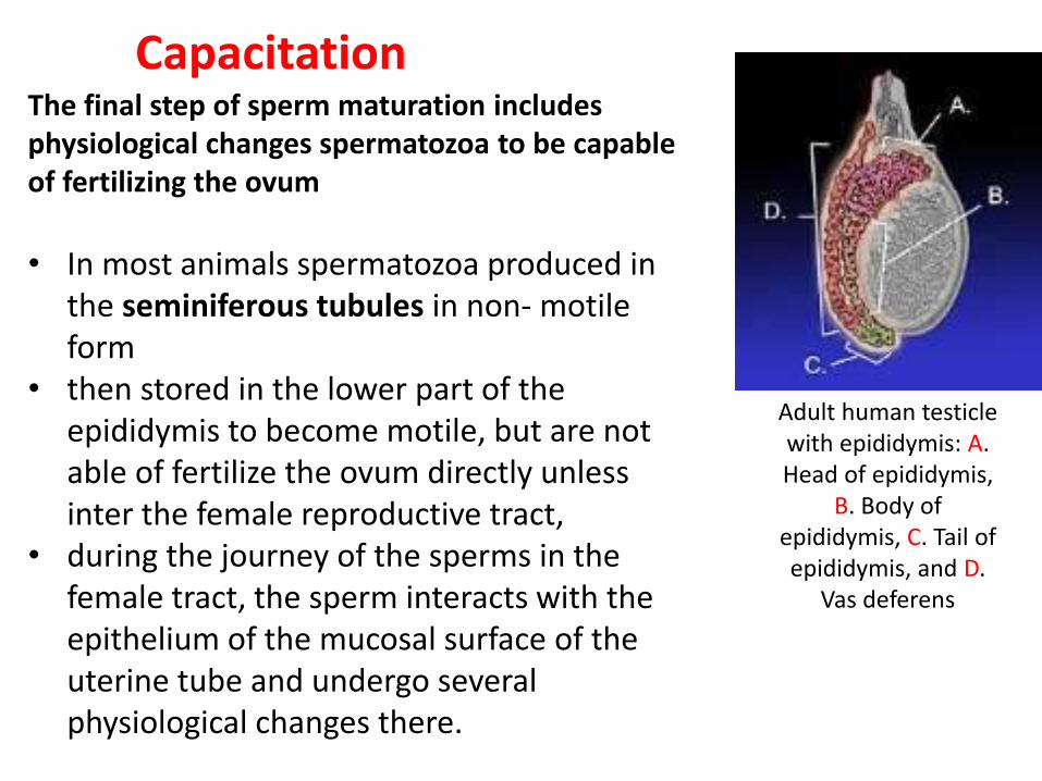
CapacitationThe final step of sperm maturation includes physiological changes spermatozoa to be capable of fertilizing the ovum
Adult human testicle with epididymis: A. Head of epididymis,
B. Body of epididymis, C. Tail of epididymis, and D.
Vas deferens
• In most animals spermatozoa produced in the seminiferous tubules in non- motile form
• then stored in the lower part of the epididymis to become motile, but are not able of fertilize the ovum directly unless inter the female reproductive tract,
• during the journey of the sperms in the female tract, the sperm interacts with the epithelium of the mucosal surface of the uterine tube and undergo several physiological changes there.

Mechanisms of the capacitation
• The acrosome plays an essential role in capacitation,
• The acrosome contain hydrolytic and Proteolytic enzymes can destroy the male genital tract if all the sperms release these enzymes
• Therefore there are the fluids in the seminiferous tubules and all the male genital tract all contain floating vesicle filled with cholesterol cover the cell membrane of the sperm and prevent the releasing of the acrosome enzymes but after the sperm are release from the tract,
• The sperms swim away from the vesicles and lost the cholesterol after few hours and become capacitated and the sperm can penetrate the ovum.
• In the mammals the capacitation doesn't occur unless the sperms inter the female genital tract.

Acrosome reaction and contact of sperm and ovumAcrosome reaction :The first reaction of the sperm when it comes in contact with the egg surface by the action of the acrosome• In human females and other mammals, the fertilization occur soon, after the ovum enters
the fallopian tube
Before sperm can enter the ovum must1. Penetrate the multiple layers of granulosa cells out of the ovum2. The follicular cells bind together by a substance called hyaluronic acid so, the
acrosome will release hyaluronidase enzymes to dissolve the attachment between the granulosa cells over the ovum
3. After attachment occurs between the sperm and zona pellucida the hydrolytic enzymes are released from the acrosome. This primates the spermatozoon to penetrate the zona pellucida matrix
4. This processes is stimulated by zone pellucida enzyme5. The sperm adhere to and fuse with the plasma membrane of the oocyte

Oocyte activation occur after sperm inter the cytoplasm of the oocyte
Changes in ionic permeability of egg plasma membrane after fertilized by a sperm. This activation includes
1. Prevent enter another sperm or called block to polyspermy
2. Resumption of meiosis3. Initiation of embryonic development

• The oocyte after fertilized by a sperm, the plasma membrane is produced granules under the oocyte membrane, between the zonapellucida and the oocyte called cortical granules, therefore this process is called cortical reaction
• The cortical granules consist of many enzymes like proteases, peroxidases, in the presence of these granules, the zona pellucidaand the oocyte plasma membrane change chemically and the zonapellucida becomes Impermeable by additional sperms.
• After infusion of the sperm membrane with the oocyte membrane the oocyte resume meiosis and complete the meiosis II, in this stage the oocyte called definitive oocyte
• The nuclear membrane of both sperm and oocyte quickly disappear and fuse their genome together and form the zygote (46,2n), by a process is called the syngamy.

Cleavage
Lec.Dr.Ruwaidah F. Khaleel

CleavageThe activated egg is passed through a phase of repeated mitotic cell division to produce an increasing number of cells and build the body of the animalCleavage: the splitting or division of activated egg by a series of mitotic cell division into a number of cells which become the building units of future organism
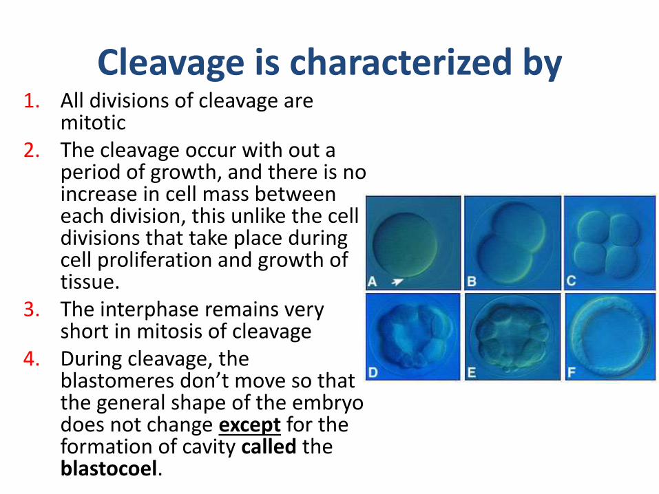
Cleavage is characterized by1. All divisions of cleavage are
mitotic2. The cleavage occur with out a
period of growth, and there is no increase in cell mass between each division, this unlike the cell divisions that take place during cell proliferation and growth of tissue.
3. The interphase remains very short in mitosis of cleavage
4. During cleavage, the blastomeres don’t move so that the general shape of the embryo does not change except for the formation of cavity called the blastocoel.

5. The produced cells from the cleavage called the blastomeres (blastos>>germ, meros>>parts)
6. The cleavage converts the egg into a compact mass of blastomers called morula which transforms into blastula, having one-layered thick blastoderm around a central cavity, blastocoel
7. The beginning of gastrulation is regarded as the end of cleavage

The amount and distribution of yolk in the oocyte effect on the cleavage
• Cleavage can be classified into the following types:
A. Holoblastic (complete) cleavage
The cleavage divides the egg completely, but two ways:
1. Equal holoblastic cleavage
When holoblastic cleavage occur in microlicithal eggs, it produces blastomeres of equal size, this found in: amphioxus, and mammals.

2. Unequal holoblasticcleavage
In mesolecithal, the holoblasticcleavage produces unequal size blastomers, which include many small-size blastomerscalled micromers, and few large-size blastomers called macromers, this type of holoblastic cleavage found in the lower fish and amphibians

B. Meroblastic (partial-incomplete) cleavageThis cleavage found in the macrolecithal eggs, divide the zygote to incomplete cleavage. In such cleavage, the division results to small amount of the active cytoplasm of animal pole or periphery of egg and most of the yolk portion of the vegetal pole or the central area of the egg remains undivided e.xchick eggs

Planes of the cleavage
1. Meridonial:Cleavage furrow passes through the center of animal-vegetal axis bisecting the egg into two equal parts, eg. The cleavage in Amphioxus and frog and first cleavage in chick.

2.VerticalThe cleavage furrows may lie on either side of the meridional plane. The furrows pass from animal to vegetal pole. The cleaved cells may be unequal in size. eg, chick

3.Equatorial cleavage
This cleavage plane bisects the egg at right angles to the main axis. It lies on the equatorial plane, it divides the egg into two halves

4. Latitudinal (transverse) cleavage
This type is similar to the equatorial plane, but it coursers through the cytoplasm on either side of the equatorial plane, parallel to the equatorial plane, it is also called transverse or horizontal plane, eg. Third cleavage in the Amphioxus and frog

Products of the cleavage
The cleavage is a fractionating process, which splits the (one-cell) zygote into smaller cells, called the blastomeres.After each cleavage. The blastomeresincreases in a doubling sequence 2,4,8,16,32…etc.

Morula
During early cleavage stages, the blastomeres tend to cluster a tightly spherical shape as solid ball of cells about 32 cells this called morula (mulberry) and the process is called morulation.No morula is found in the centrolecithal eggs like insects, while the morula in the telolecithal and isolecithal found real morula

BlastulaAfter formation of morula, the fluid begins to penetrate through the zona pellucida into the intracellular spaces, and the blastomers undergo rearrangement themselves into a true epithelium in a single cell thick, this epithelium called blastoderm gradually the intracellular spaces become a single cavity, called blastocoel, in this stage the embryo called blastocyte

Type of blastula
1. Coeloblastula
Found in lower invertebrates like, echinoderms and Amphioxus, it is in the form of central cavity called the blastocoel filled with fluid and around it a single layer of cells called the blastomere
blastocoel
blastomeres

2.DiscoblastulaFound in vertebrate like reptiles, birds and fish which have large amounts of yolk, therefore the blastula appeared at the animal pole as a small multilayered flat disc separated from the yolk by a narrow space called the subgerminal cavity
blastodisc
Subgerminal cavity yolk

3.Amphiblastola
Found in amphibians, blastula contains two types of cells micromeres and macromeres arranged into more than one layer of cells

4.Blastocyst• Such blastula found in
mammals. • The cleavage is regular and
small cavity appears inside the dividing cells, the cavity called blastocoel and later become larger,
• the fluid in this cavity push some cells towards a pole of the blastocyte, these cells called the embryoblast or innercell mass, give rise to the all embryo organs therefore called embryoblast,

• While the cells that surround the outer of the blastocytecalled outer cell mass or trophoblast give rise the epithelial layer of the blastocyte, considered the primary origin of the placenta, which attaches the fertilized ovum to the uterine wall and supplies nutrition to the embryo
• In the later stage the embryo will implant to the uterine wall by the trophoblast

Thank you

Gastrulation
Lec.Dr. Ruwaidah F. Khaleel

Gastrulation
• Is the dynamic process that deals with the re-organization or rearrangements of the cells of the embryo which convert from one-layered stage as in all vertebrates.

The single layer of epithelium in the blastula is known as blastoderm, while in the gastrula it is different:
If the gastrula was 2 layered, the outer layer is called ectoderm, and the inner layered is called endoderm,therefore this gastrula called diploblastic gastrula, like in the Hydra
If the gastrula was three layered, the outer layer is called ectoderm, the middle layered is called mesoderm, and the inner layered called endoderm, therefore this gastrula called triploblastic gastrula, like in higher animals

The process of gastrulation involves the following physiological and molecular event
1. Rearrangement of blastomeres by the morphogenetic movements
2. The cellular division or cleavage is inhibited
3. Growth. If found is insignificant
4. The nuclei become more active in controlling the activities of the embryonic cells
5. Synthesis of different kinds of protein molecules
1. Rearrangement of blastomeres by the morphogenetic movements
2. The cellular division or cleavage is inhibited
3. Growth. If found is insignificant
4. The nuclei become more active in controlling the activities of the embryonic cells
5. Synthesis of different kinds of protein molecules

The gastrulation process doesn’t occur until cleavage stop. Why?
• In the very beginning of development, during cleavage and early blastula, cells don’t be able to move under any condition, therefore the cells at the end of cleavage undergo to differentiation to starting the movement,
• during cleavage and early blastula, cells are dividing so rapidly therefore the microfilament and microtubule that the major part of the movement are so busy in the cytokinesis and not able to inter in the other activity, therefore the beginning of gastrular cell locomotion during early development of most of animal groups time happen together with a decline of rapid cell division

Morphogenetic movements
• The movement of blastomeres of embryo (blastula) during gastrulation to reorganization of the cell from one place to another and produce a new structure of the embryo called gastrula
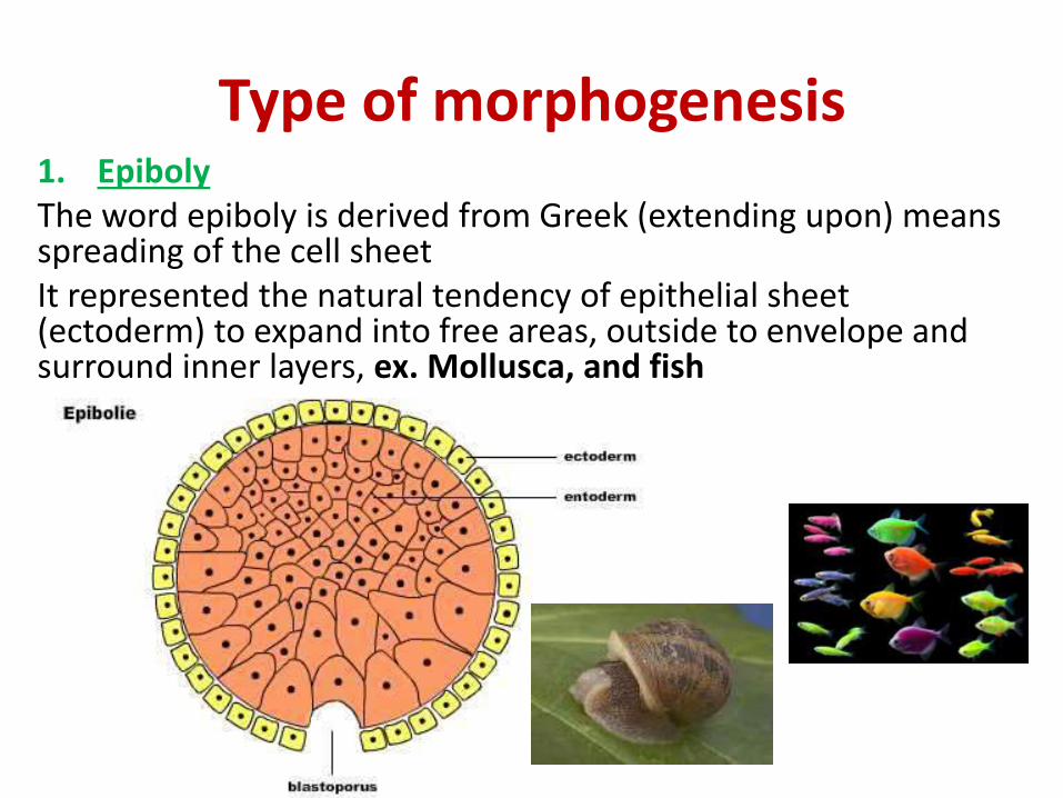
Type of morphogenesis1. EpibolyThe word epiboly is derived from Greek (extending upon) means spreading of the cell sheetIt represented the natural tendency of epithelial sheet (ectoderm) to expand into free areas, outside to envelope and surround inner layers, ex. Mollusca, and fish

2. Emboly
This is opposite to epiboly, in embryo the sheets of the cells fold or invaginate, thus the sheet migrate inside or under another layer, this type of movements occur in the mesoderm and endoderm. This type occur in Amphioxus

The embolic movement includea. Invagination
It’s a movement of layer of cell inside at a specific point. Ex. Invaginate the cell of vegetal pole in Amphioxus embryo into the blastocoel toward the animal pole.

b. InvolutionIt’s a turning of an expanding layer at a specific point,
The surface layer of the cells rolls inside itself and continue to expand along the inner surface. ex,. A lip of blastopore during the gastrulation of the amphibian the vegetal pole cells move flow over the lip and move inside

c. Ingression (polyinvagination)• Small group of cell separate from the
epithelial layer into an area.
Like the primary epithelial layer which migrate into the blastocoel cavity as new layer ex. Primitive streak in amniotes animals (reptiles, avian and mammals)

Histogenesis and organogenesis
• After gastrulation the embryo inter the next phase of development called the organogenesis during physiological and histogenesis processes
• Histogenesis: is the process by which the undifferentiated or pre-functional cells of the germ layers differentiate into specialized functional cells to form the body of the organism, and it’s the first step in organogenesis

• Each tissue consist of group of similar cell that carry out similar functions united by intracellular substances, the cells of the tissues are specialized cells, but epithelial and connective tissue are considered less specialized than nervous and muscle tissue
• The formation of an intracellular substance usually secreted by the cell and often referred to as the matrix separates the cell from each other and give the shape to the body, the intracellular substance of bone and cartilage are examples of this type of tissue.

Generally all the type of cells is derived from one of the following germinal layer
1. Ectoderm
a. Epidermal ectoderm
This type is give rise the epidermis of skin, skin derivatives (hair, feather, nails, scale, claws, sebaceous, sweat gland, and mammary gland)also give raise the epithelium of the oral cavity, nasal cavity, salivary glands and anal canal, external ear of mammal, anterior pituitary gland (adenohypophysis)

b. Neural ectoderm (neural tube)Brain
Spinal cord
Peripheral nerves
Ganglia
Roots and cones of the retina of eye
Optic nerve
Receptors of skin
Auditory and taste receptors
Posterior pituitary (neurohypophesis)

2. Endoderm Epithelium of the
digestive tract and its derivatives (liver, pancreases, gall bladder)
Epithelial lining of the respiratory tract
Epithelium of the urinary bladder
Epithelium of the auditory tube
Primordial germ cells of the some animals, Vagina, urethra and associated glands

3. Mesoderm
a. Notochord
Found in amphioxus while in the other chordates it is replaced by vertebral column (vertebrates)
b. Epimere (dorsal mesoderm)
This type of mesoderm is consist of three parts:
1. Dermatome: this give rise dermis of skin
2. Sclerotome: this give rise vertebral column
3. Myotome: skeletal muscle

c. Mesomere (intermediate mesoderm)
Excratory glands (kidney)
Excretory ducts
Urinary bladder in fish
Epithelium of gonads (epithelium of the testes and ovaries and genital ducts, and adrenal cortex
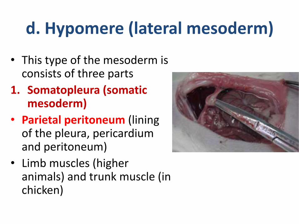
d. Hypomere (lateral mesoderm)
• This type of the mesoderm is consists of three parts
1. Somatopleura (somatic mesoderm)
• Parietal peritoneum (lining of the pleura, pericardium and peritoneum)
• Limb muscles (higher animals) and trunk muscle (in chicken)

2. Splanchnopleur (visceral mesoderm)
• Visceral peritoneum• Mesenteries• Heart• Cardiac muscles• Bone marrow• Blood cells• Blood vessels• Smooth muscles3. Coelom mesoderm, give rise the body cavity

Origin of organ systems
• Groups of specialized cells (tissue) combine to form the organs of the animal,
• Certain organs, in turn corporate with other related organs to constitute organ system

the organ systems of adult vertebrate body can be divided according to their general germ layer origin as follow
A. Ectodermal layer1. Integumentary system2. Nervous system
1. Digestive system2. Respiratory systemC. Mesodermal layer1. Muscular system2. Skeletal system3. Circulatory system4. Excretory system5. Respiratory system

• At the end of gastrulation, epidermal ectoderm covers the embryo, the neural ectoderm will differentiated from the epidermal ectoderm to form the
• The mesoderm remain closely associated with the forming the so-called chordamesodermal layer, the endoderm associated the roof of the gastrocoelto form the embryonic gut extend from the head to tail region

Thank you