Ultrastructure and cytochemistry of salivary glands of the predator Podisus nigrispinus (Hemiptera:...
9
ORIGINAL ARTICLE Ultrastructure and cytochemistry of salivary glands of the predator Podisus nigrispinus (Hemiptera: Pentatomidae) Luis Carlos Martínez & Maria do Carmo Queiroz Fialho & José Cola Zanuncio & José Eduardo Serrão Received: 28 June 2013 /Accepted: 28 August 2013 /Published online: 11 September 2013 # Springer-Verlag Wien 2013 Abstract Podisus nigrispinus Dallas (Hemiptera: Pentato- midae) is a zoophytophagous insect with a potential for use as a biological control agent in agriculture because nymphs and adults actively prey on various insects by inserting mouthparts and regurgitating the contents of the salivary glands inside the prey, causing rapid paralysis and death. However, the substances found in saliva of P. nigrispinus that causes the death of the prey are unknown. As a first step to identify the component of the saliva of P. nigrispinus , this study evaluated the ultrastructure and cytochemistry of the salivary glands of P. nigrispinus . The salivary system of P. nigrispinus has a pair of principal salivary glands, which are bilobed with a short anterior lobe and a long posterior lobe, and a pair of tubular accessory glands. The principal gland epithelium is composed of a single layer of cells enclosing a large lumen. Epithelial cells of the principal salivary gland vary from cubic to columnar shape, with one or two spherical and well-developed nuclei. Cells of the anterior lobe of the principal salivary gland have an apical surface with narrow, short, and irregular plasma membrane foldings; apical and perinuclear cytoplasm rich in rough endoplasmic reticulum; and mitochondria with tubular cristae. The basal portion of the secretory cells has mitochondria associated with many basal plasma membrane infoldings that are short but form large extra- cellular canals. Secretory granules with electron-dense core and electron-transparent peripheral are dispersed throughout the cy- toplasm. Cells of the posterior lobe of the principal salivary gland are similar to those of the anterior lobe, except for the presence of mitochondria with transverse cristae. The accessory salivary gland cells are columnar with apical microvilli, have well- developed nucleus and cytoplasm rich in rough endoplasmic reticulum, and have secretory granules. Cytochemical tests showed positive reactions for carbohydrate, protein, and acid phosphatase in different regions of the glandular system. The principal salivary glands of P. nigrispinus do not have muscle cells attached to its wall, suggesting that saliva-releasing mech- anism may occurs with the participation of some thorax muscles. The cytochemical and ultrastructural features suggest that the principal and accessory salivary glands play a role in protein synthesis of the saliva. Keywords Asopinae . Enzymes . Epithelium . Lumen . Secretory cells . Zoophytophagous Introduction Insect predators have been studied in different aspects as poten- tial agents for biological control of forest and agricultural pests. Biological control with predators is an alternative to chemical control, which reduces the cost of production and avoids the harmful environmental impact (McPherson et al. 1982; Lacerda et al. 2004; Matos Neto et al. 2004). Within Hemiptera, repre- sentatives of Asopinae (Heteroptera: Pentatomidae), Bronto- Handling Editor: Pavel Dráber L. C. Martínez : J. C. Zanuncio Departamento de Entomologia, Universidade Federal de Viçosa, 36570-000 Viçosa, Minas Gerais, Brazil L. C. Martínez e-mail: [email protected] J. C. Zanuncio e-mail: [email protected] M. do Carmo Queiroz Fialho Departamento de Morfologia, Universidade Federal do Amazonas, 69077-000 Manaus, Amazonas, Brazil e-mail: [email protected] J. E. Serrão (*) Departamento de Biologia Geral, Universidade Federal de Viçosa, Avenida Peter Henry Rolfs, Campus Universitário Viçosa, 36570-000 Viçosa, Minas Gerais, Brazil e-mail: [email protected] Protoplasma (2014) 251:535–543 DOI 10.1007/s00709-013-0549-0
-
Upload
independent -
Category
Documents
-
view
7 -
download
0
Transcript of Ultrastructure and cytochemistry of salivary glands of the predator Podisus nigrispinus (Hemiptera:...

ORIGINAL ARTICLE
Ultrastructure and cytochemistry of salivaryglands of the predator Podisus nigrispinus(Hemiptera: Pentatomidae)
Luis Carlos Martínez &Maria do Carmo Queiroz Fialho &
José Cola Zanuncio & José Eduardo Serrão
Received: 28 June 2013 /Accepted: 28 August 2013 /Published online: 11 September 2013# Springer-Verlag Wien 2013
Abstract Podisus nigrispinus Dallas (Hemiptera: Pentato-midae) is a zoophytophagous insect with a potential for use asa biological control agent in agriculture because nymphs andadults actively prey on various insects by inserting mouthpartsand regurgitating the contents of the salivary glands inside theprey, causing rapid paralysis and death. However, the substancesfound in saliva of P. nigrispinus that causes the death of the preyare unknown. As a first step to identify the component of thesaliva of P. nigrispinus , this study evaluated the ultrastructureand cytochemistry of the salivary glands of P. nigrispinus. Thesalivary system of P. nigrispinus has a pair of principal salivaryglands, which are bilobed with a short anterior lobe and a longposterior lobe, and a pair of tubular accessory glands. Theprincipal gland epithelium is composed of a single layer of cellsenclosing a large lumen. Epithelial cells of the principal salivarygland vary from cubic to columnar shape, with one or twospherical and well-developed nuclei. Cells of the anterior lobe
of the principal salivary gland have an apical surfacewith narrow,short, and irregular plasma membrane foldings; apical andperinuclear cytoplasm rich in rough endoplasmic reticulum;and mitochondria with tubular cristae. The basal portion of thesecretory cells has mitochondria associated with many basalplasma membrane infoldings that are short but form large extra-cellular canals. Secretory granules with electron-dense core andelectron-transparent peripheral are dispersed throughout the cy-toplasm. Cells of the posterior lobe of the principal salivary glandare similar to those of the anterior lobe, except for the presence ofmitochondria with transverse cristae. The accessory salivarygland cells are columnar with apical microvilli, have well-developed nucleus and cytoplasm rich in rough endoplasmicreticulum, and have secretory granules. Cytochemical testsshowed positive reactions for carbohydrate, protein, and acidphosphatase in different regions of the glandular system. Theprincipal salivary glands of P. nigrispinus do not have musclecells attached to its wall, suggesting that saliva-releasing mech-anismmay occurs with the participation of some thorax muscles.The cytochemical and ultrastructural features suggest that theprincipal and accessory salivary glands play a role in proteinsynthesis of the saliva.
Keywords Asopinae . Enzymes . Epithelium . Lumen .
Secretory cells . Zoophytophagous
Introduction
Insect predators have been studied in different aspects as poten-tial agents for biological control of forest and agricultural pests.Biological control with predators is an alternative to chemicalcontrol, which reduces the cost of production and avoids theharmful environmental impact (McPherson et al. 1982; Lacerdaet al. 2004; Matos Neto et al. 2004). Within Hemiptera, repre-sentatives of Asopinae (Heteroptera: Pentatomidae), Bronto-
Handling Editor: Pavel Dráber
L. C. Martínez : J. C. ZanuncioDepartamento de Entomologia, Universidade Federal de Viçosa,36570-000 Viçosa, Minas Gerais, Brazil
L. C. Martíneze-mail: [email protected]
J. C. Zanuncioe-mail: [email protected]
M. do Carmo Queiroz FialhoDepartamento de Morfologia, Universidade Federal do Amazonas,69077-000 Manaus, Amazonas, Brazile-mail: [email protected]
J. E. Serrão (*)Departamento de Biologia Geral, Universidade Federal de Viçosa,Avenida Peter Henry Rolfs, Campus Universitário Viçosa,36570-000 Viçosa, Minas Gerais, Brazile-mail: [email protected]
Protoplasma (2014) 251:535–543DOI 10.1007/s00709-013-0549-0
coris ,Euchistus ,Podisus , and Supputius have been successfullyused to control various insect pests (Zanuncio et al. 1994;Mohaghegh et al. 1999; Medeiros et al. 2000; Panizzi et al.2000; Tillman and Mullinix 2004; Lemos et al. 2003; Oliveiraet al. 2005).
Predation in insects is a complex behavior as it is affected byseveral factors such as predator and prey density, attack ordefense mechanisms, and feeding strategies (Cohen 1995;Mohaghegh et al. 2001; Guedes et al. 2007). Some Hete-roptera have been studied in detail for their feeding strategiesand extra-oral digestion (Boyd et al. 2002; Swart andFelgenhauer 2003; Bell et al. 2005; Fialho et al. 2009, 2012).
The Asopinae predators insert the stylet into the body of theprey and regurgitate saliva, causing rapid paralysis and death ofthe prey (Cohen 1990), after which they suck on the preycontents at leisure (Lemos et al. 2005a; Azevedo et al. 2007).The paralysis and death of the prey have been attributed to theaction of digestive enzymes, produced by the salivary glands,released inside the prey (Schmidt 1982; Cohen 1990). However,Fialho et al. (2012) did not find any digestive enzymes, exceptcollagenase, in the salivary glands. Thus, saliva compoundsresponsible for paralysis and death of the prey in Asopinaeremain unknown.
Podisus nigrispinus Dallas (Hemiptera: Pentatomidae) hasa potential as a biological control agent and is considered as ageneralist predator (Cohen 1990; Zanuncio et al. 1998; Lemoset al. 2001). The salivary system of other predatory Hemipterahas a pair of bilobed principal glands, a salivary duct, and apair of accessory glands (Puri 1924; Barth 1954; Azevedoet al. 2007; Serrão et al. 2008; Castro et al. 2013), but thesestudies describe gross morphology of the salivary glandswithout details of the fine structure of their cells.
As a first step to identify the possible substances present inthe saliva of P. nigrispinus , this study describes the anatomy,ultrastructure, and cytochemistry of its salivary gland system,which may contribute to the comprehension of the salivacompounds that induce paralysis and death of the prey duringpredation.
Materials and methods
Insects
Adults of P. nigrispinus were obtained from mass rearing in theLaboratório de Controle Biológico do Instituto de BiologiaAplicada à Agricultura e Pecuária (Bioagro, UniversidadeFederal de Viçosa, state of Minas Gerais, Brazil) and maintainedat 25±2 °C, 75±5 % relative humidity, and 12:12 h light/dayphotoperiod. These insects fed on Tenebrio molitor (L.) pupae(Coleoptera: Tenebrionidae) and Eucalyptus grandis (W.Hill ex. Maiden) leaves ad libitum (Zanuncio et al. 1994;Lemos et al. 2005b).
Scanning electron microscopy
Adults of P. nigrispinus were anesthetized at −4 °C, and thesalivary glands were dissected in saline solution for insects(0.1 M NaCl, 0.1 M KH2PO4, 0.1 M Na2HPO4) and trans-ferred to Zamboni's fixative solution (Stefanini et al. 1967) for12 h at 5 °C. Then, the samples were dehydrated in a gradedethanol series (70°, 80°, 90°, and 99°), transferred tohexamethyldisilazane for 5 min, dried at room temperature,and coated with gold (20 nm thick), and finally observedunder the LEO VP1430 scanning electron microscope.
Light microscopy
Adults of P. nigrispinus were anesthetized at −4 °C, and thesalivary glands were dissected in saline solution for insects andtransferred to the Zamboni's fixative solution for 24 h at 5 °C.Then, the samples were dehydrated in a graded ethanol series(70°, 80°, 90°, and 95°), embedded in historesin JB4, sectionedat 3 μm thickness in Leica RM2255, stained with hematoxylinand eosin, and analyzed under Leica DMLS light microscope.
Transmission electron microscopy
Salivary glands of P. nigrispinus were dissected and trans-ferred to 2.5 % glutaraldehyde in sodium cacodylate buffer(0.2 M, pH 7.2) containing 0.2 M sucrose for 4 h at roomtemperature. Then, the principal salivary gland was dividedinto anterior lobe and posterior lobe, and the accessory glandwas isolated and post-fixed in 1 % osmium tetroxide for 2 h inthe same buffer at room temperature, followed by washing inbuffer and dehydration in 70° ethanol. The samples wereembedded in LR White resin, and ultrathin sections (8–90 nm) obtained with glass knife in ultramicrotome SorvallMT2-BMT2-B were stained with 1 % aqueous uranyl acetateand lead citrate (Reynolds 1963) and examined under theZeiss EM 109 transmission electron microscope.
Cytochemistry
Carbohydrates
Some ultrathin sections of salivary glands were transferred tocopper grids, incubated in 1 % periodic acid for 20 min, andwashed in distilled water. Subsequently, the samples were trans-ferred to 0.2 % thiosemicarbazide solution for 30 min, washed in10% acetic acid solution (Thiery 1967), and examined under theZeiss EM 109 transmission electron microscope.
Acid phosphatase
Salivary glands were dissected and transferred to glutaralde-hyde–paraformaldehyde 1 % (v/v) in 0.2 M sodium cacodylate
536 L.C. Martínez et al.

buffer at pH 7.2 for 4 h at room temperature. Then, the salivaryglands were washed in 0.1M sodium acetate buffer (pH 4.8) andincubated in 0.01 M sodium β-glycerophosphate solution insodium acetate buffer (0.1 M, pH 5.0) containing 2.5 mM leadnitrate for 45 min at room temperature (Tyder and Bowen 1975).Samples were post-fixed in 1 % osmium tetroxide for 2 h atroom temperature, followed by washing in 0.1 M sodiumcacodylate buffer, dehydrated in 70° ethanol, and then em-bedded in LR White resin. Ultrathin sections were stainedwith 1 % aqueous uranyl acetate and lead citrate (Reynolds1963) and examined under the Zeiss EM 109 transmissionelectron microscope.
Lipids
The salivary glands of P. nigrispinus were dissected andtransferred to 2.5 % glutaraldehyde in 0.2 M sodiumcacodylate buffer (pH 7.2) containing 0.2 M sucrose for 4 hat room temperature. Then, salivary glands were washed insodium cacodylate buffer for 10 min and 0.1 M imidazolebuffer (pH 7.5) for 10 min. The glands were post-fixed in2 % osmium tetroxide in 0.1 M imidazole buffer for30 min at room temperature in dark (Angermüller andFahimi 1982). After further washes in imidazole buffer,the samples were dehydrated in 70° ethanol and embeddedin LR White resin. Ultrathin sections were stained with1 % aqueous uranyl acetate and lead citrate (Reynolds1963) and examined under the Zeiss EM 109 transmissionelectron microscope.
Proteins
The salivary glands were dissected and transferred to 2.5 %glutaraldehyde in 0.2 M sodium cacodylate buffer (pH 7.2)containing 0.2 M sucrose. The glands were washed in distilledwater and incubated in a 0.15 % ammoniacal silver solutionfor 5 min at room temperature. Next, the glands were trans-ferred to 3 % formaldehyde for 5 min and washed in distilledwater. Subsequently, the samples were washed in 0.1 M im-idazole buffer (pH 7.5) (McRae andMeetz 1970), followed bydehydration in a graded ethanol series. The glands wereembedded in LR White resin and the ultrathin sections werestained with 1 % aqueous uranyl acetate and lead citrate(Reynolds 1963) and then examined under the Zeiss EM109 transmission electron microscope.
Results
The salivary system of P. nigrispinus consisted of a pair ofprincipal salivary glands and a pair of accessory salivaryglands extending from the prothorax to the metathorax, whichwere translucent in saline solution for insects.
The principal salivary glands were bilobed, containingthe anterior lobe smaller than the elongated posterior lobe(Fig. 1). The anterior lobe is a semi-oval shaped with ashort projection into the insect head and enlarged towardthe posterior lobe (Fig. 1). The posterior lobe was locatedin prothorax, and it was more enlarged at the junction withthe anterior lobe and sharper at the posterior end (Fig. 1).In the hilus between the anterior and posterior lobes, anarrow salivary duct was inserted (Fig. 2), which connectedinside the head with the salivary duct of the other pair ofprincipal salivary gland to form a single salivary duct thatopens in the mouthpart stylet.
Accessory salivary glands were tubular and narrowerthan the principal salivary glands (Fig. 1), opening by along and narrow glandular duct in the hilus of the principalsalivary gland (Fig. 2). In the portion near the hilus, theduct of accessory salivary gland had a regular U-shapedfold (Fig. 2).
Figs. 1, 2 Scanning electronic micrographs of salivary glands of P.nigrispinus (Hemiptera: Pentatomidae): 1 General view showing princi-pal gland (PG) and accessory gland (AG). 2 Detail of hylus betweenanterior (Al) and posterior (Pl) lobes with ducts of accessory gland (DA)and principal gland (DP)
Ultrastructure and cytochemistry of salivary glands 537

Principal salivary glands
The epithelium of the principal salivary gland had a singlelayer of cubic or columnar cells (Figs. 3, 4), with one or twowell-developed spherical nuclei with a predominance ofuncondensed chromatin and evident nucleolus (Fig. 4). Thecytoplasm showed homogeneous aspect with few granules.The lumen content was homogeneous and acidophill (Fig. 4).Externally, the principal salivary glands were coated with athin basement membrane (Fig. 4).
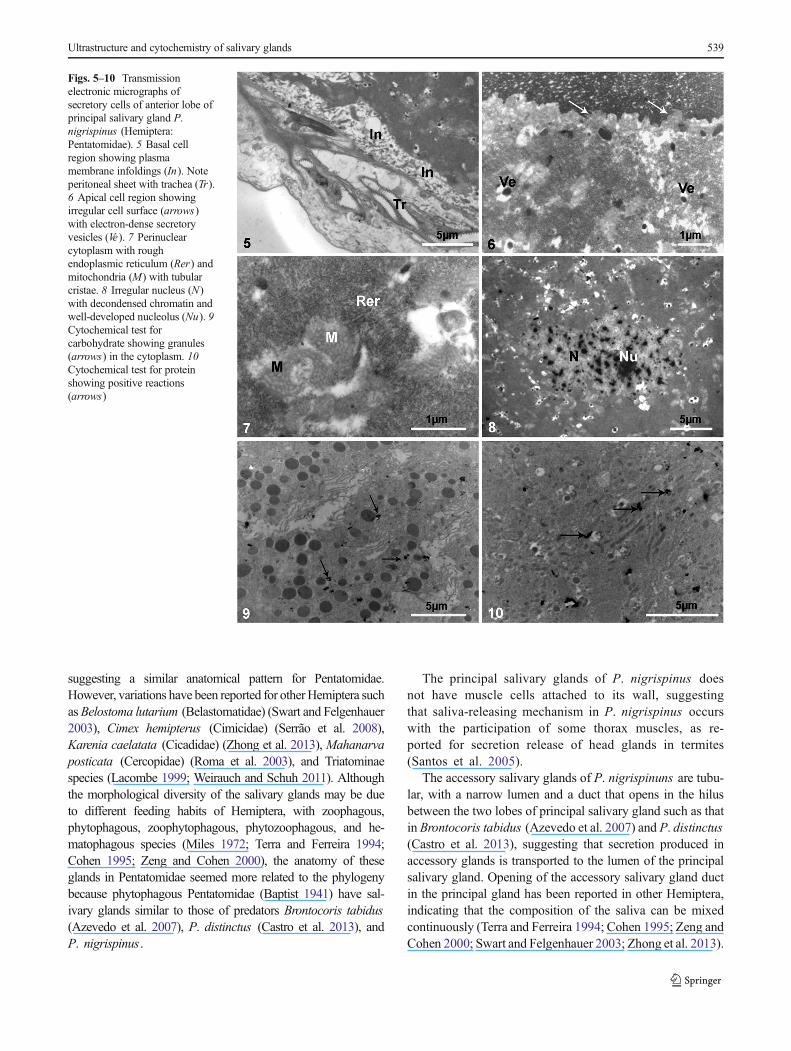
In the anterior lobe of the principal salivary glands, thebasal portion of secretory cells had short plasma membraneinfoldings that formed large canals (Fig. 5) associated withmitochondria. The cytoplasm had a high amount of roughendoplasmic reticulum (Figs. 6, 7), Golgi apparatus, andvesicles with electron-dense core and an electron-transparentperiperical halo (Fig. 8) similar to the luminal content (Fig. 6).Mitochondria were numerous and well developed, with manytubular cristae that resulted in a multivesicular compartmentbounded by double membrane (Fig. 7). The nucleus wasmedian and irregular with uncondensed chromatin and well-developed nucleolus (Fig. 9). The apical surfaces of the se-cretory cells had narrow, short, and irregular plasma mem-brane foldings (Fig. 5). The luminal content was granular and
electron-dense. Cytochemical tests showed positive reactionsfor carbohydrates near the electron-dense granules (Fig. 9) andfor protein in the electron-dense granules (Fig. 10). The cyto-chemical tests for lipids and acid phosphatase were negative inthe anterior lobe of the principal salivary gland tissues.
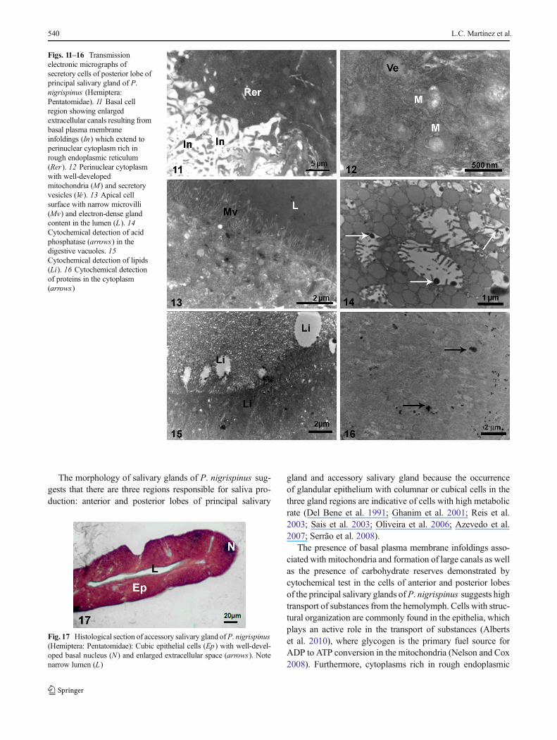
In the posterior lobe of the principal gland, the basal region ofthe secretory cell was similar to that found in the anterior lobe,with short plasma membrane infoldings associated with mito-chondria that formed large canals (Fig. 11). The cytoplasm wasrich in rough endoplasmic reticulum, Golgi complex, mitochon-dria with many cristae, and vesicles with granular content(Fig. 12). The well-developed nucleus showed a predominanceof uncondensed chromatin and multiple nucleoli. The apicalsurface of the cell was characterized by numerous narrow andlong microvilli, reaching the luminal content that showed lowerelectron density in comparison with the luminal content of theanterior lobe (Fig. 13). Cytochemical tests showed positive reac-tions for acid phosphatase in vacuoles of different sizes (Fig. 14).Lipids were found in the apical cytoplasm (Fig. 15), and proteinswere scattered in the cytoplasm with a high concentration in thebasal cell region close to the rough endoplasmic reticulum(Fig. 16). Carbohydrates were weakly identified in these cells.
The secretory cells of both anterior and posterior lobeswere onto a thin basal lamina that had an extensive amountof trachea and lacked muscle cells (data not shown).
Accessory salivary glands
The epithelium of accessory salivary glands was composed ofcolumnar cells lining a narrow lumen, whose contentremained unstained by hematoxylin and eosin staining(Fig. 17). The cytoplasm of secretory cells of the accessorysalivary gland had large vacuoles with electron-transparent orelectron-lucent content (Fig. 18), rough endoplasmic reticu-lum, and mitochondria (Fig. 19). The irregular nucleusshowed a predominance of uncondensed chromatin and nu-cleoli. The basal surface of the cell was characterized by theplasma membrane infoldings (Fig. 20). The apical surface ofthe glandular cell had narrow and short microvilli (Fig. 21).The secretory epithelium was onto a thin basal lamina rich intrachea. Cytochemical tests showed positive reactions forlipids that were homogeneously distributed in the cytoplasm(Figs. 21, 22) and proteins in the lumen and extracellularspaces (Fig. 23).
Discussion
The salivary system of P. nigrispinus is formed by a pair ofprincipal salivary glands and accessory salivary glands, withanatomy similar to that described for Podisus distinctus (Castroet al. 2013), Brontocoris tabidus (Heteroptera: Pentatomidae)(Azevedo et al. 2007), and other Pentatomidae (Baptist 1941),
Figs. 3, 4 Histological sections of salivary glands of P. nigrispinus(Hemiptera: Pentatomidae). 3 Anterior lobe of principal gland showingcolumnar epithelium (Ep) with well-developed nucleus (n) and basophil-ic cytoplasm (Cy). Note acidophill gland content in the lumen (L). 4Posterior lobe of principal gland showing cubical epithelium (Ep) withsome cells containing two nucleus (n) and basophilic cytoplasm (Cy)
538 L.C. Martínez et al.
suggesting a similar anatomical pattern for Pentatomidae.However, variations have been reported for otherHemiptera suchasBelostoma lutarium (Belastomatidae) (Swart and Felgenhauer2003), Cimex hemipterus (Cimicidae) (Serrão et al. 2008),Karenia caelatata (Cicadidae) (Zhong et al. 2013), Mahanarvaposticata (Cercopidae) (Roma et al. 2003), and Triatominaespecies (Lacombe 1999; Weirauch and Schuh 2011). Althoughthe morphological diversity of the salivary glands may be dueto different feeding habits of Hemiptera, with zoophagous,phytophagous, zoophytophagous, phytozoophagous, and he-matophagous species (Miles 1972; Terra and Ferreira 1994;Cohen 1995; Zeng and Cohen 2000), the anatomy of theseglands in Pentatomidae seemed more related to the phylogenybecause phytophagous Pentatomidae (Baptist 1941) have sal-ivary glands similar to those of predators Brontocoris tabidus(Azevedo et al. 2007), P. distinctus (Castro et al. 2013), andP. nigrispinus.
The principal salivary glands of P. nigrispinus doesnot have muscle cells attached to its wall, suggestingthat saliva-releasing mechanism in P. nigrispinus occurswith the participation of some thorax muscles, as re-ported for secretion release of head glands in termites(Santos et al. 2005).
The accessory salivary glands of P. nigrispinuns are tubu-lar, with a narrow lumen and a duct that opens in the hilusbetween the two lobes of principal salivary gland such as thatin Brontocoris tabidus (Azevedo et al. 2007) and P. distinctus(Castro et al. 2013), suggesting that secretion produced inaccessory glands is transported to the lumen of the principalsalivary gland. Opening of the accessory salivary gland ductin the principal gland has been reported in other Hemiptera,indicating that the composition of the saliva can be mixedcontinuously (Terra and Ferreira 1994; Cohen 1995; Zeng andCohen 2000; Swart and Felgenhauer 2003; Zhong et al. 2013).
Figs. 5–10 Transmissionelectronic micrographs ofsecretory cells of anterior lobe ofprincipal salivary gland P.nigrispinus (Hemiptera:Pentatomidae). 5 Basal cellregion showing plasmamembrane infoldings (In). Noteperitoneal sheet with trachea (Tr).6 Apical cell region showingirregular cell surface (arrows)with electron-dense secretoryvesicles (Ve). 7 Perinuclearcytoplasm with roughendoplasmic reticulum (Rer) andmitochondria (M) with tubularcristae. 8 Irregular nucleus (N)with decondensed chromatin andwell-developed nucleolus (Nu). 9Cytochemical test forcarbohydrate showing granules(arrows) in the cytoplasm. 10Cytochemical test for proteinshowing positive reactions(arrows)
Ultrastructure and cytochemistry of salivary glands 539
The morphology of salivary glands of P. nigrispinus sug-gests that there are three regions responsible for saliva pro-duction: anterior and posterior lobes of principal salivary
gland and accessory salivary gland because the occurrenceof glandular epithelium with columnar or cubical cells in thethree gland regions are indicative of cells with high metabolicrate (Del Bene et al. 1991; Ghanim et al. 2001; Reis et al.2003; Sais et al. 2003; Oliveira et al. 2006; Azevedo et al.2007; Serrão et al. 2008).
The presence of basal plasma membrane infoldings asso-ciated with mitochondria and formation of large canals as wellas the presence of carbohydrate reserves demonstrated bycytochemical test in the cells of anterior and posterior lobesof the principal salivary glands ofP. nigrispinus suggests hightransport of substances from the hemolymph. Cells with struc-tural organization are commonly found in the epithelia, whichplays an active role in the transport of substances (Albertset al. 2010), where glycogen is the primary fuel source forADP to ATP conversion in the mitochondria (Nelson and Cox2008). Furthermore, cytoplasms rich in rough endoplasmic
Figs. 11–16 Transmissionelectronic micrographs ofsecretory cells of posterior lobe ofprincipal salivary gland of P.nigrispinus (Hemiptera:Pentatomidae). 11 Basal cellregion showing enlargedextracellular canals resulting frombasal plasma membraneinfoldings (In) which extend toperinuclear cytoplasm rich inrough endoplasmic reticulum(Rer). 12 Perinuclear cytoplasmwith well-developedmitochondria (M) and secretoryvesicles (Ve). 13 Apical cellsurface with narrow microvilli(Mv) and electron-dense glandcontent in the lumen (L). 14Cytochemical detection of acidphosphatase (arrows) in thedigestive vacuoles. 15Cytochemical detection of lipids(Li). 16 Cytochemical detectionof proteins in the cytoplasm(arrows)
Fig. 17 Histological section of accessory salivary gland ofP. nigrispinus(Hemiptera: Pentatomidae): Cubic epithelial cells (Ep) with well-devel-oped basal nucleus (N) and enlarged extracellular space (arrows). Notenarrow lumen (L)
540 L.C. Martínez et al.

reticulum and granules containing proteins indicate the occur-rence of protein synthesis. Protein secretion by principalsalivary glands has been reported for other Hemiptera (Miles1972; Cohen 1998; Swart and Felgenhauer 2003; Nunes andCamargo-Mathias 2006; Azevedo et al. 2007; Serrão et al.2008; Fialho et al. 2012; Zhong et al. 2013), showing that thefunction of the principal salivary glands is conserved in theseinsects, regardless of their feeding habits.
Epithelial cells of accessory salivary gland of P. nigrispinushave ultrastructure suggesting involvement in the transport ofsubstances from hemolymph and proteins secretion. This is anintriguing finding, as the narrow lumen of this gland makes itunlikely to store substances that may be constitutively releasedin the lumen of the principal salivary glands because the duct ofthe accessory salivary gland opens in the hilum between theanterior and posterior lobes of the principal salivary gland.Thus, our model suggests that the accessory salivary gland of
P. nigrispinus has a different function from that suggested byMiles and Slowiak (1976), who pointed out that this glandplays a role in addition of water to saliva.
Cytochemical differences in the quantity and distributionof carbohydrates, acid phosphatase, lipids, and proteins werefound in the secretory cells of the anterior and posterior lobesof the principal and accessory salivary glands of P.nigrispinus , indicating that different substances in differentquantities are produced in these gland compartments. Thesalivary gland complex of predatory stink-bugs is importantin the production of enzymes required for extra-oral digestion(Swart and Felgenhauer 2003; Azevedo et al. 2007). In pred-atory bugs, the principal salivary glands may play a role in theproduction of digestive enzymes that cause liquefaction ofprey body for its ingestion, which characterizes extra-oraldigestion (Cohen 1990, 1995, 1998; Mohaghegh et al. 2001;Eubanks et al. 2003). However, in P. nigrispinus , the only
Figs. 18–23 Transmissionelectronic micrographs ofsecretory cells of accessorysalivary gland of P. nigrispinus(Hemiptera: Pentatomidae). 18General view of secretory cellshowing nucleus (N) andelectron-lucent granules (Gr). 19Basal cell region showing nucleus(N) with decondensed chromatinand perinuclear cytoplasm rich inrough endoplasmic reticulum(Rer). 20 Secretory cell withelectron-lucent and electron-dense granules (Gr) and irregularplasma membrane foldings(arrow). 21 Apical cell surfacewith short microvilli (Mv) andelectron-lucent granules (Gr).Inset , detail of electron-lucentgranule. 22 Cytochemicaldetection of lipids (Li). 23Cytochemical detection of protein(Pr)
Ultrastructure and cytochemistry of salivary glands 541
digestive enzyme present in the salivary glands is col-lagenase (Fialho et al. 2012). Thus, the cellular apparatusfor protein synthesis in the principal salivary glands of P.nigrispinus synthesizes other proteins/peptides than digestiveenzymes.
The diversity of substances produced by the salivary glandsof P. nigrispinus suggests that this predator can feed on largenumbers of prey. Moreover, the complexity of the salivacompounds, functional morphology of ingestion organs, andthe ecological implications of insect biology need additionalstudies to extend our understanding of salivary gland functionin the feeding behavior as well as in the production of sub-stances responsible for prey paralysis and death.
Acknowledgments The authors are grateful to the Conselho Nacional deDesenvolvimento Científico e Tecnológico CNPq (Brazil), Coordenação deAperfeiçoamento de Pessoal de Nível Superior CAPES (Brazil), Fundaçãode Amparo a Pesquisa do Estado de Minas Gerais FAPEMIG (Brasil), andNúcleo de Microscopia e Microanálise of Universidade Federal de Viçosa.
Conflict of interest We declare that there are no conflicts of interestwith the organization that sponsored the research.
References
Alberts B, Johson A, Lewis J, Raff M, Roberts K, Walter P (2010)Molecular Biology of the Cell. Garland Science, New York, pp 1–1601
Angermüller S, Fahimi DH (1982) Imidazole-buffered osmium tetroxide:an excellent stain for visualization of lipids in transmission electronmicroscopy. Histochem J 14:823–825
Azevedo DO, Zanuncio JC, Zanuncio JJS, Martins GF, Marques-Silva S,Sossai MF, Serrão JE (2007) Biochemical and morphological as-pects of salivary glands of the predator Brontocoris tabidus(Heteroptera: Pentatomidae). Braz Arch Biol Techn 50:469–477
Baptist BA (1941) Themorphology and physiology of the salivary glandsof Hemiptera-Heteroptera. Q J Microsc Sci 83:91–139
Barth R (1954) Estudos anatômicos e histológicos sobre a subfamiliaTriatominae (Heteroptera-Reduvidae). IV Parte: O complexo dasglândulas salivares de Triatoma infestans . Mem I Oswaldo Cruz53:517–585
Bell HA, Down RE, Edwards JP, Gatehouse JA, Gatehouse AMR (2005)Digestive proteolytic activity in the gut and salivary glands of thepredatory bug Podisus maculiventris (Heteroptera: Pentatomidae),effect of proteinase inhibitors. Eur J Entomol 102:139–145
Boyd DWJ, Cohen AC, Alverson DR (2002) Digestive enzymes andstylet morphology of Deraeocoris nebulosus (Hemiptera: Miridae),a predaceous plant bug. Ann Entomol Soc Am 95:395–401
Castro AA, Canevari GC, Pikart TG, Ribeiro RC, Serrão JE, ZanuncioTV, Zanuncio JC (2013) Salivary gland histology of the predatorSupputius cincticeps (Heteroptera: Pentatomidae). Ann EntomolSoc Am 106:273–277
Cohen AC (1990) Feeding adaptations of some predaceous Heteroptera.Ann Entomol Soc Am 83:1215–1223
Cohen AC (1995) Extra-oral digestion in predaceous terrestrial Arthropoda.Annu Rev Entomol 40:85–103
Cohen AC (1998) Solid-to-liquid feeding: the inside story of extra-oraldigestion in predaceous Heteroptera. Am Entomol 44:103–117
Del Bene G,Dallai R,Marchini D (1991) Ultrastructure of themidgut andthe adhering tubular salivary gland of Frankliniella occidentalis(Pergande) (Thysanoptera: Thripidae). Int J Insect MorpholEmbryol 20:15–24
Eubanks MD, Styrsky JD, Denno RF (2003) The evolution of omnivoryin heteropteran insects. Ecology 84:2549–2556
Fialho MCQ, Zanuncio JC, Neves CA, Ramalho FS, Serrão JE (2009)Ultrastructure of the digestive cells in the midgut of the predatorBrontocoris tabidus (Heteroptera: Pentatomidae) after different feed-ing periods on prey and plants. Ann Entomol Soc Am 102:119–127
FialhoMCQ, Zanuncio JC, Neves CA, Ramalho FS, Serrão JE (2012) Preydigestion in the midgut of the predatory bug Podisus nigrispinus(Hemiptera: Pentatomidae). J Insect Physiol 58:850–856
GhanimM, Rosell RC, Campbell LR, Czosnek H, Brown JK, UllmanDE(2001) Digestive, salivary, and reproductive organs of Bemisiatabaci (Gennadius) (Hemiptera: Aleyrodidae) B type. J Morphol248:22–40
Guedes BAM, Zanuncio JC, Ramalho FS, Serrão JE (2007) Midgutmorphology and enzymes of the obligate zoophytophagous stink-bug Brontocoris tabidus (Signoret, 1963) (Heteroptera:Pentatomidae). Pan-Pac Entomol 83:229–235
Lacerda MC, Ferreira AMRM, Zanuncio TV, Zanuncio JC, BernardioAS, Espindula MC (2004) Development and reproduction ofPodisus distinctus (Heteroptera: Pentatomidae) fed on larva ofBombyx mori (Lepidoptera: Bombycidae). Braz J Biol 64:237–242
Lacombe D (1999) Anatomia e histologia das glândulas salivares nostriatomíneos. Mem I Oswaldo Cruz 94:557–564
Lemos WP, Medeiros RS, Ramalho FS, Zanuncio JC (2001) Effects ofplant feeding on the development, survival and reproduction ofPodisus nigrispinus (Dallas) (Heteroptera: Pentatomidae). Int JPest Manage 47:89–93
Lemos WP, Ramalho FS, Serrão JE, Zanuncio JC (2003) Effects of dieton development of Podisus nigrispinus (Dallas) (Heteroptera:Pentatomidae), a predator of the cotton leafworm. J Appl Entomol127:389–395
LemosWP, Ramalho FS, Serrão JE, Zanuncio JC (2005a)Morphology offemale reproductive tract of the predator Podisus nigrispinus(Dallas) (Heteroptera: Pentatomidae) fed on different diets. BrazArch Biol Techn 48:129–138
Lemos WP, Zanuncio JC, Serrão JE (2005b) Attack behavior of Podisusrostralis (Heteroptera, Pentatomidae) adults on caterpillars ofBombyx mori (Lepidoptera, Bombycidae). Braz Arch Biol Techn48:975–981
Matos Neto FC, Oliveira HN, Zanuncio JC, Holtz AM (2004) Ganânciade peso del depredador Podisus distinctus (Heteroptera:Pentatomidae) en combinaciones de las presas Tenebrio molitor(Coleoptera: Tenebrionidae) e Musca domestica (Diptera:Muscidae). Rev Biol Trop 52:1–8
McPherson RM, Smith JC, Allen WA (1982) Incidence of arthropodpredators in different soybean cropping systems. Environ Entomol11:685–689
McRae EK,Meetz GD (1970) Electron microscopy of ammoniacal silverreaction for histones in erythropoietic cells of chick. J Cell Biol 45:235–245
Medeiros RS, Ramalho FS, Lemos WP, Zanuncio JC (2000) Age-dependent fecundity and life-fertility tables for Podisusnigrispinus (Dallas) (Heteroptera: Pentatomidae). J ApplEntomol 124:319–324
Miles PW (1972) The saliva of Hemiptera. Adv Insect Physiol 9:183–255Miles PW, Slowiak D (1976) The accessory salivary gland as the source
of water in the saliva of Hemiptera: Heteroptera. Experientia 15:1011–1012
Mohaghegh J, De Clercq P, Tirry L (1999) Effects of rearing history andgeographical origin on reproduction and body size of the predatorPodisus nigrispinus (Heteroptera: Pentatomidae). Eur J Entomol 96:69–72
542 L.C. Martínez et al.

Mohaghegh J, De Clercq P, Tirry L (2001) Functional response of thepredators Podisus maculiventris (Say) and Podisus nigrispinus(Dallas) (Heteroptera: Pentatomidae) to the beet armyworm,Spodoptera exigua (Hübner) (Lepidoptera: Noctuidae): effect oftemperature. J Appl Entomol 125:131–134
Nelson DE, Cox MM (2008) Lehninger principles of biochemistry, 5thedn. W.H Freeman, New York
Nunes PH, Camargo-Mathias MI (2006) Ultrastructural study of thesalivary glands of the sugarcane spittlebug Mahanarva fimbriolata(Stal, 1854) (Euhemiptera: Cercopidae). Micron 37:57–66
Oliveira I, Zanuncio JC, Serrão JE, Zanuncio TV, Pinon TBM, FialhoMCQ (2005) Effect of female weight on reproductive potential ofthe predator Brontocoris tabidus (Signoret 1852) (Heteroptera:Pentatomidae). Braz Arch Biol Techn 48:975–981
Oliveira JA, Oliveira MGA, Guedes RNC, Soares MJ (2006) Morphologyand preliminary enzyme characterization of the salivary glands from thepredatory bug Podisus nigrispinus (Heteroptera: Pentatomidae). BullEntomol Res 96:251–258
Panizzi AR, McPherson JE, James DG, Javahery M, McPherson RM(2000) Economic importance of stink bugs (Pentatomidae). In:Schaefer CW, Panizzi AR (eds) Heteroptera of economic impor-tance. CRC, Boca Raton, pp 421–474
Puri IM (1924) Studies on the anatomy of Cimex lectularius L.Parasitology 16:84–97
ReisMMM,Meirelles RM, SoaresMJ (2003) Fine structure of the salivaryglands of Triatoma infestans (Hemiptera, Reduviidae). Tissue Cell35:393–400
Reynolds ES (1963) The use of lead citrate at high pH as an electron-opaque stain in electron microscopy. J Cell Biol 17:208–2012
RomaGC, Camargo-MathiasMI, Arrigoni EB,Marin-MoralesMA (2003)Little cicada of sugarcane Mahanarva posticata (Homoptera,Cercopidae). A Brazilian agricultural pest. Morpho-histological studyof salivary glands. Cytologia 68:101–114
Sais TC,Moraes RM, Ribolla PE, Bianchi AG,Marinotti O, Bijovsky AT(2003) Morphological aspects of Culex quinquefasciatus salivaryglands. Arthropod Struct Dev 32:219–226
Santos CA, Costa-Leonardo AM, Serrão JE (2005) Morphology of thehead and frontal gland in Neotropical Nasutitermitinae (Isoptera,Termitidae). Sociobiology 46:579–593
Schmidt JO (1982) Biochemistry of insect venoms. Annu Rev Entomol27:339–368
Serrão JE, Castrillon MI, Santos-Mallet JR, Zanuncio JC, Gonçalves TC(2008) Ultrastructure of the salivary gland in Cimex hemipterus(Hemiptera: Cimicidae). J Med Entomol 45:991–999
Stefanini M, De Martino C, Zamboni L (1967) Fixation of ejac-ulated spermatozoa for electron microscopy. Nature 216:173–174
Swart CC, Felgenhauer BE (2003) Structure and function of the mouth-parts and salivary gland complex of the giant waterbug, Belostomalutarium (Stall) (Hemiptera: Belostomatidae). Ann Entomol SocAm 96:870–882
Terra WR, Ferreira C (1994) Insect digestive enzymes: properties,compartmentalization and function. Comp Biochem Physiol B109:1–62
Thiery JP (1967) Mise em evidence des polysaccharides sur coupes finesen microscopie electronique. J Microsc 6:987–1018
Tillman PG, Mullinix BG (2004) Comparison of susceptibility ofpest Euschistus servus and predator Podisus maculiventris(Heteroptera: Pentatomidae) to selected insecticides. J EconEntomol 97:800–806
Tyder TA, Bowen ID (1975) A method for the fine structural localizationof acid phosphatase activity using rho-nitrophenyl phosphate assubstrate. J Histochem Cytochem 23:235–237
Weirauch C, Schuh RT (2011) Systematics and evolution of Heteroptera:25 years of progress. Annu Rev Entomol 56:487–510
Zanuncio JC, Alves JB, Zanuncio TV, García JL (1994) Hemipterouspredators of eucalypt defoliator caterpillars. Forest Ecol Manag 65:65–73
Zanuncio TV, Torres JB, Zanuncio JC, Santos GP (1998) Ciclo devida e reprodução de Podisus distinctus (Stal) (Heteroptera:Pentatomidae) alimentado com dois tipos de presas. Rev BrasEntomol 41:335–337
Zeng F, Cohen AC (2000) Comparison of a-amylase and protease activ-ities of a zoophytophagous and two phytozoophagous Heteroptera.Comp Biochem Physiol A 126:101–106
ZhongH,Wei C, ZhangY (2013) Gross morphology and ultrastructure ofsalivary glands of the mute cicada Karenia caelatata distant(Hemiptera: Cicadoidea). Micron 45:83–91
Ultrastructure and cytochemistry of salivary glands 543