molecular studies of the salivary glands of the pea aphid
182
MOLECULAR STUDIES OF THE SALIVARY GLANDS OF THE PEA APHID, ACYRTHOSIPHON PISUM (HARRIS) by NAVDEEP S. MUTTI M. S., Punjab Agricultural University, India, 1998 AN ABSTRACT OF A DISSERTATION submitted in partial fulfillment of the requirements for the degree DOCTOR OF PHILOSOPHY Department of Entomology College of Agriculture KANSAS STATE UNIVERSITY Manhattan, Kansas 2006
-
Upload
khangminh22 -
Category
Documents
-
view
4 -
download
0
Transcript of molecular studies of the salivary glands of the pea aphid
MOLECULAR STUDIES OF THE SALIVARY GLANDS OF THE PEA APHID,
ACYRTHOSIPHON PISUM (HARRIS)
by
NAVDEEP S. MUTTI
M. S., Punjab Agricultural University, India, 1998
AN ABSTRACT OF A DISSERTATION
submitted in partial fulfillment of the requirements for the degree
DOCTOR OF PHILOSOPHY
Department of Entomology College of Agriculture
KANSAS STATE UNIVERSITY Manhattan, Kansas
2006
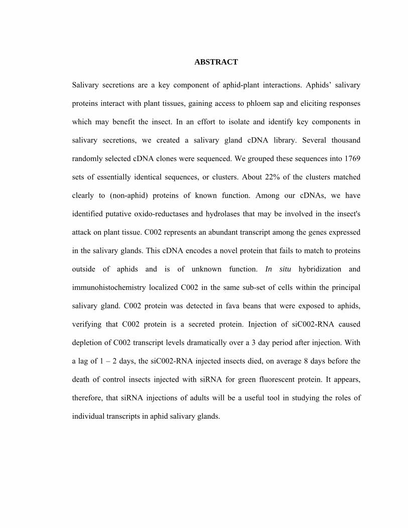
ABSTRACT
Salivary secretions are a key component of aphid-plant interactions. Aphids’ salivary
proteins interact with plant tissues, gaining access to phloem sap and eliciting responses
which may benefit the insect. In an effort to isolate and identify key components in
salivary secretions, we created a salivary gland cDNA library. Several thousand
randomly selected cDNA clones were sequenced. We grouped these sequences into 1769
sets of essentially identical sequences, or clusters. About 22% of the clusters matched
clearly to (non-aphid) proteins of known function. Among our cDNAs, we have
identified putative oxido-reductases and hydrolases that may be involved in the insect's
attack on plant tissue. C002 represents an abundant transcript among the genes expressed
in the salivary glands. This cDNA encodes a novel protein that fails to match to proteins
outside of aphids and is of unknown function. In situ hybridization and
immunohistochemistry localized C002 in the same sub-set of cells within the principal
salivary gland. C002 protein was detected in fava beans that were exposed to aphids,
verifying that C002 protein is a secreted protein. Injection of siC002-RNA caused
depletion of C002 transcript levels dramatically over a 3 day period after injection. With
a lag of 1 – 2 days, the siC002-RNA injected insects died, on average 8 days before the
death of control insects injected with siRNA for green fluorescent protein. It appears,
therefore, that siRNA injections of adults will be a useful tool in studying the roles of
individual transcripts in aphid salivary glands.
MOLECULAR STUDIES OF THE SALIVARY GLANDS OF THE PEA APHID,
ACYRTHOSIPHON PISUM (HARRIS)
by
NAVDEEP S. MUTTI
M. S., Punjab Agricultural University, India, 1998
A DISSERTATION
submitted in partial fulfillment of the requirements for the degree
DOCTOR OF PHILOSOPHY
Department of Entomology College of Agriculture
KANSAS STATE UNIVERSITY Manhattan, Kansas
2006
Approved by: Approved by:
Co-Major Professor Co-Major Professor
Dr. John C. Reese Dr. Gerald R. Reeck

COPYRIGHT
Navdeep S. Mutti
2006

ABSTRACT
Salivary secretions are a key component of aphid-plant interactions. Aphids’ salivary
proteins interact with plant tissues, gaining access to phloem sap and eliciting responses
which may benefit the insect. In an effort to isolate and identify key components in
salivary secretions, we created a salivary gland cDNA library. Several thousand
randomly selected cDNA clones were sequenced. We grouped these sequences into 1769
sets of essentially identical sequences, or clusters. About 22% of the clusters matched
clearly to (non-aphid) proteins of known function. Among our cDNAs, we have
identified putative oxido-reductases and hydrolases that may be involved in the insect's
attack on plant tissue. C002 represents an abundant transcript among the genes expressed
in the salivary glands. This cDNA encodes a novel protein that fails to match to proteins
outside of aphids and is of unknown function. In situ hybridization and
immunohistochemistry localized C002 in the same sub-set of cells within the principal
salivary gland. C002 protein was detected in fava beans that were exposed to aphids,
verifying that C002 protein is a secreted protein. Injection of siC002-RNA caused
depletion of C002 transcript levels dramatically over a 3 day period after injection. With
a lag of 1 – 2 days, the siC002-RNA injected insects died, on average 8 days before the
death of control insects injected with siRNA for green fluorescent protein. It appears,
therefore, that siRNA injections of adults will be a useful tool in studying the roles of
individual transcripts in aphid salivary glands.
vi
TABLE OF CONTENTS
LIST OF FIGURES ........................................................................................................... ix
LIST OF TABLES.............................................................................................................. x
ACKNOWLEDGEMENTS............................................................................................... xi
DEDICATION.................................................................................................................. xii
CHAPTER 1 ....................................................................................................................... 1
Chapter 1: Review of Literature ......................................................................................... 2
Aphid Taxonomy ............................................................................................................ 2
Aphid Feeding................................................................................................................. 3
Salivary Glands............................................................................................................... 6
Aphid Saliva and Salivary Proteins ................................................................................ 9
Plant Resistance and Defense Response to Aphids ...................................................... 14
Economic Importance of Aphids .................................................................................. 17
Aphids as Carriers of Viruses ....................................................................................... 18
Symbionts of Aphids..................................................................................................... 19
RNA Interference in Insects.......................................................................................... 22
Specific Objectives ........................................................................................................... 25
References..................................................................................................................... 26
CHAPTER 2 ..................................................................................................................... 58
Chapter 2: An EST library from Salivary Glands of the Pea Aphid, Acyrthosiphon pisum.
........................................................................................................................................... 59
Abstract ......................................................................................................................... 60
Introduction................................................................................................................... 61
Material and Methods ................................................................................................... 62
Plants and Aphids ..................................................................................................... 62
Phagmid cDNA Library Construction ...................................................................... 62
Sequencing, Sequence Processing, and Annotation ................................................. 63
Results........................................................................................................................... 64
Salivary Transcript Catalog for A. pisum.................................................................. 64

vii
Classification of Salivary Transcripts of A. pisum.................................................... 64
Proteases and Other Hydrolases in the Salivary Glands of A. pisum........................ 66
Oxidoreducatases in the Salivary Glands of A. pisum .............................................. 67
Discussion..................................................................................................................... 68
Acknowledgements....................................................................................................... 72
References..................................................................................................................... 73
Chapter 3: A Novel Transcript and Protein from the Salivary Glands of Pea Aphid,
Acyrthosiphon pisum......................................................................................................... 96
Abstract ......................................................................................................................... 97
Introduction................................................................................................................... 98
Material and Methods ................................................................................................... 99
Plants and Aphids ..................................................................................................... 99
Phagmid cDNA Library Construction and Sequencing.......................................... 100
RNA Isolation and RT-PCR ................................................................................... 100
RNA Isolation and Northern Blotting..................................................................... 101
Genomic DNA Isolation and Southern Blotting..................................................... 102
In situ Hybridization ............................................................................................... 102
Expression of Recombinant Protein in E. coli for Antibody Preparation............... 104
Western Blotting ..................................................................................................... 105
Immunohistochemistry ........................................................................................... 106
Preparation of dsRNA, siRNA and siRNA Injections ............................................ 107
Results......................................................................................................................... 107
The C002 Transcript and Protein............................................................................ 107
Expression of C002 in the Salivary Gland and Gut................................................ 109
Northern and Southern Blot Analyses .................................................................... 109
In situ Hybridization ............................................................................................... 110
Immunohistochemistry ........................................................................................... 110
Western Blotting ..................................................................................................... 111
Effects of RNAi on Feeding on Artificial Diet....................................................... 111
Discussion................................................................................................................... 112
Acknowledgments....................................................................................................... 115
viii
References................................................................................................................... 117
CHAPTER 4 ................................................................................................................... 137
Chapter 4: RNAi Knockdown of a Salivary Transcript Leading to Lethality in the Pea
Aphid, Acyrthosiphon pisum........................................................................................... 138
Abstract ....................................................................................................................... 139
Introduction................................................................................................................. 140
Materials and Methods................................................................................................ 142
Plants and Aphids ................................................................................................... 142
Preparation of dsRNA and siRNA.......................................................................... 143
siRNA Injections..................................................................................................... 143
Examining Transcript Levels by RT-PCR.............................................................. 144
Results and Discussion ............................................................................................... 145
Acknowledgments....................................................................................................... 148
References................................................................................................................... 149
Appendix 1...................................................................................................................... 158
Supplemental Information to Chapter 4...................................................................... 158
SUMMARY.................................................................................................................... 169
ix
LIST OF FIGURES
Figure 1. Diagnostic morphological features of an aphid................................................. 48
Figure 2. Anatomy of the salivary gland of the green peach aphid, M. persicae. ............ 50
Figure 3. Feulgen stained principal salivary gland of M. persicae. .................................. 52
Figure 4. Pea aphid, A. pisum C002 sequence. ............................................................... 119
Figure 5. Multiple sequence alignments. ........................................................................ 122
Figure 6. Expression of C002 in salivary glands and gut of pea aphid. ......................... 125
Figure 7. Northern and Southern blot analysis of C002 transcript and gene.................. 127
Figure 8. Detection of C002 mRNA in the salivary glands by in situ hybridization...... 129
Figure 9. Immunohistochemical localization of C002 protein. ...................................... 131
Figure 10. Detection of C002 protein using purified C002 antibodies........................... 133
Figure 11. Survival of siRNA injected aphids on artificial diet. .................................... 135
Figure 12. Survival of pea aphids after injection of siRNA. .......................................... 152
Figure 13. Knockdown of the C002 transcript after siRNA injections........................... 154
Figure 14. Timing of knockdown of C002 transcript after siRNA injection.................. 156
Figure 15. Survival of siRNA injected pea aphids. ........................................................ 161
Figure 16. Knockdown of the C002 transcript after siRNA injections........................... 163
Figure 17. Reproduction of pea aphids after siRNA injections. ..................................... 166
x
LIST OF TABLES
Table 1. Classification of selected aphid species.............................................................. 54
Table 2. Classification of cell types of salivary glands of M. persicae. ........................... 55
Table 3. EST projects within Hemiptera........................................................................... 56
Table 4. Plant genes up-regulated in response to aphid feeding based on microarray
analysis...................................................................................................................... 57
Table 5. Classifications of 377 clusters based on BLASTX against UniProt database.... 78
Table 6. Classification of 72 clusters encoding “hypothetical proteins.” ......................... 80
Table 7. Clusters with three or more sequences of known function................................. 84
Table 8. Clusters with more than 2 sequences with unknown function............................ 88
Table 9. Functional annotation of proteases and other hydrolases. .................................. 92
Table 10. Functional annotation of oxidoreductases. ....................................................... 94
xi
ACKNOWLEDGEMENTS
I wish to thank Dr. John C. Reese and Dr. Gerald Reeck for giving me this wonderful
opportunity to pursue my doctoral studies in Entomology at K-State. Whatever I have
transformed into today is because of them! When I say that, it is not just for the scientific
skills they have taught me, but also for various fine points in life coming out of their
sheer professionalism and experience. Drs. Reese and Reeck strived to look beyond my
work in lab and helped shape my career. It was my pleasure to work in close association
with Dr. Michael Kanost and his lab. I owe my research progress to my committee
members, Dr B.S. Gill, Dr Ming-Shun Chen, who constantly provided inputs that were in
best of my interests. I would like to extend special thanks to Dr. Yoonsoeng Park for his
helpful advice and guidance. Special thanks to Dr. Sonny Ramaswamy for creating a
stimulating environment for students in the department. I would also like to extend my
gratitude to Dr. Neal Dittmer and Dr. Maureen Gorman, who were always willing to help
me with my experiments. I would also like to thank my lab members Marisol Castaneto
and David Liang for their help with dissections. For folks at the front office, both in
Entomology and Biochemistry - you are simply the best! Every time, they greeted me
with a smile and helped me out. I would also like to thank the entire departments of
Entomology and Biochemistry for making me a part of the family and making me feel
very much at home. Finally, I would like to thank my brother Jasdeep, my wife Dilpreet
and my daughter Navnoor for their constant support and encouragement!
xii
DEDICATION
Dedicated to my parents, Dr. Darshan Singh and Mrs. Narinder Kaur, for their constant
support and encouragement.

1
CHAPTER 1
2
Chapter 1: Review of Literature
Aphid Taxonomy
Approximately, 4000 species of aphids have been described (Dixon, 1998). Of these, 250
species are considered pest species (Blackman and Eastop, 2000). Species belonging to
tribe Macrosiphini include important agricultural pests, such as the green peach aphid
(Myzus persicae), the Russian wheat aphid (Diuraphis noxia) and the pea aphid (A.
pisum) (von Dohlen and Moran, 2000; Martinez-Torres et al., 2001; Ortiz-Rivas et al.,
2004). Detailed classification of important aphid species is described in Table 1. The pea
aphid, Acyrthosiphon pisum (Harris), is a member of superfamily Aphidoidae and family
Aphididae, within the order Hemiptera (Sorensen, 1995; von Dohlen and Moran, 1995).
Diagnostic morphological features of aphids are shown in Fig. 1. (A) The base of the
proboscis lies between and behind the fore coxae; (B) the antennae have two short thick
basal segments and a thinner flagellum; (C) there is an ocular tubercle made up of three
lenses (a triommatidium) situated behind each compound eye; (D) there are two tarsal
segments; (E) the wings have only one prominent longitudinal vein; and (F) there is a
pair of siphunculi on the dorsum of the fifth abdominal segment (Heie, 1980; Dixon,
1998).
It is estimated from fossil evidence that Aphidoidae appeared 280 million year ago, in the
Carboniferous era (Dixon, 1998). Reproduction by means of unfertilized eggs

3
(parthogenesis) may have appeared in the late carboniferous or early Permian, over 200
million years ago (Heie, 1967). Viviparity, and other characteristics like shape and
venation of their wings and the structure of their proboscis and legs, appeared by the
Jurassic (146 million years ago), whereas the cauda and siphunculi appeared later, in the
Cretaceous (65 million years ago) (Shaposhnikov, 1977).
Aphids have a soft cuticle; wings, if present, are membranous. Winged aphids are known
as alatae and wingless aphids as apterae. They have both sexual (which produce fertilized
eggs that overwinter) and parthenogenetic reproduction. Short developmental time and
ability of adult females to reproduce several nymphs per day enable aphids to achieve
very high rates of increase. Aphids produce the phenotype they require to suit the
environmental circumstances they encounter. These kinds of environmentally induced
discrete variants are called polyphenisms. If aphids develop on a plant that is crowded
with many other aphids, they may develop with wings and fly to a new host plant
(Blackman, 1987; Braendle et al., 2005). The mechanisms that allow aphids to switch
between alternative morphs have remained obscure. It is believed that well-known insect
hormones (like juvenile hormone and ecdysone hormone) regulate these switches
(Hardie, 1980; Nijhout, 1999).
Aphid Feeding
Many aphids have a narrow host range. For example, the mustard aphid, Lipaphis erysimi
feeds only on cruciferous plants. The pea aphid, A. pisum, feeds on leguminous host
plants, including peas and alfalfa (Blackman and Eastop, 2000). Some aphids have a
4
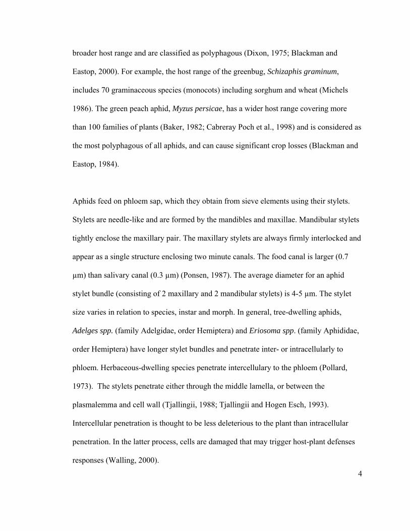
broader host range and are classified as polyphagous (Dixon, 1975; Blackman and
Eastop, 2000). For example, the host range of the greenbug, Schizaphis graminum,
includes 70 graminaceous species (monocots) including sorghum and wheat (Michels
1986). The green peach aphid, Myzus persicae, has a wider host range covering more
than 100 families of plants (Baker, 1982; Cabreray Poch et al., 1998) and is considered as
the most polyphagous of all aphids, and can cause significant crop losses (Blackman and
Eastop, 1984).
Aphids feed on phloem sap, which they obtain from sieve elements using their stylets.
Stylets are needle-like and are formed by the mandibles and maxillae. Mandibular stylets
tightly enclose the maxillary pair. The maxillary stylets are always firmly interlocked and
appear as a single structure enclosing two minute canals. The food canal is larger (0.7
µm) than salivary canal (0.3 µm) (Ponsen, 1987). The average diameter for an aphid
stylet bundle (consisting of 2 maxillary and 2 mandibular stylets) is 4-5 µm. The stylet
size varies in relation to species, instar and morph. In general, tree-dwelling aphids,
Adelges spp. (family Adelgidae, order Hemiptera) and Eriosoma spp. (family Aphididae,
order Hemiptera) have longer stylet bundles and penetrate inter- or intracellularly to
phloem. Herbaceous-dwelling species penetrate intercellulary to the phloem (Pollard,
1973). The stylets penetrate either through the middle lamella, or between the
plasmalemma and cell wall (Tjallingii, 1988; Tjallingii and Hogen Esch, 1993).
Intercellular penetration is thought to be less deleterious to the plant than intracellular
penetration. In the latter process, cells are damaged that may trigger host-plant defenses
responses (Walling, 2000).
5
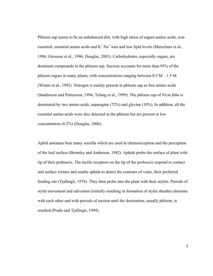
Phloem sap seems to be an unbalanced diet, with high ratios of sugars:amino acids, non-
essential: essential amino acids and K+:Na+ ions and low lipid levels (Marschner et al.,
1996; Girousse et al., 1996; Douglas, 2003). Carbohydrates, especially sugars, are
dominant compounds in the phloem sap. Sucrose accounts for more than 95% of the
phloem sugars in many plants, with concentrations ranging between 0.5 M – 1.5 M
(Winter et al., 1992). Nitrogen is mainly present in phloem sap as free amino acids
(Sandstrom and Pettersson, 1994; Telang et al., 1999). The phloem sap of Vicia faba is
dominated by two amino acids, asparagine (72%) and glycine (10%). In addition, all the
essential amino acids were also detected in the phloem but are present at low
concentration (8.2%) (Douglas, 2006).
Aphid antennae bear many sensilla which are used in chemoreception and the perception
of the leaf surface (Bromley and Anderson, 1982). Aphids probe the surface of plant with
tip of their proboscis. The tactile receptors on the tip of the proboscis respond to contact
and surface texture and enable aphids to detect the contours of veins, their preferred
feeding site (Tjallingii, 1978). They then probe into the plant with their stylets. Periods of
stylet movement and salivation (initially resulting in formation of stylet sheaths) alternate
with each other and with periods of suction until the destination, usually phloem, is
reached (Prado and Tjallingii, 1994).
6
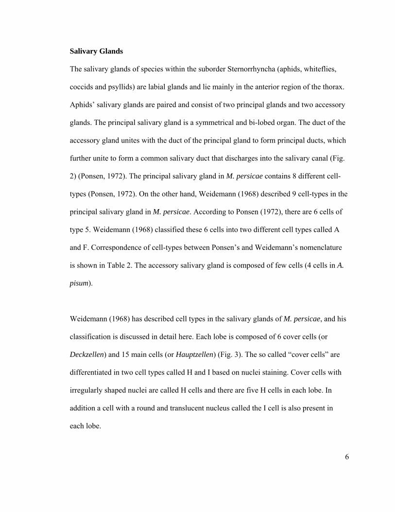
Salivary Glands
The salivary glands of species within the suborder Sternorrhyncha (aphids, whiteflies,
coccids and psyllids) are labial glands and lie mainly in the anterior region of the thorax.
Aphids’ salivary glands are paired and consist of two principal glands and two accessory
glands. The principal salivary gland is a symmetrical and bi-lobed organ. The duct of the
accessory gland unites with the duct of the principal gland to form principal ducts, which
further unite to form a common salivary duct that discharges into the salivary canal (Fig.
2) (Ponsen, 1972). The principal salivary gland in M. persicae contains 8 different cell-
types (Ponsen, 1972). On the other hand, Weidemann (1968) described 9 cell-types in the
principal salivary gland in M. persicae. According to Ponsen (1972), there are 6 cells of
type 5. Weidemann (1968) classified these 6 cells into two different cell types called A
and F. Correspondence of cell-types between Ponsen’s and Weidemann’s nomenclature
is shown in Table 2. The accessory salivary gland is composed of few cells (4 cells in A.
pisum).
Weidemann (1968) has described cell types in the salivary glands of M. persicae, and his
classification is discussed in detail here. Each lobe is composed of 6 cover cells (or
Deckzellen) and 15 main cells (or Hauptzellen) (Fig. 3). The so called “cover cells” are
differentiated in two cell types called H and I based on nuclei staining. Cover cells with
irregularly shaped nuclei are called H cells and there are five H cells in each lobe. In
addition a cell with a round and translucent nucleus called the I cell is also present in
each lobe.
7
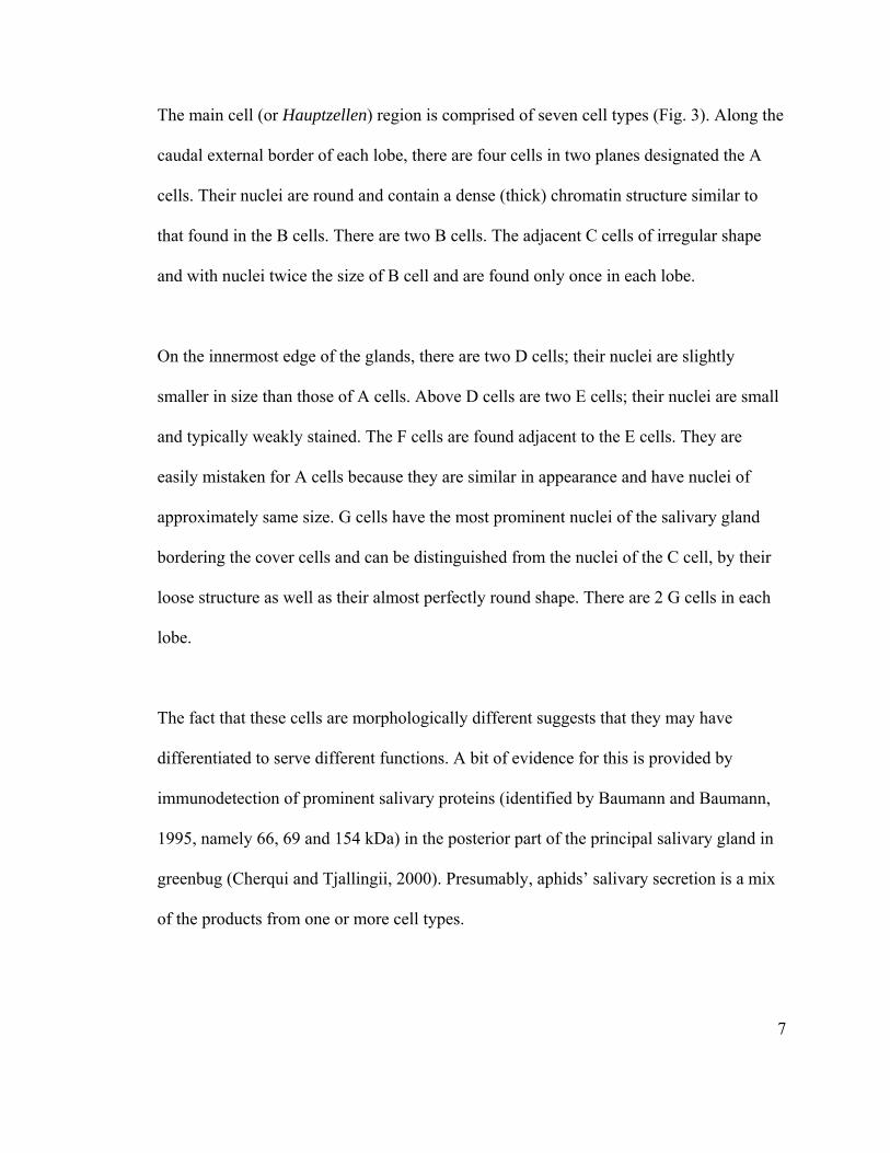
The main cell (or Hauptzellen) region is comprised of seven cell types (Fig. 3). Along the
caudal external border of each lobe, there are four cells in two planes designated the A
cells. Their nuclei are round and contain a dense (thick) chromatin structure similar to
that found in the B cells. There are two B cells. The adjacent C cells of irregular shape
and with nuclei twice the size of B cell and are found only once in each lobe.
On the innermost edge of the glands, there are two D cells; their nuclei are slightly
smaller in size than those of A cells. Above D cells are two E cells; their nuclei are small
and typically weakly stained. The F cells are found adjacent to the E cells. They are
easily mistaken for A cells because they are similar in appearance and have nuclei of
approximately same size. G cells have the most prominent nuclei of the salivary gland
bordering the cover cells and can be distinguished from the nuclei of the C cell, by their
loose structure as well as their almost perfectly round shape. There are 2 G cells in each
lobe.
The fact that these cells are morphologically different suggests that they may have
differentiated to serve different functions. A bit of evidence for this is provided by
immunodetection of prominent salivary proteins (identified by Baumann and Baumann,
1995, namely 66, 69 and 154 kDa) in the posterior part of the principal salivary gland in
greenbug (Cherqui and Tjallingii, 2000). Presumably, aphids’ salivary secretion is a mix
of the products from one or more cell types.
8
The ultra-structure of the cells of the principal salivary gland show the presence of a well
developed rough endoplasmic reticulum and also the presence of secretory granules,
suggesting that they are involved in the synthesis of salivary proteins (Moericke and
Wohlfarth-Bottermann, 1960 and 1963; Wohlfarth-Bottermann and Moericke, 1960). On
the other hand, function of the accessory gland is largely unknown; but it is involved in
virus transmission, based on presence of potato leaf roll virus particles as seen by
electron microscopy studies (Glidow et al., 2000).
Like those in aphids, Drosophila embryos salivary glands consist of two major cell types:
secretory cells and duct cells. Secretory cells are columnar epithelial cells that synthesize
and secrete high levels of proteins. At the onset of metamorphosis they also secrete the
glue to paste the pupae to the substrate. The high production level of glue proteins is
achieved by genome amplification. The chromosomes of the salivary gland nuclei
undergo endoreplication (DNA replication without division) and become giant polytene
chromosomes (Andrew, 1998). The duct cells are cuboidal epithelial cells that form the
simple tubes connecting the secretory cells and form a common duct that discharges into
salivary canal.
The initial specification of salivary cells in Drosophila occurs within a two-dimensional
sheet of cells, the ectoderm, with no known induction from underlying layers. The
salivary gland primordium is bilaterally symmetric and consists of approximately 100
cells on either side of the ventral midline (Andrew, 1998; Campos-Ortega and
Hartenstein, 1997; Andrew et al., 2000). Salivary glands arise from two ventral
9
ectodermal plates, in the region of the presumptive posterior head (Panzer et al., 1992;
Andrew et al., 2000). Salivary glands of Drosophila differentiate without further cell
division and increase in size simply by increasing the volume of individual cells (Andrew
et al., 2000). Salivary gland development begins at 4.5 h of development and finishing by
10 h of development. This initial specification, which is complete by embryonic stage 10
(about 5.5 h of development), occurs only within a specific region of the anterioposterior
axis: parasegment two. In contrast little is known about the embryonic development of
the salivary glands of aphids besides the anatomical studies by Ponsen (1972) and
Weidemann (1968) on the salivary gland.
Aphid Saliva and Salivary Proteins
Electric penetration graph (EPG) studies have shown four phases of salivary secretion
during penetration of host plants by aphids: (1) intercellular sheath secretion, (2)
intracellular salivation into cells along the stylet path, (3) initial phloem salivation (into
sieve elements); and (4) phloem feeding salivation (Tjallingii, 1988; Tjallingii, 1990;
Prado and Tjallingii, 1994; Cherqui and Tjallingii, 2000). Thus, there is ample
opportunity for salivary secretions to elicit plant responses, block wound responses or
detoxify phytochemicals. Aphid saliva holds the potential to better understand the co-
evolution of insect-host interactions (Miles, 1998; Miles, 1999).
Aphids inject a variety of physiologically and biochemically active substances into host
plants to facilitate feeding (Miles, 1968; Miles, 1999). Aphid saliva is a mix of ions,
amino acids, hemolymph (pumped from myoepithelioid cell) (Ponsen, 1972) and salivary
10
proteins / enzymes secreted from principal and accessory salivary gland (Miles, 1998;
Miles, 1999). The proteins of aphid saliva are of two types, structural and enzymatic. The
structural proteins provide a tube-like sheath (Miles 1999; Cherqui and Tjallingi 2000)
and are probably few in number, corresponding to major bands on gels with estimated
molecular masses of 154 kDa and 66/69 kDa (Baumann and Baumann, 1995). Polyclonal
antibodies against these proteins stained sheaths (Cherqui and Tjallingi 2000). Enzymatic
assays have been carried out on diluted saliva of aphids (Adams and McAllan, 1956 and
1958; Madhusudhan et al., 1994; Miles, 1999; Cherqui and Tjallingii, 2000). The
secreted salivary enzymes fall into two broad categories: hydrolases (pectinases,
cellulases, oligosaccharases) and oxidation / reduction enzymes (phenol oxidase (E.C.
1.14.18.1) and peroxidases) (Campbell and Dreyer, 1985; Miles amd Peng, 1989; Miles
1999). The roles of these enzymes during aphid attack on plants are not well understood.
Aphid saliva is believed to perform multiple functions; including creation of the stylet
sheath, assisting the penetrations of substrate for food (by the action of pectinases,
cellulases, β-glucosidases etc), digesting nutrients (polysaccharases), detoxification of
phenolic glycosides ingested during feeding by the action of polyphenol oxidases or
peroxidases (oxidation-reduction enzymes) and suppression of host defenses or elicitation
of host responses (Miles, 1972; Miles, 1987; Urbanska et al., 1998; Miles, 1999). The
salivary sheath is formed as stylets penetrate plant tissue and are left behind as a solid
structure of salivary origin after aphid feeding in host plants and on parafilm when
feeding on artificial diet (Miles, 1959; Miles 1964a). The sheath material that encases the
11
stylets is at least partly proteinaceous and begins to gel immediately after it leaves the
tips of the stylets (Miles, 1965; Miles, 1990; Miles and Harrewijn, 1991).
Aphid saliva may play a role in the ability of aphids to counter resistance factors in
plants, since some species or biotypes of aphids can feed on plants that are resistant to
other species or biotypes (Miles, 1999). Additionally, aphid saliva may enhance the bulk
flow of solutes across the sieve plates of phloem, and components of saliva may diffuse
from one sieve tube and affect the physiology of phloem transport (Miles, 1965; Prado
and Tjallingii, 1997; Miles, 1999) and/or block wound response in sieve elements.
Plants cope with a wide variety of physical and chemical, abiotic and biotic stresses.
Sieve elements are sensitive to injury; they immediately react to damage in which P-
proteins gel in response to the change in the redox condition of the cell (Alosi et al.,
1988; Wil and van Bel, 2006). P-proteins, PP1 (96 kDa) and PP2 (48 kDa) have 16 and 6
cysteine residues respectively. Droplets of phloem exudates may form gel due to
oxidation of sulfhydryls of the cysteine residues of the P-proteins leading to the
formation of intermolecular disulfide bonds (Read and Northcote, 1983, Alosi et al.,
1988; Knoblauch et al., 2001). Puncturing of the sieve element of fava beans by a glass
microelectrode (with diameter of ~0.1 µm) evokes plugging of its sieve plate within
minutes (Knoblauch and van Bel, 1998); whereas an aphid is not only able to puncture
but also suck sap from a sieve element for hours and even days (Tjallingii, 1995).
12
The “redox hypothesis” proposed by Miles and Oertli (1993) states that the oxidative
processes in healthy plants are subject to control by reducing systems of the plant such as
antioxidants like glutathione and ascorbic acid, and the aphid salivary enzymes serve to
change the natural redox equilibrium in the plant to the aphid’s advantage. Plants respond
to damage by sucking insects by mobilizing and oxidizing phenolic compounds
especially monomeric o-quinones or phenolic compounds, which are deterrent to insects.
Miles and Oertli (1993) proposed that the effective defense by the plant requires
oxidation of phenolics at a controlled rate that maintains a deterrent titer of the monomers
and at the same time allows controlled oxidation of monomeric quinones and phenols to
form polymers and phenol-protein conjugates, which are non-toxic, but serve to seal off
damaged cells. Aphid salivary oxidases on the other hand may act by enhancing
oxidation in the affected tissue, thereby decreasing concentrations of monomeric phenols
and quinones, which may be toxic to the aphid (as electrophilic o-quinones can be
alkylated by cellular nucleophiles leading to the formation of reactive oxygen species or
isomerization of quinones can lead to quinone methides, which could cause cellular
damage) (Miles, 1964b; Miles and Oertli, 1993).
Injected saliva may play a crucial role in the prevention of the plant’s wound responses
but it may also act as an elicitor of a plant’s reaction, resulting in damage during a later
stage of the infestation. Greenbugs cause necrotic spots and red spots on wheat and
sorghum, respectively (Ma et al., 1990; Girma et al., 1992). Wheat infested by Russian
wheat aphid exhibits white streaks (Deol et al., 2001). Some species of aphids can cause
chlorosis and necrosis on the growing tip of their host plants whereas other species can
13
form galls and stunt the growth of fruits (Miles, 1999). These symptoms are attributed to
aphid saliva but it is also possible that symptoms are due to the hypersensitive reaction by
the host plant.
Recently, researchers have employed a functional genomics approach in order to identify
proteins by sequencing of randomly selected clones from various cDNA libraries (whole
body, head, gut or salivary glands). A list of the expressed sequence tag (EST) projects
undertaken within the order Hemiptera is shown in Table 3. A large scale sequencing of
40,904 ESTs from the pea aphid was carried out (Sabater-Muñoz et al., 2006) leading to
12,082 unique transcript. About 59% (7,146 sequences) showed no match to any protein
of known function. Among the 4,936 annotated sequences, 4,080 and 3,977 has a
significant match in D. melanogaster and Anopheles gambiae respectively (Sabater-
Muñoz et al., 2006). A similar approach using ESTs to study of the regulation of
reproductive modes in aphids was carried out in the cereal aphid, Rhopalosiphum padi.
The majority of the ESTs sequenced were without matches or encoded hypothetical
proteins (56%) followed by housekeeping polypeptides (38%) (Tagu et al., 2004).
The likely reason of for such a high proportion of unknown sequences can be either the
sequences are too short or may correspond to 5’ or 3’ untranslated regions. It is also
possible that these partial sequences may correspond to a non-conserved domain of a
polypepetide, and a longer sequence will allow better identification.
Most of our knowledge of salivary proteins in insects comes from blood feeding insects.
Large scale sequencing of ESTs from salivary gland cDNA libraries have led to the
14
identification of proteins expressed in salivary glands that may play a vital role in blood
feeding (Francischetti et al., 2002; Valenzuela et al., 2003; Calvo et al., 2004). The
secreted proteins identified from sequencing of salivary glands of mosquitoes contains α-
glucosidases and α-amylases that initiate the digestion of carbohydrates present in dietary
sugar sources and also other enzymes and peptides involved in blood feeding and
ingestion, such as anticoagulants, vasodilators, and platelet aggregation inhibitors (Stark
and James, 1996). Some mosquito salivary proteins are immunogens that elicit allergic
reactions in the vertebrate hosts (Peng et al., 1995; Peng and Simons, 1997).
Plant Resistance and Defense Response to Aphids
Plant-herbivore relationships are the product of long evolutionary struggles between host
and predator (Schoonhoven et al., 1998). Plant conditions can affect probing behavior of
aphids, due to change in plant properties, chemical contents of the sap, and/or
physiological changes induced by aphid saliva (Hays et al., 1999; Harborne, 1988;
Karban and Baldwin, 1997; Prado and Tjallingii, 1997; Ponder et al., 2001; Pegadaraju et
al., 2005). Aphid feeding induces changes in plant metabolism and gene expression
(Moran and Thompson, 2001; Walling, 2000; Moran et al., 2002).
Plant defenses against insect herbivores can be divided into “static” or constitutive
defenses and “active” or induced defenses (Kessler and Baldwin, 2002). A constitutive
defense can be a physical barrier, as in lignification or resin production, or an
allelochemical that reduces growth and development, or can be a biochemical signal
perceived by the herbivore, as in deterrents of feeding or egg deposition, or can act as a
15
toxin (Harborne, 1988; Bennett and Wallsgrove, 1994). On the other hand, an active or
induced mechanism results in the synthesis of proteins, which could act as toxins, or have
potential to disrupt pest metabolism (Ryan, 1978). Active defenses normally involve
systemic induction. The systemic response may result in the production of defensive
proteins (Lamb and Dixon, 1997; Durner et al., 1998; Walling, 2000; Kessler and
Baldwin, 2002).
Coordination of these pathways is complex, since the wound and defense response
pathways communicate at several levels (Kessler and Baldwin, 2002). First, wound-
induced and salicylic acid-activated, mitogen-activated protein kinases appear to
coordinate activity of these pathways (Seo et al., 1995; Romeis et al., 1999; Kumar and
Klessig, 2000; Petersen et al., 2000). Second, salicylic acid interferes with jasmonic acid
biosynthesis, blocking expression of wound-response genes (Pena-Cortes et al., 1993;
Dempsey et al., 1999). Third, in Arabidopsis the jasmonic acid/ethylene- and salicylic
acid-dependent defense pathways appear to converge at regulatory junctions that involve
the NPR1, SSI1, and CPR6 gene products (Clarke et al., 1998; Shah et al., 1999;
Staswick et al., 1998; Pegadaraju et al., 2005). Thus, plants appear to perceive phloem-
feeding herbivores (such as aphids) similar to pathogens and can activate the salicylic
acid and jasmonic acid/ethylene signaling pathways (Moran and Thompson, 2001). On
the other hand, chewing insects and cell content feeders activate a wound-signaling
pathway mediated by jasmonic acid and ethylene (Walling, 2000; Kaloshian and Walling,
2005).
16
Aphid feeding induces defense response leading to the expression of specific genes.
Transcript profiling using cDNA microarrays containing 240 genes from tobacco,
Nicotiana attenuata revealed that aphid attack (Myzus nicotianae) upregulated the
expression of defense related and proteinase inhibitor genes but down regulated the
expression of photosynthesis regulated genes (Voelckel et al., 2004). Zhu-Salzman and
co-workers (2004) used cDNA microarrays with 672 cDNA fragments from sorghum,
observed that S. graminum elicited a strong induction of salicyclic acid regulated
pathogenesis related genes and a weak induction of jasmonic acid pathway regulated
genes. Heidel and Baldwin (2004) used oligo-microarrays with 789 genes from tobacco
observed that M. nicotianae elicited few responses, with up-regulation of genes involved
in nitrogen assimilation and transport but it did not alter the expression of threonine
deaminase, jasmonic acid methyl transferase and proteinase inhibitor genes (jasmonic
acid pathway genes). Moran and co-workers (2002) used cDNA microarrays with 105
ESTs from Arabidopsis revealed that aphid attack (M. persicae and Brevicoryne
brassicae) upregulated genes involved in oxidative stress pathway, pathogenesis related
proteins and tryptophan biosynthesis. A detailed list of microarray studies done on aphid-
plant interactions is provided in Table 4. In addition to the upregulation of various genes
upon aphid feeding, there was also down regulation of genes involved in oxidative stress
pathway like superoxide dismutase and peroxidase and signaling pathway genes like
alpha-dioxygenase and endo-transglycosylase (Moran et al., 2002). Voelckel and co-
workers (2004) also observed down regulation of germin and light-harvesting protein.
Many photosynthetic pathway genes like RUBISCO, a protein in photosystem II and
plastidic aldolase were down regulated upon aphid attack (Heidel and Baldwin, 2004).
17
Similarly, Bede and co-workers (2006) found that Spodoptera exigua salivary factors
(possibly glucose oxidase) can act to suppress genes involved in plant defense pathway.
Plant resistance to insect herbivores in some instances can be mediated via constitutive
gene effects. For example the tomato gene, Mi 1.2 encodes a 1,257 amino acid residues
cytoplasmic protein that is a member of leucine zipper, nucleotide-binding, leucine-rich
repeat family of R genes. The gene confers multiple resistance to a biotype of potato
aphid, Macrosiphon esculentum and three species of root-knot nematodes (Melodogyne
arenaria, M. incognita, M. jaranica and two biotypes of whitefly, Bemisia tabaci
(Milligan et al., 1998; Rossi et al., 1998; Nombela et al., 2003). In melon, another gene,
Vat (virus aphid transmission), encodes a protein with 1,473 amino acid residues and is
member of the coiled-coils, nucleotide binding, leucine-rich repeat family of R gene
(Dogimont et al., 2003). This gene confers resistance in melon to the cotton melon aphid,
Aphis gossypii and also to the transmission of certain non-persistent viruses by this aphid
(Chen et al., 1997; Martin et al., 1997).
Economic Importance of Aphids
Aphids are among the most important insect pests of temperate agriculture and cause
significant losses to U.S. argriculture and also worldwide (Blackman and Eastop, 2000).
They damage crops by transmitting pathogenic viruses, depleting photoassimilates,
covering plants with honeydew, and altering normal plant physiology (Blackman and
Eastop, 2000). Total world insecticide market is worth about $6 billion dollars, of which
18
$2 billion dollars are spent for the control of sucking pests (Robert Lind, Syngenta;
personal communication).
Aphids as Carriers of Viruses
Accessory salivary glands of aphids are important in virus transmission (Glidow et al.,
2000). Virus particles are observed in the lumen of the salivary duct. Aphids transmit
viruses by one of two general processes (Kennedy et al., 1962). Non-persistent viruses
are concentrated in the epidermis of the plant, and aphids acquire the virus when they
probe the surface of infected plants. Aphids can acquire these viruses with a single probe,
within seconds, and also can subsequently transmit it to a healthy plant within seconds.
However, non-persistent viruses are retained by the aphid for only a short period –
usually only an hour or two. After that point the aphid no longer can transmit the virus
unless it feeds on another infected plant (Gray and Gildow, 2003; Reavy and Mayo,
2002). Because of the rapid acquisition and transmission of the non-persistent viruses,
insecticides have little or no effect on reducing spread by aphids. Examples of non-
persistent viruses spread by aphids include potato virus Y and alfalfa mosaic virus. Potato
aphid and green peach aphid are highly efficient vectors of non-persistent viruses, other
aphid species can also transmit these viruses.
Persistent viruses are concentrated in the phloem, and aphids acquire the virus only after
feeding on the phloem for a while. This process takes a minimum of 30 minutes after
probing a plant and often considerably longer. Once an aphid has acquired a persistent
virus, the virus moves internally in the insect and eventually migrates to the accessory
19
salivary gland (Ponsen, 1972). Completion of this circulation within the insect can take
days after feeding on an infected plant. However, once the virus begins to appear in the
salivary glands the aphid will transmit it for the remainder of its life. Insecticides can be
somewhat more effective in reducing spread of persistent viruses than non-persistent
viruses, particularly if the insecticide rapidly incapacitates the aphid vector. Examples of
persistent viruses spread by aphids include potato leafroll virus and beet western yellows
virus (Gray and Gildow, 2003). Aphids do not transmit the mechanically transmitted
viruses like potato virus X.
The pea aphid is an important vector of viral diseases of legumes (Zitter and Provvidenti,
1984). Peas are susceptible to a large number of aphid- transmitted viruses. Pea enation
mosaic virus infects legumes in the temperate regions of the world. In addition to pea,
pea enation mosaic virus also infects broadbean, sweet pea, and alfalfa. The virus is
spread in nature most efficiently by the pea aphid and to a lesser extent by the green
peach aphid. The virus is transmitted in a persistent (circulative) manner. Pea leafroll
mosaic virus, red clover vein mosaic virus, clover yellow vein virus and bean yellow
mosaic virus are also transmitted by pea aphid but in non-persistent manner (Zitter and
Provvidenti, 1984).
Symbionts of Aphids
As mentioned earlier, phloem sap provides aphids an unbalanced diet. Aphids overcome
this imbalance partly through the nutritional contribution from their symbiotic micro-
organisms. Neither an aphid nor its symbionts can fix atmospheric nitrogen (Douglas,
20
1998; Dixon, 1998). Therefore, the aphid has to ingest the necessary amount of nitrogen
for protein synthesis from the phloem sap. Thus, the symbiont can improve aphid
nutrition only by “correcting” the composition of ingested amino acids in the phloem sap,
using its broader biosynthetic capabilities (Wilkinson and Ishikawa, 1999). Aphids
feeding on different plants appear to vary depending on their symbionts for their overall
essential amino acid synthesis, due to the large variation in proportion of essential amino
acids in phloem sap from different plant species (Sandstrom and Moran, 1999). Generally
methionine and leucine are always present in low concentration in the phloem sap,
suggesting a higher dependence on the symbiont for the synthesis of these amino acids.
The term symbiosis was first introduced by Anton de Bary in 1879 as “the permanent
association between two or more specifically distinct organisms, at least during a part of
the life cycle.” Symbiosis is only when both partners benefit from the association. It is
estimated that at least 15-20% of all insects live in symbiotic relationships with bacteria
(Buchner, 1965). Symbiotic relationship between insects and bacteria could be the key
factor in the evolutionary success of insects (Moran and Baumann, 2000). Insect
endosymbionts live inside specialized host cells called bacteriocytes, in the body cavity
of insects (Douglas, 1989). Endosymbionts cannot be cultured outside of host and host
needs the bacteria for normal growth and reproduction (Gil et al., 2002). Bacteriocyte-
associated endosymbionts are vertically transmitted from mother to the offspring through
developing egg or embryo (Buchner 1965; Houk and Griffiths, 1980).
21
The mutualism between aphids and their primary (obligate) bacterial endosymbiont
Buchnera aphidicola is well characterized (Munson et al., 1991; Wilkinson et al., 2001;
Douglas, 2006). Buchnera lives only within specialized aphid cells called bacteriocytes
and can synthesize essential amino acids and can supplement nutrients present at low
concentration in the phloem sap (Douglas, 1998). Removal of Buchnera with antibiotics
severely debilitates aphid performance and fecundity (Prosser and Douglas, 1991). The
sequencing of three B. aphidicola genomes revealed the presence of genes coding for the
biosynthesis of essential nutrients (especially amino acids) that are lacking in the aphids’
diet (Shigenobu et al., 2000; Tamas et al., 2002; van Ham et al., 2003).
B. aphidicola is believed to complement an aphid’s diet by synthesizing vitamins, sterols
and certain amino acids (Douglas, 2003; Douglas, 2006). In particular, in M. persicae, the
symbionts incorporate inorganic sulphate into the methionine and cysteine (Douglas,
1988). Symbionts synthesize tryptophan in A. pisum and S. graminum (Douglas and
Prosser, 1992; Munson and Baumann, 1993). The gene (trpEG) responsible for
tryptophan biosynthesis in S. graminum is present in multiple copies in Buchnera (Moran
et al., 2003).
In addition to the primary symbiont, some aphids harbor other intercellular symbionts.
They are called secondary (facultative) symbionts. It is likely that they have been
acquired independently many times in various aphid species beacuse they are not
confined to a particular group of aphids. The pea aphid can lack secondary symbionts or
contain various combinations of at least five kinds of secondary symbionts: three γ-3
22
proteobacteria designated as the R, T and U types, a Rickettsia and a Spiroplasma
(Sandstrom et al., 2001, Chen et al., 1996; Fukatsu et al., 2001). Endosymbionts are
vertically transmitted (from mother to daughter), and the infection status of a particular
parthenogenetic aphid lineage is stable in the laboratory (Sandstorm et al., 2001). Within
hosts, secondary symbionts are found in and near bacteriocytes, sporadically in other cell
types, and free in the hemolymph (Oliver et al., 2003). A vertically transmitted symbiont,
or one with low levels of horizontal transmission, will be lost from a population if
carrying it imposes a cost on the host, so it must have some beneficial effects of carrying
secondary symbionts. Secondary symbiont infection with γ-proteobacterium called pea
aphid U-type symbiont plays a vital role in the host plant specialization of pea aphid,
thereby improving growth and reproduction of the pea aphid on non host white clover
(Tsuchida et al., 2004). Pea aphid U-type symbiont also plays role in providing resistance
to pea aphid against major fungal pathogen Pandora neoaphidis (Scarborough et al.,
2005).
RNA Interference in Insects
The term RNA interference or “RNAi” was coined by Fire and coworkers to describe the
observation that gene expression can be blocked by double-stranded RNA (dsRNA) in
Caenorhabditis elegans (Fire et al., 1998). RNAi occurs posttranscriptionally and
involves mRNA degradation by complementary siRNAs, small (21-23 nucleotide)
double-stranded RNAs thus can act as specific determinants for down-regulation of gene
expression. Therefore, siRNA provides a valuable reagent for inactivation of gene
expression. The most important feature of the mechanism of RNAi is the processing of
23
long dsRNA into duplexes of 21-23 nucleotide RNAs (Zamore et al., 2000). RNAi has
become an important tool for down-regulating specific gene expression in many species.
RNAi appears to be related to the posttranscriptional gene silencing mechanism of
cosuppression in plants (Cogoni and Macino, 1999; Fagard et al., 2000). Cosuppression is
the ability of some transgenes to silence both themselves and homologous chromosomal
loci simultaneously. The initiator molecule for cosuppression is believed to be aberrant
RNA, possibly dsRNA, and some components of the RNAi machinery are required for
posttranscriptional silencing by cosuppression (Catalantto et al., 2000; Ketting and
Plasterk, 2000; Dernburg et al., 2000).
Dicer, a cytosolic ribonuclease III, digests long double-stranded RNA into
oligonucleotides of length 21-23-nucleotide units (Elbashir et al., 2001; Hamilton and
Baulcombe, 1999). The two strands of the siRNA are generated but the antisense strand,
relative to the mRNA target, exhibits greater silencing efficiency if it has a relatively
thermodynamically unstable 5' end (Martinez et al., 2002). Recent evidence suggests that
binding of RNA-induced silencing complex to siRNA is coordinated with Dicer cleavage.
Moreover, the loss of Dicer 2 in Drosophila melanogaster also results in loss of RNAi
activity mediated by siRNA (Kim et al., 2005). The RNA-induced silencing complex
contains the Argonaute 2 catalytic subunit that binds siRNA and mediates mRNA target
recognition and inactivation (Yan et al., 2003). The success of RNAi, hinges on the
affinity of siRNA molecule for its target mRNA (Miyagishi and Taira, 2005). The
24
regulatory targets of siRNAs are usually very similar in sequence to the target gene
(Hammond et al., 2000; Allen et al., 2005).
RNAi has been successfully used in arthropods. Injections of dsRNA or siRNA in post-
embryonic stages have been used successfully in: the honeybee, Apis mellifera (Beye et
al., 2002; Amdam et al., 2003); the giant silkmoth, Hyalophora cecropia (Bettencourt et
al., 2002); the fall armyworm, Spodoptera litura (Rajagopal et al., 2002); the silkmoth,
Bombyx mori (Uhlirova et al., 2003); the malarial mosquito, Anopheles gambiae (Osta et
al., 2004); the yellow fever mosquito, Aedes aegypti (Attardo et al., 2003); the tobacco
hornworm, Manduca sexta (Levin et al., 2005); and the red flour beetle (Tribolium
castaneum (Tomoyasu et al., 2005). Apparently injected dsRNA and /or siRNA can move
from the hemolymph into various tissues or organs, and can lead to target mRNA
degradation.

25
Specific Objectives
In my research I have undertaken a functional genomics approach to identify components
of aphid saliva. Identification of secreted proteins from the salivary glands is essential in
understanding the interaction between aphid and its host plant. We have chosen to do this
work with pea aphid, A. pisum, because of its large size (compared with other aphid
species), thus making dissections of salivary glands relatively easy and also it is a model
aphid species and is chosen for genome sequencing
(http://www.hgsc.bcm.tmc.edu/projects/aphid/).
a) To build a salivary gland cDNA library and sequence several thousand randomly
selected clones and analyze ESTs.
b) To clone and characterize C002 an abundant cDNA in our library.
c) To examine the effect of C002 transcript levels on survival and fecundity.
26
References
Adams, J.B. and McAllan, J.W. 1956. Pectinase in the saliva of Myzus persicae (Sulz.)
(Homoptera: Aphididae). Canadian Journal of Zoology, 34:541-543.
Adams, J.B. and McAllan, J.W. 1958. Pectinase in certain insects. Canadian Journal of
Zoology, 36:305-308.
Allen, E., Xie, Z., Gustafson, A. M. and Carrington, J.C. 2005. microRNA-directed
phasing during trans-acting siRNA biogenesis in plants. Cell, 121:207-221.
Alosi, M.C., Melroy, D.L. and Park, R.B. 1988. The regulation of gelating of phloem
exudate from Cucurbita fruit by dilution, glutathione and glutathione reductase.
Plant Physiology, 86:1089-1094.
Amdam, G.V., Simoes, Z.L.P., Guidugli, K.R., Norberg, K. and Omholt, S.W. 2003.
Disruption of vitellogenin gene function in adult honeybees by intra-abdominal
injection of double-stranded RNA. BMC Biotechnology, 3:1.
Andrew, D.J. 1998. Regulation and formation of the Drosophila salivary glands. Annals
of the New York Academy of Sciences, 842:55-69.
Andrew, D.J., Henderson, K.D. and Seshaiah, P. 2000. Salivary gland development in
Drosophila melanogaster. Mechanisms of Development, 92:5-17.
Anton de Bary, H. 1879. Die Erscheinung der Symbios. In English: "The phenomenon of
symbiosis". Privately printed in Strasburg.
Attardo G.M., Higgs, S., Klingler K.A., Vanlandingham, D.L, and Raikhel, A.S. 2003.
RNA interference-mediated knockdown of a GATA factor reveals a link to
anautogeny in the mosquito Aedes aegypti. Proceedings of the National Academy
of Sciences USA, 100:13374-13379.
27
Baker, J.R. 1982. Insect and related pests of flowers and foliage plants. The North
Carolina Agricultural Extension Service, pp 75.
Baumann, L. and Baumann, P. 1995. Soluble salivary proteins secreted by Schizaphis
graminum. Entomologia Experimentalis et Applicata, 7:56-60.
Bede, J.C., Musser, R.O., Felton, G.W. and Korth, K.L. 2006. Caterpillar herbivory and
salivary enzymes decrease transcript levels of Medicago truncatula genes
encoding early enzymes in terpenoid biosynthesis. Plant Molecular Biology.
60:519-531.
Bennett, R.N. and Wallsgrove, R.M. 1994. Secondary metabolites in plant defense-
mechanisms. New Phytopathology, 127:617-633.
Bettencourt, R., Terenius, O. and Faye, I. 2002. Hemolin gene silencing by ds-RNA
injected into Cecropia pupae is lethal to next generation embryos. Insect
Molecular Biology, 11:267-271.
Beye, M., Hartel, S., Hagen, A., Hasselmann, M. and Omholt, S.W. 2002. Specific
developmental gene silencing in the honey bee using a homeobox motif. Insect
Molecular Biology, 11:527-532.
Blackman, R.L. 1987. Reproduction, cytogenetics and development. In A.K. Minks and
P. Harrewijn (eds) Aphids: Their Biology, Natural Enemies and Control,
Amsterdam, Elsevier, 2A:450.
Blackman, R.L. and Eastop, V.F. 1984. Aphids on the World’s Crops: An Identification
Guide. John Wiley and Sons, Chichester, UK.
Blackman, R.L. and Eastop, V.F. 2000. Aphids on the World's Crops: An Identification
and Information Guide, Ed 2. John Wiley, Chichester, UK.
28
Braendle, C., I. Friebe, M. C. Caillaud, & D. L. Stern. 2005. Genetic variation for an
aphid wing polyphenism is genetically linked to a naturally occurring wing
polymorphism. Proceedings of the Royal Society of London, 272:657-664.
Bromley, A.K. and Anderson, M. 1982. An electrophysiological study of olfaction in the
aphid Nasonovia ribis-nigri. Entomologia Experimentalis et Applicata, 32:101-
110.
Buchner, P. 1965. Endosymbiosis of Animals with Plant Microorganisms. Interscience
Publishers, New York.
Cabreray Poch, H.L., Ponz, F. and Fereres, A. 1998. Searching for resistance in
Arabidopsis thaliana to the green peach aphid Myzus persicae. Plant Science,
138:209-216.
Calvo, E., Andersen, J., Francischetti, I.M., deL Capurro, M., deBianchi, A.G., James,
A.A., Ribeiro, J.M.C., and Marinotti, O. 2004. The transcriptome of adult female
Anopheles darlingi salivary glands. Insect Molecular Biology, 13:73-88.
Campbell, B.C. and Dreyer, D.L. 1985. Host-plant resistance of sorghum: differential
hydrolysis of sorghum pectic substances by polysaccharases of greenbug biotypes
(Schizaphis graminum, Homoptera: Aphididae). Archives of Insect Biochemistry
and Physiology, 2:203-215.
Campos-Ortega, J. A. and Hartenstein, V. 1997. The embryonic development of
Drosophila melanogaster. Springer-Verlag. Berlin.
Catalanotto, C., Azzalin, G., Macino, G., and Cogoni, C. 2000. Gene silencing in worms
and fungi. Nature, 404:245.
29
Chen, D.Q., Campbell, B.C. and Purcell, A.H. 1996. A new rickettsia from a herbivorous
insect, the pea aphid Acyrthosiphon pisum (Harris). Current Microbiology,
33:123-128.
Chen, J.Q., Rahbe, Y., Delobel, B., Sauvion, N., Guillaud, J. and Febvay, G. 1997. Melon
resistance to the aphid Aphis gossypii: behavioural analysis and chemical
correlations with nitrogenous compounds. Entomologia Experimentalis et
Applicata, 85:33-44.
Cherqui, A. and Tjallingii, W.F. 2000. Salivary protein of aphids, a pilot study on
identification, separation and immunolocalisation. Journal of Insect Physiology,
46:1177-1186.
Clarke, J.D., Liu, Y.D., Klessig, D.F. and Dong, X.N. 1998. Uncoupling PR gene
expression from NPR1 and bacterial resistance: Characterization of the dominant
Arabidopsis cpr6-1 mutant. Plant Cell, 10:557–569.
Cogoni, C. and Macino, G. 1999. Homology-dependent gene silencing in plants and
fungi: A number of variations on the same theme. Current Opinions in
Microbiology, 2:657-662.
Deol, G.S., Reese, J.C., Gill, B.S., Wilde, G.E. and Campbell, L.R. 2001. Comparative
chlorophyll losses in susceptible wheat leaves fed upon by Russian wheat aphids
or greenbugs (Homoptera: Aphididae). Journal of Kansas Entomological Society.
74:192-198.
Dempsey, D.A., Shah, J. and Klessig, D.F. 1999. Salicylic acid and disease resistance in
plants. Critical Reviews in Plant Sciences, 18:547-575.
30
Dernburg, A.F., Zalevsky, J., Colaiacovo, M.P. and Villeneuve, A.M. 2000. Transgene-
mediated co-suppression in the C. elegans germ line. Genes and Development,
14:1578-1583.
Dixon, A.F.G. 1975. Encyclopedia of Plant Physiology, 1:154-170.
Dixon A.F.G. 1998. Aphid Ecology: An Optimization Approach, Ed 2. Chapman and
Hall, New York.
Dogimont, C., Bendahmane, A., Pauquet, J., Burget, E., Desloire, S., Hagen, L., Caboch,
M. and Pitrat, M. 2003. Map-based cloning of the Vat melon gene that confers
resistance to both aphid colonization and virus transmission. In 11th International
Congress on Molecular Plant-Microbe Interactions, July 18-25, Petersburg,
Russia.
Douglas, A.E. 1988. Sulphate utilisation in an aphid symbiosis. Insect Biochemistry,
18:599-605.
Douglas, A.E. 1989. Mycetocyte symbiosis in insects. Biological Reviews of the
Cambridge Philosophical Society, 64:409-434.
Douglas, A. E. 1998. Nutritional interactions in insect–microbial symbioses: aphids and
their symbiotic bacteria Buchnera. Annual Review of Entomology, 43:17-37.
Douglas, A.E. 2003. Nutritional physiology of aphids. Advances in Insect Physiology,
31:73-140.
Douglas, A.E. 2006. Phloem-sap feeding by animals: problems and solutions. Journal of
Experimental Botany, 57:747-754.
31
Douglas, A.E. and Prosser, W.A. 1992. Synthesis of the essential amino acid tryptophan
in the pea aphid (Acyrthosiphon pisum) symbiosis. Journal of Insect Physiology,
38:565-568.
Durner, J., Wendehenne, D. and Klessig, D.F. 1998. Defense gene induction in tobacco
by nitric oxide, cyclic GMP, and cyclic ADP-ribose. Proceedings of the National
Academy of Sciences USA, 95:10328-10333.
Elbashir, S.M., Harborth, J., Lendeckel, W., Yalcin, A., Weber, K. and Tuschl, T. 2001.
Duplexes of 21-nucleotide RNAs mediate RNA interference in mammalian cell
culture. Nature, 411:494-498.
Fagard, M., Boutet, S., Morel, J.B., Bellini, C., and Vaucheret, H. 2000. AGO1, QDE1,
and RDE-1 are related proteins required for post-transcriptional gene silencing in
plants, quelling in fungi, and RNA interference in animals. Proceedings of the
National Academy of Sciences USA, 97:11650-11654.
Fire, A., Xu, S., Montgomery, M.K., Kostas, S.A., Driver, S.E. and Mello, C.C. 1998.
Potent and specific genetic interference by double-stranded RNA in
Caenorhabditis elegans. Nature, 391:806-811.
Francischetti, I.M., Valenzuela, J.G., Andersen, J.F., Mather, T.N. and Ribeiro, J.M.C.
2002. Ixolaris, a novel recombinant tissue factor pathway inhibitor (TFPI) from
the salivary glands of the tick, Ixodes scapularis: identification of factor X and
factor Xa as scaffolds for the inhibition of factor VIIa/tissue factor complex.
Blood, 99:3602-3612.

32
Fukatsu, T., Tsuchida, T., Nikoh, N. and Koga, R. 2001. Spiroplasma symbiont of the
pea aphid, Acyrthosiphon pisum (Insecta: Homoptera). Applied and
Environmental Microbiology, 67:1284-1291.
Gil, R., Sabater-Muñoz, B., Latorre, A., Silva, F.J. and Moya, A. 2002. Extreme genome
reduction in Buchnera spp.: toward the minimal genome needed for symbiotic
life. Proceedings of the National Academy of Sciences USA, 99:4454-4458.
Gildow, F. E., Reavy, B., Mayo, M. A., Duncan, G. H., Woodford, J. A.T., Lamb, J. W.,
and Hay, R. T. 2000. Aphid acquisition and cellular transport of Potato leafroll
virus-like particles lacking P5 read through protein. Phytopathology, 90:1153-
1161.
Girousse, C., Bournoville, R. and Bonnemain, J.L. 1996. Water deficit-induced changes
in concentrations in proline and some other amino acids in the phloem sap of
alfalfa. Plant Physiology, 111:109-113.
Girma M., Wilde G.E. and Reese J.C. 1992. Russian wheat aphid (Homoptera:
Aphididae) feeding behaviour on host and non-host plants. Journal of Economic
Entomology, 85:397-401.
Gray, S. and Gildow, F.E. 2003. Luteovirus–aphid interactions. Annual Review of
Phytopathology, 41:539-566.
Hamilton, A.J. and Baulcombe, D.C. 1999. A novel species of small antisense RNA in
post-transcriptional gene silencing. Science, 286:950-952.
Hammond, S.M., Bernstein, E., Beach, D., and Hannon, G.J. 2000. An RNA-directed
nuclease mediates post-transcriptional gene silencing in Drosophila cells. Nature,
404:293-296.
33
Harborne, J.B. 1988. The Flavonoids: Recent Advances in Plant Pigments. Academic
Press, NY, pp. 299-343.
Hardie, J. 1980. Juvenile hormone mimics the photoperiodic apterization of the alate
gynopara of aphid Apis fabae. Nature, 286:602-604.
Hays, D.B., Porter, D.R., Webster, J.A. and Carver, B.F. 1999. Feeding behavior of
biotypes E and H greenbug (Homoptera: Aphididae) on previously infested near-
isolines of barely. Journal of Econonic Entomology, 92:1223-1229.
Heie, O.E. 1967. Studies on fossil aphids (Homoptera: Aphidoidea), especially in the
Copenhagen collection of fossils in Baltic amber. Spolia Zoologica Musei
Hauniensis, 26:1-274.
Heie, O. E. 1980. The Aphidoidea (Hemiptera) of Fennoscandia and Denmark. I. General
Part. The families Mindaridae, Hormaphididae, Thelaxidae, Anoeciidae, and
Pemphigidae. Scandinavian Science Press Ltd. Klampenborg, Denmark.
Houk, E.J. and Griffiths, G.W. 1980. Intracellular symbiotes of the Homoptera: Annual
Review of Entomology, 25:161-187.
Hunter, W.B., Dang, P.M., Bausher, M.G., Chaparro, J.X., McKendree, W., Shatters,
R.G., McKenzie, C.L. and Sinisterra, X.H. 2003. Aphid biology: expressed genes
from the alate Toxoptera citricida, the brown citrus aphid. Journal of Insect
Science, 3:23.
Kaloshian, I. and Walling L. 2005. Hemipteran as plant pathogens. Annual Review of
Phytopathology, 43:491-521.
Karban, R. and Baldwin, I.T. 1997. Induced Responses to Herbivory. Chicago
University Press, Chicago, IL., pp 319.
34
Kennedy, J.S., Day, M.F. and Eastop, V.F. 1962. A conspectus of aphids as vectors of
plant viruses. Commonwealth Institute of Entomology, London, pp 114.
Kessler, A., and Baldwin, I.T. 2002. Plant responses to insect herbivory: The Emerging
Molecular Analysis. Annual Review of Plant Biology, 53:299-328.
Ketting, R.F. and Plasterk, R.H. 2000. A genetic link between co-suppression and RNA
interference in C. elegans. Nature, 404:296-298.
Kim, D.H., Behlke, M. A., Rose, S. D., Chang, M.S., Choi, S. and Rossi, J. 2005.
Synthetic dsRNA Dicer substrates enhance RNAi potency and efficacy. Nature
Biotechnology, 23:222-226.
Knoblauch, M. and van Bel, A.J.E. 1998. Sieve tubes in action. Plant cell, 10:35-50.
Knoblauch, M., Peters, W.S., Ehlers, K. and van Bel, A.J.E. 2001. Reversible calcium-
regulated stopcocks in legume sieve tubes. Plant Cell, 13:1221-1230.
Kumar, D. and Klessig, D.F. 2000. Differential induction of tobacco MAP kinases by the
defense signals nitric oxide, salicylic acid, ethylene, and jasmonic acid. Molecular
Plant-Microbe Interactions, 13:347-351.
Lamb, C. and Dixon, R.A. 1997. The oxidative burst in plant disease resistance. Annual
Review of Plant Physiology and Plant Molecular Biololgy, 48:251-275.
Levin, D.M., Breuer, L.N., Zhuang, S., Anderson, S.A., Nardi, J.B. and Kanost, M.R.
2005. A hemocyte-specific integrin required for hemocytic encapsulation in the
tobacco hornworm, Manduca sexta. Insect Biochemistry and Molecular Bology,
35:369-80.
35
Ma, R., Reese, J.C., Black, W.C. and Bramel-Cox, P. 1990. Detection of pectinesterase
and polygalacturonase from salivary secretions of living greenbugs, Schizaphis
graminum (Homoptera: Aphididae). Journal of Insect Physiology, 36:507-512.
Madhusudhan, M.A., Taylor, G.S. and Miles, P.W. 1994. The detection of salivary
enzymes of phytophagous Hemiptera: a compilation of methods. Annals of
Applied Biology, 124:405-412.
Marschner, H., Kirkby, E.A. and Cakmak, I. 1996. Effect of mineral nutritional status on
shoot-root partitioning of photoassimilates and cycling of mineral nutrients.
Journal of Experimental Botany, 47:1255-1263.
Martin, B., Collar, J.L., Tjallingii, W.F. and Fereres, A. 1997. Intracellular ingestion and
salivation by aphids may cause the acquisition and inoculation of non-persistently
transmitted plant viruses. The Journal of General Virology, 78:2701-2705.
Martinez, J., Patkaniowska, A., Urlaub, H., Lührmann, R. and Tuschl, T. 2002. Single-
stranded antisense siRNAs guide target RNA cleavage in RNAi. Cell, 110:563-
574.
Martínez-Torres, D., Buades, C., Latorre, A. and Moya, A. 2001. Molecular systematics
of aphids and their primary endosymbionts. Molecular Phylogenetics and
Evolution, 20:437-449.
Michels Jr, G.J. 1986. Graminaceous North American host plants of the greenbug with
notes on biotypes. Southwestern Entomologist, 11: 55-66.
Miles, P. W. 1959. The secretion of two types of saliva by an aphid. Nature, 183:756.
Miles, P. W. 1964a. Studies on the salivary physiology of plantbugs: the chemistry of
formation of the sheath material. Journal of Insect Physiology, 10:121-129.
36
Miles, P. W. 1964b. Studies on the salivary physiology of plantbugs: oxidase activity in
the salivary apparatus and saliva. Journal of Insect Physiology, 10:147-160.
Miles, P. W. 1965. Studies on the salivary physiology of plantbugs: the saliva of aphids.
Journal of Insect Physiology, 11:1261-1268.
Miles, P. W. 1968. Insect secretions in plants. Annual Review of Phytopathology,
13:1787-1801.
Miles, P.W. 1972. The saliva of hemiptera. Advances in Insect Physiology, 9:183-255.
Miles, P. W. 1987. Feeding process of Aphidoidea in relation to effects on their food
plants. In A. K. Minks and P. Harrewijn (eds) Aphids: their Biology, Natural
Enemies and Control, Elsevier, Amsterdam, vol. 2A pp. 321-339.
Miles, P. W. 1990. Aphid salivary functions and their involvement in plant toxicoses. In
R.K. Campbell and R.D. Eikenbary (eds) Aphid-Plant Genotype Interactions,
Elsevier, Amsterdam, pp. 131-147.
Miles, P. W. 1998. Aphid salivary functions: the physiology of deception. In J.M. Neito
and A.F.G. Dixon (eds) Aphids in Natural and Managed Ecosystems Universidad
de Leon, Secretario de Publicaciones, pp. 255-263.
Miles, P.W. 1999. Aphid saliva. Biological Reviews of the Cambridge Philosophical
Society, 74:41-85.
Miles, P.W. and Harrewijn, P. 1991. Discharge by aphids of soluble secretions into
dietary sources. Entomologia Experimentalis et Applicata, 59:123-134.
Miles, P. W. and Oertli, J.J. 1993. The significance of antioxidants in the aphid-plant
interactions: the redox hypothesis. Entomologia Experimentalis et Applicata,
67:285-273.
37
Miles, P.W. and Peng, Z. 1989. Studies on the salivary physiology of plant-bugs:
detoxification of phytochemicals by the salivary peroxidase. Journal of Insect
Physiology, 35:865-872.
Milligan, S.B., Bodeau, J. Yaghoobi, J. Kaloshian, I. and Zabel, P. et al., 1998 The root
knot nematode resistance gene Mi from tomato is a member of the leucine zipper,
nucleotide binding, leucine-rich repeat family of plant genes. Plant Cell, 10:1307-
1319.
Miyagishi, M. and Taira, K. 2005. siRNA becomes smart and intelligent. Nature
Biotechnology, 23:946-947.
Moericke, V. and Wohlfarth-Bottermann, K.E. 1960. Zur funktionellen morphologie der
speicheldrusen von Homopteren. I. Die Hauptezellen der Hauptdrusen von Myzus
persicae (Sulz.), Aphididae. Zeitschrift Zellforschung, 51:157-184.
Moericke, V. and Wohlfarth-Bottermann, K.E. 1963. Zur funktionellen morphologie der
speicheldrusen von Homopteren. II. Die Deck-und die zentralzellen der
speicheldrusen von Myzus persicae (Sulz.), Aphididae. Zeitschrift Zellforschung,
59:165-183.
Moran, P.J., Cheng ,Y.F., Cassell, J.L. and Thompson, G.A. 2002. Gene expression
profiling of Arabidopsis thaliana in compatible plant-aphid interactions. Archives
of Insect Biochemistry and Physiology, 51:182-203.
Moran, N.A. and Baumann, P. 2000. Bacterial endosymbionts in animals. Current
Opinions in Microbiology, 3:270-275.
38
Moran, P.J. and Thompson, G.A. 2001. Molecular responses to aphid feeding in
Arabidopsis in relation to plant defense pathways. Plant Physiology, 125:1074-
1085.
Moran, N.A., Plague, G.R., Sandström, J.P. and Wilcox, J.L. 2003. A genomic
perspective on nutrient provisioning by bacterial symbionts of insects.
Proceedings of the National Academy of Sciences USA, 100:14543-14548.
Munson, M.A. and Baumann, P. 1993. Molecular cloning and nucleotide sequence of a
putative trpDC(F)BA operon in Buchnera aphidicola (endosymbiont of the aphid,
Schizaphis graminum). Journal of Bacteriology, 175:6426–6432.
Munson, M.A., Baumann, P., Clark, M.A., Baumann, L., Moran, N.A., Voegtlin, D.J. and
Campbell, B.C. 1991. Evidence for the establishment of aphid-eubacterium
endosymbiosis in an ancestor of four aphid families. Journal of Bacteriology,
173:6321-6324.
Nijhout, H.F. 1999. Control mechanisms of polyphenic development in insects:
BioScience, 49:181-192.
Nombela, G., Williamson, V.M. and Muñiz, M. 2003. The root-knot nematode resistance
gene Mi-1.2 of tomato is responsible for resistance against the whitefly Bemisia
tabaci. Molecular Plant-Microbe Interactions, 16:645-649.
Oliver, K.M., Russell, J.A., Moran, N.A. and Hunter, M.S. 2003. Facultative bacterial
symbionts in aphids confer resistance to parasitic wasps. Proceedings of the
National Academy of Sciences USA, 100:1803-1807.

39
Ortiz-Rivas, B., Moya, A. and Martínez-Torres, D. 2004. Molecular systematics of
aphids (Homoptera: Aphididae): new insights from the long-wavelength opsin
gene. Molecular Phylogenetics and Evolution, 30:24-37.
Osta, M.A., Christophides, G.K. and Kafatos, F.C. 2004. Effects of mosquito genes on
Plasmodium development. Science, 303:2030-2032.
Panzer, S., Weigel, D. and Beckendorf, S.K. 1992. Organogenesis in Drosophila
melanogaster: embryonic salivary gland determination is controlled by homeotic
and dorsoventral patterning genes. Development, 114:49-57.
Pegadaraju, V., Knepper, C., Reese, J.C. and Shah, J. 2005. Premature leaf senescence
modulated by the Arabidopsis PAD4 gene ss associated with defense against the
phloem-feeding green peach aphid. Plant Physiology, 139:1927-1934.
Pena-Cortes, H., Albrecht, T., Prat, S., Weiler, E.W. and Willmitzer, L. 1993. Aspirin
prevents wound-induced gene expression in tomato leaves by blocking jasmonic
acid biosynthesis. Planta, 191:123-128.
Peng, Z.M.D., Yang, M. and Simons, F.E.R.S. 1995. Measurement of mosquito Aedes
vexans salivary gland-specific IgE and IgG antibodies and the distribution of
these antibodies in human sera. Annals of Allergy, Asthma, and Immunology,
74:259-264.
Peng, Z.M.D. and Simons, F.E.R. 1997. Cross-reactivity of skin and serum specific IgE
responses and allergen analysis for three mosquito species with worlwide
distribution. The Journal of Allergy and Clinical Immunology, 100:192-198.
40
Petersen, M., Brodersen, P., Naested, H., Andreasson, E., Lindhardt,U., Johansen, B.,
Nielsen, H.B., Lacy, M., Austin, M.J., Parker, J.E., et al. 2000. Arabidopsis MAP
Kinase 4 negatively regulates systemic aquired resistance. Cell, 103:1111-1120.
Pollard, D.G. 1973. Plant penetration by feeding aphids (Hemiptera, Aphidoidea): a
review. Bulletin of Entomological Research, 62:631-714.
Ponder, K.L., Pritchard, J., Harrington, R. and Bale, J.S. 2001. Feeding behavior of the
aphid Rhopalosiphum padi (Hemiptera, Aphididae) on nitrogen and water-
stressed barley (Hordeum vulgare) seedlings. Bulletin of Entomological Research,
91:125-130.
Ponsen, M.B. 1972. The site of potato leafroll virus multiplication in its vector, Myzus
persicae: an anatomical study. Mededelingen Landbouwhogeschool Wageningen,
72:1-147.
Ponsen, M.B. 1987. Alimentary Tract. In A.K. Minks and P. Harrewijn (eds) Aphids.
Their Biology, Natural Enemies and Control. World Crop Pests Vol 2A, Elsevier,
NY pp. 79-96.
Prado, E. and Tjallingii, W.F. 1994. Aphid activities during sieve element punctures.
Entomologia Experimentalis et Applicata, 72:157-165.
Prado, E. and Tjallingii, W.F. 1997. Effects of previous plant infestation on sieve element
acceptance by two aphids. Entomologia Experimentalis et Applicata, 82:189-200.
Prosser, W.A. and Douglas, A.E. 1991. The aposymbiotic aphid: an analysis of
chlortetracycline-treated pea aphid, Acyrthosiphon pisum. Journal of Insect
Physiology, 37:713-719.
41
Rajagopal, R., Sivakumar, S., Agrawal, N., Malhotra, P. and Bhatnagar, R.K. 2002.
Silencing of midgut aminopeptidase N of Spodoptera litura by double-stranded
RNA establishes its role as Bacillus thuringiensis toxin receptor. The Journal of
Biological Cemistry, 277:46849-46851.
Read, S.M. and Northcote, D.H. 1983. Subunit structure and interactions of the phloem
proteins of Cucurbita maxima (Pumpkin). European Journal of Biochemistry,
134:561-569.
Reavy, B. and Mayo, M. A. 2002. Persistent transmission of luteoviruses by aphids.
Advances in Botanical Research, 36:21-46.
Ribeiro, J.M.C., Andersen, J., Silva-Neto, M.A.C., Pham, V.M., Garfield, M.K.
and Valenzuela, J.G. 2004. Exploring the sialome of the blood-sucking bug
Rhodnius prolixus. Insect Biochemistry and Molecular Bology, 34:61-79.
Romeis, T., Piedras, P., Zhang, S., Klessig, D.F., Hirt, H. and Jones, J.D.G. 1999. Rapid
Avr9- and Cf-9-dependent activation of MAP kinases in tobacco cell cultures and
leaves: Convergence of resistance gene, elicitor, wound, and salicylate responses.
Plant Cell, 11:273-287.
Rossi, M., Goggin, F.L., Milligan, S.B., Kaloshian, I., Ullman, D.E. and Williamson,
V.M. 1998. The nematode resistance gene Mi of tomato confers resistance against
the potato aphid: Proceedings of the National Academy of Sciences USA,
95:9750-9754.
Ryan, C. 1978. Proteinase inhibitors in plant leaves: a biochemical model for pest-
induced natural plant protection. Trends in Biochemical Sciences, 7:148-150.
42
Sabater-Muñoz, B., Legeai, F., Rispe, C., Bonhomme, J., Dearden, P, Dossat, C., Duclert,
A., Gauthier, J.P., Ducray, D.G., Hunter, W., Dang, P., Kambhampati, S.,
Martinez-Torres, D, Cortes, T., Moya, A., Nakabachi, A., Philippe, C., Prunier-
Leterme, N., Rahbé, Y., Simon, J.C., Stern, D.L., Wincker, P. and Tagu, D. 2006.
Large-scale gene discovery in the pea aphid Acyrthosiphon pisum (Hemiptera).
Genome Biology, 7:R21.
Sandstrom, J. and Moran, N. 1999. How nutritionally imbalanced is phloem sap for
aphids? Entomologia Experimentalis et Applicata, 91:203-210.
Sandstrom, J., and Pettersson, J. 1994. Amino acid composition of phloem sap and the
relation to intraspecific variation in pea aphid (Acyrthosiphon pisum)
performance. Journal of Insect Physiology, 40:947-955.
Sandstrom, J.P., Russell, J.A., White, J.P. and Moran, N.A. 2001. Independent origins
and horizontal transfer of bacterial symbionts of aphids. Molecular Ecology,
10:217-228.
Scarborough, C.L., Ferrari, J. and Godfray, H.C.J. 2005. Aphid protected from pathogen
by endosymbiont. Science, 310:1781.
Schoonhoven, L.M., Jermy, T. and van Loon, J.J.A. 1998. Insect-Plant Biology. From
Physiology to Evolution. Chapman and Hall.
Shaposhnikov, G.K. 1977. The trend of evolution. Zhurnal Obshchei Biologii, 38:649-
655.
Seo, S., Okamoto, M., Seto, H., Ishizuka, K., Sano, H. and Ohashi, Y. 1995. Tobacco
MAP kinase: a possible mediator in wound signal transduction pathways. Science,
270:1988-1992.

43
Shah, J., Kachroo, P. and Klessig, D.F. 1999. The Arabidopsis ssi1 mutation restores
pathogenesis-related gene expression in npr1 plants and renders defensin gene
expression salicylic acid dependent. Plant Cell, 11:191-206.
Shigenobu, S., Watanabe, H., Hattori, M., Sakaki, Y. and Ishikawa, H. 2000. Genome
sequence of the endocellular bacterial symbiont of aphids Buchnera sp. APS.
Nature, 407:81-86.
Sorensen J.T. 1995. Non-monophyly of auchenorrhyncha (homoptera), based upon 18s
rdna phylogeny - eco-evolutionary and cladistic implications within pre-
Heteropterodea Hemiptera and a proposal for new monophyletic suborders. Pan-
Pacific Entomologist, 71:31.
Stark, K.R. and James, A.A. 1996. Salivary gland anticoagulants in Culicine and
Anopheline mosquitoes (Diptera: Culicidae). Journal of Medical Entomology,
33:645-650.
Staswick, P.E., Yuen, G.Y. and Lehman, C.C. 1998. Jasmonate signaling mutants of
Arabidopsis are susceptible to the soil fungus Pythium irregulare. Plant Journal,
15:747-754.
Tagu, D., Prunier-Leterme, N., Legeai, F., Gauthier, J.P., Duclert, A., Sabater-Muñoz, B.,
Bonhomme, J. and Simon, J.C. 2004. Annotated expressed sequence tags for
studies of the regulation of reproductive modes in aphids. Insect Biochemistry and
Molecular Bology, 34:809-22.
Tamas, I., Klasson, L., Canback, B., Naslund, A.K., Eriksson, A.S., Werenegreen, J.J.,
Sandström, J.P., Moran, N.A. and Andersson, S.G.E. 2002. 50 million years of
genomic stasis in endosymbiotic bacteria. Science, 296:2376-2379.

44
Telang, A., Sandström, J., Dyreson, E. and Moran, N.A. 1999. Feeding damage by
Diuraphis noxia results in nutritionally enhanced phloem diet. Entomologia
Experimentalis et Applicata, 91:403-412.
Tjallingii, W.F. 1978. Electronic recording of penetration behavior by aphids.
Entomologia Experimentalis et Applicata, 24:721-730.
Tjallingii, W.F., 1988. Electrical recording of stylet penetration activities. In: A. K.
Minks and P. Harrewijn (eds), Aphids. Their Biology, Natural Enemies and
Control. Vol. B. Elsevier Science Publishers B.V. Amsterdam, pp. 95-108.
Tjallingii, W.F. 1990. Continuous recording of stylet penetration activities by aphids. In
R.K. Campbell and R.D. Eikenbary (eds), Aphid-Plant Genotype Interactions.
Elsevier, New York, pp 89-99.
Tjallingii, W.F. 1995. Regulation of phloem sap feeding by aphids. In R. F. Chapman and
G. de Boer (eds), Regulatory Mechanisms in Insect Feeding. Chapman and Hall,
New York, pp. 190-209.
Tjallingii, W.F. and Hogen Esch, T. 1993. Fine structure of aphid stylet routes in plant
tissues in correlation with EPG signals. Physiological Entomology, 18:317-328.
Tomoyasu, Y., Wheeler, S.R. and Denell, R.E. 2005. Ultrabithorax is required for
membranous wing identity in the beetle Tribolium castaneum. Nature, 433: 643-
647.
Tsuchida, T., Koga, R. and Fukatsu, T. 2004. Host plant specialization governed by
facultative symbiont. Science, 303:1989.
Uhlirova, M., Foy, B.D., Beaty, B.J., Olson, K.E., Riddiford, L.M. and Jindra. M. 2003.
Use of Sindbis virus-mediated RNA interference to demonstrate a conserved role
45
of Broad-Complex in insect metamorphosis. Proceedings of the National
Academy of Sciences USA, 100:15607–15612.
Urbanska, A., Tjallingii, W. F., Dixon, A.F.G. and Leszczynski, B. 1998. Phenol
oxidising enzymes in the grain aphid's saliva. Entomologia Experimentalis et
Applicata, 86:197-203.
Valenzuela, J.G., Francischetti, I.M., Pham, V.M., Garfield, M.K. and Ribeiro, J.M.
2003. Exploring the salivary gland transcriptome and proteome of the Anopheles
stephens mosquito. Insect Biochemistry and Molecular Bology, 33:717-732.
Voelckel, C. and Baldwin, I.T. 2004. Herbivore-induced plant vaccination: array-studies
reveal the transience of herbivore-specific transcriptional imprints and a distinct
imprint from stress combinations. Plant Journal, 38:650-663.
Van Ham, R.C.H.J., Kamerbeek, J., Palacios, C., et al. 2003. Reductive genome
evolution in Buchnera aphidicola. Proceedings of the National Academy of
Sciences USA, 100:581-586.
von Dohlen, C.D. and Moran, N.A. 1995. Molecular phylogeny of the homoptera - a
paraphyletic taxon. Journal of Molecular Evolution, 41:211-223.
von Dohlen, C.D. and Moran, N.A. 2000. Molecular data support a rapid radiation of
aphids in the Cretaceous and multiple origins of host alternation. Biological
Journal of the Linnean Society, 71:689-717.
Walling, L. L. 2000. The myriad plant responses to herbivores. Journal of Plant Growth
Regulation, 19:195-216.
46
Wohlfarth-Bottermann, K.E. and Moericke, V. 1960. Zur funktionellen Morphologie der
Speicheldrusen von Homopteren. III. Die Nebendruse von Myzus persicae (Sulz.),
Aphididae. Zeitschrift Zellforschung, 52:346-361.
Weidemann, H.L. 1968. Zur morohologie der hauptspeicheldruse von Myzus persicae
(Sulz.) Entomologia Experimentalis et Applicata, 11:450-454.
Wil, T. and van Bel, A.J.E. 2006. Physical and chemical interactions between aphids and
plants. Journal of Experimental Botany, 57:729-737.
Wilkinson, T.L., Adams, D., Minto, L.B. and Douglas, A.E. 2001. The impact of host
plant on the abundance and function of symbiotic bacteria in an aphid. Journal of
Experimental Biology, 204:3027-3038.
Wilkinson, T. L. and Ishikawa, H. 1999. The assimilation and allocation of nutrients by
symbiotic and aposymbiotic pea aphids, Acyrthosiphon pisum. Entomologia
Experimentalis et Applicata, 91:195 -201.
Winter, H., Lohaus, G. and Heldt, H.W. 1992. Phloem transport of amino acids in
relation to their cytosolic levels in barley leaves. Plant Physiology, 99:996–1004.
Yan, K.S., Yan, S., Farooq, A., Han, A., Zeng, L. and Zhou, M.M. 2003. Structure and
conserved RNA binding of the PAZ domain. Nature, 426:468 -474.
Zamore, P.D., Tuschl, T., Sharp, P.A. and Bartel, D.P. 2000. RNAi: Double-stranded
RNA directs the ATP-dependent cleavage of mRNA at 21 to 23 nucleotide
intervals. Cell, 101:25-33.
Zitter, T. A. and Provvidenti, R. 1984. Viral diseases of pea. Cornell Coop. Extention
Fact. Sheet, p. 738-800.

47
Zhu-Salzman, K., Salzman, R.A., Ahn, J.E. and Koiwa, H. 2004. Transcriptional
regulation of sorghum defense determinants against a phloem-feeding aphid.
Plant Physiology, 134:420-431.
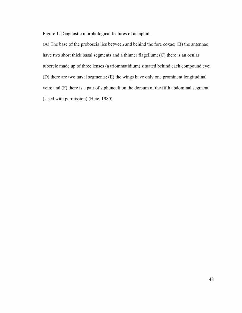
48
Figure 1. Diagnostic morphological features of an aphid.
(A) The base of the proboscis lies between and behind the fore coxae; (B) the antennae
have two short thick basal segments and a thinner flagellum; (C) there is an ocular
tubercle made up of three lenses (a triommatidium) situated behind each compound eye;
(D) there are two tarsal segments; (E) the wings have only one prominent longitudinal
vein; and (F) there is a pair of siphunculi on the dorsum of the fifth abdominal segment.
(Used with permission) (Heie, 1980).
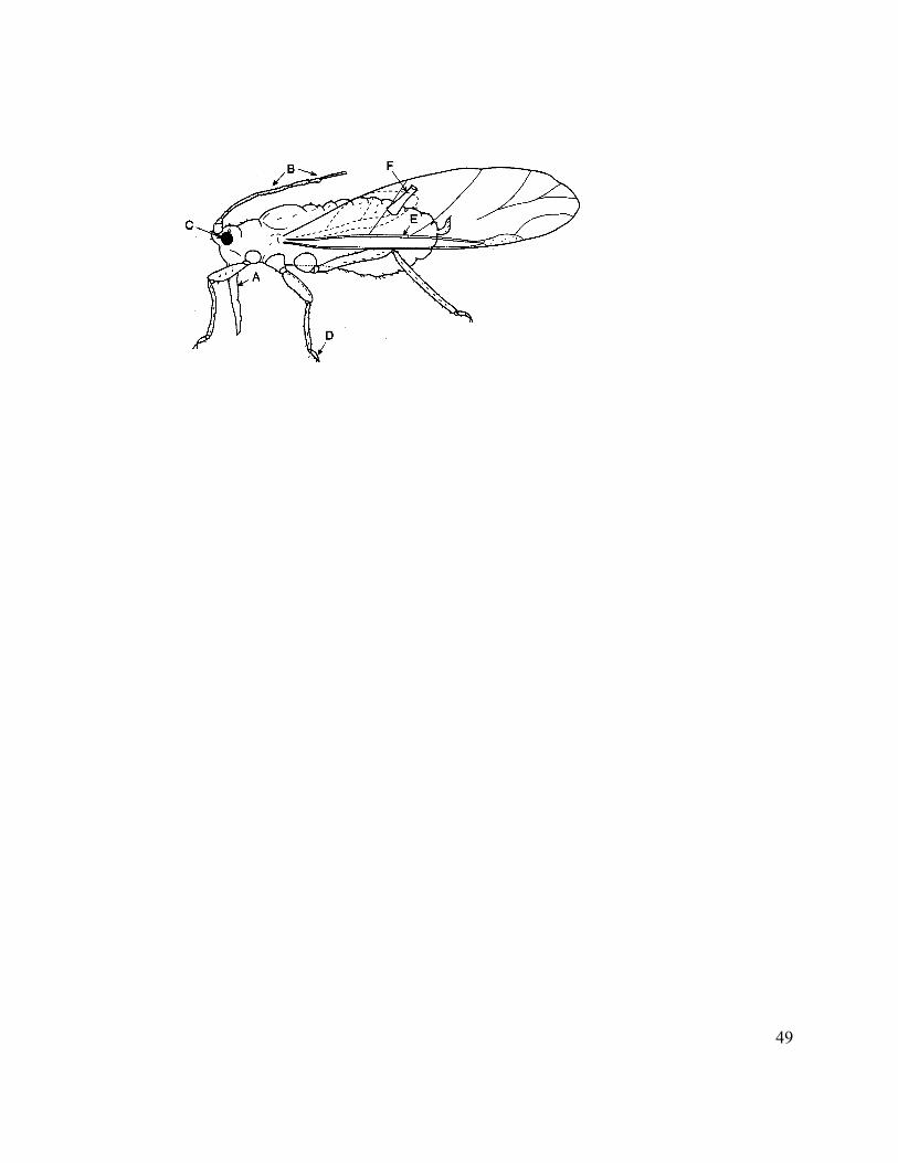
49

50
Figure 2. Anatomy of the salivary gland of the green peach aphid, M. persicae.
(A) Graphical representation of the salivary gland from transverse section of a five day
old M. persicae showing the principal salivary gland (psg), the accessory salivary gland
(asg), the salivary canal (sc) and the common salivary duct (csd). Each lobe of the
principal gland is composed of 8 cell types. Cell types 1 and 2 represents Deckzellen (dz)
and cell types 3-8 represents Hauptzellen (h). (B) Transverse section of the common
salivary duct (csd). (C) Transverse section of the middle region of the principal gland.
(D) Transverse section of the posterior region of the principal gland.
sdc: salivary duct cell; n: nucleus; mc: myoepitheloid cell; isc: intercellular secretory
canaliculum; ic: intracellular canaliculi; N2: branch of medial dorsal nerve (Used with
permission) (Ponsen, 1987).
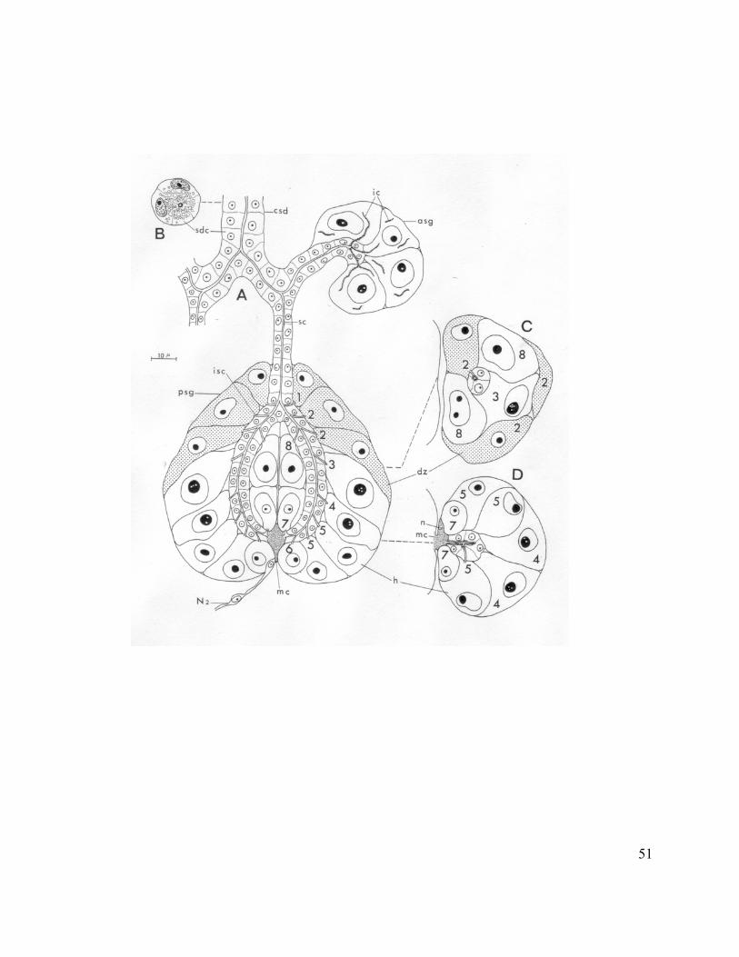
51

52
Figure 3. Feulgen stained principal salivary gland of M. persicae.
Cell types H and I represent Deckzellen and cell types A, B, C, D, E, F, and G represent
Hauptzellen (Used with permission) (Weidemann, 1968).
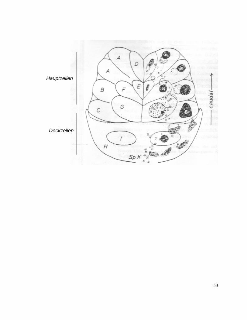
53
Deckzellen
Hauptzellen
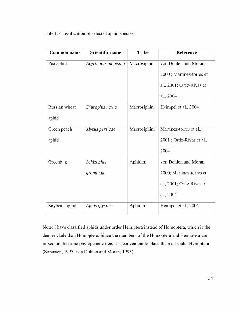
54
Table 1. Classification of selected aphid species.
Common name Scientific name Tribe Reference
Pea aphid Acyrthopisum pisum Macrosiphini von Dohlen and Moran,
2000 ; Martinez-torres et
al., 2001; Ortiz-Rivas et
al., 2004
Russian wheat
aphid
Diuraphis noxia Macrosiphini Heimpel et al., 2004
Green peach
aphid
Myzus persicae Macrosiphini Martinez-torres et al.,
2001 ; Ortiz-Rivas et al.,
2004
Greenbug Schizaphis
graminum
Aphidini von Dohlen and Moran,
2000; Martinez-torres et
al., 2001; Ortiz-Rivas et
al., 2004
Soybean aphid Aphis glycines Aphidini Heimpel et al., 2004
Note: I have classified aphids under order Hemiptera instead of Homoptera, which is the
deeper clade than Homoptera. Since the members of the Homoptera and Hemiptera are
mixed on the same phylogenetic tree, it is convenient to place them all under Hemiptera
(Sorensen, 1995; von Dohlen and Moran, 1995).
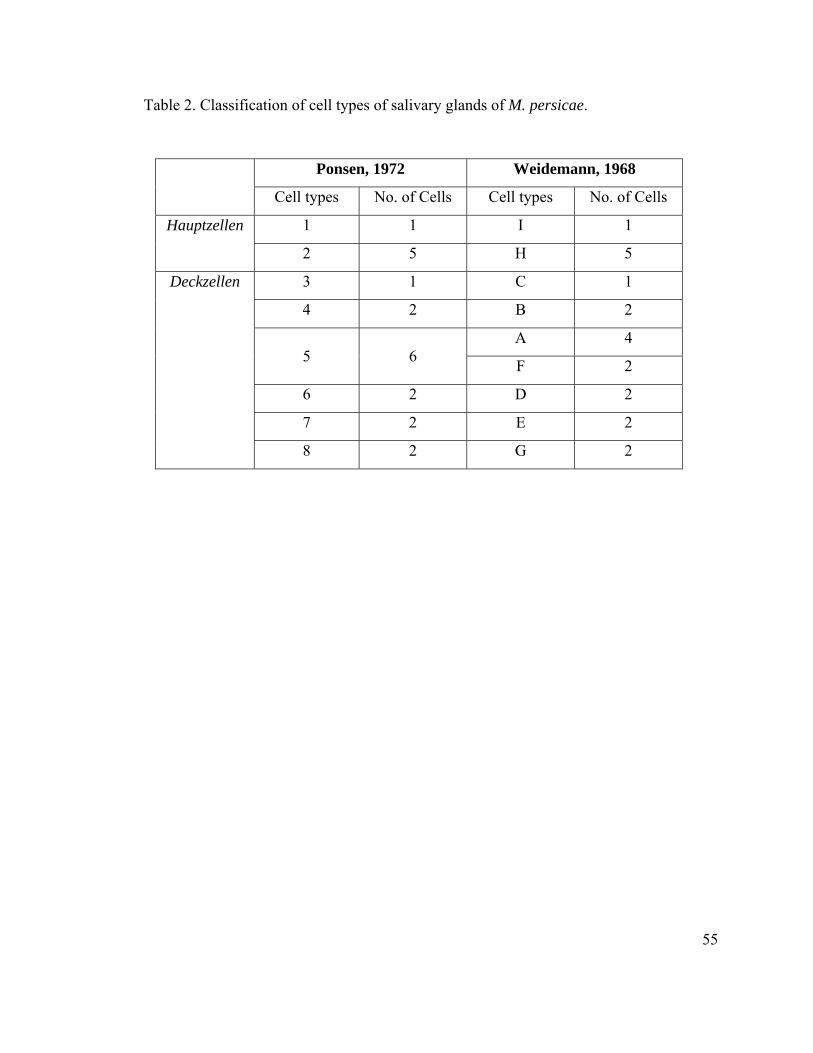
55
Table 2. Classification of cell types of salivary glands of M. persicae.
Ponsen, 1972 Weidemann, 1968
Cell types No. of Cells Cell types No. of Cells
1 1 I 1 Hauptzellen
2 5 H 5
3 1 C 1
4 2 B 2
A 4
5
6 F 2
6 2 D 2
7 2 E 2
Deckzellen
8 2 G 2
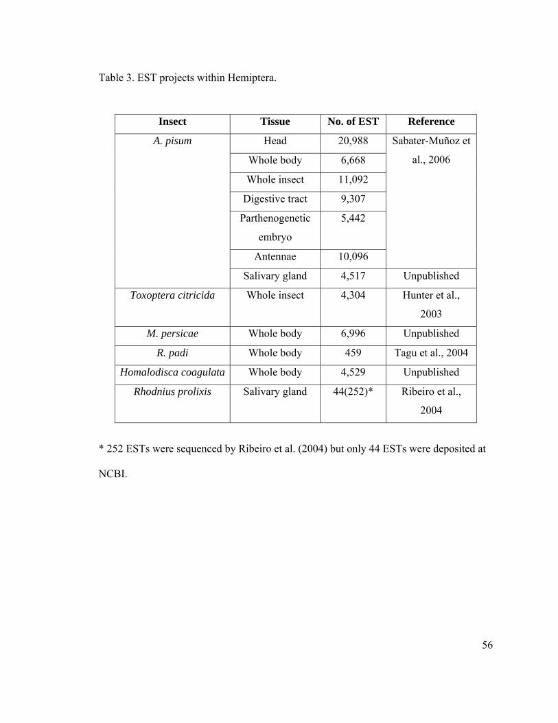
56
Table 3. EST projects within Hemiptera.
Insect Tissue No. of EST Reference
Head 20,988
Whole body 6,668
Whole insect 11,092
Digestive tract 9,307
Parthenogenetic
embryo
5,442
Antennae 10,096
Sabater-Muñoz et
al., 2006
A. pisum
Salivary gland 4,517 Unpublished
Toxoptera citricida Whole insect 4,304 Hunter et al.,
2003
M. persicae Whole body 6,996 Unpublished
R. padi Whole body 459 Tagu et al., 2004
Homalodisca coagulata Whole body 4,529 Unpublished
Rhodnius prolixis Salivary gland 44(252)* Ribeiro et al.,
2004
* 252 ESTs were sequenced by Ribeiro et al. (2004) but only 44 ESTs were deposited at
NCBI.
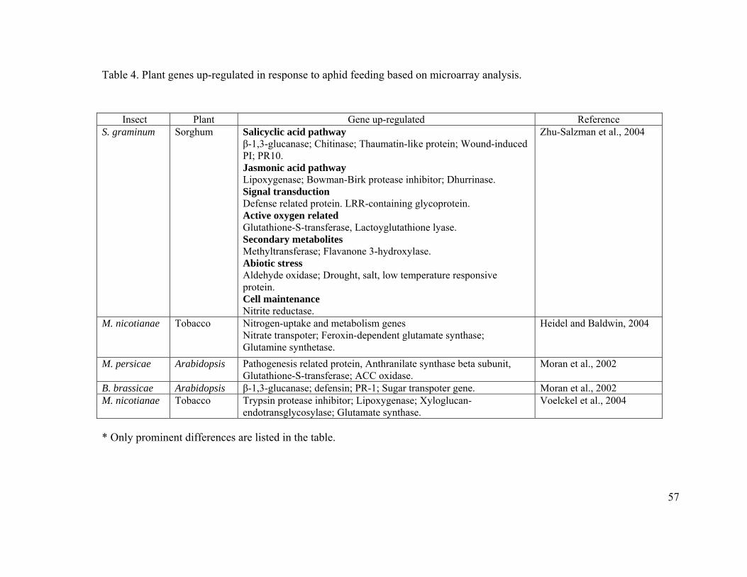
57
Table 4. Plant genes up-regulated in response to aphid feeding based on microarray analysis.
Insect Plant Gene up-regulated Reference S. graminum Sorghum Salicyclic acid pathway
β-1,3-glucanase; Chitinase; Thaumatin-like protein; Wound-induced PI; PR10. Jasmonic acid pathway Lipoxygenase; Bowman-Birk protease inhibitor; Dhurrinase. Signal transduction Defense related protein. LRR-containing glycoprotein. Active oxygen related Glutathione-S-transferase, Lactoyglutathione lyase. Secondary metabolites Methyltransferase; Flavanone 3-hydroxylase. Abiotic stress Aldehyde oxidase; Drought, salt, low temperature responsive protein. Cell maintenance Nitrite reductase.
Zhu-Salzman et al., 2004
M. nicotianae Tobacco Nitrogen-uptake and metabolism genes Nitrate transpoter; Feroxin-dependent glutamate synthase; Glutamine synthetase.
Heidel and Baldwin, 2004
M. persicae Arabidopsis Pathogenesis related protein, Anthranilate synthase beta subunit, Glutathione-S-transferase; ACC oxidase.
Moran et al., 2002
B. brassicae Arabidopsis β-1,3-glucanase; defensin; PR-1; Sugar transpoter gene. Moran et al., 2002 M. nicotianae Tobacco Trypsin protease inhibitor; Lipoxygenase; Xyloglucan-
endotransglycosylase; Glutamate synthase. Voelckel et al., 2004
* Only prominent differences are listed in the table.

58
CHAPTER 2
59
Chapter 2: An EST library from Salivary Glands of the Pea Aphid, Acyrthosiphon
pisum.
Navdeep S. Mutti1,2, Kirk Pappan2,3, Ming-Shun Chen4, John C. Reese1, Gerald R.
Reeck2*
1 Department of Entomology, Kansas State University, Manhattan, Kansas 66506.
2 Department of Biochemistry, Kansas State University, Manhattan, Kansas 66506.
3 Current address: Department of Biochemistry, Washington University School of
Medicine, St. Louis, MO 63110.
4USDA-ARS and Department of Entomology, Kansas State University, Manhattan,
Kansas 66506.
Key words: pea aphid, salivary glands.
Abbreviations: EST, expressed sequence tag; NCBI, National Center for Biotechnology
Information. PCR, polymerase chain reaction; PBS, phosphate buffered saline; ORF,
open reading frame
60
Abstract
Several thousand ESTs in a salivary gland cDNA library of the pea aphid, Acyrthosiphon
pisum have been sequenced. A cluster analysis based on sequence similarity grouped
5,098 sequences into 1,769 clusters. A majority of the clusters (about 78% or 1,392
clusters) did not match to any sequence, only about 22% (or 377 clusters) were assigned
putative functions based on BLASTX against the UniProt database. These 377 clusters
encode proteins with putative secretion signals, housekeeping genes or hypothetical
proteins. Among secreted proteins, we found 14 clusters predicted to code for proteases,
11 clusters predicted to code for oxidoreductases, and 8 clusters predicted to code for
other hydrolases. Among the housekeeping cDNAs, we found clusters coding for heat
shock proteins, cytochrome oxidases, transcription factors, polymerases, calcium or metal
binding proteins, ATPases and enzymes of electron transport and DNA binding proteins.

61
Introduction
Aphid saliva holds the potential to better understand the co-evolution of insect-host
interactions (Miles, 1999). Aphid saliva is believed to perform multiple functions,
including the creation of the stylet sheath, assisting the penetration of substrate for food
(by the action of pectinases, cellulases, β-glucosidases, etc.), digesting nutrients
(polysaccharases and proteases), detoxification of phenolic glycosides ingested during
feeding by the action of polyphenol oxidases or peroxidases (oxidation-reduction
enzymes), and the suppression of host defenses or the elicitation of host responses (Miles,
1972; Miles, 1987; Urbanska et al., 1998; Miles, 1999). The fact that aphid saliva is only
available in very small quantities makes the direct study of salivary components difficult
(Miles, 1965; Madhusudhan et al., 1994; Miles and Harrewijn, 1991).
In an attempt to reveal the complexity of the pea aphid, Acyrthosiphon pisum (Harris)
salivary glands, a high-throughput approach designed to identify a large number of
cDNAs in the salivary glands has been employed in the present work. ESTs have become
an effective means of gene discovery. In the past, similar approaches have been very
successful in salivary glands of blood-sucking bug, Rhodnius prolixus or mosquito,
Anopheles stephens (Valenzuela et al., 2003; Ribeiro et al., 2004), particularly when the
cDNA libraries have been prepared from tissues with high activity for the respective
enzymes.
We have chosen to work with the pea aphid, A. pisum, because of its large size
(compared with other aphid species), thus making dissections of salivary glands less
62
difficult. Generation of a set of pea aphid salivary cDNAs, along with the availability of
the pea aphid genome later this year (http://www.hgsc.bcm.tmc.edu/projects/aphid/), will
provide an indispensable tool for the systematic analysis of proteins/enzymes that may
play roles in the aphid-plant interactions. Here we describe the annotation of 1,726
clusters representing 5,098 mRNA sequences. Remarkably, only about 22% of our cDNA
sequences match to sequences of known functions.
Material and Methods
Plants and Aphids
Aphids were originally collected from alfalfa plants in the summer of 1999 by Dr. Marina
Caillaud at Cornell University. Thereafter, the aphids were reared at KSU on fava beans
(Vicia fabae) grown in pots (10 cm diameter) at room temperature under high intensity
sodium lights with a L:D of 16:8. Salivary glands from adult aphids were dissected and
separated from the brain tissue overlaying the glands. Dissected glands were transferred
to 50µl of PBS (137 mM NaCl, 2,7 mM KCl, 10 mM Na2HPO4, 1.8 mM KH2PO4; pH
7.4) for 50 paired glands and washed 3 times in PBS to remove hemocytes. Salivary
glands were kept at –75oC until needed.
Phagmid cDNA Library Construction
Total RNA was isolated from 250 salivary glands using the Micro RNA isolation kit from
Stratagene, La Jolla, CA. The PCR-based cDNA library was made following instructions
with the SMART cDNA library construction kit (Clontech, Palo Alto, CA). Total RNA

63
was reverse transcribed using PowerScript reverse transcriptase and CDS III primer
provided in the kit (Clontech, Palo Alto, CA). Second strand synthesis was performed
through the PCR-based protocol using SMART III and CDS III primers from Clontech.
Double strand synthesis was followed by proteinase K digestion. Double strand cDNA
was ligated into a Lambda TriplEx2 vector (Clontech, Palo Alto, CA), and the resulting
ligation reaction was packaged using Gigapack Gold III from Stratagene following the
manufacturer’s specifications.
Sequencing, Sequence Processing, and Annotation
Sequencing of the cDNA clones was done either at the Kansas State University
sequencing facility or at Genoscope, Evry cedex, France (http://www.cns.fr/). Raw EST
data were analyzed using the Lucy program (Chou and Holmes, 2001) for sequence
quality and vector sequence removal. Then ESTs were clustered into groups of nearly
identical sequences using the CAP3 software tool using default settings (Huang and
Madan, 1999). The non-redundant set of clusters (contigs and singletons) was searched
against the UniProt reference database (Bairoch et al., 2005) using the BLASTX program
downloaded from NCBI. A query sequence was annotated using the best hit in UniProt
with E value threshold set to 1e-5. The functional annotation included text description as
well as gene ontology terms of the matched reference sequence (Camon et al., 2003).
cDNAs were translated and analyzed for signal peptide and cleavage information using
SignalP 3.0 Server (Nelson et al., 1997) using default parameters
(http://www.cbs.dtu.dk/services/SignalP/).
64
Results
Salivary Transcript Catalog for A. pisum
We have sequenced 5,098 ESTs from the salivary gland cDNA library. A cluster analysis
based on sequence similarity grouped 5,098 EST sequences into 1,769 clusters. Cluster
refers to both contigs (containing 2 or more ESTs) and singletons. Putative functions
corresponding to these salivary gland ESTs collection was assigned by comparing these
ESTs with UniProt database using BLASTX with E value threshold set to 1e-5. Among
the 1,769 clusters, 1392 (78%) showed no sequence similarity with any other protein
sequences and only 377 clusters (22%) were assigned putative functions.
Classification of Salivary Transcripts of A. pisum
Classification of 377 clusters with significant matches to sequences in UniProt database
is provided in Table 5. 72 clusters correspond either to hypothetical proteins or to
proteins that were not annotated. There are several clusters corresponding to ribosomal
proteins (42), ATPase/ATP binding proteins (27), polymerases (20), cytochromes (16),
DNA binding proteins (15), proteases (14), metal binding proteins (14), mitochondrial
proteins (14), oxidoreductases (11), transcription factors (10), protein kinases (9), heat
shock proteins (9) and other hydrolases (8). We also found several clusters encoding
putative secreted proteins including several oxidoreductases, several proteases and
several carbohydrases. Among other secreted proteins identified included glutathione-S-
transferase, apolipophorin precursor protein, carbonic anhydrase, 11 kDa salivary protein,
65
odorant binding protein, armet like protein. There is a group of 13 clusters which were
previously assigned under structural component of cell wall, cell membrane and nucleus,
now listed listed as a part of Table 6 under group X for miscellaneous proteins.
We also found two clusters corresponding to viral sequences, and one cluster
corresponding to a plant sequence. The two viral clusters are non-overlapping fragments
of a 89.2 kDa capsid protein from an aphid-infecting virus, aphid lethal paralysis virus
(van Munster et al., 2002). This virus belongs to the recently recognized family
Dicistroviridae (Mayo, 2002). The plant cluster corresponds to a phospholipase C from
Arabidopsis thaliana and is probably the result of contamination from the host plant. It
was not found in the latest sequencing of 4,517 ESTs.
Further analysis using TBLASTX against NR database at NCBI of 72 clusters
corresponding to hypothetical or unannotated proteins is shown in Table 6. Signal P
analysis performed on homologs from other species identified 8 clusters encoding
secreted proteins. We also found three clusters encoding proteins from a pea aphid
secondary endosymbiont (Candidatus hamiltonella). In total putative functions were
assigned to 57 clusters (not just looking at the top match but also looking other matches
with significant e value). Only 15 clusters (out of 72) were not assigned any putative
function and encoded either a hypothetical protein or protein of unknown function.
The clusters with putative functions were classified based on the number of ESTs in a
contig (Table 7). Contig 90 contains 109 ESTs and is an aphid-infecting virus, aphid
66
lethal paralysis virus. Contig 33 represents a cluster with 57 EST sequences and is
cytochrome oxidase subunit I. Other clusters in Table 7 include ribosomal protein
(ribosomal protein S15A, contig 94 with18 EST sequences) and hypothetical protein
(contig 97 with 40 EST sequences). We also found clusters representing glucose
dehydrogenase (14 ESTs), angiotensin-converting enzyme (15 ESTs), glutathione
peroxidase (10 ESTs), apolipophorin (8 ESTs) and superoxide dismutase (6 ESTs).
Many clusters (1,392) showed no sequence similarity with any other sequences. Most of
the clusters either have short ORF’s or entirely lack ORF (Table 8). There are 21 clusters
which, despite having an ORF of 200 or more amino acids residues, showed no similarity
to any other sequence. The presence of ORF drops with number of ESTs represented in a
cluster. Most of the clusters with three ESTs have no ORF or very short ORF. Clusters
with two or one EST sequence are not listed in Table 8. This high number of unmatched
clusters might reflect the limited sequence quality delivered by single-pass sequencing
(for example, too short sequences, wrong base identified leading to frame shift errors).
Among these clusters of unknown function, an abundant cluster (contig 32) representing
46 EST sequences referred to as C002 was further characterized (Chapter 3) and also
used to develop RNAi as a tool to study gene expression in pea aphid (Chapter 4).
Proteases and Other Hydrolases in the Salivary Glands of A. pisum
Annotation of clusters revealed the presence of 14 clusters representing putative
proteases and 5 clusters representing other putative hydrolases (Table 9). Among
proteases we have identified subtilisin related protease, a member angiotensin-converting
67
enzyme family, peptidase M1, cathepsin L, cathepsin B, signal peptidase, endoprotease
FURIN and ubiquitin specific protease. SignalP analysis done on homologs from other
species (as our pea aphid salivary sequences are not full length) identified in BLAST
analysis at NCBI shows that some encode proteins with a putative signal peptide.
Therefore based on SignalP analysis, 8 clusters out of 14 clsuter encoding putative
proteases represent putative secreted proteases. Among other hydrolases we have
identified putative S-adenosyl homocysteine hydrolase, prolyl 4-hydroxylase alpha
subunit, alpha-glucosidase, trehalase, carbon-nitrogen hydrolase and phosphoesterase.
Based on SignalP analysis described above, 3 clusters represent putative secreted other
hydrolases.
Oxidoreducatases in the Salivary Glands of A. pisum
Annotation of clusters also revealed the presence of 11 clusters representing putative
oxidoreductases (Table 10). These clusters represent putative glucose dehydrogenase,
glutathione peroxidase, aldehyde dehyrogenase, thioredoxin peroxidase,
phosphoglycerate dehydrogenase, peroxiredoxin-like protein, dimethylaniline
monooxygenase, superoxide dismutase and peroxidase. SignalP analysis done on the
homologs (as our pea aphid salivary sequences are not full length) identified in BLAST
analysis at NCBI, shows that some encode a putative secreted protein and has a putative
signal peptide. Therefore, based on SignalP analysis, 5 clusters represent putative
secreted oxidoreductases.
68
Discussion
To our knowledge, this is the first attempt to create a catalog of the cDNAs from the
salivary glands of the pea aphid, A. pisum or any other aphid species. The majority of the
clusters (78%) could not have their putative function annotated. The most likely reason
for this lack of similarity is that some of the sequences are too short or may represent 5’-
or 3’-untranslated regions. Second it is possible that these partial sequences correspond to
a non-conserved domain of polypeptide: a longer sequence should allow a better
identification of these clusters. Finally, this might reflect limitations of single pass
sequencing (too short sequences, wrong base calling leading to frame shift errors). For
the clusters represented under hypothetical proteins or unannotated proteins (75 clusters),
it is possible that some of these proteins without matches correspond to aphid specific
proteins of cellular functions not yet elucidated. Genes involved in environmental
adaptation evolve quickly and might correspond to unannotated sequences (Domazet-
Loso and Tautz, 2003). As aphids are highly sensitive to environmental changes, it is
possible that these clusters may correspond to rapidly evolving sequences.
The largest proportion of functionally annotated sequences falls into following
categories: ribosomal proteins represented in 42 clusters, there are several clusters
corresponding to ATPase/ATP binding protein (27), polymerases (20), cytochromes (17),
DNA binding proteins (15), proteases (14), metal binding proteins (14), mitochondrial
proteins (14), oxidoreductases (11), transcription factors (10), protein kinases (9) heat
shock proteins (9) and other hydrolases (8). We also found several clusters encoding
putative secreted proteins including several oxidoreductases, several proteases and
69
several carbohydrases. Among other secreted proteins identified included glutathione-S-
transferase, apolipophorin precursor protein, carbonic anhydrase, 11 kDa salivary protein,
odorant binding protein, armet like protein.
A large scale sequencing of 40,904 ESTs from the pea aphid was carried out (Sabater-
Muñoz et al., 2006) leading to 12,082 unique transcripts. About 59% (7,146 sequences)
showed no match to any protein of known function. Among the 4,936 annotated
sequences, 4,080 and 3,977 has a significant match in D. melanogaster and Anopheles
gambiae respectively (Sabater-Munoz et al., 2006). A similar approach using ESTs to
study of the regulation of reproductive modes in aphids was carried out in the cereal
aphid, Rhopalosiphum padi. The majority of the ESTs sequenced were without matches
or encoded hypothetical proteins (56%) followed by housekeeping polypeptides (38%)
(Tagu et al., 2004).
The “redox hypothesis” proposed by Miles and Oertli (1993) states that the oxidative
processes in healthy plants are subject to control by reducing systems of the plant such as
antioxidants like glutathione and ascorbic acid, and that the aphid salivary enzymes serve
to change the natural redox equilibrium in the plant to the aphid’s advantage. Plants
respond to damage by sucking insects by mobilizing and oxidizing phenolic compounds
especially monomeric o-quinones or phenolic compounds, which are deterrent to insects
(Miles and Oertli 1993; Harmatha and Nawrot 2002). Several putative oxidoreductases
were identified in our study (Table 9). Five of them encode enzymes with putative

70
secretion signals; these salivary oxidases can act by enhancing oxidation, thereby
decreasing the concentration of monomeric phenols and quinones.
In Helicoverpa zea, glucose oxidase present in saliva suppresses jasmonic acid related
plant defense, presumably by the production of hydrogen peroxide (Musser et al., 2002).
Glucose oxidase and glucose dehyrogenase are also present in our salivary gland library.
They may play a similar role in suppressing jasmonic acid mediated plant defense, as
jasmonic acid regulated pathway genes are up-regulated in sorghum upon greenbug
feeding (Zhu-Salzman et al., 2004).
In addition, glycosyl hydrolases present in the salivary gland hydrolyse glycosidic bonds
in carbohydrates. Alpha-amylase hydrolyses α-1,4 linkages in starch to maltose, which is
then hydrolysed to glucose by an α-glucosidase. Amylase activity has been detected in
the salivary glands of other phytophagous heteropterans (Zeng and Cohen, 2000; Boyd,
2003). Thus, it is possible that aphids are capable of partially digesting starches before
ingestion. In addition to serving as energy and carbon sources, sugars function as
messengers in signal transduction (Rolland et al., 2002). Sucrose and trehalose are two
sugars that are involved in signal transduction in plants. Trehalose is also the
predominant hemolymph sugar in insects (Becker et al., 1996). Trehalose can be
hydrolysed into two glucose molecules by trehalase. Putative secreted trehalase is present
in the salivary glands of the pea aphid and may play a crucial role in breakdown of plant
trehalose, thereby disrupting signal transduction in plant and thus can aphid feed
continuously.
71
Recently in the social aphid, Tuberaphis styraci, cathepsin B protein was preferentially
expressed in soldiers and was localized in the midgut of soldiers. It is injected (probably
from gut) into the body of prey during attack (Kutsukake et al., 2004), although it is
possible that enzyme is also synthesized in salivary glands. We have also identified a
putative secreted protein encoding cathepsin B. In bird cherry-oat aphid, Rhopalosiphum
padi, there were changes in the activity of glutathione-S-transferase and glutathione
reductase, enzymes involved in detoxification of plant allelochemicals (Laskowska et al.,
1999). In most cases, the activities of these enzymes depend upon the composition of
aphid’s diet, when fed on cereals, glutathione-S-transferase activity further increased and
glutathione reductase activity decreased (Laskowska et al., 1999). We have identified
putative secreted glutathione peroxidase and glutathione-S-transferase in the salivary
glands of pea aphid. It is possible that these enzymes are involved in the detoxification of
plant allelochemicals and thus may play a vital role in aphid feeding.
In addition, one of our cluster is apparently a homolog of an 11 kDa salivary protein
identified in our studies is also present in the salivary glands of sand fly, Lutzomyia
longipalpis (Oliveira et al., 2006; Valenzuela et al., 2004). In the sand fly, 11 kDa protein
encodes a novel protein. Its function has not been elucidated (Oliveira et al., 2006). We
have also identified a putative carbonic anhydrase; carbonic anhydarse has been
previously reported in the salivary glands of the cockroach, Periplaneta americana (Just
and Walz, 1994). It functions by maintaining pH homeostasis in various tissues by
catalyzing the hydration of CO2 and dehydration of bicarbonate (Kivela et al., 1999). It is
72
possible that the putative secreted carbonic anhydarse in aphid saliva may regulate pH of
the sieve element.
We have also identified 12 full-length putative secreted cDNAs encoding proteins with
unknown functions. An abundant cluster, C002 was further characterized and RNAi
studies show that C002 is important for survival and reproduction of the pea aphid (see
Chapter 3 and 4). It is possible that these putative secreted proteins of unknown function
may play similar important function in pea aphid performance on the host plant.
Proteins including enzymes, identified in our study with putative secretion signals may
constitute aphid saliva. Together these enzymes may play roles in aphid-plant interaction.
This information derived from the large sequencing of ESTs from the salivary glands of
pea aphid, A. pisum along with the avaliability of the pea aphid genome later this year
will provide an indispensable tool to study the molecular basis of aphid-plant
interactions.
Acknowledgements
We thank our colleague Dr. L.J. Wang for his help with annotation of ESTs and we
would like acknowledge K-INBRE Bioinformatics Core, NIH grant number
P20RR016475. This work was supported by USDA-CREES NRI grant 2001-35302-
09975 and by the Kansas Agricultural Experiment Station (publication number 05-XXX-
J). Pea aphids (voucher specimen number - 173) were deposited at the Kansas State
University Museum of Entomological and Prairie Arthropod Research (KSU-MEPAR).
73
References
Bairoch, A., Apweiler, R., Wu, C.H., Barker, W.C., Boeckmann, B., Ferro, S., Gasteiger,
E., Huang, H., Lopez, R., Magrane, M., Martin, M.J., Natale, D.A., O’Donovan,
C., Redaschi, N. and Yeh, L.S. 2005. The universal protein resource (UniProt).
Nucleic Acids Research, 33:154-159.
Becker, A., Schloder, P., Steele, J.E. and Wegener, G. 1996. The regulation of trehalose
metabolism in insects. Experientia, 52:433-439.
Boyd, D.W. 2003. Digestive enzymes and stylet morphology of Deraeocoris nigritulus
(Uhler) (Hemiptera: Miridae) reflect adaptation for predatory habits. Annals of the
Entomological Society of America, 96:667-671.
Camon, E., Magrane, M., Barrell, D., Binns, D., Fleischmann, W., Kersey, P., Mulder,
N., Oinn, T., Maslen, J., Cox, A. and Apweiler, R. 2003. The gene ontology
annotation (GOA) project: implementation of GO in SWISS-PROT, TrEMBL,
and InterPro. Genome Research, 13:662-672.
Chou, H.H. and Holmes, M.H. 2001. DNA sequence quality trimming and vector
removal. Bioinformatics, 17:1093-1104.
Domazet-Loso, T. and Tautz, D. 2003. An evolutionary analysis of orphan genes in
Drosophila. Genome Research, 13:2213-2219.
Harmatha, J. and Nawrot, J. 2002. Insect feeding deterrent activity of lignans and related
phenylpropanoids with a methylenedioxyphenyl (piperonyl) structure moiety
Entomologia Experimentalis et Applicata, 104:51-60
Huang, X. and Madan, A. 1999. CAP3: a DNA sequence assembly program. Genome
Research, 9:868-877.
74
Just, F. and Walz, B 1994. Localization of carbonic anhydrase in the salivary glands of
the cockroach, Periplaneta americana. Histochemistry, 102:271-277.
Kivela, J., Parkkila, S., Parkkila, A.K., Leinonen, J. and Rajaniemi, H. 1999. Salivary
carbonic anhydrase isoenzyme VI. The Journal of Physiology, 520:315-320.
Kutsukake, M., Shibao, H., Nikoh, N., Morioka, M., Tamura, T., Hoshino, T., Ohgiya, S.
and Fukatsu, T. 2004. Venomous protease of aphid soldier for colony defense.
Proceedings of the National Academy of Sciences USA, 101:11338-11343.
Laskowska, I., Leszcynski, B. and Markowski, J. 1999. Activity of glutathion transferase
and reductase in tissues of bird cherry-oat aphid during its host-plant alteration.
Experimental Toxicology and Pathology, 51:357-359.
Madhusudhan, M.A., Taylor, G.S. and Miles, P.W. 1994. The detection of salivary
enzymes of phytophagous Hemiptera: a composition of methods. Annals of
Applied Biology, 124:405-412.
Mayo, M. A. 2002. Virology Division news: virus taxonomy – Houston 2002. Archives of
Virology, 147:1071-1076.
Miles, P. W. 1965. Studies on the salivary physiology of plantbugs: the saliva of aphids.
Journal of Insect Physiology, 11:1261-1268.
Miles, P.W. 1972. The saliva of hemiptera. Advances in Insect Physiology, 9:183-255.
Miles, P. W. 1987. Feeding process of Aphidoidea in relation to effects on their food
plants. In A. K. Minks and P. Harrewijn (eds) Aphids: Their Biology, Natural
Enemies and Control, Elsevier, Amsterdam, vol. 2A pp. 321-339.
Miles PW. 1999. Aphid saliva. Biological Reviews of the Cambridge Philosophical
Society, 74:41-85.
75
Miles, P.W. and Harrewijn, P. 1991. Discharge by aphids of soluble secretions into
dietary sources. Entomologia Experimentalis et Applicata, 59:123-134.
Miles, P. W. and Oertli, J.J. 1993. The significance of antioxidants in the aphid-plant
interactions: the redox hypothesis. Entomologia Experimentalis et Applicata,
67:285-273.
Musser, R.A., Hum-Musser, S., Eichenseer, H., Peiffer, M., Ervin, G., Murphy, B. and
Felton, G.W. 2002. Caterpillar saliva beats plant defences: a new weapon emerges
in the evolutionary arms race between plants and herbivores. Nature, 416:599-
600.
Nelson, H., Engelbrech, J., Brunak, S. and Heijne, G.V. 1997. Identification of
prokaryotic and eukaryotic signal peptides and prediction of their cleavage sites.
Protein Engineering, 10:1-6.
Oliveira, F., Kamhawi, S., Seitz, A.E., Pham, V.M., Guigal, P.M., Fischer, L., Ward, J.
and Valenzuela, J.G. 2006. From transcriptome to immunome: Identification of
DTH inducing proteins from a Phlebotomus ariasi salivary gland cDNA library.
Vaccine, 24:374-390.
Ribeiro, J.M.C., Andersen, J., Silva-Neto, M.A.C., Pham, V.M., Garfield, M.K. and
Valenzuela, J.G. 2004. Exploring the sialome of the blood-sucking bug Rhodnius
prolixus. Insect Biochemistry and Molecular Bology, 34:61-79.
Rolland, F., Moore, B. and Sheen, J. 2002. Sugar sensing and signaling in plants. Plant
Cell Supplement, 185-205.
Sabater-Munoz, B., Legeai, F., Rispe, C., Bonhomme, J., Dearden, P, Dossat, C., Duclert,
A., Gauthier, J.P., Ducray, D.G., Hunter, W., Dang, P., Kambhampati, S.,
76
Martinez-Torres, D, Cortes, T., Moya, A., Nakabachi, A., Philippe, C., Prunier-
Leterme, N., Rahbe, Y., Simon, J.C., Stern, D.L., Wincker, P. and Tagu, D. 2006.
Large-scale gene discovery in the pea aphid Acyrthosiphon pisum (Hemiptera).
Genome Biology, 7:R21.
Tagu, D., Prunier-Leterme, N., Legeai, F., Gauthier, J.P., Duclert, A., Sabater-Muñoz, B.,
Bonhomme, J. and Simon, J.C. 2004. Annotated expressed sequence tags for
studies of the regulation of reproductive modes in aphids. Insect Biochemistry and
Molecular Bology, 34:809-22.
Urbanska, A., Tjallingii, W. F., Dixon, A.F.G. and Leszczynski, B. 1998. Phenol
oxidising enzymes in the grain aphid's saliva. Entomologia Experimentalis et
Applicata, 86:197-203.
Valenzuela, J.G., Francischetti, I.M., Pham, V.M., Garfield, M.K. and Ribeiro, J.M.
2003. Exploring the salivary gland transcriptome and proteome of the Anopheles
stephens mosquito. Insect Biochemistry and Molecular Bology, 33:717-732.
Valenzuela, J.G., Garfield, M.K., Rowton, E.D. and Pham, V.M. 2004. Identification of
the most abundant secreted proteins from the salivary glands of the sand fly
Lutzomyia longipalpis, vector of Leishmania chagasi. Journal of Experimental
Biology, 207:3717-3729.
van Munster, M., Dullemans, A. M., Verbeek, M., van den Heuvel, J. F. J. M., Clerivet
A.and van der Wilk F. 2002. Sequence analysis and genomic organization of
Aphid lethal paralysis virus: a new member of the family Dicistroviridae. Journal
of General Virology, 83:3131-3138.
77
Zeng, F. and Cohen, A.C. 2000. Comparison of α-amylase and protease activities of a
zoophytophagus and two phytophagous Heteroptera. Comparative Biochemistry
and Physiology Part A, 126:101-106.
Zhu-Salzman, K., Salzman, R.A., Ahn, J.E. and Koiwa, H. 2004. Transcriptional
regulation of sorghum defense determinants against a phloem-feeding aphid.
Plant Physiology, 134:420-431.
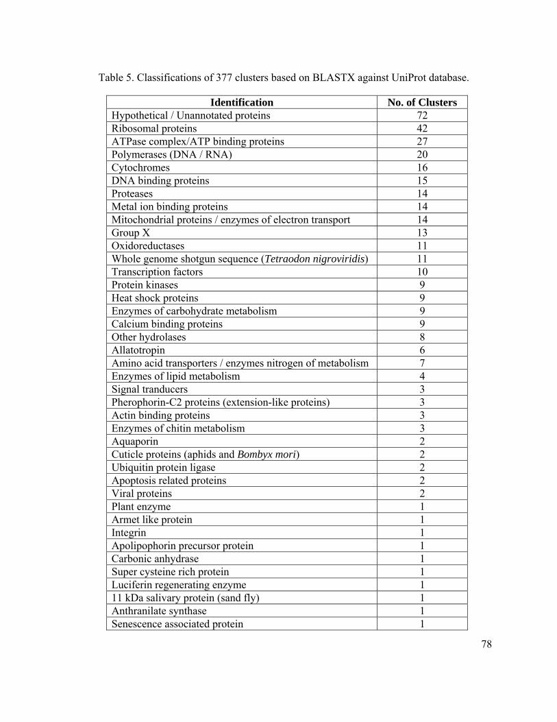
78
Table 5. Classifications of 377 clusters based on BLASTX against UniProt database.
Identification No. of Clusters Hypothetical / Unannotated proteins 72 Ribosomal proteins 42 ATPase complex/ATP binding proteins 27 Polymerases (DNA / RNA) 20 Cytochromes 16 DNA binding proteins 15 Proteases 14 Metal ion binding proteins 14 Mitochondrial proteins / enzymes of electron transport 14 Group X 13 Oxidoreductases 11 Whole genome shotgun sequence (Tetraodon nigroviridis) 11 Transcription factors 10 Protein kinases 9 Heat shock proteins 9 Enzymes of carbohydrate metabolism 9 Calcium binding proteins 9 Other hydrolases 8 Allatotropin 6 Amino acid transporters / enzymes nitrogen of metabolism 7 Enzymes of lipid metabolism 4 Signal tranducers 3 Pherophorin-C2 proteins (extension-like proteins) 3 Actin binding proteins 3 Enzymes of chitin metabolism 3 Aquaporin 2 Cuticle proteins (aphids and Bombyx mori) 2 Ubiquitin protein ligase 2 Apoptosis related proteins 2 Viral proteins 2 Plant enzyme 1 Armet like protein 1 Integrin 1 Apolipophorin precursor protein 1 Carbonic anhydrase 1 Super cysteine rich protein 1 Luciferin regenerating enzyme 1 11 kDa salivary protein (sand fly) 1 Anthranilate synthase 1 Senescence associated protein 1

79
Ornithine decarboxylase inhibitor 1 Odorant binding protein 1 Glutathione-S-transferase 1 S-adenosyl-L-methionine decarboxylases 1
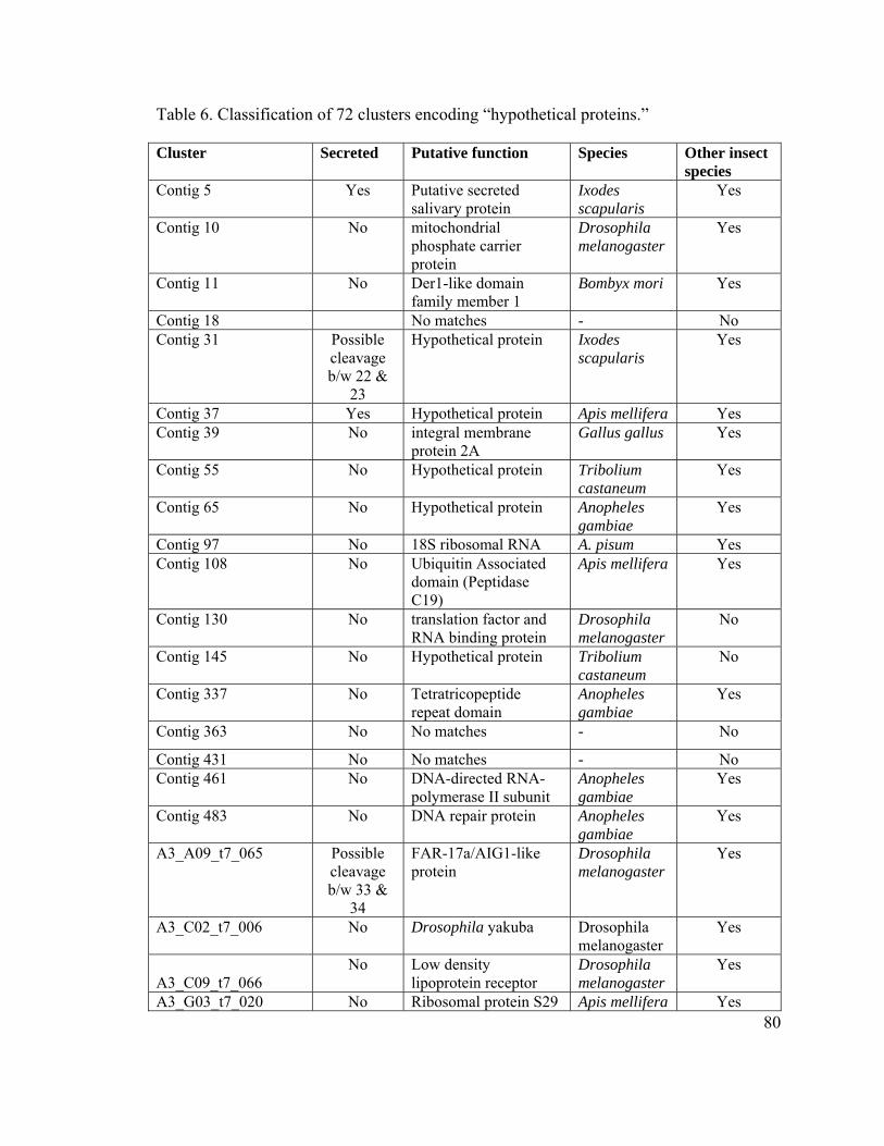
80
Table 6. Classification of 72 clusters encoding “hypothetical proteins.”
Cluster Secreted Putative function Species Other insect species
Contig 5 Yes Putative secreted salivary protein
Ixodes scapularis
Yes
Contig 10 No mitochondrial phosphate carrier protein
Drosophila melanogaster
Yes
Contig 11 No Der1-like domain family member 1
Bombyx mori
Yes
Contig 18 No matches - No Contig 31 Possible
cleavage b/w 22 &
23
Hypothetical protein Ixodes scapularis
Yes
Contig 37 Yes Hypothetical protein Apis mellifera Yes Contig 39 No integral membrane
protein 2A Gallus gallus Yes
Contig 55 No Hypothetical protein Tribolium castaneum
Yes
Contig 65 No Hypothetical protein Anopheles gambiae
Yes
Contig 97 No 18S ribosomal RNA A. pisum Yes Contig 108 No Ubiquitin Associated
domain (Peptidase C19)
Apis mellifera Yes
Contig 130 No translation factor and RNA binding protein
Drosophila melanogaster
No
Contig 145 No Hypothetical protein Tribolium castaneum
No
Contig 337 No Tetratricopeptide repeat domain
Anopheles gambiae
Yes
Contig 363 No No matches - No
Contig 431 No No matches - No Contig 461 No DNA-directed RNA-
polymerase II subunit Anopheles gambiae
Yes
Contig 483 No DNA repair protein Anopheles gambiae
Yes
A3_A09_t7_065 Possible cleavage b/w 33 &
34
FAR-17a/AIG1-like protein
Drosophila melanogaster
Yes
A3_C02_t7_006 No Drosophila yakuba Drosophila melanogaster
Yes
A3_C09_t7_066
No Low density lipoprotein receptor
Drosophila melanogaster
Yes
A3_G03_t7_020 No Ribosomal protein S29 Apis mellifera Yes

81
AphidB1_A12_t7_085 No Adaptin N terminal region
Anopheles gambiae
Yes
AphidB1_C07_t7_050 Yes Unknown function Tribolium castaneum
Yes
AphidB1_D08_t7_062 No Cytochrome c oxidase Bos Taurus No AphidB1_F01_t7_011 Yes Aldehyde
dehydrogenase Homo sapiens Yes
AphidB1_H02_t7_016 No Protein kinase C Anopheles gambiae
Yes
PlateI_D10 No Cytochrome b Anopheles gambiae
Yes
PlateI_F09 No E3 ubiquitin ligase Apis mellifera Yes PlateI_G01 No ADP-ribosylation
factor Homo sapiens Only in
Anopheles PlateKEP_C05 No hydroxypyruvate
isomerase Homo sapiens Yes
PlateKEP_G09 No TatD DNase domain Apis mellifera Yes PlateKEP_A07 No Bladder cancer-related
protein BC10 Anopheles gambiae
Yes
PlateKEP_C06 Yes Odorant binding protein
Drosophila melanogaster
Yes
PlateKEP_A05 Possible cleavage b/w 17 &
18
Eukaryotic protein of unknown function
Apis mellifera Yes
PlateKEP_B04 No Protein of unknown function
Drosophila melanogaster
Yes
PlateKEP_E09 No Ezrin/radixin/moesin family
Anopheles gambiae
Yes
ID0AAH1DD03ZM1 No Sybindin-like family Tribolium castaneum
Yes
ID0AAH10CA01ZM1 No ribosome-associated membrane protein
Bombyx mori
Yes
ID0AAH11DD09ZM1 No aphid secondary symbionts (Candidatus hamiltonella)
A. pisum No
ID0AAH11DG05ZM1 No DnaA from secondary symbiont
Glossina morsitans
No
ID0AAH12AE10ZM1 Leucine-rich repeat (LRR) protein
Anopheles gambiae
Yes
ID0AAH13CD06ZM2 No No matches - No ID0AAH14AA08ZM1 No Unknown function Mus musculus No ID0AAH14BA05ZM1 No No matches - No ID0AAH14BC12ZM1 No peroxisomal biogenesis
factor Homo sapiens Yes
ID0AAH14DE12ZM1 No Enhancer-of-zeste, (Trithorax) domain
Apis mellifera Yes
ID0AAH14DH11ZM1 No aphid secondary A. pisum No
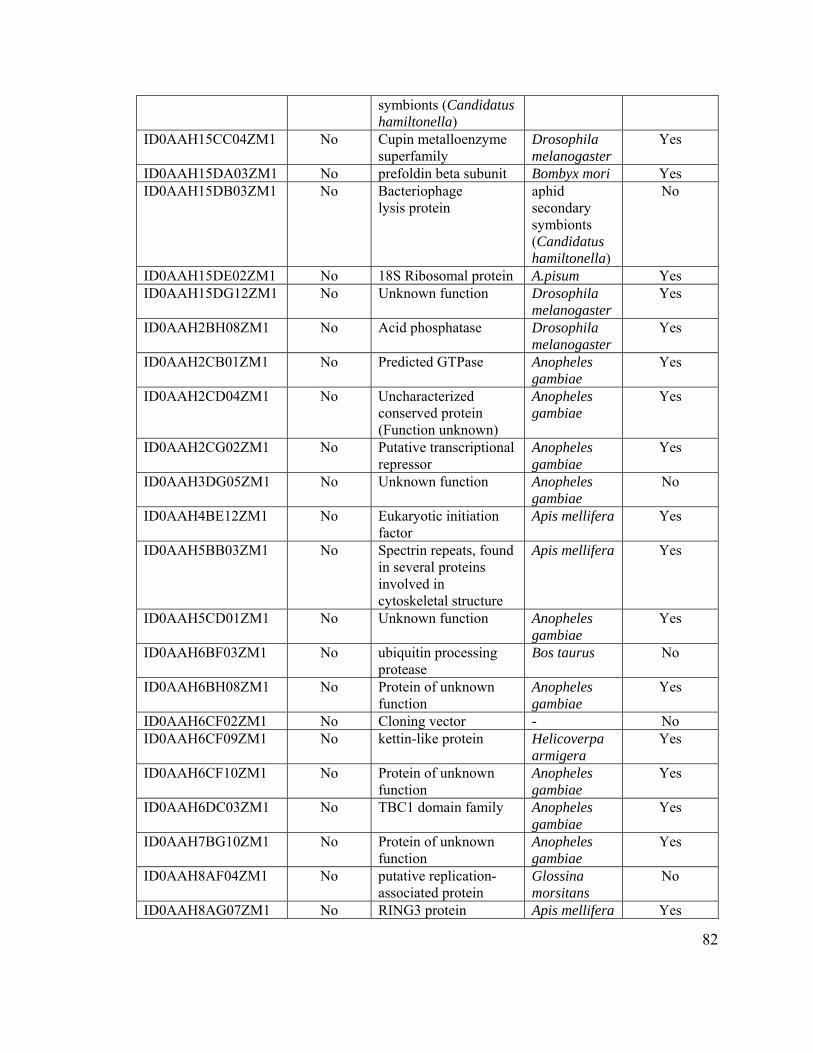
82
symbionts (Candidatus hamiltonella)
ID0AAH15CC04ZM1 No Cupin metalloenzyme superfamily
Drosophila melanogaster
Yes
ID0AAH15DA03ZM1 No prefoldin beta subunit Bombyx mori Yes ID0AAH15DB03ZM1 No Bacteriophage
lysis protein aphid secondary symbionts (Candidatus hamiltonella)
No
ID0AAH15DE02ZM1 No 18S Ribosomal protein A.pisum Yes ID0AAH15DG12ZM1 No Unknown function Drosophila
melanogaster Yes
ID0AAH2BH08ZM1 No Acid phosphatase Drosophila melanogaster
Yes
ID0AAH2CB01ZM1 No Predicted GTPase Anopheles gambiae
Yes
ID0AAH2CD04ZM1 No Uncharacterized conserved protein (Function unknown)
Anopheles gambiae
Yes
ID0AAH2CG02ZM1 No Putative transcriptional repressor
Anopheles gambiae
Yes
ID0AAH3DG05ZM1 No Unknown function Anopheles gambiae
No
ID0AAH4BE12ZM1 No Eukaryotic initiation factor
Apis mellifera Yes
ID0AAH5BB03ZM1 No Spectrin repeats, found in several proteins involved in cytoskeletal structure
Apis mellifera Yes
ID0AAH5CD01ZM1 No Unknown function Anopheles gambiae
Yes
ID0AAH6BF03ZM1 No ubiquitin processing protease
Bos taurus
No
ID0AAH6BH08ZM1 No Protein of unknown function
Anopheles gambiae
Yes
ID0AAH6CF02ZM1 No Cloning vector - No ID0AAH6CF09ZM1 No kettin-like protein Helicoverpa
armigera Yes
ID0AAH6CF10ZM1 No Protein of unknown function
Anopheles gambiae
Yes
ID0AAH6DC03ZM1 No TBC1 domain family Anopheles gambiae
Yes
ID0AAH7BG10ZM1 No Protein of unknown function
Anopheles gambiae
Yes
ID0AAH8AF04ZM1 No putative replication-associated protein
Glossina morsitans
No
ID0AAH8AG07ZM1 No RING3 protein Apis mellifera Yes
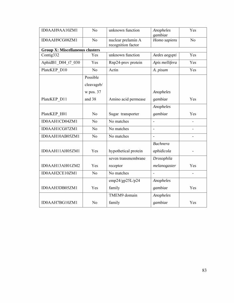
83
ID0AAH9AA10ZM1 No unknown function Anopheles gambiae
Yes
ID0AAH9CG08ZM1 No nuclear prelamin A recognition factor
Homo sapiens No
Group X: Miscellaneous clusters Contig332 Yes unknown function Aedes aegypti Yes
AphidB1_D04_t7_030 Yes Rnp24-prov protein Apis mellifera Yes
PlateKEP_D10 No Actin A. pisum Yes
PlateKEP_D11
Possible
cleavageb/
w pos. 37
and 38 Amino acid permease
Anopheles
gambiae Yes
PlateKEP_H01 No Sugar transporter
Anopheles
gambiae Yes
ID0AAH1CD04ZM1 No No matches - -
ID0AAH1CG07ZM1 No No matches - -
ID0AAH10AB05ZM1 No No matches - -
ID0AAH11AH05ZM1 Yes hypothetical protein
Buchnera
aphidicola -
ID0AAH13AH01ZM2 Yes
seven transmembrane
receptor
Drosophila
melanogaster Yes
ID0AAH2CE10ZM1 No No matches - -
ID0AAH3DB05ZM1 Yes
emp24/gp25L/p24
family
Anopheles
gambiae Yes
ID0AAH7BG10ZM1 No
TMEM9 domain
family
Anopheles
gambiae Yes
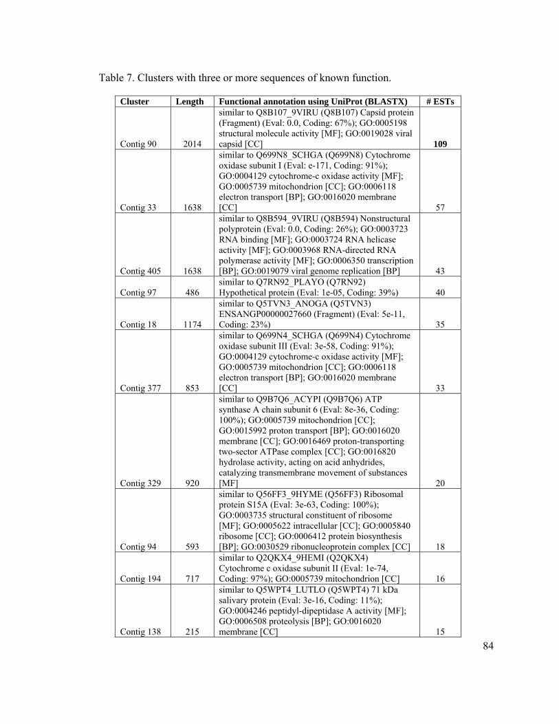
84
Table 7. Clusters with three or more sequences of known function.
Cluster Length Functional annotation using UniProt (BLASTX) # ESTs
Contig 90 2014
similar to Q8B107_9VIRU (Q8B107) Capsid protein (Fragment) (Eval: 0.0, Coding: 67%); GO:0005198 structural molecule activity [MF]; GO:0019028 viral capsid [CC] 109
Contig 33 1638
similar to Q699N8_SCHGA (Q699N8) Cytochrome oxidase subunit I (Eval: e-171, Coding: 91%); GO:0004129 cytochrome-c oxidase activity [MF]; GO:0005739 mitochondrion [CC]; GO:0006118 electron transport [BP]; GO:0016020 membrane [CC] 57
Contig 405 1638
similar to Q8B594_9VIRU (Q8B594) Nonstructural polyprotein (Eval: 0.0, Coding: 26%); GO:0003723 RNA binding [MF]; GO:0003724 RNA helicase activity [MF]; GO:0003968 RNA-directed RNA polymerase activity [MF]; GO:0006350 transcription [BP]; GO:0019079 viral genome replication [BP] 43
Contig 97 486 similar to Q7RN92_PLAYO (Q7RN92) Hypothetical protein (Eval: 1e-05, Coding: 39%) 40
Contig 18 1174
similar to Q5TVN3_ANOGA (Q5TVN3) ENSANGP00000027660 (Fragment) (Eval: 5e-11, Coding: 23%) 35
Contig 377 853
similar to Q699N4_SCHGA (Q699N4) Cytochrome oxidase subunit III (Eval: 3e-58, Coding: 91%); GO:0004129 cytochrome-c oxidase activity [MF]; GO:0005739 mitochondrion [CC]; GO:0006118 electron transport [BP]; GO:0016020 membrane [CC] 33
Contig 329 920
similar to Q9B7Q6_ACYPI (Q9B7Q6) ATP synthase A chain subunit 6 (Eval: 8e-36, Coding: 100%); GO:0005739 mitochondrion [CC]; GO:0015992 proton transport [BP]; GO:0016020 membrane [CC]; GO:0016469 proton-transporting two-sector ATPase complex [CC]; GO:0016820 hydrolase activity, acting on acid anhydrides, catalyzing transmembrane movement of substances [MF] 20
Contig 94 593
similar to Q56FF3_9HYME (Q56FF3) Ribosomal protein S15A (Eval: 3e-63, Coding: 100%); GO:0003735 structural constituent of ribosome [MF]; GO:0005622 intracellular [CC]; GO:0005840 ribosome [CC]; GO:0006412 protein biosynthesis [BP]; GO:0030529 ribonucleoprotein complex [CC] 18
Contig 194 717
similar to Q2QKX4_9HEMI (Q2QKX4) Cytochrome c oxidase subunit II (Eval: 1e-74, Coding: 97%); GO:0005739 mitochondrion [CC] 16
Contig 138 215
similar to Q5WPT4_LUTLO (Q5WPT4) 71 kDa salivary protein (Eval: 3e-16, Coding: 11%); GO:0004246 peptidyl-dipeptidase A activity [MF]; GO:0006508 proteolysis [BP]; GO:0016020 membrane [CC] 15
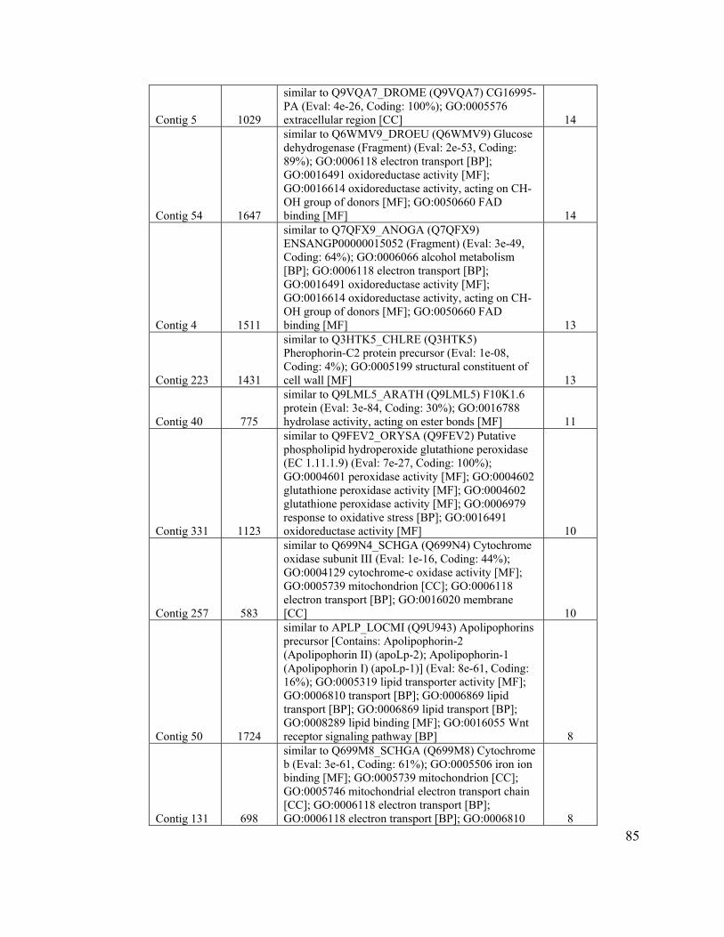
85
Contig 5 1029
similar to Q9VQA7_DROME (Q9VQA7) CG16995-PA (Eval: 4e-26, Coding: 100%); GO:0005576 extracellular region [CC] 14
Contig 54 1647
similar to Q6WMV9_DROEU (Q6WMV9) Glucose dehydrogenase (Fragment) (Eval: 2e-53, Coding: 89%); GO:0006118 electron transport [BP]; GO:0016491 oxidoreductase activity [MF]; GO:0016614 oxidoreductase activity, acting on CH-OH group of donors [MF]; GO:0050660 FAD binding [MF] 14
Contig 4 1511
similar to Q7QFX9_ANOGA (Q7QFX9) ENSANGP00000015052 (Fragment) (Eval: 3e-49, Coding: 64%); GO:0006066 alcohol metabolism [BP]; GO:0006118 electron transport [BP]; GO:0016491 oxidoreductase activity [MF]; GO:0016614 oxidoreductase activity, acting on CH-OH group of donors [MF]; GO:0050660 FAD binding [MF] 13
Contig 223 1431
similar to Q3HTK5_CHLRE (Q3HTK5) Pherophorin-C2 protein precursor (Eval: 1e-08, Coding: 4%); GO:0005199 structural constituent of cell wall [MF] 13
Contig 40 775
similar to Q9LML5_ARATH (Q9LML5) F10K1.6 protein (Eval: 3e-84, Coding: 30%); GO:0016788 hydrolase activity, acting on ester bonds [MF] 11
Contig 331 1123
similar to Q9FEV2_ORYSA (Q9FEV2) Putative phospholipid hydroperoxide glutathione peroxidase (EC 1.11.1.9) (Eval: 7e-27, Coding: 100%); GO:0004601 peroxidase activity [MF]; GO:0004602 glutathione peroxidase activity [MF]; GO:0004602 glutathione peroxidase activity [MF]; GO:0006979 response to oxidative stress [BP]; GO:0016491 oxidoreductase activity [MF] 10
Contig 257 583
similar to Q699N4_SCHGA (Q699N4) Cytochrome oxidase subunit III (Eval: 1e-16, Coding: 44%); GO:0004129 cytochrome-c oxidase activity [MF]; GO:0005739 mitochondrion [CC]; GO:0006118 electron transport [BP]; GO:0016020 membrane [CC] 10
Contig 50 1724
similar to APLP_LOCMI (Q9U943) Apolipophorins precursor [Contains: Apolipophorin-2 (Apolipophorin II) (apoLp-2); Apolipophorin-1 (Apolipophorin I) (apoLp-1)] (Eval: 8e-61, Coding: 16%); GO:0005319 lipid transporter activity [MF]; GO:0006810 transport [BP]; GO:0006869 lipid transport [BP]; GO:0006869 lipid transport [BP]; GO:0008289 lipid binding [MF]; GO:0016055 Wnt receptor signaling pathway [BP] 8
Contig 131 698
similar to Q699M8_SCHGA (Q699M8) Cytochrome b (Eval: 3e-61, Coding: 61%); GO:0005506 iron ion binding [MF]; GO:0005739 mitochondrion [CC]; GO:0005746 mitochondrial electron transport chain [CC]; GO:0006118 electron transport [BP]; GO:0006118 electron transport [BP]; GO:0006810 8
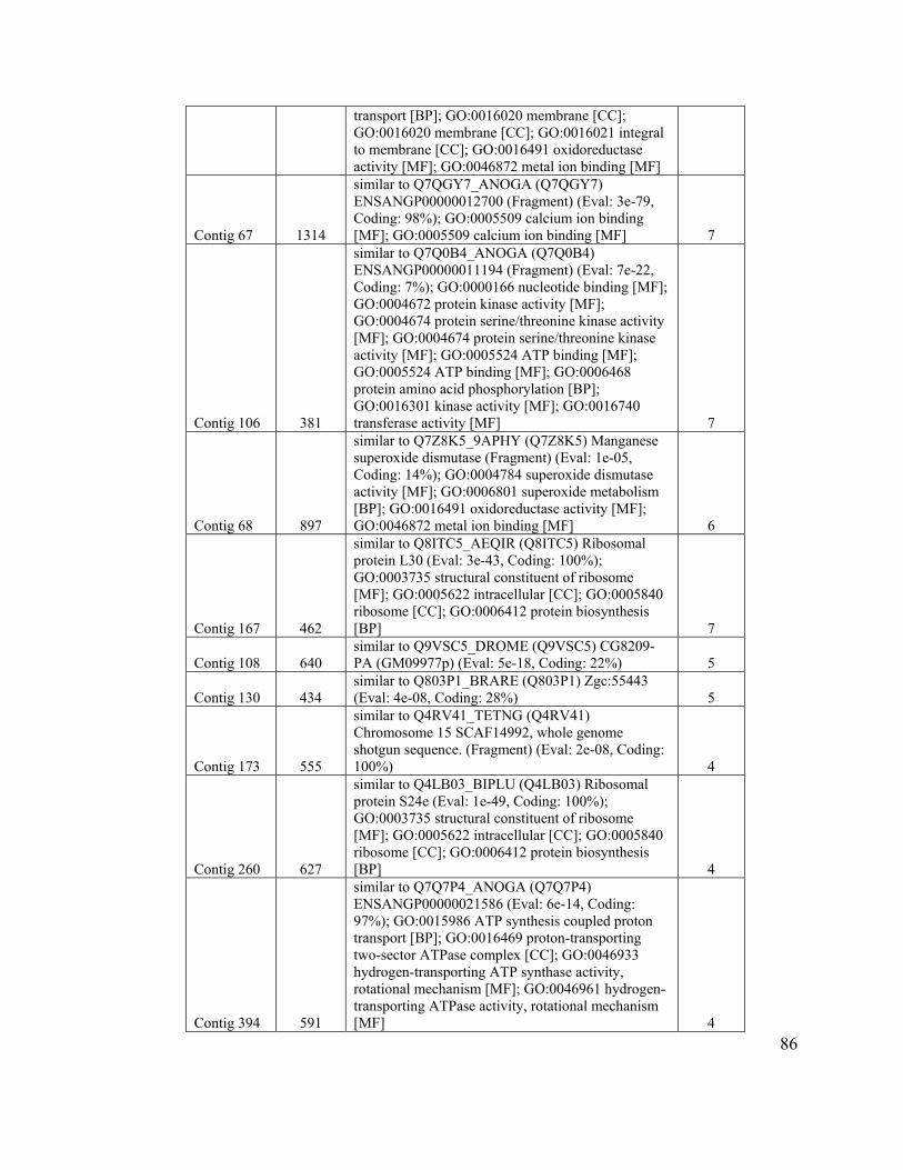
86
transport [BP]; GO:0016020 membrane [CC]; GO:0016020 membrane [CC]; GO:0016021 integral to membrane [CC]; GO:0016491 oxidoreductase activity [MF]; GO:0046872 metal ion binding [MF]
Contig 67 1314
similar to Q7QGY7_ANOGA (Q7QGY7) ENSANGP00000012700 (Fragment) (Eval: 3e-79, Coding: 98%); GO:0005509 calcium ion binding [MF]; GO:0005509 calcium ion binding [MF] 7
Contig 106 381
similar to Q7Q0B4_ANOGA (Q7Q0B4) ENSANGP00000011194 (Fragment) (Eval: 7e-22, Coding: 7%); GO:0000166 nucleotide binding [MF]; GO:0004672 protein kinase activity [MF]; GO:0004674 protein serine/threonine kinase activity [MF]; GO:0004674 protein serine/threonine kinase activity [MF]; GO:0005524 ATP binding [MF]; GO:0005524 ATP binding [MF]; GO:0006468 protein amino acid phosphorylation [BP]; GO:0016301 kinase activity [MF]; GO:0016740 transferase activity [MF] 7
Contig 68 897
similar to Q7Z8K5_9APHY (Q7Z8K5) Manganese superoxide dismutase (Fragment) (Eval: 1e-05, Coding: 14%); GO:0004784 superoxide dismutase activity [MF]; GO:0006801 superoxide metabolism [BP]; GO:0016491 oxidoreductase activity [MF]; GO:0046872 metal ion binding [MF] 6
Contig 167 462
similar to Q8ITC5_AEQIR (Q8ITC5) Ribosomal protein L30 (Eval: 3e-43, Coding: 100%); GO:0003735 structural constituent of ribosome [MF]; GO:0005622 intracellular [CC]; GO:0005840 ribosome [CC]; GO:0006412 protein biosynthesis [BP] 7
Contig 108 640 similar to Q9VSC5_DROME (Q9VSC5) CG8209-PA (GM09977p) (Eval: 5e-18, Coding: 22%) 5
Contig 130 434 similar to Q803P1_BRARE (Q803P1) Zgc:55443 (Eval: 4e-08, Coding: 28%) 5
Contig 173 555
similar to Q4RV41_TETNG (Q4RV41) Chromosome 15 SCAF14992, whole genome shotgun sequence. (Fragment) (Eval: 2e-08, Coding: 100%) 4
Contig 260 627
similar to Q4LB03_BIPLU (Q4LB03) Ribosomal protein S24e (Eval: 1e-49, Coding: 100%); GO:0003735 structural constituent of ribosome [MF]; GO:0005622 intracellular [CC]; GO:0005840 ribosome [CC]; GO:0006412 protein biosynthesis [BP] 4
Contig 394 591
similar to Q7Q7P4_ANOGA (Q7Q7P4) ENSANGP00000021586 (Eval: 6e-14, Coding: 97%); GO:0015986 ATP synthesis coupled proton transport [BP]; GO:0016469 proton-transporting two-sector ATPase complex [CC]; GO:0046933 hydrogen-transporting ATP synthase activity, rotational mechanism [MF]; GO:0046961 hydrogen-transporting ATPase activity, rotational mechanism [MF] 4
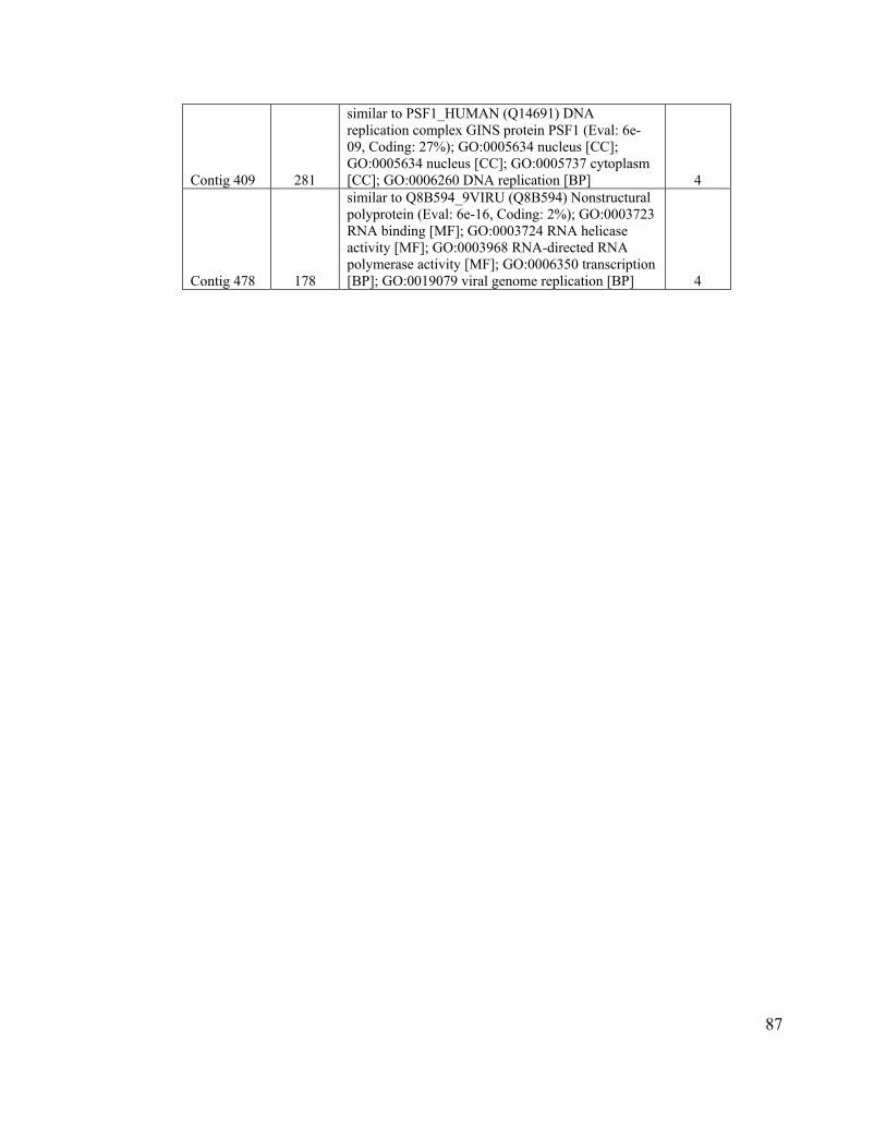
87
Contig 409 281
similar to PSF1_HUMAN (Q14691) DNA replication complex GINS protein PSF1 (Eval: 6e-09, Coding: 27%); GO:0005634 nucleus [CC]; GO:0005634 nucleus [CC]; GO:0005737 cytoplasm [CC]; GO:0006260 DNA replication [BP] 4
Contig 478 178
similar to Q8B594_9VIRU (Q8B594) Nonstructural polyprotein (Eval: 6e-16, Coding: 2%); GO:0003723 RNA binding [MF]; GO:0003724 RNA helicase activity [MF]; GO:0003968 RNA-directed RNA polymerase activity [MF]; GO:0006350 transcription [BP]; GO:0019079 viral genome replication [BP] 4

88
Table 8. Clusters with more than 2 sequences with unknown function.
Cluster Length Secreted # ESTs ORF Contig 14 1385 236 - Contig 7 2236 134 690 Contig 78 610 Full-length (secreted) cleavable site (19-20) 74 141 Contig 181 719 72 -
Contig 164 921 May be secreted, possible cleavable site (27-28) 49
154
Contig 32 1054 C002, Full-length (secreted) cleavable site (23-24) 46
213
Contig 140 1041 46 232 Contig 9 1535 Full-length (secreted) cleavable site (27-28) 35 227 Contig 34 1413 Full-length (secreted) cleavable site (28-29) 35 132 Contig 6 1452 34 294 Contig 86 2948 34 441 Contig 356 762 26 - Contig 62 1627 21 402 Contig 498 876 Full-length (secreted) cleavable site (23-24) 20 208 Contig 122 1151 Full-length (secreted) cleavable site (19-20) 19 141 Contig 283 635 19 128 Contig 20 1178 17 267 Contig 42 1481 16 492 Contig 475 950 16 247 Contig 462 821 16 64 Contig 81 716 15 - Contig 235 1038 14 - Contig 422 360 14 - Contig 38 1319 13 271 Contig 358 1302 Full-length (secreted) cleavable site (25-26) 13 263 Contig 15 813 13 155 Contig 258 577 13 78 Contig 216 488 13 - Contig 159 952 12 178 Contig 339 865 12 179 Contig 300 445 11 148 Contig 160 251 11 69 Contig 73 138 11 - Contig 154 1865 10 - Contig 3 1336 10 344 Contig 12 936 10 - Contig 470 731 10 - Contig 36 673 10 109 Contig 171 623 10 67 Contig 85 422 10 139 Contig 349 417 10 - Contig 510 660 9 219 Contig 275 645 Full-length (no signal peptide) 9 107 Contig 308 565 9 - Contig 334 543 9 - Contig 186 189 9 -
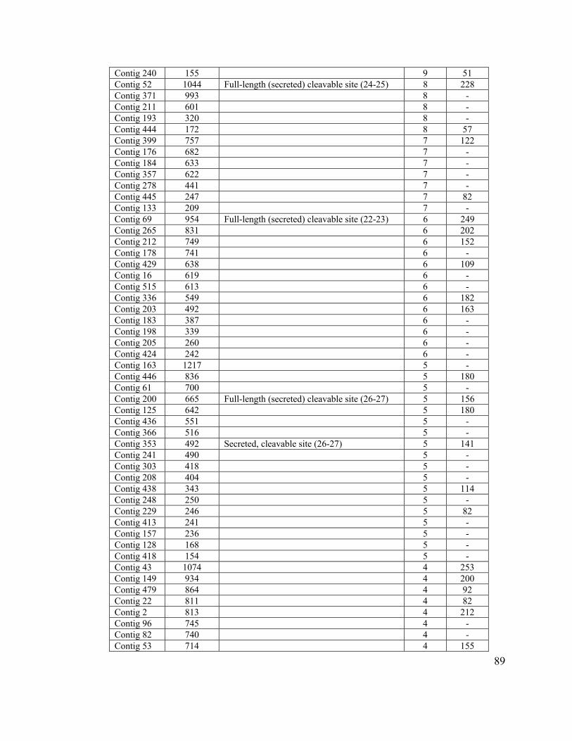
89
Contig 240 155 9 51 Contig 52 1044 Full-length (secreted) cleavable site (24-25) 8 228 Contig 371 993 8 - Contig 211 601 8 - Contig 193 320 8 - Contig 444 172 8 57 Contig 399 757 7 122 Contig 176 682 7 - Contig 184 633 7 - Contig 357 622 7 - Contig 278 441 7 - Contig 445 247 7 82 Contig 133 209 7 - Contig 69 954 Full-length (secreted) cleavable site (22-23) 6 249 Contig 265 831 6 202 Contig 212 749 6 152 Contig 178 741 6 - Contig 429 638 6 109 Contig 16 619 6 - Contig 515 613 6 - Contig 336 549 6 182 Contig 203 492 6 163 Contig 183 387 6 - Contig 198 339 6 - Contig 205 260 6 - Contig 424 242 6 - Contig 163 1217 5 - Contig 446 836 5 180 Contig 61 700 5 - Contig 200 665 Full-length (secreted) cleavable site (26-27) 5 156 Contig 125 642 5 180 Contig 436 551 5 - Contig 366 516 5 - Contig 353 492 Secreted, cleavable site (26-27) 5 141 Contig 241 490 5 - Contig 303 418 5 - Contig 208 404 5 - Contig 438 343 5 114 Contig 248 250 5 - Contig 229 246 5 82 Contig 413 241 5 - Contig 157 236 5 - Contig 128 168 5 - Contig 418 154 5 - Contig 43 1074 4 253 Contig 149 934 4 200 Contig 479 864 4 92 Contig 22 811 4 82 Contig 2 813 4 212 Contig 96 745 4 - Contig 82 740 4 - Contig 53 714 4 155
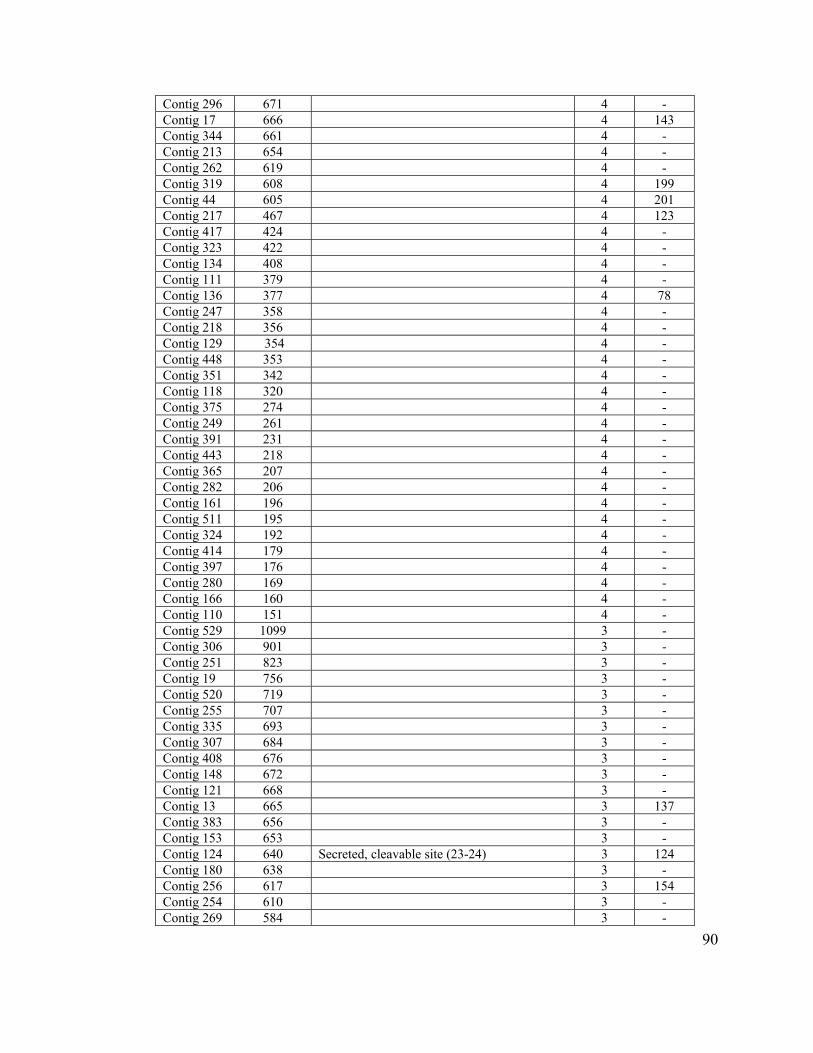
90
Contig 296 671 4 - Contig 17 666 4 143 Contig 344 661 4 - Contig 213 654 4 - Contig 262 619 4 - Contig 319 608 4 199 Contig 44 605 4 201 Contig 217 467 4 123 Contig 417 424 4 - Contig 323 422 4 - Contig 134 408 4 - Contig 111 379 4 - Contig 136 377 4 78 Contig 247 358 4 - Contig 218 356 4 - Contig 129 354 4 - Contig 448 353 4 - Contig 351 342 4 - Contig 118 320 4 - Contig 375 274 4 - Contig 249 261 4 - Contig 391 231 4 - Contig 443 218 4 - Contig 365 207 4 - Contig 282 206 4 - Contig 161 196 4 - Contig 511 195 4 - Contig 324 192 4 - Contig 414 179 4 - Contig 397 176 4 - Contig 280 169 4 - Contig 166 160 4 - Contig 110 151 4 - Contig 529 1099 3 - Contig 306 901 3 - Contig 251 823 3 - Contig 19 756 3 - Contig 520 719 3 - Contig 255 707 3 - Contig 335 693 3 - Contig 307 684 3 - Contig 408 676 3 - Contig 148 672 3 - Contig 121 668 3 - Contig 13 665 3 137 Contig 383 656 3 - Contig 153 653 3 - Contig 124 640 Secreted, cleavable site (23-24) 3 124 Contig 180 638 3 - Contig 256 617 3 154 Contig 254 610 3 - Contig 269 584 3 -

91
Contig 284 578 3 119 Contig 204 561 3 - Contig 263 552 3 - Contig 372 544 3 - Contig 458 509 3 108 Contig 192 505 3 - Contig 139 501 3 - Contig 210 488 3 - Contig 76 482 3 - Contig 132 472 3 - Contig 385 468 3 - Contig 355 463 3 - Contig 126 454 3 - Contig 233 440 3 - Contig 309 426 3 - Contig 158 416 3 - Contig 177 414 3 109 Contig 84 409 3 69 Contig 273 407 3 - Contig 103 399 3 - Contig 271 395 3 - Contig 150 386 3 - Contig 250 372 3 - Contig 313 334 3 - Contig 419 333 3 - Contig 295 320 3 - Contig 361 313 3 - Contig 352 311 3 - Contig 435 311 3 - Contig 185 304 3 - Contig 314 298 3 - Contig 514 294 3 - Contig 496 290 3 - Contig 406 281 3 - Contig 318 278 3 - Contig 484 270 3 - Contig 195 245 3 - Contig 197 232 3 - Contig 364 229 3 - Contig 322 224 3 - Contig 362 223 3 - Contig 302 215 3 - Contig 343 210 3 - Contig 467 209 3 - Contig 503 199 3 - Contig 398 192 3 - Contig 387 190 3 - Contig 77 168 3 - Contig 79 168 3 - Contig 127 155 3 - Contig 370 145 3 -
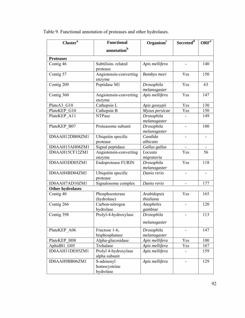
92
Table 9. Functional annotation of proteases and other hydrolases.
Clustera Functional
annotationb
Organismc Secretedd ORFe
Proteases Contig 46 Subtilisin- related
protease Apis mellifera - 140
Contig 57 Angiotensin-converting enzyme
Bombyx mori Yes 150
Contig 209 Peptidase M1 Drosophila melanogaster
Yes 63
Contig 360 Angiotensin-converting enzyme
Apis mellifera Yes 147
PlateA3_G10 Cathepsin L Apis gossypii Yes 130 PlateKEP_G10 Cathepsin B Myzus persicae Yes 150 PlateKEP_A11 NTPase Drosophila
melanogaster - 149
PlateKEP_B07 Proteasome subunit Drosophila melanogaster
- 180
ID0AAH12DB08ZM1 Ubiquitin specific protease
Candida albicans
- -
ID0AAH15AH08ZM1 Signal peptidase Gallus gallus - - ID0AAH15CF12ZM1 Angiotensin-converting
enzyme Locusta migratoria
Yes 56
ID0AAH3DD05ZM1 Endoprotease FURIN Drosophila melanogaster
Yes 118
ID0AAH4BD04ZM1 Ubiquitin specific protease
Danio rerio - -
ID0AAH7AD10ZM1 Signalosome complex Danio rerio - 177 Other hydrolases Contig 40 Phosphoesterase
(hydrolase) Arabidopsis thialiana
Yes 165
Contig 266 Carbon-nitrogen hydrolase
Anopheles gambiae
- 120
Contig 398 Prolyl-4-hydroxylase Drosophila
melanogaster
- 113
PlateKEP_A06 Fructose 1-6, bisphosphatase
Drosophila melanogaster
- 147
PlateKEP_H08 Alpha-glucosidase Apis mellifera Yes 100 AphidB1_G05 Trehalase Apis mellifera Yes 167 ID0AAH11DE05ZM1 Prolyl 4-hydroxylase
alpha subunit Apis mellifera - 159
ID0AAH9BB06ZM1 S-adenosyl homocysteine hydrolase
Apis mellifera - 129

93
a Cluster (comprising contig and singlets) in pea aphid salivary database. b Functional annotation based to top hit at NR database at NCBI. c Top match at NR database using TBLASTX. d SignalP analysis using full-length homolog of pea aphid cluster. e ORF represent number of amino acid residues in pea aphid cluster.
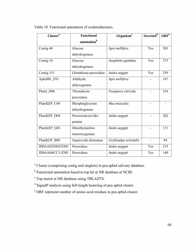
94
Table 10. Functional annotation of oxidoreductases.
Clustera Functional
annotationb
Organismc Secretedd ORFe
Contig 40 Glucose
dehydrogenase
Apis mellifera Yes 503
Contig 54 Glucose
dehydrogenase
Anopheles gambiae Yes 375
Contig 331 Glutathione peroxidase Aedes aegypti Yes 259
AphidB1_F01 Aldehyde
dehyrogenase
Apis mellifera - 167
PlateI_D06 Thioredoxin
peroxidase
Toxoptera citricida - 254
PlateKEP_C04 Phosphoglycerate
dehydrogenase
Mus musculus - -
PlateKEP_D04 Peroxiredoxin-like
protein
Aedes aegypti - 202
PlateKEP_G03 Dimethylaniline
monooxygenase
Aedes aegypti - 171
PlateKEP_B09 Superoxide dismutase Gryllotalpa orientalis - 94
ID0AAH2DE03ZM1 Peroxidase Aedes aegypti Yes 219
ID0AAH6CC11ZM1 Peroxidase Aedes aegypti Yes 140
a Cluster (comprising contig and singlets) in pea aphid salivary database. b Functional annotation based to top hit at NR database at NCBI. c Top match at NR database using TBLASTX. d SignalP analysis using full-length homolog of pea aphid cluster. e ORF represent number of amino acid residues in pea aphid cluster.

95
CHAPTER 3
96
Chapter 3: A Novel Transcript and Protein from the Salivary Glands of Pea Aphid,
Acyrthosiphon pisum.
Navdeep S. Mutti1,2, Loretta K. Pappan2,4, Khurshida Begum1, Kirk Pappan2,3, Ming-
Shun Chen5, Yoonseong Park1, John C. Reese1, Gerald R. Reeck2*
1 Department of Entomology, Kansas State University, Manhattan, Kansas 66506.
2 Department of Biochemistry, Kansas State University, Manhattan, Kansas 66506.
3 Current address: Department of Biochemistry, Washington University School of
Medicine, St. Louis, MO 63110.
4 Current address: Department of Pathology, Washington University School of Medicine,
St. Louis, MO 63110.
5USDA-ARS and Department of Entomology, Kansas State University, Manhattan,
Kansas 66506.
Key words: pea aphid, salivary glands.
Abbreviations: PCR, polymerase chain reaction; RT-PCR, reverse-transcriptase
polymerase chain reaction; EST, expressed sequence tag; NCBI, National Center for
Biotechnology Information; PBS, phosphate buffered saline; DIG, digoxigenin; LB, luria
broth; IPTG, isopropyl-β-D thiogalactoside; SDS, Sodium dodecyl sulfate.
97
Abstract
Salivary secretions are a key component of aphid-plant interactions. Aphid salivary
proteins interact with plant tissues, enabling aphids to gain access to phloem sap and
possibly eliciting responses which may benefit the insect. In an effort to isolate and
identify key components in salivary secretions, we created a salivary gland cDNA library.
Several thousand randomly selected cDNA clones were sequenced as described in
Chapter 2. The transcript corresponding to an abundant cDNA (called C002), was
characterized. Based on in situ hybridization and immunohistochemistry, transcript as
well as protein was localized to the same sub-set of cells within principal salivary glands.
The encoded protein fails to match any protein of known function and any sequence
outside of the family Aphididae. The protein encodes a secreted protein and is injected
into the host plant during aphid feeding. RNAi directed toward the C002 transcript had
no affect on growth and development on liquid diet, though, as shown in the next chapter
it causes lethality when aphids are placed on fava bean leaves.
98
Introduction
Aphid saliva plays a major role in the interaction of aphids and host plants (Miles, 1999).
Although, aphid-plant interactions have been studied extensively, not much is known at
the molecular level. The availability of aphid saliva in small quantities makes the direct
study of salivary components difficult (Miles, 1965; Madhusudhan et al., 1994; Miles and
Harrewijn, 1991). The proteins of aphid saliva are of two types, structural and enzymatic.
The structural proteins provide a tube-like sheath (Miles 1999; Cherqui and Tjallingi
2000) and are probably few in number corresponding to major bands on gels, with
estimated molecular masses of 154 kDa and 66/69 kDa (Cherqui and Tjallingi 2000).
Limited N-terminal sequence information is available on these bands (Baumann and
Baumann 1995). The secreted salivary enzymes fall into two broad categories: hydrolases
(pectinases, cellulases, oligosaccharases) and oxidation/reduction enzymes (phenol
oxidase (E.C. 1.14.18.1) and peroxidases) (Miles 1999). The role of these enzymes
during aphid attack on plants is not well-understood.
Aphid saliva is believed to perform multiple functions; including creation of the stylet
sheath, assisting the penetration of substrate for food (by the action of pectinases,
cellulases, β-glucosidases etc), digesting nutrients (polysaccharases), detoxification of
phenolic glycosides ingested during feeding by the action of polyphenol oxidases or
peroxidases (oxidation-reduction enzymes), and suppression of host defenses or
elicitation of host responses (Miles, 1972; Miles, 1987; Urbanska et al., 1998; Miles,
1999). Aphid saliva may also play a role in the ability of aphids to counter resistance
99
factors in plants, since some species or biotypes of aphids can feed on plants that are
resistant to other species or biotypes (Miles, 1999).
We have undertaken a functional genomics approach to identify components of aphid
saliva. We have chosen to do this work with pea aphid, A. pisum, because of its large size
(compared with other aphid species), thus making dissections of salivary glands less
difficult. In this paper, we report cloning of an abundant cDNA (C002) from a cDNA
library of pea aphid salivary glands. The cDNA encodes a protein that fails to match to
proteins outside of aphids, is of an unknown function, but appears to be vital to pea aphid
feeding on plant tissue.
Material and Methods
Plants and Aphids
Aphids were originally collected from alfalfa plants in the summer of 1999 by Dr. Marina
Caillaud at Cornell University. Thereafter, the aphids were reared at KSU on fava beans
(Vicia fabae) grown in pots (10 cm diameter) at room temperature under high intensity
sodium lights with a L:D of 16:8. Salivary glands from adult aphids were dissected and
separated from the brain tissue overlaying the glands. Dissected glands were transferred
to 50µl of PBS (137mM NaCl, 2,7mM KCl, 10mM Na2HPO4, 1.8mM KH2PO4; pH 7.4)
for 50 paired glands and washed 3 times in PBS to get rid of hemocytes. Salivary glands
were kept at –75oC until needed.
100
Phagmid cDNA Library Construction and Sequencing
Total RNA was isolated from 250 salivary glands using the Micro RNA isolation kit from
Stratagene, La Jolla, CA. The PCR-based cDNA library was made following instructions
with the SMART cDNA library construction kit (Clontech, Palo Alto, CA). Total RNA
was reverse transcribed using PowerScript reverse transcriptase and CDS III primer
provided in the kit. Second strand synthesis was performed through the PCR-based
protocol using SMART III and CDS III primers from Clontech. Double strand synthesis
was followed by proteinase K digestion. Double strand cDNA was ligated into a Lambda
TriplEx2 vector (Clontech, Palo Alto, CA), and the resulting ligation reaction was
packaged using Gigapack Gold III from Stratagene following the manufacturer’s
specifications.
Sequencing of the cDNA clones was done either at the Kansas State University
sequencing facility or at Genoscope, France. Vector stripped sequences were blasted
against the Gen Bank non redundant protein database from the National Center for
Biotechnology Information using TBLASTX. cDNAs were analyzed for signal peptide
and cleavage information using SignalP 3.0 Server (Nelson et al., 1997) using default
parameters (http://www.cbs.dtu.dk/services/SignalP/).
RNA Isolation and RT-PCR
Total RNA was isolated from salivary glands, guts or adult aphids using TRI reagent
(Molecular Research Center, Inc. Cincinnati, OH) following the procedure provided by
the manufacturer. For RT-PCR, total RNA was treated with DNaseI (Ambion, Austin,
101
TX)) following standard instructions. AMV reverse transcriptase was used along with
oligoDT primers to synthesize single-stranded cDNA following procedure from Promega
technical Bulletin no. 099. PCR was done using 5’ -- CCA GTG CGA TAG CGA TAA
TTT ACA AC -- 3’ and 5’ -- CAC CTC TCT TAT GAT GAA CGC CAA C -- 3’ for
C002 forward and reverse primers, respectively, giving a final product of 397 base pairs,
and using 5’ -- CCG AAA AGC TGT CAT AAT GAA GAC C -- 3’ and 5’ -- GGT GAA
ACC TTG TCT ACT GTT ACA TCT TG -- 3’ for ribosomal protein L27 forward and
reverse, primers, respectively, giving a final product of 231 base pairs.
RNA Isolation and Northern Blotting
Total RNA was extracted from adult aphids using TRI reagent (Molecular Research
Center, Inc. Cincinnati, OH). Total RNA (10 µg) was separated on a 1.2% agarose gel
containing formaldehyde and blotted on to GeneScreen membrane (Perkin Elmer,
Beltsville, MD). The RNA was fixed onto the membrane using UV-light. [32P]-labeled
RNA probes generated using a random labeling kit from Stratagene (La Jolla, CA).
Hybridization was carried out overnight at 420C in a hybridization bottle containing a
15 ml hybridization solution (10% dextran sulphate/1% SDS/1 m NaCl, pH 8.0). After
hybridization, the membranes were washed twice with 2X SSC at room temperature for
30 min, followed by 2 washes with 2X SSC plus 1% SDS at 650C for 30 min, and finally
2 washes with 0.1X SSC plus 1% SDS at room temperature for 30 min. The membranes
were then exposed to Kodak SR-5 X-ray film.
102
Genomic DNA Isolation and Southern Blotting
DNA was extracted from adult aphids following the procedure of Bender et al. (1983).
Southern blotting was carried out according to the protocol of Shen et al (2003) with
minor modifications. The genomic DNA (10 µg) was digested with various restriction
enzymes, separated by agarose gel electrophoresis, and then transferred and cross-linked
to a GeneScreen membrane (Perkin Elmer, Beltsville, MD). The membrane was treated
with hybridization solution (25 mM phosphate buffer (pH 7.0), 5X SSC, 5X Denhardt's
solution, 50 µg/ml salmon sperm DNA, 50 % formamide) at 42° C for 2 h. The cDNA
was [32P]-labeled using random labeling kit from Stratagene (La Jolla, CA) and, after
boiling for 5 min, was added immediately to the hybridization solution. The membrane
was hybridized for 15 h at 42° C. Following hybridization the membrane was washed
twice for 10 min in 2X SSC and 0.1 % SDS at room temperature and once for 30 min in
0.1X SSC and 0.5 % SDS at 37° C. Finally, the membrane was washed in 0.1X SSC and
0.5 % SDS at 68° C for 30 min and then was exposed to Kodak SR-5 X-ray film.
In situ Hybridization
Whole mount in situ hybridization of salivary glands of the pea aphid was done with
RNA digoxigenin-labeled probe. Salivary gland were dissected in PBS and fixed in 4%
paraformaldehyde in PBS for 1 hr at room temperature. All experimental procedures
were performed in a humidified chamber. After fixing, salivary glands were washed 3
times at 10 min interval in PBST (1X PBS and 0.6% Triton-X) at room temperature.
Thereafter tissues were sequentially dehydrated for 2 min with 50%, 75%, and 100%
ethanol, respectively. Again, salivary glands were sequentially rehydrated for 2 min with
103
100%, 75%, and 50% ethanol. Prehybridization was performed without probe in
hybridization solution (5XSSC, 2% blocking reagent, 0.1% N-lauroylsarcosine, 0.02%
SDS, and 50% formamide) at 600C for 1 h. Salivary glands were hybridized with
digoxigenin-labeled single-strand sense or antisense RNA probes (100ng/ml) in
hybridization solution at 600C for 16-18 h. Sense probe was used as negative control. The
DIG-labeled RNA probes were prepared with the DIG RNA Labeling Kit (Roche
Molecular Biochemicals). A 397-bp fragment of clone C002 was amplified by PCR and
subcloned into a pGEM-T Easy vector and was used as a template to generate RNA
probes. Right insertion was confirmed by EcoR1 digestion and insertion direction was
determined by PCR. This plasmid was linearized with SpeI and ApaI and transcribed with
SP6 RNA polymerase and T7 RNA polymerase to generate antisense and sense probes,
respectively. After hybridization, glands were washed with hybridization solution at 550C
for 2-4 h followed by 3 washes at 10 min interval with PBST. Salivary glands were then
incubated with alkaline phosphatase conjugated anti-DIG antibody (1:300 dilution in
PBST) for over night at 40C. The salivary glands were then washed in PBST, followed
by 3 washes in detection buffer (Roche Molecular Biochemicals). Finally the salivary
glands were stained by adding nitroblue tetrazolium salt/5-bromo-4-chloro-3-indoyl
phosphate (NBT/BCIP, 1:50 dilution in detection buffer). Staining was monitored in a
dissection microscope. Color reaction was stopped by repeated washes with PBS and
mounted in 100% glycerol. Photographs were taken using digital camera attached to the
compound microscope (Nikon Eclipse E800).
104
Expression of Recombinant Protein in E. coli for Antibody Preparation
A cDNA encoding a full-length C002 was amplified by PCR. We used a forward primer
5'--TAG CTG TAG CCA TGG AAG TTA GAT GCG--3' containing an NcoI site and a
reverse primer 5'--GTA TGG ACA AGC TTA TTA AAA ACG TCG--3' containing a
HindIII site. The resulting DNA encoded residues 1-196 of the mature protein. The PCR
product (632 bp) was ligated into a pGEM-T easy vector and used to transform E. coli
strain JM109. LB/ampicillin/IPTG/X-Gal (100 µg/mL ampicillin, 0.5mM IPTG, 80
µg/mL X-Gal) plates were used to grow transformed bacteria. Resulting white colonies
were selected for DNA sequencing. The PCR product was excised from the vector by
digestion with NcoI and HindIII and purified by low melting point agarose gel
electrophoresis, ligated into vector H6pQE60 (Lee et al., 1994), and then used to transform
E.coli strain JM109. JM109 cells were spread on an LB agar plate containing ampicillin.
Correct insertion for C002 was confirmed sequencing. A single colony from the plate was
used to inoculate 3 mL 2xYT medium containing 100 µg/mL ampicillin. Culture was
shaken at 300 rpm, 37oC overnight. The 3 mL overnight culture was then used to
inoculate 200 mL 2 x YT medium with ampicillin and still incubated at 300 rpm, 37oC
until A600 was ~ 0.7. Recombinant protein expression was then induced by adding IPTG
to 1 mM final concentration, and culture was incubated for another five hours. Bacteria
were harvested by centrifuging at 5,000 rpm, 20 min at 4oC and then resuspended in 4
mL lysis buffer (8 M urea; 0.1 M NaH2PO4; 0.01 M Tris.Cl, pH 8.0). A 5 µL sample was
reserved for western blot analysis. The bacteria were incubated on ice for 30 min with
Triton X-100 at a final concentration of 2% and lysozyme to a final concentration of
1mg/mL and then sonicated on ice (6x10 sec bursts at 200 Watts, with 1 min interval
105
between bursts). The lysates were centrifuged at 12,000 rpm for 20 min at 4oC.
Supernatants and pellets were saved and 5 µl of each sample was used for western blot
analysis.
Recombinant protein expressed from a total of 2 l. medium was purified under
denaturing conditions by affinity chromatography with nickel-nitrilotriacetic acid (Ni-
NTA) resin (Qiagen, Valencia, CA). The protein was concentrated with an YM-10
centricon (Millipore, Bedford, MA) to 400 µL, mixed with 2 x SDS loading buffer,
separated by electrophoresis in a 12.5% acrylamide gel (ISC Bioexpress, Kaysville, UT)
and stained with 0.025% Commassie blue R-250 in water. Then the protein band
(approximately 600 µg) was cut out and sliced into pieces for injection into rabbit to
generate antisera (Cocalico Biologicals, Reamstown, PA).
Western Blotting
Polyclonal rabbit antibodies were purified by immobilizing recombinant protein (C002)
to matrix composed of cross-linked 4% beaded agarose activated to form aldehyde
functional groups using AminoLink Plus Immobilization Kit (Pierce, Rockford, IL)
following manufacturer’s instructions. For western blot, approximately 500 hundred pea
aphids were placed on fresh fava bean plants and all the aphids were removed after 24 h.
1.5 g of plants tissue was homogenized in PBS on liquid nitrogen. Frequent freeze-thaw
cycles were used for complete extraction, and the extract was centrifuged at 12,000 xg for
5 min. About 2 ml of supernatant was concentrated to 200 µl using YM3 microcon
centrifugal filter devices (Millipore, Bedford, MA) and were also filtered through YM50
106
microcon centrifugal filter devices (Millipore, Bedford, MA) to get rid of contaminating
large protein. 6X SDS-PAGE sample buffer was added to concentrate and subjected to
SDS-PAGE on 4-20% gradient gels (ISC Bioexpress, Kaysville, UT) and then transferred
onto PVDF membrane. Non-specific protein binding sites were blocked with 5% instant
non-fat dry milk (BestChoice®) and membranes were incubated with purified polyclonal
antibody (1:200) overnight followed by extensive washing for 3 h with frequent changes
of 1xPBST. The antigen-antibody complexes were visualized with horse radish
peroxidase-conjugated goat anti-rabbit IgG (Pierce, Rockford, IL) at a dilution of
1:15,000 and detected with SuperSignal West Femto maximum sensitivity substrate kit
(Pierce, Rockford, IL) on X-ray film.
Immunohistochemistry
Pea aphid salivary glands were dissected in 1X PBS and washed three times in PBST
(137mM NaCl, 2.7mM KCl, 10mM Na2HPo4, 1.8mM KH2Po4, 0.1% Triton-X100; pH
7.4). Thereafter, salivary glands were fixed in Bouin (71% saturated picric acid, 24%
formaldehyde (37-40%) and 5% glacial acetic acid) for 10 min at room temperature in a
humified chamber. Salivary glands were washed extensively with PBST and incubated
with primary antibody (raised in rabbit against recombinant C002 expressed in E. coli) at
1:100 dilution overnight at 4oC. Following morning salivary glands were washed 3 times
at 15 min interval with PBST and were blocked with 5% normal goat serum in PBST for
one hour and then washed 3 times at 15 min interval with PBST and followed by
incubation with secondary antibody Cy-3 conjugated goat anti-rabbit (Jackson Immuno
107
Research Laboratories, West Grove, PA) at 1:500 dilution overnight at 4oC. Salivary
glands were washed extensively with PBST at 15 min interval. Nuclei staining was done
using TO-PRO-3 (Molecular probes, Invitrogen, CA) at 5µm conc. for 30 min in dark at
room temperature. Glands were washed extensively with PBST and mounted on
mounting media (Gel/MountTM; Biomeda corp., Foster city, CA) on a glass slide.
Photographs were taken using Nikon Zeiss LSM 5 Pascal (Laser Scanning Confocal
Microscope).
Preparation of dsRNA, siRNA and siRNA Injections
The detailed procedure is described Chapter 4. Injected aphids were placed on artificial
aphid diet (Mittler and Dadd, 1965).
Results
The C002 Transcript and Protein
C002 is an abundant cDNA in our salivary gland cDNA library from the pea aphid. The
C002 cDNA and its predicted amino acid sequence are shown in Fig. 4a. The predicted
protein contains 219 amino acid residues. The N-terminal sequence of the protein was
predicted by SignalP to be a signal peptide for an extracellular protein,
(http://www.cbs.dtu.dk/services/SignalP/), with cleavage predicted between residues 23
and 24. The predicted mass of the mature protein is 21.8 kDa. There are no potential O-
glycosylation sites (http://www.cbs.dtu.dk/services/NetOGlyc/) or N-glycosylation sites
(http://www.cbs.dtu.dk/services/NetNGlyc/).
108
Alignment of all 46 cDNA sequences reveals two forms of the transcript differing at 4
positions resulting in 4 amino-acid substitutions as shown in Fig. 4b. There are 22 cDNA
clones representing the C002a isoform and 24 cDNA clones representing the C002b
isoform. These can be either variants or alleles of the same gene, or can be two separate
genes. The encoded protein fails to match anything of known function and to any other
sequence outside of the family Aphididae. BLAST search against EST-database aligns
the pea aphid, C002 with its homologs from other aphid species. ClustalW alignment of
the pea aphid C002, with the sequences from brown citrus aphid, Toxoptera citricida,
cotton aphid, Aphis gossypii, green peach aphid, Myzus persicae and Russian wheat
aphid, Diuraphis noxia is shown in Fig. 5a. The signal cleavage site in all three proteins
is conserved. Brown citrus aphid, cotton aphid and green peach aphid sequences are not
full-length. The pea aphid, C002 and brown citrus aphid sequences are 63.8% identical
and the pea aphid C002 and cotton aphid sequences are 58.8 % identical. The pea aphid,
C002 and green peach aphid sequence are 71.5% identical. The pea aphid, C002 and
Russian wheat aphid sequences are 53.8% identical. The highest level of sequence
similarity pea aphid C002 is to green peach aphid sequences, as reflected in the
phylogram in Figure 5b.
In addition BLAST searches using three different short conserved regions taken from the
alignments of all aphid C002 sequences had a top match to a hypothetical protein. This
approach also failed to match a conserved region of C002 protein to a putative conserved
domain in other known proteins.
109
Expression of C002 in the Salivary Gland and Gut
To determine the transcript levels in salivary gland and gut of the pea aphid, RT-PCR
was conducted. As shown in Fig. 6a, the levels of C002 after 35 cycles, is significantly
higher in the salivary gland than gut of the pea aphid. Equal amounts of RNA were
loaded as shown as L27, which serves as internal RNA control. Transcript from salivary
glands can be seen on an agarose gel only after 20 cycles of PCR whereas it takes 28
cycles of PCR for band to appear in gut RT-PCR. The C002 transcript is 256 times more
abundant in salivary glands than in guts of the pea aphid.
RT-PCR on RNA isolated from various aphid species; greenbug (Schizaphis graminum),
green peach aphid (M. persicae), Russian wheat aphid (D. noxia), and soybean aphid
(Aphis glycine) using the primers from the pea aphid C002, amplified the fragment of
397bp in all the species as was expected for the pea aphid C002 transcript (Fig. 6b).
Northern and Southern Blot Analyses
Northern blot analysis of total pea aphid RNA using full-length C002 probe revealed a
single band of 1126 bases (Fig. 7a). In Southern analysis using multiple enzymes that
don’t cut within entire length of C002 clone, we observed a single band, consistent with
one gene (or locus) for pea aphid C002 (Fig. 7b Lanes 1, 2, 3 and 4). The size of the band
seen in EcoRV digest is about 1.2 kb (Lane 3, Fig. 7b) is almost same as the transcript
size of C002 based on northern blot (Fig. 7a). The possibility that there may be two genes
is eliminated. Thus, two isoforms C002a and C002b are alleles of the same gene.
110
In situ Hybridization
The pea aphid salivary glands were probed with digoxygenin-labeled C002 RNA (anti-
sense) revealed that the C002 transcript occurred in the principal salivary glands (Fig 8a
and b). There was no hybridization to the accessory salivary glands. Within principal
salivary glands only a sub-set of cells were positively stained for C002 transcript. The
negative control (with a sense probe) showed no hybridization to RNA within salivary
glands (Fig. 8c and d).
Immunohistochemistry
Recombinant C002 was expressed in E. coli and was purified using Ni-NTA resin under
denaturing conditions. This purified protein (as a gel band) was used to raise antibodies
in rabbit. Rabbit polyclonal antibodies were used to localize C002 protein in the salivary
glands of pea aphid (Fig. 9a). No staining of the accessory salivary gland was observed.
This is consistent with the in situ hybridization results shown in Fig. 8. Nuclei staining
with TO-PRO-3 followed by analysis of different z-sections taken by confocal
microscope show 10 cells in each principal lobe (5 on each side, as the principal salivary
gland is symmetrical) stained positive for C002 protein. Under higher magnification,
C002 protein appeared to be located in secretory vesicles (Fig. 9b). Pre-immune serum
was used as a negative control and showed no significant labeling in the salivary glands
(Fig. 9c).
111
Western Blotting
In odder to verify that C002 is indeed a secreted protein, western blot analysis using
purified C002 antibodies was done. Approximately five hundred pea aphids were placed
on fresh fava bean plants and after 24 h of feeding, aphids were removed from plants.
Leaf tissue was extracted and western blot was done to probe for C002 protein using
purified polyclonal rabbit antibodies. C002 protein was detected in the plants that were
exposed to aphids and was not detected in plants not exposed to aphids (Fig. 10a, lane 2
versus lane 3). Recombinant protein is shown in Lane 1 (Fig. 10a). This verifies the fact
that C002 protein is a secreted protein. It is part of pea aphid saliva and is injected into
the host-plant during feeding. Further as positive control, C002 protein was also detected
from protein extracts from 5 pea aphid heads and 5 salivary glands (Fig. 10a, lanes 4 and
5 respectively). In addition there was an additional band of about 75 kDa detected in all
lanes (expect the salivary gland extract), this band was also detected in negative control
with pre-immune rabbit serum in plant extracts with and without aphid feeding (Fig.
10b). Nothing was detected on X-ray film in a western blot with 20 antibody only.
Effects of RNAi on Feeding on Artificial Diet
We injected siC002-RNA and the injected pea aphids were placed onto artificial diet
(Mittler and Dadd, 1965) and allowed to feed across stretched parafilm membrane.
siC002-RNA injected aphids had comparable survival when compared to siGFP-RNA
injected aphids (Fig. 11). Over 70% of siC002-RNA injected aphids survived to day 7.
After day 7, aphids still alive were removed from diet and placed onto fava bean leaves in
112
agar. Over 70% of siC002-RNA injected aphids died within 2 days on leaves compared
to less that 25% for siGFP-RNA injected aphids (Fig. 11).
Discussion
Physiological and biochemical studies have established the importance of salivary
secretions in the attack of aphids on plant tissue (Miles, 1999). Although aphid-plant
interactions have been studied extensively, not much is known at the molecular level. The
availability of aphid saliva in small quantities makes the direct study of salivary
components difficult (Miles, 1965; Madhusudhan et al., 1994; Miles and Harrewijn,
1991).
In this work, a functional genomics approach was undertaken to identify components of
aphid saliva. Several thousand randomly selected cDNA clones from a pea aphid salivary
gland cDNA library were sequenced. cDNA were arranged into clusters of identical
sequences. A cluster representing 46 clones (C002) was characterized. The encoded
protein does not match any protein of known function. The encoded protein has a mass of
21.8 kDa and has a predicted secretion signal peptide, suggesting that it is a secreted
protein.
The gene encoding C002 is apparently a single copy in the pea aphid genome based on
southern blot analysis (Fig. 7b). Thus the two forms of C002 are two different alleles of
the same gene. This heterozygosity could reflect the heterogeneity of the aphid colony
used for library construction. Alternatively, this could also reflect a permanent
113
heterozygosity in individuals of this colony. Considering the fact that the ratio for the two
alleles is 1 to 1 (22:24 respectively) and there has been no sexual reproduction in the
colony, it is more likely that the two forms coexist in individual aphids rather than an
equal distribution of alleles within the population. Also if one allele had a fitness
advantage over a second allele, then the second allele would have been lost in the
population. Thus it is highly likely that both alleles are present in individual aphids and
thus the alleles appear to coexist with stable equilibrium. This would be due to balancing
selection, where the heterozygote has the highest fitness. This is very different than from
the ultimate result of random genetic drift, which would be fixation of one allele and the
loss of all others, which would happen unless there is constant input of alleles into the
population by processes such as mutation or migration. These can be ruled out, as our
colony has been maintained for 7 years from initial population collected, there has been
no migration into the colony, and 7 years is too short a time to accumulate mutations.
Thus it appears that the polymorphism is actively maintained by a balancing selection
(Graur and Li, 2000).
C002 was first identified in a positive screen for pectin methylesterase. In order to test for
the activity, a pectin methylesterase assay using purified soluble recombinant protein was
performed using procedure from Shen et al., 1999. However, no activity was observed
and we conclude that C002 is not a pectin methylesterease.
C002 transcript and protein were localized in the same sub-set of 10 cells within one lobe
of the principal salivary gland by both in situ hybridization and immunohistochemistry.

114
C002 protein can be detected in Cherqui and Tjallingii (2000) localized three salivary
proteins identified by Baumann and Baumann (1995), (namely 66, 69 and 154 kDa
proteins) within principal salivary glands and the pattern of staining observed is clearly
different from the pattern of staining observed with C002 protein in the principal salivary
gland. Thus, it seems reasonable to speculate that morphologically different cell types
identified by Weidemann (1968) and Ponsen (1972), may perform different functions and
at any given time (or stage of feeding). In other words aphids’ saliva can be mix of the
products from one or more cell types of principal salivary gland.
C002 protein was detected in fava bean plants that were exposed to aphids (Fig. 10a).
This verifies directly that C002 is a secreted protein. It is part of pea aphid saliva and is
injected into the host-plant during feeding. Being in aphid saliva, it is postulated that it
may play a role in aphid-plant interactions. RNAi experiment verifies importance of the
protein, as it is not needed to feed on articial diet but it is necessary for survival on host
plants.
C002 transcript is also present in other aphid species. But the absence of a homolog of
C002 outside of aphids, in spite of the presence of huge amount of sequence information
in various database (Non-redundant-NCBI, EST-database and UniProt Knowledgebase
database at ExPASy) and also with the availability of genome sequences of many insects
(Drosophila melanogaster, Anopheles gambiae, Tribolium castaneum, Bombyx mori and
Apis mellifera), suggests that C002 might be specific to Aphididae or phloem feeding
Hemiptera.

115
C002 could have any of many functions in aphid-plant interactions. It is apparently not a
digestive enzyme for any component of artificial diet, as it is not required to feed on such
a diet. It is possible that C002 protein is a putative hydrolase and assists an aphid’s
penetration of the leaf tissue or helps divert plant responses. It could be an inhibitor that
functions to inhibit potential toxic plant products (may be by conjugation with C002) or
C002 protein could function to avoid a potential response from phloem (callose
formation) as the siC002-RNA injected aphids are unable to feed continuously and tend
to move around a good deal, whereas uninjected aphids or aphids injected with siGFP-
RNA stay quite still on the underside of the fava bean leaves, or it could be part of the
mechanism of preventing “plugging” of the sieve element once stylets has entered
phloem. Further experiments are needed to elucidate the role of C002 in aphid-plant
interactions. Using electrical penetration graph methods on the siC002-RNA injected
aphids, we hope to understand the effect of C002 transcript knockdown on detailed
aspects of the insects’ feeding behavior.
Acknowledgments
We thank our colleagues Subbaratnam Muthukrishnan and Michael Kanost for valuable
discussions and suggestions. This work was supported by USDA-CREES NRI grant
2001-35302-09975 and by the Kansas Agricultural Experiment Station (publication
number 05-XXX-J). Pea aphids (voucher specimen number -173) were deposited at the

116
Kansas State University Museum of Entomological and Prairie Arthropod Research
(KSU-MEPAR).
117
References
Bender, W., Spierer, P. and Hogness, D.S. 1983. Chromosomal walking and jumping to
isolate DNA from the ace and rosy loci and the bithorax complex in Drosophila
melanogaster. Journal of Molecular Biology, 168:17-33.
Baumann, L. and Baumann, P. 1995. Soluble salivary proteins secreted by Schizaphis
graminum. Entomologia Experimentalis et Applicata, 7:56-60.
Cherqui, A. and Tjallingii, W.F. 2000. Salivary protein of aphids, a pilot study on
identification, separation and immunolocalisation. Journal of Insect Physiology,
46: 1177-1186.
Graur, D and Li, W.H. 2000. Fundamentals of Molecular Evolution, 2nd Edition, pp. 481.
Lee, E., Linder, M. E. and Gilman, A. G. 1994. Expression of G-protein alpha subunits
in Escherichia coli. Methods Enzymology, 237:146-164.
Madhusudhan, M.A., Taylor, G.S. and Miles, P.W. 1994. The detection of salivary
enzymes of phytophagous Hemiptera: a composition of methods. Annals of
Applied Biology, 124:405-412.
Miles, P. W. 1965. Studies on the salivary physiology of plantbugs: the saliva of aphids.
Journal of Insect Physiology, 11:1261-1268.
Miles, P.W. 1972. The saliva of hemiptera. Advances in Insect Physiology, 9: 183-255.
Miles, P. W. 1987. Feeding process of Aphidoidea in relation to effects on their food
plants. In A. K. Minks and P. Harrewijn (eds) Aphids: their Biology, Natural
Enemies and Control, Elsevier, Amsterdam, vol. 2A pp. 321-339.
Miles PW. 1999. Aphid saliva. Biological Reviews of the Cambridge Philosophical
Society, 74:41-85.
118
Miles, P.W. and Harrewijn, P. 1991. Discharge by aphids of soluble secretions into
dietary sources. Entomologia Experimentalis et Applicata, 59:123-134.
Mittler, T.E. and Dadd, R.H. 1965. Differences in the probing responses of Myzus
persicae (Sulzer) elicited by different feeding solutions behind a parafilm
membrane. Entomologia Experimentalis et Applicata, 8:107-122.
Nelson, H., Engelbrech, J., Brunak, S. and Heijne, G.V. 1997. Identification of
prokaryotic and eukaryotic signal peptides and prediction of their cleavage sites.
Protein Engineering, 10:1-6.
Ponsen, M.B. 1972. The site of potato leafroll virus multiplication in its vector, Myzus
persicae: an anatomical study. Mededelingen Landbouwhogeschool Wageningen,
72:1-147.
Shen, Z.C., Manning, G., Reese, J.C. and Reeck, G.R. 1999. Pectin methylesterase from
the rice weevil, Sitophilus oryzae (L.) (Coleoptera: Curculionidae): Purification
and characterization. Insect Biochemistry and Molecular Biology, 29:209-214.
Shen, Z., Denton, M., Mutti, N., Pappan, K., Kanost, M.R., Reese, J.C. and Reeck, G.R.
2003. Polygalacturonase from Sitophilus oryzae: Possible horizontal transfer of a
pectinase gene from fungi to weevils. Journal of Insect Science. 3:24, Available
online: insectscience.org/3.24.
Urbanska, A., Tjallingii, W. F., Dixon, A.F.G. and Leszczynski, B. 1998. Phenol
oxidising enzymes in the grain aphid's saliva. Entomologia Experimentalis et
Applicata, 86:197-203.
Weidemann, H.L. 1968. Zur morohologie der hauptspeicheldruse von Myzus persicae
(Sulz.) Entomologia Experimentalis et Applicata, 11:450-454.

119
Figure 4. Pea aphid, A. pisum C002 sequence.
a) Nucleotide and deduced amino acid sequence of pea aphid, C002 cDNA clone. N-
terminal signal peptide by SignalP is shown in bold. Arrow indicates a signal-peptide
cleavage site. b) Alignment of two alleles/variants of C002. The two alleles differ at four
positions, which are shown in bold.
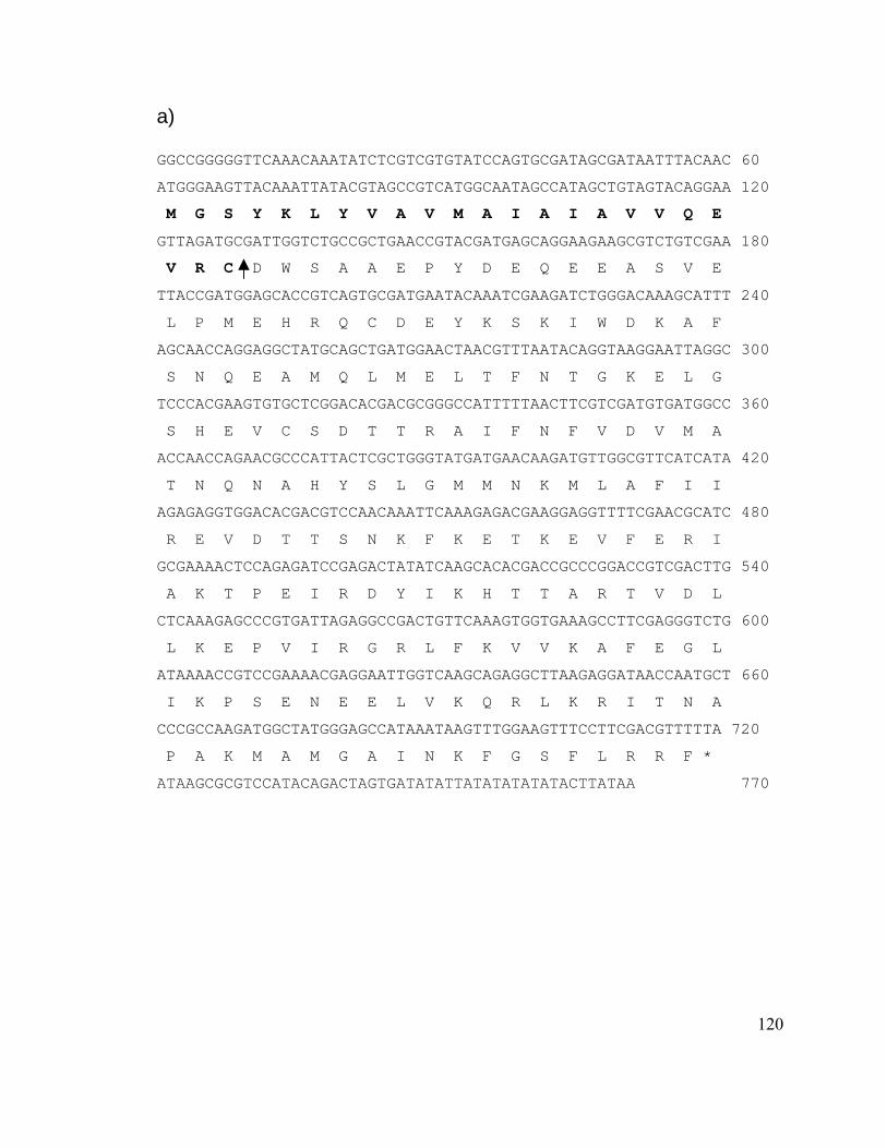
120
a)
GGCCGGGGGTTCAAACAAATATCTCGTCGTGTATCCAGTGCGATAGCGATAATTTACAAC 60
ATGGGAAGTTACAAATTATACGTAGCCGTCATGGCAATAGCCATAGCTGTAGTACAGGAA 120
M G S Y K L Y V A V M A I A I A V V Q E
GTTAGATGCGATTGGTCTGCCGCTGAACCGTACGATGAGCAGGAAGAAGCGTCTGTCGAA 180
V R C D W S A A E P Y D E Q E E A S V E
TTACCGATGGAGCACCGTCAGTGCGATGAATACAAATCGAAGATCTGGGACAAAGCATTT 240
L P M E H R Q C D E Y K S K I W D K A F
AGCAACCAGGAGGCTATGCAGCTGATGGAACTAACGTTTAATACAGGTAAGGAATTAGGC 300
S N Q E A M Q L M E L T F N T G K E L G
TCCCACGAAGTGTGCTCGGACACGACGCGGGCCATTTTTAACTTCGTCGATGTGATGGCC 360
S H E V C S D T T R A I F N F V D V M A
ACCAACCAGAACGCCCATTACTCGCTGGGTATGATGAACAAGATGTTGGCGTTCATCATA 420
T N Q N A H Y S L G M M N K M L A F I I
AGAGAGGTGGACACGACGTCCAACAAATTCAAAGAGACGAAGGAGGTTTTCGAACGCATC 480
R E V D T T S N K F K E T K E V F E R I
GCGAAAACTCCAGAGATCCGAGACTATATCAAGCACACGACCGCCCGGACCGTCGACTTG 540
A K T P E I R D Y I K H T T A R T V D L
CTCAAAGAGCCCGTGATTAGAGGCCGACTGTTCAAAGTGGTGAAAGCCTTCGAGGGTCTG 600
L K E P V I R G R L F K V V K A F E G L
ATAAAACCGTCCGAAAACGAGGAATTGGTCAAGCAGAGGCTTAAGAGGATAACCAATGCT 660
I K P S E N E E L V K Q R L K R I T N A
CCCGCCAAGATGGCTATGGGAGCCATAAATAAGTTTGGAAGTTTCCTTCGACGTTTTTA 720
P A K M A M G A I N K F G S F L R R F *
ATAAGCGCGTCCATACAGACTAGTGATATATTATATATATATACTTATAA 770
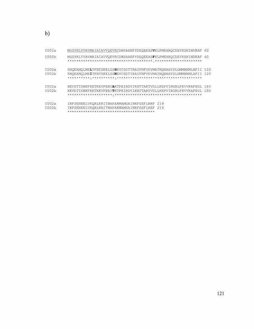
121
b) C002a MGSYKLYVAVMAIAIAVVQEVRCDWSAAEPYDEQEEASVELPMEHRQCDEYKSKIWDKAF 60
C002b MGSYKLYVAVMAIAIAVVQEVRCDWSAAEPYDEQEEASFELPMEHRQCDEYKSKIWDKAF 60 **************************************.********************* C002a SNQEAMQLMELTFNTGKELGSHEVCSDTTRAIFNFVDVMATNQNAHYSLGMMNKMLAFII 120 C002b SNQEAMQLMEITFNTGKELGSNEVCSDTTRAIFNFVDVMATNQNAHYSLGMMNKMLAFII 120 **********:**********:************************************** C002a REVDTTSNKFKETKEVFERIAKTPEIRDYIKHTTARTVDLLKEPVIRGRLFKVVKAFEGL 180 C002b REVDTTSNKFKETKEVFERITKTPEIRDYIKHTTARTVDLLKEPVIRGRLFKVVKAFEGL 180 ********************:*************************************** C002a IKPSENEELVKQRLKRITNAPAKMAMGAINKFGSFLRRF 219 C002b IKPSENEELVKQRLKRITNAPAKMAMGAINKFGSFLRRF 219 ***************************************
122
Figure 5. Multiple sequence alignments.
a) Alignment of C002 amino acid sequences from pea aphid, A. pisum, with sequences of
cotton aphid, A. gossypii, brown citrus aphid, T. citricida, green peach aphid, M. persicae
and Russian wheat aphid, D. noxia. The sequences were aligned using ClustalW version
1.82 and followed by BOXSHADE server design.
http://searchlauncher.bcm.tmc.edu/multi-align/multi-align.html
b) Phylogram of C002 sequences corresponding to the conserved regions in the
alignment. The phylogram was created starting with a multiple alignment created with
ClustalW version 1.82.
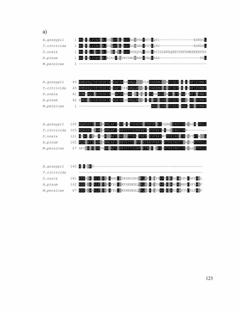
123
a) A.gossypii 1 MGRYQLYVAVMAISSLAVIQKASCAGGPNAYNTTEQYI----------------ESKDGL
T.citricida 1 MGRYQLYVAVVAISSLAVIQKASCAGGSNAYPSTEQYD----------------ESKDKL
D.noxia 1 MGSYKLYLAVIAIACIAAVQEASCSDEQVQYDDGEEVIGLEKEQEEVSEPDMEEKEEPES
A.pisum 1 MGSYKLYVAVMAIAIAVVQEVRCDWSAAEPYDEQEEAS-------------------FEL
M.persicae 1 ------------------------------------------------------------
A.gossypii 45 EMEHHQCDEYKSKIWNKAFSNPAAMQLVDVVLKTAKEMGTDNVCSDTIRVLSNFIDVMAT
T.citricida 45 EMEHHQCDEYKSKIWNKAFSYPAAMQLMDVIFETAKEMGTNDVCSDTIRVLSNFIDVMAT
D.noxia 61 EMEYHQCEEYKSKIWDNAFSNKDAMDMMKLTFITAEKMGSDAVCTDTARAFINFIEVMAT
A.pisum 42 PMEHRQCDEYKSKIWDKAFSNQEAMQLMEITFNTGKELGSNEVCSDTTRAIFNFVDVMAT
M.persicae 1 ----------------------------------AKELGSNEVCSDTTRALFNFVDVMAT
A.gossypii 105 NQNSHYSVGMLAKMLAFIAREADMTSDKFRDTKEVFDRIVQNADIRDYIRNTASRVVDLL
T.citricida 105 NQNSHYVVGMLGKMLAFIAREVDTTSDKFRETTEVFERIAKNADIRDYIRH---------
D.noxia 121 NSNSQYTRSMFKKLVAFIVRELNTTSDNFRETSEVFERIWTTPEIRDFIRDSVTRTNNVL
A.pisum 102 NQNAHYSLGMMNKMLAFIIREVDTTSNKFKETKEVFERITKTPEIRDYIKHTTARTVDLL
M.persicae 27 SPYAHFSLGMFNKMVAFILREVDTTSDKFKETKQVVDRISKTPEIRDYIRNSAAKTVDLL
A.gossypii 165 KLPVMRN---------------------------------------------------
T.citricida ----------------------------------------------------------
D.noxia 181 KEPRMRSRLFKVIEAAMDLMSKSKDGESMKQKFKGMYRAPTKMARXAMDKVGNFFRKL
A.pisum 162 KEPVIRGRLFKVVKAFEGLIKPSENEELVKQRLKRITNAPAKMAMGAINKFGSFLRRF
M.persicae 87 KEPKIRARLFRVMKAFESLIKPNENEALVKQKIKGLTNAPVKLAKGAMKTVGRLFRHF
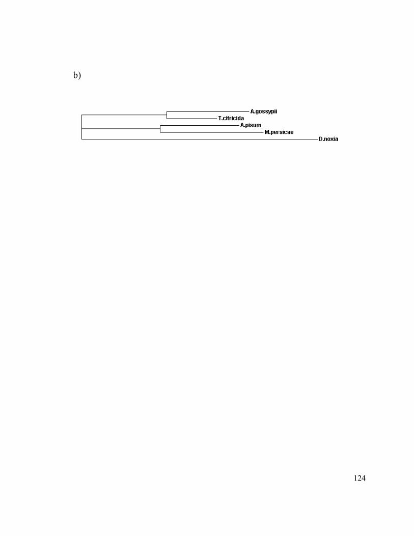
124
b)
125
Figure 6. Expression of C002 in salivary glands and gut of pea aphid.
a) RT-PCR of the C002 transcript from pea aphid, salivary glands (Lane 1) and guts
(Lane 2). PCR products after 35 cycles. b) RT-PCR of the C002 transcript from various
aphid species using primer pair from the pea aphid clone. Pea aphid, A. pisum (Lane 1),
greenbug, S. graminum (Lane 2), green peach aphid, M. persicae (Lane 3), Russian
wheat aphid, D. noxia (Lane 4), soybean aphid, A. glycine (Lane 5).
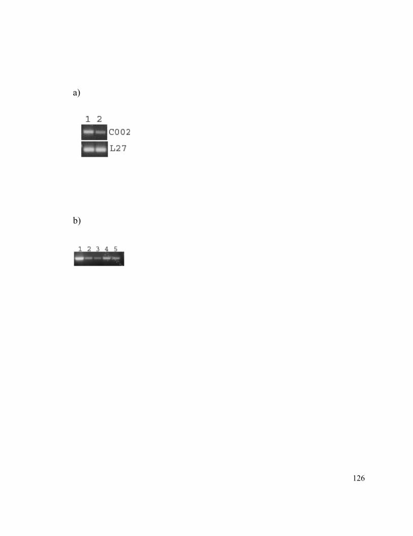
126
a)
b)
127
Figure 7. Northern and Southern blot analysis of C002 transcript and gene.
a) Northern blot analysis of the pea aphid C002 transcript. b) Southern blot analysis of
the pea aphid C002. Genomic DNA was extracted from adult aphids. The cDNA shown
in Figure 1 was radiolabelled and used as the probe. Lane 1, XbaI; Lane 2, NcoI; Lane 3
EcoRV and Lane 4 EcoRI.
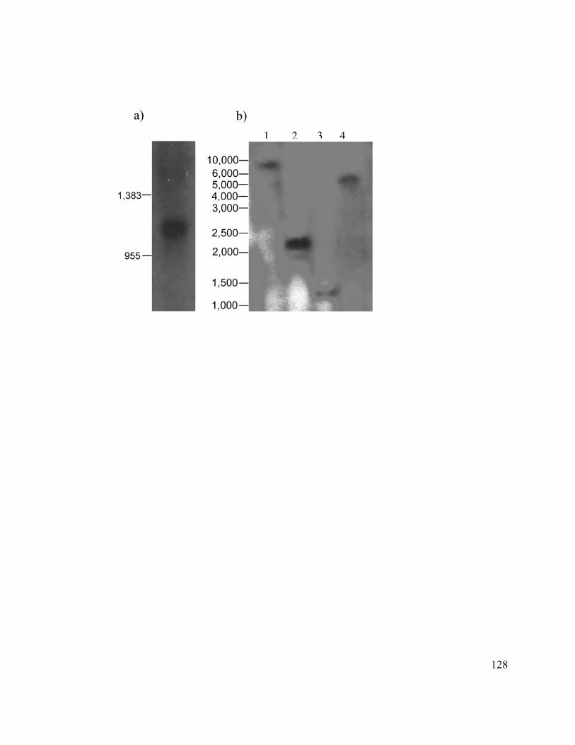
128
a) b) 1 2 3 4

129
Figure 8. Detection of C002 mRNA in the salivary glands by in situ hybridization.
Paraformaldehyde fixed salivary glands were treated with a digoxigenin (DIG)-labeled
RNA, antisense (A and B) and control, using sense probe (C and D) followed by
incubation with an anti-DIG antibody.

130
131
Figure 9. Immunohistochemical localization of C002 protein.
Localization of C002 protein within principal salivary gland of the pea aphid. a) TO-
PRO-3 staining of nuclei and Cy-3 antibody staining of C002 protein of principal and
accessory salivary glands (at 20X magnification). b) Cy-3 antibody staining of C002
protein of principal salivary glands under higher magnification (40X) showing C002
protein in secretory vesicles in four positively stained cells. (c) Negative control using
pre-immune rabbit serum instead of polyclonal C002 antibody.
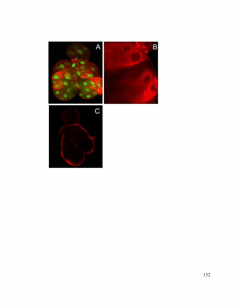
132
133
Figure 10. Detection of C002 protein using purified C002 antibodies.
a) Western blot indicating that C002 is injected into the fava bean plants during feeding.
Lane 1. Recombinant C002 protein. Lane 2. Fava bean plant without aphid feeding,
which serves as negative control. Lane 3. Fava bean plant extract after pea aphid feeding.
Lane 4. Protein extract from 5 aphid heads. Lane 5. Protein extract from 5 salivary
glands. b) Negative control using pre-immune rabbit serum instead of polyclonal C002
antibody. Lanes 1 and 2. Fava bean plant extract without aphid feeding. Lane 3. Fava
bean plant extract after aphid feeding.
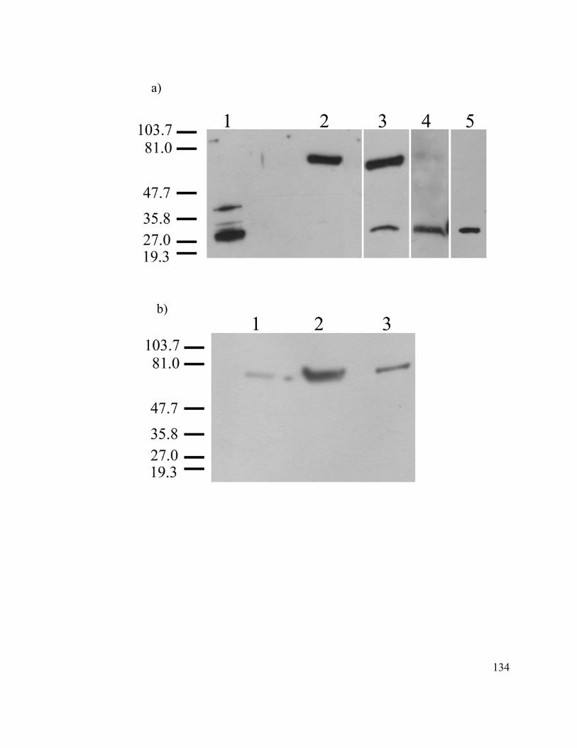
134
a)
b)
135
Figure 11. Survival of siRNA injected aphids on artificial diet.
siRNA injected aphids kept on aphid diet for 7 days after injection, thereafter, they were
moved to fava bean leaves in Petri plates on agar. Green line: injections with siC002-
RNA; Red line: injections with siGFP-RNA (control). Data from 3 independent
experiments are shown; there were 12 aphids in each experiment. The bars depict
standard errors.

136
0 2 4 6 8 10 12 14 16 18 200
2
4
6
8
10
12
14
On diet On leaves in agar
Days after injection
Num
ber o
f liv
e ap
hids

137
CHAPTER 4
138
Chapter 4: RNAi Knockdown of a Salivary Transcript Leading to Lethality in the
Pea Aphid, Acyrthosiphon pisum.
Navdeep S. Mutti1,2, Yoonseong Park1, John C. Reese1, Gerald R. Reeck2*
1 Department of Entomology, Kansas State University, Manhattan, Kansas 66506
2 Department of Biochemistry, Kansas State University, Manhattan, Kansas 66506
Key words: pea aphid, salivary gland, RNA interference, RNAi, small interfering RNA,
siRNA
Abbreviations: RNAi, RNA interference; siRNA, small interfering RNA; dsRNA,
double-stranded RNA; PCR, polymerase chain reaction; RT-PCR, reverse-transcriptase
polymerase chain reaction; EST, expressed sequence tag; GFP, green fluorescent protein;
NCBI, National Center for Biotechnology Information
139
Abstract
Injection of siRNA (small interfering RNA) into parthenogenetic adult pea aphids
(Acyrthosiphon pisum) is shown here to lead to depletion of a target salivary-gland
transcript. The siRNA was generated from double stranded RNA that covered most of the
open reading frame of the transcript, which we have called C002. The C002 transcript
level decreases dramatically over a 3-day period after injection of siRNA. With a lag of 1
to 2 days, the siC002-RNA injected insects died, on average 8 days before the death of
control insects injected with siRNA for green fluorescent protein. It appears, therefore,
that siRNA injections into adults will be a useful tool in studying the roles of individual
transcripts in aphid salivary glands, and our results suggests that siC002-RNA injections
can be a useful positive control in such studies.
140
Introduction
Double-stranded RNA (dsRNA), when injected into or ingested by an organism or
introduced into cells in culture, can specifically lower the level of the transcript of a
target gene. This method, initially documented in C. elegans and named RNA
interference, or RNAi, has become a very powerful tool to examine the role of individual
genes (Fire et al. 1998; Zamore et al. 2000). Among insects, injections of interfering
RNAi in post-embryonic stages have been used successfully in the honeybee, Apis
mellifera (Amdam et al. 2003), the moths Hyalophora cecropia (Bettencourt et al. 2002),
Spodoptera litura (Rajagopal et al. 2002), Bombyx mori (Uhlirova et al. 2003) and
Manduca sexta (Levin et al. 2005), the mosquitoes, Anopheles gambiae (Osta et al. 2004)
and Aedes aegypti (Attardo et al. 2003), the fruit fly, Drosophila melanogaster (Goto et
al. 2003), a grasshopper, Schistocerca americana (Dong and Friedrich 2005), the red
flour beetle Tribolium castaneum (Arkane et al. 2005a and 2005b, Tomoyasu et al. 2005,
) and a termite, Reticulitermes flavipes (Zhou et al. 2006). ). Evidently, in some insect
species, injected interfering RNA can move from the hemolymph into tissues or organs,
where it then exerts its transcript-lowering effect, presumably by promoting degradation
of the target mRNA. Extending this method to an aphid, and in particular, to the aphid
salivary gland, is the objective of the work reported here.
Relatively long dsRNA is often used as an interfering RNA, but in two cases listed above
(Levin et al. 2005 and Zhou et al. 2006) small interfering RNA (siRNA) was injected
instead. siRNAs of 21-base pairs are highly effective in eliciting RNAi (Elbashir et al.
141
2001) and can be produced in vitro using the enzyme Dicer, a form of RNase III and the
enzyme believed to produce siRNA in vivo (Bernstein et al. 2001).
Saliva is very important in the interaction between aphids and host plants. Proteins,
including enzymes, of aphid saliva have been postulated to play several roles, including
the formation of a sheath around the stylets, the creation of an extracellular path by the
stylet, overcoming plant defense and possibly stimulating plant defense in non-host plants
(Miles 1999; Cherqui and Tjallingii 2000). As transcripts of potential interest are found in
cDNA libraries of aphid salivary glands (for instance, among the roughly 4500 pea aphid
salivary expressed sequence tags (ESTs) recently deposited at NCBI as accession
numbers DV747494 through DV752010), a method will be needed to examine the
importance of transcripts of individual genes, and RNAi is a potentially powerful
approach for doing so.
The most abundant cDNA from a salivary gland cDNA library prepared from the
peaaphid, Acrythosiphon pisum (Reeck et al., unpublished observations), was selected as
the target transcript for this study. This cDNA was arbitrarily designated C002 (i.e.,
Cluster 2). This transcript was also found, infrequently, among whole-body and whole-
insect pea aphid ESTs, where there are 7 occurrences in approximately 17,800 such ESTs
deposited at the National Center for Biotechnology Information (Sabeter-Munoz et al.
2006). As a point of comparison, ESTs for cytochrome oxidase subunit-1, a widely
distributed protein, occur over 160 times among the whole-body and whole-insect pea
aphid ESTs. The translated nucleotide sequence of C002 does not match other sequences

142
except in aphids, where there are matches to translated ESTs from Aphis gossypii and
Toxoptera citricida. The entire open reading frame of the C002 transcript can be found at
accession number CN763138. The encoded pea aphid protein includes a predicted signal
peptide, and the protein is of entirely unknown function.
To test the efficacy of RNAi in aphids, siRNA coding for C002 was injected into adult
aphids. It was found that injection of siRNA leads to knockdown of the C002 transcript
level in salivary glands and results in a greatly reduced lifespan of the injected insects.
These results provide the basis for the use of this technique in studies of other salivary
transcripts and, possibly, transcripts in other organs.
Materials and Methods
Plants and Aphids
Aphids were originally collected from alfalfa plants in the summer of 1999 by Dr. Marina
Caillaud at Cornell University. Thereafter, the aphids were reared at KSU on fava beans
(Vicia fabae) grown in pots (10 cm diameter) at room temperature under high intensity
sodium lights with a L:D of 16:8. For the RNAi experiments, even-aged cohorts were
established by collecting nymphs from young parthenogenetic females over a 24-h
period. Cohorts formed in this way were maintained on plants for 7 days and then used
for siRNA injections.

143
Preparation of dsRNA and siRNA
PCR primers with T7 promoter sequences were used to prepare double-stranded RNA
(Tomoyasu et al. 2005). For C002 RNA, the primers had the following sequences: 5'--
TAA TAC GAC TCA CTA TAG GGA AGT TA--3' and 5'--TAA TAC GAC TCA CTA
TAG GGA AAC TT--3' (forward and reverse, respectively). The two primers cover a
region that extends from position 5 to position 637 in the open reading frame that, in its
entirety, is 660 bases. Primers for green fluorescent protein RNA that were used in
controls had the following sequences: 5'--TAA TAC GAC TCA CTA TAG GGC GAT
GC--3' and 5'--TAA TAC GAC TCA CTA TAG GGC GGA CT -- 3' (forward and
reverse, respectively). These cover a region of 520 bases in the open reading frame for
the green fluorescent protein.
PCR products were gel purified using Qiaquick Gel Extraction Kit (Qiagen, Valencia,
CA). dsRNA was then made using Megascript RNAi Kit (Ambion, Austin, TX)
following the manufacturer’s protocol. dsRNA was purified using phenol:choloform
extraction. siRNA was generated from dsRNA using the Dicer siRNA Generation Kit
T5200002 (Genetherapy Systems) and purified using siRNA purification columns of
Genlantis. Products of Dicer digestion were checked for size (21 – 23 base pairs) on 15%
acrylamide gels.
siRNA Injections
Glass needles (outer diameter of 1.0 mm, inner diameter of 0.50 mm; Sutter Instruments,
Navato, CA) were made using a micropipette puller (Model P-87, Sutter Instruments) at
144
settings of: heat, 355; velocity, 50; time, 150. A PMI-200 pressure microinjector (Dagan)
was used for siRNA injections. Aphids were held on their dorsa over a small hole in
piece of plastic tubing (5 mm inner diameter) that was blocked at one end, held in place
on a flat surface and connected at the other end to a small vacuum pump (Pro-Craft,
Grobet USA, Carlstadt, NJ). Aphids were injected at the suture joining the ventral
mesothorax and metathorax, at an angle of about 45 degrees, aimed toward the head of
the aphid. We estimate that 5 nl of siRNA (10µg/µl) was injected into each aphid.
Leaves were cut from healthy, intact fava beans and put into a sterilized 2% agar (Fisher
Scientific) supplemented with 0.1% Miracle Grow fertilizer and 0.03% methyl 4-
hydroxybenzoate (Sigma-Aldrich, St. Louis, MO) as a fungicide, essentially as described
on the David Stern website (http://www.princeton.edu/%7Edstern/PlatesProtocol.htm) for
Medicago leaves. About 7 ml of medium was placed in a Petri plate (100 x 15 mm) and
one leaf was inserted into the agar as it cooled. One injected aphid was placed on each
leaf. The plates were placed under GE Utility Shoplite with F48/25 watt/UTSL
fluorescent lights, with 16:8 L:D, at a temperature of 23o C. Plates were checked several
times a day for dead aphids, which were identified by lack of movement, being off the
leaves and, after several hours, darkened coloration.
Examining Transcript Levels by RT-PCR
Total RNA was isolated from individual pea aphid heads using TRI reagent (Molecular
Research Center, Inc. Cincinnati, OH) following the procedure provided by the
manufacturer. RNA was treated with DNase I (Ambion, Austin, TX) following the
145
company’s instructions. AMV reverse transcriptase was used with oligo-dT primers to
synthesize single-stranded cDNA following the procedure in Technical Bulletin 099 of
Promega. PCR was done using 5'--CCA GTG CGA TAG CGA TAA TTT ACA AC--3'
and 5'--CAC CTC TCT TAT GAT GAA CGC CAA C--3' for C002 forward and reverse
primers, respectively, giving a final product of 397 base pairs, and using 5'--CCG AAA
AGC TGT CAT AAT GAA GAC C--3' and 5'--GGT GAA ACC TTG TCT ACT GTT
ACA TCT TG--3' for ribosomal protein L27 forward and reverse, primers, respectively,
giving a final product of 231 base pairs. The sequence of pea aphid L27 has accession
number CN584974. Both primer pairs were used in each PCR, with L27 serving as an
internal control. PCR was performed for one cycle at 95o C for 2 min followed by 26
cycles of: 95o C for 30 s, 54o C for 30 s and 72o C for 35 s. Primers were used at 0.3 µM
and PCR master mix from Promega (Madison, WI) was used in a final volume of 50 µl.
PCR products were separated on 1% agarose gels prepared in 40 mM Tris-acetate (pH
8.3) and 1 mM EDTA. Ethidium bromide was added to a final concentration of 0.7 µg/ml
before allowing the agarose to solidify. The gels were photographed under ultraviolet
light and band intensities were obtained using SigmaScan’s Pro5 image measurement
software.
Results and Discussion
Injection of siC002-RNA into adult parthenogenetic pea aphids led to greatly reduced
life-span, as shown in Figure 1. Aphids injected with siC002-RNA died well before
control aphids injected with green fluorescent protein si-RNA. Half of the aphids injected
with siC002-RNA had died at 3 days after injection whereas 11 days was required for
146
death of half of the aphids injected with control siRNA. The survival of uninjected aphids
was similar to that of aphids injected with control siRNA, indicating that the small
injection wound was tolerated by the aphids and that injection of control siRNA and
buffer components not have a toxic effect.
RT-PCR was used to assess C002 transcript levels in RNA extracted from heads. The
signal obtained by RT-PCR using head RNA as template is from transcripts in salivary
glands, since using RNA from heads from which salivary glands had been removed by
injected aphids are shown in Fig. 2. Using the transcript for ribosomal protein L27 as
internal control, we found that transcript levels from C002 dropped dramatically within 3
days after injection with siC002-RNA. On the other hand, in control siRNA injected
insects, C002 transcript levels, normalized to L27 transcript levels, did not change
significantly.
Semiquantitative data from such measurements are plotted in Figure 3 (open triangles),
where, for purposes of comparison of the timing, the decline in the number of live insects
(from Fig. 1) is again shown. We note that the measurements of C002 transcript levels
may somewhat overestimate the levels of the transcript and thus underestimate the timing
of the knockdown, since in examining transcript levels only insects that were alive at
each time point were used. Insects that had already died might well have had extensive
knockdown of the C002 transcript.
147
These data demonstrate that siRNA injection into adult pea aphids can lower the
transcript level of a target gene, C002, expressed in the salivary gland. The injections
have a profound effect on lifespan, lowering the time to 50% survival from 11 days in
control injections to about 3 days for injection of siC002-RNA. Several questions remain
unanswered. Will the same procedure work for other salivary transcripts? There are no
data at this time regarding this point, but we will soon undertake siRNA studies on other
salivary transcripts. Will the same procedure work for lowering transcript levels in other
organs? Again, there are no data on this point. Judging from results in other insects, it
seems likely that gut (Rajagapol et al. 2002, Osta et al. 2004, Arakane et al. 2005), fat
body (Attardo et al. 2003, Amdam et al. 2003) and hemocytes (Levin et al. 2005) would
be sensitive to the effects of injected interfering RNA. Does siRNA enter aphid embryos
developing within the injected adult female? We have not studied this systematically,
but, informally, we have noticed that a significant fraction of nymphs from adults
injected with siC002 RNA do die prematurely. This suggests the possibility that siRNA
does indeed enter at least some embryos.
Unanswered by these experiments is the function of C002. We had no way of anticipating
the profound effect of knockdown of the C002 transcript and in light of that effect, we
will be investigating the function of C002 in ongoing experiments. One interesting piece
of information that we have at this point is that siC002-RNA-injected aphids exhibit a
peculiar behavior. Whereas uninjected aphids or aphids injected with siGFP-RNA stay
quite still on the underside of the fava bean leaves, the siC002-RNA-injected aphids
move around a good deal and do not stay confined to the underside of the leaves. Using
148
electrical penetration graph methods we hope to understand the effect of C002 transcript
knockdown on detailed aspects of the insects’ feeding behavior.
Acknowledgments
We thank our colleagues Subbaratnam Muthukrishnan, Michael Kanost and Maureen
Gorman for valuable discussions and suggestions. This work was supported by USDA-
CREES NRI grant 2001-35302-09975 and by the Kansas Agricultural Experiment Station
(publication number 05-XXX-J). Aphids (voucher specimen number-173) were deposited
at the Kansas State University Museum of Entomological and Prairie Arthropod
Research.
149
References
Amdam, G.V., Simoes, Z.L.P., Guidugl,i K.R., Norberg, K. and Omholt, S.W. 2003.
Disruption of vitellogenin gene function in adult honeybees by intra-abdominal
injection of double-stranded RNA. BMC Biotechnology, 3:1.
Arkane, Y., Muthukrishnan, S., Kramer, K.J., Specht, C.A., Tomoyasu, Y., Lorenzen,
M.D., Kanost, M. and Beeman, R.W. 2005a. The Tribolium chitin synthase genes
TcCHS1 and TcCHS2 are specialized for synthesis of epidermal cuticle and
midgut peritrophic matrix. Insect Molecular Biology, 14:453-463.
Arakane, Y., Muthukrishnan, S., Beeman, R.W., Kanost, M.R. and Kramer, K.J. 2005b.
Laccase 2 is the phenoloxidase gene required for beetle cuticle tanning.
Proceedings of the National Academy of Sciences USA, 102:11337-11342.
Attardo, G.M., Higgs, S., Klingler, K.A., Vanlandingham, D.L. and Raikhel, A.S. 2003.
RNA interference-mediated knockdown of a GATA factor reveals a link to
anautogeny in the mosquito Aedes aegypti. Proceedings of the National Academy
of Sciences USA, 100:13374-13379.
Bernstein, E., Caudy, A.A., Hammond, S.M. and Hannon, G.J. 2001. Role for a bidentate
ribonuclease in the initiation step of RNA interference. Nature, 409:363-366.
Bettencourt, R., Terenius, O. and Faye, I. 2002. Hemolin gene silencing by ds-RNA
injected into Cecropia pupae is lethal to next generation embryos. Insect
Molecular Biology, 11:267-271.
Cherqui, A. and Tjallingii, W.F. 2000. Salivary protein of aphids, a pilot study on
identification, separation and immunolocalisation. Journal of Insect Physiology,
46:1177-1186.

150
Dong, Y. and Friedrich, M. 2005. Nymphal RNAi: systemic RNAi mediated gene
knockdown in juvenile grasshopper. BMC Biotechnology, 5:25.
Elbashir, S.M., Harborth, J., Lendeckel, W., Yalcin, A., Weber, K. and Tuschl, T. 2001.
Duplexes of 21-nucleotide RNAs mediate RNA interference in cultured
mammalian cells. Nature, 411:495-498.
Fire, A., Xu, S., Montgomery, M.K., Kostas, S.A., Driver, S.E. and Mello, C.C. 1998.
Potent and specific genetic interference by double-stranded RNA in
Caenorhabditis elegans. Nature, 391:806-811.
Goto, A., Blandin, S., Royet, J., Reichhart, J-M. and Levanshina, E.A. 2003. Silencing of
Toll pathway components by direct injection of double-strated RNA into
Drosophila adult flies. Nucleic Acids Research, 22:6619-6623.
Levin, D.M., Breuer, L.N., Zhuang, S., Anderson, S.A., Nardi, J.B. and Kanost, M.R.
2005. A hemocyte-specific integrin required for hemocytic encapsulation in the
tobacco hornworm, Manduca sexta. Insect Biochemistry and Molecular Biology,
35:369-80.
Miles, P.W. 1999. Aphid saliva. Biological Reviews of the Cambridge Philosophical
Society, 74:41-85.
Osta, M,A., Christophides, G.K. and Kafatos, F.C. 2004. Effects of mosquito genes on
Plasmodium development. Science, 303:2030-2032.
Rajagopal, R., Sivakumar, S., Agrawal, N., Malhotra, P. and Bhatnagar, R.K. 2002.
Silencing of midgut aminopeptidase N of Spodoptera litura by double-stranded
RNA establishes its role as Bacillus thuringiensis toxin receptor. The Journal of
Biological Chemistry, 277:46849-46851.
151
Sabater-Munoz, B., Legeai, F., Rispe, C., Bonhomme, J., Dearden, P., Dossat, C.,
Duclert, A., Gauthier, J-P., Ducray, D.G., Hunter, W., Dang, P., Kambhampati,
S., Martinez-Torrez, D., Cortes, T., Moya, A., Nakabachi, A., Philippe, C.,
Prunier-Leterme, N., Rahbe, Y., Simon, J-C., Stern, D.L., Wincker, P. and Tagu,
D. 2006. Large-scale gene discovery in the pea aphid Acyrthosiphon pisum
(Hemiptera). Genome Biology, 7:R21.
Tomoyasu, Y., Wheeler, S.R. and Denell, R.E. 2005. Ultrabithorax is required for
membranous wing identity in the beetle Tribolium castaneum. Nature, 433:643-
647.
Uhlirova, M., Foy, B.D., Beaty, B.J., Olson, K.E., Riddiford, L.M. and Jindra, M. 2003.
Use of Sindbis virus-mediated RNA interference to demonstrate a conserved role
of Broad-Complex in insect metamorphosis. Proceedings of the National
Academy of Sciences USA, 100:15607-15612.
Zamore, P.D., Tuschl, T., Sharp, P.A. and Bartel, D.P. 2000. RNAi: Double-stranded
RNA directs the ATP-dependent cleavage of mRNA at 21 to 23 nucleotide
intervals. Cell, 101:25-33.
Zhou, X., Oi, F.M. and Scharf, M.E. 2006. Social exploitation of hexamerin: RNAi
reveals a major caste-regulatory factor in termites. Proceedings of the National
Academy of Sciences USA, 103: 4499-4504.
152
Figure 12. Survival of pea aphids after injection of siRNA.
The graph shows the number of surviving aphids at daily intervals after injection. Green
line and open triangles: injections with siC002-RNA. Red line and closed triangles:
injections with siGFP-RNA (control). Blue line and open squares: uninjected insects. For
the siRNA-injected insects, the data points are averages from three experiments, each of
which began with 12 insects in the experimental and control groups. The bars depict
standard errors.
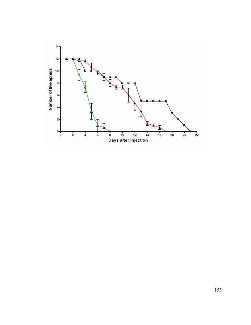
153
154
Figure 13. Knockdown of the C002 transcript after siRNA injections.
Insects were injected either with siC002-RNA or siGFP-RNA. RNA from heads of
injected insects was used in RT-PCR in which two primer-pairs were included, for C002
itself and for the transcript encoding ribosomal protein L27. The L27 PCR product serves
as an internal control. The results shown (agarose gels after ethidium bromide staining)
are of individual aphids at time points from 1 to 5 days after injection. Panel A: PCR
products from reactions (26 cycles) with head RNA from siC002-RNA-injected insects.
Panel B: PCR products from reactions (26 cycles) with head RNA from siGFP-RNA-
injected insects. Panel C: PCR products from reactions (26 cycles) carried out on RNA
extracted from heads from which salivary glands had been removed. Analysis of samples
from 4 separate insects are shown.
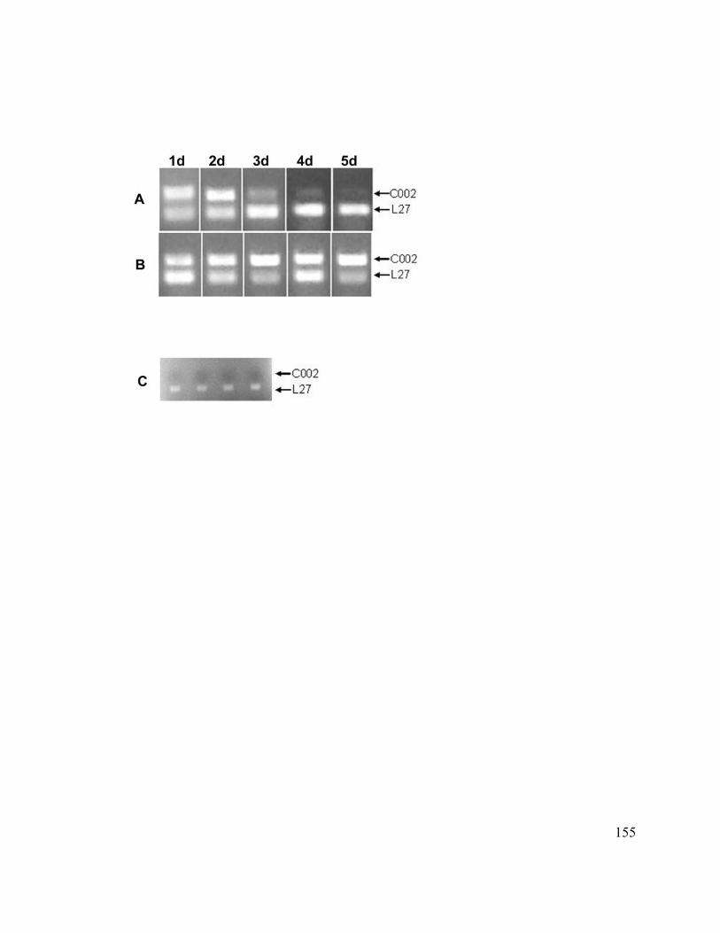
155
156
Figure 14. Timing of knockdown of C002 transcript after siRNA injection.
Data from RT-PCR analysis of C002 transcript levels (normalized against L27 transcript
levels) are plotted over a several day period after injection with either siC002-RNA (blue
line, closed triangles) or siGFP-RNA (red line, open squares). Data are averages of
normalized intensities from several individual insects at each time point and bars depict
standard errors. The green line (open triangles) shows the survival of siC002-RNA-
injected insects from Figure 1.
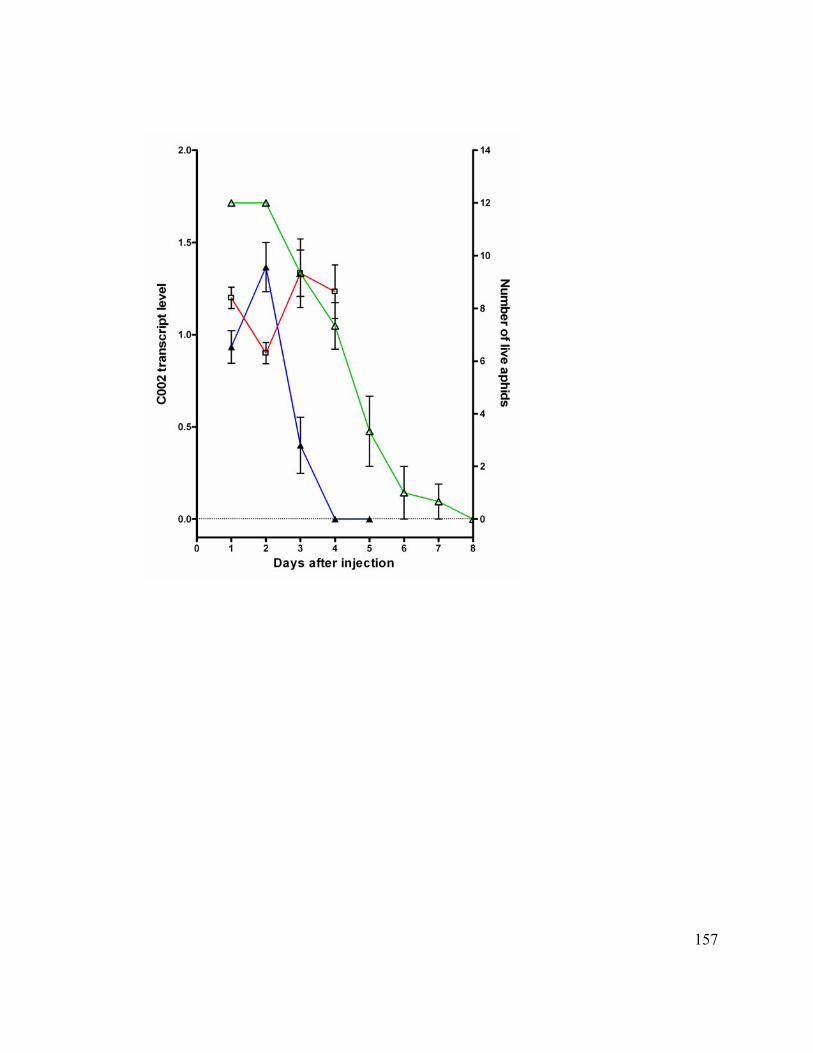
157
158
Appendix 1
Supplemental Information to Chapter 4
Injection of siC002-RNA into adult parthenogenetic pea aphids led to greatly reduced
life-span, as shown in Fig. 15. Aphids injected with siC002-RNA died well before
aphids injected with siGFP-RNA (as a control). Results from 3 experiments (each with 12
replications) at different time intervals (Feb 03, Feb 13 and Feb 18) and 3 experiments
with aged cohorts (March 20, May 21 and June 13) also with 12 replication for
experimental group (siC002-RNA injections) and control group (siGFP-RNA injections)
are shown in Fig. 15. Half of the aphids injected with siC002-RNA had died at about 3
days after injection whereas 11 days was required for death of half of the aphids injected
with siGFP-RNA. The survival of uninjected aphids was similar to that of aphids
injected with siGFP-RNA, indicating that the wound created was tolerated by the aphids
and that injection of control siRNA and buffer components not have toxic effects.
RT-PCR data from individual aphid heads injected with siC002-RNA is shown in Fig.
16a and with siGFP-RNA injections in Figure 16b. There was no effect on the transcript
level within 48 h of injection but the transcript levels decrease rapidly thereafter to almost
undetectable levels at 120 h after siC002-RNA injections (Fig. 16a). On the other hand
siGFP-RNA injections had no effect on transcript levels of C002 (Fig. 16b).
159
Suppression of C002 transcript had a drastic effect on the reproduction of siC002-RNA
injected aphids, when compared with siGFP-RNA injected aphids. Different experiments
conducted over time and with different experimental set-up are shown in Fig. 17(a, b, c
and d). Aphids injected with dsC002-RNA had on an average of 1.5 – 2.0 nymphs / day
of life, whereas, dsGFP-RNA injected aphids has 3.5 – 4.5 nymphs / day of life (Fig.
17a). RT-PCR on dsC002-RNA injected aphids did not show reduction in transcript
levels. Therefore, injections were done using siRNA instead of dsRNA and RT-PCR
analysis on siC002-RNA injected aphids showed the reduction in the transcript level.
Injections done at various time points (Nov 27, Dec 06, Dec 22, Jan 09 and Jan 26) show
a dramatic decrease in the reproduction of siC002-RNA injected aphids. Aphids injected
with siC002-RNA had on an average 0.5 – 1.5 nymphs / day of life, when compared to
4.0 – 5.0 nymphs / day of life, in case of siGFP-RNA injected aphids (Fig. 17b). For
these experiments, data on the survival was not thoroughly recorded, as the experiment
was terminated, when all siC002-RNA injected aphids were dead.
Reproduction data from 3 different experiments done on Feb 03, Feb 13 and Feb 18, and
the corresponding survival data on these injected aphids is part of Fig. 15 (yellow and
orange line for siC002-RNA and siGFP-RNA injected aphids respectively) is shown in
Fig. 17c. siC002-RNA injected aphids had an average of 1.3 - 1.8 nymphs / day of life,
whereas siGFP-RNA injected aphids had 3.9 – 4.6 nymphs / day of life (Fig. 17c).
Finally 3 different experiments with aged cohorts are shown in Fig. 17d. All the injected
aphids were of same age i.e. 7-8 days old. siC002-RNA injected aphids had 0.7 – 1.1
160
nymphs / day of life when compared to 4.4 – 4.7 nymphs / day of life in case of siGFP-
RNA injected aphids (Fig. 17d).
Behavior of the siC002-RNA injected aphids was considerably different from siGFP-
RNA injected aphids. siC002-RNA injected aphids did tend to move around (in petri-
plate, on agar and on the upper-side of the fava bean leaves). This behavior was evident
within two hours of injection in siC002-RNA injected aphids, whereas, in contrast the
siGFP-RNA injected aphids, settled down within an hour of injection and thereafter
stayed on the underside of the leaves. This behavioral change may explain the decrease in
reproduction observed in case of siC002-RNA injected aphids. It is therefore
hypothesized that the less spent on feeding on leaves led to the decrease in the survival
and reproduction of a siC002-RNA injected aphids.
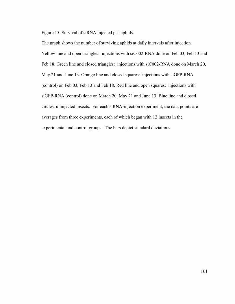
161
Figure 15. Survival of siRNA injected pea aphids.
The graph shows the number of surviving aphids at daily intervals after injection.
Yellow line and open triangles: injections with siC002-RNA done on Feb 03, Feb 13 and
Feb 18. Green line and closed triangles: injections with siC002-RNA done on March 20,
May 21 and June 13. Orange line and closed squares: injections with siGFP-RNA
(control) on Feb 03, Feb 13 and Feb 18. Red line and open squares: injections with
siGFP-RNA (control) done on March 20, May 21 and June 13. Blue line and closed
circles: uninjected insects. For each siRNA-injection experiment, the data points are
averages from three experiments, each of which began with 12 insects in the
experimental and control groups. The bars depict standard deviations.
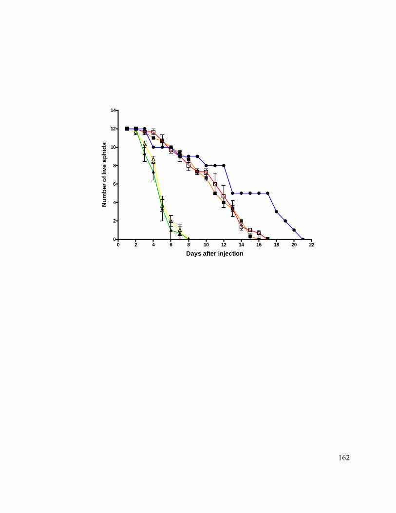
162
0 2 4 6 8 10 12 14 16 18 20 220
2
4
6
8
10
12
14
Days after injection
Num
ber o
f liv
e ap
hids
163
Figure 16. Knockdown of the C002 transcript after siRNA injections.
Insects were injected either with a) siC002-RNA b) siGFP-RNA. RNA from heads of
injected insects was used in RT-PCR in which two primer-pairs were included, for C002
itself and for the transcript encoding ribosomal protein L27. The L27 PCR product
serves as an internal control. The results shown are of individual aphids at time points
from 24hours to 120 hours after injection. PCR products from reactions (26 cycles) with
head RNA either from siC002-RNA-injected insects (a) or from siGFP-RNA-injected
insects (b) are shown after agarose gel electrophoresis and ethidium bromide staining.
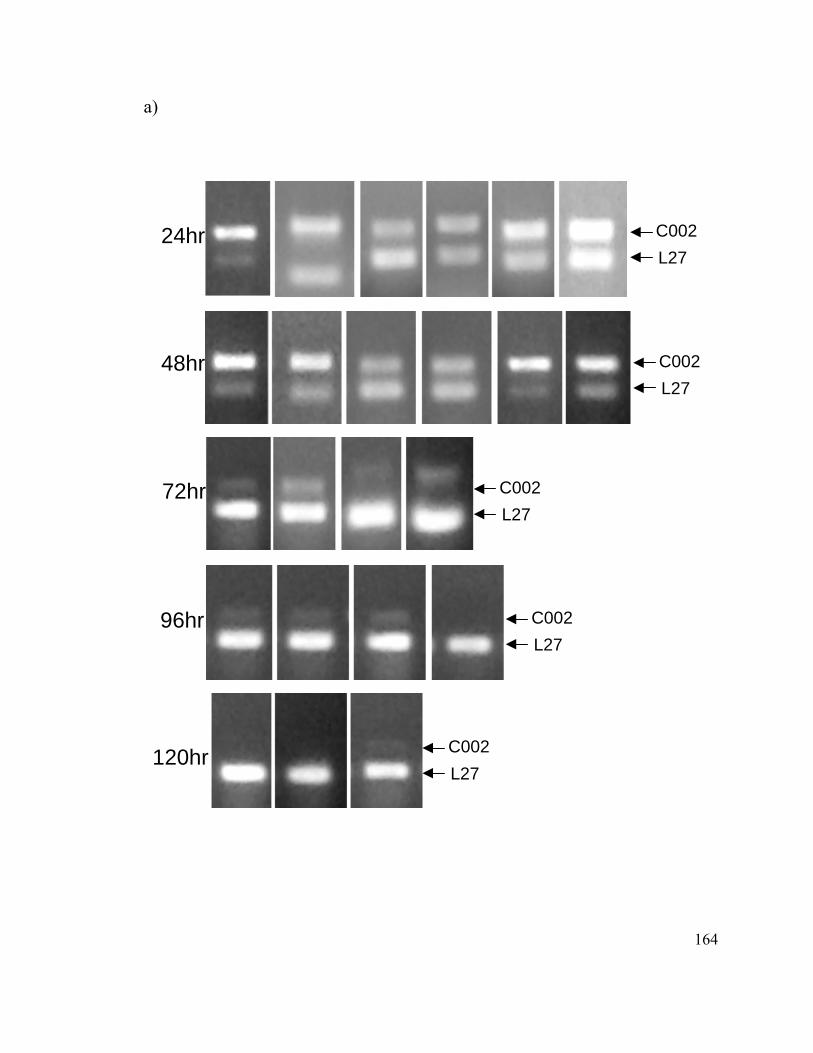
164
a)
24hr
48hr
72hr
96hr
120hr
C002L27
C002L27
C002L27
C002 L27
C002L27
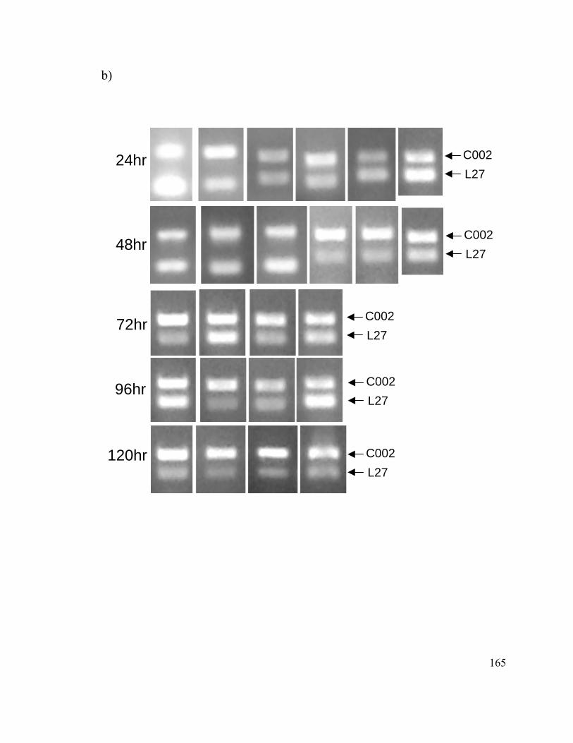
165
b)
24hr
48hr
72hr
96hr
120hr
C002L27
C002L27
C002L27
C002L27
C002L27
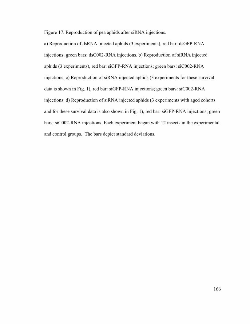
166
Figure 17. Reproduction of pea aphids after siRNA injections. a) Reproduction of dsRNA injected aphids (3 experiments), red bar: dsGFP-RNA
injections; green bars: dsC002-RNA injections. b) Reproduction of siRNA injected
aphids (3 experiments), red bar: siGFP-RNA injections; green bars: siC002-RNA
injections. c) Reproduction of siRNA injected aphids (3 experiments for these survival
data is shown in Fig. 1), red bar: siGFP-RNA injections; green bars: siC002-RNA
injections. d) Reproduction of siRNA injected aphids (3 experiments with aged cohorts
and for these survival data is also shown in Fig. 1), red bar: siGFP-RNA injections; green
bars: siC002-RNA injections. Each experiment began with 12 insects in the experimental
and control groups. The bars depict standard deviations.
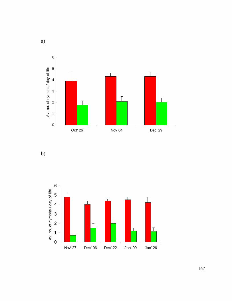
167
a)
0
1
2
3
4
5
6
Oct' 26 Nov' 04 Dec' 29
Av.
no.
of n
ymph
s / d
ay o
f life
s
b)
0
1
2
3
4
5
6
Nov' 27 Dec' 06 Dec' 22 Jan' 09 Jan' 26
Av. n
o. o
f nym
phs
/ day
of l
ife
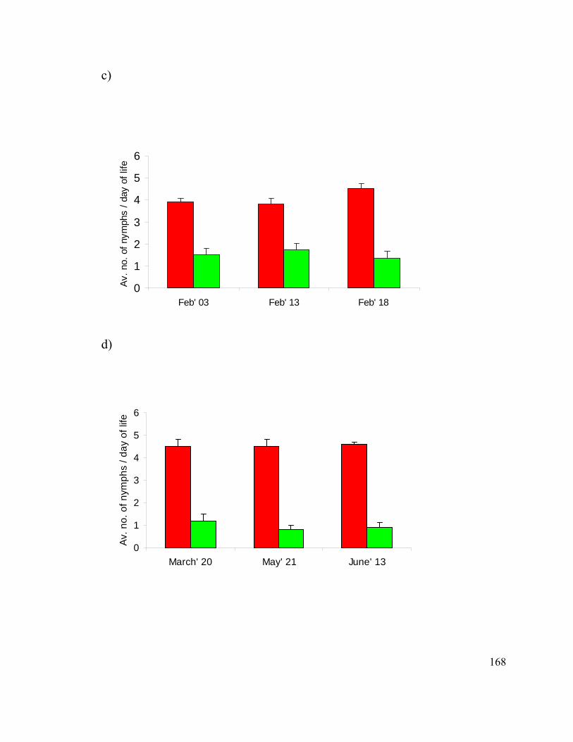
168
c)
0
1
2
3
4
5
6
Feb' 03 Feb' 13 Feb' 18
Av. n
o. o
f nym
phs
/ day
of l
ife
d)
0
1
2
3
4
5
6
March' 20 May' 21 June' 13
Av. n
o. o
f nym
phs
/ day
of l
ife
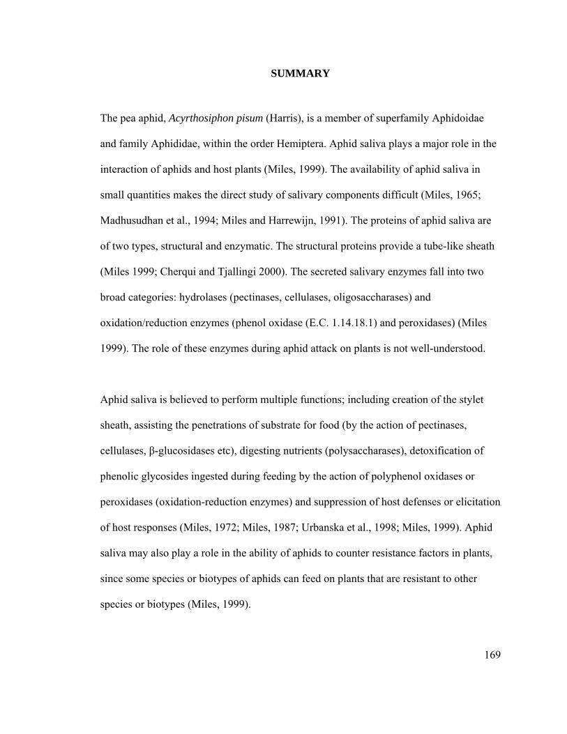
169
SUMMARY
The pea aphid, Acyrthosiphon pisum (Harris), is a member of superfamily Aphidoidae
and family Aphididae, within the order Hemiptera. Aphid saliva plays a major role in the
interaction of aphids and host plants (Miles, 1999). The availability of aphid saliva in
small quantities makes the direct study of salivary components difficult (Miles, 1965;
Madhusudhan et al., 1994; Miles and Harrewijn, 1991). The proteins of aphid saliva are
of two types, structural and enzymatic. The structural proteins provide a tube-like sheath
(Miles 1999; Cherqui and Tjallingi 2000). The secreted salivary enzymes fall into two
broad categories: hydrolases (pectinases, cellulases, oligosaccharases) and
oxidation/reduction enzymes (phenol oxidase (E.C. 1.14.18.1) and peroxidases) (Miles
1999). The role of these enzymes during aphid attack on plants is not well-understood.
Aphid saliva is believed to perform multiple functions; including creation of the stylet
sheath, assisting the penetrations of substrate for food (by the action of pectinases,
cellulases, β-glucosidases etc), digesting nutrients (polysaccharases), detoxification of
phenolic glycosides ingested during feeding by the action of polyphenol oxidases or
peroxidases (oxidation-reduction enzymes) and suppression of host defenses or elicitation
of host responses (Miles, 1972; Miles, 1987; Urbanska et al., 1998; Miles, 1999). Aphid
saliva may also play a role in the ability of aphids to counter resistance factors in plants,
since some species or biotypes of aphids can feed on plants that are resistant to other
species or biotypes (Miles, 1999).
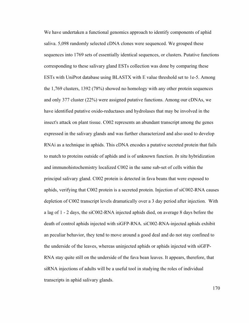
170
We have undertaken a functional genomics approach to identify components of aphid
saliva. 5,098 randomly selected cDNA clones were sequenced. We grouped these
sequences into 1769 sets of essentially identical sequences, or clusters. Putative functions
corresponding to these salivary gland ESTs collection was done by comparing these
ESTs with UniProt database using BLASTX with E value threshold set to 1e-5. Among
the 1,769 clusters, 1392 (78%) showed no homology with any other protein sequences
and only 377 cluster (22%) were assigned putative functions. Among our cDNAs, we
have identified putative oxido-reductases and hydrolases that may be involved in the
insect's attack on plant tissue. C002 represents an abundant transcript among the genes
expressed in the salivary glands and was further characterized and also used to develop
RNAi as a technique in aphids. This cDNA encodes a putative secreted protein that fails
to match to proteins outside of aphids and is of unknown function. In situ hybridization
and immunohistochemistry localized C002 in the same sub-set of cells within the
principal salivary gland. C002 protein is detected in fava beans that were exposed to
aphids, verifying that C002 protein is a secreted protein. Injection of siC002-RNA causes
depletion of C002 transcript levels dramatically over a 3 day period after injection. With
a lag of 1 - 2 days, the siC002-RNA injected aphids died, on average 8 days before the
death of control aphids injected with siGFP-RNA. siC002-RNA-injected aphids exhibit
an peculiar behavior, they tend to move around a good deal and do not stay confined to
the underside of the leaves, whereas uninjected aphids or aphids injected with siGFP-
RNA stay quite still on the underside of the fava bean leaves. It appears, therefore, that
siRNA injections of adults will be a useful tool in studying the roles of individual
transcripts in aphid salivary glands.