Histological features and histochemistry of the mucous glands ...
Upload
khangminh22Category
view
0download
0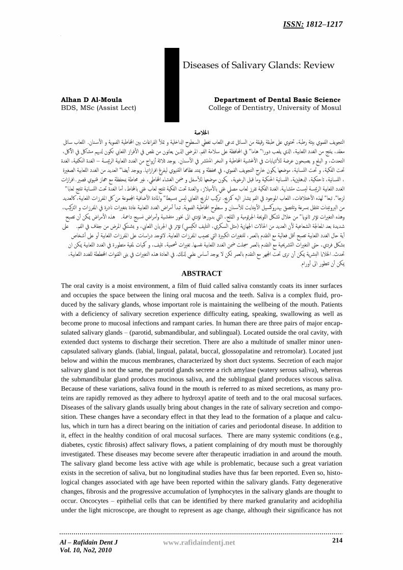
214
Alhan D Al-Moula Department of Dental Basic Science
BDS, MSc (Assist Lect) College of Dentistry, University of Mosul
اخلالضة
ة و األس نان ف امفموي تُئة رطبة، حتخوي ػىل طبلة ركِلة من امسائل ثدغى انوؼاب ثغطي امسطوح ادلاخوَة و متأل امفراغات تني اخملاطَة امفمًو انوؼاب سائل .امخجًو
املرىض اذلٍن ًؼاهون من هلص يف األفراز انوؼايب حكون دلهيم مشبلك يف األلك، . يف احملافظة ػىل سالمة امفم" ىاما" مؼلد، ًنذج من امغدد انوؼاتَة، اذلي ًوؼة دورا
امغدة امنكفِة، امغدة – ًوخد ثالثة أزواج من امغدد انوؼاتَة امرئُسة .امخحدث، و امبوع و ًطبحون غرضة مألههتاابت يف األغش َة اخملاطَة و امنخر املندرش يف األس نان
ف امفموي، يف حمفظة و ميخد هظاهما املنَوي مَفرغ افرازاهتا وخد أًضا. حتت امفكِة، و حتت انوساهَة، موضؼيا ٍكون خارج امخجًو امؼدًد من امغدد انوؼاتَة امطغرية " ًو
ة، ٍكون موضؼيا مألسفل و مضن امغشاء اخملاطي، غري حماطة مبحفظة مع هجاز كنَوي كطري ة، انوساهَة احلنكِة وما كبل امرخًو افرازات .، انوساهَة، اتحنكِة، ادلىوزًي
" امغدة امفكِة ثفرز مؼاب مطيل غين ابألمِالز، وامغدة حتت امفكِة ثنذج مؼاب غين ابخملاط، أما امغدة حتت انوساهَة ثنذج مؼااب. امغدد انوؼاتَة امرئُسة مُست مدشاهبة
واملادة األضافِة اجملموػة من لك املفرزات انوؼاتَة، اكمؼدًد " حركَة املزجي انوؼايب مُس ثس َطا. ميذه األخذالفات، انوؼاب املوحود يق امفم ٌشار امَو مكزجي" ثبؼا". مزخا
ة ثبدأ أمراض امغدد انوؼاتَة ػادة تخغريات اندرة يف املفرزات و امرتكَة، .من امربوثُنات ثنذلل ثرسػة وثوخطق هبدروكس َل األتُذاًت مألس نان و سطوح اخملاطَة امفمًو
ىذه األمراض ميكن أن ثطبح .من خالل جشلك انووحية اجلرثومِة و املوح، اميت تدورىا ثؤدي اىل خنور مذفش َة وأمراض وس َج دامعة" وىذه امخغريات ثؤثر اثهواي
ة ػىل . ثؤثر يف اجلراين انوؼايب، و ٌش خيك املرض من حفاف يف امفم (مثل امسكري، امخوَف اهكُيس)شدًدة تؼد املؼاجلة امشؼاغَة ألن امؼدًد من احلاالت اجلياًز
الثوخد دراسات ػىل املفرزات انوؼاتَة أو ػىل أشخاص . أًة حال امغدد انوؼاتَة ثطبح أكل فؼامَة مع امخلدم ابمؼمر، نوخغريات اهكبرية اميت ثطُة املفرزات انوؼاتَة
ثغريات حشمَة، ثوَف، و مكَات ملفِة مذطورة يف امغدد انوؼاتَة ميكن ان . ثشلك فردي، حىت امخغريات امدرشحيَة مع امخلدم ابمؼمر جسوت مضن امغدد انوؼاتَة هفسيا
يف امؼادة ىذه امخغريات يف تىن املنوات اخملططة نوغدد انوؼاتَة، . اخلالاي امبرشًة ميكن أن حرى حتت اجملير مع امخلدم ابمؼمر هكن ال ًوخد أساس ػومي ذلكل .حتدث
.ميكن أن ثخطور اىل أورام
ABSTRACT
The oral cavity is a moist environment, a film of fluid called saliva constantly coats its inner surfaces
and occupies the space between the lining oral mucosa and the teeth. Saliva is a complex fluid, pro-
duced by the salivary glands, whose important role is maintaining the wellbeing of the mouth. Patients
with a deficiency of salivary secretion experience difficulty eating, speaking, swallowing as well as
become prone to mucosal infections and rampant caries. In human there are three pairs of major encap-
sulated salivary glands – (parotid, submandibular, and sublingual). Located outside the oral cavity, with
extended duct systems to discharge their secretion. There are also a multitude of smaller minor unen-
capsulated salivary glands. (labial, lingual, palatal, buccal, glossopalatine and retromolar). Located just
below and within the mucous membranes, characterized by short duct systems. Secretion of each major
salivary gland is not the same, the parotid glands secrete a rich amylase (watery serous saliva), whereas
the submandibular gland produces mucinous saliva, and the sublingual gland produces viscous saliva.
Because of these variations, saliva found in the mouth is referred to as mixed secretions, as many pro-
teins are rapidly removed as they adhere to hydroxyl apatite of teeth and to the oral mucosal surfaces.
Diseases of the salivary glands usually bring about changes in the rate of salivary secretion and compo-
sition. These changes have a secondary effect in that they lead to the formation of a plaque and calcu-
lus, which in turn has a direct bearing on the initiation of caries and periodontal disease. In addition to
it, effect in the healthy condition of oral mucosal surfaces. There are many systemic conditions (e.g.,
diabetes, cystic fibrosis) affect salivary flows, a patient complaining of dry mouth must be thoroughly
investigated. These diseases may become severe after therapeutic irradiation in and around the mouth.
The salivary gland become less active with age while is problematic, because such a great variation
exists in the secretion of saliva, but no longitudinal studies have thus far been reported. Even so, histo-
logical changes associated with age have been reported within the salivary glands. Fatty degenerative
changes, fibrosis and the progressive accumulation of lymphocytes in the salivary glands are thought to
occur. Oncocytes – epithelial cells that can be identified by there marked granularity and acidophilia
under the light microscope, are thought to represent as age change, although their significance has not
ISSN: 1812–1217
Diseases of Salivary Glands: Review
Al – Rafidain Dent J
Vol. 10, No2, 2010
www.rafidaindentj.net
215
A B
been established, beside accumulation of structurally altered mitochondria. Oncocytes are found in
acini intercalated and striated ducts of salivary glands and which may give rise to neoplasms. The aim
of this review is to provide athorough knowledge of anatomy, embryology and pathophysiology in ne-
cessary to treat patient appropriately. Examines the cause, diagnostic methodology, radiographic evalu-
ation and management of a variety of salivary gland.
Keyword: Salivary gland, Histopathology of salivary glands, salivary gland disorders.
Al- Moula AD. Diseases of Salivary Glands: Review. Al–Rafidain Dent J. 2010; 10(2):214-230. Received: 11/1/2009 Sent to Referees: 12/1/2009 Accepted for Publication:16/3/2009
INTRODUCTION
Anatomy of Salivary Glands (Figure 1):
Figure (1): Anatomy of the major salivary glands. A, Parotid gland. B, Submandibular and
sublingual glands (Snell R.S.)
Parotid Gland:
The parotid gland is the largest sali-
vary glands, which lies superficial to the
posterior aspect of the masseter muscle
and the ascending ramus of the mandi-
ble.(1)
The deep part of the gland may ex-
tend forward between the medial pterygoid
process.(1)
The parotid gland is a lobulated mass
surrounded by a connective tissue capsule,
and enclosed by a dense fibrous capsule
derived from the investing layer of deep
cervical fascia.(1)
Blood Supply:
The external carotid artery and its ter-
minal branches: 1) posterior auricular arte-
ries, 2) superficial temporal 3) and trans-
verse facial arteries, while the veins drain
into the retromandibular vein.(1)
Lymph drainage:
The lymph vessels drain into the paro-
tid lymph nodes and the deep cervical
lymph nodes.(1)
Nerve supply:
Parasympathetic secretomotor fibers
from the inferior salivatory nucleus of the
ninth cranial nerve supply the parotid
gland. The nerve fibers pass to the otic
ganglion via the tympanic branch of the
ninth cranial nerve and the lesser petrosal
nerve. Postganglionic parasympathetic
fibers reach the parotid gland via the auri-
culotemporal nerve, which lies in contact
with the deep surface of the gland. Post-
ganglionic sympathetic fibers reach the
gland as a plexus of nerves around the ex-
ternal carotid artery.(1)
Submandibular Gland: The submandibular gland is located in
the submandibular triangle of the neck,
this triangle is formed by the anterior and
posterior bellies of the diagastric muscles
and the inferior border of the mandible.(1)
Blood supply:
The arteries are branches of the facial
and lingual arteries. The veins drain into
the facial and lingual veins.
Lymph drainage:
Al – Rafidain Dent J
Vol. 10, No2, 2010
Al- Moula AD
216
The lymph vessels drain into the sub-
mandibular and deep cervical lymph
nodes.(1)
Nerve supply:
Parasympathetic secretomotor supply
from the superior salivatory nucleus of the
seventh cranial nerve. The nerve fibers
pass to the submandibular ganglion and
other small ganglia close to the duct via
the chorda tympani nerve and the lingual
nerve. Postganglionic parasympathetic
fibers reach the gland either directly or
along the duct. Postganglionic sympathetic
fibers reach the gland as a plexus of nerves
around the facial and lingual arteries, from
superior cervical ganglia.(1)
Sublingual Gland: The sublingual gland is the smallest of
the three main salivary glands, it lies be-
neath the mucous membrane of the floor
of the mouth, close to the midline. The
sublingual ducts are 8-20 in number and
most of them open into the oral cavity on
the summit of the sublingual fold, but a
few may open into the submandibular
duct.(1)
Blood supply:
The gland is supplied by branches of
the facial and lingual arteries. The veins
drain into the facial and lingual veins. (1)
Lymph drainage:
The lymph vessels drain into the sub-
mandibular and deep cervical lymph
nodes. (1)
Nerve supply:
Parasympathetic secretomotor supply
from the superior salivatory nucleus of the
facial nerve. The fibers pass to the sub-
mandibular ganglion via the chorda tym-
pani nerve and the lingual nerve. Postgan-
glionic parasympathetic fibers pass to the
gland via the lingual nerve. Postganglionic
sympathetic fibers reach the gland as a
plexus of nerves around the facial and lin-
gual arteries, from superior cervical gan-
glia.(1)
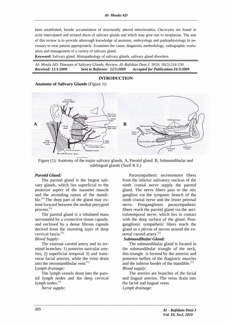
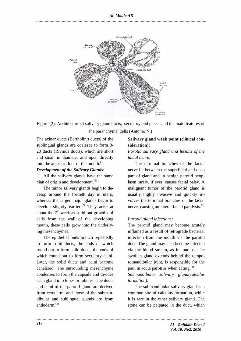
Histological feature of the Major Sali-
vary Glands:
Parotid Gland:
In the parotid gland, the spherical se-
cretory end pieces are all of serous type,
and their pyramidally shaped acinar cells
have a spherical, basally situated nucleus
and surround a small, central lumen. The
basal cytoplasm stains with basophilic
dyes, and the secretory granules in the
apical cytoplasms usually stain with aci-
dophilic dyes. Fat cell spaces are often
seen in sections of the parotid gland.(2)
In-
tercalated ducts are numerous and long in
the parotid gland, they are lined with cu-
boidal epithelial cells and have lumina that
are larger than those of acini. The striated
ducts are numerous and appear as slightly
acidophilic. The ducts consist of a simple
columnar epithelium, with round, centrally
placed nuclei, the lumina are larger rela-
tive to overall size of the ducts.(2)
Small
ducts from various regions of the gland
coalesce at the anterosuperior aspect of it
to form (major Stensen's duct), which is
about 1-3mm in diameter and 6 cm in
length, this duct opened intra orally adja-
cent to the upper molar tooth(2)
.
Submandibular Gland: The submandibular gland composed of
a mixture of serous end pieces and mucous
tubules capped with serous demilunes.
Although the serous end pieces are similar
in structure to those found in the parotid
gland, with abundant secretory granules, a
spherical nucleus and basophilic cytop-
lasm. The mucous secretory cells are filled
with pale – staining secretory material, and
little cytoplasm, while the nucleus is com-
pressed against the basal cell membrane
and contains densely stained chromatin.
The intercalated and striated ducts are less
numerous than those in the parotid gland,
and its major duct known as Warton's duct
passes forward in the sublingual space,
and opened into the mouth behind lower
incisor teeth.(3)
Sublingual Gland: The sublingual gland is also a mixed
gland, but mucous secretory cells predo-
minate, while the mucous tubules and ser-
ous demilunes resemble those of the sub-
mandibular gland. The intercalated ducts
are short and difficult to recognize. Whe-
reas the Interlobular ducts are fewer in
numbers than in the parotid or submandi-
bular glands.(2)
(Figure 2).
Al – Rafidain Dent J
Vol. 10, No2, 2010
Diseases of salivary glands
217
Figure (2): Architecture of salivary gland ducts, secretory end pieces and the main features of
the paranchymal cells (Antonio N.)
The acinar ducts (Bartholin's ducts) of the
sublingual glands are coalesce to form 8-
20 ducts (Rivinus ducts), which are short
and small in diameter and open directly
into the anterior floor of the mouth.(3)
Development of the Salivary Glands:
All the salivary glands have the same
plan of origin and development.(2)
The minor salivary glands begin to de-
velop around the fortieth day in utero,
whereas the larger major glands begin to
develop slightly earlier.(2)
They arise at
about the 7th week as solid out growths of
cells from the wall of the developing
mouth, these cells grow into the underly-
ing mesenchymes.
The epithelial buds branch repeatedly
to form solid ducts, the ends of which
round out to form solid ducts, the ends of
which round out to form secretory acini.
Later, the solid ducts and acini become
canalized. The surrounding mesenchyme
condenses to form the capsule and divides
each gland into lobes or lobules. The ducts
and acini of the parotid gland are derived
from ectoderm, and those of the subman-
dibular and sublingual glands are from
endoderm.(2)
Salivary gland weak point (clinical con-
sideration):
Parotid salivary gland and lesions of the
facial nerve:
The terminal branches of the facial
nerve lie between the superficial and deep
part of gland and a benign parotid neop-
lasm rarely, if ever, causes facial palsy. A
malignant tumor of the parotid gland is
usually highly invasive and quickly in-
volves the terminal branches of the facial
nerve, causing unilateral facial paralysis.(1)
Parotid gland infections:
The parotid gland may become acutely
inflamed as a result of retrograde bacterial
infection from the mouth via the parotid
duct. The gland may also become infected
via the blood stream, as in mumps. The
swollen gland extends behind the tempo-
romandibular joint, is responsible for the
pain in acute parotitis when eating.(1)
Submandibular salivary gland(calculus
formation):
The submandibular salivary gland is a
common site of calculus formation, while
it is rare in the other salivary gland. The
stone can be palpated in the duct, which
Al – Rafidain Dent J
Vol. 10, No2, 2010
Al- Moula AD
218
lies below the mucous membrane of the
floor of the mouth.(1)
Sublingual salivary gland and cyst for-
mation:
The sublingual salivary gland, which
lies beneath the sublingual fold of the floor
of the mouth, opens into the mouth by
numerous small ducts. Blockage of one of
these duct is believed to be the cause of
cysts under the tongue.(1)
Developmental disturbances of salivary
glands:
1. Xerostomia:
Dry mouth or xerostomia, is a frequent
clinical complaint. A loss of salivary func-
tion or a reduction in the volume of se-
creted saliva may lead to the sensation of
oral dryness. Many drugs cause central or
peripheral inhibition of salivary secretion
and destruction of salivary gland tissue is
another common cause of xerostomia.
Loss of gland function occurs after radia-
tion therapy for head and neck cancer, be-
cause the salivary glands often are in-
cluded in the radiation fields. Chemothe-
rapy for cancer or associated with bone
marrow transplantation also may cause
reduced salivary function. Autoimmune
diseases, in particular Sjogren's syndrome,
may cause progressive loss of salivary
function from the invasion of lymphocytes
into the gland and the destruction of epi-
thelial cells. Treatment patient with xeros-
tomia may include genetic modification of
salivary gland cells to increase fluid and
protein secretion.(2,4)
2. Hyperplasia of the palatal glands:
It is a non neoplastic enlargement of
the minor salivary glands of the hard pa-
late, with unknown cause, although there
is some evidence to suggest that trauma
plays a role.(5,6)
Clinical features:
The palate is the main site of involve-
ment of this salivary gland hyperplasia.
There is a male predominance and age
ranges from 24-63 years. The clinical
presentation is a unilateral swelling of the
hard and/or soft palate. This lesion is
asymptomatic, broad based and covered
with intact mucosa of normal color and
quality.(5,6)
3. Other disturbances are aplasia, atresia,
aberrancy which are rarely caused(2)
.
Physiology of salivary glands:
Secretion of saliva:
Daily secretion of saliva normally
ranges between 800-1500 milliliters, with
average value of 1000 milliliters, and con-
tain two major types of protein secretion:
1. A serous secretion that contain ptyalin
(an α-amylase) which is an enzyme for
digesting starches.
2. Mucus secretion that contains mucin
for lubricating and for surface protec-
tive purposes.
Saliva has a ph between 6.0 and
7.0 a favorable range for the digestive ac-
tion of ptyalin.(7)
Secretion of ions in saliva:
Saliva contains especially large quanti-
ties of potassium and bicarbonate ions.
The concentration of both sodium and
chloride ions are several times less than in
saliva than in plasma. Under resting condi-
tions; the concentration of sodium and
chloride ions in the saliva are only about
15mEq/L each and about 1/7 to 1/10 their
concentration in plasma. Conversely, the
concentration of potassium ions is about
30mEq/L, seven times as in plasma, while
the concentration of bicarbonate ions is
about 50-70 mEq/L, about two to three
times that of plasma. During maximal se-
cretion; the salivary ionic concentration
change considerably because the rate of
formation of primary secretion by the acini
can increase as much as 20 –fold. This
acinar secretion then flows through the
ducts as rapidly that the ductal recondi-
tioning of the secretion is considerably
reduced. The sodium chloride
Al – Rafidain Dent J
Vol. 10, No2, 2010
Diseases of salivary glands

219
concentration rises only to one half of 2/3
that of plasma, and the potassium concen-
tration rises to only four times that of
plasma.(7,8)
Table (1):
Table (1) Daily saliva production by salivary gland
Submandibular gland 70%
Parotid gland 25%
Sublingual gland 3 – 4%
Minor gland Trace
Function of saliva for oral hygiene:
1- Helps wash away pathogenic bacteria as
well as food particles that provide
their metabolic support (Cleansing ac-
tion).
2- Thiocyanate ions and anothers are sev-
eral proteolytic enzymes –most impor-
tant, lysozyme – that (a) attach the
bacteria, (b) aid the thiocyanate ions in
entering the bacteria where these ions
in turn become bacteriocidal, and (c)
digest food particles, thus helping fur-
ther to remove the bacterial metabolic
support.
3- Saliva often contain significant amounts
of protein antibodies that can destroy
oral bacteria.(7)
(antibacterial action).
Nervous regulation of salivary secre-
tion:
The salivary glands are controlled
mainly by parasympathetic nervous sig-
nals from the superior and inferior saliva-
tory nuclei in the brain stem. Many taste
stimuli, especially the sour taste (caused
by acids), elicit copies secretion of saliva
often 8 - 20 times the basal rate of secre-
tion, also, certain tactile stimuli, such as
the presence of smooth objects in the
mouth (e.g., a pebble), cause marked sali-
vation, where as rough objects cause less
salivation and occasionally even inhibit
salivation. Salivation can also be stimu-
lated or inhibited by nervous signals aris-
ing in the salivatory nuclei from higher
centers of the central nervous system. For
instance, when a person smells or eats fa-
vorite foods, salivation is greater than
when disliked food is smelled or eaten.
Salivation also occurs in response to ref-
lexes originating in the stomach and upper
small intestines. Sympathetic stimulation
can also increase salivation a slight
amount, much less so than does parasym-
pathetic stimulation. The sympathetic
nerves originate from the superior cervical
ganglia and travel along the surface of the
blood vessel walls to the salivary glands.
A secondarily factor that also affects sali-
vary secretion is the blood supply to the
glands because secretion always requires
adequate nutrients from the blood. The
parasympathetic nerve signals that induce
copious salivation also moderately dilate
the blood vessels. In addition, salivation
itself directly dilates the blood vessels,
thus providing increased salivary glands
nutrition as needed by the secretory cells.
Part of this additional vasodilator effect is
caused by kallikrein secreted by the acti-
vated salivary cells, which in turn acts as
an enzyme to split one of the blood pro-
teins, an alph 2 – globulin, to form brady-
kinin, a strong vasodilator.(7)
Salivary gland radiology:
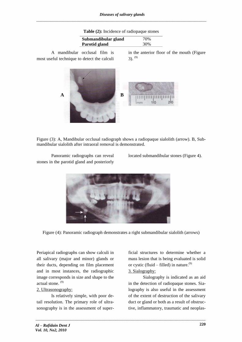
1. Plain film radiographs:
The primary purpose of plain films in
the assessment of salivary gland disease is
to identify salivary stones (calculi) al-
though only 80% to 85% of all stones are
radiopaque and therefore visible radio-
graphically Table (2).
Al – Rafidain Dent J
Vol. 10, No2, 2010
Al- Moula AD
220
Table (2): Incidence of radiopaque stones
A mandibular occlusal film is
most useful technique to detect the calculi
in the anterior floor of the mouth (Figure
3). (9)
Figure (3): A, Mandibular occlusal radiograph shows a radiopaque sialolith (arrow). B, Sub-
mandibular sialolith after intraoral removal is demonstrated.
Panoramic radiographs can reveal
stones in the parotid gland and posteriorly
located submandibular stones (Figure 4).
Figure (4): Panoramic radiograph demonstrates a right submandibular sialolith (arrows)
Periapical radiographs can show calculi in
all salivary (major and minor) glands or
their ducts, depending on film placement
and in most instances, the radiographic
image corresponds in size and shape to the
actual stone. (9)
2. Ultrasonography:
Is relatively simple, with poor de-
tail resolution. The primary role of ultra-
sonography is in the assessment of super-
ficial structures to determine whether a
mass lesion that is being evaluated is solid
or cystic (fluid – filled) in nature.(9)
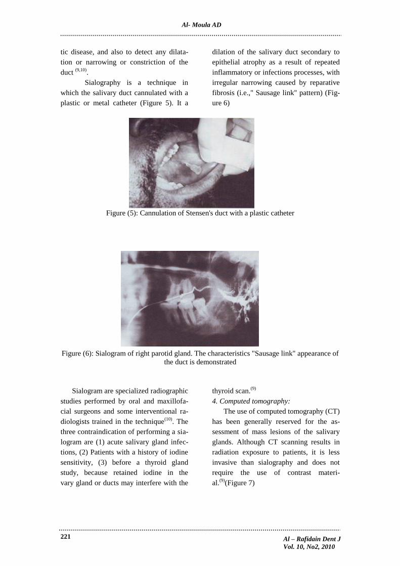
3. Sialography:
Sialography is indicated as an aid
in the detection of radiopaque stones. Sia-
lography is also useful in the assessment
of the extent of destruction of the salivary
duct or gland or both as a result of obstruc-
tive, inflammatory, traumatic and neoplas-
Submandibular gland 70%
Parotid gland 30%
A B
Al – Rafidain Dent J
Vol. 10, No2, 2010
Diseases of salivary glands
221
tic disease, and also to detect any dilata-
tion or narrowing or constriction of the
duct (9,10)
.
Sialography is a technique in
which the salivary duct cannulated with a
plastic or metal catheter (Figure 5). It a
dilation of the salivary duct secondary to
epithelial atrophy as a result of repeated
inflammatory or infections processes, with
irregular narrowing caused by reparative
fibrosis (i.e.," Sausage link" pattern) (Fig-
ure 6)
Figure (5): Cannulation of Stensen's duct with a plastic catheter
Figure (6): Sialogram of right parotid gland. The characteristics "Sausage link" appearance of
the duct is demonstrated
Sialogram are specialized radiographic
studies performed by oral and maxillofa-
cial surgeons and some interventional ra-
diologists trained in the technique(10)
. The
three contraindication of performing a sia-
logram are (1) acute salivary gland infec-
tions, (2) Patients with a history of iodine
sensitivity, (3) before a thyroid gland
study, because retained iodine in the
vary gland or ducts may interfere with the
thyroid scan.(9)
4. Computed tomography:
The use of computed tomography (CT)
has been generally reserved for the as-
sessment of mass lesions of the salivary
glands. Although CT scanning results in
radiation exposure to patients, it is less
invasive than sialography and does not
require the use of contrast materi-
al.(9)
(Figure 7)
Al – Rafidain Dent J
Vol. 10, No2, 2010
Al- Moula AD

222
Figure (7): Computerized tomographic scan of the mandible and floor of mouth shows a post-
erior submandibular sialolith (arrow)
5. Magnetic resonance imaging (MRI):
Is superior to CT scanning in delineat-
ing the soft tissue detail of salivary gland
lesions, specifically tumors, with no radia-
tion exposure to the patient or the necessi-
ty of contrast enhancement.(91)
6. Salivary scintigraphy (radioactive iso-
tope scanning):
The use of nuclear imaging in the form
of radioactive isotope scanning, or salivary
scintigraphy, allows a thorough evaluation
of the salivary gland parenchyma, with
respect to the presence of mass lesions and
the function of the gland itself. This study
uses a radioactive isotope [usually, techne-
tium (TC) 99m] injected intravenously
(IV), which is distributed throughout the
body and taken up by a variety of tissues,
including the salivary glands.(9)
7. Sialochemistry:
An examination of the electrolyte
composition of the saliva. Principally the
concentration of sodium and potassium, an
elevated sodium concentration with a de-
creased potassium concentration may indi-
cate an inflammatory sialadenitis.(9)
8. Salivary gland biopsy:
A salivary gland biopsy, either inci-
sional or excisional, can be used to diag-
nose a tumor of one of the major salivary
glands, but it is usually performed as an
aid in the diagnosis of Sjogren syndrome
(ss). The lower lip labial salivary gland
biopsy has been shown to demonstrate
certain characteristic histopathologic
changes that are seen in the major gland in
ss. The procedure is performed using local
anesthesia, and approximately 10 minor
salivary glands are removed for histologi-
cal examination.(3,9,11)
9. Fine – needle aspiration biopsy:
This procedure has a high accuracy
rate for distinguishing between benign and
malignant lesions in superficial locations.
Fine – needle aspiration biopsy is per-
formed using a syringe with a 20 –gauge
or smaller needle. After local anesthesia
the needle is advanced into the mass le-
sion, the plunger is activated to create a
vacuum in the syringe, and the needle is
moved back and forth throughout the
mass, with pressure maintained on the
plunger. The needle is withdrawn, and the
cellular material with fluid are expelled
onto a side and fixed for histological ex-
amination. (3,9)
Salivary gland infections:
Viral parotitis or mumps:
Mumps is the most common viral in-
fection which characterized by a painful,
non erythematous swelling of one or both
parotid glands that begins 2-3 weeks (in-
cubation period) after exposure to the vi-
rus. This disease occurs most commonly in
children between ages of 6-8 years and its
signs and symptoms include preauricular
Al – Rafidain Dent J
Vol. 10, No2, 2010
Diseases of salivary glands
223
pain and swelling, fever, chills and head-
ache. Viral parotitis usually resolves in 5-
12 days after its onset. Providing suppor-
tive and symptomatic care for fever, head-
ache and malaise with antipyretic, analges-
ic and adequate hydration treats viral paro-
titis. (3,6,12)
Sialadenitis:
Inflammation of the salivary glands
(sialadenitis) can arise from various and
non infectious causes. The most bacterial
infections arise as a result of ductal ob-
struction or decreased salivary flow.(13)
One of the more common causes of
sialadenitis is the recent surgery (especial-
ly abdominal surgery), when acute paroti-
tis (surgical mumps) may arise after such
operation because the patient has been
kept without food or fluids and received
atrophy during the surgical proce-
dure.(3,6,13)
Acute bacterial sialadenitis:
Histopathological feature:
Patient with acute sialadenitis, charac-
terized by accumulation of neutrophilis
within the ductal system and acini. (3,13,14)
Chronic sialadenitis:
Recurrent or persistent ductal ob-
struction (most commonly caused by sialo-
liths) can lead to chronic sialadentitis. Pe-
riodic swelling and pain occur within the
affected gland, which usually developed at
mealtime when salivary flow is stimu-
lated.(15)
Histopathological feature:
Chronic sialadentitis is characterized
by scattered or patchy infilteration of the
salivary parenchyma by lymphocytes and
plasma cells. Atrophy of the acini is com-
mon, as ductal dilatation and if associated
with fibrosis, the term chronic sclerosing
sialadentitis is used.(13)
Subacute necrotizing sialadenitis:
Is a recently recognized form of sali-
vary inflammation that occurs most com-
monly in teenagers and young adults. The
lesion usually involves the minor salivary
glands of the hard or soft palate, present-
ing as a painful nodule that is covered by
intact, erythematous mucosa. (13,16)
Histopathological feature:
Subacute necrotizing sialadenitis is
characterized by a heavy mixed inflamma-
tory infiltrate consisting of neutrophils,
lymphocytes, histocytes and eosinophils.
There is loss of most of acinar cells and
many of remaining exhibit necrosis, while
the ducts tend to be atrophic and do not
show hyperplasia or squamous metaplasia.
(13)
Chelitis glandularis:
Is a rare inflammatory condition
of the minor salivary glands, with uncer-
tain cause, although several etiologic fac-
tors have been suggested, including actinic
damage, tobacco, syphilis, poor hygiene,
and hereditary.(3,13,17)
Clinical features:
Chelitis glandularis characteristically
occurs on the lower lip and the affected
individuals experience swelling and rever-
sion of lower lip as a result of hypertrophy
and inflammation of the gland. The open-
ings of the minor salivary ducts are in-
flamed and silated, beside pressure on
glands may produce mucopurulent secre-
tions from the ductal openings. The condi-
tion most often has been reported in mid-
dle – aged and older men, although cases
have been described in women and child-
ren. However, some of the childhood cases
may represent other enities such as exfo-
liative cheilitis. Chelitis glandularis has
been classified into three types, based on
the severity of the disease:
1. Simple.
2. Superficial suppurative (Baelz's) disease.
3. Deep suppourative (Chelitis glandulais
apostematosa).
The latter two types represent progres-
sive stages of the disease with bacterial
involvement and are characterized by in-
Al – Rafidain Dent J
Vol. 10, No2, 2010
Al- Moula AD
224
creasing inflammation, suppuration, ulce-
ration, and swelling of the lip.(13,17)
Sialolithiasis (salivary calculi; salivary
stones):
Sialoliths are calcified structures that
develop within the salivary ductal system.
They are believed to arise from deposition
of calcium salts around a nidus of debris
within the duct lumen and this debris may
include inspissated mucus, bacteria, ductal
epithelial cells, or foreign bodies. The
cause of sialoliths is unclear, but their
formation can be promoted by chronic sia-
ladenitis and partial obstruction. Their de-
velopment is not related to any systemic
derangement in calcium and phosphorus
metabolism.(13,18,19)
The pathogenesis of salivary calculi
progresses through a series of stages be-
ginning with an abnormality in calcium
metabolism and salt precipitation, with
formation of organic and inorganic materi-
al to form a calcified mass. .(3,18,19)
The in-
cidence of stone formation varies from
80%, 19% and 1% in submandibular, pa-
rotid and sublingual glands respectively (3)
A variety of factors contributes to the
higher incidence of submandibular calculi.
Salivary gland secretion contain water,
electrolytes, urea, ammonia, glucose, fats,
proteins and other substances. In general,
parotid secretions are more concentrated
than those of the other salivary glands and
the main exception is the concentration of
calcium, which is about twice as abundant
in submandibular saliva as in parotid sali-
va. In addition, the alkaline ph of subman-
dibular saliva may further support stone
formation. Also the Wharton's duct of
submandibular gland is the longest sali-
vary duct, therefore saliva has a greater
distance to travel before emptied into the
oral cavity and it has two sharp curves in
its course and, as well as the flow of saliva
secretion occurs against the force of gravi-
ty.(3)
The occurrence of salivary gland
stones is twice as common in men between
age 30-50 years. But it is rare in children
and the boys are more commonly affected
than girls whereas the submandibular
gland is most commonly affected. Mul-
tiple stone formation occurs in approx-
imately 25% of patients.(3)
Histopathological feature:
On gross examination, sialoliths ap-
pear as hard masses that are round, oval,
or cylindrical and typically yellow, al-
though they may be white or yellowish –
brown. Submandibular stones tend to be
larger than those of the parotid or minor
glands. Sialoliths are usually solitary, al-
though occasionally two or more stones
may be discovered at surgery. Micro-
scopically, the calcified mass exhibits
concentric laminations that may surround
anidus of a amorphous debris. The duct
obstruction frequently is associated with
an acute or chronic sialadenitis of the feed-
ing gland.(3,13,18)
The sublingual gland is examined by
obstruction and bimanual palpation of the
anterior third of the floor of the mouth.
The minor salivary glands are ex-
amined by observation and palpation of
the mucosal surfaces of the lips, buccal
mucosa, palate, and floor of the mouth.
Obstruction of the sublingual gland is
unusual, but if it occurs, it is usually sec-
ondary to obstruction of Wharton's duct on
the same side of the oral cavity. Although
stone formation is rare in the sublingual
and minor salivary glands, the treatment is
simple excision of the stone and associated
gland.(3)
Other diseases that affect salivary gland:
Sjogren's syndrome:
Sjogren's syndrome (ss) is the expres-
sion of an autoimmune process that results
principally in dry eyes (xerophthalmia)
and dry mouth (xerostomia) owing to
lymphocyte – mediated destruction of la-
crimal and salivary gland parenchyma, in
Al – Rafidain Dent J
Vol. 10, No2, 2010
Diseases of salivary glands
225
addition rheumatoid arthritis, may also be
seen in this syndrome.(6)
So (ss) is a multi-
system disease process with available
presentation and classified into two types:
1. Primary (ss) (sicca syndrome); characte-
rized by xerostomia (dry mouth) and (xe-
rophthalmia) (dry eye).
2. Secondary (ss): composed of primary
(ss), associated with connective tissue dis-
order, most commonly rheumatoid arthri-
tis. Although the cause of (ss) is unknown,
there appear to be a strong autoimmune
influence, and it shows a female predilec-
tion of 9:1 with over 80% of affected indi-
viduals being females with a mean age of
50 years.(3,6,20)
Generally, the first symptoms to ap-
pear are arthritis complaints, followed by
ocular symptoms and late in the disease
process, salivary gland symptoms. The
involvement of the salivary and lacrimal
glands results from a lymphocytic re-
placement of the normal glandular ele-
ments, therefore, xerostomia results from a
decreased function of both the major and
minor salivary glands, with the parotid
gland being the most sensitive.(3,6)
The histopathologic changes seen in
the minor glands are similar to those in the
major (parotid) glands. Keratoconjuctivitis
sicca is suggested by the patient's com-
plaints and a schirmer's test for lacrimal
flow.(3,4,6,11,20)
Diagnosis depends on the correlation
between the patient history and laboratory
data, clinical examination and assessment
of salivary function. An important consid-
eration concerns the clinical manifestation
of xerostomia, although this is the main
oral symptom and clinical sign in (ss),
other considerations of dry mouth must be
evaluated. In addition, major salivary
gland enlargement is a feature of (ss) but
may be episodic in nature and in some pa-
tients it may not be present at all.(3,6,11,20)
The prognosis of (ss): is complicated
by an associated with malignant transfor-
mation to lymphoma. This may occur in
approximately 6% - 7% of cases and it is
more common in those with only the sicca
component of the syndrome. Less com-
monly observed is transformation of the
epithelial component to undifferentiated
carcinoma. Generally, the course for (ss) is
one of chronicity, requiring long – term
symptomatic management and careful fol-
low-up by a dentist, ophthalmologist and
rheumatologist.(3,6,20)
Traumatic salivary gland injuries:
Traumatic injuries, particularly lacera-
tions, involving the salivary glands and
their ducts may accompany a variety of
facial injuries, including fractures. Injuries
that occur in close proximity to one of the
major salivary glands or ducts require
careful evaluation. Facial lacerations may
involve not only parotid gland and its duc-
tal system, but also branches of the facial
nerve and branches of major facial vessels.
These structures require meticulous atten-
tion for appropriate diagnosis and prompt
repair. Repair may include ductal anasto-
moses, in which the proximal and distal
portions of the duct are identified, a plastic
or metal catheter is placed as astent, and
the duct is sutured over the stent. The ca-
theter usually remains in place for 10 - 14
days for epithelization of the duct to occur.
Additionally, nerve anastomoses may be
required and performed by placing epi-
neural sutures, using magnification, to
reapproximate the nerve stimps. The lace-
rations are closed in a usual layered fa-
shion, after debridment of the soft tissue
wounds to cleanse the site of entrapped
particles, such as glass or dirt. Potential
sequelae of trauma involving the major
salivary glands include infections, facial
paralysis, cutaneous salivary gland fistula,
sialocele formation and duct obstruction as
a result of scar formation, with eventual
glandular atrophy and decreased function.
Al – Rafidain Dent J
Vol. 10, No2, 2010
Al- Moula AD
226
The involved gland may eventually require
surgical removal.(3,13)
(Reactive Lesions): Mucous retention
and extravasation phenomena:
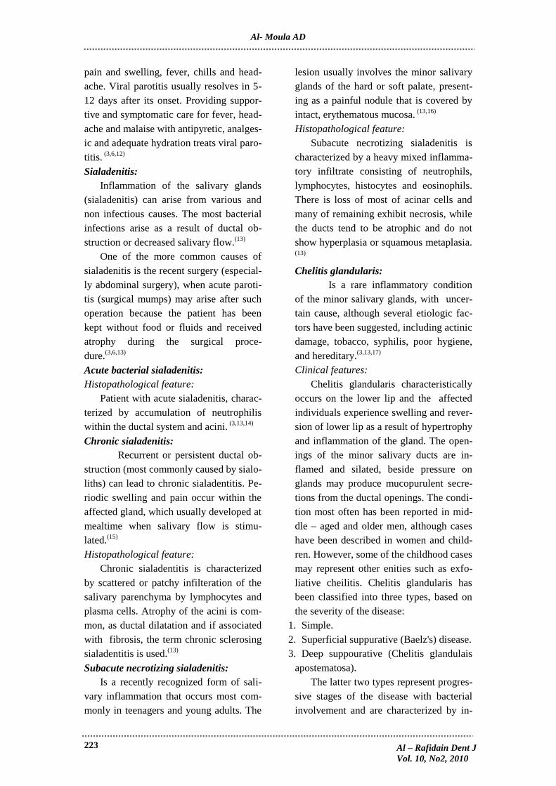
Mucocele:
Is a clinical term that include mucus
extravasation phenomenon and mucus re-
tention cyst. Because each has a distinctive
pathogenesis and microscopy, they are
considered separately.
Salivary ducts, especially those of the
minor salivary glands, are occasionally
traumatized, commonly by lip bitting, and
severed beneath the surface mucosa. Sub-
sequant saliva production may then extra-
vasate beneath the surface mucosa into the
soft tissues. Overtime, secretions accumu-
late within the tissues and produce a pseu-
docyst (without a true epithelial lin-
ing)(2,3,8,21)
that contain thick, viscous sali-
va and these lesions are most common in
the mucosa of the lower lip and known as
mucoceles (Figure 8). The second most
common site of mucocele formation is the
buccal mucosa. Which results in an ele-
vated, thinned, stretched overlying mucosa
that appears as a vesicle filled with a clear
or blue – grey mucus. The patient fre-
quently relates a history of the lesion fill-
ing with fluid, rupture of fluid collection,
and refilling of these lesions. Many
stances of mucocele formation regress
spontaneously without surgery, while in
case of persistent or recurrent lesions,
ferred treatment is required for excision of
the mucocel and the associated minor sali-
vary glands that contributed to its forma-
tion. Usually local anesthesia is adminis-
tered via a mental nerve block, and an
cision is made through the mucosa.
ful dissection around the mucocele may
permit its complete removal, however, in
many cases the thin lining ruptures and
decompresses the mucocele before remov-
al. The regional associated minor salivary
glands are removed and sent for
thological evaluation. After surgical
moval the recurrence rates of mucocele
may be as high as 15% - 30%, which
sibly caused by incomplete removal or
repeat trauma to the minor salivary
glands.(3,21)
Figure (8): Mucocele of left lower lip
Ranula:
The most common lesion of the sub-
lingual gland is the ranula, which may be
considered a mucocele of the sublingual
salivary gland. Ranulas result from either
mucous retention in the sublingual gland
ductal system or mucous extravasation as
a result of ductal disruption and divided
into simple and plunging types.
The simple ranula is confined to the
area occupied by the sublingual gland in
the sublingual space, superior to the mylo-
hyoid muscle. The progression to a plung-
ing ranula occurs when the lesion extend
beyond the level of the mylohyoid muscle
into the submandibular space. Ranulas
may reach a larger size than mucoceles,
because their overlying mucosa is thicker
Al – Rafidain Dent J
Vol. 10, No2, 2010
Diseases of salivary glands

227
and the trauma that would cause their rep-
ture is less likely in the floor of the mouth.
As a result a plunging ranula has the po-
tential to extend into the neck and com-
promise the airway, resulting in a medical
emergency.(3,13, 6,22-25)
Sialocyst (salivary duct cyst):
The salivary duct cyst is an epithelium
– lined cavity that arises from salivary
gland tissue and unlike the more common
mucocele, it is a true cyst because it is
lined by epithelium. The cause of such
cysts is uncertain, but some cases may
represent ductal dilation secondary to duc-
tal obstruction (e.g., mucus plug) which
creates increased intraluminal pressure.
Some authors refer to such lesions as mu-
cus retention cyst, while other cases ap-
pear separate from the adjacent normal
salivary ducts.(13)
Clinical features:
Salivary duct cysts usually occur in
adults and can arise with either the major
or minor glands. Cysts of the major glands
are most common within the parotid gland,
presenting as slowly growing, asympto-
matic swellings. Intraoral cysts can occur
at any minor gland site, but most frequent-
ly they develop in the floor of the mouth,
buccal mucosa, and lips. They are often
look like mucoceles, and characterized by
soft, fluctuant swelling that may appear
bluish, depending on the depth of the cyst
below the surface and some cysts may feel
relatively firm to palpation. Cysts in the
floor of the mouth often arise adjacent to
the submandibular duct and sometimes
have an amber color. (13)
Sialorrhea:
Sialorrhea, or excessive salivation, is
an uncommon condition that has various
causes. Minor sialorrhea may result from
local irritations, such as apthous ulcers or
ill – fitting dentures. Patients with new
dentures often experience excess saliva
production until they become accustomed
to the prosthesis. Sialorrhea is a well –
known clinical feature of rabies and heavy
metal poisoning, as well as a consequences
of certain medications, such as lithium and
cholinergic agonists.(13)
Clinical features:
The excess saliva production typically
produce drooling and choking, which may
cause social embarrassment. In children
with mental retardation or cerebral palsy,
the uncontrolled salivary flow may lead to
macerated sores around the mouth, chin
and neck that can become secondarily in-
fected.
An interesting type of supersalivation
of unknown cause has been tremed idi-
opathic paroxysmal sialorrhea. Individuals
with this condition experience short epi-
sodes of excessive salivation lasting from
2 - 5 minutes, and associated with a pro-
dram of nausea or epigastric pain.(13)
Salivary gland tumor:
A- Benign salivary gland tumors:
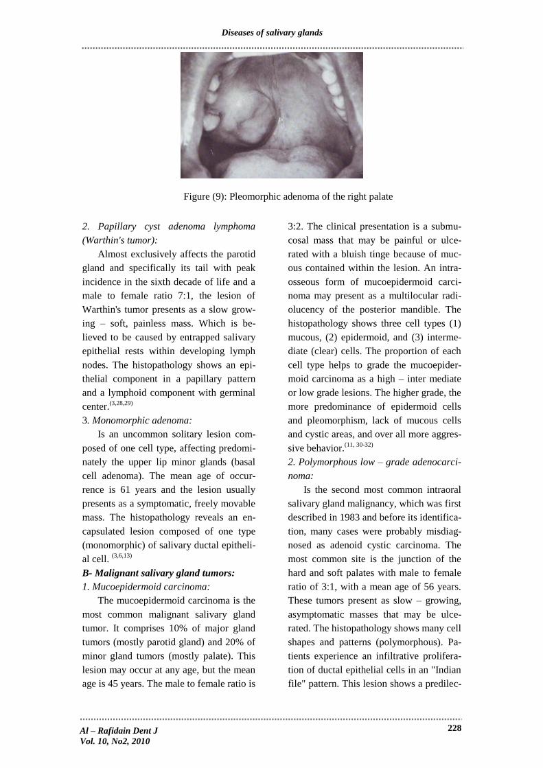
1. Benign mixed tumor or pleomorphic
adenoma (Figure 9):
Is the most common salivary gland
tumor with mean age of occurance 45
years, and a male to female ratio of 3:2. In
the major glands, the parotid gland is in-
volved in over 80% of cases, in the minor
glands the most common intraoral site is
the palate. Pleomorphic adenomas are
usually slow – growing, painless masses
with two cell type; 1) the ductal epithelial
cell and 2) the myoepithelial cell, which
may differentiate along a variety of cell
lines pleomorphic mean many form. The
treatment involves complete surgical exci-
sion with a margin of normal involved tis-
sue.(3,13,26,27)
Al – Rafidain Dent J
Vol. 10, No2, 2010
Al- Moula AD
228
Figure (9): Pleomorphic adenoma of the right palate
2. Papillary cyst adenoma lymphoma
(Warthin's tumor):
Almost exclusively affects the parotid
gland and specifically its tail with peak
incidence in the sixth decade of life and a
male to female ratio 7:1, the lesion of
Warthin's tumor presents as a slow grow-
ing – soft, painless mass. Which is be-
lieved to be caused by entrapped salivary
epithelial rests within developing lymph
nodes. The histopathology shows an epi-
thelial component in a papillary pattern
and a lymphoid component with germinal
center.(3,28,29)
3. Monomorphic adenoma:
Is an uncommon solitary lesion com-
posed of one cell type, affecting predomi-
nately the upper lip minor glands (basal
cell adenoma). The mean age of occur-
rence is 61 years and the lesion usually
presents as a symptomatic, freely movable
mass. The histopathology reveals an en-
capsulated lesion composed of one type
(monomorphic) of salivary ductal epitheli-
al cell. (3,6,13)
B- Malignant salivary gland tumors:
1. Mucoepidermoid carcinoma:
The mucoepidermoid carcinoma is the
most common malignant salivary gland
tumor. It comprises 10% of major gland
tumors (mostly parotid gland) and 20% of
minor gland tumors (mostly palate). This
lesion may occur at any age, but the mean
age is 45 years. The male to female ratio is
3:2. The clinical presentation is a submu-
cosal mass that may be painful or ulce-
rated with a bluish tinge because of muc-
ous contained within the lesion. An intra-
osseous form of mucoepidermoid carci-
noma may present as a multilocular radi-
olucency of the posterior mandible. The
histopathology shows three cell types (1)
mucous, (2) epidermoid, and (3) interme-
diate (clear) cells. The proportion of each
cell type helps to grade the mucoepider-
moid carcinoma as a high – inter mediate
or low grade lesions. The higher grade, the
more predominance of epidermoid cells
and pleomorphism, lack of mucous cells
and cystic areas, and over all more aggres-
sive behavior.(11, 30-32)
2. Polymorphous low – grade adenocarci-
noma:
Is the second most common intraoral
salivary gland malignancy, which was first
described in 1983 and before its identifica-
tion, many cases were probably misdiag-
nosed as adenoid cystic carcinoma. The
most common site is the junction of the
hard and soft palates with male to female
ratio of 3:1, with a mean age of 56 years.
These tumors present as slow – growing,
asymptomatic masses that may be ulce-
rated. The histopathology shows many cell
shapes and patterns (polymorphous). Pa-
tients experience an infiltrative prolifera-
tion of ductal epithelial cells in an "Indian
file" pattern. This lesion shows a predilec-
Al – Rafidain Dent J
Vol. 10, No2, 2010
Diseases of salivary glands

229
tion for invasion of surrounding
nerves.(3,13,30,33,34)
3. Adenoid cystic carcinoma:
Is the third most common intraoral sali-
vary gland malignancy, with a mean age
of 53 years and a male to female ratio of
3:2. Approximately 50% of these tumors
occur in the parotid gland, whereas the
other 50% occur in the minor glands of the
palate. These present as slow growing, non
ulcerated masses, with an associated
chronic dull pain. Occasionally, parotid
lesions may result in facial paralysis as a
result of facial nerve involvement. The
histopathology demonstrates an infiltrative
proliferation of basaloid cells arranged
distant in a "cribriform" (Swiss cheese)
pattern. As seen in the polymorphous low
– grade adenocarcinoma, there may be
perineural invasion.(3)
Because the tumor
is prone to late recurrence and metastasis,
as well as 5 years survival rate has little
significance and does not equate to a cure,
this rate may be as high as 70%, but it con-
tinues to decrease overtime and by 20
years, only 20% of patients are still a live.
Tumors with a solid histopathologic pat-
tern are associated with a worse out look
than those with a cribriform or tubular ar-
rangement. With respect to site, the prog-
nosis is poorest for tumors arising in the
maxillary sinus and submandibular gland.
Most studies have shown than microscopic
identification of perineural invasion has
little effect on the prognosis. Death usually
results from local recurrence or distant
metastasis, which commonly occurs to the
lungs and bones, while tumors of the pa-
late or maxillary sinus eventually may in-
vade up ward to the base of the brain.(13)
REFERENCES
1. Snell RS. Clinical Anatomy for Medical
Students. 7th edition. SMHS Washing-
ton. 2004; Pp. 773 – 788.
2. Antonio N. Ten Cate's Oral Histology,
Development, Structure and Function.
6th edition. 2003; Pp. 299 – 328.
3. Larry J P, Edward E, James R. H. Diag-
nosis and Management of Salivary
Gland Disorders Contemporary Oral
and Maxillofacial Surgery, 4th edition.
Mosby. 2003; Pp. 434 – 455.
4. Vanderwaal IE. Diseases of the Salivary
Glands. J Oral Maxillofacial Surg.
1997; 34: 15.
5. Arafat A, Brannon RB, Ellis GL. Ade-
nomatoid hyperplasia of mucous sali-
vary glands. Oral Surg Oral Med Oral
Path. 1981; 52: 51 – 55.
6. Joseph AR, James JS, Richard CKJ,
Fred C. Oral Pathology (Clinical Pa-
thologic Correlation), 4th edition. WD
Saunders, 2003; Pp. 183 – 217.
7. Arthur CG, John EH. Secretory Func-
tions of the Alimentary Tract: Text
Book of Medical Physiology. 11th edi-
tion. Vol. 2, 2006; Pp. 793 – 806.
8. Tandler B, Gresik EW, Nagato T, Phi-
lips CJ. Secretion by striated ducts of
mammalian major salivary gland: re-
view from histological, functional and
evolutionary prespective. Anat Rec.
2001; 264: 121.
9. Noyek AM. Head and Neck Radiology.
Gower medical publishing. London.
3rd
edition. 1991; 312 – 324.
10. Stephens LC, Schulthesis TE, Price
RE, et al. Radiation apoptosis of ser-
ous acinar cells of salivary and lacrim-
al glands. Cancer. 1991; 67: 1539 –
1543.
11. Abaza N. The role of labial salivary
gland biopsy in the diagnosis of Sjo-
gren's syndrome: report of three cases.
J Oral Maxillofacial Surg. 1993;
51(5): 574.
12. Goldberg MH, Bevilacqua RG Infec-
tions of the salivary glands. J Oral
Maxillofac Surg. 1995; 7: 423.
13. Brad W N, Douglas DD, Carl MA,
Jerry E.: Oral and Maxillofacial Pa-
thology. 2nd
edition. 2002; Pp. 675 –
679.
14. Blitzer A. Inflammatory and obstruc-
tive disorders of salivary glands. J
Dent Res. 1987; 66 (Supp 1): 675 –
679.
15. Koudelka BM. Obstructive Disorders:
Ellis GL, Auclair PL, Gnepp DR (eds).
Surgical pathology of the salivary
Al – Rafidain Dent J
Vol. 10, No2, 2010
Al- Moula AD

230
glands. WB Saunders, Philadelphia.
1991; Pp. 95 – 96.
16. Fowler CB, Brannon RB. Subacute
necrotizing sialadenitis: report of 7
cases and a review of the literature.
Oral Surg Oral Med Oral Pathol.
2000; 89: 600 – 609.
17. Cohen DM, Green JG, Dickmann SL
Concurrent anomalies: cheilitis glan-
dularis and double lip: report of a case.
Oral Surg Oral med Oral Pathol.
1988; 66: 397 – 399.
18. Narang R and Dixon RA. Surgical
management of submandibular siala-
denitis and sialolithiasis. Oral Surg
Oral Med Oral Pathol. 1977; 43: 201
– 210.
19. Yoshimura Y, Morishita T and Sugiha-
ra T. Salivary gland functions after
sialothiasis: scintigraphic examination
of submandibular glands with teper-
technetate. J Oral Maxillofac Surg.
1989; 47: 704 – 710.
20. Linvall AM and Jonsson R. The sali-
vary gland component of Sjogren's
syndrome: an evaluation of diagnosis
methods. Oral Surg Oral Med Oral
Pathol. 1986; 62: 32 – 42.
21. Jensen JL. Superficial mucoceles of
the oral mucosa. Am J Dermatopathol.
1990; 12: 88 – 92.
22. Baurmash HD. Marsupialization for
treatment of oral ranula: a second look
at the procedure. J Oral Maxillofac
Surg. 1992; 50: 1274.
23. Galloway RH, Gross PD, Thompson
SH, et al. Pathogenesis and treatment
of ranula: report of three cases: J Oral
Maxillofac Surg. 1989; 47: 299 – 302.
24. McClatchey KD, Appelblatt NH, Zar-
bo RJ. Plunging ranula. Oral Surg
Oral Med Oral Pathol. 1984; 57: 408
– 412.
25. Yoshimura Y. A comparison of three
methods used for treatment of ranula.
J Oral Maxillofac Surg. 1995; 53: 280.
26. Chau MY, Radden BG. A clinical –
pathological study of 53 intraoral
pleomorphic adenomas. J Oral Maxil-
lofac Surg. 1989; 18: 158 – 162.
27. Kanazawa H, Faruya T, Watanabe Te.
Plasma cytoid myoepitheloma of the
palate. J Oral Maxillofac Surg. 1999;
57: 857 – 860.
28. Kotwall CA. Smoking as an etiologic
factor in the development of Warthin's
tumor of the parotid gland. Am J Surg.
1992; 164: 646 – 647.
29. Vanderwaal IE, Davids JJ, Vander-
waal I Extra parotid Warthin's tumors
– report of 10 cases. J Oral Maxillofac
Surg. 1993; 31: 43 – 44.
30. Caslle JT, Thompson LR, Frommelt
RA.: Polymorphous low grade adeno-
carcinoma. A clinicopathologic study
of 164 cases. Cancer. 1999; 86: 207 –
219.
31. Ellis GL, Auclair PL, Gnepp DR.:
Surgical pathology of the salivary
glands, (Clinical Pathologic Correla-
tion), 1st edition. WB Saunders. 1991;
Pp. 45-47.
32. Stene T, Koppang HS Intraoral adeno-
carcinomas. J Oral Pathol. 1981; 10:
216 – 225.
33. Evan HL. Mucoepidermoid carcinoma
of salivary glands: a study of 69 cases
with special attention to histologic
grading. Am J Pathol. 1984; 81: 696 –
701.
34. Vincent SD, Hammond HL, Finkels-
tein MW Clinical and therapeutic fea-
tures of polymorphous low – grade
adenocarcinoma. Oral Surg Oral Med
Oral Pathol. 1994; 77: 41 – 47.
Al – Rafidain Dent J
Vol. 10, No2, 2010
Diseases of salivary glands
Copyright © 2022 FDOKUMEN